Embed Size (px)
Citation preview
Molecular Cell, Vol. 16, 991–1002, December 22, 2004, Copyright ©2004 by Cell Press
DNA Damage Response Pathway UsesHistone Modification to Assemblea Double-Strand Break-Specific Cohesin Domain
CHK1/Chk1p and CHK2/Rad53p (reviewed in Jackson[2002] and Shiloh [2003]). Substrates of these kinasesinclude DNA repair-related proteins such as Mre11p, asubunit of the conserved MRX (Mre11/Rad50/Xrs2) andMRN (MRE11/RAD50/NBS1) complexes in budding yeast
Elcin Unal,1,2 Ayelet Arbel-Eden,3 Ulrike Sattler,3
Robert Shroff,4 Michael Lichten,4 James E. Haber,3
and Douglas Koshland1,*1Howard Hughes Medical InstituteDepartment of Embryology
and humans, respectively (reviewed in D’Amours andThe Carnegie Institution of WashingtonJackson [2002]). The idea that DNA damage repair mayBaltimore, Maryland 21210also involve proteins linked to chromosome structure2 Department of Biologyand dynamics is supported by the fact that H2AX, aJohns Hopkins Universityvariant of the core H2A histone, is phosphorylated inBaltimore, Maryland 21218chromatin surrounding the site of damage (Rogakou et3 Department of Biologyal., 1998, 1999; Shroff et al., 2004).Rosenstiel Basic Medical Sciences Research Center
A key structural component of chromosomes that isBrandeis Universityalso linked to DNA damage is the cohesin complex.Waltham, Massachusetts 02454Cohesin is an evolutionarily conserved multisubunit4 Laboratory of Biochemistrycomplex that consists of four proteins: Mcd1p (Scc1/National Cancer InstituteRad21), Scc3p (SA1/SA2), Smc1p, and Smc3p (reviewedNational Institutes of Healthin Nasmyth [2002]). During an unperturbed cell cycle,Bethesda, Maryland 20892cohesin is loaded onto chromosomes during S phaseby Scc2p/Scc4p complex and other factors (Ciosk et al.,2000). This loading results in a large domain of cohesinsSummaryaround the centromere and small (1 kb) domains on thearm. In budding yeast, these small domains are calledThe postreplicative repair of double-strand breaksCARs (cohesin-associated regions) and are spaced at(DSBs) is thought to require sister chromatid cohesion,10–15 kb intervals and generally located in AT-rich in-provided by the cohesin complex along the chromo-tergenic sequences (Blat and Kleckner, 1999; Lalorayasome arms. A further specialized role for cohesin inet al., 2000; Megee et al., 1999; Tanaka et al., 1999).DSB repair is suggested by its de novo recruitment toThe loading of cohesin serves to hold sister chromatidsregions of DNA damage in mammals. Here, we showtogether from the time of their replication until meta-in budding yeast that a single DSB induces the forma-phase-anaphase transition, thus ensuring the biorienta-tion of a �100 kb cohesin domain around the lesion.tion of sister chromatids on the spindle apparatus andOur analyses suggest that the primary DNA damagetheir proper segregation (reviewed in Koshland and Gu-checkpoint kinases Mec1p and Tel1p phosphorylateacci [2000]).histone H2AX to generate a large domain, which is
In addition to cohesin’s role in chromosome segrega-permissive for cohesin binding. Cohesin binding totion, a role in DNA repair is suggested by observationsthe phospho-H2AX domain is enabled by Mre11p, afrom yeast and mammals. In budding yeast, sister chro-component of a critical repair complex, and Scc2p, amatids are the preferred partners for mitotic DSB repaircomponent of the cohesin loading machinery that isin diploid cells (Kadyk and Hartwell, 1992). Mutants withnecessary for sister chromatid cohesion. We also pro-hypomorphic alleles of cohesin subunits have de-vide evidence that the DSB-induced cohesin domaincreased viability in response to � irradiation (Birkenbihlfunctions in postreplicative repair.and Subramani, 1992; Heo et al., 1998; Sonoda et al.,2001). Furthermore, inactivation of cohesin in S or M
Introduction phase leads to a reduced efficiency of postreplicativeDSB repair. This defect has been attributed to lack of
DNA damage, including DSBs, triggers a conserved cel- cohesion (Sjogren and Nasmyth, 2001). Because sisterlular response that involves slowing of cell cycle pro- chromatid cohesion is established during S phase, thegression, transcriptional and posttranslational regula- cohesin loaded along the chromosome arms during Stion of repair-related proteins, and the recruitment of phase is thought to be necessary and sufficient forthese proteins to the sites of damage (reviewed in Brad- proper postreplicative repair.bury and Jackson [2003] and Rouse and Jackson [2002]). Recent cytological studies in mammalian cells indi-This response has been intensively studied because of cate that cohesin is recruited to regions of DNA damageits importance in fundamental aspects of cell division (Kim et al., 2002a). This suggests that efficient DNAand its potential relevance to human disease (reviewed repair in mammalian cells may require cohesins at thein Digweed et al. [1999]; Michelson and Weinert [2000]; site of DNA damage. Although the damage-inducedand Rotman and Shiloh [1998]). cohesin binding is intriguing, many questions remain
The DNA damage response is regulated by numerous unanswered. What promotes this binding and how is itkinases. The earliest activated kinases, ATM/Tel1p and regulated in response to damage? Is damage-inducedATR/Mec1p, subsequently activate additional ones like cohesin binding important for DNA repair and, if so, what
aspect of repair? Is damage-induced cohesin bindingunique to mammalian cells, or is it a part of the con-*Correspondence: [email protected]
Molecular Cell992
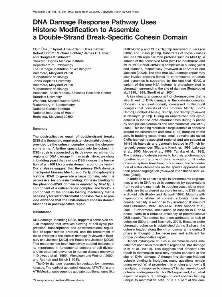
Figure 1. Cohesin Is Enriched around a DSB in M Phase
Cells growing exponentially at 30�C were treated with nocodazole to induce an M phase arrest that was maintained during the entire experiment.After 3 hr 20 min, the culture was split, and galactose was added to one half (HO cut), whereas the other half was grown in the absence ofgalactose (uncut). After 90 min, cells were fixed with 1% formaldehyde for 2 hr and used for chromatin immunoprecipitation (ChIP, seeExperimental Procedures). Input DNA and DNA coimmunoprecipitated with �-HA antibody (IP) were amplified by using primer sets correspond-ing to sequences around MAT or LEU2. To ensure the linearity of the PCR signal, appropriate dilutions of IP and input samples were used inPCR amplifications.(A) The binding of Mcd1p around MAT on ChrIII in JKM179-101A (MCD1HA6) with (HO cut) or without (uncut) a DSB at MAT. Top left, schematicdepiction of ChrIII denoting relative positions of MAT and LEU2. Bottom left, PCR amplified DNA from the left (1–11) and right side (13–23)of HO site were displayed by agarose gel electrophoresis and visualized by ethidium bromide (reverse images are shown). The PCR fragmentcontaining the HO site (12) is only present in samples without a DSB. Right, Mcd1p binding profile around MAT in samples with (HO cut, black
DSB-Induced Cohesin Domain993
served DNA damage response common to all eu- in chromatin bound and input samples. The amplifiedproduct of each primer pair was also assigned a chromo-karyotes?somal coordinate based upon its sequence alignmentTo address these questions, we examined cohesinwith the Saccharomyces Genome Database (SGD). De-binding in response to DSBs in Saccharomyces cerevis-termination of the relative Mcd1p binding for a giveniae. We show here that a single DSB induces a largechromosomal coordinate allowed us to assess Mcd1pdomain of cohesin binding near the lesion, demonstra-localization at specific chromosome locations in the ab-ting that the association of cohesin with sites of DSB issence or presence of a DSB.an evolutionarily conserved response and likely to be
In the absence of a DSB at MAT, there is low Mcd1pan important part of DNA damage response in all eukary-binding in a 20 kb region surrounding MAT, except forotes. We also report analyses of this DSB-inducedtwo CAR sites to the left of HO site (Figure 1A, uncut).cohesin domain that both define its size, kinetics ofAfter 90 min of HO induction, �95% of the DNA is cutassembly, and cell cycle regulation, and that identify(data not shown). At this time, Mcd1p binding aroundevolutionarily conserved factors necessary for its forma-the DSB site has two distinct features. Immediately adja-tion. Finally, we provide evidence for one potential rolecent to the DSB site is a 2 kb region of low binding thatof the DSB-induced cohesin domain in DNA repair.is flanked on either side by 8 kb regions of high bindingBased on these studies, we propose a model for the(Figure 1A). In the 8 kb flanking regions, the maximumassembly of the DSB-induced cohesin domain that isDSB-induced increase in Mcd1p binding is approxi-likely to be of general relevance to postreplicative repairmately 10-fold, with an average of 7-fold for regions thatin other eukaryotes.do not contain a CAR site. Thus the presence of a DSBinduces a dramatic increase in Mcd1p binding in a do-Resultsmain at least 16 kb in size.
The low Mcd1p binding in the 2 kb region immediatelyCohesin Is Enriched around a DSB Siteproximal to the DSB partly correlates with a 2-fold reduc-To investigate cohesin localization in response to a DSB,tion in the amplification of input DNA in this region com-we used the HO endonuclease to create a single DSBpared to the 8 kb flanking regions. This reduction is notin the genome of Saccharomyces cerevisiae. This site-observed in cells without a DSB. Loss of DNA proximalspecific restriction endonuclease cleaves a 24 bp de-to a DSB at MAT has been shown previously, resultinggenerate sequence present at the MAT locus on chro-from a 5�–3� resection activity (Frank-Vaillant and Mar-
mosome III (ChrIII) (Nickoloff et al., 1986). By using ancand, 2002; Lee et al., 1998). Thus, nonduplex DNA
HO gene under the control of a galactose inducible pro-structure and/or the presence of other break-associated
moter, it was possible to control the timing of HO expres- proteins could account for the low Mcd1p binding insion and DSB formation (Haber, 2002; Jensen and Her- the 2 kb region proximal to this DSB.skowitz, 1984). This allowed us to study potential Having established that a domain of Mcd1p bindingchanges in cohesin binding at the DSB site both at is induced in a region surrounding the DSB at MAT,different stages of the cell cycle and after inactivation we asked three questions about the generality of thisof potentially relevant gene products. Repair of the DSB observation: (1) whether this induction is specific to thisby homologous recombination (HR) was prevented by cohesin subunit or the cohesin complex, (2) whether thedeletion of the HML and HMR loci (Moore and Haber, domain is limited to the DSB region, and (3) whether1996). The persistence of the DSB facilitated the detec- domains are induced around DSBs at other regions oftion of potential DSB-induced changes in cohesin the genome. To investigate whether the DSB-associatedbinding. increase in Mcd1p binding reflected an increase in bind-
We first wanted to study cohesin association around ing of the whole cohesin complex, we used ChIP toa DSB in a cell cycle stage with sister chromatids, a monitor the chromatin binding of another subunit oftime when cohesin is known to function. Therefore, cells cohesin, Smc1p. To facilitate ChIP, cells were engi-were arrested in M phase with nocodazole, and then neered to express only Smc1p-6HA, the Smc1 proteina DSB was created by HO induction. The cells were with six copies of the hemaglutanin A epitope tag.engineered to express only Mcd1p-6HA, the Mcd1 pro- Smc1p association at MAT locus in cells with and with-tein with six copies of the hemaglutanin A epitope tag. out a DSB show very similar characteristics to Mcd1pChromatin binding of this cohesin subunit was assayed association (Figure 1B), with both low binding in the 2 kbby chromatin immunoprecipitation (ChIP). DNA se- region immediately proximal to the break and high levelsquences were amplified from the input chromatin and in the 8 kb flanking regions. We therefore conclude thatchromatin immunoprecipitated by anti-HA antibody with during M phase, the cohesin complex is enriched around23 primer pairs on either side of the DSB site. For each MAT locus in response to a DSB. Furthermore, becauseprimer pair, we determined the relative Mcd1p binding these strains cannot repair the DSB by HR, there is no
repair-associated DNA synthesis. Therefore, the cohesinby calculating the ratio of the amounts of PCR products
bar) and without (uncut, gray bar) a DSB. Percentage of input chromatin in Mcd1p IP is plotted on the y axis versus ChrIII coordinates (kb)on the x axis. Arrow indicates the position of the DSB.(B) The binding of Smc1p around MAT on ChrIII in EU1239 (SMC1HA6) with and without a DSB at MAT.(C) The binding of Mcd1p around LEU2 on ChrIII in JKM179-101A (MCD1HA6) with and without a DSB at MAT.(D) Mcd1p binding around LEU2 on ChrIII. YFP17-101A (leu2::HO cut-site, MCD1HA6) and YMV150-101A (leu2, MCD1HA6) were treated with2% galactose for HO induction. A DSB at leu2 was created in YFP17-101A containing the HO site, but not in YMV150-101A.
Molecular Cell994
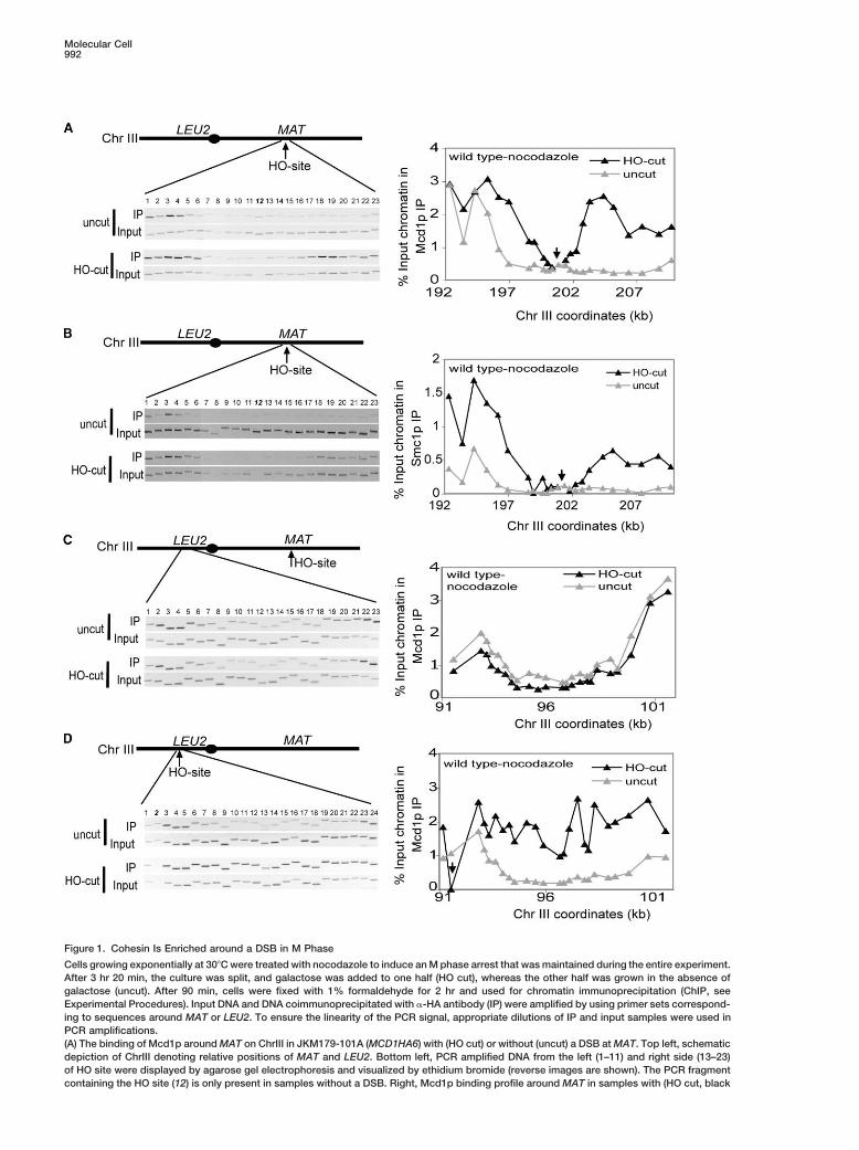
enrichment around the DSB is independent of DNA syn- similar for the scc2-4 and wild-type (wt) cells, confirmingthat S phase cohesin loading is not impaired (Sup-thesis.plemental Figure S1 available online at http://www.To address whether the increased levels of cohesinmolecule.org/cgi/content/full/16/6/991/DC1/). Cellsbinding around the DSB at MAT was unique to thiswere shifted to the restrictive temperature for 1 hr, andregion, we asked whether a DSB at MAT caused anHO was induced. The kinetics of DSB formation underincrease in cohesin binding at sites on ChrIII very distalthese conditions were similar in wt and scc2-4 cellsto the DSB or even on other chromosomes. We exam-(data not shown). After 90 min of HO induction, we ob-ined Mcd1p binding around LEU2, which is �110 kbserved no substantial increase of cohesin levels aroundaway from MAT on the left arm of ChrIII, on the oppositethe DSB site in scc2-4 cells at the restrictive temperatureside of the centromere. We detected no significant dif-(Figure 2B). Therefore, Scc2p is needed for DSB-specificference in cohesin binding in this region between cellscohesin recruitment as well as for cohesin depositionwith and without DSB at MAT (Figure 1C). Similarly, noduring S phase. Furthermore, the Scc2p requirementincrease in cohesin binding was observed in a region ofindicates that the cohesin domain at the DSB resultsChrXII (data not shown). Therefore, the elevated cohesinfrom de novo loading rather than the reorganization ofbinding around the DSB at MAT is a local response.already bound cohesin by sliding.This local response is not unique to a DSB at MAT.
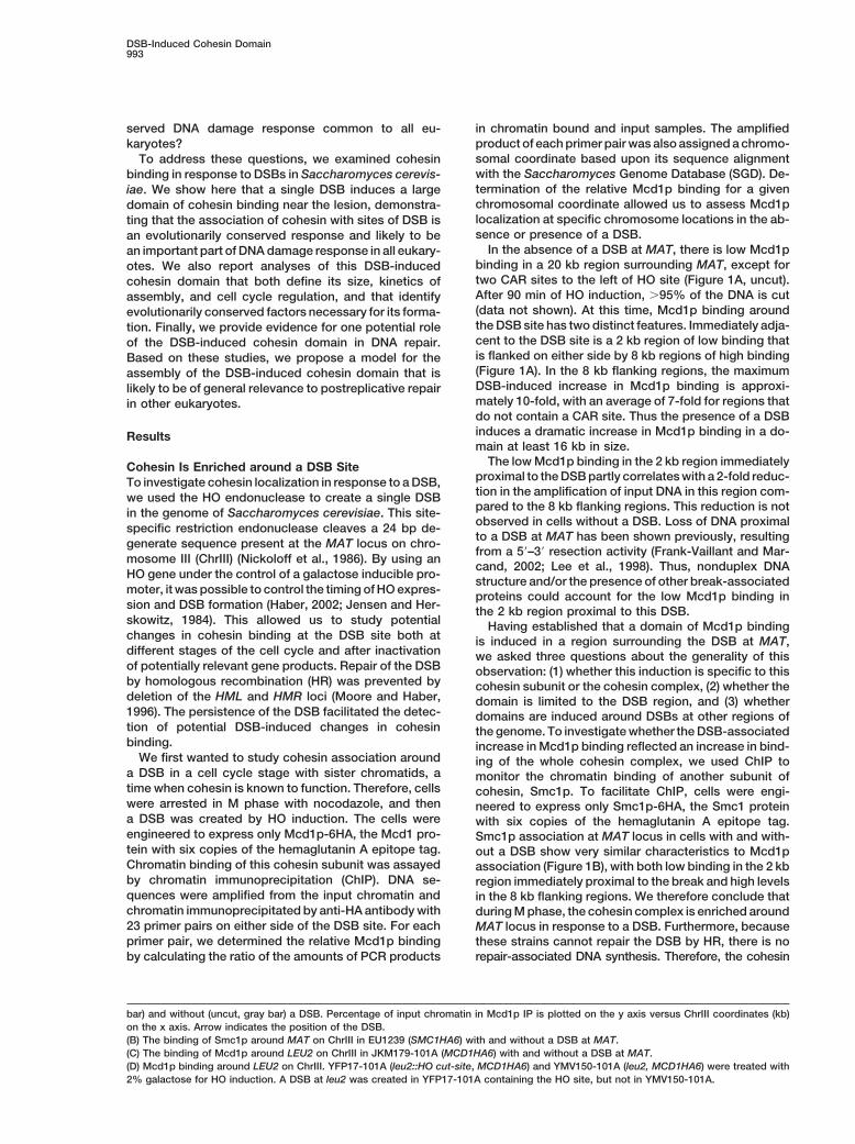
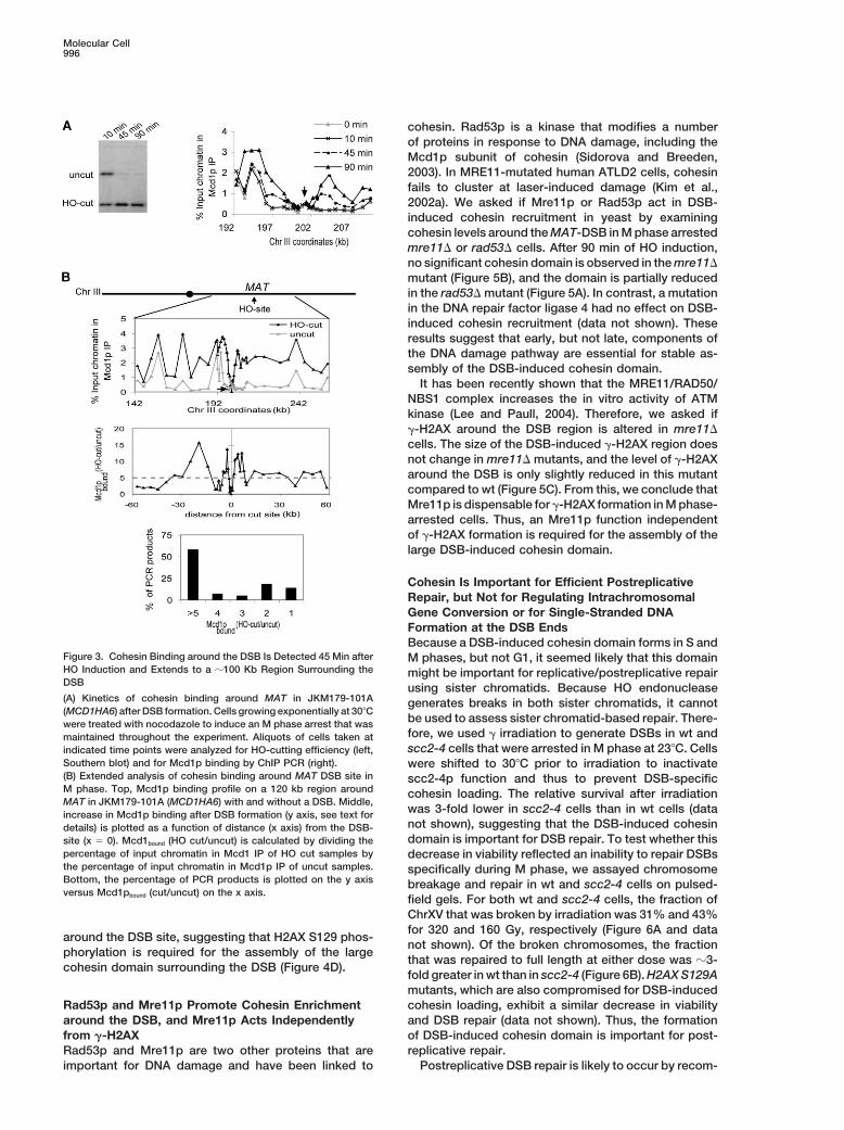
We examined a strain in which the HO recognition siteThe Assembly of a 100 Kb Cohesin Domain Lagsfrom MAT was deleted and moved to LEU2 (Paques etafter DSB Formation but Occurs Prioral., 1998), the region that fails to show increase into Completion of DSB Repaircohesin binding when the DSB is at MAT. In this region,To better characterize the DSB-induced cohesin do-cohesin binding was monitored on the right side of themain, we asked two additional questions: how large isDSB, because the left of HO site corresponds to �7 kbthe domain and how quickly does it form? To investigateregion of repetitive DNA within the Ty element. Whenthe kinetics of cohesin enrichment around the DSB site,the DSB is generated at LEU2, cohesin associationM phase-arrested cells were collected at different timearound the DSB increased in a manner very similar topoints after HO induction. The amount of HO-cut DNAthat observed at MAT (Figure 1D), with the exceptionwas determined by Southern blotting and the levels ofthat cohesin binding was elevated at the LEU2 DSBcohesin were compared by ChIP against Mcd1p subunit.even in the 2 kb region immediate proximal to the DSB.
After 10 min of HO induction, �50% of the cells con-This result shows that cohesins can bind near the endstain a DSB (Figure 3A, left). At this time point, there isof linear DNA molecules. Furthermore, these resultsno detectable increase in cohesin binding around thefrom the MAT and LEU2 loci suggest that a DSB any-DSB site. Increased cohesin binding is apparent 45 minwhere in the genome induces the formation of a localafter HO induction and intensifies at 90 min (Figure 3A,domain of cohesin.right). This result shows that there is a lag period forcohesin loading after the formation of a DSB, suggestingCohesin Enrichment around the DSB Is Cell Cyclethat cohesin loading may require a prior activation ofRegulated and Requires the Scc2p/Scc4p Complexanother DSB-induced event. In addition, in strains capa-
Given that the expression and function of the cohesinble of repair by HR, DSB-induced cohesin enrichment
complex is cell cycle regulated (Guacci et al., 1997;and HR occur in a similar time frame (data not shown).
Michaelis et al., 1997; Uhlmann and Nasmyth, 1998), we Thus, cohesin enrichment around the DSB occurs withinnext asked whether cohesin binding at a DSB is also a period required for repair by HR.cell cycle regulated. To address this question, we ar- Our initial studies on DSB induced cohesin domainrested cells at stages of the cell cycle other than M, examined a region limited to 10 kb on both sides of theinduced a DSB, and then examined cohesin binding. DSB at MAT locus. To see how far away from the DSB
In cells arrested in S phase by hydroxyurea treatment, site cohesin enrichment spreads, we looked at Mcd1pcohesin binding shows increase in the region of a DSB levels in M phase-arrested cells around the MAT locus.at MAT (Figure 2A) and LEU2 (data not shown), similar Cohesin binding in this region is low, making the ex-to the DSB-specific cohesin loading seen at these loci tended analysis of DSB-induced cohesin binding easierin M phase. In contrast, no enrichment of cohesin is (Blat and Kleckner, 1999; Glynn et al., 2004).observed surrounding the DSB site in G1-arrested cells 90 min after HO induction, the DSB-induced cohesin(data not shown). Thus, DSB-specific cohesin localiza- domain extends �40 kb to the left and �50 kb to thetion is cell cycle regulated and occurs in S and M, but right of the DSB (Figure 3B, top). More than half of thenot in G1, phase. This is consistent with the absence of sites assayed within the domain have at least a 5-foldthe cohesin subunit Mcd1p in G1 phase. increase in Mcd1p binding after induction of the DSB
The loading of cohesin on chromosomes at centro- (Figure 3B, bottom); sites with no increase in Mcd1pmeres and CAR sites during S phase requires the activity binding correspond to preexisting cohesin sites or se-of Scc2p/Scc4p complex (Ciosk et al., 2000). We asked quences immediately adjacent to the DSB (Figure 3B).whether Scc2p/Scc4p is also required for the DSB-spe- Sites with a greater than 5-fold increase are distributedcific loading of cohesin. To address this question, strains throughout the domain in regions of high GC contentcarrying the scc2-4 temperature-sensitive allele were and in both intra and intergenic regions (data not shown).arrested at permissive temperature during M phase, This data show that in response to a single DSB, cohes-allowing the loading of cohesin at centromeres and CAR ins bind apparently irrespective of sequence over a largesites during the preceding S phase. Indeed, the analysis domain around this lesion, about one-third of the size
of ChrIII.of cohesin binding at the CARC1 and CARC2 region is
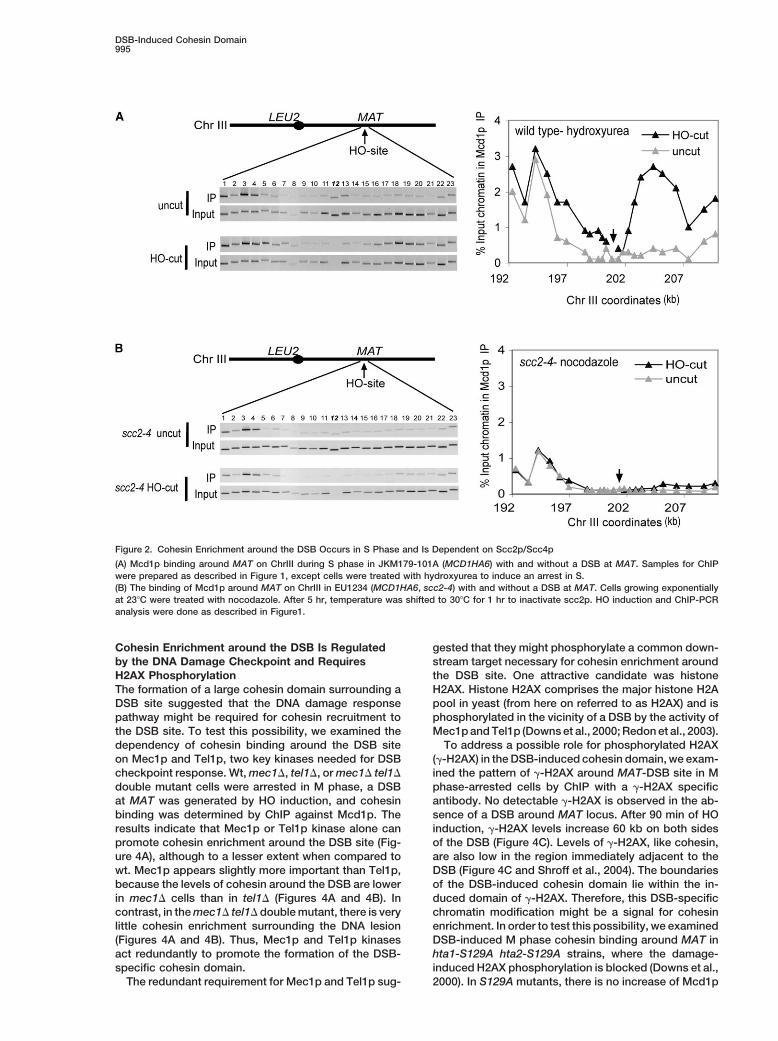
DSB-Induced Cohesin Domain995
Figure 2. Cohesin Enrichment around the DSB Occurs in S Phase and Is Dependent on Scc2p/Scc4p
(A) Mcd1p binding around MAT on ChrIII during S phase in JKM179-101A (MCD1HA6) with and without a DSB at MAT. Samples for ChIPwere prepared as described in Figure 1, except cells were treated with hydroxyurea to induce an arrest in S.(B) The binding of Mcd1p around MAT on ChrIII in EU1234 (MCD1HA6, scc2-4) with and without a DSB at MAT. Cells growing exponentiallyat 23�C were treated with nocodazole. After 5 hr, temperature was shifted to 30�C for 1 hr to inactivate scc2p. HO induction and ChIP-PCRanalysis were done as described in Figure1.
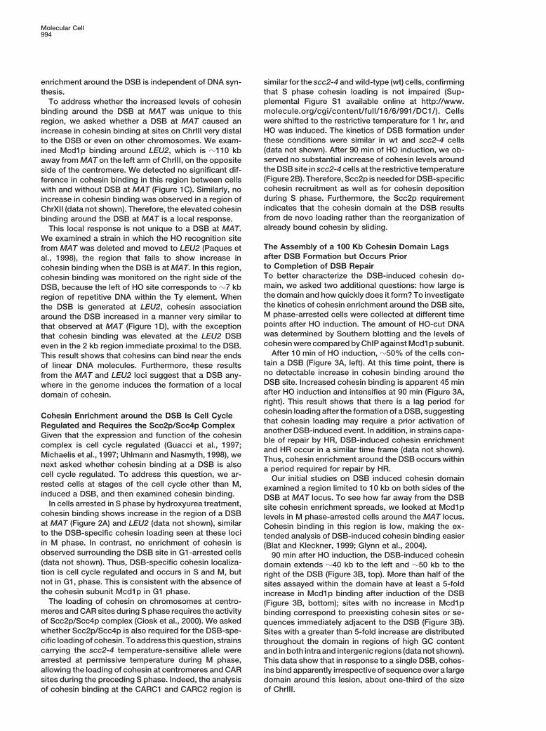
Cohesin Enrichment around the DSB Is Regulated gested that they might phosphorylate a common down-stream target necessary for cohesin enrichment aroundby the DNA Damage Checkpoint and Requires
H2AX Phosphorylation the DSB site. One attractive candidate was histoneH2AX. Histone H2AX comprises the major histone H2AThe formation of a large cohesin domain surrounding a
DSB site suggested that the DNA damage response pool in yeast (from here on referred to as H2AX) and isphosphorylated in the vicinity of a DSB by the activity ofpathway might be required for cohesin recruitment to
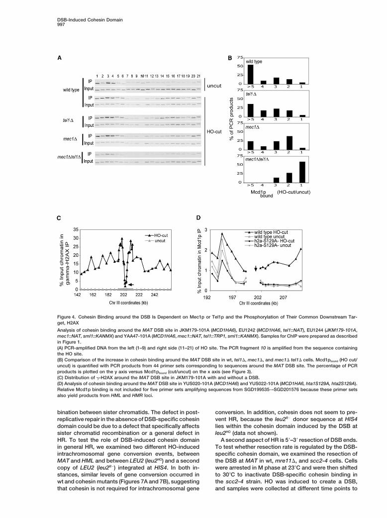
the DSB site. To test this possibility, we examined the Mec1p and Tel1p (Downs et al., 2000; Redon et al., 2003).To address a possible role for phosphorylated H2AXdependency of cohesin binding around the DSB site
on Mec1p and Tel1p, two key kinases needed for DSB (�-H2AX) in the DSB-induced cohesin domain, we exam-ined the pattern of �-H2AX around MAT-DSB site in Mcheckpoint response. Wt, mec1�, tel1�, or mec1� tel1�
double mutant cells were arrested in M phase, a DSB phase-arrested cells by ChIP with a �-H2AX specificantibody. No detectable �-H2AX is observed in the ab-at MAT was generated by HO induction, and cohesin
binding was determined by ChIP against Mcd1p. The sence of a DSB around MAT locus. After 90 min of HOinduction, �-H2AX levels increase 60 kb on both sidesresults indicate that Mec1p or Tel1p kinase alone can
promote cohesin enrichment around the DSB site (Fig- of the DSB (Figure 4C). Levels of �-H2AX, like cohesin,are also low in the region immediately adjacent to theure 4A), although to a lesser extent when compared to
wt. Mec1p appears slightly more important than Tel1p, DSB (Figure 4C and Shroff et al., 2004). The boundariesof the DSB-induced cohesin domain lie within the in-because the levels of cohesin around the DSB are lower
in mec1� cells than in tel1� (Figures 4A and 4B). In duced domain of �-H2AX. Therefore, this DSB-specificchromatin modification might be a signal for cohesincontrast, in the mec1� tel1� double mutant, there is very
little cohesin enrichment surrounding the DNA lesion enrichment. In order to test this possibility, we examinedDSB-induced M phase cohesin binding around MAT in(Figures 4A and 4B). Thus, Mec1p and Tel1p kinases
act redundantly to promote the formation of the DSB- hta1-S129A hta2-S129A strains, where the damage-induced H2AX phosphorylation is blocked (Downs et al.,specific cohesin domain.
The redundant requirement for Mec1p and Tel1p sug- 2000). In S129A mutants, there is no increase of Mcd1p
Molecular Cell996
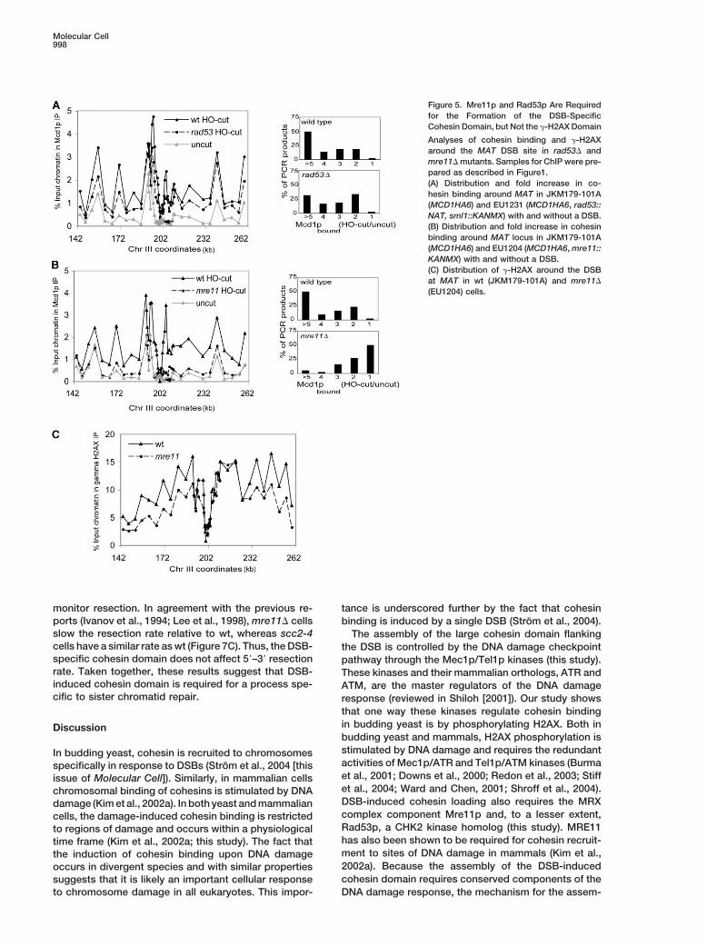
cohesin. Rad53p is a kinase that modifies a numberof proteins in response to DNA damage, including theMcd1p subunit of cohesin (Sidorova and Breeden,2003). In MRE11-mutated human ATLD2 cells, cohesinfails to cluster at laser-induced damage (Kim et al.,2002a). We asked if Mre11p or Rad53p act in DSB-induced cohesin recruitment in yeast by examiningcohesin levels around the MAT-DSB in M phase arrestedmre11� or rad53� cells. After 90 min of HO induction,no significant cohesin domain is observed in the mre11�mutant (Figure 5B), and the domain is partially reducedin the rad53� mutant (Figure 5A). In contrast, a mutationin the DNA repair factor ligase 4 had no effect on DSB-induced cohesin recruitment (data not shown). Theseresults suggest that early, but not late, components ofthe DNA damage pathway are essential for stable as-sembly of the DSB-induced cohesin domain.
It has been recently shown that the MRE11/RAD50/NBS1 complex increases the in vitro activity of ATMkinase (Lee and Paull, 2004). Therefore, we asked if�-H2AX around the DSB region is altered in mre11�cells. The size of the DSB-induced �-H2AX region doesnot change in mre11� mutants, and the level of �-H2AXaround the DSB is only slightly reduced in this mutantcompared to wt (Figure 5C). From this, we conclude thatMre11p is dispensable for �-H2AX formation in M phase-arrested cells. Thus, an Mre11p function independentof �-H2AX formation is required for the assembly of thelarge DSB-induced cohesin domain.
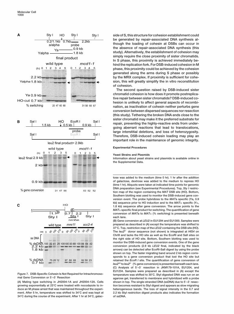
Cohesin Is Important for Efficient PostreplicativeRepair, but Not for Regulating IntrachromosomalGene Conversion or for Single-Stranded DNAFormation at the DSB EndsBecause a DSB-induced cohesin domain forms in S and
Figure 3. Cohesin Binding around the DSB Is Detected 45 Min after M phases, but not G1, it seemed likely that this domainHO Induction and Extends to a �100 Kb Region Surrounding the might be important for replicative/postreplicative repairDSB using sister chromatids. Because HO endonuclease(A) Kinetics of cohesin binding around MAT in JKM179-101A generates breaks in both sister chromatids, it cannot(MCD1HA6) after DSB formation. Cells growing exponentially at 30�C
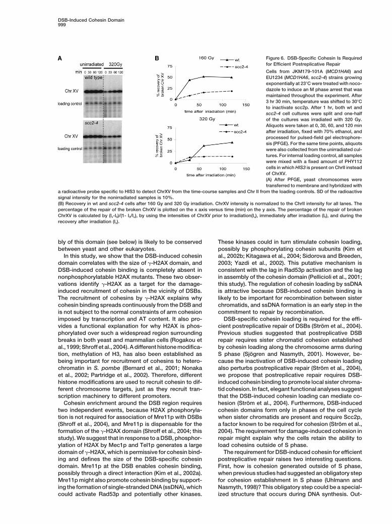
be used to assess sister chromatid-based repair. There-were treated with nocodazole to induce an M phase arrest that wasfore, we used � irradiation to generate DSBs in wt andmaintained throughout the experiment. Aliquots of cells taken atscc2-4 cells that were arrested in M phase at 23�C. Cellsindicated time points were analyzed for HO-cutting efficiency (left,
Southern blot) and for Mcd1p binding by ChIP PCR (right). were shifted to 30�C prior to irradiation to inactivate(B) Extended analysis of cohesin binding around MAT DSB site in scc2-4p function and thus to prevent DSB-specificM phase. Top, Mcd1p binding profile on a 120 kb region around cohesin loading. The relative survival after irradiationMAT in JKM179-101A (MCD1HA6) with and without a DSB. Middle,
was 3-fold lower in scc2-4 cells than in wt cells (dataincrease in Mcd1p binding after DSB formation (y axis, see text fornot shown), suggesting that the DSB-induced cohesindetails) is plotted as a function of distance (x axis) from the DSB-domain is important for DSB repair. To test whether thissite (x � 0). Mcd1bound (HO cut/uncut) is calculated by dividing the
percentage of input chromatin in Mcd1 IP of HO cut samples by decrease in viability reflected an inability to repair DSBsthe percentage of input chromatin in Mcd1p IP of uncut samples. specifically during M phase, we assayed chromosomeBottom, the percentage of PCR products is plotted on the y axis breakage and repair in wt and scc2-4 cells on pulsed-versus Mcd1pbound (cut/uncut) on the x axis.
field gels. For both wt and scc2-4 cells, the fraction ofChrXV that was broken by irradiation was 31% and 43%for 320 and 160 Gy, respectively (Figure 6A and data
around the DSB site, suggesting that H2AX S129 phos-not shown). Of the broken chromosomes, the fraction
phorylation is required for the assembly of the largethat was repaired to full length at either dose was �3-
cohesin domain surrounding the DSB (Figure 4D).fold greater in wt than in scc2-4 (Figure 6B). H2AX S129Amutants, which are also compromised for DSB-inducedcohesin loading, exhibit a similar decrease in viabilityRad53p and Mre11p Promote Cohesin Enrichment
around the DSB, and Mre11p Acts Independently and DSB repair (data not shown). Thus, the formationof DSB-induced cohesin domain is important for post-from �-H2AX
Rad53p and Mre11p are two other proteins that are replicative repair.Postreplicative DSB repair is likely to occur by recom-important for DNA damage and have been linked to
DSB-Induced Cohesin Domain997
Figure 4. Cohesin Binding around the DSB Is Dependent on Mec1p or Tel1p and the Phosphorylation of Their Common Downstream Tar-get, H2AX
Analysis of cohesin binding around the MAT DSB site in JKM179-101A (MCD1HA6), EU1242 (MCD1HA6, tel1::NAT), EU1244 (JKM179-101A,mec1::NAT, sml1::KANMX) and YAA47-101A (MCD1HA6, mec1::NAT, tel1::TRP1, sml1::KANMX). Samples for ChIP were prepared as describedin Figure 1.(A) PCR-amplified DNA from the left (1–9) and right side (11–21) of HO site. The PCR fragment 10 is amplified from the sequence containingthe HO site.(B) Comparison of the increase in cohesin binding around the MAT DSB site in wt, tel1�, mec1�, and mec1� tel1� cells. Mcd1pbound (HO cut/uncut) is quantified with PCR products from 44 primer sets corresponding to sequences around the MAT DSB site. The percentage of PCRproducts is plotted on the y axis versus Mcd1pbound (cut/uncut) on the x axis (see Figure 3).(C) Distribution of �-H2AX around the MAT DSB site in JKM179-101A with and without a DSB.(D) Analysis of cohesin binding around the MAT DSB site in YUS020-101A (MCD1HA6) and YUS022-101A (MCD1HA6, hta1S129A, hta2S129A).Relative Mcd1p binding is not included for five primer sets amplifying sequences from SGD199535→SGD201576 because these primer setsalso yield products from HML and HMR loci.
bination between sister chromatids. The defect in post- conversion. In addition, cohesin does not seem to pre-vent HR, because the leu2R� donor sequence at HIS4replicative repair in the absence of DSB-specific cohesin
domain could be due to a defect that specifically affects lies within the cohesin domain induced by the DSB atleu2HO (data not shown).sister chromatid recombination or a general defect in
HR. To test the role of DSB-induced cohesin domain A second aspect of HR is 5�–3� resection of DSB ends.To test whether resection rate is regulated by the DSB-in general HR, we examined two different HO-induced
intrachromosomal gene conversion events, between specific cohesin domain, we examined the resection ofthe DSB at MAT in wt, mre11�, and scc2-4 cells. CellsMAT and HML and between LEU2 (leu2HO) and a second
copy of LEU2 (leu2R�) integrated at HIS4. In both in- were arrested in M phase at 23�C and were then shiftedto 30�C to inactivate DSB-specific cohesin binding instances, similar levels of gene conversion occurred in
wt and cohesin mutants (Figures 7A and 7B), suggesting the scc2-4 strain. HO was induced to create a DSB,and samples were collected at different time points tothat cohesin is not required for intrachromosomal gene
Molecular Cell998
Figure 5. Mre11p and Rad53p Are Requiredfor the Formation of the DSB-SpecificCohesin Domain, but Not the �-H2AX Domain
Analyses of cohesin binding and �-H2AXaround the MAT DSB site in rad53� andmre11� mutants. Samples for ChIP were pre-pared as described in Figure1.(A) Distribution and fold increase in co-hesin binding around MAT in JKM179-101A(MCD1HA6) and EU1231 (MCD1HA6, rad53::NAT, sml1::KANMX) with and without a DSB.(B) Distribution and fold increase in cohesinbinding around MAT locus in JKM179-101A(MCD1HA6) and EU1204 (MCD1HA6, mre11::KANMX) with and without a DSB.(C) Distribution of �-H2AX around the DSBat MAT in wt (JKM179-101A) and mre11�
(EU1204) cells.
monitor resection. In agreement with the previous re- tance is underscored further by the fact that cohesinports (Ivanov et al., 1994; Lee et al., 1998), mre11� cells binding is induced by a single DSB (Strom et al., 2004).slow the resection rate relative to wt, whereas scc2-4 The assembly of the large cohesin domain flankingcells have a similar rate as wt (Figure 7C). Thus, the DSB- the DSB is controlled by the DNA damage checkpointspecific cohesin domain does not affect 5�–3� resection pathway through the Mec1p/Tel1p kinases (this study).rate. Taken together, these results suggest that DSB- These kinases and their mammalian orthologs, ATR andinduced cohesin domain is required for a process spe- ATM, are the master regulators of the DNA damagecific to sister chromatid repair. response (reviewed in Shiloh [2001]). Our study shows
that one way these kinases regulate cohesin bindingin budding yeast is by phosphorylating H2AX. Both inDiscussionbudding yeast and mammals, H2AX phosphorylation isstimulated by DNA damage and requires the redundantIn budding yeast, cohesin is recruited to chromosomesactivities of Mec1p/ATR and Tel1p/ATM kinases (Burmaspecifically in response to DSBs (Strom et al., 2004 [thiset al., 2001; Downs et al., 2000; Redon et al., 2003; Stiffissue of Molecular Cell]). Similarly, in mammalian cellset al., 2004; Ward and Chen, 2001; Shroff et al., 2004).chromosomal binding of cohesins is stimulated by DNADSB-induced cohesin loading also requires the MRXdamage (Kim et al., 2002a). In both yeast and mammaliancomplex component Mre11p and, to a lesser extent,cells, the damage-induced cohesin binding is restrictedRad53p, a CHK2 kinase homolog (this study). MRE11to regions of damage and occurs within a physiologicalhas also been shown to be required for cohesin recruit-time frame (Kim et al., 2002a; this study). The fact thatment to sites of DNA damage in mammals (Kim et al.,the induction of cohesin binding upon DNA damage2002a). Because the assembly of the DSB-inducedoccurs in divergent species and with similar propertiescohesin domain requires conserved components of thesuggests that it is likely an important cellular response
to chromosome damage in all eukaryotes. This impor- DNA damage response, the mechanism for the assem-
DSB-Induced Cohesin Domain999
Figure 6. DSB-Specific Cohesin Is Requiredfor Efficient Postreplicative Repair
Cells from JKM179-101A (MCD1HA6) andEU1234 (MCD1HA6, scc2-4) strains growingexponentially at 23�C were treated with noco-dazole to induce an M phase arrest that wasmaintained throughout the experiment. After3 hr 30 min, temperature was shifted to 30�Cto inactivate scc2p. After 1 hr, both wt andscc2-4 cell cultures were split and one-halfof the cultures was irradiated with 320 Gy.Aliquots were taken at 0, 30, 60, and 120 minafter irradiation, fixed with 70% ethanol, andprocessed for pulsed-field gel electrophore-sis (PFGE). For the same time points, aliquotswere also collected from the unirradiated cul-tures. For internal loading control, all sampleswere mixed with a fixed amount of PHY112cells in which HIS3 is present on ChrII insteadof ChrXV.(A) After PFGE, yeast chromosomes weretransferred to membrane and hybridized with
a radioactive probe specific to HIS3 to detect ChrXV from the time-course samples and Chr II from the loading controls. SD of the radioactivesignal intensity for the nonirradiated samples is 10%.(B) Recovery in wt and scc2-4 cells after 160 Gy and 320 Gy irradiation. ChrXV intensity is normalized to the ChrII intensity for all lanes. Thepercentage of the repair of the broken ChrXV is plotted on the x axis versus time (min) on the y axis. The percentage of the repair of brokenChrXV is calculated by (Ix-I0)/(1- I0/Iu), by using the intensities of ChrXV prior to irradiation(Iu), immediately after irradiation (I0), and during therecovery after irradiation (Ix).
bly of this domain (see below) is likely to be conserved These kinases could in turn stimulate cohesin loading,possibly by phosphorylating cohesin subunits (Kim etbetween yeast and other eukaryotes.
In this study, we show that the DSB-induced cohesin al., 2002b; Kitagawa et al., 2004; Sidorova and Breeden,2003; Yazdi et al., 2002). This putative mechanism isdomain correlates with the size of �-H2AX domain, and
DSB-induced cohesin binding is completely absent in consistent with the lag in Rad53p activation and the lagin assembly of the cohesin domain (Pellicioli et al., 2001;nonphosphorylatable H2AX mutants. These two obser-
vations identify �-H2AX as a target for the damage- this study). The regulation of cohesin loading by ssDNAis attractive because DSB-induced cohesin binding isinduced recruitment of cohesin in the vicinity of DSBs.
The recruitment of cohesins by �-H2AX explains why likely to be important for recombination between sisterchromatids, and ssDNA formation is an early step in thecohesin binding spreads continuously from the DSB and
is not subject to the normal constraints of arm cohesion commitment to repair by recombination.DSB-specific cohesin loading is required for the effi-imposed by transcription and AT content. It also pro-
vides a functional explanation for why H2AX is phos- cient postreplicative repair of DSBs (Strom et al., 2004).Previous studies suggested that postreplicative DSBphorylated over such a widespread region surrounding
breaks in both yeast and mammalian cells (Rogakou et repair requires sister chromatid cohesion establishedby cohesin loading along the chromosome arms duringal., 1999; Shroff et al., 2004). A different histone modifica-
tion, methylation of H3, has also been established as S phase (Sjogren and Nasmyth, 2001). However, be-cause the inactivation of DSB-induced cohesin loadingbeing important for recruitment of cohesins to hetero-
chromatin in S. pombe (Bernard et al., 2001; Nonaka also perturbs postreplicative repair (Strom et al., 2004),we propose that postreplicative repair requires DSB-et al., 2002; Partridge et al., 2002). Therefore, different
histone modifications are used to recruit cohesin to dif- induced cohesin binding to promote local sister chroma-tid cohesion. In fact, elegant functional analyses suggestferent chromosome targets, just as they recruit tran-
scription machinery to different promoters. that the DSB-induced cohesin loading can mediate co-hesion (Strom et al., 2004). Furthermore, DSB-inducedCohesin enrichment around the DSB region requires
two independent events, because H2AX phosphoryla- cohesin domains form only in phases of the cell cyclewhen sister chromatids are present and require Scc2p,tion is not required for association of Mre11p with DSBs
(Shroff et al., 2004), and Mre11p is dispensable for the a factor known to be required for cohesion (Strom et al.,2004). The requirement for damage-induced cohesion information of the �-H2AX domain (Shroff et al., 2004; this
study). We suggest that in response to a DSB, phosphor- repair might explain why the cells retain the ability toload cohesins outside of S phase.ylation of H2AX by Mec1p and Tel1p generates a large
domain of �-H2AX, which is permissive for cohesin bind- The requirement for DSB-induced cohesin for efficientpostreplicative repair raises two interesting questions.ing and defines the size of the DSB-specific cohesin
domain. Mre11p at the DSB enables cohesin binding, First, how is cohesion generated outside of S phase,when previous studies had suggested an obligatory steppossibly through a direct interaction (Kim et al., 2002a).
Mre11p might also promote cohesin binding by support- for cohesion establishment in S phase (Uhlmann andNasmyth, 1998)? This obligatory step could be a special-ing the formation of single-stranded DNA (ssDNA), which
could activate Rad53p and potentially other kinases. ized structure that occurs during DNA synthesis. Out-
Molecular Cell1000
side of S, this structure for cohesion establishment couldbe generated by repair-associated DNA synthesis al-though the loading of cohesin at DSBs can occur inthe absence of repair-associated DNA synthesis (thisstudy). Alternatively, the establishment of cohesion maysimply require the close proximity of sister chromatids.In S phase, this proximity is achieved immediately be-hind the replication fork. For DSB-induced cohesion in Mphase, this proximity could be achieved by the cohesiongenerated along the arms during S phase or possiblyby the MRX complex. If proximity is sufficient for cohe-sion, this will greatly simplify the in vitro reconstitutionof cohesion.
The second question raised by DSB-induced sisterchromatid cohesion is how does it promote postreplica-tive repair between sister chromatids? DSB-induced co-hesion is unlikely to affect general aspects of recombi-nation, as inactivation of cohesin neither perturbs geneconversion between dispersed sequences nor resection(this study). Tethering the broken DNA ends close to thesister chromatid may make it the preferred substrate forrepair, preventing the highly-reactive ends from under-going aberrant reactions that lead to translocations,large interstitial deletions, and loss of heterozygosity.Therefore, DSB-induced cohesin loading may play animportant role in the maintenance of genomic integrity.
Experimental Procedures
Yeast Strains and PlasmidsInformation about yeast strains and plasmids is available online inthe Supplemental Data.
tose was added to the medium (time 0 hr). 1 hr after the additionof galactose, dextrose was added to the medium to repress HO(time 1 hr). Aliquots were taken at indicated time points for genomicDNA preparation (see Experimental Procedures). Top, Sty I restric-tion map of the region containing the MAT DSB site (HO). Bottom,Southern blotting was used to monitor the DSB-induced gene con-version event. The probe hybridizes to the MATa specific (Ya, 0.9kb) sequence prior to HO induction and to the MAT� specific (Y�,1.8 kb) sequence after gene conversion. The arrow points to theMAT� specific final product for switching. The quantification of geneconversion of MATa to MAT� (% switching) is presented beneatheach lane.(B) Gene conversion at LEU2 in EU1294 and EU1300. Samples wereprepared as described in (A) except the temperature was shifted to37�C. Top, restriction map of the LEU2 containing the DSB site (HO).The leu2R� donor sequence (not shown) is integrated at HIS4 onChrIII and lacks the HO site as well as the EcoRI and SalI sites onthe right side of HO site. Bottom, Southern blotting was used tomonitor the DSB-induced gene conversion events. One of the geneconversion products (2.9 kb LEU2 final, indicated by the blackarrows) can be detected after EcoRI-SalI digest by using the probeshown on top. The faster migrating band around 2 kb region corre-sponds to a gene conversion product that lost the HO site butretained the EcoR I site. The quantification of gene conversion ofleu2HO to leu2R� (% gene conversion) is presented beneath each lane.(C) Analysis of 5�–3� resection in JKM179-101A, EU1204, andEU1234. Samples were prepared as described in (A) except the
Figure 7. DSB-Specific Cohesin Is Not Required for Intrachromoso- temperature was shifted to 30�C. StyI digested DNA was run on anmal Gene Conversion or 5�–3� Resection agarose gel, transferred to membrane and hybridized with a probe(A) Mating type switching in JH2004-1A and JH2004-12A. Cells shown in top. The single-stranded DNA (ssDNA) due to 5�–3� resec-growing exponentially at 23�C were treated with nocodazole to in- tion becomes resistant to StyI digest and appears as slow migratingduce an M phase arrest that was maintained throughout the experi- heterogeneous bands. The loss of signal intensity in the 0.7 andment. After 5 hr, temperature was shifted to 34�C and was kept at 2.2 kb StyI restriction digest products also indicates the formation34�C during the course of the experiment. After 1 hr at 34�C, galac- of ssDNA.
DSB-Induced Cohesin Domain1001
Cell Synchronization for communicating results prior to publication; Yixian Zhang, OrnaCohen-Fix, Margaret Hoang, Hong-Guo Yu, and Dongli Huang forExponentially dividing cell cultures were grown in YEP media
(Guthrie and Fink, 1991) with 3% glycerol (EMD, 30% v/v stock) and constructive comments on the manuscript; Eileen Hogan, CathyMistrot, Ellen Cammon, and Patricia Cammon for technical support;2% lactic acid (Fisher, 40% v/v stock [pH 5.7]) and then arrested in
G1, S, or M phase by the addition of 3 10�6 M �-factor (Sigma), and Terence Murphy, Judith Yanowitz, and members of our labora-tories for advice and helpful discussions. D.K. is supported by grants130 mM hydroxyurea (HU) (Sigma), or 15 g/ml nocodazole (Sigma),
respectively. Cells used for irradiation experiments were grown in from the Howard Hughes Medical Institutes and J.E.H is supportedby National Institutes of Health grants GM20056 and GM61766 andYEP media supplemented with 2% dextrose (EM Science, 20%
w/v stock). by Department of Energy grant 01ER63229.
Culture Conditions for HO Induction Received: July 27, 2004For HO induction, cells were grown to approximately 0.6 107 cells/ Revised: October 6, 2004ml. In strains with a stably integrated GAL10::HO sequence, cells Accepted: November 5, 2004were grown in YEP media with 3% glycerol and 2% lactic acid. In Published: December 21, 2004strains carrying the GAL10::HO sequence on a centromeric plasmid,selective synthetic medium supplemented with 3% glycerol and 2% Referenceslactic acid was used to retain the plasmid. Cells were then arrestedas described above, and subsequently D-galactose (Sigma, 20%w/v Bernard, P., Maure, J.F., Partridge, J.F., Genier, S., Javerzat, J.P.,stock) was added to the culture at a final concentration of 2% w/v. and Allshire, R.C. (2001). Requirement of heterochromatin for cohe-For analysis of gene conversion, in order to repress HO, dextrose sion at centromeres. Science 294, 2539–2542.was added to the culture at a final concentration of 2% w/v, 1 hr
Birkenbihl, R.P., and Subramani, S. (1992). Cloning and characteriza-after D-galactose addition.tion of rad21 an essential gene of Schizosaccharomyces pombeinvolved in DNA double-strand-break repair. Nucleic Acids Res.ChIP20, 6605–6611.ChIP was performed as described (Megee et al., 1999). Immunopre-Blat, Y., and Kleckner, N. (1999). Cohesins bind to preferential sitescipitations (IPs) were done with 12CA5 anti-HA antibody (Roche) oralong yeast chromosome III, with differential regulation along armsrabbit polyclonal antiserum against the yeast-specific H2AX phos-versus the centric region. Cell 98, 249–259.phopeptide (kindly provided by Christophe Redon, NCI, NIH). Input
DNA was diluted 100-fold (HA IP) or 6.25-fold (�-H2AX IP) relative Bradbury, J.M., and Jackson, S.P. (2003). The complex matter ofto immunoprecipitated DNA before PCR analysis. DNA double-strand break detection. Biochem. Soc. Trans. 31,
40–44.PCR and Data Analysis Burma, S., Chen, B.P., Murphy, M., Kurimasa, A., and Chen, D.J.Information about the primers used in this study is available upon (2001). ATM phosphorylates histone H2AX in response to DNA dou-request. A 50 l PCR reaction contained 3 l template DNA, 50 mM ble-strand breaks. J. Biol. Chem. 276, 42462–42467.KCl, 10 mM Tris-HCl ([pH 9.0] at 25�C), 0.1% Triton X-100, 1.5 mM
Ciosk, R., Shirayama, M., Shevchenko, A., Tanaka, T., Toth, A., andMgCl2, 0.2 mM of each dNTP, 50 pmole of each oligonucleotide,Nasmyth, K. (2000). Cohesin’s binding to chromosomes dependsand 2.5 units of Taq polymerase (Promega). PCR reactions were 26on a separate complex consisting of Scc2 and Scc4 proteins. Mol.cycles of 95�C, 30 s; 57�C, 45 s; and 72�C, 60 s done with a PTC-Cell 5, 243–254.200 Peltier thermal cycler from MJ research. PCR products wereD’Amours, D., and Jackson, S.P. (2002). The Mre11 complex: at theresolved on 2.5% agarose gels (NuSieve) in 1 TBE buffer with 0.15crossroads of dna repair and checkpoint signalling. Nat. Rev. Mol.g/ml ethidium bromide. Gel images were acquired by using theCell Biol. 3, 317–327.Q-image digital imaging gel documentation system, and the band
intensities were quantified by using IP Labs software. All experi- Digweed, M., Reis, A., and Sperling, K. (1999). Nijmegen breakagements were done at least twice and a representative dataset is syndrome: consequences of defective DNA double strand breakshown. repair. Bioessays 21, 649–656.
Downs, J.A., Lowndes, N.F., and Jackson, S.P. (2000). A role forDSB Analysis Saccharomyces cerevisiae histone H2A in DNA repair. NatureGenomic DNA was prepared as described (Sambrook and Russell, 408, 1001–1004.2001). The restriction enzymes used for HO cutting, gene conversion
Frank-Vaillant, M., and Marcand, S. (2002). Transient stability of DNAand 5�–3� resection analysis are StyI (New England Biolabs), EcoRI,ends allows nonhomologous end joining to precede homologousHindIII, and SalI (Roche). The digested fragments were resolved onrecombination. Mol. Cell 10, 1189–1199.agarose gels (SeaKem ME agarose) and transferred to GeneScreenGlynn, E.F., Megee, P.C., Yu, H., Mistrot, C., Unal, E., Koshland,Plus hybridization membrane either by vacuum or capillary wetD.E., DeRisi, J.D., and Gerton, J.L. (2004). Genome-Wide Mappingtransfer. The DNA probes used for Southern blotting are preparedof the Cohesin Complex in the Yeast Saccharomyces cerevisiae.by Amersham DNA labeling beads (�dCTP) and purified by Amer-PLoS Biol 2, 1325–1339.sham G-50 Micro Columns. Blot radioactivity was detected with a
Molecular Dynamics Phosphor Imager and quantified with IP Labs Guacci, V., Koshland, D., and Strunnikov, A. (1997). A direct linksoftware. between sister chromatid cohesion and chromosome condensation
revealed through the analysis of MCD1 in S. cerevisiae. Cell 91,PFGE Analysis 47–57.Total chromosomal DNA agarose plugs were prepared as described Guthrie, C., and Fink, G.R. eds. (1991). Guide to Yeast Genetics and(Schwartz and Cantor, 1984) and resolved on a 1% agarose gel by Molecular Biology (New York: Academic Press, Inc.).using Bio-Rad CHEF-DR III system at 6 V/cm for 24 hr with a switch
Haber, J.E. (2002). Uses and abuses of HO endonuclease. Methodstime ramped from 60 to 120 s at 14�C.Enzymol. 350, 141–164.
Heo, S.J., Tatebayashi, K., Kato, J., and Ikeda, H. (1998). The RHC21Gamma Irradiationgene of budding yeast, a homologue of the fission yeast rad21�Gamma irradiation was done by using a 137Cs source with a radiationgene, is essential for chromosome segregation. Mol. Gen. Genet.dose of 16 Gy/min (Mark I irradiator; J.L. Shepherd and Associates).257, 149–156.
Ivanov, E.L., Sugawara, N., White, C.I., Fabre, F., and Haber, J.E.Acknowledgments(1994). Mutations in XRS2 and RAD50 delay but do not preventmating-type switching in Saccharomyces cerevisiae. Mol. Cell. Biol.We thank Christophe Redon for providing the �-H2AX antiserum and
for his help during the gamma irradiation studies; Camilla Sjogren 14, 3414–3425.
Molecular Cell1002
Jackson, S.P. (2002). Sensing and repairing DNA double-strand base chromatin domains involved in DNA double-strand breaksin vivo. J. Cell Biol. 146, 905–916.breaks. Carcinogenesis 23, 687–696.
Rotman, G., and Shiloh, Y. (1998). ATM: from gene to function. Hum.Jensen, R.E., and Herskowitz, I. (1984). Directionality and regulationMol. Genet. 7, 1555–1563.of cassette substitution in yeast. Cold Spring Harb. Symp. Quant.
Biol. 49, 97–104. Rouse, J., and Jackson, S.P. (2002). Interfaces between the detec-tion, signaling, and repair of DNA damage. Science 297, 547–551.Kadyk, L.C., and Hartwell, L.H. (1992). Sister chromatids are pre-
ferred over homologs as substrates for recombinational repair in Sambrook, J., and Russell, D.W. (2001). Molecular Cloning: A Labo-Saccharomyces cerevisiae. Genetics 132, 387–402. ratory Manual, Third Edition (Cold Spring Harbor, NY: Cold Spring
Harbor Laboratory Press).Kim, J.S., Krasieva, T.B., LaMorte, V., Taylor, A.M., and Yokomori,K. (2002a). Specific recruitment of human cohesin to laser-induced Schwartz, D.C., and Cantor, C.R. (1984). Separation of yeast chro-DNA damage. J. Biol. Chem. 277, 45149–45153. mosome-sized DNAs by pulsed field gradient gel electrophoresis.
Cell 37, 67–75.Kim, S.T., Xu, B., and Kastan, M.B. (2002b). Involvement of thecohesin protein, Smc1, in Atm-dependent and independent re- Shiloh, Y. (2001). ATM and ATR: networking cellular responses tosponses to DNA damage. Genes Dev. 16, 560–570. DNA damage. Curr. Opin. Genet. Dev. 11, 71–77.
Kitagawa, R., Bakkenist, C.J., McKinnon, P.J., and Kastan, M.B. Shiloh, Y. (2003). ATM and related protein kinases: safeguarding(2004). Phosphorylation of SMC1 is a critical downstream event in genome integrity. Nat. Rev. Cancer 3, 155–168.the ATM-NBS1-BRCA1 pathway. Genes Dev. 18, 1423–1438. Shroff, R., Arbel-Eden, A., Pilch, D., Ira, G., Bonner, W.M., Petrini,Koshland, D.E., and Guacci, V. (2000). Sister chromatid cohesion: J.H., Haber, J.E., and Lichten, M. (2004). Distribution and dynamicsthe beginning of a long and beautiful relationship. Curr. Opin. Cell of chromatin modification induced by a defined DNA double-strandBiol. 12, 297–301. break. Curr. Biol. 14, 1703–1711.
Sidorova, J.M., and Breeden, L.L. (2003). Rad53 checkpoint kinaseLaloraya, S., Guacci, V., and Koshland, D. (2000). Chromosomalphosphorylation site preference identified in the Swi6 protein ofaddresses of the cohesin component Mcd1p. J. Cell Biol. 151, 1047–Saccharomyces cerevisiae. Mol. Cell. Biol. 23, 3405–3416.1056.
Sjogren, C., and Nasmyth, K. (2001). Sister chromatid cohesion isLee, J.H., and Paull, T.T. (2004). Direct activation of the ATM proteinrequired for postreplicative double-strand break repair in Saccharo-kinase by the Mre11/Rad50/Nbs1 complex. Science 304, 93–96.myces cerevisiae. Curr. Biol. 11, 991–995.Lee, S.E., Moore, J.K., Holmes, A., Umezu, K., Kolodner, R.D., andSonoda, E., Matsusaka, T., Morrison, C., Vagnarelli, P., Hoshi, O.,Haber, J.E. (1998). Saccharomyces Ku70, mre11/rad50 and RPAUshiki, T., Nojima, K., Fukagawa, T., Waizenegger, I.C., Peters, J.M.,proteins regulate adaptation to G2/M arrest after DNA damage. Cellet al. (2001). Scc1/Rad21/Mcd1 is required for sister chromatid co-94, 399–409.hesion and kinetochore function in vertebrate cells. Dev. Cell 1,Megee, P.C., Mistrot, C., Guacci, V., and Koshland, D. (1999). The759–770.centromeric sister chromatid cohesion site directs Mcd1p bindingStiff, T., O’Driscoll, M., Rief, N., Iwabuchi, K., Lobrich, M., and Jeggo,to adjacent sequences. Mol. Cell 4, 445–450.P.A. (2004). ATM and DNA-PK function redundantly to phosphory-Michaelis, C., Ciosk, R., and Nasmyth, K. (1997). Cohesins: chromo-late H2AX after exposure to ionizing radiation. Cancer Res. 64, 2390–somal proteins that prevent premature separation of sister chroma-2396.tids. Cell 91, 35–45.Strom, L., Betts Lindroos, H., Shirahige, K., and Sjogren, C. (2004).Michelson, R.J., and Weinert, T. (2000). Closing the gaps among aPostreplicative recruitment of cohesin to double-strand breaks isweb of DNA repair disorders. Bioessays 22, 966–969.required for DNA repair. Mol. Cell 16, this issue, 1003–1015.
Moore, J.K., and Haber, J.E. (1996). Capture of retrotransposonTanaka, T., Cosma, M.P., Wirth, K., and Nasmyth, K. (1999). Identifi-DNA at the sites of chromosomal double-strand breaks. Naturecation of cohesin association sites at centromeres and along chro-383, 644–646.mosome arms. Cell 98, 847–858.
Nasmyth, K. (2002). Segregating sister genomes: the molecular biol-Uhlmann, F., and Nasmyth, K. (1998). Cohesion between sister chro-
ogy of chromosome separation. Science 297, 559–565.matids must be established during DNA replication. Curr. Biol. 8,
Nickoloff, J.A., Chen, E.Y., and Heffron, F. (1986). A 24-base-pair 1095–1101.DNA sequence from the MAT locus stimulates intergenic recombina-
Ward, I.M., and Chen, J. (2001). Histone H2AX is phosphorylated intion in yeast. Proc. Natl. Acad. Sci. USA 83, 7831–7835.
an ATR-dependent manner in response to replicational stress. J.Nonaka, N., Kitajima, T., Yokobayashi, S., Xiao, G., Yamamoto, M., Biol. Chem. 276, 47759–47762.Grewal, S.I., and Watanabe, Y. (2002). Recruitment of cohesin to
Yazdi, P.T., Wang, Y., Zhao, S., Patel, N., Lee, E.Y., and Qin, J.heterochromatic regions by Swi6/HP1 in fission yeast. Nat. Cell Biol.
(2002). SMC1 is a downstream effector in the ATM/NBS1 branch of4, 89–93.
the human S-phase checkpoint. Genes Dev. 16, 571–582.Paques, F., Leung, W.Y., and Haber, J.E. (1998). Expansions andcontractions in a tandem repeat induced by double-strand breakrepair. Mol. Cell. Biol. 18, 2045–2054.
Partridge, J.F., Scott, K.S., Bannister, A.J., Kouzarides, T., and Allsh-ire, R.C. (2002). cis-acting DNA from fission yeast centromeres medi-ates histone H3 methylation and recruitment of silencing factorsand cohesin to an ectopic site. Curr. Biol. 12, 1652–1660.
Pellicioli, A., Lee, S.E., Lucca, C., Foiani, M., and Haber, J.E. (2001).Regulation of Saccharomyces Rad53 checkpoint kinase during ad-aptation from DNA damage-induced G2/M arrest. Mol. Cell 7,293–300.
Redon, C., Pilch, D.R., Rogakou, E.P., Orr, A.H., Lowndes, N.F., andBonner, W.M. (2003). Yeast histone 2A serine 129 is essential forthe efficient repair of checkpoint-blind DNA damage. EMBO Rep.4, 678–684.
Rogakou, E.P., Pilch, D.R., Orr, A.H., Ivanova, V.S., and Bonner,W.M. (1998). DNA double-stranded breaks induce histone H2AXphosphorylation on serine 139. J. Biol. Chem. 273, 5858–5868.
Rogakou, E.P., Boon, C., Redon, C., and Bonner, W.M. (1999). Mega-


