Embed Size (px)
Citation preview
DNA-Length-Dependent Quenching of Fluorescently Labeled IronOxide Nanoparticles with Gold, Graphene Oxide and MoS2NanostructuresMustafa Balcioglu,† Muhit Rana,† Neil Robertson,† and Mehmet V. Yigit*,†,‡
†Department of Chemistry and RNA Institute, University at Albany, SUNY, 1400 Washington Avenue, Albany, New York 12222,United States‡College of Nanoscale Science & Engineering, University at Albany, SUNY, 257 Fuller Road, Albany, New York 12203, United States
*S Supporting Information
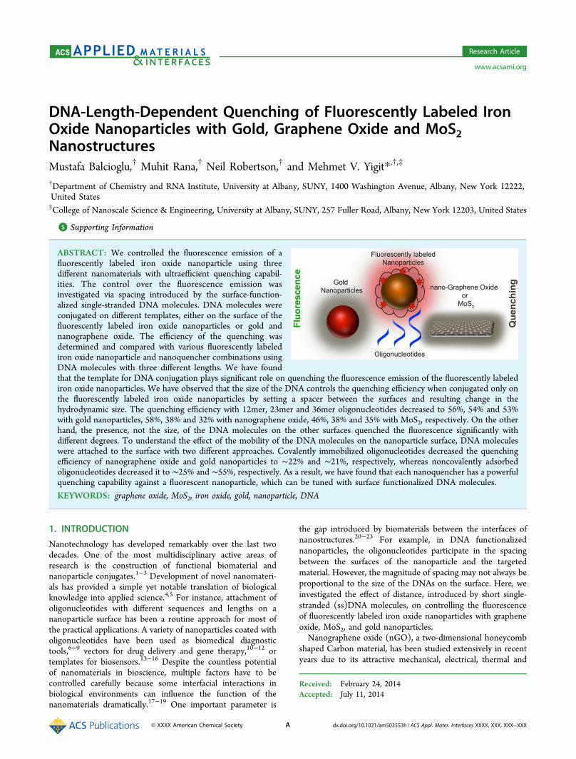
ABSTRACT: We controlled the fluorescence emission of afluorescently labeled iron oxide nanoparticle using threedifferent nanomaterials with ultraefficient quenching capabil-ities. The control over the fluorescence emission wasinvestigated via spacing introduced by the surface-function-alized single-stranded DNA molecules. DNA molecules wereconjugated on different templates, either on the surface of thefluorescently labeled iron oxide nanoparticles or gold andnanographene oxide. The efficiency of the quenching wasdetermined and compared with various fluorescently labelediron oxide nanoparticle and nanoquencher combinations usingDNA molecules with three different lengths. We have foundthat the template for DNA conjugation plays significant role on quenching the fluorescence emission of the fluorescently labelediron oxide nanoparticles. We have observed that the size of the DNA controls the quenching efficiency when conjugated only onthe fluorescently labeled iron oxide nanoparticles by setting a spacer between the surfaces and resulting change in thehydrodynamic size. The quenching efficiency with 12mer, 23mer and 36mer oligonucleotides decreased to 56%, 54% and 53%with gold nanoparticles, 58%, 38% and 32% with nanographene oxide, 46%, 38% and 35% with MoS2, respectively. On the otherhand, the presence, not the size, of the DNA molecules on the other surfaces quenched the fluorescence significantly withdifferent degrees. To understand the effect of the mobility of the DNA molecules on the nanoparticle surface, DNA moleculeswere attached to the surface with two different approaches. Covalently immobilized oligonucleotides decreased the quenchingefficiency of nanographene oxide and gold nanoparticles to ∼22% and ∼21%, respectively, whereas noncovalently adsorbedoligonucleotides decreased it to ∼25% and ∼55%, respectively. As a result, we have found that each nanoquencher has a powerfulquenching capability against a fluorescent nanoparticle, which can be tuned with surface functionalized DNA molecules.
KEYWORDS: graphene oxide, MoS2, iron oxide, gold, nanoparticle, DNA
1. INTRODUCTION
Nanotechnology has developed remarkably over the last twodecades. One of the most multidisciplinary active areas ofresearch is the construction of functional biomaterial andnanoparticle conjugates.1−3 Development of novel nanomateri-als has provided a simple yet notable translation of biologicalknowledge into applied science.4,5 For instance, attachment ofoligonucleotides with different sequences and lengths on ananoparticle surface has been a routine approach for most ofthe practical applications. A variety of nanoparticles coated witholigonucleotides have been used as biomedical diagnostictools,6−9 vectors for drug delivery and gene therapy,10−12 ortemplates for biosensors.13−16 Despite the countless potentialof nanomaterials in bioscience, multiple factors have to becontrolled carefully because some interfacial interactions inbiological environments can influence the function of thenanomaterials dramatically.17−19 One important parameter is
the gap introduced by biomaterials between the interfaces ofnanostructures.20−23 For example, in DNA functionalizednanoparticles, the oligonucleotides participate in the spacingbetween the surfaces of the nanoparticle and the targetedmaterial. However, the magnitude of spacing may not always beproportional to the size of the DNAs on the surface. Here, weinvestigated the effect of distance, introduced by short single-stranded (ss)DNA molecules, on controlling the fluorescenceof fluorescently labeled iron oxide nanoparticles with grapheneoxide, MoS2, and gold nanoparticles.Nanographene oxide (nGO), a two-dimensional honeycomb
shaped Carbon material, has been studied extensively in recentyears due to its attractive mechanical, electrical, thermal and
Received: February 24, 2014Accepted: July 11, 2014
Research Article
www.acsami.org
© XXXX American Chemical Society A dx.doi.org/10.1021/am503553h | ACS Appl. Mater. Interfaces XXXX, XXX, XXX−XXX
optical properties.24−27 nGO has remarkable potential inbiomedical applications28 due to its exceptional ssDNAadsorption29 and ultraefficient fluorescence quenching capa-bilities.30−32 These properties have attracted significantattention to nGO for biosensor development,33−41 biomarkerdetection42,43 and antisense gene delivery.44−46 Currently,DNA functionalized graphene-based systems are under heavyinvestigation for numerous purposes,4 including engineeringfunctional hybrid nanostructures.31 Therefore, the interactionof the interfaces of DNA functionalized nGOs needs to bestudied carefully for construction of graphene-based biosys-tems. MoS2, which is a two-dimensional graphene-like material,has recently received great attention due to its exciting physicalproperties comparable to nGO.50 It has been used fornanoelectronics, optoelectronics, transistor designs, energystoring devices and biosensing systems.47,48 Similar to nGO,MoS2 quenches the fluorescence of fluorophore labeledmaterials, which makes them attractive two-dimensionalnanosized templates for photonic or biomedical applications.49
Gold nanoparticles (AuNP) have several common propertieswith nGO and MoS2 regardless of the entirely different three-dimensional structure and elemental composition. Goldnanoparticles have been studied considerably due to theirunique physical properties.50−52 Several analytical and bio-medical studies have demonstrated that (1) both AuNP andnGO are ultraefficient fluorescence quenchers53−56 and (2)ssDNA molecules can be immobilized or adsorbed on bothsurfaces very efficiently via specific chemical interactions oraromatic stacking.57−60 Because the attachment of DNAmolecules constructs a biological shield on the surface ofthese two nanoparticles, nanoquenchers, we investigatedwhether this barrier can be used to control the fluorescenceemission of a fluorescent nanoparticle.Dextran coated fluorescently labeled iron oxide nanoparticles
are a class of imaging agents that are suitable for biomedicalapplications.61−63 They have been used for many differentapplications including whole body in vivo optical and MRIimaging.8,64 The strong NIR fluorescence signals of thesenanoparticles (MNcy5.5 − cy5.5 dye labeled form) providenoninvasive optical tissue imaging in live animals. Theattachment of siRNA or antisense miRNA molecules providesadditional therapeutic functionality, which makes these nano-particles attractive particularly for theranostics.65 Due to itsmultiple functionalities along with the biocompatible andbiodegradable nature, MNcy5.5 has been one of the major toolsfor theranostics in biomedicine.66,67 Here, we controlled thefluorescence emission of MNcy5.5 using nGO, MoS2 and AuNPs,promising theranostic nanomaterials, with surface-function-alized ssDNA molecules. The covalently or noncovalentlyattached ssDNA molecules on the nanoparticle surface serve asbiological shields between the interfaces, which could increasethe hydrodynamic sizes of the nanoparticles.Although significant effort has been devoted to study the
biophysical properties and the applications of individualoligonucleotide-functionalized nanoparticles, their interactionwith other nanoparticle types with systematic spacing has notbeen investigated in depth.20−23 We observed that the surfacebound short ssDNA molecules interfere with the interactionbetween nanoparticle interfaces however the length of the DNAdoes not always determine the degree of interaction. We believethat the coverage and orientation of DNA molecules on thesurface, and the three-dimensional structures of the nanoma-
terials, could be important factors in their biophysicochemicalinterfacial interactions.
2. EXPERIMENTAL SECTION2.1. Materials. All DNA sequences were purchased from
Integrated DNA Technologies (IDT), Coralville, IA, USA, with thefollowing sequence information and modifications, Thiol modifiedsequences: (1) 12mer, 5′- /5ThioMC6-D/ CCC AGG TTC TCT -3′;(2) 23mer, 5′- /5ThioMC6-D/ CAC AAA TTC GGT TCT ACAGGG TA-3′; (3) 36mer, 5′- /5ThioMC6-D/ TAC GAG TTG AGACCG TTA AGA CGA GGC AAT CAT GCA-3′. Amine modifiedsequences: (4) 12mer, 5′- /5AmMC6/ CCC AGG TTC TCT -3′; (5)23mer, 5′- /5AmMC6/ CAC AAA TTC GGT TCT ACA GGG TA-3′; (6) 36mer, 5′- /5AmMC6/ TAC GAG TTG AGA CCG TTAAGA CGA GGC AAT CAT GCA-3′. (7) Mercury Aptamer, 5′- /TTCTTT CTT CCC CTT GTT TGT T-3′.
Carboxyl graphene water dispersion was purchased from ACSMaterial, Medford, MA, USA. Dextan-T10 was purchased fromPharmacosmos, Holbaek, Denmark. cy5.5 monoreactive NHS esterwas purchased from GE Healthcare, Piscataway, NJ, USA. MoS2powder (<2 μm) was purchased from Sigma-Aldrich. All otherreagents were purchased form from Sigma-Aldrich, St. Louis, MO,USA, and used without further purification. Double distilled water wasused in preparation of all solutions.
2.2. Synthesis of cy5.5 Labeled Iron Oxide Nanoparticlesand Functionalization with Oligonucleotides. Synthesis of ironoxide nanoparticle (MN) was adapted from our earlier publication.31
The cy5.5 monoreactive NHS ester (1 mg) was dissolved in 200 μL ofdimethyl sulfoxide (DMSO) and incubated with 1 mL of thenanoparticles (MN, 10.0 mg/mL Fe) in 20 mM sodium citrate buffer(pH 8.0) overnight. The labeled nanoparticles (MNcy5.5) were purifiedusing Sephadex PD-10 column (GE Healthcare) against 10 mMphosphate buffered saline (PBS, pH 7.4). The number of cy5.5 dyemolecules per nanoparticle was determined as 10−12 by UVspectroscopy as described previously.68 The hydrodynamic size ofnanoparticle was determined as 33.1 (±4.1 nm) using dynamic lightscattering (DLS) measurements with DynaPro Titan (WyattTechnology Corporation, Goleta, CA, USA).
The covalent conjugation of DNA to MNcy5.5 was performedthrough a heterobifunctional linker SPDP (N-succinimidyl 3-(2-pyridyldithio)propionate)68,69 purchased from Pierce Biotechnology,Rockford, IL, USA. Briefly, 10 mg of succinimidyl 3-(2-pyridyldithio)-propionate (SPDP) was dissolved in 500 μL of anhydrous DMSO andincubated with MNcy5.5. The thiolated 5′ terminus of theoligonucleotides was activated with 3% tris(2-carboxyethyl)phosphine)(TCEP) treatment in nuclease-free 10 mM PBS (pH 7.4). The DNAmolecules were purified using microspin G-25 columns (GEHealthcare). After TCEP-activation and purification, the oligonucleo-tides were resuspended in 10 mM PBS (pH 7.4) and incubated withthe SPDP conjugated MNcy5.5 overnight. The resulting probe waspurified using a disposable MACS separation magnetic columns;Miltenyi Biotec Inc., Auburn, CA, USA. The DNA per MNcy5.5 wasprepared in a 10:1 molar ratio as described previously.68
2.3. Synthesis of Gold Nanoparticles and Functionalizationwith Oligonucleotides. The negatively charged citrate-stabilizedgold nanoparticles were synthesized and functionalized witholigonucleotides according to previous reports.70 Briefly, 2 mL of 50mM HAuCl4 was added into 98 mL of boiling DI water in anErlenmeyer flask on a hot plate. 10 mL of 38.8 mM sodium citratesolution was rapidly added into the stirring mixture. The reaction isstopped until the solution turned a wine-red color. The solution wascooled to room temperature and stored at 4 °C. The hydrodynamicsize of nanoparticle was determined as 13 nm (±1.4 nm) usingdynamic light scattering (DLS) measurements with DynaPro Titan(Wyatt Technology Corporation, Goleta, CA, USA).
Immobilization of Oligonucleotides on AuNP. 10 μL of 1 mMTCEP-treated thiol-modified DNA molecules was incubated with 3mL of AuNP overnight at room temperature (RT).70 Next, 100 mMNaCl was added into the DNA and AuNP mixture with gentle stirring.
ACS Applied Materials & Interfaces Research Article
dx.doi.org/10.1021/am503553h | ACS Appl. Mater. Interfaces XXXX, XXX, XXX−XXXB
The samples were stored in the dark for 24 h. The samples were thencentrifuged at 15000 rpm for 15 min at RT. The supernatant wasremoved and discarded. The AuNP−DNA conjugate at the bottom ofthe microcentrifuge tube was dispersed in 100 mM PBS (pH 7.4) and150 mM NaCl. The samples were washed and centrifuged two timesto ensure complete removal of free DNA. Finally, the AuNP−DNAconjugate was resuspended in 100 mM PBS (pH 7.4) and 150 mMNaCl.Noncovalant Adsorption of Oligonucleotides on AuNP. 2.5 μL of
1 mM unmodified DNA stock solution was dissolved in 50 μL of 100mM MES buffer (pH 5.6). The DNA solution was then added to 450μL of citrate-stabilized AuNPs. After 20 min of incubation at RT, MESbuffer pH 5.6 (final conc. 10 mM MES, 100 mM NaCl) was added tostabilize the DNA adsorption on AuNP. The sample was incubated foranother 10 min. Finally, the DNA−AuNP mixture was centrifuged at15000 rpm, and the supernatant was discarded. The pellet was washedwith 10 mM PBS pH 7.4 to remove any excess free DNA. Thecentrifugation and washing procedure was repeated two times toensure complete removal of free DNA. Finally AuNP-DNA conjugatewas resuspended in 100 mM PBS (pH 7.4) and 150 mM NaCl.Mercury detection was performed with 2.5 μL of 1 mM mercury
aptamer incubated with or without 1 μM mercury(II) perchlorate for10 min. The aptamer solutions were incubated with 450 μL of citrate-stabilized AuNPs for 20 min. After the washing steps, the MNcy5.5suspension was titrated with AuNPs and the fluorescence wasmeasured as described below.2.4. Preparation of Nanographene Oxide (nGO) and MoS2
via Sonication and Functionalization with Oligonucleotides.To prepare nanometer sized GO (0.5 mg/mL) and MoS2 (0.5 mg/mL), the starting materials were dispersed in DI water and sonicatedwith the ultrasonic processor for 7 h (120 W 20 kHz with pulse on for2 s and pulse off for 4 s) in an ice bath to prevent overheating
generated from sonication. The average particle size of the obtainednGO and MoS2 was determined as 100 ± 15 nm using dynamic lightscattering (DLS) DynaPro Titan (Wyatt Technology Corporation,Goleta, CA, USA). The transmission electron microscopy (TEM)micrographs were taken using a JEOL JEM-2010 transmission electronmicroscope. The characterization of each nanomaterial is provided inFigure S8 (Supporting Information).
Immobilization of Oligonucleotides on nGO. The couplingreaction was carried out in a glass vial with 200 μL of 500 μg/mLnGO, 2 μL of 1 mM amine-modified DNA, 2 mg of EDC (N-(3-(dimethylamino)propyl)-N′-ethylcarbodiimide hydrochloride) and 25mM MES (pH 5.6) and 25 mM NaCl.23 The mixture was rotated atRT for 3 h using a Glas-Col mini rotator. The solutions werecentrifuged at 15000 rpm for 20 min to remove nonattached DNAs.The GO−DNA conjugates were then washed with 500 μL of DI watertwice to further remove nonassociated DNAs. Finally, the conjugateswere redispersed in 1 mL of 25 mM (4-(2-hydroxyethyl)-1-piperazineethanesulfonic acid) (HEPES), 100 mM NaCl (pH 7.6)and with a final nGO concentration of 100 μg/mL. The samples werestored at 4 °C before use. Sonication was performed occasionally toassist redispersing.
Noncovalent Adsorption of Oligonucleotides on nGO. 2 μM ofDNA was incubated with nGO (100 μg/mL) in buffer (25 mMHEPES, 100 mM NaCl, pH 7.6) for 1 h at RT.23 Nonadsorbed DNAswere removed by centrifugation and washing cycles as describedabove. Finally, the purified nGO−DNA complex was resuspended inbuffer (25 mM HEPES, 100 mM NaCl, pH 7.6).
2.5. Fluorescence Measurements. The fluorescence measure-ments (excitation, 670 nm; emission, 695 nm) were performed usingthe Fluorolog-3 spectrofluorometer (Horiba Jobin-Yvon, Inc., Edison,NJ, USA). A typical experiment was performed with 20 nM MNcy5.5 in
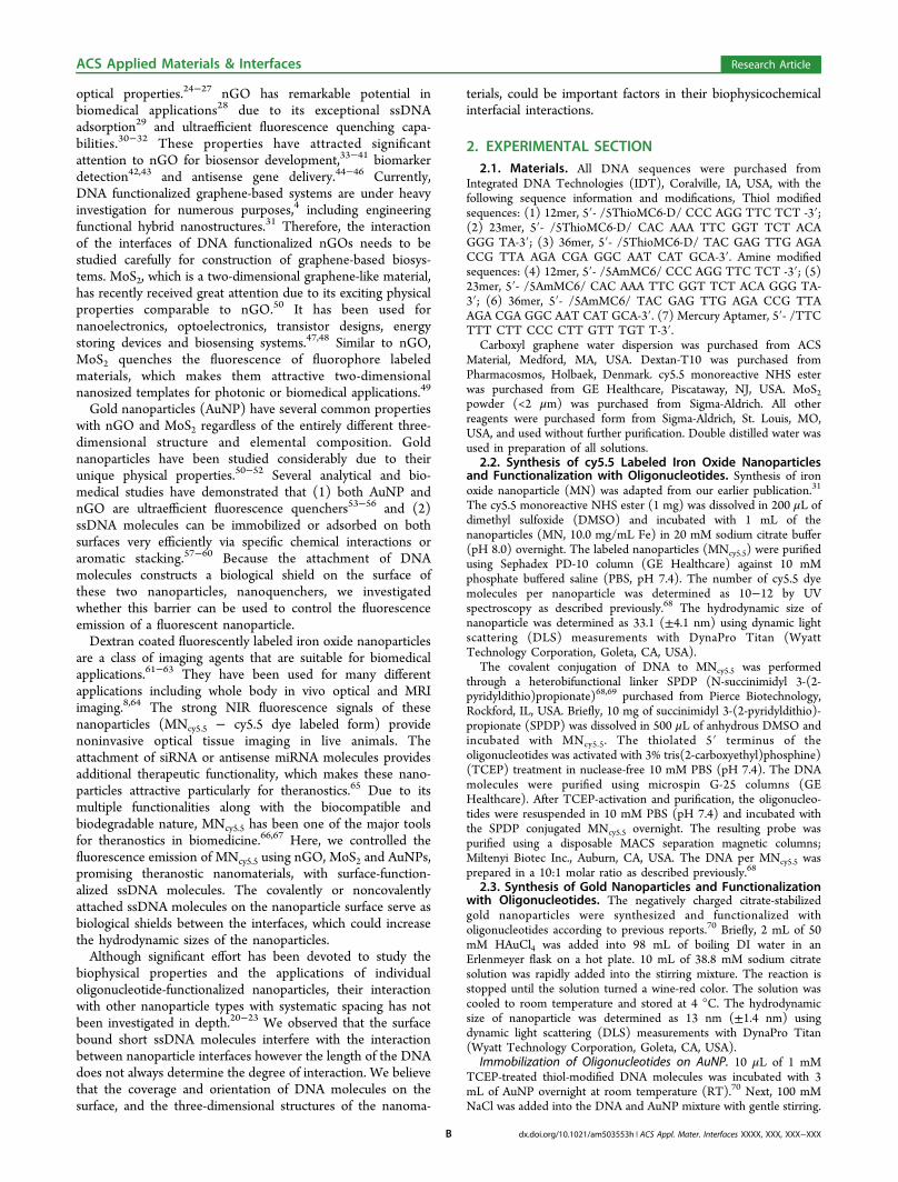
Figure 1. AuNP, nGO or MoS2 quenches the fluorescence emission of cy5.5 labeled iron oxide nanoparticles, MNcy5.5. (a) A schematicrepresentation of the interaction between the MNcy5.5 and nanoquenchers demonstrates the quenching of the cy5.5 fluorophore on MNcy5.5. (b)Fluorescence emission spectra of 20 nM MNcy5.5 (orange line) with 4 nM AuNP (red line), 1.8 μg/mL nGO (blue line) or 20 μg/mL MoS2 (greenline). (c) Titration of MNcy5.5 with AuNP, nGO or MoS2 at 200th second of the time-dependent fluorescence measurements at 695 nm. Thefluorescence signals were quenched immediately upon titration with nanoquenchers and stabilized with no significant change overtime.
ACS Applied Materials & Interfaces Research Article
dx.doi.org/10.1021/am503553h | ACS Appl. Mater. Interfaces XXXX, XXX, XXX−XXXC
10 mM PBS (pH 7.4) in a 2 mL quartz cuvette with gentle stirring.The fluorescence emission value at this concentration was determinedto be approximately ∼6.5 × 10⁶. The kinetic studies on MNcy5.5 wereperformed on for 30 min with one reading per 3 s resolution. At the200 s time point, 1.8 μg/mL of nGO, 20 μg/mL of MoS2 or 4 nM ofAuNP was added into the MNcy5.5 to monitor the quenching efficiencyof these nanomaterials on the fluorescence of MNcy5.5.The quenching efficiency was calculated according to following
equation.23 Q (%) = [1 − Fo/F] where Q is the quenching efficiency,Fo is the fluorescence intensity in the absence of quencher and F is thefluorescence intensity in the presence of quencher.Fluorescence lifetime measurements were performed on MNcy5.5
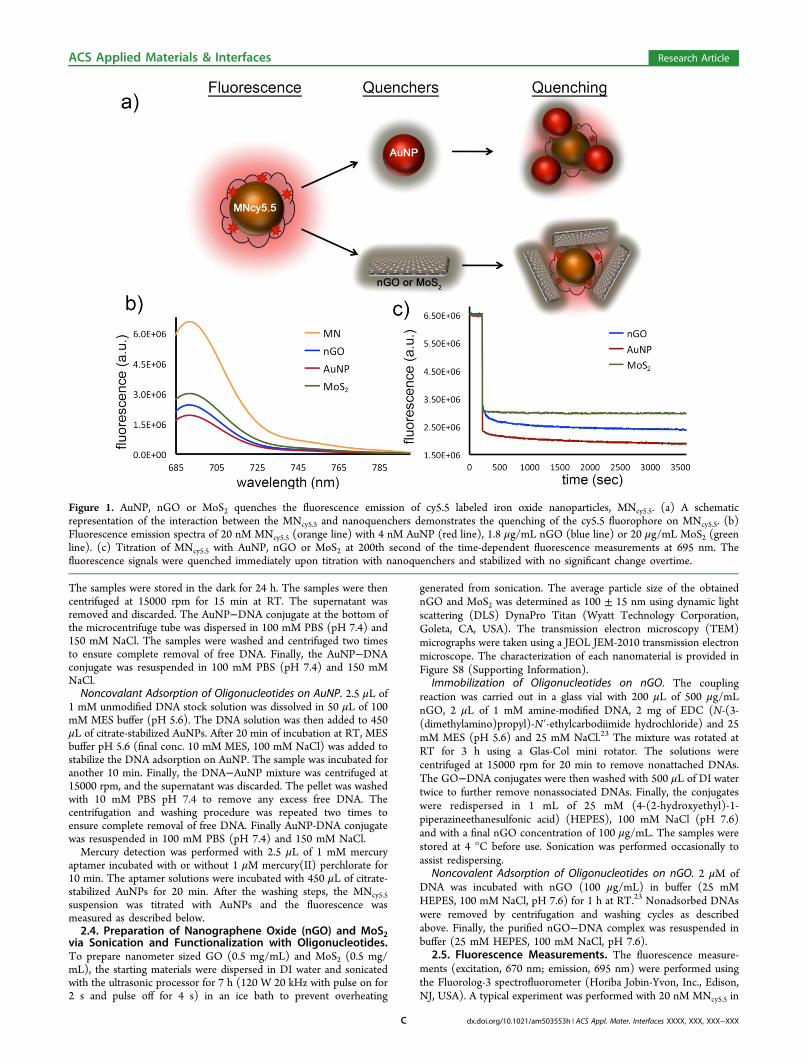
with nGO, MoS2 and AuNPs using time-correlated single-photoncounting (TCSPC) spectroscopy on Fluorolog Horiba Jobin Yvonequipment with NanoLED (λ = 635 ± 10 nm) excitation sources. Thefluorescence lifetime values were analyzed by deconvoluting theinstrument response function with biexponential decay using DAS6decay analysis software. The quality of the fit has been judged by thefitting parameters such as χ2 (<1.2). Stern−Volmer Plots weredetermined by τ0/τ vs concentration of nanoquenchers.
3. RESULTS AND DISCUSSION
Here, we investigated the interaction of AuNP, nGO or MoS2with fluorescently labeled iron oxide nanoparticles (MNcy5.5) ina distance-dependent manner using fluorescence spectroscopy.The spacing between the species was controlled using shortssDNA molecules, which changed the hydrodynamic size of thenanoparticles or served as biological shields between interfaces.Comparisons were made between two nanomaterials; oneserving as fluorescence quenchers, nanoquenchers: AuNPs,nGO or MoS2, and the other, MNcy5.5, serving as a fluorescencecarrier.First, we investigated the quenching of bare MNcy5.5 with
nGO, MoS2 or AuNP, (Figure 1a). MNcy5.5 presents a strong
steady fluorescence at pH 7.4, (Figure 1b). After the titrationwith nGO, MoS2 or AuNP solutions, significantly weakerfluorescence emission was observed due to fluorescencequenching, (Figure 1b). The emission spectra were collectedon 20 nM MNcy5.5 before and after the titration with 1.8 μg/mLnGO, 4 nM AuNP or 20 μg/mL MoS2. These nanoquencherconcentrations were determined to be ideal for obtaining theoptimum quenching yield with a minimum nanoquencherconcentration, Figure S1 (Supporting Information). Beyondthese concentrations, slightly higher fluorescence quenchingwas observed; however, the nanoquencher concentrations inthese emission ranges were dramatically higher, particularly forMoS2. This effect could be due to the saturation of the MNcy5.5fluorescence emission environment with the nanoquenchers inthe solution.The kinetics studies were performed similarly by titration of
MNcy5.5 with AuNP, nGO or MoS2 suspension and monitoredfor 1 h. It has been previously shown that the bare nGO, AuNPand MNcy5.5 interact with each other through electrostaticinteraction, which leads to an immediate quenching of thefluorescence.31 As seen in Figure 1c, the fluorescence wasquenched immediately and stabilized shortly after the nano-quencher titration without any major fluctuation. We observedthat AuNP, nGO and MoS2 have similar quenching perform-ance on MNcy5.5 fluorescence in this experimental condition.The fluorescence lifetime measurements were performed onMNcy5.5 with different concentrations of AuNP, nGO or MoS2as seen in Figure 2. The results showed that the nanoquenchersresult in a decrease in the fluorescence lifetime of MNcy5.5 (0.84ns) with increasing concentrations, (Figure 2a,b,c and Table S1,Supporting Information). All the decays were faster thanMNcy5.5 only, which indicates the presence of dynamic
Figure 2. Fluorescence lifetime decay traces of MNcy5.5 with different concentrations of (a) AuNP, (b) nGO and (c) MoS2. Stern−Volmer plots of(d) AuNP,( e) nGO and( f) MoS2 show a linear correlation in the insets. Time calibration =5.578 023 × 10−2 nanosecond/channel. Note: theconcentration vs volume table is provided in Figure S1 (Supporting Information). Nanoquencher volume: 10 μL of stock MoS2 (620 μg/mL), nGO(72 μg/mL) and AuNP (84 nM) solution is equivalent to 3.1 μg/mL, 0.4 μg/mL and 0.4 nM final concentrations, respectively.
ACS Applied Materials & Interfaces Research Article
dx.doi.org/10.1021/am503553h | ACS Appl. Mater. Interfaces XXXX, XXX, XXX−XXXD
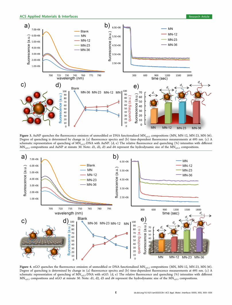
Figure 3. AuNP quenches the fluorescence emission of unmodified or DNA functionalized MNcy5.5 compositions (MN, MN-12, MN-23, MN-36).Degree of quenching is determined by change in (a) fluorescence spectra and (b) time-dependent fluorescence measurements at 695 nm. (c) Aschematic representation of quenching of MNcy5.5-DNA with AuNP. (d, e) The relative fluorescence and quenching (%) intensities with differentMNcy5.5 compositions and AuNP at minute 30. Note: d1, d2, d3 and d4 represent the hydrodynamic size of the MNcy5.5 compositions.
Figure 4. nGO quenches the fluorescence emission of unmodified or DNA functionalized MNcy5.5 compositions (MN, MN-12, MN-23, MN-36).Degree of quenching is determined by change in (a) fluorescence spectra and (b) time-dependent fluorescence measurements at 695 nm. (c) Aschematic representation of quenching of MNcy5.5-DNA with nGO. (d, e) The relative fluorescence and quenching (%) intensities with differentMNcy5.5 compositions and nGO at minute 30. Note: d1, d2, d3 and d4 represent the hydrodynamic size of the MNcy5.5 compositions.
ACS Applied Materials & Interfaces Research Article
dx.doi.org/10.1021/am503553h | ACS Appl. Mater. Interfaces XXXX, XXX, XXX−XXXE
quenching caused by the nanoquenchers.23 As discussed above(Figure S1, Supporting Information), after a threshold thechange in the fluorescence lifetime lost the linear correlationwith increasing concentration, which could be due to thesaturation with nanoquenchers, or the presence of differentquenching environments possibly due the limited diffusion ofspherical or two-dimensional nanoquenchers into the polymercoating of the MNcy5.5 with multiple fluorophores (Figures2d−f).Afterward, in order to understand the macromolecular
spacing effect of immobilized oligonucleotides on theinteraction between the nanoquenchers and MNcy5.5, weattached three different sizes of single-stranded DNA(ssDNA-12-23-36mer) molecules on the surface of MNcy5.5.The dynamic light scattering measurements indicated that thehydrodynamic size of the MNcy5.5 increased with the attach-ment of ssDNA on the surface and a greater increase wasobserved with longer ssDNA sequences, (Figure S2, SupportingInformation). First, we investigated the interaction between theMNcy5.5-DNA and unmodified spherical AuNPs, (Figure 3c).The fluorescence emission spectra indicate that AuNPs quenchthe MNcy5.5-DNA dramatically regardless of the size of threedifferent DNA molecules, (Figure 3a). However, a significantdifference between the emission spectra of MNcy5.5-DNA andbare MNcy5.5 was observed. The kinetics studies showed thatthe quenching of the fluorescence signal is very rapid andstabilizes quickly after titration with AuNP, (Figure 3b). Wehave also represented our observation by the plotting relativequenching and fluorescence with different MNcy5.5 composi-tions, (Figure 3d,e). The overall observations indicate thatthere is an efficient and fast quenching of the fluorescencesignals in all four cases with a greater quenching efficiency withunmodified MNcy5.5. The immobilized oligonucleotides influ-ence the quenching yield of MNcy5.5, which, however, wasindependent of the size of DNAs. We speculate that DNAlength-independent quenching of MNcy5.5 at these particularDNA sizes could be due to the (1) greater size of MNcy5.5 overAuNP and (2) relatively small number of DNAs on the MNcy5.5
surface. We postulate that the negatively charged goldnanoparticles can diffuse into the grooves of the positivelycharged MNcy5.5 surface regardless of the size of immobilizedDNAs and interfere with the emission of cy5.5 on the surface.Moreover, due to the absorption of ssDNA on AuNP viaspecific chemical interactions,15,19 the ssDNA on the MNcy5.5-DNA could contribute to the interaction between thenanoparticles.
Later, we investigated the interaction between the MNcy5.5-DNA and unmodified planar nGO (Figure 4c). Thefluorescence of the different MNcy5.5 compositions aresignificantly affected by extraordinary quenching capability ofnGO. We observed that nGO has a higher quenching efficiencyon unmodified MNcy5.5 compared to the MNcy5.5-DNAcompositions (Figure 4a). The kinetics studies indicate thatthe quenching of the fluorescence signal is very fast andstabilizes shortly after the addition of bare nGO (Figure 4b).Unlike gold nanoparticles, the length of the immobilized DNAmolecules on MNcy5.5-DNA surface played a significant role inaltering the magnitude of fluorescence with bare nGO (Figure4d,e). We postulate that the large two-dimensional planarsurface of nGO is unlikely to diffuse into MNcy5.5 grooves andinteract with the nanoparticle surface. In contrast, the MNcy5.5-DNA interacts with the planar surface of the bare nGO, andthus DNA coating creates spacing between surfaces (Figure4c). Overall, results suggest that the size of the immobilizedDNA molecules plays a critical role in separating twonanoparticle surfaces.Similarly, we studied a graphene-like material, MoS2, which
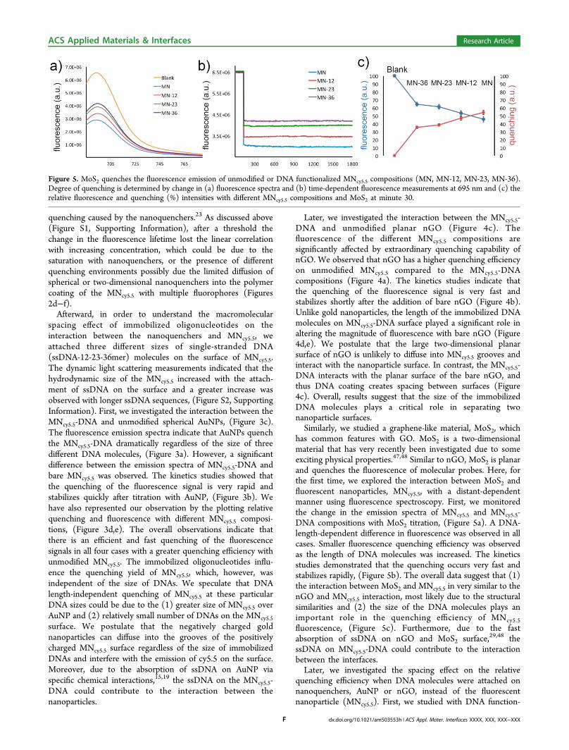
has common features with GO. MoS2 is a two-dimensionalmaterial that has very recently been investigated due to someexciting physical properties.47,48 Similar to nGO, MoS2 is planarand quenches the fluorescence of molecular probes. Here, forthe first time, we explored the interaction between MoS2 andfluorescent nanoparticles, MNcy5.5, with a distant-dependentmanner using fluorescence spectroscopy. First, we monitoredthe change in the emission spectra of MNcy5.5 and MNcy5.5-DNA compositions with MoS2 titration, (Figure 5a). A DNA-length-dependent difference in fluorescence was observed in allcases. Smaller fluorescence quenching efficiency was observedas the length of DNA molecules was increased. The kineticsstudies demonstrated that the quenching occurs very fast andstabilizes rapidly, (Figure 5b). The overall data suggest that (1)the interaction between MoS2 and MNcy5.5 in very similar to thenGO and MNcy5.5 interaction, most likely due to the structuralsimilarities and (2) the size of the DNA molecules plays animportant role in the quenching efficiency of MNcy5.5fluorescence, (Figure 5c). Furthermore, due to the fastabsorption of ssDNA on nGO and MoS2 surface,29,48 thessDNA on MNcy5.5-DNA could contribute to the interactionbetween the interfaces.Later, we investigated the spacing effect on the relative
quenching efficiency when DNA molecules were attached onnanoquenchers, AuNP or nGO, instead of the fluorescentnanoparticle (MNcy5.5). First, we studied with DNA function-
Figure 5. MoS2 quenches the fluorescence emission of unmodified or DNA functionalized MNcy5.5 compositions (MN, MN-12, MN-23, MN-36).Degree of quenching is determined by change in (a) fluorescence spectra and (b) time-dependent fluorescence measurements at 695 nm and (c) therelative fluorescence and quenching (%) intensities with different MNcy5.5 compositions and MoS2 at minute 30.
ACS Applied Materials & Interfaces Research Article
dx.doi.org/10.1021/am503553h | ACS Appl. Mater. Interfaces XXXX, XXX, XXX−XXXF
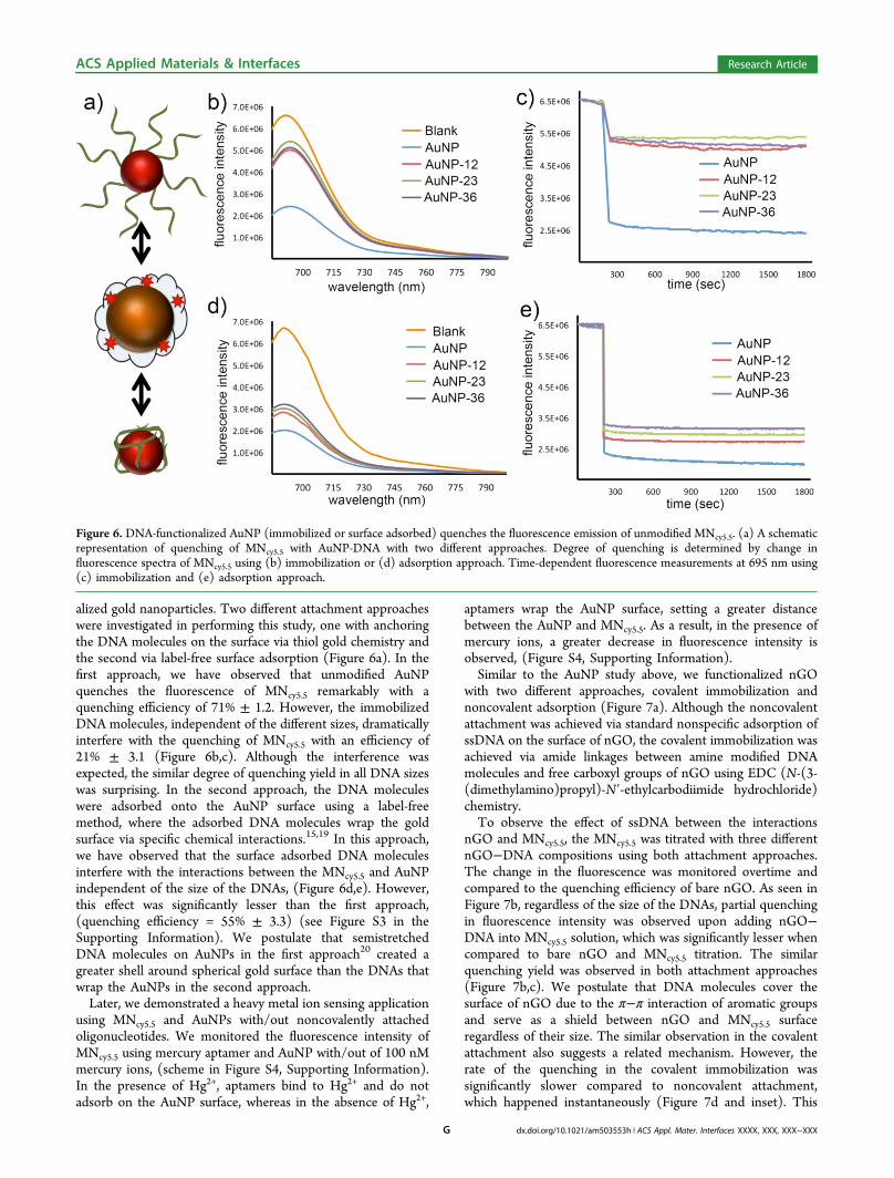
alized gold nanoparticles. Two different attachment approacheswere investigated in performing this study, one with anchoringthe DNA molecules on the surface via thiol gold chemistry andthe second via label-free surface adsorption (Figure 6a). In thefirst approach, we have observed that unmodified AuNPquenches the fluorescence of MNcy5.5 remarkably with aquenching efficiency of 71% ± 1.2. However, the immobilizedDNA molecules, independent of the different sizes, dramaticallyinterfere with the quenching of MNcy5.5 with an efficiency of21% ± 3.1 (Figure 6b,c). Although the interference wasexpected, the similar degree of quenching yield in all DNA sizeswas surprising. In the second approach, the DNA moleculeswere adsorbed onto the AuNP surface using a label-freemethod, where the adsorbed DNA molecules wrap the goldsurface via specific chemical interactions.15,19 In this approach,we have observed that the surface adsorbed DNA moleculesinterfere with the interactions between the MNcy5.5 and AuNPindependent of the size of the DNAs, (Figure 6d,e). However,this effect was significantly lesser than the first approach,(quenching efficiency = 55% ± 3.3) (see Figure S3 in theSupporting Information). We postulate that semistretchedDNA molecules on AuNPs in the first approach20 created agreater shell around spherical gold surface than the DNAs thatwrap the AuNPs in the second approach.Later, we demonstrated a heavy metal ion sensing application
using MNcy5.5 and AuNPs with/out noncovalently attachedoligonucleotides. We monitored the fluorescence intensity ofMNcy5.5 using mercury aptamer and AuNP with/out of 100 nMmercury ions, (scheme in Figure S4, Supporting Information).In the presence of Hg2+, aptamers bind to Hg2+ and do notadsorb on the AuNP surface, whereas in the absence of Hg2+,
aptamers wrap the AuNP surface, setting a greater distancebetween the AuNP and MNcy5.5. As a result, in the presence ofmercury ions, a greater decrease in fluorescence intensity isobserved, (Figure S4, Supporting Information).Similar to the AuNP study above, we functionalized nGO
with two different approaches, covalent immobilization andnoncovalent adsorption (Figure 7a). Although the noncovalentattachment was achieved via standard nonspecific adsorption ofssDNA on the surface of nGO, the covalent immobilization wasachieved via amide linkages between amine modified DNAmolecules and free carboxyl groups of nGO using EDC (N-(3-(dimethylamino)propyl)-N′-ethylcarbodiimide hydrochloride)chemistry.To observe the effect of ssDNA between the interactions
nGO and MNcy5.5, the MNcy5.5 was titrated with three differentnGO−DNA compositions using both attachment approaches.The change in the fluorescence was monitored overtime andcompared to the quenching efficiency of bare nGO. As seen inFigure 7b, regardless of the size of the DNAs, partial quenchingin fluorescence intensity was observed upon adding nGO−DNA into MNcy5.5 solution, which was significantly lesser whencompared to bare nGO and MNcy5.5 titration. The similarquenching yield was observed in both attachment approaches(Figure 7b,c). We postulate that DNA molecules cover thesurface of nGO due to the π−π interaction of aromatic groupsand serve as a shield between nGO and MNcy5.5 surfaceregardless of their size. The similar observation in the covalentattachment also suggests a related mechanism. However, therate of the quenching in the covalent immobilization wassignificantly slower compared to noncovalent attachment,which happened instantaneously (Figure 7d and inset). This
Figure 6. DNA-functionalized AuNP (immobilized or surface adsorbed) quenches the fluorescence emission of unmodified MNcy5.5. (a) A schematicrepresentation of quenching of MNcy5.5 with AuNP-DNA with two different approaches. Degree of quenching is determined by change influorescence spectra of MNcy5.5 using (b) immobilization or (d) adsorption approach. Time-dependent fluorescence measurements at 695 nm using(c) immobilization and (e) adsorption approach.
ACS Applied Materials & Interfaces Research Article
dx.doi.org/10.1021/am503553h | ACS Appl. Mater. Interfaces XXXX, XXX, XXX−XXXG
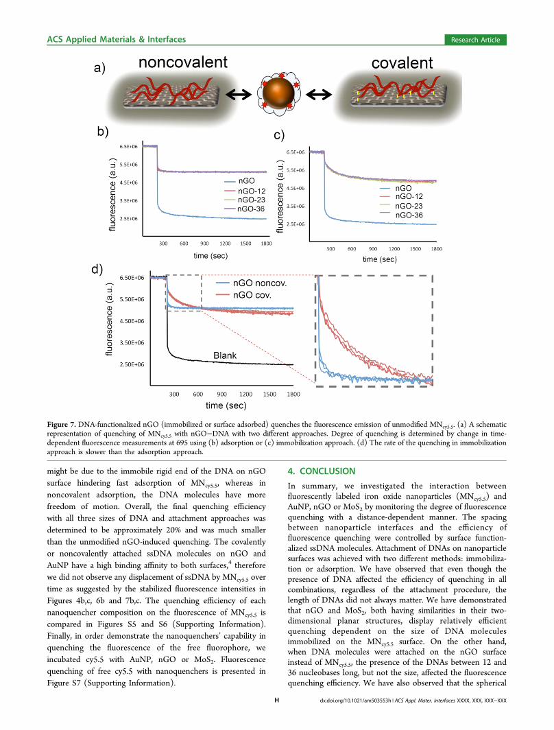
might be due to the immobile rigid end of the DNA on nGOsurface hindering fast adsorption of MNcy5.5, whereas innoncovalent adsorption, the DNA molecules have morefreedom of motion. Overall, the final quenching efficiencywith all three sizes of DNA and attachment approaches wasdetermined to be approximately 20% and was much smallerthan the unmodified nGO-induced quenching. The covalentlyor noncovalently attached ssDNA molecules on nGO andAuNP have a high binding affinity to both surfaces,4 thereforewe did not observe any displacement of ssDNA by MNcy5.5 overtime as suggested by the stabilized fluorescence intensities inFigures 4b,c, 6b and 7b,c. The quenching efficiency of eachnanoquencher composition on the fluorescence of MNcy5.5 iscompared in Figures S5 and S6 (Supporting Information).Finally, in order demonstrate the nanoquenchers’ capability inquenching the fluorescence of the free fluorophore, weincubated cy5.5 with AuNP, nGO or MoS2. Fluorescencequenching of free cy5.5 with nanoquenchers is presented inFigure S7 (Supporting Information).
4. CONCLUSION
In summary, we investigated the interaction betweenfluorescently labeled iron oxide nanoparticles (MNcy5.5) andAuNP, nGO or MoS2 by monitoring the degree of fluorescencequenching with a distance-dependent manner. The spacingbetween nanoparticle interfaces and the efficiency offluorescence quenching were controlled by surface function-alized ssDNA molecules. Attachment of DNAs on nanoparticlesurfaces was achieved with two different methods: immobiliza-tion or adsorption. We have observed that even though thepresence of DNA affected the efficiency of quenching in allcombinations, regardless of the attachment procedure, thelength of DNAs did not always matter. We have demonstratedthat nGO and MoS2, both having similarities in their two-dimensional planar structures, display relatively efficientquenching dependent on the size of DNA moleculesimmobilized on the MNcy5.5 surface. On the other hand,when DNA molecules were attached on the nGO surfaceinstead of MNcy5.5, the presence of the DNAs between 12 and36 nucleobases long, but not the size, affected the fluorescencequenching efficiency. We have also observed that the spherical
Figure 7. DNA-functionalized nGO (immobilized or surface adsorbed) quenches the fluorescence emission of unmodified MNcy5.5. (a) A schematicrepresentation of quenching of MNcy5.5 with nGO−DNA with two different approaches. Degree of quenching is determined by change in time-dependent fluorescence measurements at 695 using (b) adsorption or (c) immobilization approach. (d) The rate of the quenching in immobilizationapproach is slower than the adsorption approach.
ACS Applied Materials & Interfaces Research Article
dx.doi.org/10.1021/am503553h | ACS Appl. Mater. Interfaces XXXX, XXX, XXX−XXXH
AuNP quenches the fluorescence of MNcy5.5 regardless of thesize of the DNA molecules on either surface. Whereas theimmobilized DNA molecules on AuNP affects the quenchingefficiency the utmost.Overall, the presence of DNA molecules influences the
fluorescence emission of fluorescently labeled magnetic nano-particles dramatically with three different nanoquenchers. Thethree-dimensional structure of interacting nanoparticles and theattachment procedure of DNAs onto nanoparticle surface playimportant role in the quenching efficiency. We believe thisstudy could provide important information about engineeringhybrid bionanomaterials with tunable optical properties.
■ ASSOCIATED CONTENT
*S Supporting InformationAdditional experimental results and nanoparticle character-izations including titration of MNcy5.5 with different concen-trations of AuNP, nGO and MoS2; results of fluorescencelifetime decay fitting into a double exponential model; changein the hydrodynamic size of the MNcy5.5 compositions withdifferent lengths of ssDNA measured by dynamic lightscattering (DLS) measurements; DNA-functionalized AuNP(immobilized or surface adsorbed) quenching the fluorescenceemission of unmodified MNcy5.5; quenching of the fluorescenceof MNcy5.5 with AuNPs in the presence of single strandedmercury aptamer with or without Hg2+; quenching of MNcy5.5with AuNP, nGO or MoS2 in the presence and absence ofoligonucleotide attachments; quenching efficiency of differentnanoquencher compositions against fluorescence emission ofMNcy5.5 and its DNA conjugates with different attachmentapproaches; quenching of free cy5.5 dye with 4 nM AuNP, 1.8μg/mL of nGO or 20 μg/mL of MoS2; absorbance spectrum of76 nM MNcy5.5, 4nM AuNP, 16 μg/mL of nGO and 4.5 μg/mLof MoS2. This material is available free of charge via theInternet at http://pubs.acs.org.
■ AUTHOR INFORMATIONCorresponding Author*M. V. Yigit. Tel: (1) 518-442-3002. E-mail: [email protected].
NotesThe authors declare no competing financial interest.
■ ACKNOWLEDGMENTSWe thank Kevin Musick and Dr. Kathleen Dunn for the TEMmicrographs. We acknowledge the Ministry of NationalEducation, Republic of Turkey, for financial support to MustafaBalcioglu with full fellowship during his doctoral studies. Thiswork was supported by the SUNY Albany Start-Up Funds.
■ REFERENCES(1) Wei, Q.; Nagi, R.; Sadeghi, K.; Feng, S.; Yan, E.; Ki, S. J.; Caire,R.; Tseng, D.; Ozcan, A. Detection and Spatial Mapping of MercuryContamination in Water Samples Using a Smart-Phone. ACS Nano2014, 8, 1121−1129.(2) Hamner, K. L.; Maye, M. M. Thermal Aggregation Properties ofNanoparticles Modified with Temperature Sensitive Copolymers.Langmuir 2013, 29, 15217−15223.(3) Huang, H.; Song, W.; Chen, G.; Reynard, J. M.; Ohulchanskyy, T.Y.; Prasad, P. N.; Bright, F. V.; Lovell, J. F. Pd-Porphyrin-Cross-LinkedImplantable Hydrogels with Oxygen-Responsive Phosphorescence.Adv. Healthcare Mater. 2014, 3, 891−896.
(4) Liu, J. Adsorption of DNA onto Gold Nanoparticles andGraphene Oxide: Surface Science and Applications. Phys. Chem. Chem.Phys. 2012, 14, 10485−10496.(5) Piao, Y.; Liu, F.; Seo, T. S. A Novel Molecular Beacon Bearing aGraphite Nanoparticle as a Nanoquencher for in Situ mRNADetection in Cancer Cells. ACS Appl. Mater. Interfaces 2012, 4,6785−6789.(6) Li, J.-L.; Tang, B.; Yuan, B.; Sun, L.; Wang, X.-G. A Review ofOptical Imaging and Therapy Using Nanosized Graphene andGraphene Oxide. Biomaterials 2013, 34, 9519−9534.(7) Morales-Narvaez, E.; Merkoci, A. Graphene Oxide as an OpticalBiosensing Platform. Adv. Mater. 2012, 24, 3298−3308.(8) Yigit, M. V.; Zhu, L.; Ifediba, M. A.; Zhang, Y.; Carr, K.; Moore,A.; Medarova, Z. Noninvasive MRI-SERS Imaging in Living MiceUsing an Innately Bimodal Nanomaterial. ACS Nano 2010, 5, 1056−1066.(9) Wang, P.; Yigit, M. V.; Medarova, Z.; Wei, L.; Dai, G.; Schuetz,C.; Moore, A. Combined Small Interfering RNA Therapy and in VivoMagnetic Resonance Imaging in Islet Transplantation. Diabetes 2011,60, 565−571.(10) Alexander, C. M.; Maye, M. M.; Dabrowiak, J. C. DNA-CappedNanoparticles Designed for Doxorubicin Drug Delivery. Chem.Commun. (Cambridge, U. K.) 2011, 47, 3418−3420.(11) Alexander, C. M.; Dabrowiak, J. C.; Maye, M. M. Investigationof the Drug Binding Properties and Cytotoxicity of DNA-CappedNanoparticles Designed as Delivery Vehicles for the Anticancer AgentsDoxorubicin and Actinomycin D. Bioconjugate Chem. 2012, 23, 2061−2070.(12) Hamner, K. L.; Alexander, C. M.; Coopersmith, K.; Reishofer,D.; Provenza, C.; Maye, M. M. Using Temperature-Sensitive SmartPolymers to Regulate DNA-Mediated Nanoassembly and EncodedNanocarrier Drug Release. ACS Nano 2013, 7, 7011−7020.(13) Kundu, A.; Layek, R. K.; Kuila, A.; Nandi, A. K. HighlyFluorescent Graphene Oxide-Poly(vinyl alcohol) Hybrid: An EffectiveMaterial for Specific Au3+ Ion Sensors. ACS Appl. Mater. Interfaces2012, 4, 5576−5582.(14) Bunz, U. H.; Rotello, V. M. Gold Nanoparticle-FluorophoreComplexes: Sensitive and Discerning “Noses” for Biosystems Sensing.Angew. Chem., Int. Ed. 2010, 49, 3268−3279.(15) Xia, F.; Zuo, X.; Yang, R.; Xiao, Y.; Kang, D.; Vallee-Belisle, A.;Gong, X.; Yuen, J. D.; Hsu, B. B.; Heeger, A. J.; Plaxco, K. W.Colorimetric Detection of DNA, Small Molecules, Proteins, and IonsUsing Unmodified Gold Nanoparticles and Conjugated Polyelec-trolytes. Proc. Natl. Acad. Sci. U. S. A. 2010, 107, 10837−10841.(16) Yang, L.; Liu, C.; Ren, W.; Li, Z. Graphene Surface-AnchoredFluorescence Sensor for Sensitive Detection of Microrna Coupled withEnzyme-Free Signal Amplification of Hybridization Chain Reaction.ACS Appl. Mater. Interfaces 2012, 4, 6450−6453.(17) Chou, S. S.; De, M.; Luo, J.; Rotello, V. M.; Huang, J.; Dravid, V.P. Nanoscale Graphene Oxide (nGO) as Artificial Receptors:Implications for Biomolecular Interactions and Sensing. J. Am. Chem.Soc. 2012, 134, 16725−16733.(18) Stobiecka, M.; Hepel, M. Multimodal Coupling of OpticalTransitions and Plasmonic Oscillations in Rhodamine B ModifiedGold Nanoparticles. Phys. Chem. Chem. Phys. 2011, 13, 1131−1139.(19) Li, H.; Rothberg, L. Colorimetric Detection of DNA SequencesBased on Electrostatic Interactions with Unmodified Gold Nano-particles. Proc. Natl. Acad. Sci. U. S. A. 2004, 101, 14036−14039.(20) Parak, W. J.; Pellegrino, T.; Micheel, C. M.; Gerion, D.;Williams, S. C.; Alivisatos, A. P. Conformation of OligonucleotidesAttached to Gold Nanocrystals Probed by Gel Electrophoresis. NanoLett. 2002, 3, 33−36.(21) Dulkeith, E.; Ringler, M.; Klar, T. A.; Feldmann, J.; MunozJavier, A.; Parak, W. J. Gold Nanoparticles Quench Fluorescence byPhase Induced Radiative Rate Suppression. Nano Lett. 2005, 5, 585−589.(22) Chhabra, R.; Sharma, J.; Wang, H.; Zou, S.; Lin, S.; Yan, H.;Lindsay, S.; Liu, Y. Distance-Dependent Interactions between Gold
ACS Applied Materials & Interfaces Research Article
dx.doi.org/10.1021/am503553h | ACS Appl. Mater. Interfaces XXXX, XXX, XXX−XXXI
Nanoparticles and Fluorescent Molecules with DNA as TunableSpacers. Nanotechnology 2009, 20, 485201.(23) Huang, P.-J. J.; Liu, J. DNA-Length-Dependent FluorescenceSignaling on Graphene Oxide Surface. Small 2012, 8, 977−983.(24) Mao, H. Y.; Laurent, S.; Chen, W.; Akhavan, O.; Imani, M.;Ashkarran, A. A.; Mahmoudi, M. Graphene: Promises, Facts,Opportunities, and Challenges in Nanomedicine. Chem. Rev. 2013,113, 3407−3424.(25) Geim, A. K. Graphene: Status and Prospects. Science 2009, 324,1530−1534.(26) Novoselov, K. S.; Geim, A. K.; Morozov, S. V.; Jiang, D.; Zhang,Y.; Dubonos, S. V.; Grigorieva, I. V.; Firsov, A. A. Electric Field Effectin Atomically Thin Carbon Films. Science 2004, 306, 666−669.(27) Geim, A. K.; Novoselov, K. S. The Rise of Graphene. Nat.Mater. 2007, 6, 183−191.(28) Chung, C.; Kim, Y. K.; Shin, D.; Ryoo, S. R.; Hong, B. H.; Min,D. H. Biomedical Applications of Graphene and Graphene Oxide. Acc.Chem. Res. 2013, 46, 2211−2224.(29) Rana, M.; Balcioglu, M.; Robertson, N.; Yigit, M. V. Nano-Graphene Oxide as a Novel Platform for Monitoring the Effect ofLNA Modification on Nucleic Acid Interactions. Analyst 2014, 139,714−720.(30) Li, S.; Aphale, A. N.; Macwan, I. G.; Patra, P. K.; Gonzalez, W.G.; Miksovska, J.; Leblanc, R. M. Graphene Oxide as a Quencher forFluorescent Assay of Amino Acids, Peptides, and Proteins. ACS Appl.Mater. Interfaces 2012, 4, 7069−7075.(31) Balcioglu, M.; Rana, M.; Yigit, M. V. Doxorubicin Loading onGraphene Oxide, Iron Oxide and Gold Nanoparticle Hybrid. J. Mater.Chem. B 2013, 1, 6187−6193.(32) Li, F.; Pei, H.; Wang, L. H.; Lu, J. X.; Gao, J. M.; Jiang, B. W.;Zhao, X. C.; Fan, C. H. Nanomaterial-Based Fluorescent DNAAnalysis: A Comparative Study of the Quenching Effects of GrapheneOxide, Carbon Nanotubes, and Gold Nanoparticles. Adv. Funct. Mater.2013, 23, 4140−4148.(33) Wang, Y.; Li, Z.; Wang, J.; Li, J.; Lin, Y. Graphene and GrapheneOxide: Biofunctionalization and Applications in Biotechnology. TrendsBiotechnol. 2011, 29, 205−212.(34) Fu, X.; Lou, T.; Chen, Z.; Lin, M.; Feng, W.; Chen, L. “Turn-on” Fluorescence Detection of Lead Ions Based on AcceleratedLeaching of Gold Nanoparticles on the Surface of Graphene. ACSAppl. Mater. Interfaces 2012, 4, 1080−1086.(35) Liu, Y. M.; Punckt, C.; Pope, M. A.; Gelperin, A.; Aksay, I. A.Electrochemical Sensing of Nitric Oxide with FunctionalizedGraphene Electrodes. ACS Appl. Mater. Interfaces 2013, 5, 12624−12630.(36) He, Y.; Huang, G.; Cui, H. Quenching the Chemiluminescenceof Acridinium Ester by Graphene Oxide for Label-Free andHomogeneous DNA Detection. ACS Appl. Mater. Interfaces 2013, 5,11336−11340.(37) Shi, Y.; Yi, C.; Zhang, Z.; Zhang, H.; Li, M.; Yang, M.; Jiang, Q.Peptide-Bridged Assembly of Hybrid Nanomaterial and Its Applicationfor Caspase-3 Detection. ACS Appl. Mater. Interfaces 2013, 5, 6494−6501.(38) Dey, R. S.; Raj, C. R. Redox-Functionalized Graphene OxideArchitecture for the Development of Amperometric BiosensingPlatform. ACS Appl. Mater. Interfaces 2013, 5, 4791−4798.(39) Ryoo, S.-R.; Lee, J.; Yeo, J.; Na, H.-K.; Kim, Y.-K.; Jang, H.; Lee,J. H.; Han, S. W.; Lee, Y.; Kim, V. N.; Min, D.-H. Quantitative andMultiplexed Microrna Sensing in Living Cells Based on PeptideNucleic Acid and Nano Graphene Oxide (Pango). ACS Nano 2013, 7,5882−5891.(40) Dong, H.; Ding, L.; Yan, F.; Ji, H.; Ju, H. The Use ofPolyethylenimine-Grafted Graphene Nanoribbon for Cellular Deliveryof Locked Nucleic Acid Modified Molecular Beacon for Recognition ofMicrorna. Biomaterials 2011, 32, 3875−3882.(41) Reed, J. C.; Zhu, H.; Zhu, A. Y.; Li, C.; Cubukcu, E. Graphene-Enabled Silver Nanoantenna Sensors. Nano Lett. 2012, 12, 4090−4094.
(42) Tang, L.; Wang, Y.; Liu, Y.; Li, J. DNA-Directed Self-Assemblyof Graphene Oxide with Applications to Ultrasensitive Oligonucleo-tide Assay. ACS Nano 2011, 5, 3817−3822.(43) Ryoo, S. R.; Lee, J.; Yeo, J.; Na, H. K.; Kim, Y. K.; Jang, H.; Lee,J. H.; Han, S. W.; Lee, Y.; Kim, V. N.; Min, D. H. Quantitative andMultiplexed Microrna Sensing in Living Cells Based on PeptideNucleic Acid and Nano Graphene Oxide (Pango). ACS Nano 2013, 7,5882−5891.(44) Kim, H.; Namgung, R.; Singha, K.; Oh, I. K.; Kim, W. J.Graphene Oxide-Polyethylenimine Nanoconstruct as a Gene DeliveryVector and Bioimaging Tool. Bioconjugate Chem. 2011, 22, 2558−2567.(45) Xu, C.; Yang, D.; Mei, L.; Lu, B.; Chen, L.; Li, Q.; Zhu, H.;Wang, T. Encapsulating Gold Nanoparticles or Nanorods in GrapheneOxide Shells as a Novel Gene Vector. ACS Appl. Mater. Interfaces2013, 5, 2715−2724.(46) Shen, H.; Liu, M.; He, H.; Zhang, L.; Huang, J.; Chong, Y.; Dai,J.; Zhang, Z. PEGylated Graphene Oxide-Mediated Protein Deliveryfor Cell Function Regulation. ACS Appl. Mater. Interfaces 2012, 4,6317−6323.(47) Liu, H.; Si, M.; Deng, Y.; Neal, A. T.; Du, Y.; Najmaei, S.;Ajayan, P. M.; Lou, J.; Ye, P. D. Switching Mechanism in Single-LayerMolybdenum Disulfide Transistors: An Insight into Current Flowacross Schottky Barriers. ACS Nano 2014, 8, 1031−1038.(48) Zhu, C.; Zeng, Z.; Li, H.; Li, F.; Fan, C.; Zhang, H. Single-LayerMoS2-Based Nanoprobes for Homogeneous Detection of Biomole-cules. J. Am. Chem. Soc. 2013, 135, 5998−6001.(49) Liu, T.; Wang, C.; Gu, X.; Gong, H.; Cheng, L.; Shi, X.; Feng,L.; Sun, B.; Liu, Z. Drug Delivery with PEGylated MoS Nano-Sheetsfor Combined Photothermal and Chemotherapy of Cancer. Adv.Mater. 2014, 26, 3433−3440.(50) Wei, Q.; Qi, H.; Luo, W.; Tseng, D.; Ki, S. J.; Wan, Z.; Gorocs,Z.; Bentolila, L. A.; Wu, T. T.; Ozcan, A. Fluorescent Imaging of SingleNanoparticles and Viruses on a Smart Phone. ACS Nano 2013, 7,9147−9155.(51) Hennequin, Y.; Allier, C. P.; McLeod, E.; Mudanyali, O.;Migliozzi, D.; Ozcan, A.; Dinten, J. M. Optical Detection and Sizing ofSingle Nanoparticles Using Continuous Wetting Films. ACS Nano2013, 7, 7601−7609.(52) Rana, M.; Balcioglu, M.; Yigit, M. V. Locked Nucleic Acid-Modified Antisense Mir-10b Oligonucleotides Form Stable Duplexeson Gold Nanoparticles. Bionanoscience 2014, 4, 195−200.(53) Swierczewska, M.; Lee, S.; Chen, X. The Design and Applicationof Fluorophore-Gold Nanoparticle Activatable Probes. Phys. Chem.Chem. Phys. 2011, 13, 9929−9941.(54) Ling, J.; Huang, C. Z. Energy Transfer with Gold Nanoparticlesfor Analytical Applications in the Fields of Biochemical andPharmaceutical Sciences. Anal. Methods 2010, 2, 1439−1447.(55) Fan, C.; Wang, S.; Hong, J. W.; Bazan, G. C.; Plaxco, K. W.;Heeger, A. J. Beyond Superquenching: Hyper-Efficient EnergyTransfer from Conjugated Polymers to Gold Nanoparticles. Proc.Natl. Acad. Sci. U. S. A. 2003, 100, 6297−6301.(56) Tu, Y.; Wu, P.; Zhang, H.; Cai, C. Fluorescence Quenching ofGold Nanoparticles Integrating with a Conformation-SwitchedHairpin Oligonucleotide Probe for Microrna Detection. Chem.Commun. (Cambridge, U. K.) 2012, 48, 10718−10720.(57) Acar, H.; Genc, R.; Urel, M.; Erkal, T. S.; Dana, A.; Guler, M. O.Self-Assembled Peptide Nanofiber Templated One-Dimensional GoldNanostructures Exhibiting Resistive Switching. Langmuir 2012, 28,16347−16354.(58) Han, H.; Valle, V.; Maye, M. M. Probing Resonance EnergyTransfer and Inner Filter Effects in Quantum Dot−Large MetalNanoparticle Clusters Using a DNA-Mediated Quench and ReleaseMechanism. J. Phys. Chem. C 2012, 116, 22996−23003.(59) Han, H.; Valle, V.; Maye, M. M. Probing the Quenching ofCdSe/ZnS Qdots by Au, Au/Ag, and Au/Pd Nanoparticles.Nanotechnology 2012, 23, 435401.
ACS Applied Materials & Interfaces Research Article
dx.doi.org/10.1021/am503553h | ACS Appl. Mater. Interfaces XXXX, XXX, XXX−XXXJ
(60) Herne, T. M.; Tarlov, M. J. Characterization of DNA ProbesImmobilized on Gold Surfaces. J. Am. Chem. Soc. 1997, 119, 8916−8920.(61) Tassa, C.; Shaw, S. Y.; Weissleder, R. Dextran-Coated IronOxide Nanoparticles: A Versatile Platform for Targeted MolecularImaging, Molecular Diagnostics, and Therapy. Acc. Chem. Res. 2011,44, 842−852.(62) Yigit, M. V.; Mazumdar, D.; Lu, Y. MRI Detection of Thrombinwith Aptamer Functionalized Superparamagnetic Iron Oxide Nano-particles. Bioconjugate Chem. 2008, 19, 412−417.(63) Yigit, M. V.; Mazumdar, D.; Kim, H. K.; Lee, J. H.; Odintsov, B.;Lu, Y. Smart “Turn-on” Magnetic Resonance Contrast Agents Basedon Aptamer-Functionalized Superparamagnetic Iron Oxide Nano-particles. ChemBioChem 2007, 8, 1675−1678.(64) Sulek, S.; Mammadov, B.; Mahcicek, D. I.; Sozeri, H.; Atalar, E.;Tekinay, A. B.; Guler, M. O. Peptide Functionalized Super-paramagnetic Iron Oxide Nanoparticles as MRI Contrast Agents. J.Mater. Chem. 2011, 21, 15157−15162.(65) Yigit, M. V.; Moore, A.; Medarova, Z. Magnetic Nanoparticlesfor Cancer Diagnosis and Therapy. Pharm. Res. 2012, 29, 1180−1188.(66) Medarova, Z.; Pham, W.; Farrar, C.; Petkova, V.; Moore, A. InVivo Imaging of siRNA Delivery and Silencing in Tumors. Nat. Med.2007, 13, 372−377.(67) Pittet, M. J.; Swirski, F. K.; Reynolds, F.; Josephson, L.;Weissleder, R. Labeling of Immune Cells for in Vivo Imaging UsingMagnetofluorescent Nanoparticles. Nat. Protoc. 2006, 1, 73−79.(68) Yigit, M. V.; Ghosh, S. K.; Kumar, M.; Petkova, V.; Kavishwar,A.; Moore, A.; Medarova, Z. Context-Dependent Differences in miR-10b Breast Oncogenesis Can Be Targeted for the Prevention andArrest of Lymph Node Metastasis. Oncogene 2013, 32, 1530−1538.(69) Kumar, M.; Yigit, M.; Dai, G.; Moore, A.; Medarova, Z. Image-Guided Breast Tumor Therapy Using a Small Interfering RNANanodrug. Cancer Res. 2010, 70, 7553−7561.(70) Liu, J.; Lu, Y. Preparation of Aptamer-Linked Gold NanoparticlePurple Aggregates for Colorimetric Sensing of Analytes. Nat. Protoc.2006, 1, 246−252.
ACS Applied Materials & Interfaces Research Article
dx.doi.org/10.1021/am503553h | ACS Appl. Mater. Interfaces XXXX, XXX, XXX−XXXK


