Embed Size (px)
Citation preview
ORIGINAL ARTICLE: Experimental Endoscopy
Doppler optical coherence tomography monitoringof microvascular tissue response during photodynamic therapyin an animal model of Barrett’s esophagus
Beau A. Standish, BEng, Victor X. D. Yang, MD, PhD, Nigel R. Munce, MSc,Louis-Michel Wong Kee Song, MD, Geoffrey Gardiner, MD, Annie Lin, MD, Youxin I. Mao, PhD,Alex Vitkin, PhD, Norman E. Marcon, MD, Brian C. Wilson, PhD
Toronto, Ontario, Canada
Background: Doppler optical coherence tomography (DOCT) is an imaging modality that allows assessment ofthe microvascular response during photodynamic therapy (PDT) and may be a powerful tool for treatment mon-itoring/optimization in conditions such as Barrett’s esophagus (BE).
Objective: To assess the technical feasibility of catheter-based intraluminal DOCT for monitoring the microvas-cular response during endoluminal PDT in an animal model of BE.
Design: Thirteen female Sprague-Dawley rats underwent esophagojejunostomy to induce enteroesophageal re-flux for 35 to 42 weeks and the formation of Barrett’s mucosa. Of these, 9 received PDT by using the photosen-sitizer Photofrin (12.5 mg/kg intravenous), followed by 635-nm intraluminal light irradiation 24 hours after drugadministration. The remaining 4 surgical rats underwent light irradiation without Photofrin (controls). Anothergroup of 5 normal rats, without esophagojejunostomy, also received PDT. DOCT imaging of the esophagus byusing a catheter-based probe (1.3-mm diameter) was performed before, during, and after light irradiation in allrats.
Results: Distinct microstructural differences between normal squamous esophagus, BE, and the transition zonebetween the 2 tissues were observed on DOCT images. Similar submucosal microcirculatory effects (47%-73%vascular shutdown) were observed during PDT of normal esophagus and surgically induced BE. Controls dis-played no significant microvascular changes.
Conclusions: No apparent difference was observed in the PDT-induced vascular response between normal ratesophagus and the BE rat model. Real-time monitoring of PDT-induced vascular changes by DOCT may be ben-eficial in optimizing PDT dosimetry in patients undergoing this therapy for BE and other conditions. (Gastroint-est Endosc 2007;66:326-33.)
Doppler optical coherence tomography (DOCT)1,2 isan imaging modality that combines the high spatial resolu-tion of optical coherence tomography (OCT)3 and Dopp-ler measurement of blood flow, with a velocity resolutiondown to approximately 20 mm/s.4 This technology has en-abled the detection of blood flow noninvasively in humanretina,5 skin,6 and the GI tract,7 as well as providing cross-sectional images of tissue at near-histologic resolution. Inparticular, fiber-optic implementation allows endoscopicimaging, as previously reported by us7 in patients under-
going surveillance for Barrett’s esophagus (BE), a condi-tion in which the normal squamous mucosa is replacedby columnar, intestine-like mucosa and in which there isa marked increase in the risk of dysplasia, a precursor ofesophageal adenocarcinoma.8
Photodynamic therapy (PDT) involves light activationof an administered photosensitizer that reacts with molec-ular oxygen, generating reactive oxygen species, causingnecrosis and/or apoptosis of cells,9 activation of the hostimmune system,10 and vascular damage.11,12 These effectsdepend on the photosensitizer, the target tissue, and thespecific treatment parameters. In the case of PDT for de-struction of solid malignant tumors, vascular shutdowncan induce tissue ischemia, leading to tumor-cell death.
Copyright ª 2007 by the American Society for Gastrointestinal Endoscopy
0016-5107/$32.00
doi:10.1016/j.gie.2007.02.040
326 GASTROINTESTINAL ENDOSCOPY Volume 66, No. 2 : 2007 www.giejournal.org
If light activation is carried out shortly after photosensi-tizer administration (while the drug is still confined tothe vasculature), then vascular shutdown is the primarymode of action. This is the case, for example, in PDT forage-related macular degeneration (choroidal neovascula-ture)13 when using Visudyne (Visudyne, East Hanover,NJ) and for prostate cancer when using TOOKAD (StebaBiotech, Paris, France). PDT is approved for the treatmentof certain GI conditions, including palliation of esophagealcancer and treatment of BE with high-grade dysplasia(HGD) by using the first-generation photosensitizer Pho-tofrin (Axcan Pharma, Montreal, Quebec, Canada).14,15 Mi-crocirculatory changes, including vascular shutdown, werereported as a mechanism responsible for the PDT treat-ment effect.13 Previously, we reported on DOCT monitor-ing of the PDT-induced vascular effects during laparotomyin normal rats.16 We also recently demonstrated a novelDOCT system capable of imaging the microcirculation inboth the animal (rat)17 and human GI tracts,7 and its fea-sibility for endoscopic use.7
The primary aim of the present study was to demon-strate the technical feasibility of DOCT to monitor the vas-cular changes that occurred during endoluminal PDT ina BE rat model. With the study design including a BE armand a normal arm, a secondary aim was to assess whetherBE and normal esophageal vasculature respondeddifferently to PDT.
MATERIALS AND METHODS
Animals and PDTAll animal procedures were carried out, with institu-
tional approval, at the University Health Network, Tor-onto, Canada. Female Sprague-Dawley rats (180-250 g)were purchased (Harlan, Indianapolis, Ind) and housed2 per cage under standard laboratory conditions. To thebest of our knowledge, it has not been previously shownthat the choice of sex for inducing BE via esophagojeju-
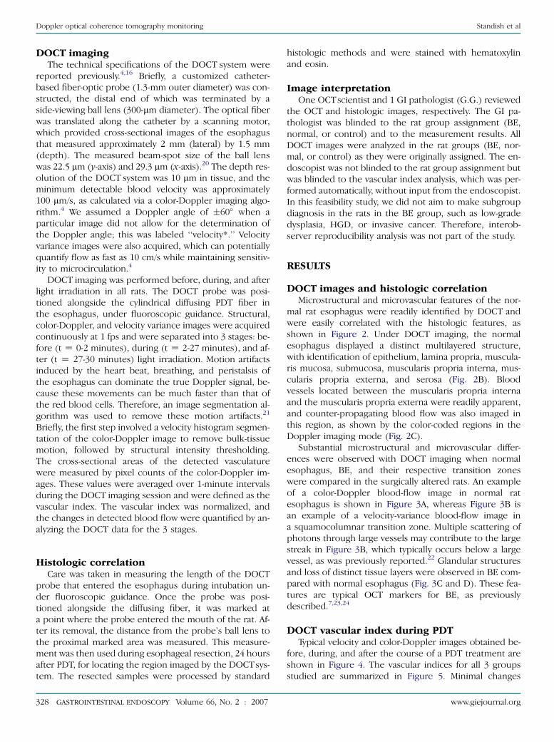
Capsule Summary
What is already known on this topic
d One of the mechanisms of Photofrin-basedphotodynamic therapy, which can be used for treatmentof Barrett’s esophagus (BE), is the vasoconstrictive effectof such therapy on tissue microvasculature andassociated tissue ischemia.
What this study adds to our knowledge
d When using Doppler optical coherence tomographyimaging in a rat model, there were distinctmicrostructural differences among normal squamousesophagus, BE, and their transition zone.
d Similar submucosal microcirculatory effects wereobserved in normal esophagus and surgically induced BEafter photodynamic therapy.
nostomy is of great importance. We, therefore, chose themore docile female sex, because the animal handling timefor this study was extensive (BE formation took O35weeks). The rats were maintained on a high protein (En-sure; Abbott, Montreal, Canada) and mushed Kibble(KMR; PetAg, Hampshire, Ill) diet. The rat’s food was with-held for 24 hours before surgical intervention and beforeDOCT imaging. Thirteen rats underwent esophagojeju-nostomy by using the Levrat technique, as previously de-scribed in detail,18,19 to induce enteroesophageal refluxfor 35 to 42 weeks and the subsequent formation ofBarrett’s epithelium. Briefly, the rats were anesthetizedwith xylazine hydrochloride (12 mg/kg intramuscular [IM])and ketamine (75 mg/kg IM). A midline laparotomy wasperformed, the gastroesophageal junction was ligated,and the distal esophagus was transected 2 mm abovethe ligature. To stabilize the esophagus, a soft siliconetube was placed into the esophagus down as far as the gas-troesophageal junction before transection. A jejunostomywas created just distal to the ligament of Treitz, and anend-to-side esophagojejunostomy was constructed withaccurate mucosa-to-mucosa approximation. Analgesia (bu-prenorphine) was administered after surgery, and the ratswere fed by the first postoperative day.
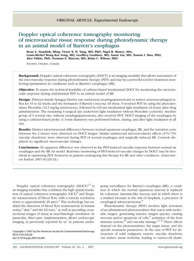
Normal and surgically altered (BE) rats were random-ized into 3 groups, as shown in Figure 1. Nine surgicallyaltered rats and 5 normal rats were administered the pho-tosensitizer Photofrin at 12.5 mg/kg intravenous, 24 hoursbefore light irradiation. For treatment, a 0.8-mm-diameter,2.7-cm-long, cylindrical, diffusing fiber was placed into theesophagus to deliver a 635-nm minimum total irradianceof 37 J cm�2 at 45 mW cm�2. Four rats in the BE groupreceived the same light irradiation without Photofrin(controls). The rats were euthanized with a 0.5 mL intra-cardiac injection of euthenol 24 hours after treatment, andthe esophagus and the stomach were resected en bloc forhistology.
Figure 1. Group allocation of normal and surgically altered rats.
Standish et al Doppler optical coherence tomography monitoring
www.giejournal.org Volume 66, No. 2 : 2007 GASTROINTESTINAL ENDOSCOPY 327
DOCT imagingThe technical specifications of the DOCT system were
reported previously.4,16 Briefly, a customized catheter-based fiber-optic probe (1.3-mm outer diameter) was con-structed, the distal end of which was terminated by aside-viewing ball lens (300-mm diameter). The optical fiberwas translated along the catheter by a scanning motor,which provided cross-sectional images of the esophagusthat measured approximately 2 mm (lateral) by 1.5 mm(depth). The measured beam-spot size of the ball lenswas 22.5 mm (y-axis) and 29.3 mm (x-axis).20 The depth res-olution of the DOCT system was 10 mm in tissue, and theminimum detectable blood velocity was approximately100 mm/s, as calculated via a color-Doppler imaging algo-rithm.4 We assumed a Doppler angle of �60� when aparticular image did not allow for the determination ofthe Doppler angle; this was labeled ‘‘velocity*.’’ Velocityvariance images were also acquired, which can potentiallyquantify flow as fast as 10 cm/s while maintaining sensitiv-ity to microcirculation.4
DOCT imaging was performed before, during, and afterlight irradiation in all rats. The DOCT probe was posi-tioned alongside the cylindrical diffusing PDT fiber inthe esophagus, under fluoroscopic guidance. Structural,color-Doppler, and velocity variance images were acquiredcontinuously at 1 fps and were separated into 3 stages: be-fore (t Z 0-2 minutes), during (t Z 2-27 minutes), and af-ter (t Z 27-30 minutes) light irradiation. Motion artifactsinduced by the heart beat, breathing, and peristalsis ofthe esophagus can dominate the true Doppler signal, be-cause these movements can be much faster than that ofthe red blood cells. Therefore, an image segmentation al-gorithm was used to remove these motion artifacts.21
Briefly, the first step involved a velocity histogram segmen-tation of the color-Doppler image to remove bulk-tissuemotion, followed by structural intensity thresholding.The cross-sectional areas of the detected vasculaturewere measured by pixel counts of the color-Doppler im-ages. These values were averaged over 1-minute intervalsduring the DOCT imaging session and were defined as thevascular index. The vascular index was normalized, andthe changes in detected blood flow were quantified by an-alyzing the DOCT data for the 3 stages.
Histologic correlationCare was taken in measuring the length of the DOCT
probe that entered the esophagus during intubation un-der fluoroscopic guidance. Once the probe was posi-tioned alongside the diffusing fiber, it was marked ata point where the probe entered the mouth of the rat. Af-ter its removal, the distance from the probe’s ball lens tothe proximal marked area was measured. This measure-ment was then used during esophageal resection, 24 hoursafter PDT, for locating the region imaged by the DOCT sys-tem. The resected samples were processed by standard
histologic methods and were stained with hematoxylinand eosin.
Image interpretationOne OCTscientist and 1 GI pathologist (G.G.) reviewed
the OCT and histologic images, respectively. The GI pa-thologist was blinded to the rat group assignment (BE,normal, or control) and to the measurement results. AllDOCT images were analyzed in the rat groups (BE, nor-mal, or control) as they were originally assigned. The en-doscopist was not blinded to the rat group assignment butwas blinded to the vascular index analysis, which was per-formed automatically, without input from the endoscopist.In this feasibility study, we did not aim to make subgroupdiagnosis in the rats in the BE group, such as low-gradedysplasia, HGD, or invasive cancer. Therefore, interob-server reproducibility analysis was not part of the study.
RESULTS
DOCT images and histologic correlationMicrostructural and microvascular features of the nor-
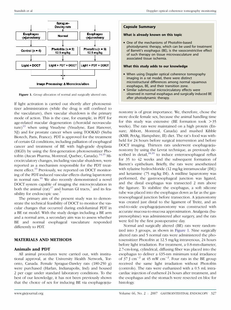
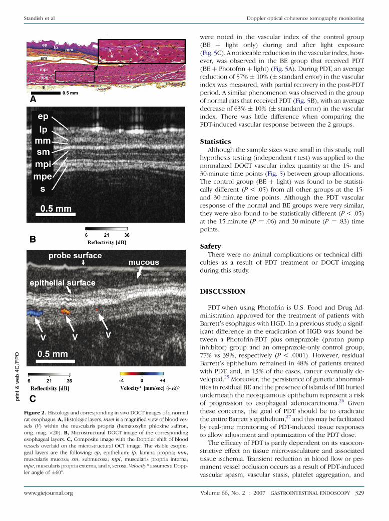
mal rat esophagus were readily identified by DOCT andwere easily correlated with the histologic features, asshown in Figure 2. Under DOCT imaging, the normalesophagus displayed a distinct multilayered structure,with identification of epithelium, lamina propria, muscula-ris mucosa, submucosa, muscularis propria interna, mus-cularis propria externa, and serosa (Fig. 2B). Bloodvessels located between the muscularis propria internaand the muscularis propria externa were readily apparent,and counter-propagating blood flow was also imaged inthis region, as shown by the color-coded regions in theDoppler imaging mode (Fig. 2C).
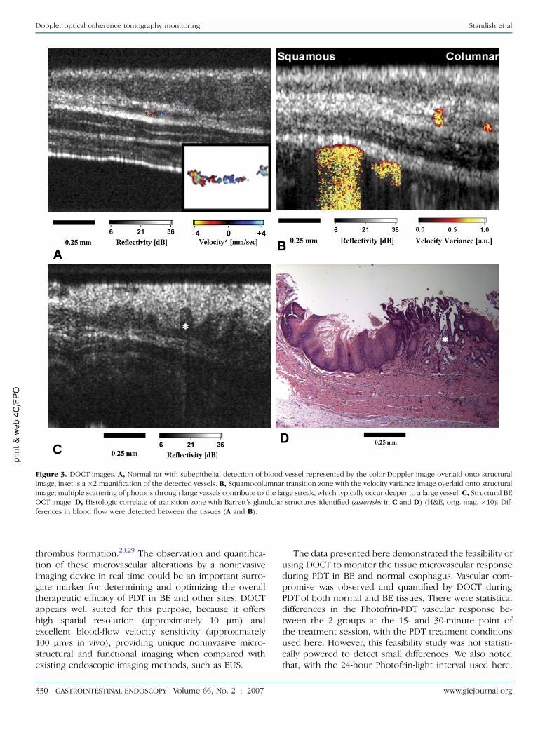
Substantial microstructural and microvascular differ-ences were observed with DOCT imaging when normalesophagus, BE, and their respective transition zoneswere compared in the surgically altered rats. An exampleof a color-Doppler blood-flow image in normal ratesophagus is shown in Figure 3A, whereas Figure 3B isan example of a velocity-variance blood-flow image ina squamocolumnar transition zone. Multiple scattering ofphotons through large vessels may contribute to the largestreak in Figure 3B, which typically occurs below a largevessel, as was previously reported.22 Glandular structuresand loss of distinct tissue layers were observed in BE com-pared with normal esophagus (Fig. 3C and D). These fea-tures are typical OCT markers for BE, as previouslydescribed.7,23,24
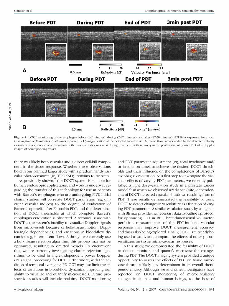
DOCT vascular index during PDTTypical velocity and color-Doppler images obtained be-
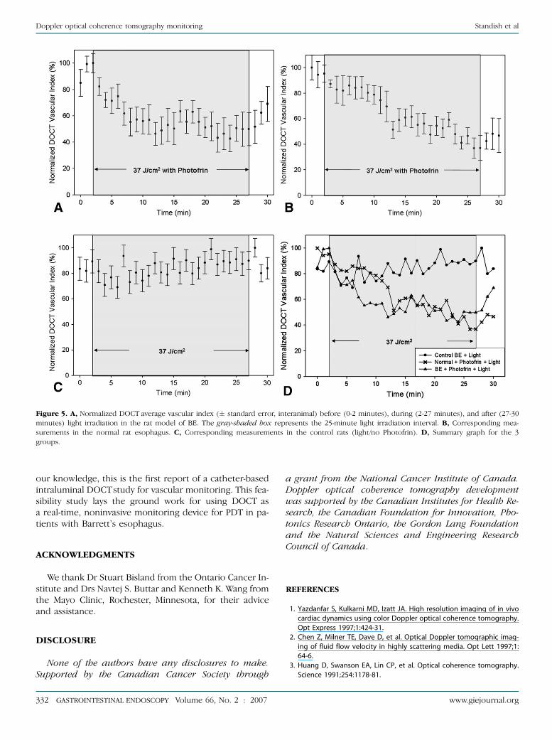
fore, during, and after the course of a PDT treatment areshown in Figure 4. The vascular indices for all 3 groupsstudied are summarized in Figure 5. Minimal changes
328 GASTROINTESTINAL ENDOSCOPY Volume 66, No. 2 : 2007 www.giejournal.org
Doppler optical coherence tomography monitoring Standish et al
were noted in the vascular index of the control group(BE þ light only) during and after light exposure(Fig. 5C). A noticeable reduction in the vascular index, how-ever, was observed in the BE group that received PDT(BE þ Photofrin þ light) (Fig. 5A). During PDT, an averagereduction of 57% � 10% (� standard error) in the vascularindex was measured, with partial recovery in the post-PDTperiod. A similar phenomenon was observed in the groupof normal rats that received PDT (Fig. 5B), with an averagedecrease of 63% � 10% (� standard error) in the vascularindex. There was little difference when comparing thePDT-induced vascular response between the 2 groups.
StatisticsAlthough the sample sizes were small in this study, null
hypothesis testing (independent t test) was applied to thenormalized DOCT vascular index quantity at the 15- and30-minute time points (Fig. 5) between group allocations.The control group (BE þ light) was found to be statisti-cally different (P ! .05) from all other groups at the 15-and 30-minute time points. Although the PDT vascularresponse of the normal and BE groups were very similar,they were also found to be statistically different (P ! .05)at the 15-minute (P Z .06) and 30-minute (P Z .83) timepoints.
SafetyThere were no animal complications or technical diffi-
culties as a result of PDT treatment or DOCT imagingduring this study.
DISCUSSION
PDT when using Photofrin is U.S. Food and Drug Ad-ministration approved for the treatment of patients withBarrett’s esophagus with HGD. In a previous study, a signif-icant difference in the eradication of HGD was found be-tween a Photofrin-PDT plus omeprazole (proton pumpinhibitor) group and an omeprazole-only control group,77% vs 39%, respectively (P ! .0001). However, residualBarrett’s epithelium remained in 48% of patients treatedwith PDT, and, in 13% of the cases, cancer eventually de-veloped.25 Moreover, the persistence of genetic abnormal-ities in residual BE and the presence of islands of BE buriedunderneath the neosquamous epithelium represent a riskof progression to esophageal adenocarcinoma.26 Giventhese concerns, the goal of PDT should be to eradicatethe entire Barrett’s epithelium,27 and this may be facilitatedby real-time monitoring of PDT-induced tissue responsesto allow adjustment and optimization of the PDT dose.
The efficacy of PDT is partly dependent on its vasocon-strictive effect on tissue microvasculature and associatedtissue ischemia. Transient reduction in blood flow or per-manent vessel occlusion occurs as a result of PDT-inducedvascular spasm, vascular stasis, platelet aggregation, and
print&web4C=FPO
Figure 2. Histology and corresponding in vivo DOCT images of a normal
rat esophagus. A, Histologic layers, inset is a magnified view of blood ves-
sels (V) within the muscularis propria (hematoxylin phloxine saffron,
orig. mag. �20). B, Microstructural DOCT image of the corresponding
esophageal layers. C, Composite image with the Doppler shift of blood
vessels overlaid on the microstructural OCT image. The visible esopha-
geal layers are the following: ep, epithelium; lp, lamina propria; mm,
muscularis mucosa; sm, submucosa; mpi, muscularis propria interna;
mpe, muscularis propria externa, and s, serosa. Velocity* assumes a Dopp-
ler angle of �60�.
www.giejournal.org Volume 66, No. 2 : 2007 GASTROINTESTINAL ENDOSCOPY 329
Standish et al Doppler optical coherence tomography monitoring
thrombus formation.28,29 The observation and quantifica-tion of these microvascular alterations by a noninvasiveimaging device in real time could be an important surro-gate marker for determining and optimizing the overalltherapeutic efficacy of PDT in BE and other sites. DOCTappears well suited for this purpose, because it offershigh spatial resolution (approximately 10 mm) andexcellent blood-flow velocity sensitivity (approximately100 mm/s in vivo), providing unique noninvasive micro-structural and functional imaging when compared withexisting endoscopic imaging methods, such as EUS.
The data presented here demonstrated the feasibility ofusing DOCT to monitor the tissue microvascular responseduring PDT in BE and normal esophagus. Vascular com-promise was observed and quantified by DOCT duringPDT of both normal and BE tissues. There were statisticaldifferences in the Photofrin-PDT vascular response be-tween the 2 groups at the 15- and 30-minute point ofthe treatment session, with the PDT treatment conditionsused here. However, this feasibility study was not statisti-cally powered to detect small differences. We also notedthat, with the 24-hour Photofrin-light interval used here,
Figure 3. DOCT images. A, Normal rat with subepithelial detection of blood vessel represented by the color-Doppler image overlaid onto structural
image, inset is a �2 magnification of the detected vessels. B, Squamocolumnar transition zone with the velocity variance image overlaid onto structural
image; multiple scattering of photons through large vessels contribute to the large streak, which typically occur deeper to a large vessel. C, Structural BE
OCT image. D, Histologic correlate of transition zone with Barrett’s glandular structures identified (asterisks in C and D) (H&E, orig. mag. �10). Dif-
ferences in blood flow were detected between the tissues (A and B).
print&web4C/FPO
330 GASTROINTESTINAL ENDOSCOPY Volume 66, No. 2 : 2007 www.giejournal.org
Doppler optical coherence tomography monitoring Standish et al
there was likely both vascular and a direct cell-kill compo-nent in the tissue response. Whether these observationshold in our planned larger study with a predominantly vas-cular photosensitizer (ie, TOOKAD), remains to be seen.
As previously shown,7 the DOCT system is suitable forhuman endoscopic applications, and work is underway re-garding the transfer of this technology for use in patientswith Barrett’s esophagus who are undergoing PDT. Initialclinical studies will correlate DOCT parameters (eg, diff-erent vascular indices) to the degree of eradication ofBarrett’s epithelia after Photofrin-PDT, and the determina-tion of DOCT thresholds at which complete Barrett’sesophagus eradication is observed. A technical issue withDOCT is the system’s inability to visualize Doppler signalsfrom microvessels because of bulk-tissue motion, Dopp-ler-angle dependencies, and variations in blood-flow dy-namics (eg, intermittent flow). Although we currently usea bulk-tissue rejection algorithm, this process may not beoptimized, resulting in omitted vessels. To circumventthis, we are currently investigating clutter rejection algo-rithms to be used in angle-independent power Doppler(PD) signal processing for OCT. Furthermore, with the ad-dition of temporal averaging, PD-OCT may also limit the ef-fects of variations in blood-flow dynamics, improving ourability to visualize and quantify microvessels. Future pro-spective studies will include real-time DOCT monitoring
and PDT parameter adjustment (eg, total irradiance and/or irradiation time) to achieve the desired DOCT thresh-olds and their influence on the completeness of Barrett’sesophagus eradication. As a first step to investigate the vas-cular effects of varying PDT parameters, we recently pub-lished a light dose–escalation study in a prostate cancermodel,30 in which we observed irradiance (rate) dependen-cies of DOCT-detected vascular shutdown resulting from ofPDT. These results demonstrated the feasibility of usingDOCT to detect changes in vasculature as a function of vary-ing PDT parameters. A similar escalation study by using ratswith BE may provide the necessary data to outline a protocolfor optimizing PDT in BE. Three-dimensional volumetricperfusion measurements of the PDT-induced vascularresponse may improve DOCT measurement accuracy,and this is also being explored. Finally, DOCTis currently be-ing used to study and compare the effects of other photo-sensitizers on tissue microvascular responses.
In this study, we demonstrated the feasibility of DOCTto detect, monitor, and quantify microvascular changesduring PDT. The DOCT imaging system provided a uniqueopportunity to assess the effects of PDT on tissue micro-vasculature, a likely key determinant in its overall thera-peutic efficacy. Although we and other investigators havereported on DOCT monitoring of microcirculatorychanges in animals and human beings, to the best of
print&web4C=FPO
Figure 4. DOCT monitoring of the esophagus before (0-2 minutes), during (2-27 minutes), and after (27-30 minutes) PDT light exposure, for a total
imaging time of 30 minutes. Inset boxes represent �1.5 magnification of the detected blood vessel. A, Blood flow is color coded by the detected velocity
variance images; a noticeable reduction in the vascular index was seen during treatment, with recovery in the posttreatment period. B, Color-Doppler
images of corresponding vessel.
www.giejournal.org Volume 66, No. 2 : 2007 GASTROINTESTINAL ENDOSCOPY 331
Standish et al Doppler optical coherence tomography monitoring
our knowledge, this is the first report of a catheter-basedintraluminal DOCT study for vascular monitoring. This fea-sibility study lays the ground work for using DOCT asa real-time, noninvasive monitoring device for PDT in pa-tients with Barrett’s esophagus.
ACKNOWLEDGMENTS
We thank Dr Stuart Bisland from the Ontario Cancer In-stitute and Drs Navtej S. Buttar and Kenneth K. Wang fromthe Mayo Clinic, Rochester, Minnesota, for their adviceand assistance.
DISCLOSURE
None of the authors have any disclosures to make.Supported by the Canadian Cancer Society through
a grant from the National Cancer Institute of Canada.Doppler optical coherence tomography developmentwas supported by the Canadian Institutes for Health Re-search, the Canadian Foundation for Innovation, Pho-tonics Research Ontario, the Gordon Lang Foundationand the Natural Sciences and Engineering ResearchCouncil of Canada.
REFERENCES
1. Yazdanfar S, Kulkarni MD, Izatt JA. High resolution imaging of in vivo
cardiac dynamics using color Doppler optical coherence tomography.
Opt Express 1997;1:424-31.
2. Chen Z, Milner TE, Dave D, et al. Optical Doppler tomographic imag-
ing of fluid flow velocity in highly scattering media. Opt Lett 1997;1:
64-6.
3. Huang D, Swanson EA, Lin CP, et al. Optical coherence tomography.
Science 1991;254:1178-81.
Figure 5. A, Normalized DOCT average vascular index (� standard error, interanimal) before (0-2 minutes), during (2-27 minutes), and after (27-30
minutes) light irradiation in the rat model of BE. The gray-shaded box represents the 25-minute light irradiation interval. B, Corresponding mea-
surements in the normal rat esophagus. C, Corresponding measurements in the control rats (light/no Photofrin). D, Summary graph for the 3
groups.
332 GASTROINTESTINAL ENDOSCOPY Volume 66, No. 2 : 2007 www.giejournal.org
Doppler optical coherence tomography monitoring Standish et al
4. Yang VXD, Gordon ML, Qi B, et al. High speed, wide velocity dynamic
range Doppler optical coherence tomography (Part 1): system design,
signal processing, and performance. Opt Express 2003;4:794-809.
5. Yazdanfar S, Rollins AM, Izatt JA. Imaging and velocimetry of the hu-
man retinal circulation with color Doppler optical coherence tomogra-
phy. Opt Lett 2000;25:1448-50.
6. Zhao Y, Chen Z, Saxer C, et al. Phase-resolved optical coherence to-
mography and optical Doppler tomography for imaging blood flow
in human skin with fast scanning speed and velocity sensitivity. Opt
Lett 2000;25:114-6.
7. Yang VXD, Tang S, Gordon ML, et al. Endoscopic Doppler optical co-
herence tomography in the human GI tract: initial experience. Gastro-
intest Endosc 2005;61:879-90.
8. Haggitt RC. Barrett’s esophagus, dysplasia, and adenocarcinoma. Hum
Pathol 1994;25:982-93.
9. Oleinick NL, Morris RL, Belichenko I. The role of apoptosis in response
to photodynamic therapy: what where, why, and how. Photochem
Photobiol Sci 2002;1:1-21.
10. Van Duijnhoven FH, Aalvers RI, Rovers JP, et al. The immunological
consequences of photodynamic treatment of cancer, a literature re-
view. Immunobiology 2003;207:105-13.
11. Chen B, Pogue BW, Goodwin IA, et al. Blood flow dynamics after pho-
todynamic therapy with verteporfin in the RIF-1 tumor. Radiat Res
2003;160:452-9.
12. Fingar V, Wiemen T, Wiehle S, et al. The role of microvascular dam-
age in photodynamic therapy: the effect of treatment on vessel con-
striction, permeability, and leukocyte adhesion. Can Res 1992;52:
4914-21.
13. van den Bergh H, Ballini JP, Sickenberg M. On the selectivity of photo-
dynamic therapy of choroidal neovascularization associated with age-
related macular degeneration. J Fr Ophtalmol 2004;27:75-8.
14. Overholt BF, Panjehpour M, DeNovo RC, et al. Photodynamic therapy
for esophageal cancer using a 180 degrees windowed esophageal bal-
loon. Lasers Surg Med 1994;14:27-33.
15. Huang Z. A review of progress in clinical photodynamic therapy. Tech-
nol Cancer Res Treat 2005;4:283-93.
16. Gordon ML, Yang VXD, Seng Yue E, et al. Doppler optical coherence
tomography for monitoring the vascular effects of photodynamic
therapy. Proc SPIE 2004;5316:147-54.
17. Yang VXD, Gordon ML, Tang S, et al. High speed, wide velocity dy-
namic range Doppler optical coherence tomography (Part III): in
vivo endoscopic imaging of blood flow in the rat and human gastro-
intestinal tracts. Opt Express 2003;11:2416-25.
18. Pera M, Trastek VF, Carpenter HA, et al. Influence of pancreatic and bil-
iary reflux on the development of esophageal carcinoma. Ann Thorac
Surg 1993;55:1386-93.
19. Buttar NS, Wang KK, Leontovich O, et al. Chemoprevention of esoph-
ageal adenocarcinoma by COX-2 inhibitors in an animal model of Bar-
rett’s esophagus. Gastroenterology 2002;122:1101-12.
20. Yang VXD, Mao YX, Munce N, et al. Interstitial Doppler optical coher-
ence tomography. Opt Lett 2005;30:1791-3.
21. Yang VX, Gordon ML, Mok A, et al. Improved phased-resolved optical
Doppler tomography using the Kasai velocity estimator and histogram
segmentation. Opto Commun 2002;208:202-14.
22. Zhao Y, Chen Z, Saxer C, et al. Phase-resolved optical coherence to-
mography and optical Doppler tomography for imaging blood flow
in human skin with fast scanning speed and high velocity sensitivity.
Opt Lett 2000;25:114-6.
23. Poneros JM, Brand S, Bouma BE, et al. Diagnosis of specialized intesti-
nal metaplasia by optical coherence tomography. Gastroenterol
2001;120:219-24.
24. Evans JA, Poneros JM, Bouma BE, et al. Optical coherence tomography
to identify intramucosal carcinoma and high-grade dysplasia in Bar-
rett’s esophagus. Clin Gastroenterol Hepatol 2006;4:38-43.
25. Overholt BF, Lightdale CJ, Wang KK, et al. International Photodynamic
Group for High-Grade Dysplasia in Barrett’s Esophagus. Photodynamic
therapy with porfimer sodium for ablation of high-grade dysplasia in
Barrett’s esophagus: international, partially blinded, randomized
phase III trial. Gastrointest Endosc 2005;62:488-98.
26. Hage M, Siersema PD, Vissers KJ, et al. Genomic analysis of Barrett’s
esophagus after ablative therapy: persistence of genetic alterations
at tumor suppressor loci. Int J Cancer 2006;118:155-60.
27. Siersema PD. Photodynamic therapy for Barrett’s esophagus: not yet
ready for the premier league of endoscopic interventions. Gastrointest
Endosc 2005;62:503-7.
28. Fingar V, Wieman T, Wiehle S, et al. The role of microvascular damage in
photodynamic therapy: the effect of treatment on vessel constriction,
permeability, and leukocyte adhesion. Cancer Res 1992;52:4914-21.
29. Chen B, Pogue BW, Goodwin IA, et al. Blood flow dynamics after pho-
todynamic therapy with verteporfin in the RIF-1 tumor. Radiat Res
2003;160:452-9.
30. Standish BA, Jin X, Smolen J, et al. Interstitial Doppler optical coher-
ence tomography monitors microvascular changes during photody-
namic therapy in a Dunning prostate model under varying
treatment conditions. J Biomed Opt 2007;12:034022.
Received October 23, 2006. Accepted February 18, 2007.
Current affiliations: Department of Medical Biophysics (B.A.S., N.R.M., A.V.,
B.C.W.), Department of Radiation Oncology (A.V.), University of Toronto,
Ontario Cancer Institute/University Health Network (V.X.D.Y., Y.I.M., A.V.,
B.C.W.), Department of Pathology (G.G.), Division of Gastroenterology
(N.E.M.), St. Michael’s Hospital, Toronto, Ontario, Canada, Division of
Gastroenterology and Hepatology (L-M.W.), Mayo Clinic, Rochester,
Minnesota, USA.
Presented at Digestive Disease Week, May 14-19, 2005, Chicago, Illinois
(Gastroenterology 2005;128[Suppl 2]:A27).
Reprint requests: Beau A. Standish, Division of Biophysics and Bioimaging,
Ontario Cancer Institute, 610 University Ave, Toronto, ON, Canada M5G
2M9.
www.giejournal.org Volume 66, No. 2 : 2007 GASTROINTESTINAL ENDOSCOPY 333
Standish et al Doppler optical coherence tomography monitoring


