Embed Size (px)
Citation preview
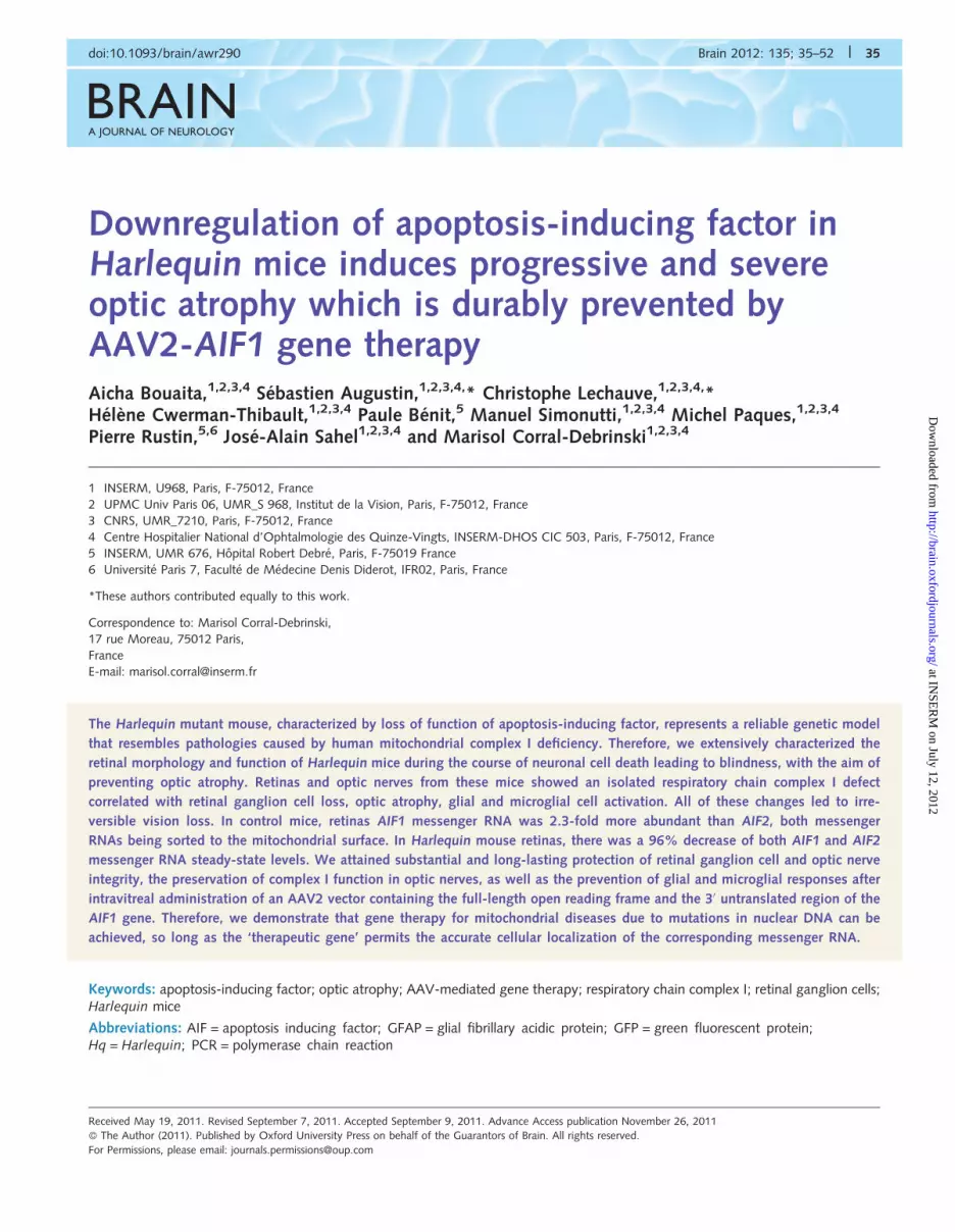
BRAINA JOURNAL OF NEUROLOGY
Downregulation of apoptosis-inducing factor inHarlequin mice induces progressive and severeoptic atrophy which is durably prevented byAAV2-AIF1 gene therapyAicha Bouaita,1,2,3,4 Sebastien Augustin,1,2,3,4,* Christophe Lechauve,1,2,3,4,*Helene Cwerman-Thibault,1,2,3,4 Paule Benit,5 Manuel Simonutti,1,2,3,4 Michel Paques,1,2,3,4
Pierre Rustin,5,6 Jose-Alain Sahel1,2,3,4 and Marisol Corral-Debrinski1,2,3,4
1 INSERM, U968, Paris, F-75012, France
2 UPMC Univ Paris 06, UMR_S 968, Institut de la Vision, Paris, F-75012, France
3 CNRS, UMR_7210, Paris, F-75012, France
4 Centre Hospitalier National d’Ophtalmologie des Quinze-Vingts, INSERM-DHOS CIC 503, Paris, F-75012, France
5 INSERM, UMR 676, Hopital Robert Debre, Paris, F-75019 France
6 Universite Paris 7, Faculte de Medecine Denis Diderot, IFR02, Paris, France
*These authors contributed equally to this work.
Correspondence to: Marisol Corral-Debrinski,
17 rue Moreau, 75012 Paris,
France
E-mail: [email protected]
The Harlequin mutant mouse, characterized by loss of function of apoptosis-inducing factor, represents a reliable genetic model
that resembles pathologies caused by human mitochondrial complex I deficiency. Therefore, we extensively characterized the
retinal morphology and function of Harlequin mice during the course of neuronal cell death leading to blindness, with the aim of
preventing optic atrophy. Retinas and optic nerves from these mice showed an isolated respiratory chain complex I defect
correlated with retinal ganglion cell loss, optic atrophy, glial and microglial cell activation. All of these changes led to irre-
versible vision loss. In control mice, retinas AIF1 messenger RNA was 2.3-fold more abundant than AIF2, both messenger
RNAs being sorted to the mitochondrial surface. In Harlequin mouse retinas, there was a 96% decrease of both AIF1 and AIF2
messenger RNA steady-state levels. We attained substantial and long-lasting protection of retinal ganglion cell and optic nerve
integrity, the preservation of complex I function in optic nerves, as well as the prevention of glial and microglial responses after
intravitreal administration of an AAV2 vector containing the full-length open reading frame and the 30 untranslated region of the
AIF1 gene. Therefore, we demonstrate that gene therapy for mitochondrial diseases due to mutations in nuclear DNA can be
achieved, so long as the ‘therapeutic gene’ permits the accurate cellular localization of the corresponding messenger RNA.
Keywords: apoptosis-inducing factor; optic atrophy; AAV-mediated gene therapy; respiratory chain complex I; retinal ganglion cells;Harlequin mice
Abbreviations: AIF = apoptosis inducing factor; GFAP = glial fibrillary acidic protein; GFP = green fluorescent protein;Hq = Harlequin; PCR = polymerase chain reaction
doi:10.1093/brain/awr290 Brain 2012: 135; 35–52 | 35
Received May 19, 2011. Revised September 7, 2011. Accepted September 9, 2011. Advance Access publication November 26, 2011
� The Author (2011). Published by Oxford University Press on behalf of the Guarantors of Brain. All rights reserved.
For Permissions, please email: [email protected]
at INSE
RM
on July 12, 2012http://brain.oxfordjournals.org/
Dow
nloaded from
IntroductionMitochondrial disorders, thought initially to be rare, now appear
to be relatively common (Schaefer et al., 2008). Despite spectacu-
lar progress in elucidating the molecular basis of mitochondrial
respiratory chain diseases in the last decade, a substantial
amount remains to be done regarding therapy (Kerr, 2010; Schon
et al., 2010). Thus, innovative approaches, including gene therapy,
which may offer the real prospect of developing treatments that
improve the underlying condition of patients, should be explored.
Various obstacles may limit success in this goal: (i) the scarcity of
animal models that truly resemble human diseases; (ii) conditional
tissue targeting by gene therapy so as to minimize systemic distri-
bution and avoid harmful side-effects; (iii) difficulty of efficient mito-
chondrial delivery of the gene product; and (iv) as for any Mendelian
disorder, the choice of vectors, delivery method and dosage.
Since the eye is particularly suitable as a target organ for gene
therapy (Colella et al., 2009) and ocular involvement is a frequent
feature in mitochondrial respiratory chain diseases (Carelli et al.,
2009; Yu-Wai-Man et al., 2009), we decided to develop a gene
therapy strategy for preventing optic atrophy in the Harlequin
(Hq) mouse. Hq mice exhibit common characteristics of human
neurodegenerative mitochondriopathies due to respiratory chain
complex I deficiency, such as the degeneration of the cerebellum,
retina, optic nerve, thalamic, striatal and cortical regions (Klein
et al., 2002; Vahsen et al., 2004; Benit et al., 2008). The Hq
phenotype is caused by a severe reduction of apoptosis-inducing
factor (AIF) gene expression due to a retroviral insertion in the first
intron of the gene (Klein et al., 2002). AIF is a flavoprotein
with nicotinamide adenine dinucleotide (NADH) oxidase activity
localized to the mitochondrial inner membrane and involved in
respiratory chain complex I biogenesis and/or stability.
Interestingly, AIF was first discovered as a caspase-independent
death effector able to induce features of apoptosis in isolated
nuclei (Susin et al., 1999; Ye et al., 2002). Recently, a homozy-
gous trinucleotide deletion in exon 5 of the human AIF gene,
leading to the ablation of an arginine residue at position 201 of
the AIF polypeptide, has been described as responsible for a severe
X-linked mitochondrial encephalopathy and oxidative phosphoryl-
ation (OXPHOS) failure in two infant patients (Ghezzi et al.). By
extensively evaluating Hq mouse eyes, we determined that retinal
ganglion cells reached up to 36% of loss relative to control mice,
associated with the disappearance of optic nerve fibres.
Furthermore, the residual fibres presented a clear respiratory
chain complex I deficiency. The overall injury evidenced in the
ganglion cell layer of Hq mice led to progressive vision loss;
thus, 10-month-old mice were permanently blind.
At least four different isoforms of AIF are possibly generated by
alternative splicing of the messenger RNA precursor; in the ner-
vous system AIF1 and AIF2 (Hangen et al., 2010) are the most
abundant. Here we show that in Hq retinas, AIF1 and AIF2
messenger RNA steady-state levels were 96% inferior to control
animals; in controls, AIF1 messenger RNA is 2.3-fold more
abundant than AIF2. Both AIF1 and AIF2 messenger RNAs are
sorted to the mitochondrial surface, as previously reported for
several nuclear transcripts encoding mitochondrial proteins in
human cells (Sylvestre et al., 2003). In an attempt to prevent
retinal ganglion cell loss and optic nerve atrophy in Hq mice we
have constructed a recombinant adeno-associated virus type 2
(AAV2/2_AIF1) which incorporates the mouse AIF1 open reading
frame with its full-length 30-UTR (untranslated region), ensuring
the sorting of the messenger RNA to the mitochondrial surface
and the efficient mitochondrial translocation of the protein. This
point is particularly important because of the pro-apoptotic prop-
erties of AIF if localized to the cytosol (Scovassi et al., 2009). The
prevention of retinal ganglion cell degeneration and the preserva-
tion of respiratory chain complex I activity in optic nerve were
durably achieved in eyes injected intravitreally with AAV2/
2_AIF1. Hence, we established the proof of principle that
rAAV2/2-mediated gene therapy can lead to the prevention of
respiratory chain complex I defects and optic atrophy in Hq mice.
Materials and methods
AnimalsHemizygous (Hq/Y) males were obtained by mating Hq/X females
with either Hq/Y or wild-type males from the colony shipped from
The Jackson Laboratory at �2 months of age.
The Hq strain was B6CBACaAw-J/A-Pdc8Hq/J (http://jaxmice.jax
.org/strain/000501.html). All mice used in this study were F1 mice
bred from founders having a mixed genetic background. Only hemizy-
gous (Hq/Y) males received evaluations and gene therapy; they were
compared exclusively with littermate males from the colony. The mice
were housed from one to four per cage in a temperature-controlled
environment, with a 12-h light/dark cycle and free access to food and
water. Studies were conducted in accordance with the statements on
the care and use of animals in research of the guidelines issued by the
French Ministry of Agriculture and the Veterinarian Department of Paris
(Permit number DF/DF_2010_PA1000298), the French Ministry of
Research (Approval number 5575) and the ethics committees of the
University Paris 6 and the INSERM, Institut National de la Sante et
de la Recherche Medicale (Authorization number 75-1710).
Adeno-associated viral vectors androute for ocular administrationThe entire Mus musculus apoptosis-inducing factor, mitochondrion-
associated 1 (Aifm1) messenger RNA sequence (http://www.ncbi
.nlm.nih.gov/nuccore/NM_012019) of 1926 base pairs (bp) was
synthesized by Genscript Corp, encompassing the original 87 bp of
the 50-UTR, the entire open reading frame encoding a 612 amino
acid-long protein and two restriction sites at the extremities: EcoR1
at the 50 and XhoI at the 30 for cloning into the pAAV internal ribo-
some entry site-humanized recombinant green fluorescent protein
(hrGFP) vector (Stratagene) in which we had earlier replaced the
hGH [human growth hormone 1 (MIM 139250)] polyadenylation
signal with the 176 bp full-length AIF1 30-UTR (http://www.ncbi.
nlm.nih.gov/nuccore/NM_012019) by using BglII and RsrII unique re-
striction sites. AIF1 transcription is under the control of the CMV pro-
moter and the b-globin intron to ensure high levels of expression. The
open reading frame is in frame with the 3� FLAG� sequence at the
C-terminus. The pAAV-internal ribosome entry site-humanized recom-
binant GFP vector also contains a dicistronic expression cassette in
which the humanized recombinant GFP is expressed as a second
36 | Brain 2012: 135; 35–52 A. Bouaita et al.
at INSE
RM
on July 12, 2012http://brain.oxfordjournals.org/
Dow
nloaded from

open reading frame translated from the encephalomyocarditis virus
internal ribosome entry site. The final vector, named AAV2/2_AIF1,
contains adeno-associated virus type 2 (AAV2) inverted terminal
repeats, which direct viral replication and packaging. The expression
cassettes, flanked by the two AAV2 inverted terminal repeats, were
encapsidated into AAV2 shells. The final construct was sequenced for
accuracy (Genome Express).
In a previous report, we demonstrated that intravitreal injection of
vector DNA followed by electroporation is successful in transducing rat
retinal ganglion cells and leads to a sustained transgene expression for
up to 90 days (Ellouze et al., 2008). In vivo electroporation of the
AIF1 vector DNA in control mice was efficient in transducing retinal
ganglion cells, which express AIF1 for at least 2 months without any
harm to retinal architecture, optic fibre density or visual function
(data not shown). Nevertheless, since our aim is to prevent durably
the absence of AIF in Hq mice and its deleterious effect on retinal
ganglion cell integrity, we need a protocol permitting both the efficient
transduction of retinal ganglion cells and the long-lasting expression of
the AIF1 transgene. It has been demonstrated that, to date, AAV2/2
represents the best choice in terms of overall transduction efficiency
and tropism for adult rodent retinal ganglion cells (Harvey et al., 2009;
Hellstrom et al., 2009). Moreover, it is well known that AAV2 shows
strong dependence for transduction on heparan sulphate proteogly-
cans that are present in retinal ganglion cells and in the inner limiting
membrane. The inner limiting membrane is a meshwork of extracellu-
lar matrix proteoglycans located at the interface of the vitreous and
the ganglion cell layer. Thus, this first physical barrier may also effect-
ively localize the virus and prevent it from being cleared from the
vitreous via the trabecular meshwork, thereby facilitating subsequent
retinal ganglion cell transduction (Dalkara et al., 2009). For all these
reasons, we decided to target retinal ganglion cells of Hq mice with
the AAV2/2_AIF1 vector, by administration into the vitreous body of
mouse eyes.
Vectors were produced in the Vector Core at the University Hospital
of Nantes (http://www.vectors.nantes.inserm.fr). The AAV titres were
determined by dot blot and expressed as vector genomes per millilitre.
During the course of this study we used two independent productions
with the following titres: 7.5 � 1010 and 2.37 � 1011 vector genomes/
ml. For intravitreal injections, after dilatation of the pupil with topical
1% tropicamide (CibaVision), mice were subjected to anaesthesia with
isoflurane (40 mg/kg body weight). The tip of a 15-mm 33-gauge
needle, mounted on a 10 ml Hamilton syringe (Hamilton Bonaduz
AG) was advanced through the sclera �1 mm posterior to the
corneoscleral limbus in the superior region of the eye, and 2–3 ml of
vector suspension was injected intravitreally, avoiding retinal structure
disruption, bleeding or lens injury. All the injections were performed
under visual control using an ophthalmic surgical microscope. Viral
particles were mixed with 1/10 000 of sterile fluorescein to follow
the homogenous dissemination of the suspension into the vitreous.
Forty-three mice (4- to 8-weeks old) were subjected to AAV2/
2_AIF1 administration; 33 animals received 2.25 � 108 vector gen-
omes/eye (lowest titre batch) and 10 received 7.11 � 108 vector gen-
omes/eye (highest titre batch). Two animals developed a cataract due
to the surgery and three animals died �7 weeks after eye surgery from
natural causes; these five animals were discarded from the study.
Tissue homogenate preparation andrespiratory chain enzymatic assaysAfter rapid and careful dissection, optic nerves or retinas were kept
frozen (�80�C). Samples were prepared at ice-melting temperature by
homogenization of tissues using a 1-ml hand-driven glass–glass Potter
in 200ml of extraction buffer (0.25 mM sucrose, 40 mM KCl, 2 mM
ethylene glycol tetraacetic acid (EGTA), 1 mg/ml bovine serum albu-
min and 20 mM Tris–HCl, pH 7.2). Large cellular debris was spun
down by a low speed centrifugation (3100 rpm, 8 min) and tissue
homogenates were used immediately. Respiratory chain complex I
and V activities were measured using a Cary 50 spectrophotometer
equipped with an 18-cell holder maintained at 37�C (Varian Australia)
as previously described (Benit et al., 2008). Each assay was made in
triplicate with 50 ml of the homogenates obtained. All chemicals were
of the highest grade from Sigma-Aldrich. Complex I
(NADH-ubiquinone oxidoreductase) activity values were either normal-
ized by complex V or converted to specific activities after protein
quantification by the Bradford method. Optic nerves were dissected
from 40 mice subjected to AAV2/2_AIF1 injection; optic nerves from
seven mice were subjected to histological studies. Respiratory chain
assessments obtained from only 28 mice were included in Table 3,
since we failed to measure complex I and complex V activities from
five mouse eye samples due to technical problems encountered during
the assessment.
Fundus imaging by confocal scanninglaser ophthalmoscopy and opticalcoherence tomographyA confocal scanning laser ophthalmoscope (Heidelberg Retina
Angiograph, Heidelberg Engineering) with green laser illumination
was used to examine mice before treatment and each month until
euthanasia. To monitor the nerve fibre layer at each session, up to
four pictures, each from an average of eight images, corresponding to
cardinal areas of the eye fundus were performed. The built-in software
was used for post-processing the images, including alignment,
adjustment of contrast, construction of a mean image and/or of a
composite image (Paques et al., 2006). Spectral-domain optical coher-
ence tomography was performed using the Spectralis� system from
Heidelberg Engineering. The acquisition time per tomographic image,
which includes summation of 10 raw images, was in the order of 1 s
(Grieve et al., 2004). Neural retina thickness for control and Hq eyes
was compared by measuring the distance from the anterior boundary
of the retinal nerve fibre layer and the posterior boundary of the
photoreceptors layer. Throughout the examination process, a quiet
ambiance with dim illumination was maintained. Pupil dilation was
performed with topical 1% tropicamide (CibaVision). Careful slit-lamp
examination before the procedure ruled out the presence of any
corneal or lens opacities. Mice were manually held in front of either
apparatus, in an upright position. As a rule, restraint and rest periods
alternated approximately every 30 s. The examination was interrupted
if the animal showed any sign of tiredness.
Retinal and optic nerve histologyAnimals were sacrificed under deep ketamine (50 mg/kg) xylazine
(10 mg/kg) anaesthesia. After the ocular globes were removed, they
were pierced in the cornea with a needle and fixed for 1 h in 4%
paraformaldehyde at 4�C. After removal of the cornea and lens,
eyecups were post-fixed overnight in 4% paraformaldehyde at 4�C,
cryoprotected by overnight incubation in phosphate-buffered saline
containing 30% sucrose (Sigma-Aldrich) at 4�C. Then, retinas were
embedded in Optimal Cutting Temperature medium (Neg 50;
Richard-Allan Scientific), frozen in liquid nitrogen, and stored at
�20�C. Optic nerves were carefully collected and fixed overnight in
Gene therapy to prevent optic neuropathy Brain 2012: 135; 35–52 | 37
at INSE
RM
on July 12, 2012http://brain.oxfordjournals.org/
Dow
nloaded from

4% paraformaldehyde at 4�C. Next day, after washes in phosphate-
buffered saline, tissues were cryoprotected by incubation overnight in
30% sucrose in phosphate-buffered saline. Subsequently, optic nerves
were incubated in a 7.5% gelatin solution from porcine skin; Type A
(Sigma-Aldrich) and 10% sucrose. Samples were frozen between
�50�C and �60�C in 2-methyl-butane solution and then stored at
�20�C. Sections of retinas and optic nerves (transverse and longitudin-
al, near the globe) with a thickness of 10 mm were cut on a cryostat
(Microm Microtech) at �20�C and mounted on SuperFrost�Plus slides.
For immunochemistry, sections of retinas and optic nerves were
washed with phosphate buffer pH 7.4, permeabilized with 1%
Triton X-100 in phosphate buffer for 15 min at room temperature
and treated with 10% normal goat serum, 3% bovine serum albumin,
0.5% Triton and 0.05% Tween-20 in phosphate buffer for 1 h. They
were then incubated with primary antibody overnight at 4�C. The next
day, sections were washed three times in phosphate buffer and
incubated with the appropriate secondary antibodies and DAPI (40,6-
diamidino-2-phenylindole; 2 mg/ml) for 90 min at room temperature
with 3% normal goat serum, 3% bovine serum albumin, 0.5%
Triton and 0.05% Tween-20 in phosphate buffer. Finally, they were
washed three times in phosphate buffer, rinsed with sterile water and
mounted on a glass slide. Primary and secondary antibodies used are
shown in Supplementary Table 1.
Microscopic observations and retinalganglion cell countsImage acquisition of fluorescence labelling was monitored with a fluor-
escence microscope (Leica DM 6000 B) using the MetaVue software
for image acquisitions or a confocal laser scanning microscope
Olympus FV1000. Images were captured with Olympus software
(Olympus). Images were then analysed with Adobe Photoshop� CS
and ImageJ (National Institutes of Health) software. Slides from retinas
and optic nerve sections were also scanned with the Hamamatsu
Nanozoomer Digital Pathology (NDP) 2.0 HT, its fluorescence unit
option (L11600-05) and the NanoZoomer’s 3-CCD TDI camera
(Hamamatsu Photonics). A resolution of 0.92 mm/pixel (�40) was
routinely used.
Retinal ganglion cell counts were performed from each retina on
three entire consecutive cryostat sections with depth at �400 mm
from the optic nerve, using BRN3A immunolabelling as previously
described (Ellouze et al., 2008). BRN3A is a transcription factor
expressed in �80% of all retinal ganglion cells in mouse retina
(Badea et al., 2009) and 490% in rat retina (Nadal-Nicolas et al.,
2009). Manual counting of immunopositive cells was performed after
capturing each whole section in 13–15 images; the identity of each
mouse was unknown for the counting. BRN3A-positive cell numbers
were further validated, for a subset of animals in each group, by per-
forming two additional immunostainings of different retinal sections
(four sections apart from the one used for the first labelling). Retinal
ganglion cell numbers was estimated as a mean for 20 control eyes,
26 eyes from Hq untreated mice and both eyes from 16 Hq mice
subjected to AAV2/2_AIF1 administration in one of their eyes
(12 out of 16 received the vector in their left eyes).
Optomotor testsThe head-tracking method is based on an optomotor test (Thaung
et al., 2002). Vertical black-and-white lines of three varying widths,
subtending 0.0625, 0.125 and 0.25 cycle/�, were presented to the
animal and rotated alternatively clockwise and counterclockwise,
each for 60 s with an interval of 10 s between the two drum rotations.
Head movements were recorded with a video camera mounted above
the apparatus. Animals were scored only when the speed of the head
turn corresponded with the speed of rotation of the stripes (12�/s).
Light levels were kept constant (240 lux).
RNA extractions and real-timequantitative polymerase chain reactionassayRNA preparations from mitochondrion-bound polysomes and free-
polysomes were obtained from 12 brains (cortical region) from
8-week-old C57Bl/6NCrlBR male mice with some modifications of
the protocol previously published for HeLa cells (Sylvestre et al.,
2003). Briefly, after euthanasia, tissues were rinsed in phosphate-
buffered saline before homogenization with a glass Potter homogen-
izer in the freshly prepared extraction buffer [0.32 M sucrose, 30 mM
Tris–HCl pH 7.5, 5 mM AcMg, 100 mM KCl, 1% (vol/vol) protease
inhibitor cocktail (Sigma), 5 mM b-mercapthoethanol, 1 mM PMFS
(Fluka), 0.1% (w/vol) bovine serum albumin fraction V in ultra-pure
distilled water]. The homogenate was centrifuged (1000 rpm, 10 min,
4�C) to eliminate nucleus and unbroken cells. The supernatant was
conserved at 4�C and the pellet was subjected to two additional hom-
ogenizations for optimal cell lysis. The three supernatants were pooled
and were centrifuged at slow speed twice to remove nucleus and
unbroken cells. The supernatants obtained were then centrifuged at
12 500 rpm for 30 min at 4�C; the pellet contained mitochondria and
the supernatant represented the cytosolic fraction. The mitochondrial
fraction was washed three times in the extraction buffer (lacking
bovine serum albumin). The last mitochondrial pellet, containing poly-
somes bound to the outer mitochondrial membrane, was conserved
at �80�C. The cytosolic fraction was gently deposed on a SW41 tube
that contained two layers of 2.0 M sucrose and 0.5 M sucrose
prepared in the extraction buffer. SW41 tubes were centrifuged at
4�C for 20 h at 40 000 rpm. The pellet represented the free-polysome
fraction.
Total RNAs were prepared from mitochondrion-bound polysomes,
free-polysome fractions as well as from wild-type or Hq mouse retinas
using the RNA isolation kit (Qiagen). RNA concentration was deter-
mined with the Nanodrop ND-2000 Spectrophotometer (Thermo
Fisher Scientific) and RNA integrity was verified using agarose gel
electrophoresis. Real-time polymerase chain reaction (PCR) analyses
using mitochondrion-bound polysome and free-polysome fractions
were performed with the Superscript III one-step real-time PCR kit
with Platinum Taq (Invitrogen, Life Technologies).
For real-time quantitative PCR analyses, 1 mg of total RNA from
wild-type or Hq mouse retinas was reverse-transcribed with oligo-dT
in a 20ml final reaction volume using Superscript� II Reverse
Transcriptase (Invitrogen, Life Technologies) following the manufactur-
er’s instructions. The mouse AIF1, AIF2 and AIF1tr (AIF1 messenger
RNA produced from the vector) primers were customized to be highly
specific for each messenger RNA specie. RNAs were purified from
21 mice subjected to AAV2/2_AIF1 intravitreal injection, 13 received
the lowest vector dose and eight received the highest vector dose.
One of the samples from the lowest dose was discarded, since a mix-
ture with another sample led to a confusion of its real identity. Thus,
the results for 20 mice subjected to AAV2/2_AIF1 administration were
presented. In treated mice, the measurement of AIF1 corresponds to
the overall amount of AIF1 messenger RNA (endogenous gene and
AAV2/2_AIF1 driven expression) and the measurement AIF1tr repre-
sents only the abundance of messenger RNA transcribed from the
38 | Brain 2012: 135; 35–52 A. Bouaita et al.
at INSE
RM
on July 12, 2012http://brain.oxfordjournals.org/
Dow
nloaded from
vector since one of the primers used recognizes the FLAG epitope
sequence. The primers used in this study are shown in
Supplementary Table 2 and were synthesized by Invitrogen, Life
Technologies.
Quantitative real-time PCR reactions were performed using ABI
7500 Fast (Applied Biosystems). For microplate experiments, 1/100
and 1/500 of the reverse transcription product was used per gene
as template for quantitative PCR reactions with Power Sybr� green
PCR Master Mix (Applied Biosystems, Life Technologies,). Real-time
PCR amplifications were carried out as follows: 10 min 95�C incubation
followed by 40 cycles of 15 s at 95�C and 1 min at 60�C. Each bio-
logical sample was subjected to the assay in triplicate. Obtained Ct
values for each sample allowed AIF1, AIF2, AIF1tr (AIF1 messenger
RNA synthesized from the AAV2/2_AIF1 vector), BRN3A and
gamma-synuclein (SNCG) messenger RNA quantifications using
7500 v.2.0.4 software (Applied Biosystems). The mitochondrial ATP6
gene was selected as the most stable, regarding messenger RNA
steady-state levels, to normalize the messenger RNA species evaluated
and to obtain relative messenger RNA abundance in Hq retinas.
Several reports indicated that BRN3A and SNGC messenger RNA
levels can be used to assess retinal ganglion cell number (Soto et al.,
2008; Surgucheva et al., 2008; Torero Ibad et al., 2011). Therefore,
their estimation in both Hq and control retinas aimed at completing
retinal ganglion cell counts obtained by histological evaluation of
retinal sections.
Statistical analysesValues are expressed as mean � SEM. Data were compared using
Student’s t-test (http://www.u707.jussieu.fr/biostatgv/index.html).
P-values of 50.05 were considered as significant, and 50.01 as
highly significant.
Results
Phenotypic hallmarks of Harlequinmouse eyesWe first studied the viability of retinal ganglion cells in Hq mice
since their long axons form the optic nerve. We examined
eye fundus at different ages using a full-field optical coherence
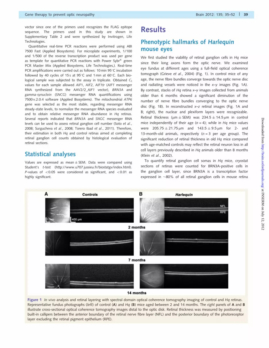
tomograph (Grieve et al., 2004) (Fig. 1). In control mice of any
age, the nerve fibre bundles converge towards the optic nerve disc
and radiating vessels were noticed in the x–y images (Fig. 1A).
By contrast, stacks of Hq retina x–y images collected from animals
older than 6 months showed a significant diminution of the
number of nerve fibre bundles converging to the optic nerve
disc (Fig. 1B). In reconstructed x–z retinal images (Fig. 1A and
B; right), the nuclear and plexiform layers were recognizable.
Retinal thickness (mm � SEM) was 234.5 � 14.5mm in control
mice independently of their age (n = 4); while in Hq mice values
were 205.75 � 21.75 mm and 143.5 � 9.5 mm for 2- and
13-month-old animals, respectively (n = 3 per age group). The
significant reduction of retinal thickness in old Hq mice compared
with age-matched controls may reflect the retinal neuron loss in all
cell layers previously described in Hq animals older than 8 months
(Klein et al., 2002).
To quantify retinal ganglion cell somas in Hq mice, cryostat
sections of retinas were counted for BRN3A-positive cells in
the ganglion cell layer, since BRN3A is a transcription factor
expressed in �80% of all retinal ganglion cells in mouse retina
Figure 1 In vivo analysis and retinal layering with spectral domain optical coherence tomography imaging of control and Hq retinas.
Representative fundus photographs (left) of control (A) and Hq (B) mice aged between 2 and 14 months. The right panels of A and B
illustrate cross-sectional optical coherence tomography images distal to the optic disk. Retinal thickness was measured by positioning
built-in callipers between the anterior boundary of the retinal nerve fibre layer (NFL) and the posterior boundary of the photoreceptor
layer excluding the retinal pigment epithelium (RPE).
Gene therapy to prevent optic neuropathy Brain 2012: 135; 35–52 | 39
at INSE
RM
on July 12, 2012http://brain.oxfordjournals.org/
Dow
nloaded from

Figure 2 Immunofluorescence analysis of retinal and optic nerves from control and Hq mice. (A) Immunostaining for GFAP (green)
and BRN3A (red) of retinas from control and Hq mice aged 1 month; GFAP expression was restricted to the ganglion cell layer (GCL).
The nuclei were contrasted with DAPI (blue). (B) With ageing, Hq mice exhibited a progressive increase in GFAP expression as illustrated in
Hq mice aged 7 and 13 months, respectively. By contrast, age-matched controls only showed a GFAP signal restricted to the ganglion cell
layer. It also appears in this figure that with ageing the Hq mouse inner nuclear layer (INL) and outer nuclear layer (ONL) have thinned
considerably. Scale bar = 50mm. (C) Low magnification images of retinas from 5-month-old and 8-month-old Hq mice and age-matched
controls, GFAP (green) and DAPI (blue) staining are shown. Scale bar = 1 mm. In merge reconstitutions, a 3-fold zoom of the same region is
also illustrated. (D) Independent proximal optic nerve transversal sections from 8-month-old control and Hq mice were immunolabelled with
antibodies against the heavy chain (200 kDa) subunit of neurofilaments, (NF200, green), the tubulin b-III, a microtubule protein (b-III, red)
and the basic myelin protein (MBP, red). Immunoreactivity for the two axonal markers (NF200 and tubulin b-III) shows that axons are largely
lost in Hq mice relative to age-matched controls. Myelin basic protein immunoreactivity was also significantly reduced in Hq mouse optic
nerves, suggesting myelin alterations due to the absence of AIF1 in these mice. Scale bars = 100 and 50mm. IPL = inner plexiform layer.
40 | Brain 2012: 135; 35–52 A. Bouaita et al.
(continued)
at INSE
RM
on July 12, 2012http://brain.oxfordjournals.org/
Dow
nloaded from

Figure
2C
ontinued
.
Gene therapy to prevent optic neuropathy Brain 2012: 135; 35–52 | 41
at INSE
RM
on July 12, 2012http://brain.oxfordjournals.org/
Dow
nloaded from

(Badea et al., 2009) and 490% in rat retina (Nadal-Nicolas et al.,
2009). We found that retinal ganglion cell somas were signifi-
cantly reduced in Hq retinas: 149 � 27 (n = 26) relative to
233 � 39 in control retinas (n = 20), a diminution of 436% of
the total amount of BRN3A-positive cells in the ganglion cell layer
(P =2.2 � 10�7). Accordingly, the total number of cells in the
ganglion cell layer was also significantly lower in Hq mice:
502.7 � 50.8 relative to 608.4 � 76 in control retinas
(P = 0.00021).
Glial cell activation is an important factor contributing to retinal
ganglion cell death in experimental models of glaucoma (Jiang
et al., 2007; Zhou et al., 2009). In retinas from 1-month-old
control and Hq mice, glial fibrillary acidic protein (GFAP), which
is a sensitive marker of glial activation immunofluorescence, was
confined exclusively to the ganglion cell layer, presumably corres-
ponding to the end-feet of Muller cells and a few astrocytes (Fig.
2A). An obvious increase in GFAP immunoreactivity across the
entire thickness of the retina was observed in Hq retinas from
7 - and 13-month-old mice; while 7- and 13-month-old control
retinas showed GFAP immunostaining restricted to the ganglion
cell layer (Fig. 2B). Interestingly, in retinal sections from 7- and 13-
month-old Hq mice, the diminished number of BRN3A-positive
cells in the ganglion cell layer and the thinness of both the inner
nuclear layer and outer nuclear layer are noticeable, confirming
the neuron cell loss noted in eye fundus images (Fig. 1) and re-
ported earlier (Klein et al., 2002). The reconstruction of whole
retinal sections confirmed the extensive glial response in 5- and
8-month-old Hq mice relative to age-matched controls as well as
the retinal thickness reduction in Hq eyes (Fig. 2C). Next, optic
nerve cross-sections were immunolabelled to observe axonal pro-
files and to search for myelin pathology in Hq mice. Figure 2D
illustrates a recognizable reduction in immunopositive dots in
7-month-old Hq mice relative to age-matched controls when
two antibodies against integral axonal proteins were used (b-III
tubulin and NF200). Moreover, the immunostaining with the anti-
body against the myelin basic protein, one of the most abundant
proteins of the myelin sheath, shows a very weak signal in Hq
mice; whereas intact and strong labelled red rings (myelin basic
protein) were observed in age-matched control (Fig. 2D). Thus,
AIF depletion in the optic nerve induces severe loss of axons along
with oligodendrocyte death; this latter could ultimately lead to
impaired myelination. We also evaluated longitudinal sections of
wild-type and Hq mice for gliosis and microglial activation. For the
latter, we used IBA1 (ionized calcium binding adaptor molecule 1)
since it is upregulated in activated microglia and is involved in cell
migration and phagocytosis (Sasaki et al., 2001). We observed a
weak immunoreactivity for GFAP in optic nerves from a 12-
month-old control, while a strong signal was apparent in optic
nerves from an age-matched Hq mouse. Further, it appears that
IBA1-positive cells were present in higher numbers and with an
increased staining intensity in optic nerves from Hq eyes compared
with the few scattered IBA1-positive cells observed in optic nerves
dissected from an age-matched control (Supplementary Fig. 1).
Additionally, microglial cells in control optic nerves were consist-
ently more ramified (resting state); while IBA1 immunostaining in
optic nerves from Hq mice revealed both ramified cells and round
somata cells with simplified processes (activated state). Hence,
similar to rodent models of optic nerve injury (Bosco et al.,
2008; Sivilia et al., 2009; Morrison et al., 2010) optic nerves
from Hq mice hosted activated astrocytes, microglial cells, as
well as structural disruption of myelination.
Studies of respiratory chain functionFirst, we assessed complex I and complex V activities and
estimated the complex I/complex V ratios in retinas from eight
8-month-old Hq mice and seven age-matched controls
(Table 1). A significant 60% decrease relative to control values
was observed, in both complex I-specific activity and complex
I/complex V ratio, while complex V activity was identical between
Hq and control mice. Secondly, optic nerves were collected either
from animals sacrificed before 8 months of age or older than
8 months. In control optic nerves, complex I, complex V and
their ratio did not change with age, being similar to the ones
measured in retinas (Table 1). Optic nerves from Hq mice aged
58 months already had an �50% decrease of complex I activity,
Table 1 Detection of respiratory chain complex I deficiency in retinas and optic nerves from Hq mice
Ocular tissues Specific activities (nmol/min/mg protein) � SEM Activity ratios � SEM
Complex I Complex V Complex I/V
Retinas from 8-month-old mice (number of eyes tested)
Controls (14) 16.44 � 3.7 68.48 � 8.24 0.278 � 0.071
Harlequin (16) 6.91 � 0.88 75.73 � 11.8 0.090 � 0.014
P-value controls/Harlequin 5.41 � 10�5 0.13 9.15 � 10�6
Optic nerves (age; number of eyes tested)
Controls (2–7 months; 32) 14.12 � 4.53 72.24 � 20.9 0.235 � 0.05
Harlequin (2–7 months; 28) 7.22 � 1.91 70.64 � 29.37 0.143 � 0.025
P-value controls/Harlequin 0.0025 0.898 7.35 � 10�9
Controls (8–13 months; 20) 13.94 � 4.18 70.93 � 17.4 0.243 � 0.055
Harlequin (8–13 months; 28) 3.78 � 1.64 72.33 � 16.9 0.0706 � 0.0176
P-value controls/Harlequin 4.67 � 10�6 0.891 2.58 � 10�10
Measurements of complex I and V activities were obtained by spectrophotometry. Complex I and complex V activities were expressed as nanomoles of oxidized NADH/min/mg protein. In retinas and optic nerves, a complex I defect was shown in young Hq mice; the activity declined in Hq with age. Values represent the mean of triplicatesper each eye sample evaluated.
42 | Brain 2012: 135; 35–52 A. Bouaita et al.
at INSE
RM
on July 12, 2012http://brain.oxfordjournals.org/
Dow
nloaded from

a decrease that worsened with age (470% decrease after
8 months of age). Complex V activity was not different between
controls and Hq mice in either age group examined. Thus, in Hq
mice not only did a significant proportion of nerve fibre bundles
disappear (Fig. 1) but complex I enzymatic activity in residual
nerve fibres was severely compromised.
Harlequin mouse visual functionWe next studied the visual function of the Hq mice by the
optokinetic drum test (Thaung et al., 2002; Davies et al., 2007).
We first examined sighted littermate controls (B6CBAC) at two
different ages (43 months and between 6 and 12 months).
They were capable of tracking the moving acuity square-wave
gratings of the three frequencies examined, by making several
head-tracking movements in the same direction and speed as
the drum (Table 2). Hq mice younger than 3 months showed
head-tracking of all three square-wave gratings, indicating that
they were not functionally blind despite their reduced visual per-
formance. Hq mice aged between 4 and 8 months of age spent
much less time tracking across the test period at all three
square-wave gratings relative to age-matched controls. Thus, by
this age there was a significant effect of genotype in the three
grating frequencies, for both the clockwise and counterclockwise
drum rotations (Table 2). Finally, eight Hq mice older than
11 months failed to make head-tracking movements at any of
the grating frequencies in either clockwise or counterclockwise
drum rotation, and could be considered totally blind. The complete
loss of visual function in animals older than 1 year is certainly the
consequence of retinal ganglion cell degeneration, the disappear-
ance of photoreceptors (Figs 1 and 2B) and the disruption of
myelin in the optic nerves (Fig. 2D).
Relative abundance of AIF1 and AIF2messenger RNAs in Hq retinasThe relative abundance of AIF1 and AIF2 transcripts (Hangen
et al., 2010) in retinas from control and Hq mice was determined
by quantitative real-time PCR using the comparative ��Ct
method and the mitochondrial ATP6 as a reference gene, since
its messenger RNA steady-state levels remained almost unchanged
in all the samples evaluated (data not shown). RNA preparations
from 24 retinas of littermate controls aged between 6 and
7 months were examined. AIF1 messenger RNA is 2.3-fold
more abundant than AIF2 in retinas from control mice
(P = 8.2 � 10�12). The difference between AIF1 and AIF2 messen-
ger RNA relative abundance was preserved in 31 Hq retinas, while
the total amount of both messenger RNAs fell to 54% of control
values (Fig. 3A). As SNCG messenger RNA is expressed at high
levels in most if not all retinal ganglion cells and in no other cells
within the adult mouse retina (Soto et al., 2008; Surgucheva et al.,
2008), we determined the relative amount of SNCG and BRN3A
messenger RNAs (Fig. 3B). The steady-state levels of these two
specific markers of retinal ganglion cells were reduced to �50%
of control values in Hq retinas (P-values of 2.3 � 10�8 and
1.8 � 10�11 for BRN3A and SNCG, respectively), confirming ourTab
le2
Opto
moto
rte
sts
of
contr
ol
and
Har
lequin
mic
e
Anim
als
Tra
ckin
gfe
ature
sLi
tter
mat
eco
ntr
ol
mic
e(5
3m
onth
sof
age)
n=
10
Litt
erm
ate
contr
ol
mic
e(6
–12
month
sof
age)
n=
10
Young
Hq
mic
e(5
3m
onth
sof
age)
n=
12
Hq
mic
e(4
–8m
onth
sof
age)
n=
10
Gra
tings
freq
uen
cy(c
ycle
s/�)
0.0
625
0.1
25
0.2
50.0
625
0.1
25
0.2
50.0
625
0.1
25
0.2
50.0
625
0.1
25
0.2
5
Clo
ckw
ise
4.3�
1.9
5.3�
1.7
6�
29.8�
29.3�
2.5
8.9�
2.2
2.6�
1.6
4.2
5�
24.8�
22.1�
1.7
82.3
5�
1.7
3.3�
1.7
5
Counte
r-cl
ock
wis
e5.4�
2.1
6.3�
0.8
58�
1.4
10.4�
2.5
8�
2.3
9.6�
2.2
4.6�
2.1
3.7
5�
1.7
53.7�
1.7
1.7
5�
1.5
52.4�
1.5
82.7�
1.9
Four
diffe
rent
gro
ups
of
mic
ew
ere
eval
uat
edfo
rhea
dtr
acki
ng
move
men
tat
anan
gula
rsp
eed
iden
tica
lto
that
of
the
dru
mro
tation.
The
mic
ew
ere
pre
sente
dw
ith
a2�,4�
and
8�
gra
ting
(corr
espondin
gto
0.2
5,0.1
25
and
0.0
625
cycl
es/�
,re
spec
tive
ly).
They
mad
ese
vera
lhea
dtr
acki
ng
move
men
tsin
the
sam
edirec
tion
and
spee
das
the
dru
m.H
ead
move
men
tssh
ow
nin
Tab
le2
are
pro
vided
per
min
ute
.D
ata
colle
cted
from
Hq
mic
eag
edbet
wee
n4
and
8m
onth
sat
the
thre
egra
ting
freq
uen
cies
for
both
the
clock
wis
ean
dco
unte
r-cl
ock
wis
edru
mro
tations
wer
esi
gnifi
cantly
diffe
rent
toth
ose
of
litte
rmat
eco
ntr
olm
ice
(6–1
2m
onth
sof
age)
:St
uden
t’s
t-te
st;
P=
3.8�
10�
6,
3�
10�
6,
0.0
015,
0.0
0094,
0.0
106,
0.0
0019.
Eight
Hq
mic
eof
about
1ye
arof
age
wer
eal
sosu
bje
cted
toth
ete
stth
ree
tim
esw
ithin
a2-w
eek
tim
ein
terv
al:
they
tota
llyfa
iled
tom
ake
hea
dtr
acki
ng
move
men
tsat
any
of
the
gra
ting
freq
uen
cies
for
eith
ercl
ock
wis
eor
counte
r-cl
ock
wis
edru
mro
tation.
Gene therapy to prevent optic neuropathy Brain 2012: 135; 35–52 | 43
at INSE
RM
on July 12, 2012http://brain.oxfordjournals.org/
Dow
nloaded from

data on retinal ganglion cell loss and nerve fibre degeneration
(Fig. 1).
Design of a gene therapy strategy forpreventing retinal ganglion cell andoptic nerve injuryTo efficiently prevent retinal ganglion cell loss and optic nerve
atrophy in Hq mice, AIF protein must be translocated to the mito-
chondrial inner membrane. By studying RNAs isolated from
mitochondrion-bound polysomes and free cytosolic polysomes
isolated from mouse brains we found an enrichment of AIF1
and AIF2 messenger RNAs in mitochondrion-bound polysomes;
indeed, �70% of the overall signal of AIF1 or AIF2 messenger
RNA was found in this fraction (Fig. 4). Next, we selected AIF1 for
gene therapy since its messenger RNA is 2.3-fold more abundant
than AIF2 messenger RNA. We constructed a recombinant AAV
vector containing both the full-length AIF1 open reading frame
and the 30-UTR of the mouse gene to ensure messenger RNA
sorting to the mitochondrial surface. The recombinant vector
was generated using the genome and capsid proteins from sero-
type 2, which has been described as able to efficiently transduce
retinal ganglion cells in mice (Guy et al., 2009; Zhou et al., 2009).
For preventing optic atrophy, 4- to 8-week-old animals were sub-
jected to a single intravitreal injection since at this age the extent
of retinal ganglion cell injury did not lead to noticeable nerve fibre
degeneration (Fig. 1) or vision loss (Table 2).
Effect of rAAV2/2_AIF1 administrationon retinal ganglion cell integrityFour to five months after AAV2/2_AIF1 treatment, we evaluated
the relative amount of the transduced AIF1 messenger RNA by
real-time quantitative PCR in retinas from 20 Hq mice. Twelve
mice received 2.25 � 108 vector genomes; the remaining eight
were treated with 3.16-fold more of AAV2/2_AIF1. We detected
specific messenger RNA species in retina preparations of all the
injected eyes; the steady-state levels of the transduced AIF1
(AIF1tr) messenger RNA contributed to a 14% increase of the
overall amount of AIF1 messenger RNA when 2.25 � 108 vector
genomes were used. Remarkably, mouse retinas from eyes
injected with the highest dose of the vector showed 600-fold
more of the transduced AIF1 messenger RNA. Consequently, the
total amount of AIF1 messenger RNA in retinas from transduced
eyes reached 25.25% of the control value (Fig. 5A). We next
estimated the relative amounts of BRN3A and SNCG messenger
RNAs in retinas from treated and untreated eyes. Independently of
the AAV2/2_AIF1 dose used, a consistent but not statistically sig-
nificant increase of 13.5 and 20%, respectively in their relative
abundance was measured between treated and untreated eyes,
whereas rhodopsin (RHO) messenger RNA abundance remained
unchanged (Fig. 5B). The high variability observed could be
ascribed to the well-known interindividual phenotypic heterogen-
eity, unevenness due to transduction efficiency, but also to the use
of whole retinas in which retinal ganglion cells represent 55% of
the overall cell population. Therefore, we decided to estimate ret-
inal ganglion cell number in retinal sections from 16 mice treated
in one of their eyes with AAV2/2_AIF1 and euthanized �4
months after vector administration. Retinal ganglion cell number
in treated eyes was 31.6% higher than in contralateral untreated
eyes from the same animals (P = 1.4 � 10�9, Fig. 5C). AIF1 ex-
pression driven by the AAV2/2 vector in Hq mice led to an in-
crease of �39% for retinal ganglion cells and 15% for ganglion
cell layer cell population relative to the values obtained in the
overall untreated Hq eye population (P = 1.2 � 10�8 and
0.00034, respectively). Thus, AAV2/2_AIF1 intravitreal administra-
tion led to a remarkable preservation of retinal ganglion cell
Figure 3 AIF1, AIF2, SNCG and BRN3A messenger RNA (mRNA) steady-state levels in control and Hq retinas. Relative fold variations
were calculated using the comparative Ct method and normalized to the mitochondrial ATP6 messenger RNA steady-state levels, since
no variations for the latter were observed among all the RNA preparations evaluated. Data obtained (A and B) correspond to three
independent quantifications (mean � SEM) of RNA preparations from mouse retinas (control n = 24, Hq n = 31). AIF1 and AIF2
messenger RNA steady-state levels are significantly different (Student’s t-test; P = 8.2 � 10�12). The steady-state levels of both BRN3A
and SNCG messenger RNAs were significantly diminished in Hq retinas relative to age-matched controls (P-values of 2.3 � 10�8 and
1.8 � 10�11, respectively). PCR amplifications were performed using specific primers for each gene (Supplementary Table 2).
44 | Brain 2012: 135; 35–52 A. Bouaita et al.
at INSE
RM
on July 12, 2012http://brain.oxfordjournals.org/
Dow
nloaded from

integrity since their number reached 89% of the control value
despite remaining statistically different (P = 0.042).
AIF1 expression prevents nerve fibreloss in Hq miceTo determine whether AAV2/2_AIF1 administration could lead to
a better preservation of nerve fibres in Hq mice, eyes fundus
images were collected using confocal scanning laser ophthalmos-
copy (Paques et al., 2006) and optical coherence tomography.
Experiments were performed before vector administration and
monthly until euthanasia, permitting us to follow over time the
disappearance of nerve fibres and look for any change related to
AIF1 transgene expression in treated eyes. Striations of the nerve
fibre layer radiating from the optic nerve disc were clearly visible
for different areas of the eye fundus (nasal, temporal, inferior,
superior). Figure 6A shows a mouse eye fundus image before
(6 weeks) and at different times after vector administration.
A diffuse loss of nerve fibre bundles is clearly visible in the
untreated eye of the mouse at the age of 22 weeks, especially
in the superior and temporal superior areas. In the bottom panel
of Fig. 6A, images collected for the eye that received AAV2/
2_AIF1 are illustrated, for the areas shown, tracks of axons were
preserved. Thus, axon loss gives the impression of being less
important than in the untreated eye, indicating that AIF1 expres-
sion protected against nerve fibre degeneration. Figure 6B illus-
trates fundus imaging obtained by optical coherence tomography
in Hq mouse before euthanasia; it is noticeable that nerve fibre
bundles are more abundant in the treated eye than in the untreat-
ed eye. Moreover, the reconstructed x–z retinal images show irre-
gularities in the retinal structure in the untreated eye, especially in
the nerve fibre layer and ganglion cell layer which appear as
broader regions of high scattering intensity, while the disorgan-
ization of cell architecture is less notable in the treated eye.
Haematoxylin and eosin staining of AAV2/2_AIF1 treated Hq
eye and contralateral untreated eye confirms the optical coherence
tomography results since it appears that the nerve fibre layer is
thicker in the treated eye, as if more retinal ganglion cell axons are
converging to the optic nerve head. The axonal profiles, detected
by immunohistochemistry for NF200 and b-III tubulin, in optic
nerve cross-sections confirmed that treated Hq eyes presented
an increase in neurofilament and microtubule-immunopositive
signals relative to untreated eyes (Fig. 6C). This indicates that
AAV2/2_AIF1 administration prevents loss of axons within the
nerve and substantiates retinal ganglion cell number estimations
in eyes subjected AAV2/2_AIF1 treatment (Fig. 5C). Conversely,
no changes in the myelin basic protein-immunostaining were
noticed in treated eyes relative to untreated ones. This result
indicates that oligodendrocytes residing in the optic nerve cannot
be transduced by the vector after intravitreal administration.
Remarkably, optic nerve sections immunolabelled for GFAP and
IBA1 showed weaker fluorescence signals in the treated eye rela-
tive to the untreated one. Immunofluorescence for both GFAP and
IBA1 in the optic nerve section shown were comparable between
the eye subjected to vector administration and a control sample
from a mouse of approximately the same age (Supplementary
Fig. 2). Accordingly, AIF1 expression driven by AAV2/2_AIF1
appears to lead to a beneficial effect on optic fibre integrity and
a diminution of gliosis and microglial activation in optic nerves.
Complex I activity is maintained in opticnerves from AAV2/2_AIF1-treatedanimalsWe next assessed complex I enzymatic activity in optic nerves
from mice expressing the AIF1 transgene to determine whether
the beneficial effects observed in both retinal ganglion cell overall
number and nerve fibre density could result from preserved
Figure 4 Subcellular distribution of AIF1 and AIF2 messenger
RNAs in mouse brains. Steady-state levels of different
messenger RNAs were compared in the mitochondrion-bound
polysome (M-P) and free-polysomes (F-P) fractions depending
on the gene examined, as follows: mitochondrial ATP6: 50 ng
of RNA for each fraction and 18 cycles; glyceraldehyde-3-
phosphate dehydrogenase, GAPDH: 50 ng for each fraction
and 23 cycles; adenylate kinase isoenzyme 4, AK4: 100 ng
for each fraction and 30 cycles; apoptosis-inducing factor,
mitochondrion-associated 1 (AIF1) encoding the isoform AIF1:
250 ng for each fraction and 30 cycles; apoptosis-inducing
factor, mitochondrion-associated 2 (AIF2) encoding the isoform
AIF2: 250 ng for each fraction and 30 cycles. PCR amplifications
were performed using specific primers for each gene
(Supplementary Table 2) and three independent RNA purifica-
tions. Each specific primer leads to the amplification of a PCR
product of 150 bp. Densitometric analyses were performed with
signals revealed by electrophoresis (top: 1/10 of the real-time
PCR reaction for the ATP6 messenger RNA and 1/5 for the other
messenger RNAs). Bar graphs (bottom) represent the amount of
each messenger RNA species in the mitochondrion-bound
polysome fraction relative to the total signal measured in
mitochondrion-bound polysome and free-polysome fractions.
The intensity of each signal was quantified using the Quantity
One� 1-D Analysis Software (Bio-Rad laboratories).
Gene therapy to prevent optic neuropathy Brain 2012: 135; 35–52 | 45
at INSE
RM
on July 12, 2012http://brain.oxfordjournals.org/
Dow
nloaded from

Figure 5 Evaluation of AIF1 transgene expression and its impact on retinal ganglion cell integrity. (A) Relative quantification of
endogenous AIF1 messenger RNA and AIF1 messenger RNA transcribed from the AAV2/2_AIF1 vector in Hq retinas. For vector
administration, intravitreal injections were performed in one eye with either 2.25 � 108 vector genomes (VG) or 7.11 � 108 vector
genomes. (B) The impact of AAV2/2_AIF1 treatment on retinal ganglion cell integrity was estimated by the evaluation of SNCG and
46 | Brain 2012: 135; 35–52 A. Bouaita et al.
(continued)
at INSE
RM
on July 12, 2012http://brain.oxfordjournals.org/
Dow
nloaded from

complex I activity. Twenty-eight mice were subjected to intravi-
treal injection of AAV2/2_AIF1 at ages between 4 and 8 weeks;
eight of these animals received the highest dose of the vector. All
the mice were euthanized at 16–20 weeks after vector adminis-
tration. AIF1 expression driven by AAV2/2_AIF1 led to a signifi-
cant protection against complex I defect, since both complex I
activity and complex I/complex V ratios in treated eyes attained
the values measured in age-matched controls and were statistically
different to those measured in the untreated fellow eyes (Table 3).
By contrast as expected, values measured in untreated eyes were
very similar to the ones measured in untreated Hq mice of
approximately the same age (group 2–7 months; Table 1) con-
firming that the observed difference is a direct consequence of the
increased AIF protein level in treated eyes. Noticeably, in all the
samples assessed, complex V enzymatic activities were unchanged.
Thus, AAV2/2_AIF1 administration to Hq mouse eyes, before an
extensive retinal ganglion cell loss, neutralized the deleterious
effect of AIF depletion on complex I function; and this was
independent of vector dose used, showing that a 14% increase
in the overall amount of cellular AIF1 messenger RNA (Fig. 5A)
is sufficient to protect against complex I deficiency.
DiscussionBecause the above experiments target the multi-faceted AIF
protein, a number of conclusions can be drawn from this study.
They first deal with AIF function. Throughout the study, it is clear
that AIF depletion in the Hq retinas and optic nerves, far from
reducing cell death processes, rather generates retinal ganglion
cell death; this latter process is robustly counteracted by AAV2/
2_AIF1. Besides, the consequence of AIF depletion seems to be
directly correlated with severely defective respiratory chain com-
plex I activity. Thus, these results fully confirm the previous
observation that the alternative function of AIF, as an assembly
factor for the respiratory chain, determines the in vivo mouse
phenotype associated with AIF depletion (Vahsen et al., 2004;
Benit et al., 2008). A similar observation was recently reported
in humans, where a deleterious mutation in AIF triggers a typical
mitochondrial respiratory chain disease hallmarked by severe
encephalopathy (Ghezzi et al., 2010).
It has been suggested that the AIF2 isoform mediates physio-
logical function only in the differentiating neurons (Hangen et al.,
2010). We observed that increased levels of AIF1, the major iso-
form we have identified in mouse retinas, are sufficient to protect
retinal ganglion cell and optic nerve integrity in Hq mice. So far,
in the context of eye diseases resulting from mitochondrial dys-
function, two approaches have been used to get round the ab-
sence of available experimental models. First, mitochondrial
dysfunction has been triggered by blockade of complex I by rote-
none. Complex I inhibition was subsequently counteracted by
the allotopic expression of the yeast Ndi1 protein which success-
fully counteracts the effect of rotenone by providing a bypass
for matrix NADH oxidation (Marella et al., 2010). Next, impair-
ing complex I activity by the allotopic expression of a mutant
ND4 gene was shown to be readily neutralized by expressing
its wild-type counterpart (Ellouze et al., 2008). While these
approaches demonstrate the feasibility of gene therapy in
these particular models of complex I-associated eye diseases,
the Hq mouse strain provides an authentic genetic model for
a mitochondrial respiratory chain complex I disease affecting
the eye.
Down regulation of AIF gene expression in Hq mice is respon-
sible for a severe respiratory chain complex I defect and leads to
the progressive degeneration of neurons in the cerebellum, thal-
amus and striatum with a massive glial reaction (El Ghouzzi et al.,
2007). We have shown in Hq mice that, despite variable pheno-
typic progression across mice, the loss of retinal ganglion cells
reached up to 36% of control values in Hq mice aged 6–9
months. The decrease of retinal ganglion cell number is associated
with a noticeable diminution in the density of optic fibre bundles
converging to the optic nerve disc and within the optic nerve the
number of axons was greatly reduced. As in the brain (El Ghouzzi
et al., 2007), Hq retinas and optic nerves exhibited a strong gliosis
reaction that occurred early in the course of retinal ganglion cell
degeneration. We also demonstrated a marked activation of
microglia in Hq optic nerves. In this respect, Hq optic nerve path-
ology bears a resemblance to rodent models of glaucoma induced
by acute intraocular hypertension; in these animals an early and
widespread GFAP overexpression in both retina and brain occurred
(Zhang et al., 2009) as well as a marked activation of microglia in
the retina, optic nerve and optic tract (Ebneter et al., 2010). It can
be hypothesized that impairment of mitochondrial function is a
key event during the course of glaucoma as suggested recently
in the DBA/2 J mouse model. Hence, their optic nerves possess a
lower bioenergetic reserve that correlates with exposure to raised
intraocular pressure (Baltan et al., 2010). We also demonstrated
that the deleterious impact of AIF depletion in Hq optic nerves
starts in young animals, as the respiratory chain complex I defect
Figure 5 ContinuedBRN3A messenger RNA steady-state levels in untreated or treated Hq eyes. AAV2/2_AIF1 administration did not influence the
relative RHO messenger RNA amounts as shown. Data obtained correspond to three independent experiments per RNA preparation
(mean � SEM) of mouse retina [control n = 20, Hq untreated or treated eyes (2.25 � 108 vector genomes; n = 12) and Hq untreated or
treated eyes (7.11 � 108 vector genomes; n = 8)]. (C) Retinal ganglion cell (RGC) number estimations were performed after immuno-
labelling for BRN3A antibody and DAPI staining; this latter allowed estimation of total nuclei in the ganglion cell layer (GCL). The
histograms showed the means � SEM obtained after all of the total and BRN3A cells in the ganglion cell layer were counted in three
independent retinal sections per animal. Thirty-two Hq mouse eyes were evaluated: 16 were subjected to AAV2/2_AIF1 administration in
one eye (12 left eyes and four right eyes); their fellows were untreated. Animals were euthanized between 16 and 20 weeks after vector
administration. Values were compared with retinal ganglion cell number obtained for 20 controls and 26 Hq mice that were not subjected
to gene therapy.
Gene therapy to prevent optic neuropathy Brain 2012: 135; 35–52 | 47
at INSE
RM
on July 12, 2012http://brain.oxfordjournals.org/
Dow
nloaded from

Figure 6 Protection against retinal ganglion cell damage in Hq eyes treated with AAV2/2_AIF1. (A) Eye fundus images obtained by
scanning laser ophthalmoscopy from an Hq mouse, in which one eye has been subjected to AAV2/2_AIF1 injection when the animal was
6 weeks old. Images were collected from both eyes before vector injection and at different times until euthanasia, performed 20 weeks
after vector administration. Different regions of the retina were illustrated: face up, NI (nasal inferior), T (temporal), S (superior) and
TI (temporal inferior). It appears that the untreated eye may have undergone a more severe degenerative process of the nerve fibres
relative to the treated eye. (B) Spectral domain optical coherence tomography and light microscopic images of treated (left) and untreated
(right) eyes from one Hq mouse in which AAV2/2_AIF1 administration was performed at the age of 1 month; the animal was sacrificed
16 weeks later. En face images show more nerve fibres bundles in the treated eye than in the untreated one (left). Low-magnification
images from the treated and untreated eyes of the same Hq mouse after haematoxylin and eosin staining of retinal sections are also
illustrated. Note that more retinal ganglion cell axons converged to the optic nerve head in the treated eye relative to the untreated eye.
(C) Immunofluorescence analysis of optic nerve cross-sections from control and Hq mice. Independent proximal optic nerve transversal
sections (near the globe) from a 7-month-old control animal and an Hq mouse were immunolabelled with antibodies against the heavy
chain (200 kDa) subunit of neurofilaments, (NF200, green), the tubulin b-III, a microtubule protein (b-III, red) and the basic myelin protein
(MBP, red). The Hq mouse shown was subjected to a single intravitreal injection of AAV2/2_AIF1 (7.11 � 108 vector genomes) in its left
eye at 8 weeks of age and euthanized 5 months later. Immunoreactivity for the two axonal markers (NF200 and tubulin b-III) shows
a noticeable prevention of axon loss in the treated eye relative to the untreated one. Indeed, the density of either green (NF200) or red
(b-III) dots was similar to the one observed in control mouse. Conversely, myelin basic protein immunofluorescence signal did not
change by the AAV2/2_AIF1 administration. Scale bars = 100 and 50 mm.
48 | Brain 2012: 135; 35–52 A. Bouaita et al.
(continued)
at INSE
RM
on July 12, 2012http://brain.oxfordjournals.org/
Dow
nloaded from

Figure 6 Continued.
Gene therapy to prevent optic neuropathy Brain 2012: 135; 35–52 | 49
at INSE
RM
on July 12, 2012http://brain.oxfordjournals.org/
Dow
nloaded from

was significantly reduced in mice younger than 8 months relative
to age-matched controls (�50% of control values); the deficiency
reached its greatest level in mice older than 8 months (�30%
activity relative to controls). The significance of these observations
is 2-fold: (i) depletion of the AIF protein in retinal ganglion
cells leads to early cell death correlated with the disappearance
of optic fibres; and (ii) when animals presented up to 36% of
retinal ganglion cell loss, in the residual axons the respiratory
chain complex I defect was aggravated, suggesting that, eventu-
ally, the majority of retinal ganglion cells and their axons could
completely disappear. Retinal ganglion cell injury, optic nerve
degeneration and respiratory chain complex I defect certainly
contributed to vision loss; indeed, visual capabilities of Hq mice
progressively decline with age and 1-year-old mice can be con-
sidered completely and irreversibly blind. Moreover, in adult Hq
mice (6–8 months of age); myelin basic protein expression is
strongly diminished in optic nerves, indicating an impaired mylina-
tion process that may cause disruption of the optic nerve integrity
and contribute to visual function loss.
Interestingly, patients harbouring an AIF mutation (Ghezzi et al.,
2010) had abnormal visual evoked potentials: a delay in conduc-
tion and abnormal cortical waves with distortion of the recorded
potentials (G. Uziel and M. Zeviani, personal communication).
Since visual evoked potential measures the conduction of the
visual pathway from the optic nerve to the occipital cortex,
abnormal amplitudes can reflect retinal ganglion cell loss and
optic atrophy, as observed in Hq mice. Our data on retinal gan-
glion cell physiopathology and onset were an incentive to envisage
a gene therapy in Hq eyes before the disappearance of retinal
ganglion cells and their axons become measurable.
A number of conclusions emerge from our successful attempt at
gene therapy in Hq mouse optic atrophy. First, we strengthened
our previous observation that the specific sorting of the ‘thera-
peutic gene’ messenger RNA to the mitochondrial surface works
efficiently to ensure the delivery of the therapeutic protein inside
the organelle. This is particularly obvious in the case of AIF, whose
abnormal cytosolic distribution is known to readily trigger
apoptosis (Scovassi et al., 2009). In keeping with this, since AIF
messenger RNAs localize preferentially to the mitochondrial
surface in mouse brain, we have designed an AAV2/2 vector
specifying the AIF1 protein but also possessing the full-length
30-UTR of the AIF gene, to ensure the sorting of the correspond-
ing messenger RNA to the mitochondrial surface. Then by using
serotype 2 we confirmed numerous reports demonstrating that it
displays a high retinal ganglion cell transduction efficiency after
intravitreal injection (Harvey et al., 2002; Leaver et al., 2006;
MacLaren et al., 2006; Hellstrom et al., 2009). Thus, the present
study describes the first successful example of the ocular targeting
of a mitochondrial protein in the context of AAV-mediated gene
delivery. AIF1 gene expression driven by the vector appears stable
for up to 5 months post-injection without any negative effect on
retinal architecture or function. AIF1 messenger RNA steady-state
levels in transduced retinas increased depending on the vector
doses used; the low dose induced an �14% increase in the overall
messenger RNA AIF1 abundance while with the highest dose AIF1
messenger RNA reached �25% of the value measured in control
mouse retinas. Consequently, we assume that retinal ganglion cell
transduction yield was high and generated sustained AIF1 gene
expression. Importantly, AIF1 transgene expression was effect-
ive in: (i) improving retinal ganglion cell survival; (ii) preserving
nerve fibre integrity; (iii) reducing microglial activation and
gliosis reaction; and (iv) stabilizing respiratory chain complex I
function in optic nerves. Undeniably, retinal ganglion cell loss
was almost completely prevented since eyes subjected to
AAV2/2_AIF1 administration presented a retinal ganglion cell
population reaching �89% of the value measured in control
mice. Moreover in optic nerves from treated eyes, which axon
number was clearly superior to that in untreated animals, respira-
tory chain complex I activity was not statistically different com-
pared with the one measured in age-matched controls (P = 0.33;
Table 3).
In conclusion, the work presented here corroborates our previ-
ous study in a rat Leber’s hereditary optic neuropathy (LHON)
model in which replacement gene therapy, based on messenger
RNA sorting to the mitochondrial surface, was effective in pre-
venting the deleterious effects on retinal ganglion cell and optic
nerve integrity caused by a mutation in the mitochondrial ND4
gene (Ellouze et al., 2008).
Table 3 Complex I and complex V activity assessment in optics nerves from Harlequin mice treated withAAV2/2_AIF1
Optic nerves (n = 28) Complex I � SEM Complex V � SEM Complex I/V � SEM
Untreated eyes 7.43 � 1.98 69.12 � 14.3 0.128 � 0.042
Treated eyes 12.08 � 3.04 72.21 � 11.2 0.20 � 0.061
Student test: P-values
Treated/untreated 0.00074 0.625 0.00014
Treated/Hq 0.00043 0.89 0.0006
Treated/Controls 0.335 0.99 0.07
Untreated/Hq 0.82 0.89 0.21
The results from 28 animals subjected to intravitreal administration of AAV2/2_AIF1 at an approximate age of 6 weeks are shown. The averagetime for euthanasia was 4–5 months after the ocular intervention; thus mice were all younger than 8 months at the time of the assessment.Complex I and complex V activities were expressed as nanomoles of oxidized NADH/min/mg protein. Data were compared using Student’s t-test(http://www.u707.jussieu.fr/biostatgv/index.html). The comparison between animals subjected to vector administration and the whole controlor Hq population was performed using the values shown in Table 2 for the group of animals younger than 8 months.
50 | Brain 2012: 135; 35–52 A. Bouaita et al.
at INSE
RM
on July 12, 2012http://brain.oxfordjournals.org/
Dow
nloaded from

With a view towards future clinical trials in patients with mito-
chondrial impairment, the main objective of this study was to test
and validate preventive gene therapy of AIF1 driven by AAV2/2.
This has proven largely successful, as the vector clearly forced
expression of AIF1 in the recipient Hq mouse retinas, protected
mitochondrial respiratory chain complex I function and prevented
retinal ganglion cells and their axons from degeneration.
We have thus established the proof of principle that: (i) mes-
senger RNA sorting to the mitochondrial surface represents a
powerful tool for ensuring the efficient delivery of therapeutic
proteins inside the organelle; and (ii) the prevention of OXPHOS
failure is sufficient to protect against retinal ganglion cell and optic
nerve degeneration in Hq mouse strain; which is a faithful model
of human disease. Pivotally, since ocular involvement is a frequent
feature in mitochondrial diseases (Fraser et al., 2010; Yu-Wai-Man
et al., 2010) and since clinical trials based on AAV-mediated
gene therapy for retinal dystrophies are ongoing in three labo-
ratories (Bainbridge et al., 2008; Maguire et al., 2008;
Cideciyan, 2010) our study opens the door to counteracting
mitochondrial dysfunction in future clinical studies.
AcknowledgementsThe authors thank Dr Aubrey de Grey for careful reading and
feedback on final version of the manuscript. We are grateful
to David Godefroy and Stephane Fouquet (Cellular Imaging
Facility of the Institut de la Vision), respectively, for their help in
Nanozoomer� slides scanning, confocal laser scanning microscopy
and for useful discussions and comments on the histological
evaluations of the study.
FundingINSERM; the CNRS; l’Agence Francaise pour la Recherche (ANR);
l’Association Francaise contre les Myopathies (AFM); l’Agence
Francaise contre les Maladies Mitochondriales (AMMi); la
Fondation Voir et Entendre.
Supplementary materialSupplementary material is available at Brain online.
ReferencesBadea TC, Cahill H, Ecker J, Hattar S, Nathans J. Distinct roles of tran-
scription factors brn3a and brn3b in controlling the development,
morphology, and function of retinal ganglion cells. Neuron 2009;
61: 852–64.Bainbridge JW, Smith AJ, Barker SS, Robbie S, Henderson R, Balaggan K,
et al. Effect of gene therapy on visual function in Leber’s congenital
amaurosis. N Engl J Med 2008; 358: 2231–9.
Baltan S, Inman DM, Danilov CA, Morrison RS, Calkins DJ, Horner PJ.
Metabolic vulnerability disposes retinal ganglion cell axons to dysfunc-
tion in a model of glaucomatous degeneration. J Neurosci 2010; 30:
5644–52.
Benit P, Goncalves S, Dassa EP, Briere JJ, Rustin P. The variability of the
harlequin mouse phenotype resembles that of human mitochondrial-
complex I-deficiency syndromes. PLoS ONE 2008; 3: e3208.
Bosco A, Inman DM, Steele MR, Wu G, Soto I, Marsh-Armstrong N,
et al. Reduced retina microglial activation and improved optic nerve
integrity with minocycline treatment in the DBA/2J mouse model of
glaucoma. Invest Ophthalmol Vis Sci 2008; 49: 1437–46.
Carelli V, La Morgia C, Valentino ML, Barboni P, Ross-Cisneros FN,
Sadun AA. Retinal ganglion cell neurodegeneration in mitochondrial
inherited disorders. Biochim Biophys Acta 2009; 1787: 518–28.Cideciyan AV. Leber congenital amaurosis due to RPE65 mutations and
its treatment with gene therapy. Prog Retin Eye Res 2010; 29:
398–427.Colella P, Cotugno G, Auricchio A. Ocular gene therapy: current progress
and future prospects. Trends Mol Med 2009; 15: 23–31.Dalkara D, Kolstad KD, Caporale N, Visel M, Klimczak RR, Schaffer DV,
et al. Inner limiting membrane barriers to AAV-mediated retinal
transduction from the vitreous. Mol Ther 2009; 17: 2096–102.Davies VJ, Hollins AJ, Piechota MJ, Yip W, Davies JR, White KE, et al.
Opa1 deficiency in a mouse model of autosomal dominant optic at-
rophy impairs mitochondrial morphology, optic nerve structure and
visual function. Hum Mol Genet 2007; 16: 1307–18.
Ebneter A, Casson RJ, Wood JP, Chidlow G. Microglial activation in the
visual pathway in experimental glaucoma: spatiotemporal characteriza-
tion and correlation with axonal injury. Invest Ophthalmol Vis Sci
2010; 51: 6448–60.
El Ghouzzi V, Csaba Z, Olivier P, Lelouvier B, Schwendimann L,
Dournaud P, et al. Apoptosis-inducing factor deficiency induces early
mitochondrial degeneration in brain followed by progressive multifocal
neuropathology. J Neuropathol Exp Neurol 2007; 66: 838–47.Ellouze S, Augustin S, Bouaita A, Bonnet C, Simonutti M, Forster V, et al.
Optimized allotopic expression of the human mitochondrial ND4
prevents blindness in a rat model of mitochondrial dysfunction. Am J
Hum Genet 2008; 83: 373–87.
Fraser JA, Biousse V, Newman NJ. The neuro-ophthalmology of
mitochondrial disease. Surv Ophthalmol 2010; 55: 299–334.
Ghezzi D, Sevrioukova I, Invernizzi F, Lamperti C, Mora M, D’Adamo P,
et al. Severe X-linked mitochondrial encephalomyopathy associated
with a mutation in apoptosis-inducing factor. Am J Hum Genet
2010; 86: 639–49.
Grieve K, Paques M, Dubois A, Sahel J, Boccara C, Le Gargasson JF.
Ocular tissue imaging using ultrahigh-resolution, full-field optical
coherence tomography. Invest Ophthalmol Vis Sci 2004; 45: 4126–31.
Guy J, Qi X, Koilkonda RD, Arguello T, Chou TH, Ruggeri M, et al.
Efficiency and safety of AAV-mediated gene delivery of the human
ND4 complex I subunit in the mouse visual system. Invest Ophthalmol
Vis Sci 2009; 50: 4205–14.
Hangen E, De Zio D, Bordi M, Zhu C, Dessen P, Caffin F, et al. A
brain-specific isoform of mitochondrial apoptosis-inducing factor:
AIF2. Cell Death Differ 2010; 17: 1155–66.Harvey AR, Hellstrom M, Rodger J. Gene therapy and transplantation in
the retinofugal pathway. Prog Brain Res 2009; 175: 151–61.Harvey AR, Kamphuis W, Eggers R, Symons NA, Blits B, Niclou S, et al.
Intravitreal injection of adeno-associated viral vectors results in the
transduction of different types of retinal neurons in neonatal and
adult rats: a comparison with lentiviral vectors. Mol Cell Neurosci
2002; 21: 141–57.
Hellstrom M, Ruitenberg MJ, Pollett MA, Ehlert EM, Twisk J,
Verhaagen J, et al. Cellular tropism and transduction properties of
seven adeno-associated viral vector serotypes in adult retina after
intravitreal injection. Gene Ther 2009; 16: 521–32.
Jiang C, Moore MJ, Zhang X, Klassen H, Langer R, Young M. Intravitreal
injections of GDNF-loaded biodegradable microspheres are
neuroprotective in a rat model of glaucoma. Mol Vis 2007; 13:
1783–92.Kerr DS. Treatment of mitochondrial electron transport chain disorders: a
review of clinical trials over the past decade. Mol Genet Metab 2010;
99: 246–55.
Gene therapy to prevent optic neuropathy Brain 2012: 135; 35–52 | 51
at INSE
RM
on July 12, 2012http://brain.oxfordjournals.org/
Dow
nloaded from

Klein JA, Longo-Guess CM, Rossmann MP, Seburn KL, Hurd RE,Frankel WN, et al. The Harlequin mouse mutation downregulates
apoptosis-inducing factor. Nature 2002; 419: 367–74.
Leaver SG, Cui Q, Plant GW, Arulpragasam A, Hisheh S, Verhaagen J,
et al. AAV-mediated expression of CNTF promotes long-term survivaland regeneration of adult rat retinal ganglion cells. Gene Ther 2006;
13: 1328–41.
MacLaren RE, Buch PK, Smith AJ, Balaggan KS, MacNeil A, Taylor JS,
et al. CNTF gene transfer protects ganglion cells in rat retinaeundergoing focal injury and branch vessel occlusion. Exp Eye Res
2006; 83: 1118–27.
Maguire AM, Simonelli F, Pierce EA, Pugh EN Jr, Mingozzi F, Bennicelli J,et al. Safety and efficacy of gene transfer for Leber’s congenital
amaurosis. N Engl J Med 2008; 358: 2240–8.
Marella M, Seo BB, Thomas BB, Matsuno-Yagi A, Yagi T. Successful
amelioration of mitochondrial optic neuropathy using the yeast NDI1gene in a rat animal model. PLoS One 2010; 5: e11472.
Morrison JC, Cepurna Ying Guo WO, Johnson EC. Pathophysiology of
human glaucomatous optic nerve damage: insights from rodent
models of glaucoma. Exp Eye Res 2010; 93: 156–64.Nadal-Nicolas FM, Jimenez-Lopez M, Sobrado-Calvo P, Nieto-Lopez L,
Canovas-Martinez I, Salinas-Navarro M, et al. Brn3a as a marker of
retinal ganglion cells: qualitative and quantitative time course studies in
naive and optic nerve-injured retinas. Invest Ophthalmol Vis Sci 2009;50: 3860–8.
Paques M, Simonutti M, Roux MJ, Picaud S, Levavasseur E, Bellman C,
et al. High resolution fundus imaging by confocal scanning laserophthalmoscopy in the mouse. Vision Res 2006; 46: 1336–45.
Sasaki Y, Ohsawa K, Kanazawa H, Kohsaka S, Imai Y. Iba1 is an
actin-cross-linking protein in macrophages/microglia. Biochem
Biophys Res Commun 2001; 286: 292–7.Schaefer AM, McFarland R, Blakely EL, He L, Whittaker RG, Taylor RW,
et al. Prevalence of mitochondrial DNA disease in adults. Ann Neurol
2008; 63: 35–9.
Schon EA, DiMauro S, Hirano M, Gilkerson RW. Therapeutic prospectsfor mitochondrial disease. Trends Mol Med 2010; 16: 268–76.
Scovassi AI, Soldani C, Veneroni P, Bottone MG, Pellicciari C. Changes of
mitochondria and relocation of the apoptosis-inducing factor duringapoptosis. Ann N Y Acad Sci 2009; 1171: 12–7.
Sivilia S, Giuliani A, Fernandez M, Turba ME, Forni M, Massella A, et al.
Intravitreal NGF administration counteracts retina degeneration
after permanent carotid artery occlusion in rat. BMC Neurosci 2009;10: 52.
Soto I, Oglesby E, Buckingham BP, Son JL, Roberson ED, Steele MR,
et al. Retinal ganglion cells downregulate gene expression and lose
their axons within the optic nerve head in a mouse glaucomamodel. J Neurosci 2008; 28: 548–61.
Surgucheva I, Weisman AD, Goldberg JL, Shnyra A, Surguchov A.
Gamma-synuclein as a marker of retinal ganglion cells. Mol Vis
2008; 14: 1540–8.Susin SA, Lorenzo HK, Zamzami N, Marzo I, Snow BE, Brothers GM,
et al. Molecular characterization of mitochondrial apoptosis-inducing
factor. Nature 1999; 397: 441–6.Sylvestre J, Margeot A, Jacq C, Dujardin G, Corral-Debrinski M. The role
of the 3’ untranslated region in mRNA sorting to the vicinity of
mitochondria is conserved from yeast to human cells. Mol Biol Cell
2003; 14: 3848–56.Thaung C, Arnold K, Jackson IJ, Coffey PJ. Presence of visual head
tracking differentiates normal sighted from retinal degenerate mice.
Neurosci Lett 2002; 325: 21–4.
Torero Ibad R, Rheey J, Mrejen S, Forster V, Picaud S, Prochiantz A,et al. Otx2 promotes the survival of damaged adult retinal ganglion
cells and protects against excitotoxic loss of visual acuity in vivo.
J Neurosci 2011; 31: 5495–503.
Vahsen N, Cande C, Briere JJ, Benit P, Joza N, Larochette N, et al. AIFdeficiency compromises oxidative phosphorylation. EMBO J 2004; 23:
4679–89.
Ye H, Cande C, Stephanou NC, Jiang S, Gurbuxani S, Larochette N, et al.DNA binding is required for the apoptogenic action of apoptosis
inducing factor. Nat Struct Biol 2002; 9: 680–4.
Yu-Wai-Man P, Griffiths PG, Chinnery PF. Mitochondrial optic neuropa-
thies - disease mechanisms and therapeutic strategies. Prog Retin EyeRes 2010; 30: 81–114.
Yu-Wai-Man P, Griffiths PG, Hudson G, Chinnery PF. Inherited
mitochondrial optic neuropathies. J Med Genet 2009; 46: 145–58.
Zhang S, Wang H, Lu Q, Qing G, Wang N, Wang Y, et al. Detection ofearly neuron degeneration and accompanying glial responses in the
visual pathway in a rat model of acute intraocular hypertension.
Brain Res 2009; 1303: 131–43.Zhou X, Li F, Kong L, Chodosh J, Cao W. Anti-inflammatory effect of
pigment epithelium-derived factor in DBA/2J mice. Mol Vis 2009; 15:
438–50.
52 | Brain 2012: 135; 35–52 A. Bouaita et al.
at INSE
RM
on July 12, 2012http://brain.oxfordjournals.org/
Dow
nloaded from


