Embed Size (px)
Citation preview
DOI: 10.1002/elan.201500132
Dually Electroactive Disulfide-Confined AryldiazoniumSalt Used as a Linker Molecule for PreparingImmunosensor PlatformsMd. Mohibul Islam Khan,[a] Mithrabinda K. Poduval,[a] Tae-Hyun Kim,[a] and Kyuwon Kim*[a]
1 Introduction
Surface tailoring with desired end chemical functionalityis an important factor in chemical or biochemical sensors.Among the various surface modification strategies; mostprominent are self-assembled monolayer (SAM) forma-tion of alkane thiol on gold, silanization of organic silaneon ITO or glass and electrochemical reduction of diazoni-um salt on metal, carbon or semiconductor. Electrochem-ical grafting of diazonium salt offer advantages overthose techniques in terms of simplicity, stability and dura-tion of the process involved [1,2]. Since the first report[3], to date electro grafting of diazonium salts have beenattracted much scientific attention for the versatility ofthe method [4–11]. Important way of robust surface im-mobilization is to design highly reactive structure whichcan make covalent bond between surface and the mole-cule to be immobilized [12]. Site-specific immobilizationof biomolecules is an important step for the preparationof protein conjugates for medical therapies and biochips.Activated form of disulfide, thiosulfinate or thiosulfonatecan react with thiol (�SH) group which is important forbiomolecules immobilization on solid substrates. Ourgroup has reported disulfide tethered silane molecule formultibiofunctionalization [13]. However the use of dualelectrochemically active disulfide-confined aryl diazonium(DSAD) for biofunctionalization has not reported yet.DSAD can be electrochemically grafted on any conduc-tive surfaces and its further electrochemical oxidationmakes it reactive to different biomolecules. It is impor-tant to examine the DSAD as a linker molecule becauseof its superior features for surface grafting technique.
Graphene, sp2-hibridized single atom thick carbon layeris intriguing material due to its unique electrical, mechan-ical and optical properties [14]. Because of its low cost,biocompatibility, large surface area and high electrocata-
lytic activity makes graphene a potential electrode mate-rial for electrochemical biosensors. Functionalization ongraphene improves its applications for sensors and semi-conductors [15]. Reduced graphene oxide (RGO) is oneform of functionalized graphene. Functionalization canintroduce different functional groups and band gap on it.The covalent functionalization of graphene by aryl diazo-nium salt is well established [16]. We have covalentlymodified electrochemically reduced graphene oxide(ERGO) with DSAD by using electrochemical depositiontechnique, which is simple, fast, green and easy to control.This initiate disulfide group on ERGO.
DSAD-modified ERGO has electrochemically activat-ed to attach cleaved mouse antibody. Sandwich ELISAhas performed on the modified ERGO surfaces for thedetection of mouse antigen using a HRP labeled secon-dary antibody. In the presence of hydrogen peroxide(H2O2), HRP catalytically converted hydroquinone (HQ)to benzoquinone (BQ) which electrochemically reducedat the sensor surfaces generating a signal current propor-tional to the antigen amount. We presented that oursensor platform showed good sensitivity and specificitytowards mouse antigen detection by electrochemicalELISA protocol.
2 Experimental
2.1 Reagents and Apparatus
Lipoic acid (LA), N-Boc-1,4-phenylenediamine, triethyla-mine, hydroxybenzotriazole, 1-ethyl-3[3-(dimethyl ami-
[a] M. Mohibul Islam Khan, M. K. Poduval, T.-H. Kim, K. KimDepartment of Chemistry, Incheon National UniversityIncheon 406–772, Korea*e-mail: [email protected]
Abstract : A new electroactive disulfide-confined aryl di-azonium (DSAD) salt was synthesized and used asa linker for biomolecules immobilization to prepare twokinds of immunoassay platforms. DSAD was electrode-posited on ITO electrode surfaces by cyclic voltammetry.Disulfide group of DSAD attached on the surfaces wereelectrochemically oxidized into thiosulfinate or thiosulfo-
nate groups. For the first work, a detection of rabbit anti-gen was performed on ITO microelectrodes array by spa-tially-selective approach. In the second work, DSAD wasdeposited on electrochemically reduced graphene oxide-modified ITO surfaces, which were used as a platform forelectrochemical sandwich immunoassay for detectingmouse antigen.
Keywords: Diazonium salt · Graphene · Electrodeposition · Microelectrode array · Immunoassay
www.electroanalysis.wiley-vch.de � 2015 Wiley-VCH Verlag GmbH & Co. KGaA, Weinheim Electroanalysis 2015, 27, 1 – 9 &1&
These are not the final page numbers! ��
Full Paper
no)propyl]-carbodiimide hydrochloride (EDCl), tetrabu-tylammonium perchlorate (TBAP), Tris (2-carboxyethyl)phosphine (TCEP), graphite powder (<20 mm), H2SO4,H2O2, HCl, sodium nitrite (NaNO2), sodium chloride,rabbit antigen, mouse antigen, anti-rabbit antigen, anti-mouse antigen, anti-rabbit antigen TRITC and anti-mouse antigen HRP were purchased from Sigma Aldrich.Phosphate-buffered saline (PBS) solution consisted of0.01 M phosphate and 0.15 M NaCl. PBST is composed ofPBS and 0.05 % (w/w) Tween-20. All chemicals were ana-lytical grade and used without purification. All buffersand aqueous solutions were made with 18.2 MW purewater. Fluorescence imaging, cyclic voltammetry (CV)and X-ray photoelectron spectroscopy (XPS) were car-ried out with the same instruments as reported previously[13].
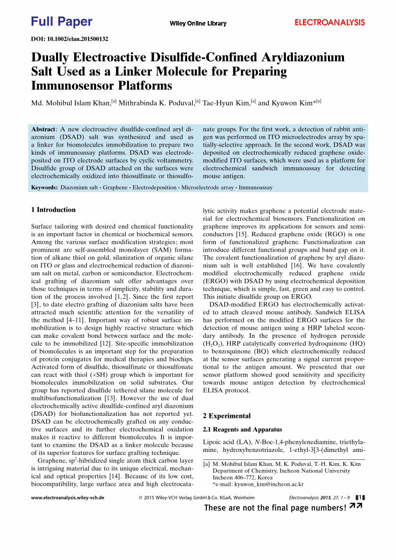
2.2 Synthesis of tert-Butyl 4-(5-(1,2-dithiolan-3-yl)pentanamide)phenylcarbamate (1a)
N-Boc-1,4-Phenylenediamine, triethylamine, hydroxyben-zotriazole, EDCl, and additional triethylamine wereadded successively to a solution of lipoic acid in dichloro-methane. The reaction mixture was stirred overnight at258 and diluted with water. The product was extractedwith dichloromethane, dried over magnesium sulfate, fil-tered and concentrated under reduced pressure. The red-dish brown powder obtained was suspended in diethylether, filtered and rinsed with ether to afford a salmonpink powder in 80.5 % yield.
1H NMR (CDCl3, 400 MHz): d ppm : 7.42 (2H, d,2xArH, J=8.0 Hz), 7.30 (2H, d, 2xArH, J=8.0 Hz); 7.18(1H, br signal, ArNHCO), 6.46 (1H, br signal, ArNH-BOC), 3.59–3.54 (1H, m, �S�CHCH2), 3.21–3.08 (2H, m,CH2), 2.50–2.42 (1H, m, �S�CH2CH2), 2.34 (2H, t,CH2CH2CO, J=7.0 Hz), 1.95–1.87 (1H, m, -S-CH2), 1.78–
1.68 (4H, m, CH2), 1.61 (1H, br signal, CH2), 1.51 (9H, s,NHBOC), 1.25 (1H, br signal, CH2). (Scheme 1)
2.3 Synthesis of N-(4-Aminophenyl)-5-(1,2-dithiolan-3-yl)pentanamide (2a)
1a (210.97 mg, 0.532 mM) was dissolved in 6.666 mL dryDCM:TFA (10 :1) mixture and stirred overnight at roomtemperature. The reaction washed with water and extract-ed by DCM. The solution dried over magnesium sulfateand concentrated under reduced pressure. Purificationwas carried out on a silica-gel column (mobile phase 5 %methanol in DCM) to get a beige colored solid in 21 %yield.
1H NMR: d ppm : 9.45 (1H, br signal, ArCONH), 7.20(2H, d, 2xArH, J=8.0 Hz), 7.47 (2H, d, 2xArH, J=8.0 Hz), 4.87 (2H, br signal, ArNH2), 3.66–3.59 (1H, m, �S�CH�), 3.21–3.10 (2H, m, �S�CHCH2), 2.44–2.39 (1H,m, CH2), 2.21 (2H, t, CH2, J=7.0 Hz), 1.98–1.84 (2H, m,CH2), 1.59–1.54 (2H, m, CH2), 1.40–1.36 (2H, m, CH2),1.23 (1H, br signal, CH2). (Scheme 2)
2.4 Synthesis of 5-(1,2-Dithiolan-3-yl)pentanamide-4-benzene Diazonium Chloride
To prepare 1.5 mM 2 mL solution, 0.889 mg of 2a was dis-solved in 1.9 mL 0.1 M HCl and cold in ice bath. ColdNaNO2 (0.23 mg in 0.1 mL H2O) 100 mL was added dropwise with stirring. Stirring continued for more 30 minutes.Freshly prepared disulfide confined diazonium (DSAD)solution was used for electrodeposition. (Scheme 3)
2.5 Immobilization of Cleaved Antibody onMicroelectrode Array and Sandwich Immunoassay
Freshly prepared DSAD was electrodeposited on the fourmicroelectrodes. Only one electrode electrochemically
Scheme 1. Synthesis of tert-butyl 4-(5-(1,2-dithiolan-3-yl)pentanamide)phenylcarbamate (1a).
Scheme 2. Synthesis of N-(4-aminophenyl)-5-(1,2-dithiolan-3-yl)pentanamide (2a).
Scheme 3. Synthesis of 5-(1,2-dithiolan-3-yl)pentanamide-4-benzene diazonium chloride.
www.electroanalysis.wiley-vch.de � 2015 Wiley-VCH Verlag GmbH & Co. KGaA, Weinheim Electroanalysis 2015, 27, 1 – 9 &2&
These are not the final page numbers! ��
Full Paper
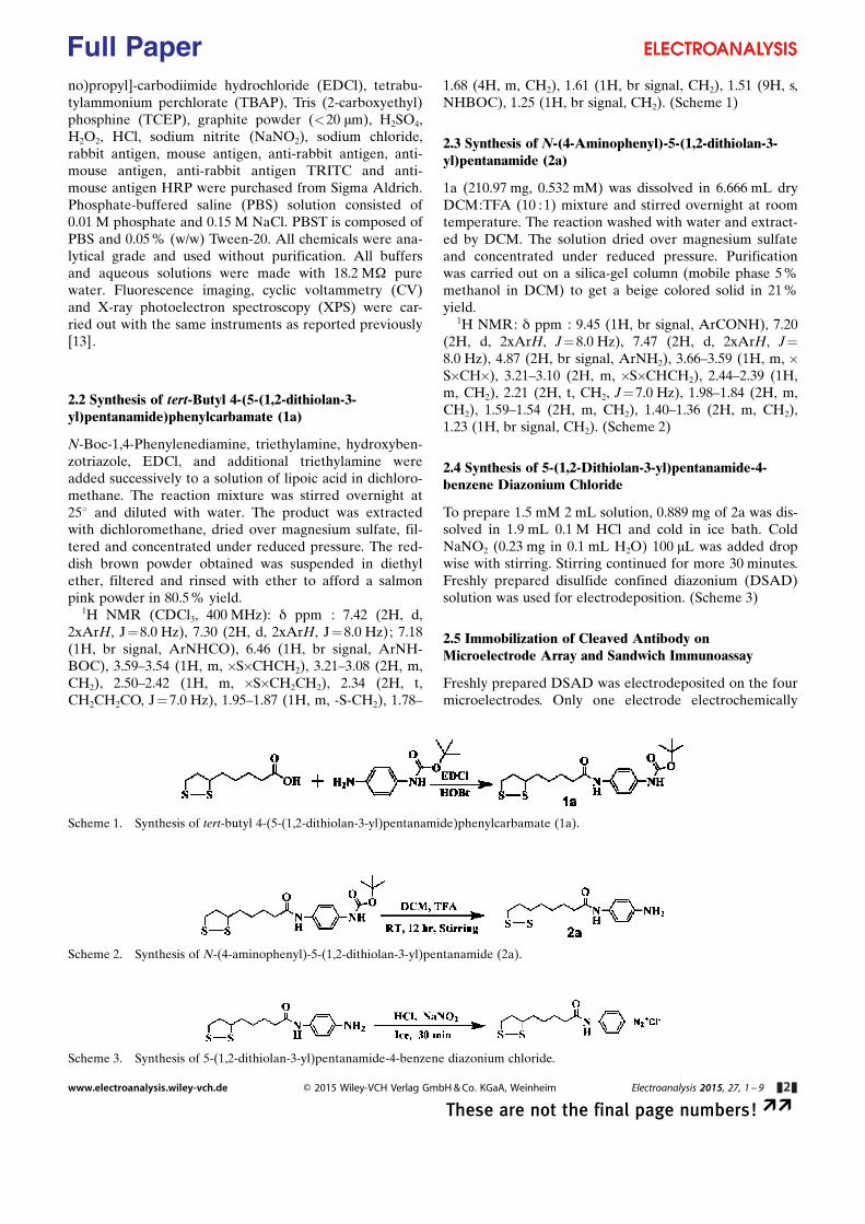
oxidized to get thiosulfinate or thiosulfonate group. Thearray surfaces were exposed with freshly preparedcleaved anti-rabbit antigen fragments in PBST for 2 h.This was followed by washing with PBST, water and driedby N2 gas. Then the surface was treated with 50 mL solu-tion of 5 mg/mL rabbit antigen in PBST for 1 h. This wasfollowed by washing with PBST, water and drying withN2 gas. 50 mL solution of 20 mg/mL TRITC-labeled anti-rabbit antigen in PBST was exposed onto the ITO arrayfor 1 h. The resulting surface was observed with fluores-cent microscope.
Antibody fragments was prepared as follows. 30 mg ofanti-rabbit IgG and 100 mg of tris(2-carboxyethyl)phos-phine were added into 500 mL of PB solution and incubat-ed for 3 h. TCEP in the mixture solution was removed bycentrifugal filtration at 14 000 g and 4 8C for 17 min (cut-off MW: 50000 Dalton). Then 200 mL of PB was addedand centrifuged again at 14 000 g for 13 min. After com-plete removal of TCEP, addition of 30 mL PBST was fol-lowed, and then used immediately.
2.6 Preparation of ODSAD/ERGO/PEI/ITO Surfaces
GO was synthesized and characterized as we reportedpreviously [17]. ITO electrode was cut into 2.5� 1 cm2
size, washed with piranha, water and dried by N2. 100 mL1 % PEI in H2O was exposed for 1 h at 25 8C in bare ITO.The surface was washed with water and dried by N2.0.2 mg/mL GO was treated on the modified surface for3 h, washed with water and dried with N2. Then GO/PEI/ITO surface was electrochemically reduced in the aque-ous solution with 0.5 M NaCl. On the ERGO modifiedsurface DSAD was electrodeposited. DSAD/ERGO/PEI/ITO was electrochemically oxidized with 0.1 M TBAP in95% ACN to get oxidized DSAD/ERGO/PEI/ITO orODSAD/ERGO/PEI/ITO surface.
2.7 Preparation of Immunosensor Electrodes andEnzyme Linked Immunosorbent Assay
For immobilization of cleaved anti-mouse antigen on theODSAD/ERGO/PEI/ITO surface 100 mL of cleaved anti-mouse antigen (100 mg/mL) was dropped on the surfaceand incubated for 2 h, followed by washing with PBST,water and dried with N2 gas. Then the surface was treatedwith 1% bovine serum albumin (BSA) solution in PBSfor 1 h to block nonspecific binding site on the surface.Then the modified surface was washed with PBS, waterand dried with N2 gas. Different concentrations of 100 mLmouse antigen in PBST was exposed on the surface for
Fig. 1. Scheme of the preperation of DSAD-modified patterned ITO electrode surfaces and spatially-selective detection of rabbit an-tigen.
www.electroanalysis.wiley-vch.de � 2015 Wiley-VCH Verlag GmbH & Co. KGaA, Weinheim Electroanalysis 2015, 27, 1 – 9 &3&
These are not the final page numbers! ��
Full Paper
1 h, followed by washed with PBST, water and dried byN2 gas. Then, 100 mL of anti-mouse antigen HRP (20 mg/mL in PBST) was exposed on the modified surface andincubated for 1 h. After that the surface was washed withPBST, water and dried with N2 gas. For negative controlexperiment no antigen or rabbit antigen were used in-stead of mouse antigen. All the steps were carried out at25 8C temperature.
2.8 Voltammetric Detection of Antigen
The active area of the working electrode was 0.45 cm2. Inthe cell freshly prepared 1.5 mM H2O2 and 2.0 mM hy-droquinone (HQ) in 0.1 M PBS was kept undisturbed for5 min at room temperature for enzymatic detection. Fi-nally electrochemical reduction current was obtained forthe reduction of BQ. Three different measurements weredone for each concentration of mouse antigen.
3 Results and Discussion
3.1 Spatially-Selective Immobilization of DSAD andDetection of Rabbit Antigen
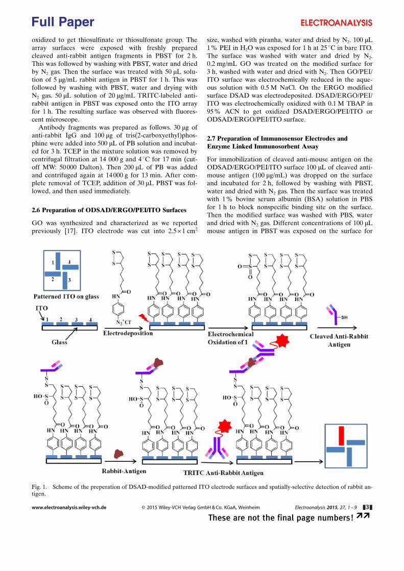
As shown in Figure 1 DSAD was grafted on four differ-ent ITO microelectrodes. Then only electrode 1 was elec-trochemically oxidized. Cleaved anti-rabbit antigen fol-lowed by rabbit antigen after that TRITC labeled anti-rabbit antigen were exposed on the four electrodes. Final-ly the surface was investigated with fluorescence micros-copy to detect the rabbit antigen. The electrodepositionof DSAD cation on ITO microelectrodes was carried outby cyclic voltammetry (CV), which shows a large irrever-sible reduction current at �0.35 V during the first cyclefollowed by greatly diminished currents on the secondcycle as shown in Figure 2A. The electrodeposition wasvery efficient on the ITO m-electrodes. DSAD modifiedITO was electrochemically oxidized. As shown in Fig-ure 2B electrochemical oxidation of DSAD produces twosuccessive irreversible waves at 0.85 V and 1.2 V duringthe first cycle which then disappeared in the next cycles.The two waves were due to two steps for oxidation of di-
Fig. 2. (A) CVs for the electrodeposition of 1.5 mM DSAD in 0.1 M HCl on ITO microelectrode at a scan rate of 50 mVs�1. (B)CVs for the electrochemical oxidation of DSAD/ITO with 0.1 M TBAP in 95% ACN at a scan rate of 50 mVs�1. (C) S 2p XPS spec-tra for DSAD modified ITO surfaces before (a) and after (b) electrochemical oxidation. (D) Fluorescence microscopic image ob-tained from specfic immoblization of cleaved anti-rabbit antigen. The broken lines indicate the position of ITO electrodes. Scale baris 150 mm.
www.electroanalysis.wiley-vch.de � 2015 Wiley-VCH Verlag GmbH & Co. KGaA, Weinheim Electroanalysis 2015, 27, 1 – 9 &4&
These are not the final page numbers! ��
Full Paper
sulfide as shown previously for other disulfide compounds[18]. The resulting surfaces were investigated by X-rayphotoelectron spectroscopy. As shown in Figure 1C-a, thesulfur 2p XPS result consists of two peaks at bindingenergy of 163.5 and 164.5 eV, corresponding to S 2p3/2 andS 2p1/2 respectively of sulfide group. However, no peakwas observed at bare electrode. A double peaks at167.5 eV and 168.5 eV corresponds to sulfonates (SO2) in-volved in in a thiosulfonate group and an additional peakat about 166.5 eV corresponds to sulfinates (SO) in thio-sulfinate group [12]. With approximation thiosulfinateproduces from 2e-loss and thiosulfonate produces from4e-loss [19]. After electrooxidation the S 2p XPS spec-trum shows a decrease in the intensity ratios of the non-oxidized S2p to oxidized one (Figure 2C-a and b). Fromthe CV and XPS results, it was confirmed that both thegroup thiosulfinate and thiosulfonate was produced onthe surface through electrochemical oxidation.
The thiol reactivity of the oxidized surface was investi-gated by immobilization of thiol containing antibody pre-pared by antibody cleavage with TCEP [20]. After elec-trooxidation of only electrode 1 in Figure 1, a solutionhaving a cleaved anti-rabbit antigen (c-Ab-R) was ex-
posed on the array surface. The solution of rabbit antigen(R-IgG) was dropped on the c-Ab-R immobilized surface.Then the array was allowed to expose to the solution ofanti-rabbit antigen labeled with TRITC as a secondaryantibody. After washing and drying, the resulting surfacewas examined with fluorescence microscope. As shown inFigure 2D highly contrasted red color is observed only inthe oxidized surface. The result suggested that our strat-egy was successful for the detection of protein on theclosely spaced microelectrode array.
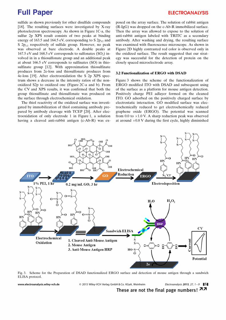
3.2 Functionalization of ERGO with DSAD
Figure 3 shows the scheme of the functionalization ofERGO modified ITO with DSAD and subsequent usingof the surface as a platform for mouse antigen detection.Positively charge PEI adlayer formed on the cleanedITO. GO adsorbed on the positively charged surface byelectrostatic interaction. GO modified surface was elec-trochemically reduced to get electrochemically reducedgraphene oxide (ERGO). The potential was scannedfrom 0.0 to �1.0 V. A sharp reduction peak was observedat around �0.8 V during the first cycle, highly diminished
Fig. 3. Scheme for the Preparation of DSAD functionalized ERGO surface and detection of mouse antigen through a sandwichELISA protocol.
www.electroanalysis.wiley-vch.de � 2015 Wiley-VCH Verlag GmbH & Co. KGaA, Weinheim Electroanalysis 2015, 27, 1 – 9 &5&
These are not the final page numbers! ��
Full Paper
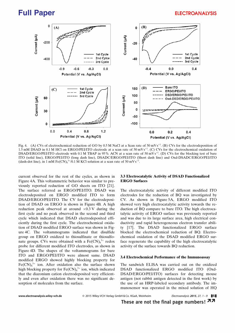
current observed for the rest of the cycles, as shown inFigure 4A. This voltammetric behavior was similar to pre-viously reported reduction of GO sheets on ITO [21].The surface referred as ERGO/PEI/ITO. DSAD waselectrodeposited on ERGO modified ITO to formDSAD/ERGO/PEI/ITO. The CV for the electrodeposi-tion of DSAD on ERGO is shown in Figure 4B. A highreduction peak observed at around �0.3 V during thefirst cycle and no peak observed in the second and thirdcycle which indicated that DSAD electrodeposited effi-ciently during the first cycle. The electrochemical oxida-tion of DSAD modified ERGO surface was shown in Fig-ure 4C. The voltammograms indicated that disulfidegroup on ERGO oxidized to thiosulfinate or thiosulfo-nate groups. CVs were obtained with a Fe(CN)6
3� redoxprobe for different modified ITO electrodes, as shown inFigure 4D. The shapes of the voltammograms for bareITO and ERGO/PEI/ITO were almost same. DSADmodified ERGO showed highly blocking property forFe(CN)6
3� ion. After oxidation also the surface shownhigh blocking property for Fe(CN)6
3� ion, which indicatedthat the diazonium cation electrodeposited very efficient-ly and even after oxidation there was no significant de-sorption of molecules from the surface.
3.3 Electrocatalytic Activity of DSAD FunctionalizedERGO Surfaces
The electrocatalytic activity of different modified ITOelectrodes for the reduction of BQ was investigated byCV. As shown in Figure 5A, ERGO modified ITOshowed very high electrocatalytic activity towards the re-duction of BQ compare to bare ITO. The high electroca-talytic activity of ERGO surface was previously reportedand was due to its large surface area, high electrical con-ductivity and rapid heterogeneous electron transfer abili-ty [17]. The DSAD functionalized ERGO surfaceblocked the electrochemical reduction of BQ. Electro-chemical oxidation of the DSAD modified ERGO sur-face regenerate the capability of the high electrocatalyticactivity of the surface towards BQ reduction.
3.4 Electrochemical Performance of the Immunoassay
The sandwich ELISA was carried out on the oxidizedDSAD functionalized ERGO modified ITO (Oxd-DSAD/ERGO/PEI/ITO) surfaces for detecting mouseantigen (not rabbit antigen detected in the first work) bythe use of an HRP-labeled secondary antibody. The im-munosensor was operated in the mixed solution of HQ
Fig. 4. (A) CVs of electrochemical reduction of GO by 0.5 M NaCl at a Scan rate of 50 mVs�1. (B) CVs for the electrodeposition of1.5 mM DSAD in 0.1 M HCl on ERGO/PEI/ITO electrode at a scan rate of 50 mVs�1. (C) CVs for the electrochemical oxidation ofDSAD/ERGO/PEI/ITO electrode with 0.1 M TBAP in 95% ACN at a scan rate of 50 mV s�1. (D) CVs for the blocking test of bareITO (solid line), ERGO/PEI/ITO (long dash line), DSADC/ERGO/PEI/ITO (Short dash line) and Oxd-DSADC/ERGO/PEI/ITO(dash-dot line), in 1 mM Fe(CN)6
3�/0.1 M KCl solution at a scan rate of 50 mV s�1.
www.electroanalysis.wiley-vch.de � 2015 Wiley-VCH Verlag GmbH & Co. KGaA, Weinheim Electroanalysis 2015, 27, 1 – 9 &6&
These are not the final page numbers! ��
Full Paper
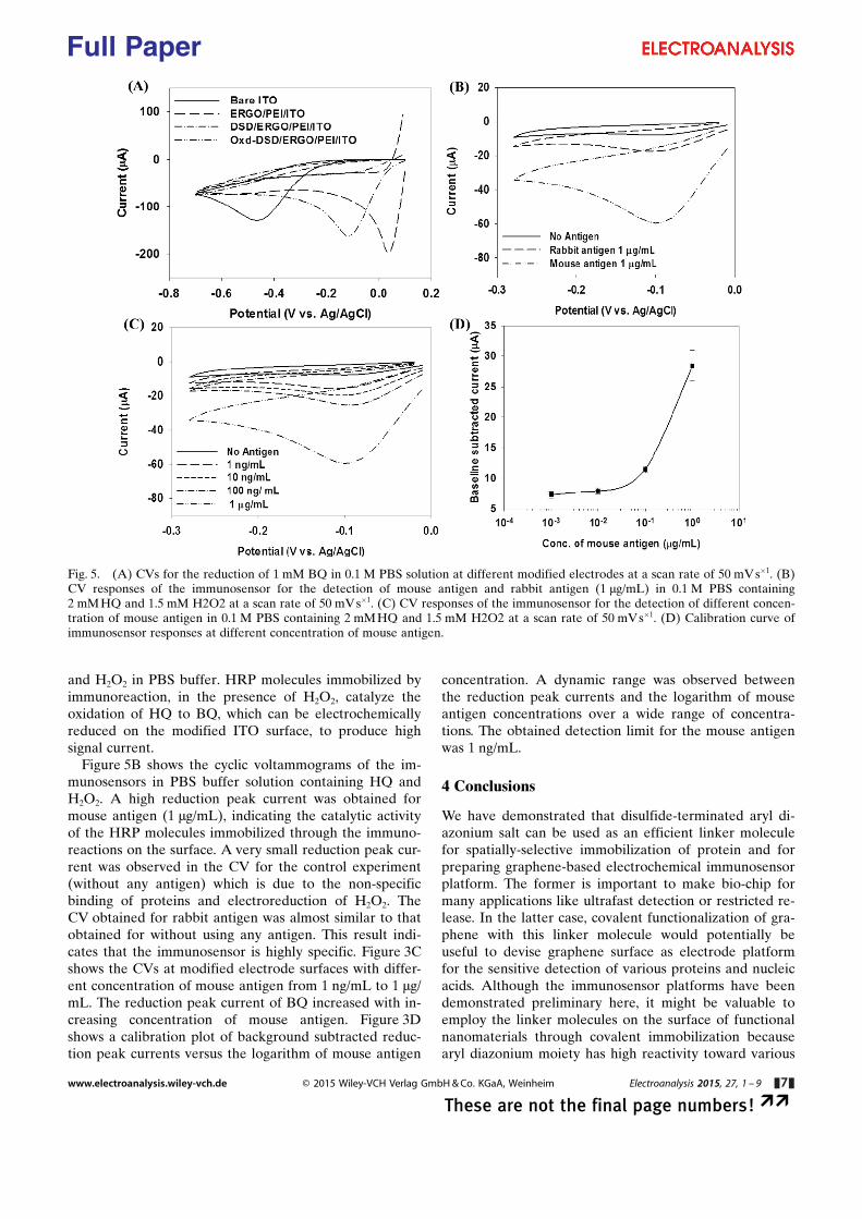
and H2O2 in PBS buffer. HRP molecules immobilized byimmunoreaction, in the presence of H2O2, catalyze theoxidation of HQ to BQ, which can be electrochemicallyreduced on the modified ITO surface, to produce highsignal current.
Figure 5B shows the cyclic voltammograms of the im-munosensors in PBS buffer solution containing HQ andH2O2. A high reduction peak current was obtained formouse antigen (1 mg/mL), indicating the catalytic activityof the HRP molecules immobilized through the immuno-reactions on the surface. A very small reduction peak cur-rent was observed in the CV for the control experiment(without any antigen) which is due to the non-specificbinding of proteins and electroreduction of H2O2. TheCV obtained for rabbit antigen was almost similar to thatobtained for without using any antigen. This result indi-cates that the immunosensor is highly specific. Figure 3Cshows the CVs at modified electrode surfaces with differ-ent concentration of mouse antigen from 1 ng/mL to 1 mg/mL. The reduction peak current of BQ increased with in-creasing concentration of mouse antigen. Figure 3Dshows a calibration plot of background subtracted reduc-tion peak currents versus the logarithm of mouse antigen
concentration. A dynamic range was observed betweenthe reduction peak currents and the logarithm of mouseantigen concentrations over a wide range of concentra-tions. The obtained detection limit for the mouse antigenwas 1 ng/mL.
4 Conclusions
We have demonstrated that disulfide-terminated aryl di-azonium salt can be used as an efficient linker moleculefor spatially-selective immobilization of protein and forpreparing graphene-based electrochemical immunosensorplatform. The former is important to make bio-chip formany applications like ultrafast detection or restricted re-lease. In the latter case, covalent functionalization of gra-phene with this linker molecule would potentially beuseful to devise graphene surface as electrode platformfor the sensitive detection of various proteins and nucleicacids. Although the immunosensor platforms have beendemonstrated preliminary here, it might be valuable toemploy the linker molecules on the surface of functionalnanomaterials through covalent immobilization becausearyl diazonium moiety has high reactivity toward various
Fig. 5. (A) CVs for the reduction of 1 mM BQ in 0.1 M PBS solution at different modified electrodes at a scan rate of 50 mV s�1. (B)CV responses of the immunosensor for the detection of mouse antigen and rabbit antigen (1 mg/mL) in 0.1 M PBS containing2 mMHQ and 1.5 mM H2O2 at a scan rate of 50 mVs�1. (C) CV responses of the immunosensor for the detection of different concen-tration of mouse antigen in 0.1 M PBS containing 2 mMHQ and 1.5 mM H2O2 at a scan rate of 50 mVs�1. (D) Calibration curve ofimmunosensor responses at different concentration of mouse antigen.
www.electroanalysis.wiley-vch.de � 2015 Wiley-VCH Verlag GmbH & Co. KGaA, Weinheim Electroanalysis 2015, 27, 1 – 9 &7&
These are not the final page numbers! ��
Full Paper

solid surfaces including carbon (graphene, graphite, anddiamond), gold, platinum, and indium-tin oxide.
Acknowledgement
This work was supported by Incheon National UniversityResearch Grant in 2011.
References
[1] J. J. Gooding, S. Ciampi, Chem. Soc. Rev. 2011, 40, 2704 –2718.
[2] S. Mahouche-Chergui, S. Gam-Derouich, C. Mangeney,M. M. Chehimi, Chem. Soc. Rev. 2011, 40, 4143–4166.
[3] M. Delamar, R. Hitmi, J. Pinson, J. M. Saveant, J. Am.Chem. Soc. 1992, 114, 5883–5884.
[4] A. Adenier, E. Cabet-Deliry, A. Chauss�, S. Griveau, F.Mercier, J. Pinson, C. Vautrin-Ul, Chem. Mater. 2005, 17,491–501.
[5] B. P. Corgier, C. A. Marquette, L. J. Blum, J. Am Chem.Soc. 2005, 127, 18328 –18332.
[6] A.-M. J. Haque, S.-R. Kwon, H. Park, T.-H. Kim, Y.-S. Oh,S.-Y. Choi, J.-D. Hong, K. Kim, Chem. Commun. 2009, 486 –4867.
[7] G. Liu, E. Luais, J. J. Gooding, Langmuir 2011, 27, 4176 –4183.
[8] S. Maldonado, T. J. Smith, R. D. Williams, S. Morin, E.Barton, K. J. Stevenson, Langmuir 2006, 22, 2884 –2891.
[9] R. Polsky, J. C. Harper, D. R. Wheeler, S. M. Dirk, D. C.Arango, S. M. Brozik, Biosens. Bioelectron. 2008, 23, 757 –764.
[10] A. -E. Radi, X. Munoz-Berbel, V. Lates, J. -L. Marty, Bio-sens. Bioelectron. 2009, 24, 1888 –1892.
[11] C. Saby, B. Ortiz, G. Y. Champagne, D. B�langer, Langmuir1997, 13, 6805 –6813.
[12] E. Pavlovic, A. P. Quist, U. Gelius, L. Nyholm, S. Oscarsson,Langmuir 2003, 19, 4217–4221.
[13] H. J. Kim, S.-R. Kwon, K. Kim, Electrochem. Commun.2012, 20, 52 –55.
[14] K. S. Novoselov, A. K. Geim, S. V. Morozov, D. Jiang, Y.Zhang, S. V. Dubonos, I. V. Grigorieva, A. A. Firsov, Science2004, 306, 666–669.
[15] V. Georgakilas, M. Otyepka, A. B. Bourlinos, V. Chandra,N. Kim, K. C. Kemp, P. Hobza, R. Zboril, K. S. Kim, Chem.Rev. 2012, 112, 6156–6214.
[16] G. L. Paulus, Q. H. Wang, M. S. Strano, Acc. Chem. Res.2013, 46, 160 –170.
[17] M. M. I. Khan, A.-M. J. Haque, K. Kim, J. Electroanal.Chem. 2013, 700, 54–59.
[18] J. Gourcy, P. Martigny, J. Simonet, G. Jeminet, Tetrahedron1981, 37, 1495 –1502.
[19] C. Terashima, T. N. Rao, B. Sarada, Y. Kubota, A. Fujishi-ma, Anal. Chem. 2003, 75, 1564–1572.
[20] G. T. Hermanson, Bioconjugate Techniques, 2nd ed., Aca-demic Press, San Diego, CA, 2008, pp. 95–97.
[21] A.-M. J. Haque, H. Park, D. Sung, S. Jon, S.-Y. Choi, K.Kim, Anal. Chem. 2012, 84, 1871.
Received: January 6, 2015Accepted: March 23, 2015
Published online: && &&, 2015
www.electroanalysis.wiley-vch.de � 2015 Wiley-VCH Verlag GmbH & Co. KGaA, Weinheim Electroanalysis 2015, 27, 1 – 9 &8&
These are not the final page numbers! ��
Full Paper
FULL PAPERS
M. Mohibul Islam Khan,M. K. Poduval, T.-H. Kim, K. Kim*
&& –&&
Dually Electroactive Disulfide-Confined Aryldiazonium Salt Usedas a Linker Molecule for PreparingImmunosensor Platforms
www.electroanalysis.wiley-vch.de � 2015 Wiley-VCH Verlag GmbH & Co. KGaA, Weinheim Electroanalysis 2015, 27, 1 – 9 &9&
These are not the final page numbers! ��
Full Paper


