Embed Size (px)
Citation preview
Dynamics of Human Cytomegalovirus Infection in CD34�
Hematopoietic Cells and Derived Langerhans-Type Dendritic Cells
Roxanne Coronel, Sachiko Takayama, Timothy Juwono, Laura Hertel
Center for Immunobiology and Vaccine Development, Children’s Hospital Oakland Research Institute, Oakland, California, USA
ABSTRACT
Acquisition of human cytomegalovirus (CMV) usually occurs by contact between contaminated bodily fluids, such as urine andsaliva, and host mucosal cells. Langerhans-type dendritic cells (LC) are the only type of immune cells found in the outermostlayers of the oral mucosae, where they not only provide a first line of defense against CMV but can easily be targeted by orallyadministered vaccines, while their bone marrow resident progenitors are important sites of virus latency. In this work, wetracked the progress of infection in CD34� progenitor cells, immature LC (iLC), and mature LC (mLC) exposed to the clinical-like strain TB40-BAC4 or to the vaccine strain AD169varATCC, prior to their long-term maintenance under either immature ormature conditions. We show that the genomes of both strains are efficiently maintained in CD34� cells during their differentia-tion into iLC, although this requires the presence of larger amounts of input AD169varATCC DNA. Lipopolysaccharide- andCD40 ligand-induced maturation of iLC derived from latently infected progenitors was not associated with robust viral genomereplication and progeny production, while maturation of directly infected iLC increased and prolonged expression of the viralimmediate early proteins. While effective replication of viral genomes from both strains occurred only in mLC, both iLC andmLC produced viral progeny, suggesting that both types of LC may contribute to CMV horizontal transmission in vivo.
IMPORTANCE
Human CMV is usually acquired via the oral and nasal mucosae. Langerhans-type dendritic cells (LC) are the only type of im-mune cells found in the outermost layers of these tissues. Understanding how CMV interacts with LC and their hematopoieticprogenitors is thus essential to develop innovative means of defense against this virus. Here we show that the genomes of a viru-lent and an attenuated strain of CMV are maintained in hematopoietic progenitor cells during their differentiation into imma-ture LC and that maturation of these cells by exposure to lipopolysaccharide and CD40 ligand is not sufficient to trigger virusreactivation. While the extents of viral protein expression and genome replication were broadest in directly infected mature LCpopulations, similar amounts of viral progeny were detected in the supernatants of immature and mature LC, suggesting thatthese immune cells of the oral mucosa are likely to be important for CMV transmission within the human population.
Human cytomegalovirus (CMV), a major cause of disease anddeath in immunocompromised and in congenitally infected
individuals, is normally acquired by contact between infectedbodily fluids such as urine and saliva and host mucosal surfaces,particularly those lining the oral and nasal cavities.
From these peripheral sites, CMV is thought to reach the cir-culation and hence the bone marrow, where latency is establishedin myeloid cells, including CD34� hematopoietic progenitors (1–5). Reactivation from latency in the myeloid progeny of thesecells is then believed to be the source of newly produced viralparticles, which upon amplification in oral epithelial cells arereleased in the saliva and contribute to CMV horizontal trans-mission in vivo (6–8).
Immature Langerhans-type dendritic cells (iLC) are the onlyprofessional antigen-presenting cells located in the outermost lay-ers of the oral mucosa (9–14). As such, iLC are bound to be thefirst immune cells to encounter invading pathogens that accesstheir hosts via the oral cavity, such as CMV. Upon contact with“danger” signals, iLC migrate toward the draining lymph nodesand begin the process of maturation, which culminates with theactivation of adaptive immune responses (15). Although matureLangerhans-type dendritic cells (mLC) usually reside withinsecondary lymphoid organs, their presence has been detectedin the oral mucosae of individuals with inflammatory diseasessuch as gingivitis, periodontitis, and oral ulcers (16, 17), sug-
gesting that under certain conditions, mLC may also get indirect contact with CMV.
As the number of latently infected mononuclear cells found inthe blood of healthy donors and the amount of live LC that can beobtained from oral tissues are usually extremely low, CMV infec-tion of CD34� progenitor cells, iLC, and mLC has been studiedpredominantly by using in vitro culture systems.
In 1994, Kondo et al. first showed that viral genomes could bemaintained in the absence of productive infection in fetal liver-derived granulocyte-macrophage progenitor cells (GM-Ps) ex-posed to the laboratory-adapted CMV strains Towne RC256 andToledo (18). As cells proliferated over a time course of 28 days,viral DNA accumulated in parallel with the number of GM-Ps.
Received 4 February 2015 Accepted 2 March 2015
Accepted manuscript posted online 11 March 2015
Citation Coronel R, Takayama S, Juwono T, Hertel L. 2015. Dynamics of humancytomegalovirus infection in CD34� hematopoietic cells and derived Langerhans-type dendritic cells. J Virol 89:5615–5632. doi:10.1128/JVI.00305-15.
Editor: R. M. Sandri-Goldin
Address correspondence to Laura Hertel, [email protected].
Copyright © 2015, American Society for Microbiology. All Rights Reserved.
doi:10.1128/JVI.00305-15
May 2015 Volume 89 Number 10 jvi.asm.org 5615Journal of Virology
Treatment with ganciclovir, an inhibitor of productive viral replica-tion, did not affect this trend. In later studies using CD34� cells pu-rified from GM-Ps, establishment of latency without production orrelease of viral particles was also observed with the attenuated CMVstrains TownevarRIT3 and AD169varATCC (19, 20).
In 2002, Goodrum et al. presented a new model to study CMVlatency, consisting of bone marrow-derived CD34� cells main-tained on irradiated murine stromal cells in the presence of serum(21). While clinical-like strains underwent latency in this system,productive replication was observed in cells infected with Towneand AD169varATCC (21–23), leading to the conclusion that viralopen reading frames (ORFs) missing from the genomes of atten-uated strains were required to establish latency (23).
A year later, we introduced a new system to study CMV inter-actions with iLC and mLC differentiated in vitro from umbilicalcord or peripheral blood CD34� progenitor cells using a highlydefined, serum-free cytokine cocktail (24), which was shown togenerate LC displaying the same morphological and ultrastruc-tural features of epithelial iLC (25–28) and expressing some of themarkers of oral LC (24, 29, 30). Using this model, we showed thatmLC are more permissive to infection onset than iLC and that thisis due not to defects in viral entry but rather to the inefficienttranscription of viral genes, particularly those encoding the imme-diate early proteins 1 and 2 (IE1/IE2), in iLC (9, 24). The fact thatboth clinical-like and laboratory-adapted strains could initiatetheir replication cycle in both LC types, moreover, implied thatnone of the proteins encoded by the ORFs lacking from the ge-nomes of attenuated strains is absolutely required for CMV infec-tion of LC (9, 24).
Using the same system, Reeves et al. then showed that differ-entiation of CD34� cells latently infected with the clinical-likestrain TB40/E was associated with the maintenance of viral ge-nomes in a transcriptionally silent state, while iLC maturationtriggered reactivation (31, 32).
In this work, we used our CD34� cell-iLC-mLC differentia-tion/maturation model to (i) quantify changes in viral and cellulargenome amounts occurring during the differentiation of latentlyinfected CD34� cells into iLC, (ii) compare the effects of matura-tion on CMV reactivation in LC derived from latently infectedCD34� cells and on CMV replication in directly infected LC, (iii)compare the kinetics of viral replication in CD34� cells, iLC, andmLC to those in human foreskin fibroblasts (HFF), a cell typehighly permissive to lytic infection, and (iv) compare the infectionkinetics of the CMV vaccine strain AD169varATCC, possessing ashorter genome and a more restricted tropism, to that of the clin-ical-like strain TB40-BAC4, which is characterized by a morecomplete genome and a broader tropism.
Currently available data provide only qualitative evidence ofviral genome carriage during the differentiation of CD34� cellsinto iLC and of viral reactivation in mLC (31, 32). As the processof CD34� cell differentiation is associated with substantial in-creases in cell numbers (up to 20- to 40-fold), the quantitativemeasurement of viral genome copy numbers present over timebecomes crucial to understand whether viral DNA is lost, main-tained, or replicated during latency. We show that, after an initialsharp drop, TB40-BAC4 genomes are maintained in proliferating/differentiating CD34� cells in the absence of IE1/IE2 proteinexpression and progeny release, while the efficient carriage ofAD169varATCC genomes requires the presence of high(er)amounts of viral genomes at the start of infection.
Thus far, the effects of LC maturation on CMV infection dy-namics have been separately evaluated within the context of viralreactivation in lipopolysaccharide (LPS)-treated iLC differenti-ated from latently infected, bone marrow-derived CD34� cells(31, 32) or of viral lytic replication in LPS- and/or CD40 ligand(CD40L)-treated iLC populations differentiated from umbilicalcord or peripheral blood derived CD34� cells (9, 24). Becausethese assays were conducted with LC populations differentiatedfrom different sets of CD34� cells donors, exposed to differentmaturation stimuli, and infected with different viral strains, re-sults are difficult to compare. In this work, we asked whether mat-uration by exposure to CD40L and LPS, stimuli known to increasethe susceptibility of iLC to infection onset, would also triggerCMV reactivation in iLC carrying latent viral genomes andwhether the delivery of maturation signals after iLC exposure toviral particles would be as effective in supporting infection prog-ress as in cells exposed to virus when already mature. We foundthat maturation of iLC differentiated from latently infected, um-bilical cord blood-derived CD34� cells was not accompanied bythe robust amplification of viral DNA, although TB40-BAC4 ge-nome amounts were larger in mature than in immature LC differ-entiated from latently infected CD34� cells. The fact that no viralprogeny was observed in culture supernatants, however, suggeststhat exposure to LPS and CD40L molecules is not sufficient totrigger vigorous CMV reactivation. Yet, maturation was associ-ated with the enhanced and prolonged expression of the IE1/IE2proteins in directly infected mLC and in iLC matured immediatelyafter infection, suggesting that LPS/CD40L signaling can rapidlyinduce the establishment of an intranuclear environment sup-portive of viral gene transcription from genomes delivered by viralparticles, but not as much from genomes already present in thenuclei of latently infected iLC. Interestingly, this enhancement inIE1/IE2 protein expression was not followed by a similar increasein viral genome amplification events. Instead, viral DNA replica-tion was most efficient in cells infected when already mature, sug-gesting that the supportive milieu promoted by maturation mustalready be present at the time of infection onset to reach highgenome replication rates.
Human fibroblasts have been traditionally used to study CMVpathogenesis on account of their high degree of permissiveness toCMV replication in vitro. Consequently, these cells have become abasis of comparison to assess CMV tropism for other cell types.Our evaluation of CMV infection progress in HFF, iLC, and mLCrevealed that the extents of viral protein expression, genome am-plification, and progeny production were dramatically lower iniLC and mLC than in HFF. This suggests that, irrespective of thesusceptibility differences between iLC and mLC, both LC types aremore resistant to infection than HFF, at least when exposed to thetwo CMV strains tested here.
Consistent with our previous results (24), we found that alarger proportion of mLC than iLC express the IE1/IE2 proteins atday two and that mLC produce and release viral progeny. Progressof infection in iLC, however, had not been previously assessed.Here we show that iLC too produce viral progeny, a quite surpris-ing finding considering the absence of widespread IE1/IE2 proteinsynthesis and of large increases in viral genome amounts, as seenin mLC and in HFF cultures. Immature LC thus appear to be lessresistant to CMV infection than previously believed, and they maycontribute to CMV horizontal transmission in vivo by releasingnewly produced virus in the oral cavity. Our finding that viral
Coronel et al.
5616 jvi.asm.org May 2015 Volume 89 Number 10Journal of Virology
yields were reduced, rather than increased, upon maintenance ofinfected iLC under maturation conditions was also quite intrigu-ing, as it may hint at the existence of cell defense mechanismsacting to restrict viral progeny production in infected iLC duringtheir migration toward the draining lymph nodes in vivo, in orderto prevent virus spread to other cell types within the host.
Finally, all studies of CMV latency making use of our CD34�
cell-iLC-mLC differentiation/maturation model (31, 32), as wellas our initial work on LC susceptibility to CMV infection andimmunomodulation (24, 33, 34), were conducted using thebroadly tropic but genotypically heterogeneous strain TB40/E(35). Consequently, no data are currently available on the behav-ior of attenuated strains in this model, except for our findings thatboth Towne and AD169varATCC can initiate infection in iLC andmLC (9, 24). Studies using these strains in latency models haveyielded contrasting results. While Towne and AD169varATCCwere reported to establish latent infections in GM-Ps (18–20),both strains were found to initiate productive infections in CD34�
cells cultured on irradiated murine stromal cells (21–23). In thiswork, we compared the infection kinetics of TB40-BAC4, a geno-typically uniform strain derived from the bacterial artificial chro-mosome cloning of a highly endotheliotropic subpopulation ofTB40/E (35), to those of AD169varATCC. The progress ofAD169varATCC infection in both iLC and mLC beyond the initialstages of infection was also tracked. No evidence of productivereplication was found in AD169varATCC-infected CD34� cellcultures, in accordance with results from GM-Ps (20) but notfrom CD34� cells/murine stromal cells cultures (23). The onlydifference we observed between the infection kinetics of TB40-BAC4 and AD169varATCC was in the efficiency of viral genomemaintenance during CD34� cells differentiation, suggesting thatno additional ORF beyond those contained in the genome ofAD169varATCC is required to support entry and replication indirectly infected iLC and mLC.
MATERIALS AND METHODSCells. Umbilical cord blood CD34� hematopoietic progenitor cells werepurchased from Stemcell Technologies Inc., Vancouver, Canada. Imma-ture LC populations were obtained by culturing CD34� cells at a densityof 1 � 104 to 2.5 � 104 cells per well in 48-well tissue culture plates usingserum-free X-VIVO 15 medium (Lonza/BioWhittaker, Allendale, NJ)supplemented with 1,500 IU/ml of granulocyte-macrophage colony-stimulating factor (GM-CSF) (Leukine Sargramostim; Immunex, Seattle,WA), 2.5 ng/ml of tumor necrosis factor alpha, 20 ng/ml of stem cellfactor, 100 ng/ml of Flt3 ligand, and 0.5 ng/ml of transforming growthfactor �1 (all purchased from Peprotech, Rocky Hill, NJ), while matura-tion was induced by cell exposure to X-VIVO 15 medium containing 10%fetal bovine serum, 200 ng/ml of CD40 ligand (Immunex, Seattle, WA),1,500 IU/ml of GM-CSF, and 250 ng/ml of lipopolysaccharide (Sigma-Aldrich, St. Louis, MO) as previously described (9, 24). Human foreskinfibroblasts were propagated in Dulbecco’s modified Eagle medium(DMEM) supplemented with 10% fetal clone serum III (HyClone), 100U/ml penicillin and 100 �g/ml streptomycin, 4 mM HEPES, and 1 mMsodium pyruvate (Gibco, Life Technologies, Grand Island, NY).
Virus strains and titrations. AD169varATCC (36) and TB40-BAC4(35), gifts from E. S. Mocarski (Emory University, Atlanta, GA) and C.Sinzger (University of Ulm, Ulm, Germany), respectively, were propa-gated on HFF and purified by ultracentrifugation as previously described(24). Titers of virus stocks and culture supernatants were determined byimmunofluorescence staining analyses of HFF harvested at 24 h postin-fection (hpi) and stained with the monoclonal antibody MAb810 (1:500;Chemicon, Temecula, CA) directed against the viral IE1/IE2 proteins.
Cell infection. HFF were plated at a density of � 2 � 104 cells/cm2 3days before infection with TB40-BAC4 at a multiplicity of infection(MOI) of 0.05 PFU/cell. The virus inoculum was left in contact with cellsat 37°C in 5% CO2 for 4 h, after which cells were washed twice prior toincubation in complete DMEM. Supernatant and cells were collected at 4hpi and at days 2, 4, 6, 8, 10, and 14. Immediately after thawing, CD34�
cells from each donor were left uninfected or were exposed toAD169varATCC or TB40-BAC4 virions at a calculated MOI of 10 for 4 hprior to washing and plating in iLC medium. At day eight, iLC wereharvested, counted, and replated in either immature or maturation me-dium. Differentiating CD34� cells and supernatants were collected at 4hpi and at days 2, 4, 6, and 8, while iLC derived from infected CD34� cellsand their supernatants were harvested at 4 hpi and at days 2, 4, 6, 8, 10, and14. Immature LC differentiated from uninfected CD34� cells were alsoharvested on day eight, counted, and exposed to AD169varATCC orTB40-BAC4 virions for 4 h, prior to washing and plating in either iLC ormLC medium, or were left uninfected and cultured under maturationconditions. These uninfected cells were then harvested at day two post-maturation, exposed to AD169varATCC or TB40-BAC4 virions for 4 h,washed, and plated again in mLC medium. Infected iLC and mLC werethen collected at 4 hpi and at days 2, 4, 6, 8, 10, and 14. For foscarnettreatment, cells were exposed to medium containing 300 �g/ml (1 mM) ofthe viral polymerase inhibitor phosphonoformic acid, as in our previouswork (37). All cells and supernatant samples were stored at �80°C prior toprocessing for titration and real-time quantitative PCR (qPCR) analyses,respectively.
Immunofluorescence staining analyses (IFA). HFF grown on 12-mm-diameter glass coverslips and LC deposited on glass slides by centrif-ugation at 500 rpm for 3 min at room temperature (RT) using a Cytospin4 (Thermo Shandon) were fixed in 1% formaldehyde for 30 min at RT,permeabilized in 0.5% Triton X-100 for 20 min on ice, treated with block-ing buffer (20% fetal bovine serum in phosphate-buffered saline [PBS])for 30 min at RT, and incubated with mouse monoclonal antibodyMAb810 (1:400; EMD Millipore), directed against an epitope encoded byexon 2 of the UL122/123 ORF and common to both the IE1 and IE2proteins (38) for 1 h at RT in a humidified chamber. After washing inblocking buffer, samples were incubated with Alexa Fluor 594-conjugatedgoat anti-mouse antibodies (1:200; Life Technologies, Carlsbad, CA) for 1h at RT, followed by nuclear labeling with Hoechst 33342 (0.2 mg/ml;Molecular Probes, Eugene, OR) for 3 min at RT. For IE1/IE2 and ppUL44double staining, fixed cells were first incubated with a monoclonal anti-ppUL44 antibody (1:500 dilution; Goodwin Institute, Plantation, FL) fol-lowed by an Alexa Fluor 594-conjugated goat anti-mouse antibody,blocked with normal mouse immunoglobulin G (1:100; Caltag, Burlin-game, CA), and then stained with an Alexa Fluor 488-conjugated anti-IE1/IE2 monoclonal antibody. Slides were then mounted in 90% glyc-erol–10% PBS containing 2.5 g/liter of 1, 4-diazabicyclo-(2,2,2)-octane(DABCO) (Alfa Aesar, Pelham, NH) and viewed on a Nikon Eclipse E600fluorescence microscope equipped with iVision-Mac imaging software.The percentage of IE1/IE2� cells was then determined using the ImageJ1.47v software.
Real-time quantitative genomic PCR. Genomic DNA was extractedfrom infected and uninfected cells using the OmniGenX PureSpin gDNAminiprep kit (E&K Scientific, Santa Clara, CA). mRNA was obtained us-ing the �MACS mRNA isolation kit (Miltenyi Biotec, Bergisch Gladbach,Germany) and was reverse transcribed using SuperScript II reverse trans-criptase (Invitrogen, Life Technologies, Grand Island, NY). Real-timequantitative PCRs were performed using the iTaq SYBR green supermixwith ROX (Bio-Rad, Hercules, CA) and an ABI7900 thermocycler (Ap-plied Biosystems, Carlsbad, CA), with primers hybridizing to exon 2 of theviral UL122 and UL123 ORFs (forward primer, 5=-GGCCGAAGAATCCTCAAAA-3=; reverse primer, 5=-TCGTTGCAATCCTCGGTCA-3=) or tothe cellular albumin gene (forward primer, 5=-GCTGTCATCTCTTGTGGGCTGT-3=; reverse primer. 5=-AAACTCATGGGAGCTGCTGGTT-3=)as in our previous work (37). The following cycling parameters were used:
CMV Infection of Hematopoietic Cells
May 2015 Volume 89 Number 10 jvi.asm.org 5617Journal of Virology
95°C for 2 min to activate the iTaq polymerase, followed by 40 cycles oftemplate denaturation at 95°C for 15 s, primer annealing at 51°C (UL122/123) or 57°C (albumin) for 30 s, and product extension at 72°C for 30 s.Absolute quantifications of viral and cellular genome amounts were ob-tained using a standard curve made by serial dilutions of plasmid pON303(39) and of plasmid pTOPO-albumin (9). The number of viral genomecopies per cell was calculated as number of viral DNA copies/(number ofalbumin DNA copies/2) (40).
Statistical analysis. Student t tests (paired, two tailed) were conductedto compare the mean values of two data sets, while the nonparametric anddistribution free Kolmogorov-Smirnov (KS) test was used to compare thecumulative distributions of any two data sets (http://www.wessa.net/rwasp_Reddy-Moores%20K-S%20Test.wasp/). Differences were con-sidered significant at a P value of �0.05.
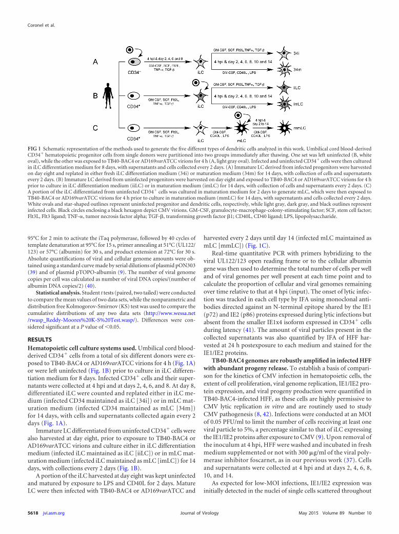
RESULTSHematopoietic cell culture systems used. Umbilical cord blood-derived CD34� cells from a total of six different donors were ex-posed to TB40-BAC4 or AD169varATCC virions for 4 h (Fig. 1A)or were left uninfected (Fig. 1B) prior to culture in iLC differen-tiation medium for 8 days. Infected CD34� cells and their super-natants were collected at 4 hpi and at days 2, 4, 6, and 8. At day 8,differentiated iLC were counted and replated either in iLC me-dium (infected CD34 maintained as iLC [34i]) or in mLC mat-uration medium (infected CD34 maintained as mLC [34m])for 14 days, with cells and supernatants collected again every 2days (Fig. 1A).
Immature LC differentiated from uninfected CD34� cells werealso harvested at day eight, prior to exposure to TB40-BAC4 orAD169varATCC virions and culture either in iLC differentiationmedium (infected iLC maintained as iLC [iiLC]) or in mLC mat-uration medium (infected iLC maintained as mLC [imLC]) for 14days, with collections every 2 days (Fig. 1B).
A portion of the iLC harvested at day eight was kept uninfectedand matured by exposure to LPS and CD40L for 2 days. MatureLC were then infected with TB40-BAC4 or AD169varATCC and
harvested every 2 days until day 14 (infected mLC maintained asmLC [mmLC]) (Fig. 1C).
Real-time quantitative PCR with primers hybridizing to theviral UL122/123 open reading frame or to the cellular albumingene was then used to determine the total number of cells per welland of viral genomes per well present at each time point and tocalculate the proportion of cellular and viral genomes remainingover time relative to that at 4 hpi (input). The onset of lytic infec-tion was tracked in each cell type by IFA using monoclonal anti-bodies directed against an N-terminal epitope shared by the IE1(p72) and IE2 (p86) proteins expressed during lytic infections butabsent from the smaller IE1x4 isoform expressed in CD34� cellsduring latency (41). The amount of viral particles present in thecollected supernatants was also quantified by IFA of HFF har-vested at 24 h postexposure to each medium and stained for theIE1/IE2 proteins.
TB40-BAC4 genomes are robustly amplified in infected HFFwith abundant progeny release. To establish a basis of compari-son for the kinetics of CMV infection in hematopoietic cells, theextent of cell proliferation, viral genome replication, IE1/IE2 pro-tein expression, and viral progeny production were quantified inTB40-BAC4-infected HFF, as these cells are highly permissive toCMV lytic replication in vitro and are routinely used to studyCMV pathogenesis (8, 42). Infections were conducted at an MOIof 0.05 PFU/ml to limit the number of cells receiving at least oneviral particle to 5%, a percentage similar to that of iLC expressingthe IE1/IE2 proteins after exposure to CMV (9). Upon removal ofthe inoculum at 4 hpi, HFF were washed and incubated in freshmedium supplemented or not with 300 �g/ml of the viral poly-merase inhibitor foscarnet, as in our previous work (37). Cellsand supernatants were collected at 4 hpi and at days 2, 4, 6, 8,10, and 14.
As expected for low-MOI infections, IE1/IE2 expression wasinitially detected in the nuclei of single cells scattered throughout
FIG 1 Schematic representation of the methods used to generate the five different types of dendritic cells analyzed in this work. Umbilical cord blood-derivedCD34� hematopoietic progenitor cells from single donors were partitioned into two groups immediately after thawing. One set was left uninfected (B, whiteoval), while the other was exposed to TB40-BAC4 or AD169varATCC virions for 4 h (A, light gray oval). Infected and uninfected CD34� cells were then culturedin iLC differentiation medium for 8 days, with supernatants and cells collected every 2 days. (A) Immature LC derived from infected progenitors were harvestedon day eight and replated in either fresh iLC differentiation medium (34i) or maturation medium (34m) for 14 days, with collection of cells and supernatantsevery 2 days. (B) Immature LC derived from uninfected progenitors were harvested on day eight and exposed to TB40-BAC4 or AD169varATCC virions for 4 hprior to culture in iLC differentiation medium (iiLC) or in maturation medium (imLC) for 14 days, with collection of cells and supernatants every 2 days. (C)A portion of the iLC differentiated from uninfected CD34� cells was cultured in maturation medium for 2 days to generate mLC, which were then exposed toTB40-BAC4 or AD169varATCC virions for 4 h prior to culture in maturation medium (mmLC) for 14 days, with supernatants and cells collected every 2 days.White ovals and star-shaped outlines represent uninfected progenitor and dendritic cells, respectively, while light gray, dark gray, and black outlines representinfected cells. Black circles enclosing a black hexagon depict CMV virions. GM-CSF, granulocyte-macrophage-colony-stimulating factor; SCF, stem cell factor;Flt3L, Flt3 ligand; TNF-, tumor necrosis factor alpha; TGF-�, transforming growth factor �1; CD40L, CD40 ligand; LPS, lipopolysaccharide.
Coronel et al.
5618 jvi.asm.org May 2015 Volume 89 Number 10Journal of Virology
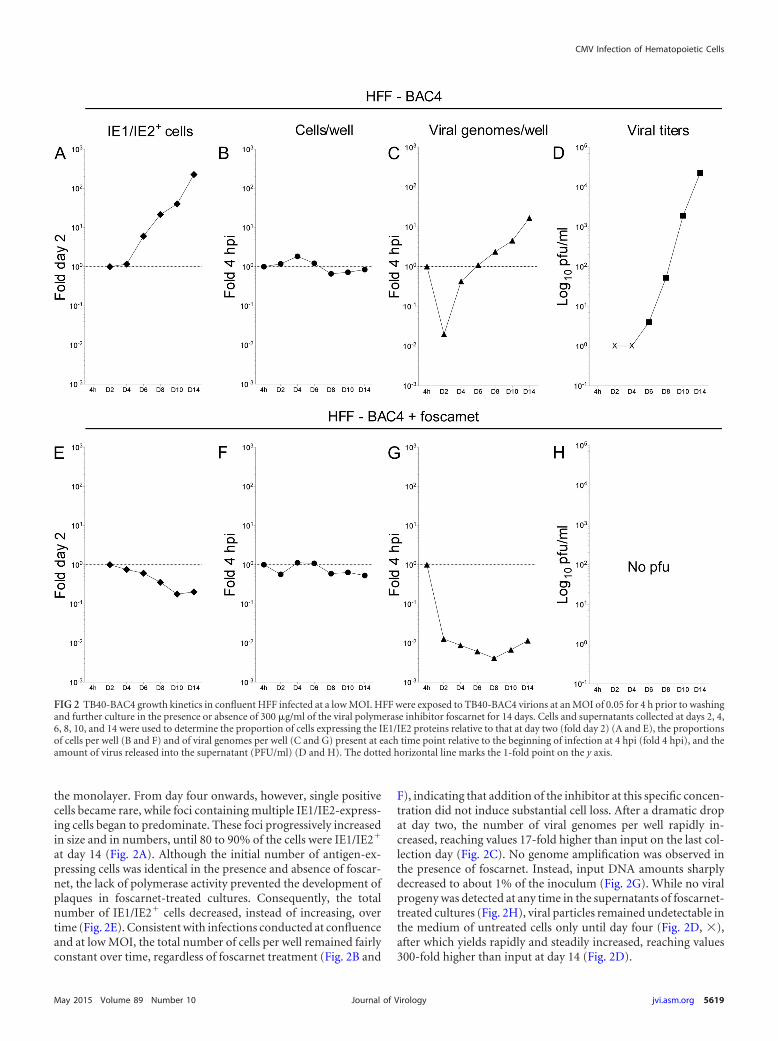
the monolayer. From day four onwards, however, single positivecells became rare, while foci containing multiple IE1/IE2-express-ing cells began to predominate. These foci progressively increasedin size and in numbers, until 80 to 90% of the cells were IE1/IE2�
at day 14 (Fig. 2A). Although the initial number of antigen-ex-pressing cells was identical in the presence and absence of foscar-net, the lack of polymerase activity prevented the development ofplaques in foscarnet-treated cultures. Consequently, the totalnumber of IE1/IE2� cells decreased, instead of increasing, overtime (Fig. 2E). Consistent with infections conducted at confluenceand at low MOI, the total number of cells per well remained fairlyconstant over time, regardless of foscarnet treatment (Fig. 2B and
F), indicating that addition of the inhibitor at this specific concen-tration did not induce substantial cell loss. After a dramatic dropat day two, the number of viral genomes per well rapidly in-creased, reaching values 17-fold higher than input on the last col-lection day (Fig. 2C). No genome amplification was observed inthe presence of foscarnet. Instead, input DNA amounts sharplydecreased to about 1% of the inoculum (Fig. 2G). While no viralprogeny was detected at any time in the supernatants of foscarnet-treated cultures (Fig. 2H), viral particles remained undetectable inthe medium of untreated cells only until day four (Fig. 2D, �),after which yields rapidly and steadily increased, reaching values300-fold higher than input at day 14 (Fig. 2D).
FIG 2 TB40-BAC4 growth kinetics in confluent HFF infected at a low MOI. HFF were exposed to TB40-BAC4 virions at an MOI of 0.05 for 4 h prior to washingand further culture in the presence or absence of 300 �g/ml of the viral polymerase inhibitor foscarnet for 14 days. Cells and supernatants collected at days 2, 4,6, 8, 10, and 14 were used to determine the proportion of cells expressing the IE1/IE2 proteins relative to that at day two (fold day 2) (A and E), the proportionsof cells per well (B and F) and of viral genomes per well (C and G) present at each time point relative to the beginning of infection at 4 hpi (fold 4 hpi), and theamount of virus released into the supernatant (PFU/ml) (D and H). The dotted horizontal line marks the 1-fold point on the y axis.
CMV Infection of Hematopoietic Cells
May 2015 Volume 89 Number 10 jvi.asm.org 5619Journal of Virology
In HFF infected at low MOI, thus, both the proportion of IE1/IE2-expressing cells and the total amounts of viral genomes perwell increase linearly over time, while progeny production ratesfollow a sigmoidal distribution, as expected for multistep lyticreplication.
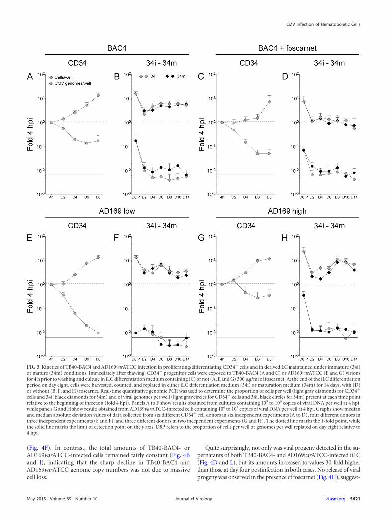
TB40-BAC4 and AD169varATCC genomes are maintainedin the absence of lytic gene expression and progeny productionin proliferating/differentiating CD34� cells. Consistent with theexpected behavior for stem cells, the total number of infectedCD34� cells per well increased over time irrespective of the virusstrain used and of the presence or absence of 300 �g/ml of foscar-net (Fig. 3A, C, E, and G, gray diamonds). The rates of cell growthwere slightly different, however, with median values of 2-fold ev-ery 2 days for TB40-BAC4- and AD169varATCC-infected cellsbut of only 1.3-fold in the presence of foscarnet. This suggests that,in contrast to the case for HFF, foscarnet may be toxic to CD34�
cells at this specific concentration, possibly due to the reducedproliferation rates of HFF compared to CD34� cells. No differ-ence was observed in the total amplification extent (about 20-foldat day eight) of CMV- and mock-infected cells (not shown), indi-cating that infection had no negative effects on cell viability.
After an initial decrease, the total number of viral genomes perwell in TB40-BAC4-infected cultures appeared to stabilize at val-ues corresponding to 20% of the initial input in the absence offoscarnet (Fig. 3A, gray circles), while in the presence of the inhib-itor, viral genome loss continued until only 5 to 7% of initial DNAamounts remained at day eight (Fig. 3C). These differences, how-ever, did not reach statistical significance in donor paired two-tailedStudent t tests (P 0.233 at day six and P 0.152 at day eight), andthe two data distributions (Fig. 3A and C, day two to day eight) didnot score as different in a two-sample KS test (P 0.249).
A similar sharp decrease was also observed in AD169varATCC-infected cultures that contained amounts (105 to 106) of genomesper well at 4 hpi comparable to those for TB40-BAC4-infectedcultures (Fig. 3E). In contrast, the loss of viral DNA in culturesderived from CD34� cells harboring larger amounts (106 to 107)of AD169varATCC genomes per well at 4 hpi was substantially lessprofound, leveling at 20 to 36% of initial input at day six to eight(Fig. 3G), with the two data distributions (Fig. 3E and G) beingsubstantially different according to a KS test (P 0.014).
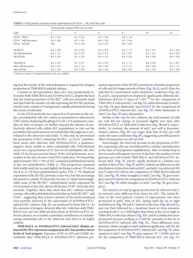
No IE1/IE2� cells were observed by IFA analysis of cytospinpreparations, and no expression of the UL122/123 genes was de-tected by real-time quantitative reverse transcription-PCR analy-ses of samples at each time postinfection (data not shown), whileextremely small amounts of viral particles were detected in thesupernatants of infected cultures, irrespective of the presence orabsence of foscarnet (Table 1). As extracellular virions were notremoved by proteolytic digestion at 4 hpi, these particles are morelikely to represent unpenetrated virions than newly produced viralprogeny.
Together, these results indicate that both TB40-BAC4 andAD169varATCC establish nonlytic infections in proliferating anddifferentiating CD34� cells, with their genomes being maintainedover time in a process that may involve the activity of the viralpolymerase, and that requires larger initial amounts ofAD169varATCC genomes to attain the same degree of efficiencyas in TB40-BAC4-infected cultures.
TB40-BAC4 and AD169varATCC genomes are maintainedat low levels in LC derived from infected CD34� cells irrespec-tive of maturation. At the end of the differentiation period at day
eight, iLC derived from infected CD34� cells were harvested andreplated in either differentiation (34i) or maturation (34m) me-dium for 14 days (Fig. 1A). Cell replating was associated with asharp 10-fold drop in both the total number of cells per well andviral genomes per well until day two postplating, after which thetotal number of cells per well started to rise again until day six toeight, irrespective of the medium used (Fig. 3B, F, and H, gray andblack diamonds), and with the exception of TB40-BAC4-infected,foscarnet-treated cells (Fig. 3D). This increase was not mirroredby the total number of viral genomes per well, which did notincrease again but remained instead at levels at or below back-ground until day 14 (Fig. 3B, D, F, and H, gray and black circles).Viral genome loss was less severe in 34i and 34m cells derived fromCD34� cells containing large amounts of AD169varATCC ge-nomes (Fig. 3H), and the cumulative distributions of data wereconsidered to be different according to the KS comparisons of 34idata from populations containing low and high viral genomeamounts (Fig. 3F and H, gray circles; P 0.003) and to KS com-parisons of 34m data from populations containing small and largeviral genome amounts (Fig. 3F and H, black circles; P 0.013).
Similarly, viral genome loss was less pronounced in 34mthan in 34i cells derived from TB40-BAC4-infected progenitors(Fig. 3B), with the two distributions scoring as different (P 0.013 for the donor paired KS comparison of 34i and 34msamples in Fig. 3B).
In contrast, treatment of 34i or 34m cells with foscarnet did notappear to have any major effect on their content in viral genomesover time. Accordingly, the distributions of data from treated anduntreated samples were not statistically different (P 0.139 forthe KS comparison of treated to untreated 34i or 34m samples[Fig. 3B to D]).
No IE1/IE2� cells were observed at any time point, and theamount of viral particles detected in the supernatants was againextremely small and very similar in cells treated or not with fos-carnet (Table 1).
Together, these findings suggest that viral genomes are main-tained at very low levels in both immature and mature LC derivedfrom infected CD34� progenitors and that this process is moreefficient in 34i and 34m cells derived from progenitors containinglarge amounts of AD169varATCC DNA and in 34m cells derivedfrom TB40-BAC4-infected CD34� cells. Although some viral ge-nome replication may be occurring, viral progeny is not producedin large quantities, if at all, by any of these cells. Thus, LPS andCD40L stimulation of iLC differentiated from infected progeni-tors does not appear to induce robust viral DNA replication andprogeny production.
TB40-BAC4- or AD169varATCC-infected iLC maintainedunder immature conditions (iiLC) produce viral progeny inthe absence of widespread IE1/IE2 gene expression and ofrobust genome amplification. We next assessed the kinetics ofviral replication in iLC directly exposed to TB40-BAC4 orAD169varATCC virions and subsequently maintained under im-mature (iiLC) or mature (imLC) conditions (Fig. 1B).
Consistent with previous results (9, 24), only a small fraction ofiiLC expressed the IE1/IE2 proteins at day two postinfection withTB40-BAC4 (0.8% � 1.5%) or AD169varATCC (1.7% � 0.7%).These proportions rapidly decreased, plummeting to zero by day14 (Fig. 4A, E, and I). The same trend was followed by the totalnumber of viral genomes per well (Fig. 4C, G, and K) and by thetotal amount of cells per well in foscarnet-treated cultures
Coronel et al.
5620 jvi.asm.org May 2015 Volume 89 Number 10Journal of Virology
(Fig. 4F). In contrast, the total amounts of TB40-BAC4- orAD169varATCC-infected cells remained fairly constant (Fig. 4Band J), indicating that the sharp decline in TB40-BAC4 andAD169varATCC genome copy numbers was not due to massivecell loss.
Quite surprisingly, not only was viral progeny detected in the su-pernatants of both TB40-BAC4- and AD169varATCC-infected iiLC(Fig. 4D and L), but its amounts increased to values 30-fold higherthan those at day four postinfection in both cases. No release of viralprogeny was observed in the presence of foscarnet (Fig. 4H), suggest-
FIG 3 Kinetics of TB40-BAC4 and AD169varATCC infection in proliferating/differentiating CD34� cells and in derived LC maintained under immature (34i)or mature (34m) conditions. Immediately after thawing, CD34� progenitor cells were exposed to TB40-BAC4 (A and C) or AD169varATCC (E and G) virionsfor 4 h prior to washing and culture in iLC differentiation medium containing (C) or not (A, E and G) 300 �g/ml of foscarnet. At the end of the iLC differentiationperiod on day eight, cells were harvested, counted, and replated in either iLC differentiation medium (34i) or maturation medium (34m) for 14 days, with (D)or without (B, F, and H) foscarnet. Real-time quantitative genomic PCR was used to determine the proportion of cells per well (light gray diamonds for CD34�
cells and 34i, black diamonds for 34m) and of viral genomes per well (light gray circles for CD34� cells and 34i, black circles for 34m) present at each time pointrelative to the beginning of infection (fold 4 hpi). Panels A to F show results obtained from cultures containing 105 to 106 copies of viral DNA per well at 4 hpi,while panels G and H show results obtained from AD169varATCC-infected cells containing 106 to 107 copies of viral DNA per well at 4 hpi. Graphs show medianand median absolute deviation values of data collected from six different CD34� cell donors in six independent experiments (A to D), four different donors inthree independent experiments (E and F), and three different donors in two independent experiments (G and H). The dotted line marks the 1-fold point, whilethe solid line marks the limit of detection point on the y axis. D8P refers to the proportion of cells per well or genomes per well replated on day eight relative to4 hpi.
CMV Infection of Hematopoietic Cells
May 2015 Volume 89 Number 10 jvi.asm.org 5621Journal of Virology
ing that the activity of the viral polymerase is required for progenyproduction in TB40-BAC4-infected cultures.
Contrary to all expectations, thus, iiLC were productively in-fected by both TB40-BAC4 and AD169varATCC, despite the factsthat the initial proportion of IE1/IE2� cells was extremely smalland that both the number of cells expressing the IE1/IE2 proteinsand the total content of viral genomes rapidly plummeted duringthe course of infection.
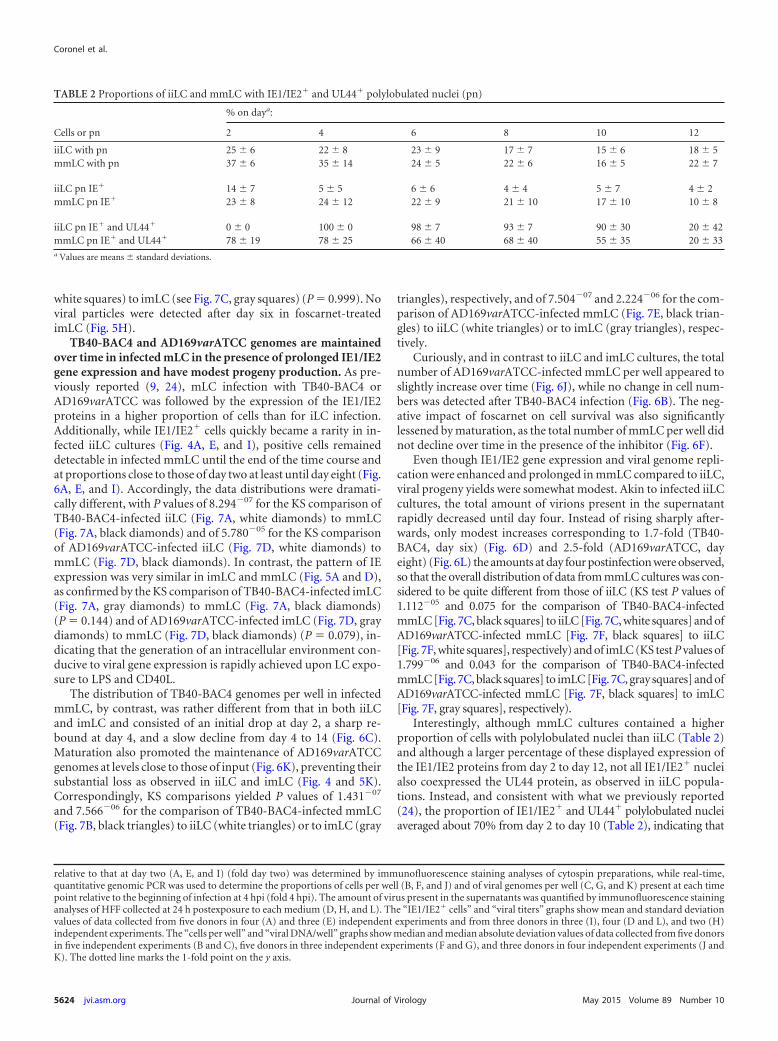
As the CD34 molecule was reported to be present on the sur-face of endothelial cells (43), which are permissive to infection byCMV strains displaying the gH/gL/UL128-131A pentameric com-plex on their envelopes, we further assessed the identity of pro-ductively infected cells within iiLC populations, to rule out thepossibility that small amounts of endothelial cells might have con-tributed to the observed viral yields. To this end, we determinedthe proportion of iiLC containing IE1/IE2� or UL44� polylobu-lated nuclei after infection with AD169varATCC, a pentamer-negative strain unable to infect endothelial cells. Polylobulatednuclei are a typical feature of LC but not of endothelial cells (44),while UL44, the processivity factor of the viral polymerase, accu-mulates at the sites of active viral DNA replication. We found thatapproximately 25% � 6% of iiLC contained polylobulated nucleiat day two postinfection (Table 2). This proportion remainedfairly stable until day six and slightly declining to about 17% fromday 8 to 12. Of these polylobulated nuclei, 14% � 7% displayedexpression of the IE1/IE2 proteins at day two, but this percentagedecreased to a steady 5% from day 4 to day 12. Quite interestingly,while none of the IE1/IE2� polylobulated nuclei expressed theUL44 protein at day two, almost all became UL44� from day fouronwards. Together, these data show that iiLC cultures actuallycontain cells with polylobulated nuclei that are both IE1/IE2� andUL44�, suggesting that at least a portion of the newly producedviral particles observed in the supernatant of AD169varATCC-infected iiLC cultures (Fig. 4L) are produced by bona fide LC. Asthe amounts of progeny detected after iiLC infection with TB40-BAC4 were not very different from those of AD169varATCC-in-fected cultures, we consider a potential contribution of contami-nating endothelial cells to the observed viral titers to be highlyunlikely.
TB40-BAC4- or AD169varATCC-infected imLC display en-hanced IE1/IE2 expression compared to iiLC but produce lowerlevels of viral progeny. Exposure of iLC to LPS and CD40L im-mediately after TB40-BAC4 or AD169varATCC infection pro-
moted expression of the IE1/IE2 proteins in a broader proportionof cells and for longer periods of time (Fig. 5A, E, and I) than forinfected iLC maintained under immature conditions (Fig. 4A,E, and I), and populations displayed significantly different dis-tributions (KS test P values of 7.293�06 for the comparison ofTB40-BAC4-infected iiLC [see Fig. 7A, white diamonds] to imLC[see Fig. 7A, gray diamonds] and of 0.017 for the comparison ofAD169varATCC-infected iiLC [see Fig. 7D, white diamonds] toimLC [see Fig. 7D, gray diamonds]).
Similar to the case for iiLC cultures, the total number of cellsper well did not change or increased slightly over time afterAD169varATCC or TB40-BAC4 infection (Fig. 5B and J, respec-tively). In contrast, the amount of imLC per well in foscarnet-treated cultures (Fig. 5F) was larger than that of iiLC per wellunder the same conditions (Fig. 4F), suggesting a possible positiveeffect of maturation on cell survival.
Interestingly, the observed increase in the proportion of IE1/IE2-expressing cells was not followed by a similar intensificationin the rates of viral genome amplification. After an initial drop atday two and a subsequent recovery at day four, the number of viralgenomes per well in both TB40-BAC4- and AD169varATCC-in-fected imLC (Fig. 5C and K) rapidly declined in a fashion verysimilar to that of iiLC (Fig. 4C and K). Indeed, no difference in thedistribution of data from infected iiLC and imLC was detected (KStest P values of 0.160 for the comparison of TB40-BAC4-infectediiLC [see Fig. 7B, white triangles] to imLC [see Fig. 7B, gray trian-gles] and of 0.644 for the comparison of AD169varATCC-infectediiLC [see Fig. 7E, white triangles] to imLC [see Fig. 7E, gray trian-gles]).
The kinetics of viral progeny production by infected imLC,in contrast, were different from those for iiLC. The initial de-cline in the viral particle content of supernatants was moreprotracted in imLC than in iiLC, lasting until day six to eightpostinfection (Fig. 5D and L) instead of day four (Fig. 4D and L),and was then followed by a moderate boost in virus amounts,reaching levels 3- to 5-fold higher than those at day six or eight inTB40-BAC4-infected cultures (Fig. 5D), and by a milder but moreprotracted increase, peaking at 2-fold the amounts at day six inAD169varATCC-infected cells (Fig. 5L). These differences were,however, robust enough to reach statistical significance only for theKS comparison of AD169varATCC-infected iiLC (see Fig. 7F, whitesquares) to imLC (see Fig. 7F, gray squares) (P 0.049) and notfor the comparison of TB40-BAC4-infected iiLC (see Fig. 7C,
TABLE 1 Viral particle contents in the supernatants of CD34�, 34i, and 34m cells
Cells
Viral particle content (PFU/ml) on daya:
2 4 6 8 10 14
CD34� BAC4 0.1 � 0.5 0.2 � 0.5 0.3 � 0.8 0.6 � 1.9CD34� with foscarnet 0.0 � 0.0 0.2 � 0.4 0.0 � 0.0 0.2 � 0.6CD34� AD169 NA 0.3 � 0.8 0.4 � 0.5 0.0 � 0.0
34i BAC4 0.2 � 0.8 0.1 � 0.4 0.3 � 0.8 0.4 � 1.7 0.1 � 0.3 0.2 � 0.634i with foscarnet 0.4 � 1.3 0.2 � 0.7 0.2 � 0.7 0.2 � 0.7 0.2 � 0.7 0.4 � 0.934i AD169 0.5 � 0.9 0.1 � 0.4 0.4 � 0.7 0.0 � 0.0 0.0 � 0.0 0.0 � 0.0
34m BAC4 0.2 � 0.5 0.1 � 0.7 0.1 � 0.3 0.3 � 1.7 0.0 � 0.0 0.1 � 0.734m with foscarnet 0.2 � 0.7 0.4 � 1.3 0.0 � 0.0 1.1 � 3.3 0.0 � 0.0 0.4 � 1.334m AD169 0.0 � 0.0 0.0 � 0.0 0.0 � 0.0 0.0 � 0.0 0.0 � 0.0 0.0 � 0.0a Values are means � standard deviations. NA, not available.
Coronel et al.
5622 jvi.asm.org May 2015 Volume 89 Number 10Journal of Virology
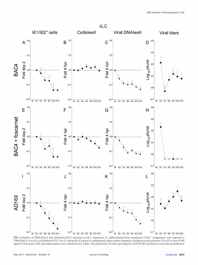
FIG 4 Kinetics of TB40-BAC4 and AD169varATCC infection in iiLC. Immature LC differentiated from uninfected CD34� progenitors were exposed toTB40-BAC4 (A to H) or AD169varATCC (I to L) virions for 4 h prior to washing and culture under immature conditions in the presence (E to H) or not of 300�g/ml of foscarnet. Cells and supernatants were collected every 2 days. The proportion of cells expressing the viral IE1/IE2 proteins at each time postinfection
CMV Infection of Hematopoietic Cells
May 2015 Volume 89 Number 10 jvi.asm.org 5623Journal of Virology
white squares) to imLC (see Fig. 7C, gray squares) (P 0.999). Noviral particles were detected after day six in foscarnet-treatedimLC (Fig. 5H).
TB40-BAC4 and AD169varATCC genomes are maintainedover time in infected mLC in the presence of prolonged IE1/IE2gene expression and have modest progeny production. As pre-viously reported (9, 24), mLC infection with TB40-BAC4 orAD169varATCC was followed by the expression of the IE1/IE2proteins in a higher proportion of cells than for iLC infection.Additionally, while IE1/IE2� cells quickly became a rarity in in-fected iiLC cultures (Fig. 4A, E, and I), positive cells remaineddetectable in infected mmLC until the end of the time course andat proportions close to those of day two at least until day eight (Fig.6A, E, and I). Accordingly, the data distributions were dramati-cally different, with P values of 8.294�07 for the KS comparison ofTB40-BAC4-infected iiLC (Fig. 7A, white diamonds) to mmLC(Fig. 7A, black diamonds) and of 5.780�05 for the KS comparisonof AD169varATCC-infected iiLC (Fig. 7D, white diamonds) tommLC (Fig. 7D, black diamonds). In contrast, the pattern of IEexpression was very similar in imLC and mmLC (Fig. 5A and D),as confirmed by the KS comparison of TB40-BAC4-infected imLC(Fig. 7A, gray diamonds) to mmLC (Fig. 7A, black diamonds)(P 0.144) and of AD169varATCC-infected imLC (Fig. 7D, graydiamonds) to mmLC (Fig. 7D, black diamonds) (P 0.079), in-dicating that the generation of an intracellular environment con-ducive to viral gene expression is rapidly achieved upon LC expo-sure to LPS and CD40L.
The distribution of TB40-BAC4 genomes per well in infectedmmLC, by contrast, was rather different from that in both iiLCand imLC and consisted of an initial drop at day 2, a sharp re-bound at day 4, and a slow decline from day 4 to 14 (Fig. 6C).Maturation also promoted the maintenance of AD169varATCCgenomes at levels close to those of input (Fig. 6K), preventing theirsubstantial loss as observed in iiLC and imLC (Fig. 4 and 5K).Correspondingly, KS comparisons yielded P values of 1.431�07
and 7.566�06 for the comparison of TB40-BAC4-infected mmLC(Fig. 7B, black triangles) to iiLC (white triangles) or to imLC (gray
triangles), respectively, and of 7.504�07 and 2.224�06 for the com-parison of AD169varATCC-infected mmLC (Fig. 7E, black trian-gles) to iiLC (white triangles) or to imLC (gray triangles), respec-tively.
Curiously, and in contrast to iiLC and imLC cultures, the totalnumber of AD169varATCC-infected mmLC per well appeared toslightly increase over time (Fig. 6J), while no change in cell num-bers was detected after TB40-BAC4 infection (Fig. 6B). The neg-ative impact of foscarnet on cell survival was also significantlylessened by maturation, as the total number of mmLC per well didnot decline over time in the presence of the inhibitor (Fig. 6F).
Even though IE1/IE2 gene expression and viral genome repli-cation were enhanced and prolonged in mmLC compared to iiLC,viral progeny yields were somewhat modest. Akin to infected iiLCcultures, the total amount of virions present in the supernatantrapidly decreased until day four. Instead of rising sharply after-wards, only modest increases corresponding to 1.7-fold (TB40-BAC4, day six) (Fig. 6D) and 2.5-fold (AD169varATCC, dayeight) (Fig. 6L) the amounts at day four postinfection were observed,so that the overall distribution of data from mmLC cultures was con-sidered to be quite different from those of iiLC (KS test P values of1.112�05 and 0.075 for the comparison of TB40-BAC4-infectedmmLC [Fig. 7C, black squares] to iiLC [Fig. 7C, white squares] and ofAD169varATCC-infected mmLC [Fig. 7F, black squares] to iiLC[Fig. 7F, white squares], respectively) and of imLC (KS test P values of1.799�06 and 0.043 for the comparison of TB40-BAC4-infectedmmLC [Fig. 7C, black squares] to imLC [Fig. 7C, gray squares] and ofAD169varATCC-infected mmLC [Fig. 7F, black squares] to imLC[Fig. 7F, gray squares], respectively).
Interestingly, although mmLC cultures contained a higherproportion of cells with polylobulated nuclei than iiLC (Table 2)and although a larger percentage of these displayed expression ofthe IE1/IE2 proteins from day 2 to day 12, not all IE1/IE2� nucleialso coexpressed the UL44 protein, as observed in iiLC popula-tions. Instead, and consistent with what we previously reported(24), the proportion of IE1/IE2� and UL44� polylobulated nucleiaveraged about 70% from day 2 to day 10 (Table 2), indicating that
relative to that at day two (A, E, and I) (fold day two) was determined by immunofluorescence staining analyses of cytospin preparations, while real-time,quantitative genomic PCR was used to determine the proportions of cells per well (B, F, and J) and of viral genomes per well (C, G, and K) present at each timepoint relative to the beginning of infection at 4 hpi (fold 4 hpi). The amount of virus present in the supernatants was quantified by immunofluorescence staininganalyses of HFF collected at 24 h postexposure to each medium (D, H, and L). The “IE1/IE2� cells” and “viral titers” graphs show mean and standard deviationvalues of data collected from five donors in four (A) and three (E) independent experiments and from three donors in three (I), four (D and L), and two (H)independent experiments. The “cells per well” and “viral DNA/well” graphs show median and median absolute deviation values of data collected from five donorsin five independent experiments (B and C), five donors in three independent experiments (F and G), and three donors in four independent experiments (J andK). The dotted line marks the 1-fold point on the y axis.
TABLE 2 Proportions of iiLC and mmLC with IE1/IE2� and UL44� polylobulated nuclei (pn)
Cells or pn
% on daya:
2 4 6 8 10 12
iiLC with pn 25 � 6 22 � 8 23 � 9 17 � 7 15 � 6 18 � 5mmLC with pn 37 � 6 35 � 14 24 � 5 22 � 6 16 � 5 22 � 7
iiLC pn IE� 14 � 7 5 � 5 6 � 6 4 � 4 5 � 7 4 � 2mmLC pn IE� 23 � 8 24 � 12 22 � 9 21 � 10 17 � 10 10 � 8
iiLC pn IE� and UL44� 0 � 0 100 � 0 98 � 7 93 � 7 90 � 30 20 � 42mmLC pn IE� and UL44� 78 � 19 78 � 25 66 � 40 68 � 40 55 � 35 20 � 33a Values are means � standard deviations.
Coronel et al.
5624 jvi.asm.org May 2015 Volume 89 Number 10Journal of Virology
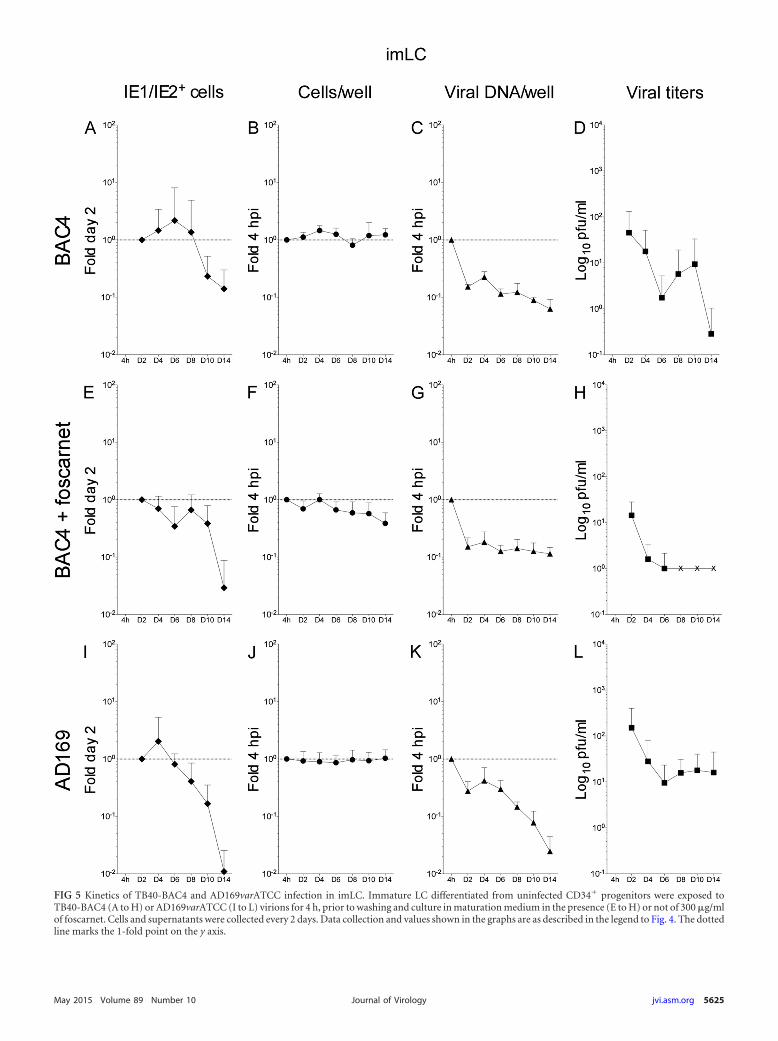
FIG 5 Kinetics of TB40-BAC4 and AD169varATCC infection in imLC. Immature LC differentiated from uninfected CD34� progenitors were exposed toTB40-BAC4 (A to H) or AD169varATCC (I to L) virions for 4 h, prior to washing and culture in maturation medium in the presence (E to H) or not of 300 �g/mlof foscarnet. Cells and supernatants were collected every 2 days. Data collection and values shown in the graphs are as described in the legend to Fig. 4. The dottedline marks the 1-fold point on the y axis.
May 2015 Volume 89 Number 10 jvi.asm.org 5625Journal of Virology
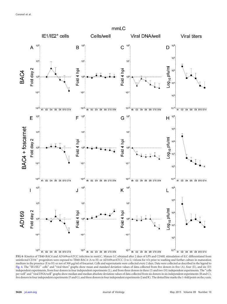
FIG 6 Kinetics of TB40-BAC4 and AD169varATCC infection in mmLC. Mature LC obtained after 2 days of LPS and CD40L stimulation of iLC differentiated fromuninfected CD34� progenitors were exposed to TB40-BAC4 (A to H) or AD169varATCC (I to L) virions for 4 h prior to washing and further culture in maturationmedium in the presence (E to H) or not of 300 �g/ml of foscarnet. Cells and supernatants were collected every 2 days. Data were collected as described in the legend toFig. 4. The “IE1/IE2� cells” and “viral titers” graphs show mean and standard deviation values of data collected from five donors in five (A), four (E), and six (D)independent experiments, from four donors in four independent experiments (L), and from three donors in three (I) and two (H) independent experiments. The “cellsper well” and “viral DNA/well” graphs show median and median absolute deviation values of data collected from six donors in six independent experiments (B and C),five donors in four independent experiments (F and G), and three donors in four independent experiments (J and K). The dotted line marks the 1-fold point on the y axis.
Coronel et al.
5626 jvi.asm.org May 2015 Volume 89 Number 10Journal of Virology
infection did not progress beyond the immediate early stages in acertain proportion of cells, possibly contributing to lower viral yields.
Consistent with previous data (24), thus, mmLC are produc-tively infected by both TB40-BAC4 and AD169varATCC, but de-spite the facts that the IE1/IE2 proteins are expressed in a larger
proportion of cells and for longer periods of time and that viralDNA is replicated and/or maintained better than in iiLC, mmLCproduce lower levels of viral progeny over time.
Together, these data indicate that the intracellular signalinginduced by exposure of iLC to LPS and CD40L promotes viral IE
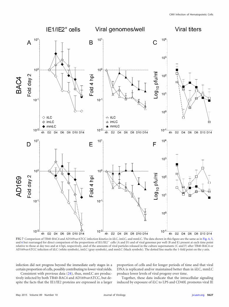
FIG 7 Comparison of TB40-BAC4 and AD169varATCC infection kinetics in iiLC, imLC, and mmLC. The data shown in this figure are the same as in Fig. 4, 5,and 6 but rearranged for direct comparison of the proportions of IE1/IE2� cells (A and D) and of viral genomes per well (B and E) present at each time pointrelative to those at day two and at 4 hpi, respectively, and of the amounts of viral particles released in the culture supernatants (C and F) after TB40-BAC4 orAD169varATCC infection of iiLC (white symbols), imLC (gray symbols), and mmLC (black symbols). The dotted line marks the 1-fold point on the y axis.
CMV Infection of Hematopoietic Cells
May 2015 Volume 89 Number 10 jvi.asm.org 5627Journal of Virology
gene transcription, leading to a more widespread and prolongedexpression of the IE1/IE2 proteins in infected cells. This permis-sive milieu is established quite rapidly after LC exposure to thematuration stimuli, as the pattern of IE1/IE2 protein expressionwas similar in LC infected when already mature (mmLC), and inLC infected when still immature but exposed to maturation stim-uli immediately afterwards (imLC). This supportive environmentis also maintained over time, preventing the loss of viral proteinexpression observed in iiLC (Fig. 7A and D).
In contrast to viral gene expression, maintenance of viral ge-nomes over time occurs only when the supportive environmentpromoted by maturation is present at the moment of infection.Indeed, exposure of iLC to LPS and CD40L immediately afterinfection did not stimulate viral genome replication (Fig. 7B andE). Despite supporting viral gene expression and genome replica-tion, maturation does not substantially stimulate viral progenyproduction, with iiLC, mmLC, and imLC all releasing virus overtime (Fig. 7C and F).
DISCUSSION
CD34� progenitor cells and their LC progeny are thought to playimportant roles in CMV pathogenesis in vivo as sites of viral la-tency, reactivation, and replication and as means of viral spreadwithin the hosts and between hosts. Despite this, many aspects ofCMV interactions with these cells remain unknown.
In this study, we sought to compare the efficiencies of infectiononset, viral genome replication, and viral progeny production inCD34� progenitor cells, iLC, and mLC derived from the same setof donors and exposed to the clinical-like CMV strain TB40-BAC4or to the laboratory-adapted strain AD169varATCC prior to theirmaintenance under immature or mature conditions. Infectionprogress was tracked for up to 14 days and was compared to that inHFF, a highly permissive cell type customarily used in CMVpathogenesis studies.
Viral genomes are maintained in CD34� cells during theirdifferentiation into iLC. This is the first report to quantify thenumbers of viral and cellular genome copies present in CD34� cellcultures during their differentiation into iLC. We show that whileCD34� cells proliferate over time, viral genome amounts remainfairly constant (Fig. 3A and G), suggesting either that viral DNA ismaintained without amplification or degradation in cells that re-main viable during the 8 days of differentiation or that it is repli-cated in a subset of cells and lost in another subset of similar size.Which one of these, and potentially other, hypotheses is correctcannot be definitely ascertained in the absence of quantitativePCR data from single cells.
Clearly, the number of viral DNA copies did not increase inparallel with the number of cellular genomes, as reported forTowne (20)- or AD169varATCC (18)-infected GM-Ps, suggestingthat the behavior of viral genomes can change depending on thephenotype and differentiation state of the hosting cell. Consistentwith data from GM-Ps (20), but in contrast to data from CD34�
cells cultured with irradiated murine stromal cells (23), no evi-dence of productive replication was found in AD169varATCC-infected CD34� cell cultures (Table 1). Rather, AD169varATCCgenome loads rapidly plummeted in cell populations containingthe same starting amounts of viral DNA as TB40-BAC4-infectedcells (Fig. 3E). The main difference we observed between clinical-like and laboratory-adapted strains, thus, was in the efficiency ofviral genome maintenance during cell replication, rather than inthe ability to establish nonlytic infections. Additionally, the defectin AD169varATCC genome maintenance was not absolute but
could be rectified by increasing the amounts of input DNA byabout 10-fold (Fig. 3G).
AD169varATCC virions do not contain the gH/gL/UL128-131A pentameric complex required for fusion of the viral envelopewith endosomal membranes (45, 46), and they enter cells largely viathe plasma membrane (47–49). Although the mechanisms used byCMV to access CD34� cells have not been described, the ability topenetrate cells via both pathways may confer some advantage to pen-tamer-positive strains, akin to what we showed for iLC and mLC(9). Increasing the load of AD169varATCC input particles maythus augment the number of virions that reach the nucleus, im-proving their retention over time. Alternatively, the observedrapid loss of viral genomes may be due to the lack of expression ofUL138 (and/or of other proteins), leading to the inefficient main-tenance of viral DNA during latency, rather than to the onset ofproductive infections as reported for CD34� cells cultured onirradiated murine stromal cells (21–23).
CMV infections in vivo are often associated with the develop-ment of thrombocytopenia (50). Whether this is due to some toxiceffect of infection acting directly on hematopoietic progenitorcells or to some indirect negative influence on stromal cell func-tions remains unclear. Infection of CD34� cell populations invitro was reported to negatively impact their proliferation rates,but this was highly dependent on the CD34� cell subtype, thevirus strain, and the MOI used (4, 22, 51–56). We did not detectany difference in the total number of iLC obtained after differen-tiation of mock- or CMV-infected CD34� cells in multiple exper-iments using TB40-BAC4 and AD169varATCC. Infected popula-tions did not express the viral IE1/IE2 proteins and did notproduce viral progeny (Table 1), suggesting that viral genomemaintenance was not associated with extensive cytopathic effectsleading to widespread cell death.
Maturation of LC differentiated from infected CD34� cellsdoes not substantially enhance the efficacy of viral genomemaintenance and the frequency of reactivation events. After aninitial and unavoidable loss in cell and viral genome numbersfollowing iLC harvest and replating procedures, cell proliferationresumed (Fig. 3B, F, and H), except in foscarnet-treated cultures(Fig. 3D), and was accompanied by the maintenance of viral ge-nomes at relatively steady amounts until day 14 (Fig. 3B, D, andH). While no statistically significant difference was observed be-tween the amounts of viral DNA present in 34i and 34m culturesderived from progenitors infected with AD169varATCC (Fig. 3Fand H), TB40-BAC4-infected 34m cells contained, on average,2.5-fold more viral genomes than 34i cells from day four onwards(Fig. 3B), and the two populations possessed statistically differentdistributions. None of the 34i or 34m cultures expressed the IE1/IE2 proteins throughout the time course, however, and theamounts of virus found in their respective supernatants were vir-tually identical (Table 1), indicating that maturation of iLC de-rived from infected progenitors was not associated with an in-crease in CMV reactivation events. These findings are in contrastto those of Reeves et al., who reported that mLC derived from theCD34� cells of seropositive individuals contained 10-fold largeramounts of viral DNA than undifferentiated CD34� cells andtranscribed the UL122/123 genes encoding the viral IE1/IE2 pro-teins (57). IE gene expression was also observed at day one (32) orday three (31) postmaturation of iLC differentiated from progen-itors experimentally infected with the clinical-like strain TB40/E,and virus was detected in mLC culture supernatants at day 10 or14 postmaturation (31). Although the culture conditions used inthese studies were more similar to the ones employed here, a num-
5628 jvi.asm.org May 2015 Volume 89 Number 10Journal of Virology
ber of key differences exist that can explain the observed discrep-ancies.
First, we used CD34� progenitor cells harvested from the cordblood of different donors, as opposed to cells from the peripheralblood or the bone marrow of adults. Although all CD34� progen-itor cells, irrespective of their anatomical origin, can differentiateinto iLC using the specific cytokine cocktail we employed in here,cord and peripheral blood CD34� cells are not completely iden-tical (58) and may thus react to CMV infection in different ways.
Second, viral genome amounts were quantified from Southernblot images of PCR-amplified genomic extracts obtained from theCD34� cells and from the mLC of a single donor and at a singletime point (57), as opposed to by real-time quantitative PCR anal-yses of genomic DNA derived from the cells of different donors,harvested at multiple time points, and in several independent ex-periments. While circulating CMV such as that found in naturallyinfected CD34� cells is likely to have superior abilities to persistand reactivate in myeloid cells, the quantification method usedmay also have contributed to the observed differences in viralgenome amounts. Additionally, both viral and cellular DNA copynumbers were simultaneously tracked in this study, as opposed toviral DNA amounts only (57).
Viral gene expression was also evaluated in a different way:while we determined the percentage of cells expressing the IE1/IE2proteins at each time point, Reeves et al. used reverse transcrip-tion-PCR to assess the presence of UL122/123 transcripts (32, 57).On the one hand, we may have missed and/or underestimated thetrue extent of viral reactivation in 34i and 34m cells, particularly ifIE1/IE2 protein expression occurred in only a very small percent-age of cells. On the other hand, transcripts are not consistentlytranslated into proteins, and no quantification of reverse tran-scription-PCR data was provided, making it difficult to gauge theactual extent of viral reactivation in mLC.
While naturally infected CD34� cells were cultured under con-ditions very similar to those used in this study (57), experimen-tally infected cells were exposed to TB40/E for 24 h, incubated inthe absence of differentiation cytokines for 3 days, and then dif-ferentiated into iLC (31, 32). The prolonged exposure to CMVvirions (24 versus 4 h), coupled to the use of TB40/E, a genotypi-cally and phenotypically heterogeneous strain (35) likely possess-ing a broader tropism than its bacterial artificial chromosomeclone TB40-BAC4 used here, may have increased the proportionof infected CD34� cells, as well as the load of viral particles per cell.The incubation of infected cells in cytokine-free medium prior totheir differentiation into iLC, moreover, may have promoted theexpansion of CD34� cell subsets more permissive to the establish-ment of infection.
While we used a combination of LPS and CD40L molecules totrigger iLC maturation, Reeves et al. used LPS only (31, 32, 57).Our recent data indicate that iLC exposure to LPS alone, CD40Lalone, or LPS plus CD40L can generate mLC with different sus-ceptibilities to direct CMV infection (not shown), suggesting thatsignaling by these molecules can trigger very different responses iniLC, with potential influences on the ability of latent virus to re-activate. Interestingly, while reactivated virus was found (albeitnot quantified) directly in the supernatant of mLC derived fromexperimentally infected CD34� cells (31), CMV reactivation inmLC originating from naturally infected CD34� cells was re-ported to occur after coculture with permissive fibroblasts (57). Inour hands, maturation of latently infected iLC by exposure to LPS
and CD40L was not sufficient to trigger robust viral reactivation,which we think may require additional stimulation beyond thatprovided by these molecules, which may be supplied by cytokinesand other soluble factors secreted by HFF or produced by LCthemselves upon contact with heterologous cell types. Indeed, co-culture with HFF has historically been the most commonly usedmethod to induce full reactivation (18–23, 59).
iLC maturation promotes and prolongs IE1/IE2 protein ex-pression. Stimulation of immature LC with LPS and CD40L for 2days before infection (mmLC) or immediately after exposure toCMV virions (imLC) enhanced and prolonged viral IE proteinexpression compared to that by unstimulated cells (iiLC) (Fig. 7Aand D). This implies that maturation promotes infection onsetand does so relatively quickly, as the kinetics of viral protein ex-pression in imLC and mmLC were almost identical. We previ-ously showed that mLC contain substantially larger amounts ofUL122/UL123 mRNA than iLC at 24, 48, and 72 h postinfectionwith different CMV strains (9). Maturation is thus likely to act byspecifically boosting viral gene transcription, although changes inthe efficiency of mRNA translation may also contribute to in-crease the proportion of IE1/IE2� cells. This transcriptional en-hancement may occur through a variety of possible mechanism,including the remodeling of repressive chromatin on the UL122/UL123 gene promoter, the disassembly of nuclear domain 10 bod-ies, and the replacement of transcriptional repressors with tran-scriptional activators on viral gene promoters. The major IEpromoter was indeed reported to be associated with makers oftranscriptionally active chromatin, such as acetylated H4 histones,but devoid of transcriptional repressors, such as the heterochro-matin protein 1, in mLC (31, 32, 57), while removal of the cellularproteins hDaxx and ATRX from the nuclear domain 10 bodies ofmLC was proposed to contribute to these cells’ susceptibility toinfection (60). In a recently completed microarray-based compar-ison of the transcriptional profile of iLC and mLC, we found thatapproximately 15% of the genes more highly expressed in iLC and6% of those more highly expressed in mLC encode known tran-scription factors or chromatin-modifying enzymes. The potentialeffects of these proteins’ activities on LC susceptibility to infectionare under investigation.
iLC maturation prior to infection enhances viral genomereplication. Viral genome amounts were larger in cells infectedwhen already mature (mmLC) than in cells matured after infec-tion (imLC) (Fig. 7B and E), indicating that the intranuclear en-vironment required for efficient viral DNA replication is not asquickly established as that promoting viral gene transcription andmust already be present at the moment of infection onset toachieve effective genome amplification. Quite interestingly, ex-pression of the viral IE1/IE2 proteins is not sufficient to generatethis milieu, as the proportions of IE1/IE2� in imLC and mmLCwere very similar (Fig. 7A and D), yet viral genome numbersplummeted in imLC. A second block to the progress of viral in-fection may thus exist in iLC, acting after the one limiting IE geneexpression and potentially shunting infection toward an abortiveoutcome. Removal of this second hindrance cannot be easily at-tained by signaling events ensuing exposure to LPS and CD40L orby the powerful activities of the IE1/IE2 proteins and may insteadrequire the presence of specific cellular functions that need time toaccumulate.
iLC maturation does not potently stimulate viral progenyproduction or release. Quite unexpectedly, viral progeny was de-
CMV Infection of Hematopoietic Cells
May 2015 Volume 89 Number 10 jvi.asm.org 5629Journal of Virology
tected in the supernatant of iiLC cultures (Fig. 7C and F). This israther remarkable considering that the already low proportion ofIE1/IE2� iiLC rapidly and dramatically decreased over time andwas mirrored by viral genome amounts. These analyses were,however, not conducted at a single-cell level. It is thus possiblethat infection may have proceeded in the few cells initially express-ing the IE1/IE2 proteins, leading to some low level of viral genomereplication that may have escaped detection and culminating inthe release of newly formed viral particles. The presence of theUL44 protein in virtually all IE1/IE2� iiLC certainly lends supportto this hypothesis, although this would require this subpopulationof permissive iLC to be particularly effective at producing viralprogeny, considering the comparatively large amounts of virusdetected in iiLC culture supernatants. Viral cycle progression inIE1/IE2� iiLC may also have been boosted following some matu-ration or activation events induced by factors secreted by unin-fected bystander cells and exerting more potent effects on viralreplication than exposure to LPS and CD40L.
Also contrary to expectations, maturation did not substantiallyenhance viral yields. Considering that a larger proportion ofmmLC expressed the IE1/IE2 proteins and that mmLC culturescontained larger amounts of viral genomes overall, we expectedyields from these cells to exceed those of iiLC. Instead, values weresimilar (TB40-BAC4) (Fig. 7C) or lower (AD169varATCC) (Fig.7F). As only virus contained in the culture medium was measured,however, mmLC may still be producing larger amounts of prog-eny than iiLC, retaining the majority of it intracellularly. Alterna-tively, infection may be blocked after IE1/IE2 protein expressionin a certain proportion of cells, as our data pertaining UL44 ex-pression seem to suggest (Table 2), leading to an overall decreasein viral yields from these cultures.
Interestingly, iLC maturation after infection reduced yieldsand delayed the time of appearance of new viral particles from daysix to day eight (Fig. 7C and F, gray squares), so that imLC were theslowest and lowest producers of progeny overall. The occurrenceof fewer viral genome replication events and the enhanced reten-tion of newly produced virions within the cell may both havecontributed to this effect.
Foscarnet’s effects on cellular and viral genome copy num-bers vary depending on the cell type. Although inhibition of theviral DNA polymerase decreased the number of viral genomespresent in CD34� cells at late times postinfection (Fig. 3C), theobserved differences did not reach statistical significance, and therate of cell proliferation was also reduced, making it difficult todraw definitive conclusions as to whether the cellular or the viralenzyme is required for viral genome maintenance and, possibly,replication during latency. Treatment with ganciclovir, anotherviral polymerase inhibitor, was also reported to have no effect onthe amplification of Towne genomes in latently infected GM-Pcultures (18). In contrast to foscarnet, however, ganciclovir re-quires activation by the viral kinase UL97 (61, 62), which is notexpressed during latency and may thus have been inactive in non-lytically infected cells.
Culture of infected iiLC in the presence of foscarnet also led tosome considerable cell loss over time (Fig. 4F), while this effectwas less pronounced in imLC cultures (Fig. 5F) and almost unde-tectable in mmLC populations (Fig. 6F), suggesting that matura-tion may be alleviating the negative impact of foscarnet on LCproliferation and/or viability. The specific bases of these toxic ef-fects are difficult to pinpoint, as foscarnet was reported to only
minimally affect the activity of host DNA polymerases at concen-trations that completely abrogate viral DNA replication (63). In-deed, no cell loss was observed in treated HFF cultures when viralprogeny production was completely blunted (Fig. 2F and H). Theantiproliferative effects we observed may thus be specific to differ-entiating CD34� cells and to iiLC, perhaps on account of theirhigher proliferation rates than those of confluent HFF popula-tions. Exposure of exponentially growing human embryo cells to 1mM foscarnet, as in the present study, was indeed reported toreduce their division rates by 50% (64).
Aside from its effect on cell numbers, foscarnet treatment sub-stantially reduced the content of viral genomes in mmLC (KS testP value of 0.016 for the comparison of untreated to treated mmLCsamples) (Fig. 6C to G) but had little effect on iiLC (KS test P valueof 0.727 for the comparison of untreated to treated iiLC samples)(Fig. 4C to G) and imLC (KS test P value of 0.076 for the compar-ison of untreated to treated iiLC samples) (Fig. 5C to G). Of note,viral genome amounts in treated mmLC cultures were larger thanthose in treated iiLC (KS test P value of 6.38�05) and imLC (KS testP value of 1.207�08), suggesting that maturation may promotemaintenance of viral DNA even in the absence of replication, pos-sibly by protecting viral genomes from degradation.
LC are substantially less permissive to infection than HFF. Adirect comparison of CMV replication kinetics in HFF and LC isdifficult to perform. Population-wise, LC are much less suscepti-ble to CMV infection than HFF. When infected at an MOI of 10,only about 2% � 0.7% of iLC and 10% � 5% of mLC (9), butbetween 80% and 100% of HFF (65), express the IE1/IE2 proteins.While the percentage of IE1/IE2� HFF can be increased by raisingthe MOI, the total numbers of LC expressing the IE1/IE2 proteinsafter infection at MOIs of 10 and 100 remain virtually identical.Finally, whereas the proportion of IE1/IE2� HFF progressivelyincreases over time until all cells become positive (Fig. 2A), onlyimLC and mmLC support prolonged IE protein expression (Fig.7A and D), but even in these populations, it never becomes uni-versal. Some of these features of LC cultures can be explained bytheir being substantially less phenotypically homogeneous thanHFF. At least two subpopulations of LC are likely to exist, onehighly resistant and one more permissive to infection, so thatwhen all cells from the latter have been colonized, no new infec-tions can take place. We also speculate that particle spread may besomewhat restricted in LC, particularly so in iiLC. New virionsbegin to appear in the supernatants of both HFF and iiLC culturesbetween day four and day six (Fig. 2D and 6D and L). In HFFpopulations, a good portion of these particles proceed to start anew cycle in neighboring uninfected cells, as attested by themarked surge in the number of IE1/IE2� cells from day four to daysix and afterwards (Fig. 2A), ensuing the development and en-largement of plaques. No such increase is observed in iiLC popu-lations (Fig. 4A and I), suggesting that the released progeny doesnot initiate new replication cycles in uninfected cells. This may bedue to the fact that no additional permissive cells are availablewithin the population, either because all susceptible iiLC havealready been infected or because bystander cells have acquiredresistance in response to soluble mediators released by infectediiLC. Maturation appears to somewhat mitigate this effect and torender iLC more susceptible to infection onset (Fig. 5 and 6A andI). At the same time, however, viral particle production and/orrelease is negatively affected in imLC and mmLC, leading to lowertiters in their supernatants than in those of iiLC (Fig. 7C and F).
Coronel et al.
5630 jvi.asm.org May 2015 Volume 89 Number 10Journal of Virology
Although to much lower levels than HFF, both iiLC and mmLCnevertheless produce viral progeny and may theoretically contrib-ute to CMV spread from the oral mucosae to other organs withinthe host, as well as to CMV transmission to new hosts via the salivain vivo. Quite interestingly, no major differences in the replicationkinetics of TB40-BAC4 and AD169varATCC were detected. Thissuggests that use of AD169varATCC in orally delivered vaccinescould potently trigger LC responses, potentially inducing the pro-duction of protective antibodies directly at the sites of CMV entry.The production of viral progeny within the oral cavity followingvaccination is, however, likely to be of concern, even if viral spreadfrom infected LC to neighboring epithelial cells is likely to remainlimited due to the absence of the pentameric complex from thesurface of AD169varATCC’s virions. The most important futuredirection in this area of research, therefore, is to determinewhether these findings, obtained using in vitro-differentiated LC,also hold true in vivo.
ACKNOWLEDGMENTS
This work was supported by NIH grant AI099372 and by a subgrant fromthe Canadian Institutes of Health Research.
REFERENCES1. Mendelson M, Monard S, Sissons P, Sinclair J. 1996. Detection of
endogenous human cytomegalovirus in CD34� bone marrow progeni-tors. J Gen Virol 77:3099 –3102. http://dx.doi.org/10.1099/0022-1317-77-12-3099.
2. Khaiboullina SF, Maciejewski JP, Crapnell K, Spallone PA, Dean StockA, Pari GS, Zanjani ED, Jeor SS. 2004. Human cytomegalovirus persistsin myeloid progenitors and is passed to the myeloid progeny in a latentform. Br J Haematol 126:410 – 417. http://dx.doi.org/10.1111/j.1365-2141.2004.05056.x.
3. Minton EJ, Tysoe C, Sinclair JH, Sissons JG. 1994. Human cytomega-lovirus infection of the monocyte/macrophage lineage in bone marrow. JVirol 68:4017– 4021.
4. Zhuravskaya T, Maciejewski JP, Netski DM, Bruening E, MackintoshFR, St Jeor S. 1997. Spread of human cytomegalovirus (HCMV) afterinfection of human hematopoietic progenitor cells: model of HCMV la-tency. Blood 90:2482–2491.
5. Slobedman B, Mocarski ES. 1999. Quantitative analysis of latent humancytomegalovirus. J Virol 73:4806 – 4812.
6. Britt W. 2008. Manifestations of human cytomegalovirus infection: pro-posed mechanisms of acute and chronic disease. Curr Top Microbiol Im-munol 325:417– 470. http://dx.doi.org/10.1007/978-3-540-77349-8_23.
7. Crough T, Khanna R. 2009. Immunobiology of human cytomegalovirus:from bench to bedside. Clin Microbiol Rev 22:76 –98. http://dx.doi.org/10.1128/CMR.00034-08.
8. Mocarski ES, Shenk T, Pass RF. 2007. Cytomegaloviruses, p 2701–2772.In Knipe DM, Howley PM, Griffin DE, Lamb RA, Martin MA, Roizman B,Straus SE (ed), Fields virology, 5th ed. Lippincott Williams & Wilkins,Philadelphia, PA.
9. Lauron EJ, Yu D, Fehr AR, Hertel L. 2014. Human cytomegalovirusinfection of Langerhans-type dendritic cells does not require the presenceof the gH/gL/UL128-131A complex and is blocked after nuclear deposi-tion of viral genomes in immature cells. J Virol 88:403– 416. http://dx.doi.org/10.1128/JVI.03062-13.
10. Allam JP, Novak N, Fuchs C, Asen S, Berge S, Appel T, Geiger E,Kochan JP, Bieber T. 2003. Characterization of dendritic cells from hu-man oral mucosa: a new Langerhans’ cell type with high constitutive Fcep-silonRI expression. J Allergy Clin Immunol 112:141–148. http://dx.doi.org/10.1067/mai.2003.1607.
11. Allam JP, Niederhagen B, Bucheler M, Appel T, Betten H, Bieber T,Berge S, Novak N. 2006. Comparative analysis of nasal and oral mu-cosa dendritic cells. Allergy 61:166 –172. http://dx.doi.org/10.1111/j.1398-9995.2005.00965.x.
12. Barrett AW, Cruchley AT, Williams DM. 1996. Oral mucosal Langer-hans’ cells. Crit Rev Oral Biol Med 7:36 –58. http://dx.doi.org/10.1177/10454411960070010301.
13. Cutler CW, Jotwani R. 2006. Dendritic cells at the oral mucosal interface.J Dent Res 85:678 – 689. http://dx.doi.org/10.1177/154405910608500801.
14. Novak N, Haberstok J, Bieber T, Allam JP. 2008. The immune privilegeof the oral mucosa. Trends Mol Med 14:191–198. http://dx.doi.org/10.1016/j.molmed.2008.03.001.
15. Steinman RM. 2007. Dendritic cells: understanding immunogenicity.Eur J Immunol 37(Suppl 1):S53–S60. http://dx.doi.org/10.1002/eji.200737400.
16. Jotwani R, Palucka AK, Al-Quotub M, Nouri-Shirazi M, Kim J, Bell D,Banchereau J, Cutler CW. 2001. Mature dendritic cells infiltrate the Tcell-rich region of oral mucosa in chronic periodontitis: in situ, in vivo,and in vitro studies. J Immunol 167:4693– 4700. http://dx.doi.org/10.4049/jimmunol.167.8.4693.
17. Farthing PM, Matear P, Cruchley AT. 1990. The activation of Langer-hans cells in oral lichen planus. J Oral Pathol Med 19:81– 85. http://dx.doi.org/10.1111/j.1600-0714.1990.tb00801.x.
18. Kondo K, Kaneshima H, Mocarski ES. 1994. Human cytomegaloviruslatent infection of granulocyte-macrophage progenitors. Proc Natl AcadSci U S A 91:11879 –11883. http://dx.doi.org/10.1073/pnas.91.25.11879.
19. Cheung AK, Abendroth A, Cunningham AL, Slobedman B. 2006. Viralgene expression during the establishment of human cytomegalovirus la-tent infection in myeloid progenitor cells. Blood 108:3691–3699. http://dx.doi.org/10.1182/blood-2005-12-026682.
20. Cheung AK, Gottlieb DJ, Plachter B, Pepperl-Klindworth S, Avdic S,Cunningham AL, Abendroth A, Slobedman B. 2009. The role of thehuman cytomegalovirus UL111A gene in down-regulating CD4� T-cellrecognition of latently infected cells: implications for virus eliminationduring latency. Blood 114:4128 – 4137. http://dx.doi.org/10.1182/blood-2008-12-197111.
21. Goodrum FD, Jordan CT, High K, Shenk T. 2002. Human cytomega-lovirus gene expression during infection of primary hematopoietic pro-genitor cells: a model for latency. Proc Natl Acad Sci U S A 99:16255–16260. http://dx.doi.org/10.1073/pnas.252630899.
22. Goodrum F, Jordan CT, Terhune SS, High K, Shenk T. 2004. Differ-ential outcomes of human cytomegalovirus infection in primitive hema-topoietic cell subpopulations. Blood 104:687– 695. http://dx.doi.org/10.1182/blood-2003-12-4344.
23. Goodrum F, Reeves M, Sinclair J, High K, Shenk T. 2007. Humancytomegalovirus sequences expressed in latently infected individuals pro-mote a latent infection in vitro. Blood 110:937–945. http://dx.doi.org/10.1182/blood-2007-01-070078.
24. Hertel L, Lacaille VG, Strobl H, Mellins ED, Mocarski ES. 2003. Sus-ceptibility of immature and mature Langerhans cell-type dendritic cells toinfection and immunomodulation by human cytomegalovirus. J Virol77:7563–7574. http://dx.doi.org/10.1128/JVI.77.13.7563-7574.2003.
25. Caux C, Dezutter-Dambuyant C, Schmitt D, Banchereau J. 1992.GM-CSF and TNF-alpha cooperate in the generation of dendritic Lang-erhans cells. Nature 360:258 –261. http://dx.doi.org/10.1038/360258a0.
26. Strobl H, Riedl E, Scheinecker C, Bello-Fernandez C, Pickl WF, Rap-persberger K, Majdic O, Knapp W. 1996. TGF-beta 1 promotes in vitrodevelopment of dendritic cells from CD34� hemopoietic progenitors. JImmunol 157:1499 –1507.
27. Strobl H, Bello-Fernandez C, Riedl E, Pickl WF, Majdic O, Lyman SD,Knapp W. 1997. flt3 ligand in cooperation with transforming growthfactor-beta1 potentiates in vitro development of Langerhans-type den-dritic cells and allows single-cell dendritic cell cluster formation underserum-free conditions. Blood 90:1425–1434.
28. Gatti E, Velleca MA, Biedermann BC, Ma W, Unternaehrer J, EbersoldMW, Medzhitov R, Pober JS, Mellman I. 2000. Large-scale culture andselective maturation of human Langerhans cells from granulocyte colony-stimulating factor-mobilized CD34� progenitors. J Immunol 164:3600 –3607. http://dx.doi.org/10.4049/jimmunol.164.7.3600.
29. Strunk D, Rappersberger K, Egger C, Strobl H, Kromer E, Elbe A,Maurer D, Stingl G. 1996. Generation of human dendritic cells/Langerhans cells from circulating CD34� hematopoietic progenitor cells.Blood 87:1292–1302.
30. Rozis G, Benlahrech A, Duraisingham S, Gotch F, Patterson S. 2008.Human Langerhans’ cells and dermal-type dendritic cells generated fromCD34 stem cells express different toll-like receptors and secrete differentcytokines in response to toll-like receptor ligands. Immunology 124:329 –338. http://dx.doi.org/10.1111/j.1365-2567.2007.02770.x.
31. Reeves MB, Lehner PJ, Sissons JG, Sinclair JH. 2005. An in vitro modelfor the regulation of human cytomegalovirus latency and reactivation in
CMV Infection of Hematopoietic Cells
May 2015 Volume 89 Number 10 jvi.asm.org 5631Journal of Virology
dendritic cells by chromatin remodelling. J Gen Virol 86:2949 –2954. http://dx.doi.org/10.1099/vir.0.81161-0.
32. Reeves MB, Sinclair JH. 2010. Analysis of latent viral gene expression innatural and experimental latency models of human cytomegalovirus andits correlation with histone modifications at a latent promoter. J Gen Virol91:599 – 604. http://dx.doi.org/10.1099/vir.0.015602-0.
33. Lee AW, Hertel L, Louie RK, Burster T, Lacaille V, Pashine A, AbateDA, Mocarski ES, Mellins ED. 2006. Human cytomegalovirus alterslocalization of MHC class II and dendrite morphology in matureLangerhans cells. J Immunol 177:3960 –3971. http://dx.doi.org/10.4049/jimmunol.177.6.3960.
34. Lee AW, Wang N, Hornell TM, Harding JJ, Deshpande C, Hertel L,Lacaille V, Pashine A, Macaubas C, Mocarski ES, Mellins ED. 2011.Human cytomegalovirus decreases constitutive transcription of MHCclass II genes in mature Langerhans cells by reducing CIITA transcriptlevels. Mol Immunol 48:1160 –1167. http://dx.doi.org/10.1016/j.molimm.2011.02.010.
35. Sinzger C, Hahn G, Digel M, Katona R, Sampaio KL, Messerle M,Hengel H, Koszinowski U, Brune W, Adler B. 2008. Cloning and se-quencing of a highly productive, endotheliotropic virus strain derivedfrom human cytomegalovirus TB40/E. J Gen Virol 89:359 –368. http://dx.doi.org/10.1099/vir.0.83286-0.
36. Chee MS, Bankier AT, Beck S, Bohni R, Brown CM, Cerny R, HorsnellT, Hutchison CA, III, Kouzarides T, Martignetti JA, Preddie E, Satch-well SC, Tomlinson P, Weston KM, Barrell BG. 1990. Analysis of theprotein-coding content of the sequence of human cytomegalovirus strainAD169. Curr Top Microbiol Immunol 154:125–169.
37. Hertel L, Mocarski ES. 2004. Global analysis of host cell gene expressionlate during cytomegalovirus infection reveals extensive dysregulation ofcell cycle gene expression and induction of Pseudomitosis independent ofUS28 function. J Virol 78:11988 –12011. http://dx.doi.org/10.1128/JVI.78.21.11988-12011.2004.
38. Mazeron MC, Jahn G, Plachter B. 1992. Monoclonal antibody E-13(M-810) to human cytomegalovirus recognizes an epitope encoded byexon 2 of the major immediate early gene. J Gen Virol 73:2699 –2703.http://dx.doi.org/10.1099/0022-1317-73-10-2699.
39. Spaete RR, Mocarski ES. 1985. Regulation of cytomegalovirus gene ex-pression: alpha and beta promoters are trans activated by viral functions inpermissive human fibroblasts. J Virol 56:135–143.
40. Bressollette-Bodin C, Coste-Burel M, Besse B, Andre-Garnier E, FerreV, Imbert-Marcille BM. 2009. Cellular normalization of viral DNA loadson whole blood improves the clinical management of cytomegalovirus orEpstein Barr virus infections in the setting of pre-emptive therapy. J MedVirol 81:90 –98. http://dx.doi.org/10.1002/jmv.21334.
41. Tarrant-Elorza M, Rossetto CC, Pari GS. 2014. Maintenance and repli-cation of the human cytomegalovirus genome during latency. Cell HostMicrobe 16:43–54. http://dx.doi.org/10.1016/j.chom.2014.06.006.
42. Fortunato EA. 2014. Use of diploid human fibroblasts as a model systemto culture, grow, and study human cytomegalovirus infection. MethodsMol Biol 1119:47–57. http://dx.doi.org/10.1007/978-1-62703-788-4_4.
43. Fina L, Molgaard HV, Robertson D, Bradley NJ, Monaghan P, Delia D,Sutherland DR, Baker MA, Greaves MF. 1990. Expression of the CD34gene in vascular endothelial cells. Blood 75:2417–2426.
44. Zelickson AS. 1965. The Langerhans cell. J Invest Dermatol 44:201–212.45. Huber MT, Compton T. 1998. The human cytomegalovirus UL74 gene
encodes the third component of the glycoprotein H-glycoprotein L-con-taining envelope complex. J Virol 72:8191– 8197.
46. Adler B, Scrivano L, Ruzcics Z, Rupp B, Sinzger C, Koszinowski U.2006. Role of human cytomegalovirus UL131A in cell type-specific virusentry and release. J Gen Virol 87:2451–2460. http://dx.doi.org/10.1099/vir.0.81921-0.
47. Hobom U, Brune W, Messerle M, Hahn G, Koszinowski UH. 2000. Fastscreening procedures for random transposon libraries of cloned herpesvi-
rus genomes: mutational analysis of human cytomegalovirus envelopeglycoprotein genes. J Virol 74:7720 –7729. http://dx.doi.org/10.1128/JVI.74.17.7720-7729.2000.
48. Vanarsdall AL, Chase MC, Johnson DC. 2011. Human cytomegalovirusglycoprotein gO complexes with gH/gL, promoting interference with viralentry into human fibroblasts but not entry into epithelial cells. J Virol85:11638 –11645. http://dx.doi.org/10.1128/JVI.05659-11.
49. Isaacson MK, Compton T. 2009. Human cytomegalovirus glycoprotein Bis required for virus entry and cell-to-cell spread but not for virion attach-ment, assembly, or egress. J Virol 83:3891–3903. http://dx.doi.org/10.1128/JVI.01251-08.
50. Almeida-Porada GD, Ascensao JL. 1996. Cytomegalovirus as a cause ofpancytopenia. Leuk Lymphoma 21:217–223.
51. Simmons P, Kaushansky K, Torok-Storb B. 1990. Mechanisms of cyto-megalovirus-mediated myelosuppression: perturbation of stromal cellfunction versus direct infection of myeloid cells. Proc Natl Acad Sci U S A87:1386 –1390. http://dx.doi.org/10.1073/pnas.87.4.1386.
52. Sing GK, Ruscetti FW. 1990. Preferential suppression of myelopoiesis innormal human bone marrow cells after in vitro challenge with humancytomegalovirus. Blood 75:1965–1973.
53. Movassagh M, Gozlan J, Senechal B, Baillou C, Petit JC, Lemoine FM.1996. Direct infection of CD34� progenitor cells by human cytomegalo-virus: evidence for inhibition of hematopoiesis and viral replication.Blood 88:1277–1283.
54. Sindre H, Tjoonnfjord GE, Rollag H, Ranneberg-Nilsen T, Veiby OP, BeckS, Degre M, Hestdal K. 1996. Human cytomegalovirus suppression of andlatency in early hematopoietic progenitor cells. Blood 88:4526–4533.
55. Maciejewski JP, St Jeor SC. 1999. Human cytomegalovirus infection ofhuman hematopoietic progenitor cells. Leuk Lymphoma 33:1–13.
56. Crapnell K, Zanjani ED, Chaudhuri A, Ascensao JL, St Jeor S, Ma-ciejewski JP. 2000. In vitro infection of megakaryocytes and their precur-sors by human cytomegalovirus. Blood 95:487– 493.
57. Reeves MB, MacAry PA, Lehner PJ, Sissons JG, Sinclair JH. 2005.Latency, chromatin remodeling, and reactivation of human cytomegalo-virus in the dendritic cells of healthy carriers. Proc Natl Acad Sci U S A102:4140 – 4145. http://dx.doi.org/10.1073/pnas.0408994102.
58. Hawley RG, Ramezani A, Hawley TS. 2006. Hematopoietic stem cells.Methods Enzymol 419:149 –179. http://dx.doi.org/10.1016/S0076-6879(06)19007-2.
59. Hahn G, Jores R, Mocarski ES. 1998. Cytomegalovirus remains latent ina common precursor of dendritic and myeloid cells. Proc Natl Acad SciU S A 95:3937–3942. http://dx.doi.org/10.1073/pnas.95.7.3937.
60. Saffert RT, Penkert RR, Kalejta RF. 2010. Cellular and viral control over theinitial events of human cytomegalovirus experimental latency in CD34�cells. J Virol 84:5594–5604. http://dx.doi.org/10.1128/JVI.00348-10.
61. Littler E, Stuart AD, Chee MS. 1992. Human cytomegalovirus UL97open reading frame encodes a protein that phosphorylates the antiviralnucleoside analogue ganciclovir. Nature 358:160 –162. http://dx.doi.org/10.1038/358160a0.
62. Sullivan V, Talarico CL, Stanat SC, Davis M, Coen DM, Biron KK.1992. A protein kinase homologue controls phosphorylation of ganciclo-vir in human cytomegalovirus-infected cells. Nature 358:162–164. http://dx.doi.org/10.1038/358162a0.
63. McVoy MA, Nixon DE. 2005. Impact of 2-bromo-5,6-dichloro-1-beta-D-ribofuranosyl benzimidazole riboside and inhibitors of DNA, RNA, andprotein synthesis on human cytomegalovirus genome maturation. J Virol79:11115–11127. http://dx.doi.org/10.1128/JVI.79.17.11115-11127.2005.
64. Stenberg K, Skog S, Tribukait B. 1985. Concentration-dependent effectsof foscarnet on the cell cycle. Antimicrob Agents Chemother 28:802– 806.http://dx.doi.org/10.1128/AAC.28.6.802.
65. Miller MS, Hertel L. 2009. Onset of human cytomegalovirus replicationin fibroblasts requires the presence of an intact vimentin cytoskeleton. JVirol 83:7015–7028. http://dx.doi.org/10.1128/JVI.00398-09.
Coronel et al.
5632 jvi.asm.org May 2015 Volume 89 Number 10Journal of Virology


