Embed Size (px)
Citation preview

Journal of Medical Genetics (1975). 12, 339.
Dyskeratosis congenita: clinical features andgenetic aspects
Report of a family and review of the literatureCHINTANA SIRINAVIN and ARTHUR A. TROWBRIDGE*
Divisions of Medical Genetics and Hematology, Department of Medicine, University of Oregon Medical School,Portland, Oregon 97201, USA
Summary. A large family with dyskeratosis congenita is reported. Therewere nine affected males, the findings in five ofwhom are reported. We review 46cases selected from the literature. The cardinal findings of this inherited multi-system disorder are delineated from these 51 cases. The complications of thedisease, including opportunistic infection, are described. The parallel is madebetween dyskeratosis congenita and Fanconi's anaemia.The X-linked transmission of dyskeratosis congenita is confirmed by the family
pedigree in this report. From the analysis ofthe families reported in the literature,there appears to be genetic heterogeneity in this disease. The study in our familyindicates absence of close linkage between the Xga locus and the X-linked recessiveform of dyskeratosis congenita.
Dyskeratosis congenita is a rare genodermatosis.The first description of the skin and nail changescharacteristic of this disease was by Zinsser in 1910when he reported two brothers, one of whom alsohad leukoplakia involving the tongue. Subse-quently other similar cases were described primarilyin the dermatology literature. We have found 46reported cases of dyskeratosis congenita in theworld's literature involving 40 males, five females,and one of unstated sex (Table I).The manifestations of the disease are multi-
systemic. The disease involves mainly the ecto-derm with recticulated hyperpigmentation of theskin, nail dystrophy, and leukoplakia of the mucousmembranes. Pancytopoenia and malignancy arethe serious and fatal complications of this disease.One half of the reported cases had a positive
family history. Unfortunately, the family his-tories were incomplete in most cases. Thus thereare insufficient data to document the mode of in-heritance clearly. The disease has occurred almost
exclusively in males and is generally thought to betransmitted as an X-linked recessive trait.We had an opportunity to study a family in which
dyskeratosis congenita segregated in three genera-tions. There were nine affected males and this isthe largest family reported to date. Four of theaffected family members have been followed in theGenetic and Hematology Clinics of the UniversityofOregon Medical School. The propositus had beenunder our care for the treatment of aplastic anae-mia for 18 months until his death at age 31. He andhis three nephews, aged 17, 14, and 11, represent thedifferent stages of dyskeratosis congenita; the find-ings in this family illustrate the course ofthe disease.
In this paper we describe the family and reviewthe previously reported cases. From the findingsin these cases, the characteristic clinical picture ofdyskeratosis congenita which has multisystem in-volvement is delineated. We report for the firsttime opportunistic infection as another fatal com-plication of this disease. Pedigree data from thepublished reports are analysed to determine themode(s) of inheritance. Finally, possible mechan-isms which will explain the different facets ofdyskeratosis congenita are proposed.
339
Received 5 November 1974.* Present address: Section of Hematology-Oncology, Scott and
White Clinic, Temple, Texas 76501, USA.
group.bmj.com on March 29, 2016 - Published by http://jmg.bmj.com/Downloaded from

Sirinavin and Trowbridge
.0A_~~~04)
*a0G
f0+e R~~~~100
0X
.07
0407
0~~~~~~4~~~~~~~~~~~~~~~~~~4
~ ~ ~ ~ ~ ~ 7,9: .~~~~~O
o~~~-a.'-0---)-0LO*o °-0o
OE E ' o O = Q or.-
Zu*(i)-EDSA.q1.t
340
group.bmj.com on March 29, 2016 - Published by http://jmg.bmj.com/Downloaded from

Dyskeratosis congenita: clinical features and genetic aspects
Family historyThe pedigree is given in Fig. 1. The maternal grand-
mother (I.2) was born in Wales, and the maternalgrandfather (I.1) came from The Netherlands. Two oftheir sons (I1.10, II.11) were reported to have 'bad skinand bad nails'. They died in their late teens or earlytwenties from causes unknown to the family. It was notcertain whether II.3 was also affected.The parents of the propositus (IL.1, I.2) were not re-
lated; the mother was a registered nurse. In additionto the propositus they had two other similarly affectedsons (III.5 and III.19). Although hospital records werenot available for review, the death certificates were in-teresting. III.5 died at 15 years of bronchopneumonia.He had anaemia for one year and 'malnutrition' for twoyears before his death. Acute pharyngitis was thecause of death in III.19 at age 13. He also had aplasticanaemia and 'severe malnutrition'. Both were mentallyretarded.
III.11, sister of the propositus, has four affected sons(IV.l1, IV.14, IV.17, IV.20). The oldest son by herfirst marriage (IV.11) was noted to have nail changeswhen he was between 2 and 5 years old. At 9-10 yearshe developed dark, 'dirty-looking' skin. She was toldby her mother, who until then had kept the family con-dition a secret, that IV. 1 1 suffered from the same condi-tion as his uncles and great uncles. He was also mentallyretarded, sickly, and thin. He had no problems with hiseyes or any lesions in his mouth. At 12 he developedinfectious hepatitis along with several members of thefamily. However, his was more severe requiringhospitalization and blood transfusions. He died at an-other hospital at 17 years of age after an acute repiratoryillness of 6 days duration. On admission he was insevere respiratory distress. Fever, cyanosis, tachy-cardia, and decreased breath sounds in both lung fieldswere noted. The liver and spleen were not enlarged by
*:...}:r:.M
*: ^:.l:%
A
palpation and the external genitalia were normal. Hishair was thin and nails rudimentary. The skin of theanterior cervical, axillary, and both lateral hip regionscontained mottled areas of hyper- and hypopigmenta-tion. Chest radiology demonstrated hazy, generalizedinfiltrate bilaterally. He died on the day of admission.Review of the necropsy slides established Pneumocystiscarinii pneumonia as the immediate cause of death.Twenty-one family members were personally ex-
amined. No similarily affected cases other than thepropositus and three ofhis nephews (IV. 14, IV. 17, IV.20)were identified. The unaffected members were of nor-mal intelligence. A coincidental finding was Poland'ssyndrome (Clarkson, 1962) in IV.10 who was 13 yearsold. He was born with cutaneous syndactyly of the 2ndto the 5th fingers of the left hand. The metacarpals andmiddle phalanges were shortened, causing the left handto be about two-thirds normal size. In addition, thepectoral musculature was absent on the left side.
Case reportsIV.14, IV.17, and IV.20. IV.14 was born on 20
January 1957, IV.17 on 24 June 1960, and IV.20 on 10June 1963. Their histories and physical findings arevery similar and will be discussed together.
These three brothers are the children of III.11 by hersecond marriage. The family appears to be socio-culturally deficient. The mother (III.11) is 42 and thefather (III.12) is 47 years of age. The parents were notrelated and have enjoyed life-long good health. Thegestations and deliveries were normal in the threechildren; birth weights were about 1800 g, being approxi-mately the same as those of the normal sibs. The skinand nails appeared to be normal at birth. The mothernoted nothing unusual about their early developmentallandmarks. After one or two years of schooling, each
...........:..................~~~~~~~~~~~~~~~~~~~~~~~~~~~~~~~~~~~~~~~~~~~~~..;-:.3
i. 2. .... ...
:.6:.. ..c......
.......*.::
FIG. 2. Atrophic skin at dorsum of hands and different degrees of nail dystrophy in IV.20 (A), IV.17 (B), and the propositus (C).
341
group.bmj.com on March 29, 2016 - Published by http://jmg.bmj.com/Downloaded from

Sirinavin and Trowbridge
was considered to be a slow learner and was thereforetransferred to a special education class. Physical de-velopment has been normal for age. IV.14 developedfacial hair and his voice deepened at 15 years of age.Their general health had been excellent. In childhoodall of them had measles and chickenpox without compli-cations; IV. 14 also had mumps parotitis. Surgical pro-cedures which include circumcisions at birth and anorchiopexy in IV. 17 at age 12 were tolerated well. Theyhave normal visual acuity, colour vision, and hearing.There was no history of recurrent infection, fracture, ordysphagia.The nail changes (Fig. 2) were initially observed at
about 2 to 5 years of age. The sequence of changes wereidentical. At first there were splitting and peeling ofnails (IV.20 represents this early stage); then they be-came longitudinally ridged, friable with irregular freeedges and grew very slowly (demonstrable in IV.17).Eventually the nails became smaller than normal and onsome digits the rudiments only were left, sometimeswithout sharp demarcation between the nails and theskin (observed in IV.14). These changes occurred inthe fingernails first, followed by similar changes of thetoenails. There was no history or evidence ofperiungualinfection.The characteristic brownish discolouration of the skin
appeared in IV.14 when he was 7 or 8 years old, and inhis two brothers at about age 9 or 10. It was first notedalong the lateral neck and progressed to involve theupper chest, arms, legs, and then the entire body. Onclose examination there were areas of hyperpigmentationforming a network pattern, and enmeshed between thesewere areas of hypopigmented skin (Fig. 3). The skinappeared atrophic. Fine telangiectases could also be
FIG. 3. Reticulated hyperpigmented skin seen on the neck of IV.17.
seen in some areas, being more prominent over the butter-fly area, pinna, and neck.The degree of skin involvement seems to be related to
age. In IV.20 (age 11) it was limited to the face, neck,upper chest, and certain areas of the extremities, but itwas noted over the entire body in IV.14 (age 17). Theskin was generally dry and scaly but not sun-sensitive.Atrophic and wrinkled skin was noted over the dorsum ofhands (Fig. 2), feet, elbows, and knees and in IV.20 alsoover the penis. There was a marked hyperkeratosis ofthe palms and soles with loss of palmar ridge patterns inIV.14 but the degree of keratosis was less in IV.17 and itwas minimal in IV.20 whose ridge patterns were clearlyidentified.
Bullae of the hands and feet were recurrent in areas oftrauma and were slow to heal. The three boys as well astheir mother complained of excessive sweating all overthe body, more marked on the forehead and in the palmsand soles, even in cool weather.At the age of 15 years IV.14 developed epiphora. This
problem was ascribed to stenotic lacrimal ducts. Subse-quent dilatation relieved the symptom. IV.17 at the ageof 14 developed a similar problem. IV.20 had no epi-phora although he also was found to have stenotic punc-tae lacrimalis.The hair, eyebrows, and eyelashes were normal.
Tooth eruption occurred at an appropriate age. Therewas wide spacing of the teeth in all of them althoughthey had very mild thrust habit. Multiple mild hypo-calcification of the terminal cusp tips was noted in IV. 14.Both IV.14 and IV.17 had a small leukoplakic patch atthe lateral border of tongue, but leukoplakia was notfound in the youngest brother.The rest of the physical examination on each was
negative. IQs were in the 55-65 range.Complete peripheral blood counts were normal in
these three patients. Bone marrow examinationsshowed varying degrees of hypocellularity.
III.20, the propositus, was born on 23 August 1943and died on 4 June 1974. He was seen at the Uni-versity of Oregon Medical School for the first time inJune 1972 at the age of 29 with a complaint of occasionalbriefepisodes of epistaxis and questionable haematemesisduring the preceding month. The erythroid valueswere within the normal range. The symptoms re-curred in December 1972 at which time he was ad-mitted for complete evaluation. His fingernails andtoenails had been rudimentary from birth and the skinhad always been dry and mottled. He also sunburnedvery easily. His teeth were extracted at age 27 due toextensive caries. About the same time there was amarked thinning of the scalp hair.
Sweating was considered normal. He had no historyof bullous eruption of the skin or dysphagia. His visionwas poor with occasional blurring; epiphora was absent.The significant past medical history was mumps in
childhood and an operation for a fracture of the distal leftfemur at age 20 years.He had been married to a 21-year-old mentally re-
342
group.bmj.com on March 29, 2016 - Published by http://jmg.bmj.com/Downloaded from

Dyskeratosis congenita: clinical features and genetic aspects
tarded woman for 7 months. He completed the eighthgrade of a special education course at 21 years.
Physical examination showed a mentally retarded male,appearing aged with sharp facial features (Fig. 4).Height was 130 cm and weight 50.9 kg. The scalp hairwas thin, short, fine, and silky; facial hair, eyebrows, andeyelashes were sparse. His eyelids were thin and thepalpebral fissures narrow. The punctae lacrimalis werestenotic. The visual acuity was severely impaired to20/200 in both eyes. Colour vision was defectivebut not consistent with any diagnostic pattern. Thefunduscopic examination showed atrophic optic discsand extensive areas of infarction and haemorrhageobliterating the macula.The skin of the face, neck, and arms had areas of
hypopigmentation surrounded by hyperpigmentedareas, forming a reticular pattern. Many fine telangiec-tases were scattered on the trunk and extremities. Theskin of the dorsum of the hands was atrophic; the palmsand soles were hyperkeratotic with absence of dermalridges. The skin was generally warm and moist exceptover the sun-exposed areas where it was dry and scaly.Local injection of methacholine bromide 1:10 000 pro-duced sweating equally in hypo- and hyperpigmentedareas. There were only rudimentary nails present onfingers and toes (Fig. 2). The tongue mucosa was atro-phic, smooth, and shiny. No leukoplakia was present onthe initial examination. However, one year later hardpalate, buccal mucosa, and tongue displayed leuko-plakia (Fig. 5). The biopsy showed epithelial hyper-plasia and hyperkeratosis with questionable premalignantdysplasia. The heart and lungs were unremarkable; theliver and spleen were not palpable. The penis, testes,and prostate were small. FIG. 4. The propositus. Note sharp facial features; sparse scalp
hair, eyebrows, and eyelashes; and abnormal skin pigmentation.
FIG. 5. Leukoplakia and atrophic mucosa of tongue in the propositus.
343
.... ..... ...... ..
group.bmj.com on March 29, 2016 - Published by http://jmg.bmj.com/Downloaded from

344 Sirinavin and Trowbridge
TABLE I
CLINICAL MANIFESTATIONS OF DYSKERATOSIS CONGENITA IN 51 CASES
*0 C ~~~~~~cisCZ
C-~~ ~ ~0
4) 0. U)~0-~ 0
C)
Report C C cis 0
U) .-.o0 200 > 0
iC) 0c00. 0. 0 -U)u ~ ~~cs 0 a
alo - 0~~~Ct4 - 0
Zinsser (1910) 1~ M 18 4 + + -
Engman (1926; 1935) 3~ M 22-32 8-9 + + __ _ ~_+ * + +
_________ ~~41 M 18-26 7 + + __ _ _ + * + -
Cole et al (1930; 1955; 5 M 20-50 Child- * + + + + + * + ++ +1957) hood
Wise (1943); Garb and 6~ M 25-36 5 + + + * + + + ±+ +Rubin (1944); Garb
(1947; 1958) 7) M 21-33 12 + + -+ + *
Jansen (1951) 8 M 21 12 + + + ++ + __-Kitamura and Hirako 9 M 14 10 * +- -+
(1955 ; case 2)
Costello andRBuncke 10 M 18-20 Birth * + + +* + + + - -(1956); Costello F(1957)
Koszewski and 1 1' M 26 Child- * + - *+ +Hubbard (1956) ~ ___ hood+
*~~~12 M 20
Basex and Dupre 13t M 11-12 7 + + - -* + - - +(1957); Calmetteset al (1957)
Pastinszky etal (1957) 14 M 21 5 + + ++ + + + + *
McDonald and 15 M 8 4 + - +Goldschmidt (1960;case 2)_
Grekin and Schwartz 16 M 11-16 5 + -* + +(1962); Inoue et al(1973)
Sato and Hannibal 17 M 20 13 *++ + + +(1962)
Sorrow and Hitch 18 F 31-33 Child- * - + * * + +(1963) hood
Bodalskietal (1963) 19 M 10 1 *+ + + + +
20 M 6 3 * + + -+ + + +
Milgrom et al(1964) 21 M 44 + + - - -+ + - + -
Georgouras (1965) 22 F 8 Birth *+ + + + + - -
BryanandNixon(1965) 23 M 31 ___ -+ _ _ _ ____ -__ + +
24~ M 39 24 + + +* + + -
25C M 16 10 + + + * - - -
261 M 14 Birth + _ -- - * + - - -
27' m 8 -* +- - - -
+ = present;-= absent;*= earliest manifestation(s); t- = positive fsmily history; I= infection; B= bleeding; M= malignancy. Relatedcases are bracketed.
group.bmj.com on March 29, 2016 - Published by http://jmg.bmj.com/Downloaded from

Dyskeratosis congenita: clinical features and genetic aspects
--'0 0. X _I~~~'0~'D0
.0 ci CU 00
I+ + + 1,~~~~~,
+ + l___ I -
+ +++ +I
The evaluation in December 1972 demonstrated no
specific bleeding site. X-rays of the gastrointestinaltract and skeleton and intravenous urogram were normal.The complete blood count was as follows: haemoglobin8.5 g/dl, haematocrit 24% , reticulocytes 0.7 %zO, platelets22 x 109/1, white blood cells 1.7 x 10⁹/1 with a normaldifferential. Bone marrow biopsy was markedly hypo-cellular.The clinical course over the subsequent one and one-
half years was without incident. He was treated withnandrolone decanoate and occasional blood transfusionswith the haemoglobin levels ranging from 4 to 10 gIdl,white blood counts 0.8 to 3.2 x 10⁹/1, platelets 1 to 110 x
109/1, and reticulocytes 0.1 to 5.40°. Proctosigmoido-scopy at 30 years revealed no leukoplakia or mass lesion.The endocrine studies to be reported were consistentwith hypogonadotrophic hypogonadism.
Approximately one month before death the patientdeveloped a low grade fever without any specific com-plaints. He gradually deteriorated and expired at theage of 31 years. Necropsy revealed disseminated cyto-megaloviral infection.
Other laboratory data. The urine was negativefor amino acids and the radiology of sella turcicaappeared normal in all four patients. They had normalimmunoglobulin levels but absence of delayed hyper-sensitivity response to several skin tests. Furtherimmunological investigations are in progress.
Studies of the propositus' lymphocytes repeatedlyshowed a poor response to phytohaemagglutinin (PHA)stimulation with a mitotic index of 5/1000. No struc-tural abnormality or increased chromosomal breakagewas observed. Bone marrow preparations were studiedon two occasions; only four out of 1000 cells had mitoticactivity; the cells' metaphases were not of adequatequality to provide a definitive result. Skin fibroblastcultures were attempted twice (one of the specimens was
obtained immediately after death), but the tissue did notrespond to the culture procedure.The peripheral lymphocytes and the bone marrow
cells of IV.14, IV.17, and IV.20, however, showed goodmitotic response to PHA stimulation. There were no
rearrangements or increased number of chromosomalbreaks. Chromosome studies of the skin fibroblastsshowed similar results.
Results
Our four patients had the characteristic mani-festations of dyskeratosis congenita. The earliestfinding was nail dystrophy which developed in earlychildhood, and was probably present at birth in thepropositus. Later the atrophic reticulated telangi-ectatic hyperpigmentation appeared; it was firstevident on the neck, upper chest, and face with laterextension over the arms, legs, and trunk. Theatrophic skin of the dorsum of hands and feet andhyperkeratosis of palms and soles were present in allof the four patients, but bullous formation of the
345
group.bmj.com on March 29, 2016 - Published by http://jmg.bmj.com/Downloaded from
346 Sirinavin and Trowbridge
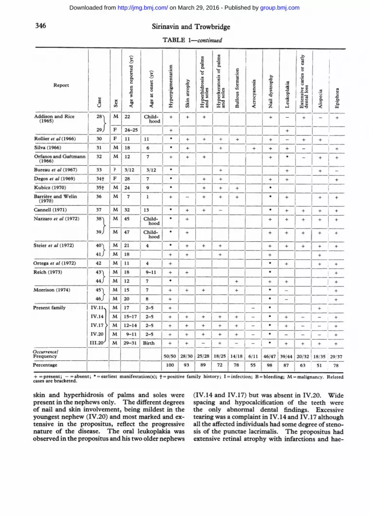
TABLE 1-continued
0CcC 0 ~~~~~~~~0 0~~~~~C) >~~~~~~~~~~~~~~~~~~~~~~~~~~~~~~~C0. 0. 0 CAReport 2 CCC) o .CCqoJ4..0~~~ ~ ~ ~ ~~~~ CC~~. CC U C'0
Cu >0u~0 .C) C)u C)C 0 0C~~co C0Caj e°0 ' u
C'->a a W - .
U o < Z 00 < 1
Addison and Rice 28l M 22 Child- ++
+ + - + - +
(1965) hood
29) F 24-25 + +__1=1= _1 __Rollier et al (1966) 30 F 11 11 * + + + + + +
Silva (1966) 31 M 18 6 * ++ + + +
Orfanos and Gaftmann 32 M 12 7 + + + + + +(1966)
Bureau et al (1967) 33 ? 3/12 3/12 *+ +
Degos et al (1969) 34t F 28 7 * + + + + +
Kubicz (1970) 35t M 24 9 + + + *
Barriere and Welin 36 M 7 1 + __ + + + * + + +(1970)
Cannell (1971) 37 M 32 13 + + + + + +
Nazzaro et al (1972) 38 M 45 Child- * + + + + + +hood
39) M 47 Child- * + + + + +hood
Steier et al (1972) 40 M 21 4 * + + + + + + + +
411 M 18 + + + + +
Ortega et al (1972) 42 M 11 4 + + + +
Reich (1973) 43l M 18 9-11 + + * +
44) M 12 7 *+ + + +
Morrison (1974) 45l M 15 7 + + + ± * +
46) M 20 8 + * _ +
Present family IV.11i M 17 2-5 + - * +
IV.141 M 15-17 2-5 + + + + + -_ * + _ +
IV.17 M 12-14 2-5 + + + + + +-* + _ - +
IV.20J M 9-11 2-5 + + + + + - * - _ +
III.20 M 29-31 Birth + + - + - - * + + + +
Occurrence/Frequency 50/50 28/30 25/28 18/25 14/18 6/11 46/47 39/44 20/32 18/35 29,137
Percentage 100 93 89 72 78 55 98 87 63 51 78
+ = present; -= absent; * = earliest manifestation(s); t = positive family history; I = infection; B = bleeding; M = malignancy. Relatedcases are bracketed.
skin and hyperhidrosis of palms and soles were (IV.14 and IV.17) but was absent in IV.20. Widepresent in the nephews only. The different degrees spacing and hypocalcification of the teeth wereof nail and skin involvement, being mildest in the the only abnormal dental findings. Excessiveyoungest nephew (IV.20) and most marked and ex- tearing was a complaint in IV.14 and IV.17 althoughtensive in the propositus, reflect the progressive all the affected individuals had some degree of steno-nature of the disease. The oral leukoplakia was sis of the punctae lacrimalis. The propositus hadobserved in the propositus and his two older nephews extensive retinal atrophy with infarctions and hae-
group.bmj.com on March 29, 2016 - Published by http://jmg.bmj.com/Downloaded from

Dyskeratosis congenita: clinical features and genetic aspects
0C
cl 00~~00.0
cis 0. 0I E 0l --- 0 0 ~~C ci 0~
- 1-+ja)+--0~ ~ ~ ~ ~ 5 0
133 01 42/0 24/46 014 05122
=~l
42 59 4 0 52 49 0644
l + + + +
_+ +
__=+_ I= + + I_ +_ __-+ +B
i_ i_ - - |_ I
I_ _ 1_- _~ ± + __ __ ._
+ -- - - - - I
- =+ + + + - - _13/31 10/17 14/29 8/20 24/46 21/43 4/25 12/27
42 159 L48 40 52 49 16 44
morrhages. All of them were mentally retardedand probably could be classified in the trainablegroup. The findings that were present only in thepropositus but not in his nephews were underweightand short stature; alopecia; sparse eyebrows, eye-lashes, and body hair; early dental loss; hypogonad-ism and aplastic anemia.
Review of the literatureFamily histories, clinical findings, and laboratory
data on 46 of the previously reported cases of dys-keratosis congenita are summarized in Table I to-gether with the findings in our four patients andIV.11 whose medical records and necropsy slideswere reviewed by us.Although approximately 60 patients were re-
ported or mentioned in the literature as havingdyskeratosis congenita, several cases are not in-cluded in Table I because of incomplete data.These are a brother of Basex and Dupre's case(1957) and of Kubicz's (1970); family members invarious studies (Schamberg, 1960; Degos et al,1969; Scoggins et al, 1971; Nazzaro et al, 1972); orbecause of incorrect diagnosis (Moon-Adams andSlatkin, 1955; Aplas, 1956). Two reports ondyskeratosis congenita could not be located (Jacobsand Tromovitch, 1962; Lobo et al, 1964). Onecase of dyskeratosis congenita reported as Fanconi'sanaemia (McDonald and Goldschmidt, 1960) isincluded in Table I.
Clinical features. The disease manifested it-self before puberty in all cases except one (case 24);it was noted at birth in four cases (10, 22, 26, 31).The earliest manifestations, in order of frequency,were skin hyperpigmentation, nail dystrophy,leukoplakia of the oral mucosa, epiphora, anaemia,and bullous eruptions of the skin. The first threefindings are characteristic of dyskeratosis congenitaand they eventually appeared in almost all the cases.Those who did not have this triad were either in ayoung age group or were incompletely described.
In addition to typical reticulated telangiectatichyperpigmentation (100%), the other skin changeswere atrophic and wrinkled skin over the dorsum ofhands, feet, genitalia, and extremities (93%); hyper-hidrosis of palms and soles (89%); and hyperkera-tosis of palms and soles (72%) causing cracking,fissure, and disappearance of dermal ridges. Thebullae often developed (78%) after even a slighttrauma. Acrocyanosis was noted in 55%/ of thepatients. Dystrophic change of nails (98%) was notof the same degree in all digits. The fingernailswere usually more severely affected than the toe-nails.
Leukoplakia (87%) could occur anywhere butleukoplakia of the oral mucosa was the most fre-quent and constant finding. Other sites of leuko-plakia were urethra (cases 3, 14), glans penis (case10), vagina (case 18), rectoanal region (cases 5, 10,14, 25).No good description of dental abnormalities was
available. This is probably because most of the
347
group.bmj.com on March 29, 2016 - Published by http://jmg.bmj.com/Downloaded from

Sirinavin and Trowbridge
patients had lost their teeth by the time they were
first seen. Early dental loss or extensive caries werereported in 63% of cases. Wide separation of teeth(cases 10, IV.14, IV.17, and IV.20), malformedteeth (cases 10, 11), crowded teeth (case 32), andhypocalcification (IV.14) may reflect abnormaldental development.
Fifty-one percent of the patients had thin, luster-less hair. The sparse body hair in some cases
could be related to hypogonadism. Eyebrows andeyelashes were sometimes absent.
Epiphora resulting from obliteration of punctaelacrimalis by epithelial hyperplasia was found in78%. Other ocular findings were conjunctivitis,blepharitis, ectropion, and loss of eyelashes. Opticpallor (case 40) and atherosclerotic change of retinalvessels (case 18) were found in one patient each.None of the previously reported cases had retino-pathy similar to our propositus.
Thirteen of 31 patients (42%) had subnormalintelligence. The level of mental development isdifficult to determine because IQ testing was notgenerally performed except in cases 5, 9, and 25whose IQs were 93, 85, and 79, respectively. TheIQs in our patients ranged from 55-65. It is ofnote that there seemed to be an intrafamilial correla-tion in the mental capability (families of Garb, 1958[cases 6 and 7], of Bryan and Nixon, 1965 [cases 24-27], of Nazzaro et al, 1972 [cases 38 and 39], and ofour family).
Dysphagia in dyskeratosis congenita (59%) was
ascribed to oesophageal diverticulum (cases 6, 21)and congenital oesophageal stenosis (cases 28, 9, 45)but in five cases the anatomic abnormality could notbe determined. One patient (not included in TableI) had suffered from dysphagia and subsequentlydeveloped carcinoma of the cervical oesophagus(Schamberg, 1960).
Forty-eight percent of the patients were of hypos-thenic build, only case 40 and our propositus(III.20) were also short. 'Sharp', 'pinched' facies,and large 'beaked' nose were described in fourpatients (cases 3, 10, 28, and 30) as well as in III.20.These findings could very well be secondary to de-creased subcutaneous fat.Although small testes and penis were mentioned
in eight patients, hypogonadism was documented inonly case 11. Cases 6 and 8 had a small sella turcicabut there was no other evidence indicating it to bepathological. Hypopituitarism and hypoadrenal-ism were mentioned in case 9. Two patients hadenlarged thyroid (cases 16, 20). III.20 had secon-
dary hypogonadism.Additional findings in dyskeratosis congenita were
abnormalities of the external ear (case 10) and mid-
dle ear (case 21), minor skeletal anomalies (cases 11,18, 19, 26), aseptic necrosis ofhips (cases 16, 26), de-formed joints (cases 10, 43), abnormal ECG (cases10, 15), and urethral stenosis (cases 10, 43).
Laboratory studies. The cytogenetic studiescarried out in nine of the reported cases (16, 21, 24,25, 26, 27, 28, 42, 44) and in our patients demon-strated no structural abnormality and no increasedbreaks or gaps. The studies in most cases were inblood lymphocytes, except case 16 and the fourpatients in this report in which bone marrow andskin fibroblasts were also studied.The chromosome studies of the peripheral lym-
phocytes in the family reported by Scoggins et al(1971) showed endoreduplications in two patients,one ofwhom also had chromatid breaks in two out of27 cells examined (personal communication). Theirkaryotypes were normal. Morrison (1974) found20%. of chromosomal breaks in one of his patients(case 45) and another patient (case 46) had 10%breakage.
Immunological investigations were reported inonly a few patients. Case 16, aged 17, and case 42,aged 10, were found on necropsy examinations tohave thymic aplasia and lymphocyte depleted spleenand lymph nodes. All the immunoglobulins wereabnormally low in case 42 (Ortega et al, 1972) as wasthe IgM in cases 40 and 41 (Steier et al, 1972). Thefamily studied by Scoggins et al (1971) was reportedto have normal immunoglobulin levels and skinhomograft rejection but absence of delayed hyper-sensitivity response to skin tests.
Aminoaciduria was reported in case 21 but wasabsent in our family.
Complications. The evidence from the litera-ture indicates that there is a definite increased inci-dence of solid malignant tumours in the patientswith dyskeratosis congenita. Leukaemia has notbeen reported in any of the cases.
Malignant changes usually developed in themucous membrane. The sites of the tumours inseven patients (5, 6, 10, 18, 29, 35, 37) were thetongue, buccal mucosa, nasopharynx, rectum, cer-vix, vagina, and the skin. A patient reported bySchamberg (1960) had carcinoma of the oesophagus.Interestingly, two of the patients had multipleprimary tumours (cases 5, 6).
In at least three of these cases (5, 6, 37) the carci-noma was found to originate in the pre-existingleukoplakia. The interval between the time whenoral leukoplakia was first noted and the malignantchange was approximately 27 years in case 5, 10years in case 6, and 16 years in case 37.
348
group.bmj.com on March 29, 2016 - Published by http://jmg.bmj.com/Downloaded from

Dyskeratosis congenita: clinical features and genetic aspects 349
TABLE IIDEVELOPMENT OF MALIGNANT TUMOURS IN DYSKERATOSIS CONGENITA
Case Age at Location TypeDetection (yr)l_l
5 (Cole et al, 1957) 47-50 Tongue Squamous cell carcinoma
Rectum AdenocarcinomaNasopharynx Squamous cell carcinoma
6 (Garb, 1958; case 2) 35 Buccal mucosa Epidermoid carcinoma (grade 3)
Rectum Mucinous carcinoma (poorly differentiated)
10 (Costello, 1967) 20 Dorsum of hand Squamous cell carcinoma
18 (Sorrow and Hitch, 1963) 31 Cervix and vagina Squamous cell carcinoma (moderately well differentiated)29 (Addison and Rice, 1965; case 2) 24 Buccal mucosa Squamous cell carcinoma (well differentiated)35 (Kubicz, 1970) 24 Coral mucosa Basal-squamous cell carcinoma
37 (Cannell, 1971) 33 Oral mucosa Anaplastic squamous cell carcinoma
(Not in Table I; Schamberg, 1960) Oesophagus (Squamous cell) carcinoma
The types and locations of the tumours are shownin Table II. The age in which the tumour wasfirst noted was usually between the third and thefifth decade.
Twenty-four patients (52%) developed anaemiaas the result of bone marrow failure. Nineteen ofthe patients also had leukopenia and/or thrombo-cytopenia. One patient (case 44) had only thrombo-cytopenia at the time of the report.
Infection and/or gastrointestinal bleeding led tothe fatal outcome in eight cases (5, 7, 11, 15, 16, 23,42, 43), usually in their second decade. Deaths inthe family reported here also occurred at an earlyage. II.13 and II.19 (not included in Table I)died from respiratory tract infections of unknownaetiology at which time II.13 was said to have'anaemia', and I.l19 'aplastic anaemia'.The immediate causes of death in IV. 11 and the
propositus were, however, not related to haemato-logical complications. IV. 1 died at age 17 fromPneumocystis carinii pneumonia at which time theperipheral blood counts showed no evidence of bonemarrow failure. The propositus, although he hadsuffered from aplastic anaemia for at least 2 yearswith occasional minor bleeding episodes, died fromcytomegaloviral infection at 31 years of age. (De-tailed pathological findings in these two patientswill be reported elsewhere.)
Mode of inheritance. Although dyskeratosiscongenita has been generally accepted to be here-ditary, the mode of inheritance was still consideredas 'unknown' or 'unclear' by several authors, evenin the more recent reports. There has in the pastbeen strong supportive evidence for an X-linked
recessive mode of inheritance; however, both auto-somal recessive and autosomal dominant trans-missions have also been suggested. We haveaccumulated the pedigree data from the publishedpapers to determine whether the segregation of thedisease supports the X-linked recessive mode ofinheritance, or if, in fact, there is genetic hetero-geneity.
Dyskeratosis congenita has been reported inseveral ethnic groups: Caucasian American, Ameri-can Negro, Jewish American, Brazilian, German,French, Dutch, Italian, Polish, Australian, Japanese,and African. Of 51 patients reported in the litera-ture, only five were females giving M: F sex ratio of10: 1; 22 cases came from 11 families and the restwere sporadic cases.The preponderance of affected males together
with the occurrence of the disease in eight sets ofmale sibs (Zinsser, 1910; Engman, 1926; Garb andRubin, 1944; Koszewski and Hubbard, 1956; Basexand Dupre, 1957; Kubicz, 1970; Nazzaro et al, 1972;Steier et al, 1972) suggest an X-linked recessivetrait. The supportive evidence for X-linkedrecessive transmission came from Bryan andNixon's family (1965), where the disease was trans-mitted through the females in three generations,with a total of five affected males. There was oneopportunity for male-to-male transmission whichdid not occur. The inheritance pattern in thefamily reported here also indicates dyskeratosiscongenita to be X-linked recessive trait. In threegenerations, nine affected males received the defec-tive gene through their mothers.
However, the relationship between this seeminglyX-linked recessive disease and the affected females
group.bmj.com on March 29, 2016 - Published by http://jmg.bmj.com/Downloaded from

Sirinavin and Trowbridge
TABLE IIISEGREGATION ANALYSIS OF 14 FAMILIES FROM THE LITERATURE
ExpectedObserved
Autosomal Recessive X-linked
1,262 4 2 2 0 4 0 4 6 1.5 4.5 2 1IL5 0 1 3 0 0 .67 4 2 2 3 2 1 1 0 1 4 1 3 3 1.513 3 2- .5 22 .
Case(s) 0 01641 3 2 1 1 2 0 24-1 0 2 119211.211 0 0 0 2 0.5.0 a 1
2021 1 1 1 0 1 ~~~~~~~~~~~~~~~~~~~~~01 2 0. 1. 1 0.
21 3 1 2 2 1 1 1 0 1 3 0.75 2.25 21~~~'0'22~~~~ ~~~~5 14.3 .5 37 .
1,2 6 2 4 2 2 0 4 0 4 6 1.5 4.5 2 16,7 4 2 2 3 2 1 1 0 1 4 1.7 3.2 3 1.4013 3 2 1 3 2 1 0 0 0 3 0.75 2.25 3 1.
225 3 0 3 1 54. 1.5 37 .
352 6 2 4 2 2 - 4 - 4 6 1.5 4.5 2 -3,7 3 1 2 2 1 1 1 0 1 3 0.7 2.2 2 1.4,13 3 2 1 2 2 0 1 0 1 3 0.75 2.25 2 1.
is not clear. Four of the five female patients weresporadic cases and one had an affected brother(Addison and Rice, 1965). One explanation maybe the Lyonization effect of the gene in the femaleheterozygotes. Steier et al (1972) reported 'milddyschromic cutaneous changes' in the mother andthe sister of his two male patients. But none of thefemales, including the obligate carriers, in Bryanand Nixon's family (1965) or in the family re-
ported here showed any sign of the disease.Another possible explanation might be that auto-
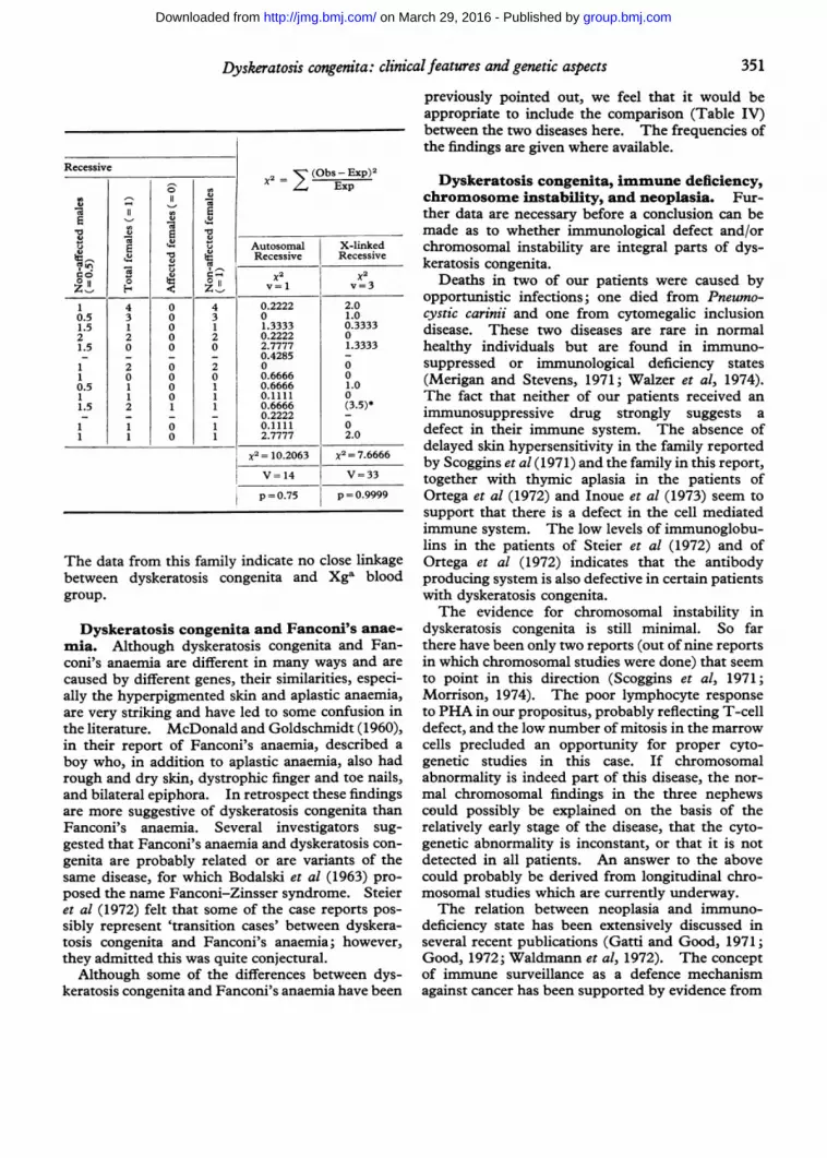
somal forms of dyskeratosis congenita also exist.One instance of parental consanguinity was reported(Costello and Buncke, 1956); there was one maleoffspring but the total number of sexes of the off-spring was not provided. Using the Chi-squaretest, pedigree analysis of 14 families (in which thedisease was present in only one sibship and thefamily history was considered adequate for analysispurpose) showed the probability to be 0.9999 forX-linked recessive and 0.75 for autosomal recessivetransmission (Table III).
In three families, the disease was said to be auto-somal dominant. One reported by Scoggins et al(1971) is a Negro family in which the propositus,his father, his three sisters, and his daughter haveevidence of dyskeratosis congenita (R. B. Scoggins,
personal communication). Degos et al (1969) re-
ported an affected woman whose sister, father, uncle,andpaternalgrandmotherwereprobablyaffectedalso.Nazzaro et al (1972) in their report of two brothersmentioned that there were five other affected mem-bers, two ofwhom were females. The authors sug-gested that the pattern fits incomplete dominanttransmission. The exact relationship ofthe affectedfamily members was not reported.
Linkage study in the present family. Thered blood cell G6PD electrophoresis was done onall family members seen by us and colour vision testwas performed with HRR pseudochromatic plate inthe male members. The G6PD electrophoresisshowed all to be type A. The colour vision testwas normal in all males except the proposituswhose colour vision defect may be secondary to thesevere retinal pathology rather than hereditary X-linked colour blindness. Therefore these twoX-linked markers are not informative.The Xga blood group is, however, informative in
this family (see Fig. 1). HII.11 is an Xga hetero-zygote. The Xga blood types of III.15 and III.17indirectly indicate that II.1 was also heterozygous.Xga type could not be determined in the probandbecause he also had a positive direct Coombs test.
350
group.bmj.com on March 29, 2016 - Published by http://jmg.bmj.com/Downloaded from
Dyskeratosis congenita: clinical features and genetic aspects
Recessive 2 2 (Obs-Exp)2X Exp
E E t u Autosomal X-linled
oWtiRecessive Recessive
Z, E4 Z_ v1 v=3
1 4 0 4 0.2222 2.00.5 3 0 3 0 1.01.5 1 0 1 1.3333 0.33332 2 0 2 0.2222 01.5 0 0 0 2.7777 1.3333- - - - 0.4285 -
1 2 0 2 0 01 0 0 0 0.6666 00.5 1 0 1 0.6666 1.01 1 0 1 0.1111 01.5 2 1 1 0.6666 (3.5)*_ - - - 0.2222 _
1 1 0 1 0.1111 01 1 0 1 2.7777 2.0
X= 10.2063 x2 = 7.6666
V=14 V=33
p = 0.75 p = 0.9999
The data from this family indicate no close linkagebetween dyskeratosis congenita and Xga bloodgroup.
Dyskeratosis congenita and Fanconi's anae-
mia. Although dyskeratosis congenita and Fan-coni's anaemia are different in many ways and arecaused by different genes, their similarities, especi-ally the hyperpigmented skin and aplastic anaemia,are very striking and have led to some confusion inthe literature. McDonald and Goldschmidt (1960),in their report of Fanconi's anaemia, described a
boy who, in addition to aplastic anaemia, also hadrough and dry skin, dystrophic finger and toe nails,and bilateral epiphora. In retrospect these findingsare more suggestive of dyskeratosis congenita thanFanconi's anaemia. Several investigators sug-gested that Fanconi's anaemia and dyskeratosis con-
genita are probably related or are variants of thesame disease, for which Bodalski et al (1963) pro-posed the name Fanconi-Zinsser syndrome. Steieret al (1972) felt that some of the case reports pos-sibly represent 'transition cases' between dyskera-tosis congenita and Fanconi's anaemia; however,they admitted this was quite conjectural.
Although some of the differences between dys-keratosis congenita and Fanconi's anaemia have been
previously pointed out, we feel that it would beappropriate to include the comparison (Table IV)between the two diseases here. The frequencies ofthe findings are given where available.
Dyskeratosis congenita, immune deficiency,chromosome instability, and neoplasia. Fur-ther data are necessary before a conclusion can bemade as to whether immunological defect and/orchromosomal instability are integral parts of dys-keratosis congenita.
Deaths in two of our patients were caused byopportunistic infections; one died from Pneumo-cystic carinii and one from cytomegalic inclusiondisease. These two diseases are rare in normalhealthy individuals but are found in immuno-suppressed or immunological deficiency states(Merigan and Stevens, 1971; Walzer et al, 1974).The fact that neither of our patients received animmunosuppressive drug strongly suggests adefect in their immune system. The absence ofdelayed skin hypersensitivity in the family reportedby Scoggins et al (1971) and the family in this report,together with thymic aplasia in the patients ofOrtega et al (1972) and Inoue et al (1973) seem tosupport that there is a defect in the cell mediatedimmune system. The low levels of immunoglobu-lins in the patients of Steier et al (1972) and ofOrtega et al (1972) indicates that the antibodyproducing system is also defective in certain patientswith dyskeratosis congenita.The evidence for chromosomal instability in
dyskeratosis congenita is still minimal. So farthere have been only two reports (out of nine reportsin which chromosomal studies were done) that seemto point in this direction (Scoggins et al, 1971;Morrison, 1974). The poor lymphocyte responseto PHA in our propositus, probably reflecting T-celldefect, and the low number of mitosis in the marrowcells precluded an opportunity for proper cyto-genetic studies in this case. If chromosomalabnormality is indeed part of this disease, the nor-mal chromosomal findings in the three nephewscould possibly be explained on the basis of therelatively early stage of the disease, that the cyto-genetic abnormality is inconstant, or that it is notdetected in all patients. An answer to the abovecould probably be derived from longitudinal chro-mosomal studies which are currently underway.The relation between neoplasia and immuno-
deficiency state has been extensively discussed inseveral recent publications (Gatti and Good, 1971;Good, 1972; Waldmann et al, 1972). The conceptof immune surveillance as a defence mechanismagainst cancer has been supported by evidence from
351
group.bmj.com on March 29, 2016 - Published by http://jmg.bmj.com/Downloaded from

Sirinavin and Trowbridge
TABLE IVCOMPARISON BETWEEN DYSKERATOSIS CONGENITA AND FANCONI'S ANAEMIA
Dyskeratosis Congenita Fanconi's Anaemia*
Mode of inheritance Usually X-linked recessive; can be Autosomal recessiveautosomal recessive or dominant
M:F sex incidence 10:1 2:1
Birth weight Normal in most cases Low (56% weighed less than 2.5 kg)
Microcephaly Absent Present (40%)Eye abnormalities Epiphora and/or stenosis of punctae Strabismus (22%,'0), microphthalmia (16%)
lacrinaalis (78%)Leukoplakia Present (87%) Absent
Alopecia Present (51%) Absent
Hyperpigmentation Present (100%) Present (77%)
Nail dystrophy Present (98%) Absent
Skeletal malformations J_Rare Present (66%), especially of the upper limbs
Internal abnormalities Oesophageal diverticulum or stricture; Renal anomalies (28%)dysphagia (59%)
Stature Hyposthenic build (54%) Short (60%), usually below 3rd centile
Mental deficiency Present (42%) Present (17%)
Hypoplasia of the bone Present (52%) Present (100%0)marrow
Malignancy Solid tumours (17%); no leukaemias Solid tumours reported in at least 7 cases;reported (Bernstein et al, 1971; Swift et al, 1971)
leukaemia reported in at least 8 cases (Dosik et at,1970)
Chromosomal breakage and Probably absent Present in lymphocytes, bone marrow, andrearrangement fibroblasts (Dosik et al, 1970; German, 1971)
Immunological defect Present Absent
* Reference: Gmyrek and Syllm-Rapoport (1964) unless otherwise indicated.
both clinical and laboratory studies. Several gene-tic disorders are known to be associated withimmunodeficiency and an increased incidence ofmalignancy. These include ataxia-telangiectasia,Wiscott-Aldrich syndrome, Bruton agammaglobu-linemia (Waldmann et al, 1972), and Bloom's syn-drome (German, 1969).
Cytogenetic abnormalities have been observed inseveral human neoplasias (Sandberg and Hossfeld,1970). In addition, chromosomal instability hasbeen demonstrated in somatic cells of patients withBloom's syndrome, Fanconi's anaemia, ataxia-telangiectasia, and xeroderma pigmentosum whichare genetic diseases with a predisposition to cancer(German, 1971). The significance of chromosomalchanges in cancer is still not known. German(1971) was of an opinion that a clone of cells, de-rived from a single progenitor cell containing markerchromosome(s), once having undergone a certainmutation becomes susceptible to conversion tocancer by some oncogenic factors.The tendency to develop malignant tumours in
the patients with dyskeratosis congenita could verywell result from immunological defect. A pre-disposition to chromosomal abnormality, if sub-stantiated, could also play a role in the developmentof cancer.
Primary defect in dyskeratosis congenita.The interrelation between the multiple system ab-normalities in dyskeratosis congenita is yet to bedetermined. There are two possibilities regardingthe relation between aplastic anaemia and im-mune deficiency in this disease. Since the lym-phocyte precursors arise from primitive pluri-potential cells located in the bone marrow(McGregor, 1968), a disorder of stem cells can,therefore, give rise to the abnormalities of both Tand B lymphocytes as well as to the three cell linesof the bone marrow. Another possible explanationis that the abnormality in the mesoderm, as evidentin the bone marrow, may lead to the abnormal de-velopment of the thymus gland. It has beenshown that epitheliomesenchymal interaction is
352
group.bmj.com on March 29, 2016 - Published by http://jmg.bmj.com/Downloaded from
Dyskeratosis congenita: clinical features and genetic aspects
necessary in the development of the thymus in vitro(Auerbach, 1960), and Peterson et al (1964) sug-gested that the abnormality of the mesoderm willexplain the thymus disorder in ataxia telangiectasia.These reasonings, however, do not explain cutaneousmanifestations and mental deficiency which are alsomanifestations of dyskeratosis congenita.
It seems that the basic defect in dyskeratosiscongenita may be at the level of cell division. Theabnormality in one of the enzymatic steps essentialto normal cell division, for example DNA poly-merase II whose activity correlates positively withthe rate of tissue regeneration (Baril et al, 1973), canlead to hypoproliferation or impaired regenerationof the skin, nails, bone marrow, and neurones indyskeratosis congenita. An example analogous tothis may be the defect in deoxyribonuclease whichis essential for DNA repair in xeroderma pigmen-tosum (Robbins et al, 1974). Tritiated thymidineincorporation by the lymphocytes and skin fibro-blasts may be one way to study the DNA synthesisin this disorder. If this is shown to be abnormal, itwill be interesting to look further for any abnormalityat the different steps of cell division.
We are grateful to Dr J. Vidgoff for the translation ofseveral articles, Mrs P. Evans for typing the manuscript,Drs G. A. Brooksby and R. G. Weleber for ophthalmo-logical examinations, Dr W. E. Gibson for dental evalu-ations, and Dr D. Linder for reviewing the pathologyslides. We thank Dr B. K. McCaw and Miss C. L.Olson for cytogenetic studies, Miss S. I. Rowe and MissS. M. Hazard for genetic marker studies, and Dr R.Vlietinck for helping with statistical analysis. We wishto thank also Drs E. W. Lovrien, R. D. Koler, and F.Hecht for their advice, Dr F. J. Storrs for reviewing themanuscript, and Drs R. L. Dimond and S. A. Ebert forreferring the propositus to us.
REFERENCESAddison, M. and Rice, M. S. (1965). The association of dyskera-
tosis congenita and Fanconi's anaemia. Medical Journal ofAustralia, 1, 797-799.
Aplas, V. (1956). Zur Kenntnis der Poikilodermie, Parapsoriasisund atrophia cutis reticularis cum pigmentatione, dystrophiaunguim et leukoplakia oris: Zinsser-'Dyskeratosis congenita'.Archivfur Klinische und Experimentelle Dermatologie, 202,224-237.
Auerbach, R. (1960). Morphogenetic interactions in the develop-ment of mouse thymus gland. Developmental Biology, 2, 271-284.
Baril, E. F., Jenkins, M. D., Brown, 0. E., Laszlo, J., and Morris,H. P. (1973). DNA polymerases I and II in regenerating ratliver and Morris hepatomas. Cancer Research, 33, 1187-1193.
Barriere, H. and Welin, J. (1970). Dyskeratose congenitale avecthrombopenie. Ses relations avec l'anemie de Fanconi. Bulletinde la Societe Franfaise de Dermatologie et de Syphiligraphie, 77,864-868.
Basex, A. and Dupre, A. (1957). Dyskeratosis congenitale (typeZinsser-Cole-Engman) associee a une myelopathie constitution-elle (purpura thrombopenique et neutropenie). Annales deDermatologie et Syphiligraphie, 84, 497-513.
Bernstein, M. S., Hunter, R. L., and Yachnin, S. (1971). Hepatomaand peliosis hepatis developing in a patient with Fanconi's anemia.New England Journal of Medicine, 284, 1135-1136.
Bodalski, J., Defecinska, E., Judiewicz, L., and Pacanowska, M.(1963). Fanconi's anaemia and dyskeratosis congenita as a syn-drome. Dermatologica, 127, 330-342.
Bryan, H. G. and Nixon, R. K. (1965). Dyskeratosis congenita andfamilial pancytopenia. Journal of the American Medical Associ-ation, 192,203-208.
Bureau, Y., Barriere, H., Litoux, P., and Bureau, B. (1967). Dys-keratose congenitale de Zinsser-Cole-Engman. Bulletin de laSocietc Francaise de Dermatologie et de Syphiligraphie, 74, 649.
Calmettes, L., Deodati, F., and Daraux, H. (1957). Dyskeratosecongenitale avec atresie des points lacrymaux (maladie de Zinsser,Engman et Cole). Archives d'Ophthalmologie, 17, 250-255.
Cannell, H. (1971). Dyskeratosis congenita. British Journal ofOral Surgery, 9, 8-10.
Clarkson, P. (1962). Poland's syndactyly. Guy's Hospital Reports,111, 335-346.
Cole, H. N., Cole, H. N., Jr., and Lascheid, W. P. (1957). Dyskera-tosis congenita. Archives ofDermatology, 76,712-719.
Cole, H. N., Rauschkolb, J. E., and Toomey, J. (1930). Dyskera-tosis congenita with pigmentation, dystrophia unguis and leuko-keratosis oris. Archives of Dermatology and Syphilology, 21, 71-95.
Cole, H. N., Rauschkolb, J., and Toomey, J. (1955). Dyskeratosiscongenita with pigmentation, dystrophia unguim and leukokera-tosis oris. Archives of Dermatology, 71,451-456.
Costello, M. J. (1957). Dyskeratosis congenita with superimposedprickle-cell epithelioma on the dorsal aspect of the left hand.Archives of Dermatology, 75, 451.
Costello, M. J. and Buncke, C. M. (1956). Dyskeratosis congenita.Archives of Dermatology, 73, 123-132.
Degos, R., Bernard, J., Belaich, S., Flandrin, G., and Varet, B.(1969). Syndrome de Zinsser-Cole-Engman (Zinsser-Fanconi).Bulletin de la Socidte Franfaise de Dermatologie et de Syphili-graphie, 76, 15-17.
Dosik, H., Hsu, L. Y., Todaro, G. J., Lee, S. L., Hirschhorn, K.,Selirio, E. S., and Alter, A. A. (1970). Leukemia in Fanconi'sanemia: cytogenetic and tumor virus susceptibility studies.Blood, 36, 341-352.
Engman, M. F. (1926). A unique case of reticular pigmentation ofthe skin with atrophy. Archives of Dermatology and Syphilology,13,685-687.
Engman, M. F., Jr. (1935). Congenital atrophy of the skin, withreticular pigmentation. Report of two cases. journal of theAmerican Medical Association, 105, 1252-1256.
Garb, J. (1947). Dyskeratosis congenita with pigmentation, dys-trophia unguium and leukoplakia oris. Archives of Dermatologyand Syphilology, 55, 242-250.
Garb, J. (1958). Dyskeratosis congenita with pigmentation, dys-trophia unguim, and leukoplakia oris. A follow-up report of twobrothers. Archives ofDermatology, 77, 704-712.
Garb, J. and Rubin, G. (1944). Dyskeratosis congenita with pig-mentation, dystrophia unguim and leukoplakia oris. Archives ofDermatology and Syphilology, 50, 191-198.
Gatti, R. A. and Good, R. A. (1971). Occurrence of malignancy inimmunodeficiency diseases. A literature review. Cancer, 28, 89-98.
Georgouras, K. (1965). Dyskeratosis congenita. Australian jour-nal of Dermatology, 8, 36-43.
German, J. (1969). Bloom's syndrome. I. Genetical and clinicalobservations in the first twenty-seven patients. American JournalofHuman Genetics, 21, 196-227.
German, J. (1972). Genes with increased chromosomal instabilityin somatic cells and predispose to cancer. In Progress in MedicalGenetics, ch. 2, p. 61, Ed. by A. G. Steinberg and A. G. Vearn.Grune and Stratton, New York and London.
Good, R. A. (1972). Relations between immunity and malignancy.Proceedings of the National Academy of Sciences, 69, 1026-1042.
Grekin, J. N. and Schwartz, 0. D. (1962). Dyskeratosis congenitawith pigmentation, dystrophia unguium and leukokeratosis oris.Archives of Dermatology, 85, 124-125.
Inoue, S., Mekanik, G., Mahallati, M., and Zuelzer, W. (1973).Dyskeralogis congenita with pancytopenia. American journal ofDiseases of Children, 126,389-396.
Jacobs, P. H. and Tromovitch, A. (1962). Patient presentations.In Proceedings of the XII International Congress of Dermatology,vol. 2, p. 1478, ed. by D. M. Pillsbury and C. S. Livingood.Excerpta Medica, Amsterdam.
Jansen, L. H. (1951). The so-called 'dyskeratosis congenita'.Dermatologica, 103, 167-177.
353
group.bmj.com on March 29, 2016 - Published by http://jmg.bmj.com/Downloaded from

Sirinavin and Trowbridge
Kitamura, K. and Hirako, T. (1955). tYber zwei japanische Filleeiner eigenartigen retikularen Pigmentierung. Dermatologica,110,97-107.
Koszewski, B. J. and Hubbard, T. F. (1956). Congenital anemia inhereditary ectodermal dysplasia. Archives of Dermatology, 74,159-166.
Kubicz, J. (1970). Syndroma Zinsser-Engman-Cole cum dys-chromia extensiva corporis. Przeglad Dermatologiczny, 57, 239-242.
Lobo, J. (1964). Doenca de Zinsser et Engman-Cole. AnaisBrasileiros de Dermatologia, 39, (3), 33.
McDonald, R. and Goldschmidt, B. (1960). Pancytopenia withcongenital defects (Fanconi's anaemia). Archives of Disease inChildhood, 35, 367-372.
McGregor, D. D. (1968). Bone marrow origin of immunologicallycompetent lymphocytes in the rats. J'ournal of ExperimentalMedicine, 127,953-966.
Merigan, T. C. and Stevens, D. A. (1971). Viral infections in manassociated with acquired immunological deficiency states. Federa-ation Proceedings, 30, 1858-1864.
Milgrom, H., Stoll, H. L., Jr., and Crissey, J. T. (1964). Dyskera-tosis congenita: a case with new features. Archives of Dermato-logy, 89, 345-349.
Moon-Adams, D. and Slatkin, M. H. (1955). Familial pigmentationwith dystrophy of the nails. Archives of Dermatology, 71, 591-598.
Morrison, J. G. L. (1974). Dyskeratosis congenita: two extremes.South African MedicalJournal, 48,223-225.
Nazzaro, P., Argentieri, R., Bassetti, F., Leonetti, F., Topi, G., andValenzano, L. (1972). Dyskeratose congenitale de Zinsser-Cole-Engmann. Bulletin de la Sociltl Franvaise de Dermatologie et deSyphiligraphie, 79, 242-244.
Orfanos, C. and Gaftmann, H. (1966). Leukoplakien, Pigment-verschiebungen und Nageldystrophie: Zinsser-Cole-Engman-Syndrom-sog. Dyskeratosis congenita. Medizinische Welt, 2,2589-2594.
Ortega, J. A., Swanson, V. L., Landing, B. H., and Mammond, G. D.(1972). Congenital dyskeratosis. Zinsser-Engman-Cole syn-drome with thymic dysplasia and aplastic anemia. AmericanJournal of Diseases of Children, 124, 701-704.
Pastinszky, I., Vankos, J., and Racz, I. (1957). Ein Beitrag zurPathologie der 'Dyskeratosis congenital Cole-Rauschkolb-Toomey. Dermatologische Wochenschrift, 135,587-593.
Peterson, R. D. A., Kelly, W. D., and Good, R. A. (1964). Ataxia-telangiectasia. Its association with a defective thymus, immuno-logical-deficiency disease, and malignancy. Lancet, 1, 1189-1193.
Reich, H. (1973). Zinsser-Cole-Engman Syndrom. MedizinischeKlinik, 68, 283-292.
Robbins, J. H., Kraemer, K. H., Lutzner, M. A., Festoff, B. W., andCoon, H. G. (1974). Xeroderma pigmentosum. An inheriteddisease with sun sensitivity, multiple cutaneous neoplasms, andabnormal DNA repair. Annals of Internal Medicine, 80, 221-248.
Rollier, M. R., Rollier, M., and Prost, A. (1966). Sur un cas desyndrome de Zinsser-Engman-Cole. Bulletin de la SocieteFran;aise de Dermnatologie et de Syphiligraphie, 73,383-386.
Sandberg, A. A. and Hossfeld, D. K. (1970). Chromosomal ab-normalities in human neoplasia. Annual Review of Medicine, 21,379.
Sato, S. I. and Hannibal, J. E., Jr. (1962). Ectodermal defect:dyskeratosis congenita with pigmentation, dystrophia unguim andleukokeratosis oris. Archives ofDermatology, 86, 114-115.
Schamberg, I. L. (1960). Dyskeratosis congenita withpigmentation,dystrophia unguis and leukokeratosis oris. Archives of Dermato-logy, 81, 266.
Scoggins, R. B., Prescott, K. J., Asher, G. H., Blaylock, W. K., andBright, R. W. (1971). Dyskeratosis congenita with Fanconi-typeanemia: investigations of immunologic and other defects. ClinicalResearch, 19,409.
Silva, J. R. E. (1966). Syndrome de Zinsser-Fanconi. Annales deDermatologie et Syphiligraphie, 93, 497-502.
Sorrow, J. M., Jr. and Hitch, J. M. (1963). Dyskeratosis congenita:first report of its occurrence in a female and a review of the litera-ture. Archives ofDermatology, 88,340-347.
Steier, W., Van Voolen, G. A., and Selmanowitz, V. J. (1972). Dys-keratosis congenita: relationship to Fanconi's anemia. Blood, 39,510-521.
Swift, M., Zimmerman, D., and McDonough, E. R. (1971). Squa-mous cell carcinomas in Fanconi's anemia. journal of the Ameri-can Medical Association, 216,325-326.
Waldmann, T. A., Strober, W., and Blasse, R. M. (1972). Immuno-deficiency disease and malignancy. Various immunologic de-ficiencies of man and the role of immune processes in the controlof malignant disease. Annals of Internal Medicine, 77, 605-628.
Walzer, P. D., Perl, D. P., Krogstad, D. J., Rawson, P. G., andSchultz, M. G. (1974). Pneumocystis carinii pneumonia in theUnited States. Epidemiologic, diagnostic, and clinical features.Annals of Internal Medicine, 80, 83-93.
Wise, F. (1943). Dyskeratosis congenita with pigmentation, dys-trophia unguis and leukokeratosis oris. Archives of Dermatologyand Syphilology, 48, 560.
Zinsser, F. (1910). Atrophia cutis reticularis cum pigmentatione,dystrophia unguim et leukoplakia oris. Ikonographia Dermato-logica (Hyoto), 5, 219-223.
NoticeSymposium on the Pathology of Pregnancy
The Royal College of Pathologists are holding a symposium on the pathology of pregnancyon 12 and 13 February 1976, at the Royal College of Physicians. The following subjectswill be covered in the two-day meeting. Fetoplacental function: its nature and assessment;haematological problems; hypertension and renal disease; infections; trophoblastic tumours;and congenital abnormalities.The symposium is open to workers in all disciplines connected with the subject. The
registration fee is £12 (sterling), which includes coffee, lunch, and tea on the two days and a
copy of the papers to be published as a special supplement to the Journal of Clinical Pathology.Those wishing to attend the symposium dinner on Thursday evening, 12 February, shouldsend with their application form the additional fee of £8 (sterling) which includes drinksbefore and with the dinner. The final programme will be sent out in February 1976.Application forms can be obtained from The Royal College of Pathologists, 2 Carlton HouseTerrace, London SW1Y 5AF.
354
group.bmj.com on March 29, 2016 - Published by http://jmg.bmj.com/Downloaded from

review of the literature.aspects. Report of a family andclinical features and genetic Dyskeratosis congenita:
C Sirinavin and A A Trowbridge
doi: 10.1136/jmg.12.4.3391975 12: 339-354 J Med Genet
http://jmg.bmj.com/content/12/4/339Updated information and services can be found at:
These include:
serviceEmail alerting
corner of the online article. this article. Sign up in the box at the top right Receive free email alerts when new articles cite
Notes
http://group.bmj.com/group/rights-licensing/permissionsTo request permissions go to:
http://journals.bmj.com/cgi/reprintformTo order reprints go to:
http://group.bmj.com/subscribe/To subscribe to BMJ go to:
group.bmj.com on March 29, 2016 - Published by http://jmg.bmj.com/Downloaded from