Embed Size (px)
Citation preview
Effect of Prenatal Glucocorticoids on Cerebral Vasculature ofthe Developing Brain
Govindaiah Vinukonda, PhD1, Krishna Dummula, MD1, Sabrina Malik, MD1, Furong Hu,BA1, Carl I. Thompson, PhD2, Anna Csiszar, MD, PhD3, Zoltan Ungvari, MD, PhD3, andPraveen Ballabh, MD1,21 Department of Pediatrics, New York Medical College-Westchester Medical Center, Valhalla, NY2 Department of Physiology, New York Medical College-Westchester Medical Center, Valhalla,NY3 Department of Anatomy & Cell Biology, New York Medical College-Westchester Medical Center,Valhalla, NY and Reynolds Oklahoma Center on Aging, Department of Geriatric Medicine,University of Oklahoma Health Science Center, Oklahoma City, Okla
AbstractBackground and Purpose—Prenatal glucocorticoids prevent germinal matrix hemorrhage inpremature infants. The underlying mechanism, however, is elusive. Germinal matrix (GM) isenriched with angiogenic vessels exhibiting paucity of pericytes and GFAP-positive astrocyeendfeet. Therefore, we asked whether glucocorticoid treatment would suppress angiogenesis, andenhance periendothelial coverage by pericytes and GFAP-positive endfeet in the GMmicrovasculature.
Methods—We treated pregnant rabbits with intramuscular betamethasone and delivered pupsprematurely by C-section at E29 (Term=32d). Endothelial turnover, vascular density, pericytecoverage, GFAP-positive endfeet, cell-death and growth factors orchestrating angiogenesis,including vascular endothelial growth factor (VEGF), angiopoietins, transforming growth factor-β(TGF-β), platelet derived growth factor-B(PDGF-B), were compared between betamethasone-treated and untreated pups. Similar comparisons were done between autopsy-materials frompremature infants exposed and unexposed to prenatal glucocorticoids.
Results—Antenatal glucocorticoid treatment reduced endothelial proliferation, vascular densityand VEGF expression in the GM of both rabbits and humans. The pericyte coverage was greater inglucocorticoid-treated rabbit pups and human infants than in controls, but not the GFAP(+)endfeet coverage. TGF-β, but not angiopoietins and PDGF-B, were elevated in glucocorticoid-treated rabbit pups compared to controls. Betamethasone treatment induced apoptosis, neuronaldegeneration and gliosis in rabbits pups. However, there was no evidence of increased cell-deathin glucocorticoid-exposed human infants.
Conclusions—Prenatal glucocortiocoid suppresses VEGF and elevates TGF-β levels, whichresults in angiogenic inhibition, trimming of neovasculature and enhanced pericyte coverage.These changes contribute to stabilizing the GM vasculature, thereby reducing its propensity tohemorrhage. Prenatal glucocorticoid exposure does not induce neural cell-death in humans, unlikerabbits.
Address for Correspondence: Praveen Ballabh, MD, Regional Neonatal Center, 2nd floor, Maria Fareri Children’s Hospital atWestchester Medical Center. Valhalla, NY 10595, Phone: 914-493-8558 Fax: 914-493-1005 [email protected] of interest: None
NIH Public AccessAuthor ManuscriptStroke. Author manuscript; available in PMC 2011 August 1.
Published in final edited form as:Stroke. 2010 August ; 41(8): 1766–1773. doi:10.1161/STROKEAHA.110.588400.
NIH
-PA Author Manuscript
NIH
-PA Author Manuscript
NIH
-PA Author Manuscript
KeywordsGerminal matrix hemorrhage-intraventricular hemorrhage; glucocorticoids; betamethasone;pericyte; germinal matrix; vasculature; VEGF; TGF-β
IntroductionPrenatal glucocorticoids (GCs) prevent respiratory distress syndrome and intraventricularhemorrhage (IVH) in preterm infants.1, 2 Indeed, NIH Consensus Development Panel on the“Effect of corticosteroids for fetal maturation on perinatal outcomes” has recommended useof prenatal GC in preterm labor.3 In the USA, the preterm birth rate is 12.5%, and 75% ofwomen in preterm labor with gestational age of 34 weeks or less are treated with GC.4 As~13 million babies are born premature worldwide every year, a huge number of preterminfants (about 5–6 millions) are exposed to prenatal steroid. This increases their survival andreduces both the incidence and severity of IVH.5,6 Yet, the molecular mechanism by whichGCs prevent IVH is elusive. Therefore, we asked how prenatal GC would reduce theincidence of IVH.
IVH typically initiates in the germinal matrix (GM). This periventricular region, located onthe head of caudate nucleus and underneath ventricular ependyma, is a richly vascularizedcollection of neural precursor cells and is selectively vulnerable to hemorrhage. IVH isattributed to intrinsic fragility of the GM vasculature and disturbance in cerebral blood flow.Our previous work has shown that a rapid angiogenesis in the GM, induced by high vascularendothelial growth factor (VEGF) and angiopoietin (ANGPT)-2 levels, contributes toincreased vascular fragility and vulnerability to hemorrhage, and that angiogenic inhibitionreduces the occurrence of IVH in rabbit pup model.7 Furthermore, angiogenic vessels of theGM exhibit paucity of pericytes, deficiency of fibronectin in the basal lamina, and reducedperivascular coverage by GFAP(+) astrocyte endfeet.8–10 Hence, the fragility of GMmicrovasculature is attributed to immature basal lamina and reduced perivascular coverageby pericytes as well as GPAP(+) endfeet.
Prenatal GC--betamethasone and its stereo-isomer, dexamethasone--are used in pretermlabor. They exhibit a wide range of pharmacological effects and toxicities on the brain ofpremature infants.11–13 However, little is known about the effects of GCs on themorphology and molecular components of the developing cerebral vasculature. The GCdownregulates VEGF in an in vitro model of the blood brain barrier and cultured cells ofvarious origin,14,15 and accordingly, the GC treatment effectively suppresses angiogenesisin various disease models.16, 17 The blockade of VEGF signaling prunes the nascent,immature and pericyte-deficient microvasculature of tumors.18 In addition, this remodels theremaining vasculature which results in less dilated blood vessels exhibiting enhancedpericyte coverage.18 Other than VEGF, growth factors angiopioetin-1, PDGF-B and TGF-βplay key role in maturation of the vasculature, particularly in the assembly of pericytesaround the immature blood vessels.19 Therefore, we hypothesized that prenatal GC wouldsuppress angiogenesis by downregulation of angiogenic growth factors including VEGF andANGPT-2, and enhance pericyte recruitment by inducing distinctive changes in theregulating growth factors--angiopioetin-1, PDGF-B and TGF-β.
There is increasing evidence that the GC treatment affect the phenotype and function ofastrocytes. For example, the dexamethasone treatment in the astrocytes cultures andtriamcinolone intravitreal injection in mice model of laser retinal photocoagulation enhanceGFAP levels in the astrocytes.20,21 Importantly, high dose of dexamethasone andmethylprednisolone induces apoptotic cell death in rats raising safety concerns with prenatal
Vinukonda et al. Page 2
Stroke. Author manuscript; available in PMC 2011 August 1.
NIH
-PA Author Manuscript
NIH
-PA Author Manuscript
NIH
-PA Author Manuscript
GC treatment.22 Thus, we postulated that prenatal GC treatment might mature the cerebralvasculature by increasing GFAP+ perivascular endfeet, but might cause undesirable adverseeffects---neural cell death and gliosis.
Material and MethodsAnimal experiment
Animal protocol was approved by Institutional Animal Care and Use Committee of NewYork Medical College, Valhalla, NY. We obtained 8 timed pregnant New Zealand rabbitsfrom Charles River Laboratories (Wilmington, MA, USA). The rabbits were sequentiallyassigned to receive either intramuscular betamethasone (n=4) or saline (n=4). The dose ofbetamethasone in pregnant women is 12.5 mg once daily for 2 days; and average weight ofpregnant women is about 60 kg.23 On this basis, we calculated a dose of 0.2 mg/kg(12.5/60= 0.2) daily for 2 days in pregnant rabbits. Thus, betamethasone (celestone;Schering Corporation, Kenilworth, NJ) was administered 0.2 mg/kg/dose every 24 hours ongestational day 27 and 28 for a total of 2 doses.
C-section was performed at day 29 of gestational age to deliver rabbit pups prematurely(term=32days). Pups were dried immediately and were kept warm in an infant incubator at35°C. After stabilization of their conditions, they were weighed and fed with puppy formula(Esbilac, Petag, Hampshire, IL, USA). Pups were sacrificed at 3 epochs--2, 6 and 48 h ofage. Brain was then dissected and cut into 2 mm coronal slices on brain matrix. All thehistological evaluations were done from coronal sections taken at the level of midseptalnucleus. The comparison groups were balanced with respect to the body weight and genderof rabbit pups.
Laser capture microdissection (LCM)—LCM is described in Online SupplementalMethods.
Human tissue collection and processing: The Institutional Review Board of New YorkMedical College approved the use of human autopsy materials for this study. Women inpreterm labor receive either betamethasone (12.5 mg once daily for 2 days) ordexamethasone (6 mg twice daily for 2 days) to prevent respiratory distress syndrome inpremature infants. The preterm infants included in the present study delivered within fewhours to 3 days after completion of GC treatment to their mothers; and infants died at 6–72hpostnatal age (Supplementary table). The wall of cerebral hemisphere in fetuses consists ofventricular zone, subventricular zone, intermediate zone, cortical plate and marginal zone, asdescribed by the Boulder Committee.24 In this study, we described intermediate zoneembryonic white matter synonymously with white matter and cortex for cortical plate for thesake of simplicity of presentation. Brain samples were processed as described.8 About 2–3mm thick coronal slices were taken at the level of thalamostriate groove from the frontallobe. The coronal blocks included frontal lobe cortex, white matter and GM. The sampleswere fixed in 4% paraformaldehyde in phosphate buffer saline for 18 h and then werecryoprotected by immersing into 20% sucrose in PBS buffer. The tissues were frozen afterembedding them into optimum cutting temperature compound. Frozen coronal blocks werecut into 15 μm sections using cryostat and saved at −80 °C until use.
Immunohistochemistry, Neuronal degeneration (Fluoro-Jade B), Fluorescentin situ detection of DNA fragmentation (TUNEL), Western Blot Analyses andQuantitative real-time polymerase chain reaction (PCR)—The techniques areillustrated in Online supplemental methods.
Vinukonda et al. Page 3
Stroke. Author manuscript; available in PMC 2011 August 1.
NIH
-PA Author Manuscript
NIH
-PA Author Manuscript
NIH
-PA Author Manuscript
Quantification of vascular density, endothelial proliferation, cell death,pericyte and astrocyte coverage—We described in Supplemental methods.
Statistics and AnalysisTo determine differences in the endothelial proliferation, vascular density, vessel area andcell degeneration between GC-exposed and unexposed human infants, two-way analysis ofvariance (ANOVA) with repeated measures was used. The repeated factor was applied tothe three brain regions--cortex, white matter and germinal matrix. To assess differences inendothelial proliferation, vessel density, vessel area, pericyte coverage and growth factors inrabbit pups, two way ANOVA was used for each of the brain regions (cortex, white matterand germinal matrix) separately. The independent factors in two way ANOVA were--postnatal age (2h vs. 48h) and treatment (betamethasone vs. no treatment). All post-hoccomparisons to test for differences between means were done using Tukey multiplecomparison test at 0.05 significance level. Student t-test was used to compare between 2groups (Western blot analyses data).
ResultsGlucocorticoids reduce endothelial proliferation in both rabbits and humans
As GC suppresses angiogenesis in various disease models,16,17 we asked whether GC wouldreduce endothelial proliferation in the GM. To this end, we double labeled the brain sectionswith Ki67 (proliferation marker) and CD31 (endothelium in rabbit) or CD34 (endotheliumin human) specific antibodies and evaluated x–z and y–z (orthogonal views) reconstructionof stacks of confocal images to verify Ki67 immunoreactivity embedded into CD34 (Fig.1A,C). The endothelial proliferation index was significantly lower in the GM ofbetamethasone-treated rabbit pups compared with untreated controls at both 2 and 48 h age(P<0.001 and 0.007, n=5 each group at each epoch, Fig. 1B). The endothelial proliferation inthe GM was also less abundant at 48h age than at 2h age in both control and betamethasone-treated pups (P<0.001 and 0.017). The endothelial turnover in the cortex and white matterwas significantly fewer relative to the GM at 2h age (data not shown).
We next compared endothelial proliferation between human premature infants exposed andunexposed to prenatal GC betamethasone or dexamethasone (Supplementary Table1). Theinfants in the two groups were of comparable gestational (23–25 weeks) and postnatal age(<72h). Similar to rabbit pups, prenatal GC exposure significantly reduced the endothelialproliferation index in the GM (P=0.019, n=5 each, Fig.1D) of premature infants. In thecortex and white matter, the endothelial proliferation was significantly less compared withthe GM in both GC-treated and untreated group (data not shown), and did not reduce onexposure to prenatal GC. Together, prenatal GC exposure diminished endothelialproliferation in the GM of both the premature rabbit pups and human infants.
Glucocorticoids prune the germinal matrix vasculature in both rabbits and humansBecause GC suppresses VEGF expression in the culture experiments14 and since VEGFinhibitors destroy the angiogenic vasculature in tumors,18 we compared vascular density andpercent blood vessel area between coronal sections (midseptal nucleus level) ofbetamethasone-treated pups and untreated controls. We found that the vascular density wassignificantly reduced in the GM, but not in the cortex or white matter, of the GC-exposedrabbit pups compared with the unexposed controls at 2 age (P<0.05), but not at 48h age (Fig.2A). The percent area of the blood vessel profiles in the GM were also significantly less inbetamethasone-treated pups at 2h age than in untreated controls (P<0.002), but not at 48hage (Fig.2B). The percent blood vessel area was also lesser at 48h than at 2h age amongcontrol pups, but not in betamethasone-exposed pups (P<0.004). In the cortex and white
Vinukonda et al. Page 4
Stroke. Author manuscript; available in PMC 2011 August 1.
NIH
-PA Author Manuscript
NIH
-PA Author Manuscript
NIH
-PA Author Manuscript
matter, the percent blood vessel area was significantly smaller than in the GM at 2h of age(Data not shown).
Accordingly, in human premature infants, both the vascular density and percent blood vesselarea in the GM were significantly less in the GC-treated infants than in untreated controls(P<0.04 and 0.008, Fig.2C,2D). In the cortex and white matter, these metrics werecomparable between the treatment and control groups. Hence, GC trims the angiogenic GMvasculature in the premature rabbit pups and human infants.
Betamethasone suppresses VEGF, not ANGPT-2Since prenatal GC pruned GM neovasculature in our experiments, we asked whetherprenatal betamethasone would suppress the angiogenic growth factors, VEGF andANGPT-2, in the GM. To this end, we measured protein levels of these two growth factorsin homogenates from a coronal slice taken at midseptal nucleus level; and we assayedmRNA expression in the laser dissected samples from the 3 brain regions--GM, cerebralcortex and white matter. Western blot analysis revealed that 24kDa VEGF was significantlyless abundant in betamethasone-treated rabbit pups compared to untreated controls (P=0.04,Fig.3A). Accordingly, real-time PCR showed that betamethasone treatment reduced VEGFmRNA level in the GM (P=0.02, Fig.3B) but not in the cortex and white matter at 2h age. At6h age, VEGF mRNA expression was significantly reduced in all the three brain regionscortex, white matter and GM of the betamethasone-treated pups compared to controls(P=0.01, 0.04 and 0.02 respectively). However, prenatal betamethasone treatment did notaffect VEGF mRNA levels at 48h age in any of the brain regions. Importantly, ANGPT-2protein and mRNA accumulation were similar in the treated and control pups (Fig. 3C,D).Together, betamethasone treatment suppressed VEGF, not ANGPT-2
Betamethasone enhances pericyte coverage in the germinal matrix vasculatureThere is paucity of pericytes in the GM vasculature;8 and VEGF inhibition prunes theimmature vessels lacking pericytes.18 Therefore, we postulated that betamethasonetreatment would enhance pericyte coverage in the GM vasculature. Thus, we assessedcoronal brain sections double labeled with NG2 (pericyte marker) and CD31 antibodies. Inrabbits, we found that betamethasone enhanced pericyte vascular coverage at both 2 and 48hage (P<0.001each, Fig.4A,B) in the GM, but not in the cortex or white matter. However, thepericyte coverage remained significantly less in the GM than in the other brain regions in thetreated pups.
We next evaluated pericyte coverage in human autopsy materials from premature infantsand obtained similar findings as in rabbit pups. The pericyte coverage was higher in the GMof premature infants exposed to prenatal GC compared with untreated infants (P=0.016, Fig.4C,D). GC exposure did not affect pericyte coverage in the cortex and white mattervasculature. Collectively, GC treatment enhanced pericyte coverage in the GM vasculature.
Betamethasone elevates TGF-β, but not ANGPT-1, PDGF-B levelsThe ligand-receptor systems that recruit pericytes include, TGF-β, ANGPT-1, PDGF-B andtheir receptors. Since betamethasone treatment augmented pericyte coverage in the GMvasculature, we determined whether TGF-β, ANGPT-1, PDGF-B levels were higher in theGM of GC-treated pups compared to untreated controls. Real-time PCR showed thatANGPT-1 and PDGF-B levels were comparable between betamethasone-treated and controlgroups at all epochs (Fig. 5A,B). However, TGF-β mRNA expression was elevated in therabbit GM exposed to betamethasone compared with unexposed controls at both 2 and 6h(P=0.04, 0.03), but not at 48h age. TGF-β was also higher in cortex of treated pupscompared to controls at 2h (P<0.05), but not at 6 and 48h age. (Fig.5C). To confirm
Vinukonda et al. Page 5
Stroke. Author manuscript; available in PMC 2011 August 1.
NIH
-PA Author Manuscript
NIH
-PA Author Manuscript
NIH
-PA Author Manuscript
elevation in TGF-β levels, we measured its protein expression by Western blot analyses andfound that TGF-β protein level was higher in betamethasone-treated rabbit pups comparedwith untreated controls (P<0.05; Fig.5D). Hence, betamethasone treatment upregulates TGF-β levels, but not ANGPT-1, PDGF-B expression.
We also assessed the receptors of VEGF, ANGPT and PDGF-B by real-time PCR. Wefound no significant difference in mRNA expression of VEGFR2, Tie-2 and PDGFRβreceptors between betamethasone-treated and control groups (data not shown).
Betamethasone enhances GFAP-positive astrocytes in the germinal matrixAs GC treatment enhances GFAP in astrocytes,20,21 we assessed GFAP expression in theGM using immunohistochemistry. Immuolabeling revealed that GFAP-positive astrocyteswere more abundant in the GM of betamethasone-treated pups compared to untreatedcontrols at both 2 and 48h of age (Fig.6A). We next performed Western blot analyses on thehomogenates from 1 mm thick brain slice at the level of midseptal nucleus. GFAP proteinexpression was significantly greater in the betamethasone-treated pups compared withuntreated controls (P<0.05, n=5 in each group, Fig.6b).
We then compared GFAP+ perivascular endfeet coverage in the GC-exposed and unexposedhuman infants. The percent GFAP+ astrocyte endfeet was 1.5 fold greater in thebetamethasone-treated pups compared to untreated controls (32.5±9.6 vs. 20.2±4.3%). Thedifference, however, was not statistically significant. In conclusion, prenatal GCsignificantly enhances GFAP expression in the astrocytes of the rabbit GM, but not GFAP+endfeet coverage in the GM of premature infants.
Betamethasone induces apoptosis, neuronal degeneration and growth retardation inrabbits
Because betamethasone treatment can induce cell death,22 we assessed apoptosis andneuronal degeneration in rabbit pups treated with prenatal betamethasone compared tountreated controls at 2h postnatal age. TUNEL-positive cells were more abundant in theGM, cortex and white matter of the treated pups compared with untreated controls (P<0.001, 0.025, 0.026 respectively; Fig. 6 and Supplemental Fig.1). Accordingly, Fluoro-JadeB positive neurons were in larger number in the GM, cortex and white matter of pups treatedwith prenatal betamethasone compared with untreated controls (P=0.001,0.022,0.003, Fig.6D, Supplemental Fig. 1). We next assessed apoptosis and neuronal degeneration in humanpremature infants exposed and unexposed to antenatal GC. In contrast to rabbits, density ofTUNEL-positive neural cells and Fluoro-Jade B positive neurons were remarkablycomparable between the two groups of human infants (Supplemental Fig.1).
The betamethasone-treated pups were markedly smaller in weight (29.3±1.6 g vs. 47.1± 0.9g; P<0.001, n=12 pups each) compared to untreated controls. Hence, prenatal GC inducescell death in premature rabbit pups, but not in preterm infants.
DISCUSSIONIVH is the most common neurological complication of prematurity affecting about 12,000infants each year in the USA alone.25 Of note, prematurity rate is escalating;26 survival ofpremature infants has remarkably increased with advances in the medical care; and the IVHrate among preterm infants has remained almost stationary during the last 10 years.27 Thus,IVH and the attendant complications, including cerebral palsy, post-hemorrhagichydrocephalus and cognitive deficits, have emerged as global health problems. No treatmentof IVH is currently available. The only widely practiced preventive strategy is the use ofprenatal GC in women in preterm labor, which reduces the occurrence of IVH in preterm
Vinukonda et al. Page 6
Stroke. Author manuscript; available in PMC 2011 August 1.
NIH
-PA Author Manuscript
NIH
-PA Author Manuscript
NIH
-PA Author Manuscript
infants by more than 50%.5 In this study, we determined the mechanistic basis of the use ofprenatal GC to prevent IVH in preterm infants. We found that the prenatal GC suppressedangiogenesis, pruned the neovasculature, and enhanced pericyte coverage, therebystabilizing the GM vasculature. We then observed that prenatal GC increased apoptoticneural cell death and neuronal degeneration in premature rabbit pups, but not in humanpremature infants.
In the present study, the GC suppressed VEGF expression in the forebrain, particularly inthe GM; and accordingly, endothelial proliferation was diminished in the GM of GC-treatedpups compared with untreated controls. Consistent with our findings, the GC downregulatesVEGF in culture model of the blood brain barrier as well as in other cell types; and GC alsosuppresses tumor angiogenesis in animal models.14–17 Importantly, our previous study hasshown that GM has high VEGF and angiopoietin-2 levels inducing rapid endothelialproliferation in the microvasculature, and that the suppression of VEGF by celecoxib, aCOX-2 inhibitor, or ZD6474, a VEGFR2 blocker, minimizes both the incidence and severityof IVH.7 Hence, this is plausible that the prenatal GC confers protection against IVH bydownregulation of VEGF and suppression of angiogenesis.
Another key finding in our study was that the GC treatment reduced vascular density andenhanced pericyte coverage in the GM microvasculature. Because angiogenic inhibitorssuppress VEGF levels resulting in apoptosis of endothelial cells not protected by pericytes,the increase in pericyte coverage on GC exposure could be secondary to selective loss ofneovasculature lacking in pericytes18. Importantly, trimming of GM vasculature mightimpair oxygenation in this brain region that could adversely affect its development. Todetermine an alternate mechanism that might augment pericyte coverage in the vasculature,we assessed levels of growth factors involved in pericyte recruitment. We observedelevation in TGF-β levels on GC treatment, but not in PDGF-B, angiopoietin or theirreceptors--Tie-2 and PDGFRβ. TGF-β promotes stabilization of the neovasculature bydifferentiation of pericytes from mesenchymal cells and by recruitment of pericytes aroundthe angiogenic blood vessels.19 TGF-β is generally antiangiogenic, but could beproangiogenic in low concentration.19 Therefore, we speculate that elevation of TGF-β inthe GM with GC exposure might assist in suppression of angiogenesis and contribute topericyte recruitment in the GM vasculature.
Our previous work has shown that perivascular coverage by GFAP(+) end-feet wasdecreased in the GM compared with the cerebral cortex and white matter in prematureinfants 23–34 wk9. We expected that GC exposure will increase GFAP (+) endfeet in theGM. However, although GFAP (+) endfeet perivascular coverage tended to be elevated inthe GM of infants exposed to GC, the difference was not statistically significant. Thesestudies performed on human autopsy material of short postmortem interval are invaluable.Nevertheless, the limitations of such studies are exposure of infants to a number of prenataland postnatal variables including mechanical ventilation, medications and others, which canpotentially confound the results. We also found elevation in GFAP levels in the rabbit brain,demonstrated by immunolabeling and Western blot analyses. The increase in GFAP on GCtreatment might be attributable to an elevation in TGF-β levels. Several other studies havealso shown an elevation in GFAP levels in astrocytes on steroid treatment.20, 21 Together,GC treatment enhances GFAP levels in the astrocytes, but this elevation may not besignificant in the perivascular endfeet.
Of note, we observed abundance of apoptotic neural cells and neuronal degeneration in therabbit pups exposed to prenatal GC. High doses of dexamethasone and methylprednisolonealso induce apoptotic cell death in hippocampal culture experiments and in rats andmonkeys.22,28,29 The apoptosis is typically mediated by GC receptors via genomic or non
Vinukonda et al. Page 7
Stroke. Author manuscript; available in PMC 2011 August 1.
NIH
-PA Author Manuscript
NIH
-PA Author Manuscript
NIH
-PA Author Manuscript
genomic pathways, and these effects differ with respect to GC preparation, dose andduration of treatment as well as with the stage of neural cell maturation.29 In contrast torabbit pups, cell death was comparable in premature infants exposed and unexposed toprenatal GC. Similar to humans, prenatal dexamethasone exposure does not affect neuralcell death in ovine fetuses at 90% gestation.30 However, at 70% gestational age, prenataldexamethasone reduces apoptosis and caspase activity in the ovine fetal cerebral cortex.30
This marked discrepancy in the effect of GC on cell death between the human, rabbit andsheep fetuses could be attributed to distinctive maturation and susceptibility of neural cellsto GC as well as to the differences in the pharmacokinetics of GC among the species. All themothers of the infants included in the present study completed the GC course within 72hours of the delivery of their infants; and these premature infants died at 6–72 h postnatalage. Therefore, it is less likely that we missed the window of apoptotic cell death andneuronal degeneration following prenatal steroid treatment. In this context, it is important tolink intrauterine cerebral development of rabbits to humans. The E29 rabbit pups (term=32d,E29=85–90% gestation) could be considered equivalent to 33 week premature infant.However, previous studies indicate that cortical and non-cortical development of E29 rabbitsequates to ~20 weeks of gestational age in humans and myelination initiates in the early 3rd
trimester in humans and at postnatal day 4–7 in rabbits.31 Thus, E29 pups might be similarto premature infants of 30±4 weeks gestational age.
This article presents the mechanistic basis of GC treatment in the prevention of IVH.Obtaining autopsy materials from premature infants of short postmortem interval withcomparable demographics for the GC-treatment and control group was a result of ourdiligently made unremitting effort of several years. The infants in the two groups were ofshort postnatal age to reflect the effect of prenatal steroid. However, the limitations ofhuman studies are exposure of premature infants to a number of pre- and post-natal variablesincluding mechanical ventilation, exposure to medications and others that can potentiallyimpact the data. The data in both rabbits and humans showed that GC augmented theperivascular pericyte coverage. However, despite the enhancement in pericyte coverage afterGC-treatment, this remained less in the GM than in the other brain regions cerebral cortexand white matter. As pericytes are the providers of structural integrity to the vasculature,strategies to further enhance the pericyte coverage might offer greater protection against thedevelopment of IVH in premature infants.
In conclusion, prenatal GC suppressed VEGF levels and elevates TGF-β, which resulted ininhibition of angiogenesis, trimming of the neovasculature and enhancement in the pericytecoverage. These morphological and molecular changes would stabilize the GM vasculature,thereby reducing its vulnerability to hemorrhage. Prenatal glucocorticoid exposure did notinduce neural cell death in premature human infants, unlike rabbit pups.
Supplementary MaterialRefer to Web version on PubMed Central for supplementary material.
AcknowledgmentsAmerican Heart Association grant-in-aid and NIH/NICHD HD061778 grant (PB). Authors thank Dr. Quihu Shi,PhD for statistical advice.
References1. Crowley P. Prophylactic corticosteroids for preterm birth. Cochrane Database Syst Rev
2000:CD000065. [PubMed: 10796110]
Vinukonda et al. Page 8
Stroke. Author manuscript; available in PMC 2011 August 1.
NIH
-PA Author Manuscript
NIH
-PA Author Manuscript
NIH
-PA Author Manuscript
2. Shankaran S, Bauer CR, Bain R, Wright LL, Zachary J. Relationship between antenatal steroidadministration and grades iii and iv intracranial hemorrhage in low birth weight infants. TheNICHD neonatal research network. Am J Obstet Gynecol 1995;173:305–312. [PubMed: 7631710]
3. Effect of corticosteroids for fetal maturation on perinatal outcomes. NIH consensus developmentpanel on the effect of corticosteroids for fetal maturation on perinatal outcomes. JAMA1995;273:413–418. [PubMed: 7823388]
4. Meadow WL, Bell A, Sunstein CR. Statistics, not memories: What was the standard of care foradministering antenatal steroids to women in preterm labor between 1985 and 2000? ObstetGynecol 2003;102:356–362. [PubMed: 12907113]
5. Elimian A, Garry D, Figueroa R, Spitzer A, Wiencek V, Quirk JG. Antenatal betamethasonecompared with dexamethasone (betacode trial): A randomized controlled trial. Obstet Gynecol2007;110:26–30. [PubMed: 17601892]
6. Sen S, Reghu A, Ferguson SD. Efficacy of a single dose of antenatal steroid in surfactant-treatedbabies under 31 weeks’ gestation. J Matern Fetal Neonatal Med 2002;12:298–303. [PubMed:12607761]
7. Ballabh P, Xu H, Hu F, Braun A, Smith K, Rivera A, Lou N, Ungvari Z, Goldman SA, Csiszar A,Nedergaard M. Angiogenic inhibition reduces germinal matrix hemorrhage. Nat Med 2007;13:477–485. [PubMed: 17401377]
8. Braun A, Xu H, Hu F, Kocherlakota P, Siegel D, Chander P, Ungvari Z, Csiszar A, Nedergaard M,Ballabh P. Paucity of pericytes in germinal matrix vasculature of premature infants. J Neurosci2007;27:12012–12024. [PubMed: 17978043]
9. El-Khoury N, Braun A, Hu F, Pandey M, Nedergaard M, Lagamma EF, Ballabh P. Astrocyte end-feet in germinal matrix, cerebral cortex, and white matter in developing infants. Pediatr Res2006;59:673–679. [PubMed: 16627880]
10. Xu H, Hu F, Sado Y, Ninomiya Y, Borza DB, Ungvari Z, Lagamma EF, Csiszar A, Nedergaard M,Ballabh P. Maturational changes in laminin, fibronectin, collagen iv, and perlecan in germinalmatrix, cortex, and white matter and effect of betamethasone. J Neurosci Res 2008;86:1482–1500.[PubMed: 18214989]
11. Aghajafari F, Murphy K, Matthews S, Ohlsson A, Amankwah K, Hannah M. Repeated doses ofantenatal corticosteroids in animals: A systematic review. Am J Obstet Gynecol 2002;186:843–849. [PubMed: 11967518]
12. Scheepens A, van de Waarenburg M, van den Hove D, Blanco CE. A single course of prenatalbetamethasone in the rat alters postnatal brain cell proliferation but not apoptosis. J Physiol2003;552:163–175. [PubMed: 12909684]
13. Huang WL, Beazley LD, Quinlivan JA, Evans SF, Newnham JP, Dunlop SA. Effect ofcorticosteroids on brain growth in fetal sheep. Obstet Gynecol 1999;94:213–218. [PubMed:10432130]
14. Kim H, Lee JM, Park JS, Jo SA, Kim YO, Kim CW, Jo I. Dexamethasone coordinately regulatesangiopoietin-1 and vegf: A mechanism of glucocorticoid-induced stabilization of blood-brainbarrier. Biochem Biophys Res Commun 2008;372:243–248. [PubMed: 18485896]
15. Iwai A, Fujii Y, Kawakami S, Takazawa R, Kageyama Y, Yoshida MA, Kihara K. Down-regulation of vascular endothelial growth factor in renal cell carcinoma cells by glucocorticoids.Mol Cell Endocrinol 2004;226:11–17. [PubMed: 15489000]
16. Yano A, Fujii Y, Iwai A, Kageyama Y, Kihara K. Glucocorticoids suppress tumor angiogenesisand in vivo growth of prostate cancer cells. Clin Cancer Res 2006;12:3003–3009. [PubMed:16707595]
17. Kasselman LJ, Kintner J, Sideris A, Pasnikowski E, Krellman JW, Shah S, Rudge JS, YancopoulosGD, Wiegand SJ, Croll SD. Dexamethasone treatment and icam-1 deficiency impair VEGF-induced angiogenesis in adult brain. J Vasc Res 2007;44:283–291. [PubMed: 17406120]
18. Jain RK. Normalization of tumor vasculature: An emerging concept in antiangiogenic therapy.Science 2005;307:58–62. [PubMed: 15637262]
19. Armulik A, Abramsson A, Betsholtz C. Endothelial/pericyte interactions. Circ Res 2005;97:512–523. [PubMed: 16166562]
Vinukonda et al. Page 9
Stroke. Author manuscript; available in PMC 2011 August 1.
NIH
-PA Author Manuscript
NIH
-PA Author Manuscript
NIH
-PA Author Manuscript
20. Dot C, Behar-Cohen F, BenEzra D, Doat M, Jonet L, May F, Jeanny JC. Influence oftriamcinolone intravitreal injection on retinochoroidal healing processes. Exp Eye Res2007;84:1081–1089. [PubMed: 17408616]
21. Avola R, Di Tullio MA, Fisichella A, Tayebati SK, Tomassoni D. Glial fibrillary acidic proteinand vimentin expression is regulated by glucocorticoids and neurotrophic factors in primary ratastroglial cultures. Clin Exp Hypertens 2004;26:323–333. [PubMed: 15195687]
22. Duksal F, Kilic I, Tufan AC, Akdogan I. Effects of different corticosteroids on the brain weightand hippocampal neuronal loss in rats. Brain Res 2009;1250:75–80. [PubMed: 19010310]
23. Lohninger AK, Bock P, Salzer H, Sevelda P, Lohninger AF. Antenatal betamethasone-dose-effectson fetal rat lung morphology and surfactant. J Perinat Med 1994;22:319–328. [PubMed: 7877069]
24. Embryonic vertebrate central nervous system: Revised terminology. The boulder committee. AnatRec 1970;166:257–261. [PubMed: 5414696]
25. Ballabh P. Intraventricular hemorrhage in premature infants: Mechanism of disease. Pediatr Res2010;67:1–8. [PubMed: 19816235]
26. Arias E, MacDorman MF, Strobino DM, Guyer B. Annual summary of vital statistics--2002.Pediatrics 2003;112:1215–1230. [PubMed: 14654589]
27. Jain NJ, Kruse LK, Demissie K, Khandelwal M. Impact of mode of delivery on neonatalcomplications: Trends between 1997 and 2005. J Matern Fetal Neonatal Med 2009;22:491–500.[PubMed: 19504405]
28. Yu S, Patchev AV, Wu Y, Lu J, Holsboer F, Zhang JZ, Sousa N, Almeida OF. Depletion of theneural precursor cell pool by glucocorticoids. Ann Neurol 2010;67:21–30. [PubMed: 20186952]
29. Uno H, Eisele S, Sakai A, Shelton S, Baker E, DeJesus O, Holden J. Neurotoxicity ofglucocorticoids in the primate brain. Horm Behav 1994;28:336–348. [PubMed: 7729802]
30. Malaeb SN, Hovanesian V, Sarasin MD, Hartmann SM, Sadowska GB, Stonestreet BS. Effects ofmaternal antenatal glucocorticoid treatment on apoptosis in the ovine fetal cerebral cortex. JNeurosci Res 2009;87:179–189. [PubMed: 18711727]
31. Clancy B, Kersh B, Hyde J, Darlington RB, Anand KJ, Finlay BL. Web-based method fortranslating neurodevelopment from laboratory species to humans. Neuroinformatics 2007;5:79–94.[PubMed: 17426354]
32. Georgiadis P, Xu H, Chua C, Hu F, Collins L, Huynh C, Lagamma EF, Ballabh P. Characterizationof acute brain injuries and neurobehavioral profiles in a rabbit model of germinal matrixhemorrhage. Stroke 2008;39:3378–3388. [PubMed: 18845808]
Vinukonda et al. Page 10
Stroke. Author manuscript; available in PMC 2011 August 1.
NIH
-PA Author Manuscript
NIH
-PA Author Manuscript
NIH
-PA Author Manuscript
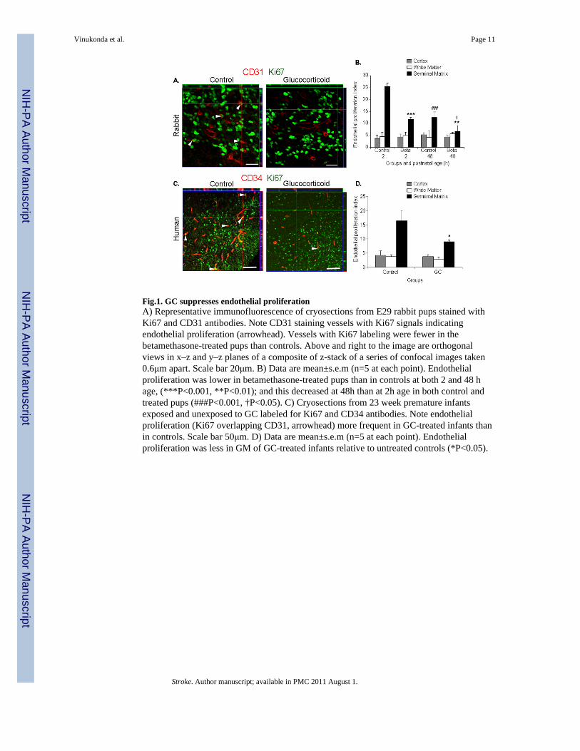
Fig.1. GC suppresses endothelial proliferationA) Representative immunofluorescence of cryosections from E29 rabbit pups stained withKi67 and CD31 antibodies. Note CD31 staining vessels with Ki67 signals indicatingendothelial proliferation (arrowhead). Vessels with Ki67 labeling were fewer in thebetamethasone-treated pups than controls. Above and right to the image are orthogonalviews in x–z and y–z planes of a composite of z-stack of a series of confocal images taken0.6μm apart. Scale bar 20μm. B) Data are mean±s.e.m (n=5 at each point). Endothelialproliferation was lower in betamethasone-treated pups than in controls at both 2 and 48 hage, (***P<0.001, **P<0.01); and this decreased at 48h than at 2h age in both control andtreated pups (###P<0.001, †P<0.05). C) Cryosections from 23 week premature infantsexposed and unexposed to GC labeled for Ki67 and CD34 antibodies. Note endothelialproliferation (Ki67 overlapping CD31, arrowhead) more frequent in GC-treated infants thanin controls. Scale bar 50μm. D) Data are mean±s.e.m (n=5 at each point). Endothelialproliferation was less in GM of GC-treated infants relative to untreated controls (*P<0.05).
Vinukonda et al. Page 11
Stroke. Author manuscript; available in PMC 2011 August 1.
NIH
-PA Author Manuscript
NIH
-PA Author Manuscript
NIH
-PA Author Manuscript
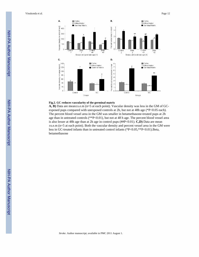
Fig.2. GC reduces vascularity of the germinal matrixA, B) Data are mean±s.e.m (n=5 at each point). Vascular density was less in the GM of GC-exposed pups compared with unexposed controls at 2h, but not at 48h age (*P<0.05 each).The percent blood vessel area in the GM was smaller in betamethasone-treated pups at 2hage than in untreated controls (**P<0.01), but not at 48 h age. The percent blood vessel areais also lesser at 48h age than at 2h age in control pups (##P<0.01). C,D) Data are mean±s.e.m (n=5 at each point). Both the vascular density and percent vessel area in the GM wereless in GC-treated infants than in untreated control infants (*P<0.05,**P<0.01).Beta,betamethasone
Vinukonda et al. Page 12
Stroke. Author manuscript; available in PMC 2011 August 1.
NIH
-PA Author Manuscript
NIH
-PA Author Manuscript
NIH
-PA Author Manuscript
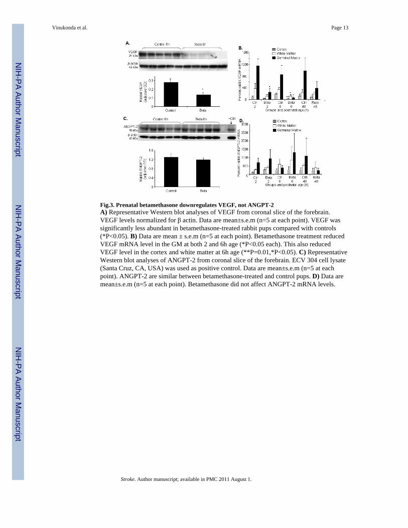
Fig.3. Prenatal betamethasone downregulates VEGF, not ANGPT-2A) Representative Western blot analyses of VEGF from coronal slice of the forebrain.VEGF levels normalized for β actin. Data are mean±s.e.m (n=5 at each point). VEGF wassignificantly less abundant in betamethasone-treated rabbit pups compared with controls(*P<0.05). B) Data are mean ± s.e.m (n=5 at each point). Betamethasone treatment reducedVEGF mRNA level in the GM at both 2 and 6h age (*P<0.05 each). This also reducedVEGF level in the cortex and white matter at 6h age (**P=0.01,*P<0.05). C) RepresentativeWestern blot analyses of ANGPT-2 from coronal slice of the forebrain. ECV 304 cell lysate(Santa Cruz, CA, USA) was used as positive control. Data are mean±s.e.m (n=5 at eachpoint). ANGPT-2 are similar between betamethasone-treated and control pups. D) Data aremean±s.e.m (n=5 at each point). Betamethasone did not affect ANGPT-2 mRNA levels.
Vinukonda et al. Page 13
Stroke. Author manuscript; available in PMC 2011 August 1.
NIH
-PA Author Manuscript
NIH
-PA Author Manuscript
NIH
-PA Author Manuscript
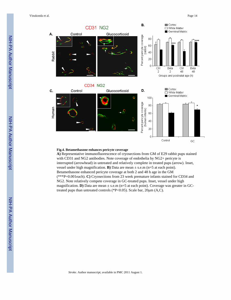
Fig.4. Betamethasone enhances pericyte coverageA) Representative immunofluorescence of cryosections from GM of E29 rabbit pups stainedwith CD31 and NG2 antibodies. Note coverage of endothelia by NG2+ pericyte isinterrupted (arrowhead) in untreated and relatively complete in treated pups (arrow). Inset,vessel under high magnification. B) Data are mean ± s.e.m (n=5 at each point).Betamethasone enhanced pericyte coverage at both 2 and 48 h age in the GM(***P=0.001each). C) Cryosections from 23 week premature infants stained for CD34 andNG2. Note relatively compete coverage in GC-treated pups. Inset, vessel under highmagnification. D) Data are mean ± s.e.m (n=5 at each point). Coverage was greater in GC-treated pups than untreated controls (*P<0.05). Scale bar, 20μm (A,C).
Vinukonda et al. Page 14
Stroke. Author manuscript; available in PMC 2011 August 1.
NIH
-PA Author Manuscript
NIH
-PA Author Manuscript
NIH
-PA Author Manuscript
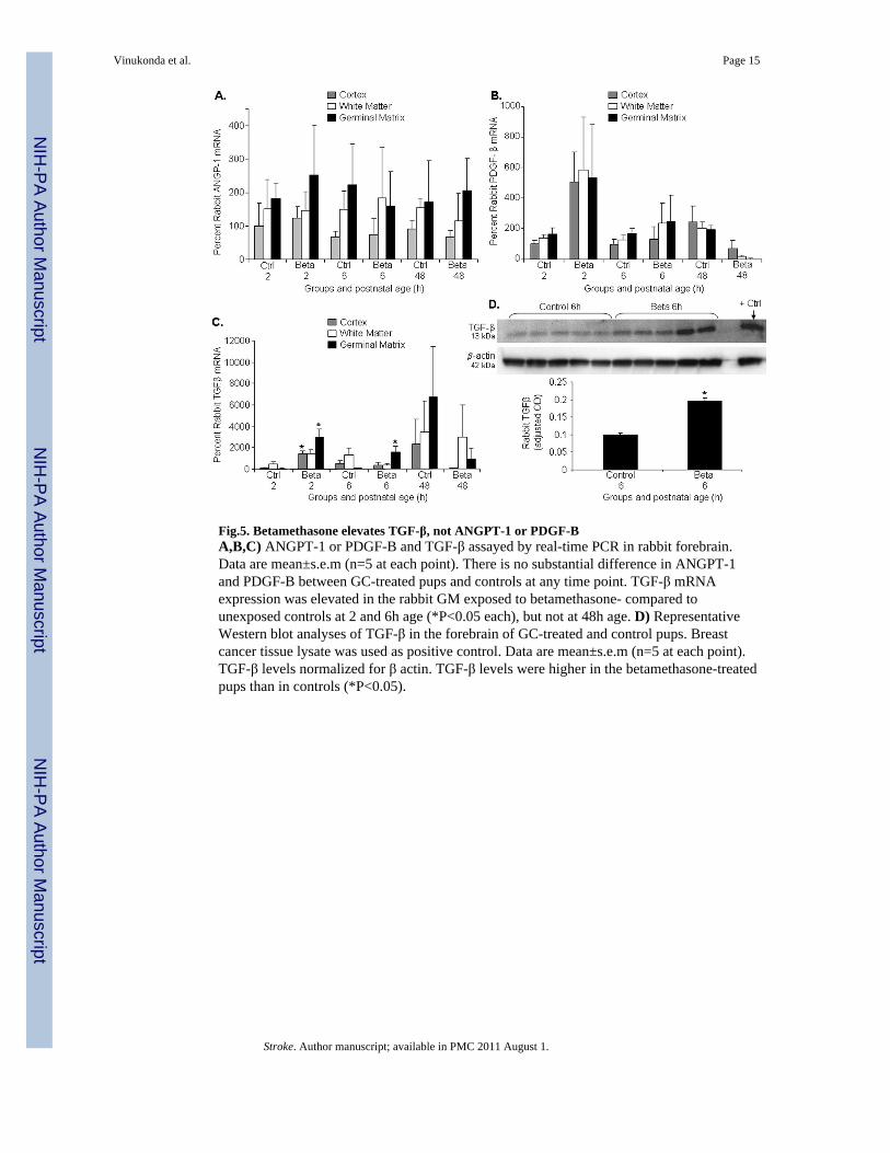
Fig.5. Betamethasone elevates TGF-β, not ANGPT-1 or PDGF-BA,B,C) ANGPT-1 or PDGF-B and TGF-β assayed by real-time PCR in rabbit forebrain.Data are mean±s.e.m (n=5 at each point). There is no substantial difference in ANGPT-1and PDGF-B between GC-treated pups and controls at any time point. TGF-β mRNAexpression was elevated in the rabbit GM exposed to betamethasone- compared tounexposed controls at 2 and 6h age (*P<0.05 each), but not at 48h age. D) RepresentativeWestern blot analyses of TGF-β in the forebrain of GC-treated and control pups. Breastcancer tissue lysate was used as positive control. Data are mean±s.e.m (n=5 at each point).TGF-β levels normalized for β actin. TGF-β levels were higher in the betamethasone-treatedpups than in controls (*P<0.05).
Vinukonda et al. Page 15
Stroke. Author manuscript; available in PMC 2011 August 1.
NIH
-PA Author Manuscript
NIH
-PA Author Manuscript
NIH
-PA Author Manuscript
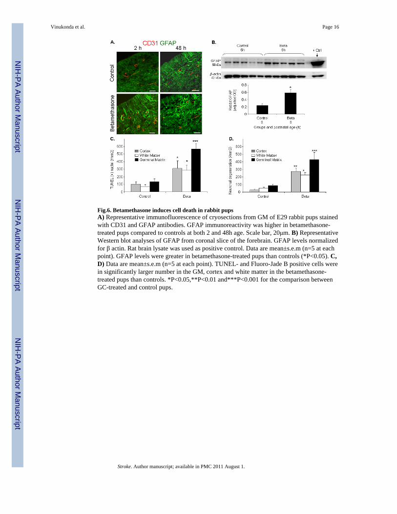
Fig.6. Betamethasone induces cell death in rabbit pupsA) Representative immunofluorescence of cryosections from GM of E29 rabbit pups stainedwith CD31 and GFAP antibodies. GFAP immunoreactivity was higher in betamethasone-treated pups compared to controls at both 2 and 48h age. Scale bar, 20μm. B) RepresentativeWestern blot analyses of GFAP from coronal slice of the forebrain. GFAP levels normalizedfor β actin. Rat brain lysate was used as positive control. Data are mean±s.e.m (n=5 at eachpoint). GFAP levels were greater in betamethasone-treated pups than controls (*P<0.05). C,D) Data are mean±s.e.m (n=5 at each point). TUNEL- and Fluoro-Jade B positive cells werein significantly larger number in the GM, cortex and white matter in the betamethasone-treated pups than controls. *P<0.05,**P<0.01 and***P<0.001 for the comparison betweenGC-treated and control pups.
Vinukonda et al. Page 16
Stroke. Author manuscript; available in PMC 2011 August 1.
NIH
-PA Author Manuscript
NIH
-PA Author Manuscript
NIH
-PA Author Manuscript


