Embed Size (px)
Citation preview

1143
Braz J Med Biol Res 36(9) 2003
Estrogen and the vascular system
Effects of estrogen on the vascular system
Grupo de Pesquisa sobre Hipertensão Arterial,Departamento de Farmacologia, Instituto de Ciências Biomédicas,Universidade de São Paulo, São Paulo, SP, Brasil
R.C. Tostes, D. Nigro,Z.B. Fortes and
M.H.C. Carvalho
Abstract
The cardiovascular protective actions of estrogen are partially medi-ated by a direct effect on the vessel wall. Estrogen is active both onvascular smooth muscle and endothelial cells where functionallycompetent estrogen receptors have been identified. Estrogen adminis-tration promotes vasodilation in humans and in experimental animals,in part by stimulating prostacyclin and nitric oxide synthesis, as well asby decreasing the production of vasoconstrictor agents such as cyclo-oxygenase-derived products, reactive oxygen species, angiotensin II,and endothelin-1. In vitro, estrogen exerts a direct inhibitory effect onsmooth muscle by activating potassium efflux and by inhibitingcalcium influx. In addition, estrogen inhibits vascular smooth musclecell proliferation. In vivo, 17ß-estradiol prevents neointimal thicken-ing after balloon injury and also ameliorates the lesions occurring inatherosclerotic conditions. As is the case for other steroids, the effectof estrogen on the vessel wall has a rapid non-genomic componentinvolving membrane phenomena, such as alteration of membraneionic permeability and activation of membrane-bound enzymes, aswell as the classical genomic effect involving estrogen receptor acti-vation and gene expression.
CorrespondenceM.H.C. Carvalho
Departamento de Farmacologia
ICB, USP
Av. Lineu Prestes, 1524
05508-900 São Paulo, SP
Brasil
Fax: +55-11-3091-7322
E-mail: [email protected]
Research supported by PRONEX (No.
125/98), FAPESP (Nos. 97/14208-8,
00/04611-4, and 00/12141-8), CAPES
and CNPq (No. 351365/1997-4).
Received March 14, 2003
Accepted May 28, 2003
Key words• Sex hormones• Estrogen• Vascular smooth muscle• Endothelium, nitric oxide• Endothelium-derived
hyperpolarizing factor• Angiotensin• Endothelin-1• Calcium channels• Potassium channels
Introduction
Extensive epidemiological observations,clinical mechanistic studies, and basic labo-ratory research have suggested that the inci-dence of cardiovascular disease increaseswith menopause and that hormone replace-ment therapy, or more specifically, estrogenreplacement therapy is associated with ben-eficial cardiovascular effects in postmeno-pausal women (1,2). Estrogen has a multi-tude of biological effects that may accountfor its apparent cardiovascular benefits(which remain to be proved in randomizedclinical trials), including favorable effectson the lipid profile, antioxidant activity, en-
hanced fibrinolysis, and a series of actionson the vasculature, which will be the focus ofour review. Since selective estrogen receptormodulators (SERMs), such as the syntheticestrogen-like compounds tamoxifen andraloxifene, are being considered as an optionin hormone replacement therapy due to theirestrogen-agonist effects on some tissues(liver, bone) and estrogen-antagonistic ef-fects on other tissues (breast, uterus), weincluded some of the already described vas-cular effects of SERMs in this review.
Effects of estrogen
The actions of steroid hormones can be
Brazilian Journal of Medical and Biological Research (2003) 36: 1143-1158ISSN 0100-879X Review

1144
Braz J Med Biol Res 36(9) 2003
R.C. Tostes et al.
divided into two types: those that are de-layed in onset and prolonged in duration arecalled “genomic” effects, and those that arerapid in onset and short in duration are called“non-genomic” effects. The early effects takeplace within minutes (e.g., changes in vaso-motor tone) and are mediated by rapid intra-cellular signaling pathways, whereas the de-layed effects (e.g., remodeling or lipid alter-ations) require hours to days to occur andrequire transcriptional events with subse-quent modulation of protein expression. Al-though the rapid and delayed effects of ster-oids are clearly distinguishable from eachother, there are actions that have onset timeof minutes and it is not clear as to whethergenomic or non-genomic mechanisms apply.
Estrogen receptors and mechanismsof action
Radioactive estrogens (tritium-labeledestrogens) were used to demonstrate for thefirst time intracellular steroid hormone re-ceptors in endocrine target organs throughthe body. Later, cells expressing the genom-ic steroid receptors were identified andmapped by binding studies, by immunocy-tochemistry, and by in situ hybridization.The receptors for estrogen and for all classesof steroidal hormones (androgens, glucocor-ticoids, mineralocorticoids, progestins, vita-min D), as well as thyroid hormones andretinoic acid, belong to the class of intracel-lular receptors and have been classicallydefined as nuclear ligand-activated trans-cription factors. Activation of these recep-tors by the corresponding hormones affectsgene expression by acting on specific se-quences in the target genes, known as estro-gen-response elements, and by modulatingtranscriptional events. Importantly, gene ex-pression may be further regulated throughpositive and negative interactions of estro-gen receptors (ER) with transcription factors(3). The discovery of intracellular hormonereceptors led to studies focusing on the long-
lasting effects of steroids (genomic effects)on cell function. However, the fact that notall actions of estrogens are delayed in onsetand prolonged in duration implies anothermechanism of action (non-genomic effects).Some of the estrogen effects are mediated byERs localized on the cell surface since mem-brane-impermeant forms of estrogen lead tothe activation of mitogen-activated proteinkinase (MAPK) as well as stimulation ofcGMP production and nitric oxide (NO) re-lease. The use of confocal microscopy andflow cytometric analysis has demonstratedthat endothelial cells contain surface recep-tors for estrogen, detectable by cell-imper-meant ligand binding and anti-ER antibodies.Activation of membrane ERs has been linkedto novel, rapid signaling pathways inducedby estrogen. More recently, it was shownthat several actions of steroid hormones onmembranes involve non-nuclear signalingpathways, such as coupling to G proteinsand generation of second messengers (4).Estrogens can induce Ca2+ mobilization, in-crease levels of cyclic nucleotide secondmessengers, and activate several kinases,including protein kinase C, phosphatidylin-ositol-3-OH kinase (PI3-kinase), and MAPKthrough incompletely defined mechanisms(5,6). Adding more complexity, non-genom-ic effects of steroid hormones may be recep-tor-mediated or receptor-independent. Ste-roid hormone genomic and non-genomic ef-fects may occur simultaneously and may actat different levels, revealing the high com-plexity of steroid hormone regulation of cellfunction.
The original studies indicated that therewas one type of steroid receptors and that thenature of the biological responses in differ-ent tissues was determined by the network ofgenes available for interaction with the acti-vated receptor. Later, two independent groupscloned the ER complementary DNA (7,8).
However, the distribution of this ER couldnot explain all of the biological effects ofestrogens, for instance in the ovaries. Later,

1145
Braz J Med Biol Res 36(9) 2003
Estrogen and the vascular system
when a second ER was cloned, a usefulexplanation for the dissociation between theER localization and effects of estrogen wassuggested (9). Two types of intracellularERs have been identified: ER-α and ER-ß,and isoforms of ER-α and ER-ß (splice vari-ants ER-ß1 and ER-ß2) have been also iden-tified.
The ligand-binding domain of ER-ßshows great homology compared with thatof ER-α. High homology is also observed inthe DNA-binding domain or estrogen-re-sponse element sites for the ER-α and ER-ßin the estrogen-sensitive genes. However,the ER domains responsible for the interac-tion of ER-α and ER-ß with transcriptionfactors, the ligand-independent N- and C-terminal transcription activation functions-1and -2, markedly differ between ER-α andER-ß. This lack of homology may explainthe different effects on an estrogen-responseelement site observed with estrogen agonistsand antagonists.
Furthermore, different ligands cause dif-ferent conformational changes in ERs,thereby affecting interaction patterns withadditional transcription factor co-regulatorsand eventually the fine control of gene ex-pression. It should also be mentioned thatsteroid receptor co-regulators react differ-ently with ER-α and ER-ß. For instance, thetwo ERs interact with an activator protein-1site in opposite ways in the presence ofdifferent ligands (10). Finally, recent workindicated that ER-ß may counteract or con-trol ER-α activity in a variety of tissues,including the uterus, and mediate estrogeniceffects other than ER-α in some tissues, suchas the bone or the immune system.
The discovery and cloning of ER-ß pro-vided a basis for understanding how theknockout of ER-α could have resulted in aviable organism and in the continued actionof estrogens in some tissues, although ani-mals are sterile and show altered sexual andother behaviors (11). On the other hand,knockout mice for ER-ß appear quite normal
and are able to reproduce, although theyshow some reduction in litter size (12).
Measurements of mRNA for ER-α andER-ß have shown distributions in the bodythat differ quite markedly from each other.Moderate to high expression of ER-α hasbeen identified in the pituitary, kidney, epi-didymis, and adrenal glands, whereas mod-erate to high expression of ER-ß has beenfound in prostate, lung, bladder, and brain.Overlapping high expression of ER-α andER-ß has been identified in ovary, testis anduterus. In the vasculature, ERs have beenidentified by different techniques in the en-dothelium, intima and adventitia and on ad-renergic nerve endings of arteries fromvarious territories and several species, in-cluding humans.
A very interesting observation is that thevascular cells are capable of expressingaromatase, the key enzyme in the estrogensynthesis pathway, suggesting that the vas-cular system is capable of local estrogenbiosynthesis in vivo, which may lead to acti-vation of ER and downstream activation oftarget genes (13). Locally produced estrogenmay therefore act in an endocrine, paracrineand autocrine manner on vascular and non-vascular cells.
Effects of estrogen on the arterialwall
Actions of steroidal hormones on thearterial wall include alteration or modula-tion of ion fluxes and of receptors on smoothmuscle cells and modulation of endotheli-um-derived factor production and activity.In this review we focused on the effects ofestrogen on components of endothelial andsmooth muscle cells. However, it is impor-tant to keep in mind that estrogen-inducedeffects depend on the vascular bed and onthe animal species being considered, indi-cating that there is regional and species het-erogeneity in the modulatory influence ofestrogen on vasomotor function. To mention

1146
Braz J Med Biol Res 36(9) 2003
R.C. Tostes et al.
few examples, it has been reported that 17ß-estradiol induces both endothelium-depend-ent and -independent relaxation in the rataorta but only endothelium-independent re-laxation in the rat mesenteric arteries. NOcontributes strongly to the endothelium-dependent relaxation induced by 17ß-estra-diol in isolated aortas, whereas in small cere-bral arteries both NO and cyclooxygenase(COX) metabolites contribute to estrogen-induced effects. Estrogen treatment increasesaortic stiffness and potentiates endothelialvasodilator function in the hindquarters, butnot in the carotid vascular bed. Differencesin the mechanisms involved in estrogen ac-tions may reflect a differential contributionof mechanisms involved in vascular toneregulation. Furthermore, there is evidencethat ER expression may change with patho-logical conditions or, inversely, that changesin ER expression may lead to abnormal vas-cular function (14).
Actions of estrogen on endothelialcells
The endothelium plays a major role invascular tone control by releasing both re-laxing and contractile factors and estrogensexert a number of effects on endothelial-derived factors, as summarized below.
Estrogens have been shown to enhanceendothelial-dependent relaxation in arterialrings from different animals and from differ-ent vascular beds, including coronary, mes-enteric, aorta and cerebral arteries. Studieson humans have demonstrated that estrogenreplacement treatment increases coronaryflow and decreases both coronary resistanceand peripheral vascular tone.
Nitric oxide
Earlier reports indicated that basal re-lease of NO is increased in females com-pared to males (15,16) and that estrogenadministration to ovariectomized rats restores
the impaired ex vivo basal release of NO.Effects of estradiol were also described inarteries from male animals. Huang et al. (17)observed that 17ß-estradiol restores endo-thelial NO release in response to shear stressin pressurized gracilis muscle arterioles ofmale spontaneously hypertensive rats (SHR)by up-regulation of endothelial nitric oxidesynthase (NOS). Conversely, it has beenreported that endothelium-dependent relax-ation elicited by carbachol and histaminewas attenuated by estradiol in preparationsfrom intact male rats. Moreover, aortic pros-tacyclin release was reduced by about 40%after estradiol treatment in tissues from theseanimals. These results showing that releaseof NO in arteries from male rats is not af-fected by estradiol treatment suggest genderspecificity for the vascular effects of estro-gen.
NO production accounts for most of theendothelium-dependent relaxation activity,and there is extensive evidence showing es-trogen-induced up-regulation of endothelialNO production. Probable mechanisms in-volved in estradiol-induced increased NOproduction include: 1) transcriptional stimu-lation of NOS gene expression, 2) inhibitionof cytokine-induced down-regulation of NOSgene expression, 3) post-translational modi-fication of NOS protein, 4) increased co-factor or L-arginine availability, 5) non-ge-nomic activation of second messengers (e.g.,Ca2+, cAMP) and tyrosine kinase, 6) translo-cation from the membrane to intracellularsites, and 7) modulation of NO degradingsystems (e.g., reactive oxygen radical gen-eration and antioxidants).
Induction of constitutive (Ca2+-depend-ent) NOS by estrogen has been demonstratedin a variety of tissues, consistent with thepresence of estrogen-response elements inthe NOS promoter. In addition to increasingNOS production, estrogen induces rapid en-hancement of NOS activity and NO releasethrough nontranscriptional mechanisms andby reducing its Ca2+ dependence (18). This

1147
Braz J Med Biol Res 36(9) 2003
Estrogen and the vascular system
effect seems to be much more intense andfunctionally relevant than the increase in NOSexpression induced by estrogen and is inhib-ited by the ER antagonist ICI 182,780, indi-cating that the effect is mediated by ERs(19). In SHR, estrogen deprivation (inducedby ovariectomy) decreases NOS activity andexpression and NO-derived metabolites (20).
Recent studies indicate that estrogen-in-duced activation of endothelial NOS is drivenby activation of the PI3-kinase/Akt pathwayresulting from direct interactions betweenthe ER and the regulatory subunit of PI3-kinase (6), and requires MAPK activation(19). Hisamoto et al. (21) observed that 17ß-estradiol, but not 17α-estradiol, caused acuteactivation of endothelial NOS both in humanumbilical vein endothelial cells and in sim-ian virus 40-transformed rat lung vascularendothelial cells. Activation of endothelialNOS involves the activation of Akt and thephosphorylation of endothelial NOS, whichis mediated by ER-α via a non-genomicmechanism.
The effects of estrogen on NOS may alsobe associated with its effects on caveolin-1expression, which inhibits endothelial NOScatalytic activity. Jayachandran et al. (22)have shown that endothelial NOS proteinexpression and nitrite/nitrate production bybovine aortic endothelial cells are enhancedby 17ß-estradiol, which also stimulatescaveolin-1 transcription and translationthrough ER-mediated mechanisms.
Similar to estrogen, the SERM raloxifenestimulates endothelial NOS mRNA expres-sion (genomic effects) and also triggers rapidactivation of NO synthesis by stimulatingendothelial NOS (non-genomic effects) viathe PI3-kinase pathway ER signaling (23). Infemoral veins, raloxifene induces acute re-laxation both by NO release and by directstimulation of vascular smooth muscle cellsdepending on the ovarian hormonal status ofthe animal. As we will discuss later, estrogenalso prevents NO degradation due to its anti-oxidant properties, consequently increasing
NO availability.The effects of estrogen on NOS activity
are suggested to be important in arterial in-jury. Local delivery of 17ß-estradiol duringpercutaneous transluminal coronary angio-plasty improved endothelial function, en-hanced re-endothelialization and endotheli-al NOS expression and decreased neointimaformation. Recently, Tolbert et al. (24) haveshown that the vasoprotective effects of es-trogen after ligation vascular injury are par-tially reduced in inducible NOS knockoutmice, suggesting that estrogen also modu-lates inducible NOS expression and plays arole in neointima formation.
Endothelium-derived hyperpolarizing factor
Endothelium-derived hyperpolarizingfactors (EDHFs) are important mediators ofvascular relaxation, more specifically in re-sistance-sized arteries where they regulatetissue blood flow. The release of EDHFs ismodulated by a number of influences includ-ing agonist stimulation, shear stress, anddisease. The chemical identification and func-tional characterization of EDHFs vary de-pending on vascular size, vascular bed andspecies. Three major candidates are the ep-oxyeicosatrienoic acids, cytochrome P450metabolites of arachidonic acid, K+ and hy-drogen peroxide. Additionally, electricalcoupling through myoendothelial gap junc-tions serves to conduct electrical changesfrom the endothelium to the smooth muscleand may mediate or propagate hyperpolar-ization.
Evidence showing that estrogen modu-lates EDHF production and release has accu-mulated over the past few years. One of thefirst lines of evidence for estrogen-inducedhyperpolarization, although not specificallyby EDHF, was provided by Harder andCoulson (25). They observed that additionof diethylstilbestrol to dog coronary arterieshyperpolarized the membrane and reducedinput resistance, which was not related to

1148
Braz J Med Biol Res 36(9) 2003
R.C. Tostes et al.
increased Na+-K+ ATPase pump activity.Later, it was shown that the hyperpolariz-
ing response to acetylcholine in mesentericarteries isolated from middle-aged rats issignificantly greater in females than in males.Ovariectomy causes a marked reduction inacetylcholine-induced hyperpolarization infemale arteries and this is improved by 17ß-estradiol replacement therapy (26). Furtherevidence for a positive modulation of EDHFrelease by estrogen is provided by the obser-vation that during pregnancy, a conditionwith high levels of estrogen, there is an up-regulation of EDHF-mediated vasodilation.On the other hand, in pial arteries from fe-male rats, ovariectomy increases the NO-and prostacyclin (PGI2)-insensitive dilation(EDHF-mediated dilation) whereas estradiolreplacement reduces it, suggesting a nega-tive modulation of estrogen on EDHF release.
Arachidonic acid metabolites
The actions of estrogen on vascular cellsalso influence the metabolism of prostaglan-dins and the activity of COX, a key enzymein the production of prostaglandins.
It has been repeatedly shown that 17ß-estradiol stimulates production of vasodila-tor prostaglandins such as PGI2 in a varietyof preparations such as ovine uterine ar-teries, ovine fetal pulmonary artery endothe-lial cells, human umbilical vein endothelialcells, and rat mesenteric and cerebral bloodvessels.
Increased levels of PGI2 upon stimula-tion with estradiol have been associated withincreased expression of the key enzymesinvolved in PGI2 production, phospholipaseA2, COX-1, and prostacyclin synthetase(PGIS), as demonstrated in rat cerebral bloodvessels, where chronic in vivo 17ß-estradioltreatment enhances basal PGI2 synthesis byincreasing COX-1 and PGIS proteins (27). Itseems that the COX-2 pathway also plays aspecific role in estradiol-induced PGI2 syn-thesis and vasodilation. In human umbilical
vein endothelial cells, 17ß-estradiol increasesthe release of PGI2 via the induction of COX-2, but not COX-1 protein (28), and in thecutaneous vasculature of postmenopausalwomen, acute 17ß-estradiol administrationenhanced the response to acetylcholine afteraspirin, diclofenac, and placebo, but not af-ter celecoxib treatment (29).
More recently, an association betweenestradiol and vasoconstrictor prostaglandinshas also been established: 1) estrogen re-placement prevents prostaglandin H synthase(PGHS)-dependent vasoconstriction, thatwas associated with augmented sensitivityof the thromboxane A2 (TXA2)/prostaglan-din H2 (PGH2) receptor, in resistance-sizedmesenteric arteries from ovariectomizedSprague-Dawley rats, 2) estrogen improvesvasodilation in isolated mesenteric arteriesfrom aged Fisher rats by decreasing PGHS-2-dependent constriction and PGHS-2 ex-pression, and 3) estrogen decreases the syn-thesis of contractile COX metabolites. Wehave shown that ovariectomy increases reac-tivity to norepinephrine and reduces sensi-tivity to acetylcholine in microvessels fromSHR whereas treatment with estradiol orestradiol + progesterone similarly restoredthese altered responses. The COX inhibitorsindomethacin, diclofenac, as well as ridogrel(a TXA2 receptor antagonist and inhibitor ofthromboxane synthase), but not dazoxiben(an inhibitor of thromboxane synthase), alsorestored norepinephrine and acetylcholineresponses in ovariectomized SHR. The re-lease of PGF2α, but not of TXB2 and 6-keto-PGF1α, was greater in ovariectomized SHRthan in control SHR microvessels upon stimu-lation with norepinephrine, suggesting thatestrogen decreases the synthesis of contractileCOX metabolites, such as PGH2/PGF2α (30).
As mentioned earlier, not all arteries re-spond in the same way to estradiol. It hasbeen reported that estradiol and progeste-rone treatments differentially alter proteinexpression of the key enzymes involved inPGI2 production, phospholipase A2, COX-1,

1149
Braz J Med Biol Res 36(9) 2003
Estrogen and the vascular system
and PGIS, in uterine and systemic (renal,coronary, mammary, omental) arteries.
The modulatory actions of estrogen onthe COX pathway seem to be mediatedthrough activation of ERs. In pulmonaryartery endothelial cells, PGI2 production ismediated by ER-ß, since it was fully blockedby both ER antagonism with ICI 182,780,which is not selective for either ER isoform,and the ER-ß-specific antagonist RR-tetrahydrochrysene. On the other hand, theobservation that estrogen increases COX-1levels in cerebral blood vessels from wild-type mice, but is ineffective in ER-α knock-out mice, suggests that ER-α regulation ofthe endothelial COX-1 pathway also appearsto contribute to the effects of estrogen.
Adhesion molecules
In an inflammatory process, cytokinesand other inflammatory mediators elicit re-markable phenotypic changes in endothelialcells. Activated endothelial cells release NOand PGI2, leading to vasodilation, which inturn facilitates leukocyte influx to the sitesof injury. Cytokine-activated endothelial cellsexpress adhesion molecules for leukocytes -E-selectin, intercellular adhesion molecule-1 (ICAM-1) and vascular cell adhesion mol-ecule-1 (VCAM-1) - and are a source ofchemokines that not only have chemotacticeffects on leukocytes, but also are potentactivators of leukocyte integrins, allowingtheir interaction with their endothelial counter-receptors.
Many cell types involved in immune andinflammatory responses are responsive toestrogen. These include T and B lympho-cytes, and cells from the myelomonocyticlineage such as monocytes, macrophages,and mast cells. Actions of estrogen on in-flammatory markers seem to depend on lev-els of estrogen. At physiological concentra-tions, estrogen increases proinflammatorycytokine production - IL-1, tumor necrosisfactor-α (TNF-α), and IL-6 - by monocytes.
However, subjects with high estrogen status(premenopausal women) have significantlylower monocyte chemoattractant protein(MCP-1) levels than subjects with low estro-gen status (postmenopausal women), andhormone replacement therapy lowers plasmalevels of MCP-1 (31). The estrogen-inducedincrease in TNF-α activates endothelial ad-hesion to leukocytes via increased expres-sion of the endothelial adhesion moleculesE-selectin, ICAM-1, and VCAM-1 (32). Ki-netic studies of mRNA for endothelial adhe-sion molecules show that, at early time points,estrogen treatment results in increased levelsof mRNA for E-selectin, ICAM-1, andVCAM-1, whereas at later time points, estro-gen-treated cells disclose lower levels ofmRNA for these cytokines (32).
However, at higher or pharmacologicaldoses and after longer periods of exposure,estrogen decreases cytokine-induced adhe-sion molecule expression by cultured endo-thelial cells (33). A prospective, random-ized, placebo-controlled 12-week study onhealthy, normotensive postmenopausal womenwho received either micronized estradiolalone, or sequentially estradiol combinedwith a progestagen showed a significant de-crease in the plasma concentrations of solubleICAM-1, soluble VCAM-1, and thrombo-modulin. In the same direction, it has beenshown that Fas ligand expression by thevascular endothelium, which inhibits themigration of inflammatory cells into the ves-sel wall, is improved by estradiol treatment,resulting in inhibition of leukocyte trafficacross the endothelium. According to theauthors, the maintenance of endothelial Fasligand expression by estradiol may representa mechanism of the apparent antiatherogeniceffect of estrogen.
Oxidative stress - reactive oxygen species
The vasculoprotective effects of estro-gen have been also partially attributed to ashift in the NO/superoxide anion (O2
-) bal-

1150
Braz J Med Biol Res 36(9) 2003
R.C. Tostes et al.
ance in the vessel wall, thereby increasingthe bioavailability of NO. In human umbili-cal vein cultured endothelial cells, 17ß-es-tradiol decreases expression of the NADPHoxidase subunit gp91phox and up-regulatesendothelial NOS expression, improving theNO/O2
- balance (34). On the other hand,estradiol increases endothelium-derived re-laxing factor activity in the thoracic aorta ofovariectomized female rats in the absence ofchanges in NOS activity or NOS gene andprotein expression. Since lucigenin-enhancedchemiluminescence, an indicator of O2
- pro-duction, of the aorta was decreased in estra-diol-treated ovariectomized rats comparedto the placebo group, it was suggested thatthe decreased endothelium-derived genera-tion of O2
- in response to estrogens accountsfor enhanced NO bioactivity and decreasedperoxynitrite release (35). Increased oxida-tive stress and decreased NO production dueto estrogen deficiency have been associatedwith higher blood pressure levels. Chronicestrogen replacement in ovariectomized ratsprevents the decrease of plasma levels ofnitrites/nitrates as well as the enhancementof blood pressure.
Wassmann et al. (36) observed that estro-gen replacement therapy and angiotensin type1 (AT1) receptor antagonism prevent theworsened endothelial dysfunction, enhancedvasoconstrictor response to angiotensin II(Ang II) and increased vascular O2
- produc-tion displayed by ovariectomized SHR. Veryrecently, it has been shown that estrogen re-duces Ang II-induced expression of NAD(P)Hoxidase and peroxynitrite in cultured endo-thelial cells. Estrogen as well as superoxidedismutase also inhibited Ang II-induced AT1receptor expression and nitrotyrosine stain-ing, effects that were not inhibited by the ERantagonist ICI 182,780 (37).
We have shown that treatment with su-peroxide dismutase also restored changes innorepinephrine and acetylcholine responsesin ovariectomized SHR (38). We have alsofound evidence that estrogen reduces O2
-
bioavailability in SHR microvessels in vivo.We observed that oxidative stress in mesen-teric arteries, evaluated by using intravitalmicroscopy and superfusion with hydroethi-dine, was significantly increased in ovariec-tomized SHR and was attenuated by estra-diol or estradiol + progesterone treatment.Treatment with the superoxide dismutasemimetic MnTMPyP, but not with mannitol,that decomposes hydroxyl radicals, or L-NAME, an NOS inhibitor, attenuated oxida-tive stress in ovariectomized SHR. Treat-ment of mesenteries with diphenyleneiodo-nium, an NAD(P)H oxidase inhibitor, butnot with oxypurinol, a xanthine oxidase in-hibitor, produced a significant reduction ofoxyradical generation in microvessels fromovariectomized SHR (38).
Similar to estrogen, the SERM idoxifeneeffectively blunts Ang II-induced reactiveoxygen species production as evaluated byconfocal laserscanning microscopy using theredox sensitive marker 2',7'-dichlorofluores-cein and measurement of NAD(P)H oxidaseactivity. This estrogen effect has been ex-plained in part by idoxifene-induced down-regulation of AT1 receptor expression, aswill be discussed later.
The estrogen-mediated induction of theprotein thiol/disulfide oxidoreductases suchas disulfide isomerase, thioredoxin, thio-redoxin reductase, and glutaredoxin in vas-cular endothelial cells may also be involvedin the antioxidant properties of estrogen ob-served in the vascular system. Recent studieshave also shown that estrogen inhibits endo-thelial cell apoptosis induced by hydrogenperoxide and that phytoestrogens (daidzein,genistein and resveratrol) attenuate the oxi-dative DNA damage induced by advancedglycation end-products in vascular smoothmuscle cells. The latter action was associ-ated with an increase in intracellular totalglutathione levels.
In humans it has been suggested that theendothelial dysfunction secondary to acuteendogenous estrogen deprivation (surgical
1151
Braz J Med Biol Res 36(9) 2003
Estrogen and the vascular system
ovariectomy) is caused by reduced NO avail-ability, which results from COX-dependentproduction of oxidative stress (39).
Angiotensin II
Estrogen acts on the renin-angiotensinsystem at different points of the cascade: atthe formation of Ang II, at the level of Ang IIreceptors and on Ang II-induced responses.
Estrogen has been shown to increase geneexpression and plasma levels of angiotensin-ogen. Estrogen also has effects on the levelsof renin in the circulation: 1) women underhormone replacement therapy with estrogendisplay lower renin levels than those notusing such therapy, 2) premenopausal womendisplay lower renin levels than postmeno-pausal women, and 3) women display lowerrenin levels than men.
Estrogen deficiency has been shown toincrease the subtype 1 Ang II receptor (AT1receptor) mRNA levels, as well as the effi-cacy of Ang II on vasoconstriction (due toincreased AT1 receptor density), whereasestrogen replacement therapy in ovariecto-mized rats reversed AT1 receptor overex-pression. Down-regulation of AT1 receptormRNA and protein expression by estrogenoccurs through activation of ERs and seemsto be mediated by NO-dependent pathways(40).
Estrogen treatment also antagonizes theAT1 receptor-mediated growth-promotingeffects of Ang II in vascular smooth musclecells and the mechanisms involved in theaction of 17ß-estradiol include attenuationof AT1 receptor-mediated extracellular sig-nal-regulated kinase activation and transac-tivation of MAPK phosphatase-1 expression(41).
As mentioned earlier, estrogen andSERMs, such as idoxifene, inhibit Ang II-induced reactive oxygen species release. Ourgroup has observed that chronic treatment offemale SHR with losartan caused similardecreases in oxyradicals in both control and
ovariectomized rats, whereas diclofenac andverapamil had no effects (38), suggestingthat, despite the contribution of Ang II to theoxidative stress in SHR, this is not the path-way that is up-regulated in the absence ofestrogen.
The action of estrogens on angiotensin-converting enzyme (ACE) activity have alsobeen reported. Brosnihan et al. (42) showedthat ACE activity in the circulation and intissues is reduced upon chronic hormonereplacement in two animal models ofpostmenopause. Reduced tissue levels (kid-ney and aorta) of ACE, paralleled by re-duced ACE mRNA concentrations, has alsobeen described with chronic estrogen re-placement therapy.
These authors also investigated the car-diovascular responses to administration ofAng-(1-7) and Ang II in female transgenic(mRen2)27 rats receiving estrogen replace-ment. The estrogen-treated rats showed lowerblood pressure levels and the magnitude ofthe depressor component of the biphasicresponse to Ang-(1-7) was significantly en-hanced, whereas the pressor component wasattenuated. Estrogen replacement also sig-nificantly attenuated the pressor response toAng II. In addition, estrogen replacementtherapy was shown to reduce plasma andtissue ACE activity in association with areduction in circulating levels of Ang II.Because estrogen increased the levels ofplasma Ang-(1-7) and amplified the vasodi-lator actions of Ang-(1-7), while reducingthe formation and vasoconstricting actionsof Ang II, the authors suggested that estro-gen shifts the vasoconstrictor-vasodilator bal-ance of the renin-angiotensin system (42).
The same group later showed that ovari-ectomized monkeys treated with conjugatedequine estrogen replacement for 30 monthsexhibited a reduction of ACE activity inassociation with a significant increase inplasma Ang I and hyperreninemia. PlasmaAng II levels were not increased in monkeystreated with estrogen, suggesting that the

1152
Braz J Med Biol Res 36(9) 2003
R.C. Tostes et al.
decrease in ACE curtailed the formation ofthe peptide. The Ang II/Ang I ratio, an invivo index of ACE activity, was significantlyreduced by estrogen treatment, further sup-porting the biochemical significance of ACEinhibition by estrogen.
Clinical data have shown that postmeno-pausal women who received conjugatedequine estrogen for 3 months exhibited asignificant reduction in plasma ACE activitythat correlated with an increase in NO-medi-ated forearm endothelium-dependent vasodi-latation (reactive hyperemia) and increasedserum levels of nitrite/nitrate. However, otherinvestigators have observed that acute ad-ministration of estradiol produces no changesin vascular ACE activity or in the vasocon-stricting responses to Ang I or Ang II.
Sex-related differences in the effects ofblockers of the renin-angiotensin system havebeen documented, with female animals dis-playing a better response. Treatment withlosartan, an AT1 antagonist, normalizes highblood pressure in 100% of female SHRagainst 53% of male SHR (43). Enalapril, anACE inhibitor, produced similar results, nor-malizing blood pressure in 71% of femaleSHR vs 46% of male SHR (44).
Endothelin
Similar to its actions on the renin-angio-tensin system, the actions of estrogen on theendothelin-1 (ET-1) pathway have beenshown at different points of the cascade: atits formation, at the level of the receptorsand on ET-1-induced responses.
It has been reported that 17ß-estradiolattenuates ET-1-induced coronary artery con-striction both in vitro and in vivo. In addi-tion, increased expression of prepro-ET-1mRNA has been observed in porcine aorticendothelial cells in the absence of femaleovarian hormones. Barber et al. (45) reportedthat endogenous fluctuations in estrogen in-fluence the affinity of ET-1 receptors incoronary arterial smooth muscle from fe-
male pigs, and gender differences in ET-1receptor density, as well as in the ratio of ET-1 receptor subtypes, have also been reported.An increase in the total number of ET-1receptors, but not in ET-1 binding Kd, hasbeen reported in human saphenous vein frommen compared to women. Furthermore, adifferential ratio of ETA to ETB receptors,favoring vasodilator effects in women, hasalso been described (46).
Our group showed that arteries from male,but not female, DOCA-salt rats display in-creased sensitivity to ET-1 and to the selec-tive ETB receptor agonist IRL-1620 both invitro and in vivo (47,48). Changes in ETB-mediated vascular responses were associ-ated with increased ET-1 and ETB receptorgene expression in male, but not in female,animals (49). Furthermore, we observed thatovariectomy increases the vasoconstrictingresponses to the ETB agonist and that estro-gen replacement therapy restores IRL-1620-induced responses, supporting our suggestionthat the ovarian hormones modulate ET-1/ETB receptor vascular responses/expressionin DOCA-salt hypertension (50). Sexual di-morphism in vascular reactivity to ET-1 wasalso reported in SHR (51).
Estradiol, as well as 2-hydroxyestradiol,or 2-methoxyestradiol, metabolites of estra-diol with little or no affinity for ERs, inhib-ited basal and ET-1-stimulated synthesis inporcine coronary artery endothelial cells.Estradiol-induced inhibition of ET-1 is pos-sibly mediated by inhibition of MAPK activ-ity, and by an ER-independent mechanism(52). On the other hand, the inhibitory ef-fects of ICI 182,780 on the 17ß-estradiol-induced decrease of ET-1 gene expressionand peptide secretion in cultured bovine ca-rotid arterial endothelial cells suggest acti-vation of ER-dependent mechanisms.
Studies conducted on healthy postmeno-pausal women who received continuous hor-mone replacement therapy (17ß-estradiolcombined with norethisterone acetate ormethoxyprogesterone) reported that post-

1153
Braz J Med Biol Res 36(9) 2003
Estrogen and the vascular system
menopausal women treated with hormonereplacement therapy had increased plasmanitrites/nitrates and decreased ET-1 levels(53). Women receiving the SERM raloxifenehad similar changes in plasma nitrites/ni-trates and ET-1 levels as well as in the ratioof NO to ET-1 (53).
Actions of estrogen on vascularsmooth muscle cells
We so far have seen that by acting onendothelial cells and modulating endotheli-um-derived relaxing and contraction factorrelease/activity, estrogen indirectly affectssmooth muscle cell tone (Figure 1). Estrogenhas also direct effects on smooth musclecells. Here we will try to summarize a seriesof observations showing that 17ß-estradiolmay play a role in selectively regulatingvascular smooth muscle tone by modulating/modifying ion channel permeability in thesecells (Figure 1). The voltage-clamp tech-nique has helped elucidate the effects ofestrogen on ion channels.
Potassium channels
The actions of estradiol on K+ channelshave been described both in smooth muscleand endothelial cells. ATP-sensitive K+
(KATP) channels seem to be involved in di-ethylstilbestrol-induced relaxation in isolatedrat aorta, since diethylstilbestrol-inducedvasodilation is inhibited by glibenclamideand tetraethylammonium, but not by paxillin.Since relaxation in response to forskolin wasantagonized by glibenclamide, it was sug-gested that diethylstilbestrol-induced relax-ation in the rat aorta is related to the modula-tion of KATP channels via cyclic AMP-de-pendent mechanisms (54).
Functional studies also demonstrated that17ß-estradiol relaxes porcine coronary ar-teries by an endothelium-independent mech-anism involving K+ efflux, and subsequentstudies employing the patch-clamp technique
have confirmed that estrogen stimulates K+
channel gating in coronary smooth muscle.Perforated-patch recordings from metaboli-cally intact coronary myocytes revealed that17ß-estradiol more than doubles steady-stateoutward currents in these cells at positivevoltages whereas studies of on-cell patchesdemonstrated a potent stimulatory effect of17ß-estradiol on the gating of the large-con-ductance, Ca2+- and voltage-activated K+
(BKCa) channels. Furthermore, blockingBKCa channels in intact arteries inhibitedestrogen-induced relaxation. Since the ef-fect of 17ß-estradiol on BKCa channels wasblocked by inhibiting cGMP-dependent pro-tein kinase activity and was mimicked by
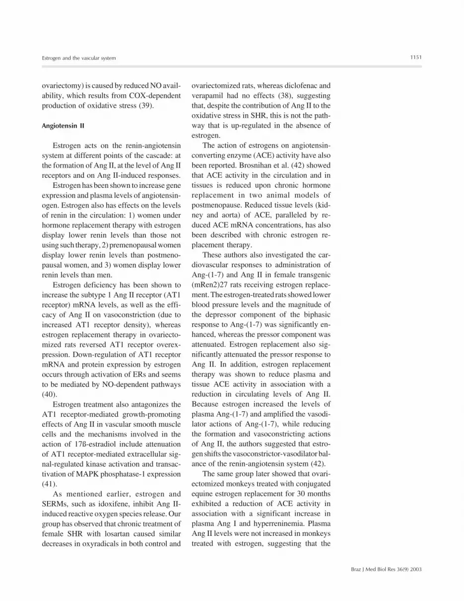
Figure 1. Putative mechanisms for the action of estrogen on the vascular system. By actingon endothelial cells, estrogen increases EDRFs (NO, EDHF and PGI2) production/releaseand decreases release/actions of EDCFs (PGF2α, TXA2, Ang II, ET-1, O2
-). By acting onvascular smooth muscle cells, estrogen activates K+ channels, leading to cell hyperpolariza-tion, and blocks activation of Ca2+ channels, decreasing intracellular Ca2+ concentration.Actions of estrogen may be mediated by both direct genomic effects (activation of nuclearreceptors and transcriptional events) and non-genomic effects (activation of rapid intracellu-lar signaling pathways). AA = arachidonic acid; ACE = angiotensin-converting enzyme; AngI = angiotensin I; Ang II = angiotensin II; Ca2+ channels = calcium channels; COX =cyclooxygenase; EC = endothelial cells; ECE = endothelin converting enzyme; EDCF =endothelium-derived contracting factor; EDHF = endothelium-derived hyperpolarizing fac-tor; EDRF = endothelium-derived relaxing factor; ER = estrogen receptor; ERE = estrogen-response element; ET-1 = endothelin-1; K+ channels = potassium channels; L-arg = L-arginine; MAPK = mitogen-activated protein kinase; NO = nitric oxide; NOS = nitric oxidesynthase; O2
- = superoxide anion; pET-1 = prepro-endothelin-1; PGF2α = prostaglandin F2alpha; PGI2 = prostacyclin; PKC = protein kinase C; SMC = smooth muscle cells; TXA2 =thromboxane A2; VOC = voltage operated Ca2+ channel.
EDRFs NOS
L-arg
NO
ER
SMC
VOC
Ca2+
Ca2+
PKCMAPK
Ang II
Ang IEDCFsACE
PGF2αTXA2
COX
AA pET-1
ECE
ET-1
EC
O2-
NAD(P)H
K+
COX
EDHFEC
K+
PGI2ER
ERE
AANucleus
Gene transcription
Hyperpolarization

1154
Braz J Med Biol Res 36(9) 2003
R.C. Tostes et al.
exogenous cGMP or by stimulating cGMP-dependent protein kinase activity, the au-thors proposed that 17ß-estradiol relaxes cor-onary arteries by opening BKCa channelsvia cGMP-dependent phosphorylation (55).
A direct interaction of estradiol with avoltage-gated channel subunit was describedby Valverde and colleagues (56). They ob-served that estradiol binds to the ß-subunit ofmaxi-K channels, which consist of a pore-forming α-subunit and a regulatory ß-sub-unit that confers a higher Ca2+ sensitivity onthe channel. Binding of estradiol to the ß-subunit activates the maxi-K channels inde-pendent of the generation of intracellularsignals and can be triggered by estradiolconjugated to a membrane-impenetrable car-rier protein (56).
In contrast to the observations on smoothmuscle cells, neither 17ß-estradiol nor es-triol affected BKCa channel activity in hu-man umbilical vein endothelial cells. How-ever, 2-methoxyestradiol, an endogenous me-tabolite of 17ß-estradiol, reversibly sup-pressed the amplitude of K+ outward cur-rents and produced a shift in the activationcurve of BKCa channels to more positivepotentials. The 2-methoxyestradiol-inducedinhibition of BKCa channels is primarilymediated by a decrease in the number oflong-lived openings.
Calcium channels
Functional evidence has suggested thatestrogen modulates Ca2+ entry or intracellu-lar Ca2+ release in vascular smooth musclecells. In rat aorta and mesenteric artery, 17ß-estradiol significantly reduced the maximumcontraction to norepinephrine and KCl with-out affecting potency. In experiments car-ried out using Ca2+-free solution in whichCa2+ stores were depleted, 17ß-estradiol sig-nificantly reduced the contraction to Ca2+
restoration in rat aorta, suggesting that 17ß-estradiol diminishes the maximum contrac-tile response to norepinephrine due to re-
striction of Ca2+ entry. Similarly, 17ß-estra-diol in isolated rabbit basilar artery inducedrelaxation due to inhibition of extracellularCa2+ influx to vascular smooth muscle cells,since estradiol inhibited CaCl2-induced con-traction. Furthermore, basal maintained phen-ylephrine- and KCl-induced [Ca2+]i and con-traction of vascular smooth muscle triggeredby Ca2+ entry from the extracellular spaceexhibit differences depending on the pres-ence or absence of female gonads. Cell con-traction and [Ca2+]i due to Ca2+ release fromthe intracellular stores are not affected bygonadectomy. However, the observation thatin the absence of extracellular Ca2+, 17ß-estradiol still relaxed arteries pre-contractedwith norepinephrine, suggests that the hor-mone inhibits intracellular Ca2+ release, aneffect that is possibly due to direct interac-tions with the cell membrane or with ionchannel proteins.
Using intact and permeabilized strips andisolated single cells of smooth muscle fromfemoral artery and portal vein, Kitazawa etal. (57) observed that estradiol attenuatedhigh KCl-induced force development andmyosin light chain phosphorylation, and pro-duced rapid and reversible relaxation. Estra-diol also rapidly inhibited voltage-depend-ent L-type Ca2+ channel currents in isolatedsmooth muscle cells, suggesting that at phar-macological concentrations estrogen prima-rily reduces Ca2+ influx through inhibition ofL-type Ca2+ channels and decreases myosinlight chain phosphorylation and contractionof smooth muscle. 17ß-estradiol also de-creased the [Ca2+]i response to ET-1 in cor-onary artery smooth muscle cells isolatedfrom gonad-intact, sexually mature female pigs,under conditions in which Ca2+ influx andsarcoplasmic reticulum Ca2+ reuptake wereblocked. The effects of estradiol were blockedby extracellular lanthanum and by the specificER antagonist ICI 182,780, indicating thatestradiol decreases [Ca2+]i in coronary ar-tery smooth muscle by affecting Ca2+ effluxvia a receptor-mediated mechanism.

1155
Braz J Med Biol Res 36(9) 2003
Estrogen and the vascular system
By using electrophysiological techniques,Ogata and colleagues (58) demonstrated that17ß-estradiol as well as the synthetic estro-gens, ethynylestradiol and diethylstilbestrol,inhibited the barium inward current throughthe voltage-dependent L-type Ca2+ channelin cultured rat thoracic aortic smooth musclecell lines (A7r5). In the rabbit basilar artery,estradiol also induces relaxation by inhibit-ing voltage-dependent Ca2+ channels. How-ever, in this preparation estradiol inhibitsboth nicardipine-sensitive and -resistant Ca2+
currents via a pertussis toxin-sensitive GTP-binding protein.
Other actions of estrogen
Other actions of estrogen have been de-scribed such as activation of the carbon mon-oxide/heme oxygenase/cGMP pathway inendothelial cells of human origin (umbilicalvein and uterine artery), up-regulation of α1-adrenergic receptors in resistance-sized mes-enteric arteries and also modulation of pro-liferation and growth of smooth muscle cells.Looking at growth responses, it has beenshown that in cultured vascular smoothmuscle cells from Sprague-Dawley rats,idoxifene, a SERM, inhibited platelet-de-rived growth factor-induced DNA synthesisand mitogenesis and protected endothelialcells from TNF-α-induced apoptosis in vi-tro. Idoxifene also modulates the balloondenudation-induced vascular injury responseby significantly enhancing reendothelializa-tion in injured carotid arteries.
Cardiovascular versus global actionsof estrogen
As mentioned earlier, extensive epide-miological observations have suggested thathormone replacement therapy is associatedwith beneficial cardiovascular effects in post-menopausal women (1,2). However, there isalso evidence that hormone replacementtherapy for secondary prevention of heart
disease may be associated with an early in-crease of arterial and venous thromboticevents: 1) the heart and estrogen/progestinreplacement study showed no differences inthe rate of primary coronary heart diseaseevents between active therapy and placeboafter an average of 4.1 years of therapy. Thestudy also showed an increased risk (50%)for nonfatal myocardial infarction and coro-nary heart disease death during the first yearof follow-up among women on active thera-py; 2) in the Nurses’ Health Study, womenwith established heart disease, but not healthywomen, who had used hormone replacementtherapy exhibited an increased risk of myo-cardial infarction recurrence or coronary heartdisease death; 3) the Puget Sound GroupHealth Cooperative showed that healthywomen on hormone replacement therapy forshort periods of time had double the risk ofmyocardial infarction than women who hadused hormone replacement therapy for longerperiods (1-2 years); 4) the Women’s HealthInitiative trial also showed, two years ago, atrend toward early increased cardiovascularrisk in healthy women, and very recently thistrial was interrupted due to a recommenda-tion from the Data and Safety MonitoringBoard. The reason was that the test statisticfor invasive breast cancer exceeded the stop-ping boundary for this adverse effect and theglobal index statistics supported risks ex-ceeding benefits among healthy postmeno-pausal women. The results indicated that thehormone replacement regimen in theWomen’s Health Initiative trial should notbe initiated or continued for primary preven-tion of coronary heart disease (59).
However, the reasons for this lack ofbenefit are not yet clear and differ from thebeneficial effects predicted in observationalstudies and in animal models of cardiovas-cular disease. Dose regimen, combination ofestrogen with progestins versus estrogenalone, the administration route and durationof treatment are some of the factors that maybe involved in the discrepancies. The pres-

1156
Braz J Med Biol Res 36(9) 2003
R.C. Tostes et al.
ence of gene polymorphisms in the setting ofestrogen therapy has also been implicated asone possible reason for the disappointingresults. Polymorphisms in genes related tothrombotic events, such as factor V Leiden,prothrombin, factor VII, fibrinogen andplasma activator inhibitor I, could increasethe risk for cardiovascular disease and throm-botic complications in a subset of womenand, therefore, would obscure the real ben-efit of estrogen replacement therapy (60). Ifthis is the case, estrogen replacement thera-py may be useful to prevent cardiovasculardisease in a large number of postmenopausalwomen, but not in a subset of women whoare at high risk for cardiovascular and throm-botic complications (60).
Conclusions
Estrogen is active both in vascular smoothmuscle and endothelial cells and may exertits cardiovascular protective actions by adirect effect on the vessel wall. Clinical andanimal studies have demonstrated the ben-eficial effects of estrogen on the vascularsystem. However, because estrogen affectsso many cellular processes, it is imperativeto gain a better understanding of the molec-ular mechanisms, both genomic and non-genomic, by which estrogen induces cellularsignals and modulates vascular responses.Furthermore, the beneficial clinical effectsof estrogen need to be confirmed in large andmulticenter randomized clinical trials.
References
1. Barret-Connor E & Bush TL (1991). Estrogen and coronary heartdisease in women. Journal of the American Medical Association,265: 1861-1867.
2. Kanel WB, Hjortland MC, McNamara PM & Gordon T (1976). Meno-pause and risk of cardiovascular disease: the Framingham study.Annals of Internal Medicine, 85: 447-452.
3. Beato M & Sanchez-Pacheco A (1996). Interaction of steroid hor-mone receptors with the transcription initiation complex. EndocrineReviews, 17: 587-609.
4. Russell KS, Haynes MP, Sinha D, Clerisme E & Bender JR (2000).Human vascular endothelial cells contain membrane binding sitesfor estradiol, which mediate rapid intracellular signaling. Proceed-ings of the National Academy of Sciences, USA, 97: 5930-5935.
5. Kelly MJ & Levin ER (2001). Rapid actions of plasma membraneestrogen receptors. Trends in Endocrinology and Metabolism, 12:152-156.
6. Simoncini T, Hafezi-Moghadam A, Brazil DP, Ley K, Chin WW & LiaoJK (2000). Interaction of oestrogen receptor with the regulatorysubunit of phosphatidylinositol-OH kinase. Nature, 407: 538-541.
7. Green S, Walter P, Kumar V, Krust A, Bornert JM, Argos P &Chambon P (1986). Human oestrogen receptor cDNA sequence,expression and homology to v-erb-A. Nature, 320: 134-139.
8. Greene GL, Gilna P, Waterfield M, Baker A, Hoter Y & Shine J(1986). Sequence and expression of human estrogen receptorcomplementary DNA. Science, 231: 1150-1154.
9. Kuiper GGJM, Enmark E, Pelto-Huikko M, Nilsson S & GustafssonJA (1996). Cloning of a novel estrogen receptor expressed in ratprostate and ovary. Proceedings of the National Academy of Sci-ences, USA, 93: 5925-5930.
10. Paech K, Webb P, Kuiper GG, Nilsson S, Gustafsson J, Kushner PJ& Scanlan TS (1997). Differential ligand activation of estrogen re-ceptors ERα and ERß at AP1 sites. Science, 277: 1508-1510.
11. Korach KS (1994). Insights from the study of animals lacking func-tional estrogen receptor. Science, 266: 1524-1527.
12. Krege JH, Hodgin JB, Couse JF, Enmark E, Warner M, Mahler JF,Sar M, Korach KS, Gustafsson JA & Smithies O (1998). Generationand reproductive phenotypes of mice lacking estrogen receptor ß.Proceedings of the National Academy of Sciences, USA, 95: 15677-15682.
13. Harada N, Sasano H, Murakami H, Ohkuma T, Nagura H & Takagi Y(1999). Localized expression of aromatase in human vascular tis-sues. Circulation Research, 84: 1285-1291.
14. Zhu Y, Bian Z, Lu P et al. (2002). Abnormal vascular function andhypertension in mice deficient in estrogen receptor beta. Science,295: 505-508.
15. Hayashi T, Fukuto JM, Ignarro LJ & Chaudhuri G (1992). Basalrelease of nitric oxide from aortic rings is greater in female rabbitsthan male rabbits: implications for atherosclerosis. Proceedings ofthe National Academy of Sciences, USA, 89: 11259-11263.
16. Nigro D, Fortes ZB, Scivoletto R & Carvalho MHC (1990). Simulta-neous release of endothelium-derived relaxing and contracting fac-tors induced by noradrenaline in normotensive rats. General Phar-macology, 21: 443-446.
17. Huang A, Sun D, Koller A & Kaley G (2000). 17beta-estradiol re-stores endothelial nitric oxide release to shear stress in arterioles ofmale hypertensive rats. Circulation, 101: 94-100.
18. Caulin-Glaser T, Garcia-Cardena G, Sarrel P, Sessa WC & Bender JR(1997). 17ß-Estradiol regulation of human endothelial cell basal nitricoxide release, independent of cytosolic Ca2+ mobilization. Circula-tion Research, 81: 885-892.
19. Chen Z, Yuhanna IS, Galcheva-Gargova Z, Karas RH, MendelsohnME & Shaul PW (1999). Estrogen receptor alpha mediates thenongenomic activation of endothelial nitric oxide synthase by estro-gen. Journal of Clinical Investigation, 103: 401-406.
20. Costa SG, Anversa P, Scavone C, Sucupira M & Carvalho MHC(1998). Nitric oxide synthase activity in microvessels of SHR andnormotensive rats: effects of estrogen. Journal of Hypertension,25: S80 (Abstract).

1157
Braz J Med Biol Res 36(9) 2003
Estrogen and the vascular system
21. Hisamoto K, Ohmichi M, Kurachi H et al. (2001). Estrogen inducesthe Akt-dependent activation of endothelial nitric-oxide synthase invascular endothelial cells. Journal of Biological Chemistry, 276:3459-3467.
22. Jayachandran M, Hayashi T, Sumi D, Iguchi A & Miller VM (2001).Temporal effects of 17beta-estradiol on caveolin-1 mRNA and pro-tein in bovine aortic endothelial cells. American Journal of Physiolo-gy, 281: H1327-H1333.
23. Simoncini T, Genazzani AR & Liao JK (2002). Nongenomic mechan-isms of endothelial nitric oxide synthase activation by the selectiveestrogen receptor modulator raloxifene. Circulation, 105: 1368-1373.
24. Tolbert T, Thompson JA, Bouchard P & Oparil S (2001). Estrogen-induced vasoprotection is independent of inducible nitric oxide syn-thase expression: evidence from the mouse carotid artery ligationmodel. Circulation, 104: 2740-2745.
25. Harder DR & Coulson PB (1979). Estrogen receptors and effects ofestrogen on membrane electrical properties of coronary vascularsmooth muscle. Journal of Cell Physiology, 100: 375-382.
26. Sakuma I, Liu MY, Sato A, Hayashi T, Iguchi A, Kitabatake A &Hattori Y (2002). Endothelium-dependent hyperpolarization and re-laxation in mesenteric arteries of middle-aged rats: influence ofoestrogen. British Journal of Pharmacology, 135: 48-54.
27. Ospina JA, Krause DN & Duckles SP (2002). 17beta-estradiol in-creases rat cerebrovascular prostacyclin synthesis by elevatingcyclooxygenase-1 and prostacyclin synthase. Stroke, 33: 600-605.
28. Akarasereenont P, Techatraisak K, Thaworn A & Chotewuttakorn S(2000). The induction of cyclooxygenase-2 by 17beta-estradiol inendothelial cells is mediated through protein kinase C. Inflamma-tion Research, 49: 460-465.
29. Calkin AC, Sudhir K, Honisett S, Williams MR, Dawood T &Komesaroff PA (2002). Rapid potentiation of endothelium-depend-ent vasodilation by estradiol in postmenopausal women is medi-ated via cyclooxygenase 2. Journal of Clinical Endocrinology andMetabolism, 87: 5072-5075.
30. Dantas AP, Scivoletto R, Fortes ZB, Nigro D & Carvalho MH (1999).Influence of female sex hormones on endothelium-derived vaso-constrictor prostanoid generation in microvessels of spontaneouslyhypertensive rats. Hypertension, 34 (Part 2): 914-919.
31. Koh KK, Son JW, Ahn JY, Lee SK, Hwang HY, Kim DS, Jin DK, AhnTH & Shin EK (2001). Effect of hormone replacement therapy onnitric oxide bioactivity and monocyte chemoattractant protein-1levels. International Journal of Cardiology, 81: 43-50.
32. Cid MC, Kleinman HK, Grant DS, Schnaper HW, Fauci AS & HoffmanGS (1994). Estradiol enhances leukocyte binding to tumor necrosisfactor (TNF)-stimulated endothelial cells via an increase in TNF-induced adhesion molecules E-selectin, intercellular adhesion mole-cule type 1 and vascular cell adhesion molecule type 1. Journal ofClinical Investigation, 93: 17-25.
33. Caulin-Glaser T, Watson CA, Pardi R & Bender JR (1996). Effects of17ß-estradiol on cytokine-induced endothelial cell adhesion mole-cule expression. Journal of Clinical Investigation, 98: 36-42.
34. Wagner AH, Schroeter MR & Hecker M (2001). 17beta-estradiolinhibition of NADPH oxidase expression in human endothelial cells.FASEB Journal, 15: 2121-2130.
35. Barbacanne MA, Rami J, Michel JB, Souchard JP, Philippe M,Besombes JP, Bayard F & Arnal JF (1999). Estradiol increases rataorta endothelium-derived relaxing factor (EDRF) activity withoutchanges in endothelial NO synthase gene expression: possible roleof decreased endothelium-derived superoxide anion production.Cardiovascular Research, 41: 672-681.
36. Wassmann S, Baumer AT, Strehlow K, van Eickels M, Grohe C,
Albory K, Rosen R, Bohm M & Nickenig G (2001). Endothelialdysfunction and oxidative stress during estrogen deficiency in spon-taneously hypertensive rats. Circulation, 103: 435-441.
37. Gragasin FS, Xu Y, Arenas IA, Kainth N & Davidge ST (2003).Estrogen reduces angiotensin II-induced nitric oxide synthase andNAD(P)H oxidase expression in endothelial cells. Arteriosclerosis,Thrombosis, and Vascular Biology, 23: 38-44.
38. Dantas AP, Tostes RC, Fortes ZB, Costa SG, Nigro D & Carvalho MH(2002). In vivo evidence for antioxidant potential of estrogen inmicrovessels of female spontaneously hypertensive rats. Hyper-tension, 39 (Part 2): 405-411.
39. Virdis A, Ghiadoni L, Pinto S, Lombardo M, Petraglia F, GennazzaniA, Buralli S, Taddei S & Salvetti A (2000). Mechanisms responsiblefor endothelial dysfunction associated with acute estrogen depriva-tion in normotensive women. Circulation, 101: 2258-2263.
40. Nickenig G, Strehlow K, Wassmann S, Baumer AT, Albory K, SauerH & Bohm M (2000). Differential effects of estrogen and progeste-rone on AT(1) receptor gene expression in vascular smooth musclecells. Circulation, 102: 1828-1833.
41. Takeda-Matsubara Y, Nakagami H, Iwai M, Cui TX, Shiuchi T,Akishita M, Nahmias C, Ito M & Horiuchi M (2002). Estrogen acti-vates phosphatases and antagonizes growth-promoting effect ofangiotensin II. Hypertension, 39: 41-45.
42. Brosnihan KB, Senanayake PS, Li P & Ferrario CM (1999). Bi-direc-tional actions of estrogen on the renin-angiotensin system. BrazilianJournal of Medical and Biological Research, 32: 373-381.
43. Nigro D, Fortes ZB, Scivoletto R, Barbeiro HV & Carvalho MH(1997). Sex-related differences in the response of spontaneouslyhypertensive rats to angiotensin-converting enzyme inhibitor. Endo-thelium, 5: 63-71.
44. Silva-Antonialli MM, Fortes ZB, Carvalho MH, Scivoletto R & NigroD (2000). Sexual dimorphism in the response of thoracic aorta fromSHRs to losartan. General Pharmacology, 34: 329-335.
45. Barber DA, Michener SR, Ziesmer SC & Miller VM (1996). Chronicincreases in blood flow upregulate endothelin-B receptors in arterialsmooth muscle. American Journal of Physiology, 270: H65-H71.
46. Ergul A, Shoemaker K, Puett D & Tackett RL (1998). Gender differ-ences in the expression of endothelin receptors in human saphen-ous vein in vitro. Journal of Pharmacology and Experimental Thera-peutics, 285: 511-517.
47. Tostes RCA, David FL, Carvalho MHC, Nigro D, Scivoletto R &Fortes ZB (2000). Gender differences in vascular reactivity to endo-thelin-1 in deoxycorticosterone acetate-salt hypertensive rats. Jour-nal of Cardiovascular Pharmacology, 35 (Suppl 6): S99-S101.
48. Tostes RCA, David FL, Fortes ZB, Nigro D, Scivoletto R & CarvalhoMHC (2000). Deoxycorticosterone acetate-salt hypertensive ratsdisplay gender-related differences in ETB receptor-mediated vascu-lar responses. British Journal of Pharmacology, 130: 1092-1098.
49. David FL, Carvalho MH, Cobra AL, Nigro D, Fortes ZB, Rebouças NA& Tostes RC (2001). Ovarian hormones modulate endothelin-1 vas-cular reactivity and mRNA expression in DOCA-salt hypertensiverats. Hypertension, 38 (Part 2): 692-696.
50. David FL, Montezano ACI, Rebouças NA, Nigro D, Fortes ZB,Carvalho MHC & Tostes RC (2002). Gender differences in vascularexpression of endothelin and ET A/ET B receptors, but not in cal-cium handling mechanisms, in deoxycorticosterone acetate-salt hy-pertension. Brazilian Journal of Medical and Biological Research,35: 1061-1068.
51. Fortes ZB, Nigro D, Scivoletto R & Carvalho MH (1991). Influence ofsex on the reactivity to endothelin-1 and noradrenaline in spontane-ously hypertensive rats. Clinical and Experimental Hypertension.

1158
Braz J Med Biol Res 36(9) 2003
R.C. Tostes et al.
Part A, Theory and Practice, 13: 807-816.52. Dubey RK, Jackson EK, Keller PJ, Imthurn B & Rosselli M (2001).
Estradiol metabolites inhibit endothelin synthesis by an estrogenreceptor-independent mechanism. Hypertension, 37 (Part 2): 640-644.
53. Saitta A, Altavilla D, Cucinotta D et al. (2001). Randomized, double-blind, placebo-controlled study on effects of raloxifene and hormonereplacement therapy on plasma NO concentrations, endothelin-1levels, and endothelium-dependent vasodilation in postmenopausalwomen. Arteriosclerosis, Thrombosis, and Vascular Biology, 21:1512-1519.
54. Martinez C, Sanchez M, Hidalgo A & Garcia de Boto MJ (2001).Involvement of K(ATP) channels in diethylstilbestrol-induced relax-ation in rat aorta. European Journal of Pharmacology, 413: 109-116.
55. White RE, Darkow DJ & Lang JL (1995). Estrogen relaxes coronaryarteries by opening BKCa channels through a cGMP-dependentmechanism. Circulation Research, 77: 936-942.
56. Valverde MA, Rojas P, Amigo J, Cosmelli D, Orio P, Bahamonde MI,
Mann GE, Vergara C & Latorre R (1999). Acute activation of Maxi-Kchannels (hSlo) by estradiol binding to the beta subunit. Science,285: 1929-1931.
57. Kitazawa T, Hamada E, Kitazawa K & Gaznabi AK (1997). Non-genomic mechanism of 17 beta-oestradiol-induced inhibition of con-traction in mammalian vascular smooth muscle. Journal of Physiol-ogy, 499 (Part 2): 497-511.
58. Ogata R, Inoue Y, Nakano H, Ito Y & Kitamura K (1996). Oestradiol-induced relaxation of rabbit basilar artery by inhibition of voltage-dependent Ca channels through GTP-binding protein. British Jour-nal of Pharmacology, 117: 351-359.
59. Writing Group for the Women’s Health Initiative Investigators(2002). Risks and benefits of estrogen plus progestin in healthypostmenopausal women. Journal of the American Medical Associa-tion, 288: 321-333.
60. Herrington DM & Klein KP (2001). Pharmacogenetics of estrogenreplacement therapy. Journal of Applied Physiology, 91: 2776-2784.


