Embed Size (px)
Citation preview
Non-Genomic Regulation of Vascular Cell Function and Growth byEstrogen
Matthias R. Meyer1, Elvira Haas1, Eric R. Prossnitz2, and Matthias Barton1,3,*
1 Departement für Innere Medizin, Klinik und Poliklinik für Innere Medizin, Universitätsspital Zürich,Switzerland 2 Department of Cell Biology and Physiology, Cancer Research and Treatment Center,University of New Mexico Health Sciences Center, Albuquerque, NM 87120, United States 3Molecular Internal Medicine, University of Zurich, 8057 Zurich
AbstractEstrogens exert rapid, non-genomic effects, which are mediated by plasma membrane-associatedestrogen receptors (mER) mERα and mERβ, and the intracellular transmembrane G protein-coupledestrogen receptor (GPER). Membrane-initiated responses contribute to transcriptional activation,resulting in a complex interplay of nuclear and extra-nuclear mechanisms that mediate the acutephysiological responses to estrogens. Non-genomic estrogen signaling also activates a variety ofintracellular estrogen signaling pathways that regulate vascular function and cell growth involvingrapid but also long-term effects. This review discusses recent advances in understanding of themechanisms of non-genomic estrogen receptor signaling in the vascular wall.
KeywordsEndothelial Cell; Estradiol; Estrogen Receptor; Genomic Signaling; GPER; GPR30; Non-GenomicSignaling; Plasma Membrane; Vascular Smooth Muscle Cell
1. Genomic and Non-Genomic Estrogen SignalingTraditionally, cellular responses to estrogens and estrogenic compounds have been consideredto be mediated by the two “classical” nuclear estrogen receptors α(ERα) and β (ERβ), knownas “genomic” estrogen signaling (Katzenellenbogen et al., 2000; Nilsson et al., 2001). Bothreceptors function as ligand-activated transcription factors that reside in the cytosol andtranslocate into the nucleus upon ligand-binding, where they form receptor homo- orheterodimers, and interact with estrogen response elements (ERE) in the promoter region oftarget genes (Figure 1) (Barton, 2001; Katzenellenbogen et al., 2000; Matthews andGustafsson, 2003; Nilsson et al., 2001). Nuclear ER-estrogen complexes also modulate thefunction of other classes of transcription factors through protein-protein interactions, thusregulating gene expression even without direct binding to DNA (O’Lone et al., 2004). In both
*Corresponding author: Matthias Barton, M.D., Professor and Head, Molecular Internal Medicine, University of Zurich, LTK Y44 G22,Winterthurer Strasse 190, 8057 Zurich, Switzerland. Tel. +41-44-635 5451 Fax +41-44-635 6875, [email protected]'s Disclaimer: This is a PDF file of an unedited manuscript that has been accepted for publication. As a service to our customerswe are providing this early version of the manuscript. The manuscript will undergo copyediting, typesetting, and review of the resultingproof before it is published in its final citable form. Please note that during the production process errors may be discovered which couldaffect the content, and all legal disclaimers that apply to the journal pertain.
NIH Public AccessAuthor ManuscriptMol Cell Endocrinol. Author manuscript; available in PMC 2010 September 24.
Published in final edited form as:Mol Cell Endocrinol. 2009 September 24; 308(1-2): 9–16. doi:10.1016/j.mce.2009.03.009.
NIH
-PA Author Manuscript
NIH
-PA Author Manuscript
NIH
-PA Author Manuscript
scenarios, cell-specific recruitment of co-activators or displacement of co-repressors to thesites of DNA binding enhances or represses gene activation and thereby modulates cell function(Figure 1) (Levin, 2005).
A variety of cellular responses to physiological concentrations of estrogens occur rapidlywithin seconds to few minutes, which cannot be mediated by transcription and protein synthesis(Haynes et al., 2002; Levin, 2005; Meyer et al., 2006; Pietras and Szego, 1977; Simoncini etal., 2004). Instead, these rapid estrogen-mediated effects are transmitted via enzymaticpathways and ion channels through activation of membrane-associated ER (mER), and arereferred to as “non-genomic” (Aronica et al., 1994; Boonyaratanakornkit and Edwards,2007; Cheskis et al., 2007; Haynes et al., 2002; Levin, 2005; Meyer et al., 2006; Moriarty etal., 2006; Simoncini et al., 2004). It should be noted that the distinction between transcriptionaland non-genomic effects is entirely arbitrary since some of the intracellular signaling pathwaysconverge and activate nuclear transcription factors (Bjornstrom and Sjoberg, 2005). As aconsequence, the combination of these actions at multiple response elements allow fine-tuningof estrogen-dependent regulation of gene transcription (Figure 1) (Bjornstrom and Sjoberg,2005). In addition, nuclear ER also represent targets of phosphorylation by mitogen-activatedprotein kinases (MAPK) (Kato et al., 1995), and their function may be affected by cross-talkbetween ERα and ERβ (Matthews and Gustafsson, 2003; Traupe et al., 2007). Thus, the cellularresponse to estrogens results from a complex interplay of transcriptional and non-transcriptional events.
Endogenous estrogens protect from vascular disease and atherosclerosis prior to menopauseinvolving beneficial effects on the cholesterol profile and blood pressure (Mendelsohn andKaras, 1999; Meyer et al., 2006). Indeed, estrogens have direct effects on the vascular wall,including inhibition of vascular smooth muscle cell proliferation, powerful vasodilator activity,inhibition of inflammation, antioxidant properties, and accelerated endothelial cell recoveryafter vascular injury (Mendelsohn and Karas, 1999; Meyer et al., 2006). Nuclear ERα andERβ mediate several of these effects, presumably by regulating distinct and largely non-overlapping sets of atheroprotective and atheropromoting ERE-containing genes (Mendelsohnand Karas, 1999; O’Lone et al., 2007). In addition, estrogens activate numerous signalingpathways in cardiovascular cells which are thought to either acutely affect cellular function,or to regulate transcription factors through protein kinase-mediated phosphorylationindependent of classical ERE (Bjornstrom and Sjoberg, 2005). We will discuss how non-nuclear estrogen receptors contribute to rapid and long-term vascular effects.
2. Current Concepts of Rapid Vascular Estrogen Signaling2.1. Plasma Membrane-Associated Estrogen Receptors (mER) mERα and mERβ
In the late 1960s, Clara Szego and associates reported increases in uterine cyclic AMPconcentrations within minutes (Szego and Davis, 1969). These investigators later alsoidentified estrogen binding sites in isolated plasma membranes of uterine endometrium (Pietrasand Szego, 1977). Similarly, increases in vascular cyclic AMP concentrations in response to17β-estradiol have been reported (Kishi and Numano, 1982; Mügge et al., 1993). Since then,a concept of rapid activation of intracellular signaling pathways via mER has emerged(Boonyaratanakornkit and Edwards, 2007; Cheskis et al., 2007; Haynes et al., 2002; Levin,2005; Meyer et al., 2006; Moriarty et al., 2006; Simoncini et al., 2004). In both native humanumbilical vein endothelial cells and human endothelial cell lines, cell surface estrogen bindingsites have been detected using antibodies against epitopes of ERα (Russell et al., 2000). Inaddition, results from transfection studies suggest that both the nuclear and plasma membrane-associated form of ERα and ERβ respectively, are derived from one transcript (Razandi et al.,1999), Endogenous ERα and ERβ are detectable in plasma membranes of primary humanendothelial cells, but not in endothelial cells derived from ERα/ERβ combined-deleted
Meyer et al. Page 2
Mol Cell Endocrinol. Author manuscript; available in PMC 2010 September 24.
NIH
-PA Author Manuscript
NIH
-PA Author Manuscript
NIH
-PA Author Manuscript
(DERKO) mice (Razandi et al., 2004). In certain vascular endothelial and smooth muscle cellsand endothelial cell lines, a role for mER activating MAPK and Akt signaling has beensuggested using membrane-impermeable 17β-estradiol-BSA linked to fluorescent dyes (Chenet al., 2004; Haynes et al., 2000; Russell et al., 2000; Somjen et al., 2004). It should be kept inmind, however, that 17β-estradiol-BSA is reportedly prone to artifacts due to contaminationwith unconjugated 17β-estradiol (Taguchi et al., 2004), thus any findings need to be interpretedwith caution. More recently, work from Levin’s laboratory has proposed that in certain cancercells, which are immortalized by nature, both membrane and nuclear 17β-estradiol-bindingproteins could be identical with the “classical” human ER by comparing nuclear- and plasmamembrane-localized ER using ER protein isolation, digestion, and tandem array massspectrometry (Pedram et al., 2006). Whether and how a subpopulation of classical ERα andERβ also translocates from the cytosol to the plasma membrane of vascular cells in vivo isunclear.
2.2. Mechanisms of mERα and mERβ-Mediated Rapid Estrogen SignalingSeveral recent studies identified potential mechanisms leading to translocation of an ERsubpopulation to the plasma membrane, as well as several scaffold proteins involved in thisprocess (Boonyaratanakornkit and Edwards, 2007; Cheskis et al., 2007; Moriarty et al.,2006). Caveolin-1 is a primary structural protein of caveolae (Okamoto et al., 1998), vesicularinvaginations of the plasma membrane that represent specialized membrane signalingorganelles, which are the preferential site of mER-centered signaling complexes in endothelialcells (Chambliss et al., 2000; Chambliss et al., 2002; Razandi et al., 2002). Characteristically,ERα co-localizes with caveolin-1 and activates endothelial nitric oxide synthase (eNOS) incaveolae of endothelial cells (Chambliss et al., 2000; Razandi et al., 2002). Moreover,overexpression of striatin, an ERα-binding protein facilitating the assembly of signalingmolecules required for rapid estrogen signaling, enhances localization of ERα to the plasmamembrane of the human aortic EAhy926 endothelial cell line (Lu et al., 2004). In line with thisdata, ERα also associates with caveolin-1 at the plasma membrane of primary vascular smoothmuscle cells upon stimulation with 17β-estradiol (Figure 2) (Razandi et al., 2002).
These findings have been extended by several studies using non-vascular cell lines.Overexpression of caveolin-1 increases ERα translocation to the plasma membrane in MCF-7breast cancer cells (Razandi et al., 2002). In addition, the adaptor protein Shc appears to interactwith and promote the translocation of ERα to the plasma membrane of these cells (Song et al.,2004). In addition to scaffold protein-mediated translocation, palmitoyl acyl transferase-dependent palmitoylation within a highly conserved 9 amino acid motif in the ligand-bindingdomain that is detectable in ERα and ERβ, as well as in the androgen and progesterone receptorproteins, promotes ER association with the plasma membrane and interaction with caveolin-1in different cell types (Acconcia et al., 2005; Pedram et al., 2007).
The exact role of mERα and mERβ in cell signaling is currently not clear. Although mERs donot have intrinsic kinase activity, they represents a central component of a membrane“signalosome” (Moriarty et al., 2006), where numerous molecules potentially important formediating rapid signaling cascades, such as G-proteins, tyrosine kinase c-Src, modulator ofnon-genomic activity of the ER (MNAR), caveolin-1, and heat shock protein 90 (Hsp90)interact in response to estrogen (Figure 2) (Boonyaratanakornkit and Edwards, 2007; Cheskiset al., 2007; Moriarty et al., 2006). In bovine aortic endothelial cells, direct interaction of theG protein Gαi with ERα is essential for the activation of c-Src (Kumar et al., 2007). Thisinteraction represents one of the initial steps in mER-mediated rapid cell signaling (Migliaccioet al., 2002). MNAR is an important scaffold molecule that is preferentially expressed in rapidlyproliferating cells (Vadlamudi and Kumar, 2007). It facilitates and stabilizes the mER-c-Srcinteraction, a critical step for sufficient activation of c-Src and subsequent MAPK signaling
Meyer et al. Page 3
Mol Cell Endocrinol. Author manuscript; available in PMC 2010 September 24.
NIH
-PA Author Manuscript
NIH
-PA Author Manuscript
NIH
-PA Author Manuscript
(Vadlamudi and Kumar, 2007). Moreover, MNAR coordinates and promotes a complexformation with ERα, c-Src and p85, the regulatory subunit of phosphatidylinositol-3-kinase(PI3K), in MCF-7 cells upon treatment with 17β-estradiol (Greger et al., 2007). Overexpressionof MNAR potentiates estrogen-mediated cell proliferation, suggesting that MNAR is animportant regulator of cell cycle progression via MAPK and PI3K/Akt pathways (Cheskis etal., 2008; Greger et al., 2007; Vadlamudi and Kumar, 2007). The role of MNAR for estrogen-related vascular physiology and pathophysiology is currently not known.
2.3. The Intracellular Transmembrane G Protein-Coupled Estrogen Receptor (GPER)GPER, formerly known as the G protein-coupled orphan receptor GPR30, is a member of theseven-transmembrane G protein-coupled receptor family and has been shown to activate rapidsignaling cascades following estrogen binding (Figure 1) (Filardo et al., 2000;Haas et al.,2009;Prossnitz et al., 2008;Revankar et al., 2005;Thomas et al., 2005). Using fluorescentestrogen derivatives (E2-Alexas), confocal microscopy revealed that GPER is primarilylocalized to the endoplasmic reticulum and that this expression correlates with endogenousGPER expression (Revankar et al., 2005). Although in contrast to the expected localization ofGPER at the plasma membrane (Filardo et al., 2007;Kanda and Watanabe, 2003;Thomas etal., 2005), intracellularly expressed GPER is capable of initiating cellular signaling (Revankaret al., 2007). This discrepancy may be explained by the observation that G protein-coupledreceptors traffic between the endoplasmic reticulum and the plasma membrane during receptorbiogenesis and internalization in response to agonist stimulation (Kleuser et al., 2008). Uponagonist binding, GPER activates heterotrimeric G proteins that initiate several effectors,including adenylate cyclase and c-Src (Filardo et al., 2002;Thomas et al., 2005). Rapidincreases in cAMP tissue content following stimulation by 17β-estradiol occur in vascular andnon-vascular tissues (Aronica et al., 1994;Mügge et al., 1993;Revankar et al., 2005). Theprotein c-Src activates matrix metalloproteinases, which are involved in transactivation ofepidermal growth factor receptors (Filardo et al., 2000). This results in multiple down-streamevents, including activation of MAPK and PI3K (Filardo et al., 2000;Revankar et al.,2005;Thomas et al., 2005). Kinase activation, in turn, may activate nuclear proteins involvedin transcriptional regulation of genes whose promoters do not necessarily contain an ERE(Figure 1) (Prossnitz et al., 2008). Interestingly, antagonists of the “classic” ER, tamoxifen,raloxifene and ICI182,780, are agonists of GPER (Filardo et al., 2000;Thomas et al., 2005), asare certain phytoestrogens such as genistein and quercetin (Maggiolini et al., 2004). TheseGPER-agonistic activities may have important implications for the clinical use of these drugs.
2.4. Mechanisms of GPER-Mediated Rapid Estrogen SignalingExpression of GPER can be detected in tissues from malignant tumors, and its role for humancancer is currently being studied (Prossnitz et al., 2008). GPER is overexpressed in certain“high-risk” breast and endometrial carcinoma cells (Filardo et al., 2006; Smith et al., 2007).However, GPER activation may inhibit tumor cell growth in certain types of breast cancer cells(Kleuser et al., 2008). In addition, GPER transcripts are widely distributed in human tissues,including the brain, liver, and male and female reproductive tract. In the latter, GPERexpression is differentially regulated during the estrous cycle (Prossnitz et al., 2008). We haverecently found high expression levels of GPER in arterial vascular smooth muscle cells andintact arteries of patients with coronary atherosclerosis (Haas et al., 2007). Recent studies incardiomyocytes have suggested that estrogen signaling involves ERα and ERβ-independentpathways (Ullrich et al., 2008). This observation is supported by the finding that activation ofGPER by ICI 182,780 or tamoxifen inhibits growth of cardiomyocytes and fibroblasts (Mercieret al., 2003). Although there was evidence for rapid calcium signaling mediated by GPER(Revankar et al., 2005), which is crucial for regulation of vascular tone (Feletou and Vanhoutte,2006), and although genetic linkage studies in human have suggested a potential role of theGPER locus on chromosome 7p22 in the susceptibility to low-renin hypertension (Lafferty et
Meyer et al. Page 4
Mol Cell Endocrinol. Author manuscript; available in PMC 2010 September 24.
NIH
-PA Author Manuscript
NIH
-PA Author Manuscript
NIH
-PA Author Manuscript
al., 2000), the role of GPER for vascular homeostasis remained unclear. We therefore set outa number of experiments to study its role in vascular cells, as well as in rodents and humansin vitro and in vivo (Haas et al., 2009). We found, using selective ligands of GPER, that thisreceptor indeed controls vascular tone by directly and indirectly promoting vasodilation andgrowth inhibition. Accordingly, injection of a GPER agonist (G-1) into normotensive rats wasassociated with a marked reduction in blood pressure (Figure 3). Of particular interest was theobservation that the time course of changes in intracellular calcium in human vascular smoothmuscle cells depends on whether the agonist was applied extra- or intracellularly, the lattercausing much faster responses in calcium increases than after extracellular application (Haaset al., 2009). These data are consistent with the notion that GPER indeed is an intracellular Gprotein-coupled receptor, in line with its localization on the endoplasmic reticulum (Prossnitzet al., 2008). Acute stimulation of human vascular smooth muscle cells devoid of ERα andERβ stimulated with the GPER agonist G-1 increases MAPK phosphorylation (Haas et al.,2009). Selective activation of GPER using different agonists also potently inhibits humanvascular smooth muscle cell growth (Figure 3, panel F), consistent with potent growth-inhibitory effects of tamoxifen in rat cardiomyocytes and fibroblasts (Mercier et al., 2003).Vascular effects of GPER-activation investigated were absent in animals deficient of GPER(Figure 3, panel B); surprisingly, we found that GPER deficiency is associated with markedabdominal obesity in both male and female mice (Figure 3, panel C), indicating that estrogensignaling via this receptor contributes to adipocyte and metabolic function independent ofgender. The mechanisms underlying the obesity phenotype have been extensively studied andresults will be presented in an upcoming publication (Dr. D.J. Clegg, personal communication).Similarly, abnormal glucose tolerance – though in the absence of an obesity phenotype – havebeen linked to GPER-deficiency (Mårtensson et al., 2009). Taken together, GPER is a noveland important regulator of vascular tone, vascular smooth muscle cell growth, and obesity.
These findings are possibly relevant to the vascular protective effects of estrogens in humans,as well as to hormone therapy. Indeed, selective estrogen receptor modulators such asraloxifene or tamoxifen, or ICI 182,780 are potent agonists of GPER while simultanouslyblocking the “classical” estrogen receptors ERα and ERβ (Filardo et al., 2000; Thomas et al.,2005). Although the Raloxifene Use for The Heart (RUTH) trial found any effects of treatmenton cardiac events in elderly women with coronary artery disease or multiple cardiovascularrisk factors (Barrett-Connor et al., 2006), it remained unclear whether there could be beneficialeffects suggested by the preclinical data (Haas et al., 2009). Indeed, a most recent publicationincluding an age-dependent sub-analysis of the RUTH trial found a significantly lowerincidence of cardiovascular events only if menopausal women were younger, i.e. below 60years of age (Collins et al., 2009), consistent with the recently proposed hypothesis that agingmight limit the vasculoprotective effect of estrogen receptor activation (Barton et al., 2007).Further studies are required to determine whether GPER activation could become a noveltherapeutic approach in cardiovascular medicine.
2.5. Rapid Estrogen Signaling via the Actin CytoskeletonRapid changes in cell shape and size confer basal physiological functions of many cell types,including neurons and podocytes, and involve changes of the morphological plasticity of theactin cytoskeleton (Faul et al., 2007; Schubert and Dotti, 2007). Rearrangement of the actincytoskeleton generally depends on binding of external stimuli to membrane-associatedreceptors, and is mediated via subsequent rapid action of different protein phosphatases andkinases (Faul et al., 2007; Schubert and Dotti, 2007). Accordingly, movement as well as growthand migration of vascular cells are characterized by dynamic remodeling of the actincytoskeleton. These processes are thought to be required for intracellular signaling, growth,angiogenesis, but also for repairing injured vascular areas and maintaining the functionalintegrity of the endothelium (Giretti and Simoncini, 2008). Interestingly, rapid signaling
Meyer et al. Page 5
Mol Cell Endocrinol. Author manuscript; available in PMC 2010 September 24.
NIH
-PA Author Manuscript
NIH
-PA Author Manuscript
NIH
-PA Author Manuscript
cascades activated by sex steroids have been implicated in these processes (Fu and Simoncini,2007; Giretti and Simoncini, 2008). For example, 17β-estradiol-induced MAPK signalingcontributes to the preservation of endothelial cell shape by preventing the stress-induceddisruption of the actin cytoskeleton (Razandi et al., 2000). Moreover, exposure of isolatedhuman endothelial cells to 17β-estradiol induces rapid remodeling of the actin cytoskeleton, aprocess that requires the interaction of ERα and the G protein Gα13 (Simoncini et al., 2006).In this context, non-genomic estrogen signaling may represent a mechanism, which enablesthe cell to rapidly adapt to surrounding stimuli.
3. Non-Genomic Estrogen Signaling and Regulation of Vascular Cell Function3.1. Endothelial Cell Function and Nitric Oxide
In the human vascular system, the prototypical non-transcriptional action of estrogens is theinduction of direct, i.e. endothelium-independent vasodilation (Mügge et al., 1993). In additionto the regulation of ion fluxes (Fu and Simoncini, 2007; Simoncini et al., 2004), 17β-estradiolrapidly activates PI3K/Akt with subsequent phosphorylation and activation of eNOS. Theseeffects are mediated by ERα but not ERβ (Chambliss et al., 2000; Haynes et al., 2000;Simoncini et al., 2000; Traupe et al., 2007). c-Src is a physiologically relevant modulator ofestrogen-induced vasodilation (Li et al., 2007), and is considered a critical upstream regulatorof the interaction of ERα and the p85α regulatory subunit of PI3K (Haynes et al., 2003). Asthese activation events occur in caveolae (Chambliss et al., 2000; Li et al., 2003), ER-mediatedactivation of eNOS is in part caveolin-1-dependent (Razandi et al., 2002), but may also befacilitated by adaptor proteins such as MNAR, Shc, and striatin (Boonyaratanakornkit andEdwards, 2007; Fu and Simoncini, 2007; Moriarty et al., 2006). The subsequent immediateeNOS activation and release of NO causes rapid, endothelium-dependent vasodilation, whilesimultaneously inhibiting vascular smooth muscle cell proliferation, leukocyte adhesion, andplatelet aggregation (Simoncini et al., 2000). Interestingly, the estrogen-activated PI3K/Akt-eNOS signaling cascade in isolated endothelial cells is also required for transcriptionalactivation of human telomerase, a protein which affects angiogenesis and aging (Grasselli etal., 2008). In this context, there is evidence to suggest that active eNOS and ERα co-localizein the nucleus and are recruited onto the ERE of a specific telomerase promoter site (Grasselliet al., 2008). This provides yet another possible mechanism whereby rapid estrogen signalingpathways can affect transcriptional and thus long-term regulation of cell growth and function.
As a consequence of structural and functional endothelial cell damage induced by hypertension,diabetes, and hypercholesterolemia, reduced NO bioavailability enhances proliferation andmigration of vascular smooth muscle cells (Barton and Haudenschild, 2001). In contrast, earlyrestoration of endothelial cell integrity by estrogens may attenuate the response to vascularsmooth muscle cell injury by increasing the availability of NO (Garg and Hassid, 1989).Accordingly, NO attenuates the endothelin-1-induced activation of MAPK as well as proteinsynthesis in vascular smooth muscle cells (Bouallegue et al., 2007). Thus, constitutiveendothelial production of NO is critical for vascular health, as it maintains the mitogenicquiescence of vascular smooth muscle cells by tonic generation of NO, which is partly derivedfrom ERα-mediated non-genomic signaling. In immortalized human endothelial cells, it hasbeen shown that a 46kDa splice variant of ERα (ER46) associates with the plasma membraneand triggers estrogen-stimulated phosphorylation of membrane eNOS more efficiently thanfull-length ERα (ER66) (Li et al., 2003). In contrast, ER66 more efficiently mediates ERE-dependent eNOS-gene transactivation (Figtree et al., 2003; Li et al., 2003). Thus, differentialregulation of expression of ER46 and ER66 could provide the endothelial cell with a stringenttool to regulate between these genomic and non-genomic pathways in health as well as indisease.
Meyer et al. Page 6
Mol Cell Endocrinol. Author manuscript; available in PMC 2010 September 24.
NIH
-PA Author Manuscript
NIH
-PA Author Manuscript
NIH
-PA Author Manuscript
3.2. Regulation of Vascular Cell GrowthNatural estrogens differently regulate vascular cell growth, including inhibition of vascularsmooth muscle cell proliferation and acceleration of endothelial cell growth, which contributesto the protective effects of estrogens on the cardiovascular system (Mendelsohn and Karas,1999). Treatment of vascular smooth muscle cells with growth factors induces proliferationand activates MAPK signaling via extracellular signal-related kinases (ERK)-1/2 (Force andBonventre, 1998), effects that are reversed by 17β-estradiol in isolated human, porcine, andrat vascular smooth muscle cells (Dubey et al., 2000; Geraldes et al., 2002; Geraldes et al.,2003; Haas et al., 2007; Morey et al., 1997). Interestingly, the inhibitory effect of 17β-estradiolon ERK1/2 activity and vascular smooth muscle cell growth are blocked by an ER antagonist(Dubey et al., 2000), indicating that these effects are mediated by ER. Treatment of porcineaortic vascular smooth muscle cells with antisense oligonucleotide sequences complementaryto ERβ mRNA abrogates the inhibitory effect of 17β-estradiol on ERK1/2 activity andproliferation, indicating that in pigs these effects are primarily mediated by ERβ (Geraldes etal., 2003). In contrast, in endothelial cells of pigs and mice, 17β-estradiol acutely stimulatesERK1/2 activity and cell proliferation (Geraldes et al., 2002; Geraldes et al., 2003; Pedram etal., 2006), and studies using antisense oligomers indicate that ERα may mediate these effects(Geraldes et al., 2003). This illustrates that in these two neighboring vascular cell typesestrogen-signaling cascades activated by different ER have opposite effects on cellularproliferation. The variable ability of 17β-estradiol to modulate ER-caveolin-1 association indifferent cell types has also been implicated in these differential effects of estrogen on ERK1/2activity and proliferation (Razandi et al., 2002).
Expression of GPER, which also mediates ERK1/2 signaling via trans-activation of theepidermal growth factor receptor (Filardo et al., 2000), can be detected in several cell types(Prossnitz et al., 2008). A number of reports indicate that estrogen-mediated proliferation ofcertain cancer cells appears to be dependent on GPER via MAPK-dependent signaling(Albanito et al., 2007; Vivacqua et al., 2006a; Vivacqua et al., 2006b). However, a more recentreport indicates that GPER can also inhibit cell growth in breast cancer cells, an effect involvingtransforming growth factor-β/Smad signaling (Kleuser et al., 2008). Because of the high GPERexpression levels observed in human vascular smooth muscle cells (Haas et al., 2007), thisreceptor could also be involved in estrogen-dependent regulation of vascular cell proliferation.Indeed, when activated with GPER-selective ligands vascular smooth muscle cell growth isstrongly inhibited (Haas et al., 2009).
3.3. Vascular Apoptosis and Cell SurvivalEstrogens have been implicated in the regulation of vascular cell survival (Barton and Kockx,2002). Apoptosis occurs during atherogenesis, and may be required by certain vascular smoothmuscle cells as a physiological regulator in order to accommodate for the altered structuralsituation in hypertensive vascular hypertrophy or restenosis (Kockx and Herman, 2000).However, apoptosis of vascular smooth muscle cells may also transform a previously stableplaque into a lesion prone to rupture (Kockx and Herman, 2000). Indeed, vascular smoothmuscle cells produce most of the interstitial collagen fibers, which are important for the tensilestrength of the fibrous cap (Kockx and Herman, 2000).
Rapid signaling through mERα and mERβ not only regulates cellular proliferation, but is alsoinvolved in mechanisms regulating cell survival (Moriarty et al., 2006). Importantly, signalingvia different MAPK pathways enables the cell to dynamically balance between these opposingevents: on the one hand the ERK1/2 pathway contributes to cell proliferation, while on theother hand activation of the p38 MAPK pathway has been linked to apoptosis (Cheng et al.,2008; Xia et al., 1995). In bovine aortic endothelial cells, estrogens have been shown to mediatecell survival via activation of ERK1/2 and inhibition of p38 signaling (Liu et al., 2002; Razandi
Meyer et al. Page 7
Mol Cell Endocrinol. Author manuscript; available in PMC 2010 September 24.
NIH
-PA Author Manuscript
NIH
-PA Author Manuscript
NIH
-PA Author Manuscript
et al., 2000). Interestingly, 17β-estradiol-BSA is able to activate p38, which is reversed by ERantagonists, suggesting that the mERs mediate these effects (Razandi et al., 2000). In contrastto endothelial cells, treatment of synthetic vascular smooth muscle cells with 17β-estradiolrapidly activates the p38 signaling cascade and induces apoptosis, while inhibiting ERK1/2phosphorylation (Mori-Abe et al., 2003). In rat aortic vascular smooth muscle cells, selectiveactivation of ERα rapidly increases ERK phosphorylation and cell proliferation, whereasselective activation of ERβ rapidly induces p38 activity and apoptosis (Cheng et al., 2008).Thus, vascular smooth muscle cell survival may also be regulated by differential expressionof ERα and ERβ in order to affect the balance between proliferation and apoptosis as requiredfor maintaining the homeostasis of the vascular wall. Whether this also plays a role in humansis unknown. In addition, studies investigating mechanisms of apoptosis in human keratinocytes(Kanda and Watanabe, 2003) and murine thymocytes (Wang et al., 2008) demonstrated animportant role for estrogen-signaling via GPER, indicating that this receptor may also have apotential role in regulation of cell survival in the cardiovascular system.
4. PerspectivesEstrogen-dependent regulation of vascular gene expression and protein function is a complexprocess involving both nuclear and membrane-associated ER signaling pathways. The finalgene response to estrogens in a particular cardiovascular cell, however, will depend on anumber of conditions, including (a) expression, cellular localization, and affinity of differentER proteins, (b) combination of transcription factors bound to a specific gene promoter, (c)availability of signaling proteins and co-regulatory factors, and (d) nature and intensity ofextracellular stimuli (Bjornstrom and Sjoberg, 2005). Moreover, estrogens affect post-transcriptional stability of RNA and post-translational modification of proteins, thereby furtheraffecting cellular phenotype and expression profile (Miller and Duckles, 2008). Regarding theemerging complexity of vascular estrogen action, it is possible that genomic estrogen signalingmight be required to maintain basic cellular functions, whereas activation of intracellularsignaling pathways may represent mechanisms that allow rapid adaptation of vascularfunctions in response to changes of the surrounding milieu (Fu and Simoncini, 2007). Rapidestrogen-mediated effects therefore may confer the ability of the cell to dynamically encounterpathological alterations, such as vascular inflammation or atherogenesis (Meyer et al., 2006).Thus, both genomic and non-genomic estrogen actions are likely to contribute to cardiovascularhomeostasis. The individual contribution of these pathways, however, to estrogen-dependentvascular effects in health and disease in vivo still remains to be defined. Moreover, there issubstantial evidence that function and expression of “classical” ERs is altered during differentstages of atherogenesis (Mendelsohn and Karas, 2005; Meyer et al., 2008). It can therefore beassumed that non-genomic and genomic estrogen signaling pathways (Figure 1) are alsoconsiderably altered in diseased vessels. This may be one of the factors contributing to the lackof therapeutic benefit in women who received hormone therapy late after menopause (Hulleyet al., 1998; Rossouw et al., 2002). At present, we do not know to what extent and how thepresence of atherosclerotic vascular disease or the cessation of endogenous estrogen productionin postmenopausal women affects vascular estrogen signaling in vivo. However, selectiveGPER activation might be suitable for the treatment and prevention of adverse clinical eventsdue to cardiovascular disease in the presence of cardiovascular risk factors or even overtatherosclerosis. The first clinical study using raloxifene as a GPER agonist suggests that thisappears indeed to be the case, at least in younger postmenopausal women (Collins et al.,2009).
AcknowledgmentsFunding
Meyer et al. Page 8
Mol Cell Endocrinol. Author manuscript; available in PMC 2010 September 24.
NIH
-PA Author Manuscript
NIH
-PA Author Manuscript
NIH
-PA Author Manuscript
Original work by the authors is supported by Swiss National Science Foundation grants 3200-108258/1 andK-33KO_122504/1 (M.B.), and by NIH grants CA116662 and CA118743 (E.R.P.).
List of AbbreviationseNOS Endothelial nitric oxide synthase
ERK1/2 Extracellular signal-related kinases-1/2
ERα Estrogen receptor alpha
ERβ Estrogen receptor beta
GPER G protein-coupled estrogen receptor 1
GPR30 G protein-coupled receptor 30
Hsp90 Heat shock protein 90
MAPK Mitogen-activated protein kinase
mER Membrane-associated estrogen receptor (α or β)
MNAR Modulator of non-genomic activity of the estrogen receptor
NO Nitric oxide
PI3K Phosphatidylinositol-3-kinase
ReferencesAcconcia F, Ascenzi P, Bocedi A, Spisni E, Tomasi V, Trentalance A, Visca P, Marino M. Palmitoylation-
dependent estrogen receptor alpha membrane localization: regulation by 17beta-estradiol. Mol BiolCell 2005;16:231–237. [PubMed: 15496458]
Albanito L, Madeo A, Lappano R, Vivacqua A, Rago V, Carpino A, Oprea TI, Prossnitz ER, Musti AM,Ando S, Maggiolini M. G protein-coupled receptor 30 (GPR30) mediates gene expression changesand growth response to 17beta-estradiol and selective GPR30 ligand G-1 in ovarian cancer cells.Cancer Res 2007;67:1859–1866. [PubMed: 17308128]
Aronica SM, Kraus WL, Katzenellenbogen BS. Estrogen action via the cAMP signaling pathway:stimulation of adenylate cyclase and cAMP-regulated gene transcription. Proc Natl Acad Sci U S A1994;91:8517–8521. [PubMed: 8078914]
Barrett-Connor E, Mosca L, Collins P, Geiger MJ, Grady D, Kornitzer M, McNabb MA, Wenger NK.Effects of raloxifene on cardiovascular events and breast cancer in postmenopausal women. N Engl JMed 2006;355:125–137. [PubMed: 16837676]
Barton M. Postmenopausal oestrogen replacement therapy and atherosclerosis: can current compoundsprovide cardiovascular protection? Expert Opin Investig Drugs 2001;10:789–809.
Barton M, Haudenschild CC. Endothelium and atherogenesis: endothelial therapy revisited. J CardiovascPharmacol 2001;38(Suppl 2):S23–25. [PubMed: 11811371]
Barton, M.; Kockx, MM. Estrogen and apoptosis in atherosclerosis. In: Samioe, G.; Skouby, S., editors.Midlife Health - Current Concepts and Challanges for the Future; Proceedings of the 5th EuropeanCongress on Menopause; Copenhagen, Denmark. July 1–5, 2000; Amsterdam: Elsevier InternationalCongress Series; 2002. p. 81-93.
Barton M, Meyer MR, Haas E. Hormone replacement therapy and atherosclerosis in postmenopausalwomen: does aging limit therapeutic benefits? Arterioscler Thromb Vasc Biol 2007;27:1669–1672.[PubMed: 17634518]
Bjornstrom L, Sjoberg M. Mechanisms of estrogen receptor signaling: convergence of genomic andnongenomic actions on target genes. Mol Endocrinol 2005;19:833–842. [PubMed: 15695368]
Meyer et al. Page 9
Mol Cell Endocrinol. Author manuscript; available in PMC 2010 September 24.
NIH
-PA Author Manuscript
NIH
-PA Author Manuscript
NIH
-PA Author Manuscript
Boonyaratanakornkit V, Edwards DP. Receptor mechanisms mediating non-genomic actions of sexsteroids. Semin Reprod Med 2007;25:139–153. [PubMed: 17447204]
Bouallegue A, Daou GB, Srivastava AK. Nitric oxide attenuates endothelin-1-induced activation ofERK1/2, PKB, and Pyk2 in vascular smooth muscle cells by a cGMP-dependent pathway. Am JPhysiol Heart Circ Physiol 2007;293:H2072–2079. [PubMed: 17644565]
Chambliss KL, Yuhanna IS, Mineo C, Liu P, German Z, Sherman TS, Mendelsohn ME, Anderson RG,Shaul PW. Estrogen receptor alpha and endothelial nitric oxide synthase are organized into afunctional signaling module in caveolae. Circ Res 2000;87:E44–52. [PubMed: 11090554]
Chambliss KL, Yuhanna IS, Anderson RG, Mendelsohn ME, Shaul PW. ERbeta has nongenomic actionin caveolae. Mol Endocrinol 2002;16:938–946. [PubMed: 11981029]
Chen DB, Bird IM, Zheng J, Magness RR. Membrane estrogen receptor-dependent extracellular signal-regulated kinase pathway mediates acute activation of endothelial nitric oxide synthase by estrogenin uterine artery endothelial cells. Endocrinology 2004;145:113–125. [PubMed: 14512434]
Cheng B, Song J, Zou Y, Wang Q, Lei Y, Zhu C, Hu C. Responses of vascular smooth muscle cells toestrogen are dependent on balance between ERK and p38 MAPK pathway activities. Int J Cardiol.2008published online
Cheskis BJ, Greger JG, Nagpal S, Freedman LP. Signaling by estrogens. J Cell Physiol 2007;213:610–617. [PubMed: 17886255]
Cheskis BJ, Greger J, Cooch N, McNally C, McLarney S, Lam HS, Rutledge S, Mekonnen B, Hauze D,Nagpal S, Freedman LP. MNAR plays an important role in ERa activation of Src/MAPK and PI3K/Akt signaling pathways. Steroids 2008;73:901–905. [PubMed: 18261753]
Collins P, Mosca L, Geiger MJ, Grady D, Kornitzer M, Amewou-Atisso MG, Effron MB, Dowsett SA,Barrett-Connor E, Wenger NK. Effects of the Selective Estrogen Receptor Modulator Raloxifene onCoronary Outcomes in The Raloxifene Use for the Heart Trial. Results of Subgroup Analyses byAge and Other Factors. Circulation. 2009
Dubey RK, Jackson EK, Gillespie DG, Zacharia LC, Imthurn B, Keller PJ. Clinically used estrogensdifferentially inhibit human aortic smooth muscle cell growth and mitogen-activated protein kinaseactivity. Arterioscler Thromb Vasc Biol 2000;20:964–972. [PubMed: 10764660]
Faul C, Asanuma K, Yanagida-Asanuma E, Kim K, Mundel P. Actin up: regulation of podocyte structureand function by components of the actin cytoskeleton. Trends Cell Biol 2007;17:428–437. [PubMed:17804239]
Feletou M, Vanhoutte PM. Endothelium-derived hyperpolarizing factor: where are we now? ArteriosclerThromb Vasc Biol 2006;26:1215–1225. [PubMed: 16543495]
Figtree GA, McDonald D, Watkins H, Channon KM. Truncated estrogen receptor alpha 46-kDa isoformin human endothelial cells: relationship to acute activation of nitric oxide synthase. Circulation2003;107:120–126. [PubMed: 12515753]
Filardo EJ, Quinn JA, Bland KI, Frackelton AR Jr. Estrogen-induced activation of Erk-1 and Erk-2requires the G protein-coupled receptor homolog, GPR30, and occurs via trans-activation of theepidermal growth factor receptor through release of HB-EGF. Mol Endocrinol 2000;14:1649–1660.[PubMed: 11043579]
Filardo EJ, Quinn JA, Frackelton AR Jr, Bland KI. Estrogen action via the G protein-coupled receptor,GPR30: stimulation of adenylyl cyclase and cAMP-mediated attenuation of the epidermal growthfactor receptor-to-MAPK signaling axis. Mol Endocrinol 2002;16:70–84. [PubMed: 11773440]
Filardo EJ, Graeber CT, Quinn JA, Resnick MB, Giri D, DeLellis RA, Steinhoff MM, Sabo E. Distributionof GPR30, a seven membrane-spanning estrogen receptor, in primary breast cancer and its associationwith clinicopathologic determinants of tumor progression. Clin Cancer Res 2006;12:6359–6366.[PubMed: 17085646]
Filardo EJ, Quinn J, Pang Y, Graeber C, Shaw S, Dong J, Thomas P. Activation of the novel estrogenreceptor G protein-coupled receptor 30 (GPR30) at the plasma membrane. Endocrinology2007;148:3236–3245. [PubMed: 17379646]
Force T, Bonventre JV. Growth factors and mitogen-activated protein kinases. Hypertension1998;31:152–161. [PubMed: 9453296]
Fu XD, Simoncini T. Non-genomic sex steroid actions in the vascular system. Semin Reprod Med2007;25:178–186. [PubMed: 17447207]
Meyer et al. Page 10
Mol Cell Endocrinol. Author manuscript; available in PMC 2010 September 24.
NIH
-PA Author Manuscript
NIH
-PA Author Manuscript
NIH
-PA Author Manuscript
Garg UC, Hassid A. Nitric oxide-generating vasodilators and 8-bromo-cyclic guanosine monophosphateinhibit mitogenesis and proliferation of cultured rat vascular smooth muscle cells. J Clin Invest1989;83:1774–1777. [PubMed: 2540223]
Geraldes P, Sirois MG, Bernatchez PN, Tanguay JF. Estrogen regulation of endothelial and smoothmuscle cell migration and proliferation: role of p38 and p42/44 mitogen-activated protein kinase.Arterioscler Thromb Vasc Biol 2002;22:1585–1590. [PubMed: 12377734]
Geraldes P, Sirois MG, Tanguay JF. Specific contribution of estrogen receptors on mitogen-activatedprotein kinase pathways and vascular cell activation. Circ Res 2003;93:399–405. [PubMed:12893737]
Giretti MS, Simoncini T. Rapid regulatory actions of sex steroids on cell movement through the actincytoskeleton. Steroids 2008;73:895–900. [PubMed: 18308352]
Grasselli A, Nanni S, Colussi C, Aiello A, Benvenuti V, Ragone G, Moretti F, Sacchi A, Bacchetti S,Gaetano C, Capogrossi MC, Pontecorvi A, Farsetti A. Estrogen receptor-alpha and endothelial nitricoxide synthase nuclear complex regulates transcription of human telomerase. Circ Res 2008;103:34–42. [PubMed: 18519947]
Greger JG, Fursov N, Cooch N, McLarney S, Freedman LP, Edwards DP, Cheskis BJ. Phosphorylationof MNAR promotes estrogen activation of phosphatidylinositol 3-kinase. Mol Cell Biol2007;27:1904–1913. [PubMed: 17194752]
Haas E, Meyer MR, Schurr U, Bhattacharya I, Minotti R, Nguyen HH, Heigl A, Lachat M, Genoni M,Barton M. Differential effects of 17beta-estradiol on function and expression of estrogen receptoralpha, estrogen receptor beta, and GPR30 in arteries and veins of patients with atherosclerosis.Hypertension 2007;49:1358–1363. [PubMed: 17452498]
Haas E, Bhattacharya I, Brailoiu E, Damjanovic M, Brailoiu GC, Gao X, Mueller-Guerre L, Marjon NA,Gut A, Minotti R, Meyer MR, Amann K, Ammann E, Perez-Dominguez A, Genoni M, Clegg DJ,Dun NJ, Resta TC, Prossnitz ER, Barton M. Regulatory role of G protein-coupled estrogen receptorfor vascular function and obesity. Circ Res 2009;104:288–291. [PubMed: 19179659]
Haynes MP, Sinha D, Russell KS, Collinge M, Fulton D, Morales-Ruiz M, Sessa WC, Bender JR.Membrane estrogen receptor engagement activates endothelial nitric oxide synthase via the PI3-kinase-Akt pathway in human endothelial cells. Circ Res 2000;87:677–682. [PubMed: 11029403]
Haynes MP, Li L, Russell KS, Bender JR. Rapid vascular cell responses to estrogen and membranereceptors. Vascul Pharmacol 2002;38:99–108. [PubMed: 12379956]
Haynes MP, Li L, Sinha D, Russell KS, Hisamoto K, Baron R, Collinge M, Sessa WC, Bender JR. Srckinase mediates phosphatidylinositol 3-kinase/Akt-dependent rapid endothelial nitric-oxide synthaseactivation by estrogen. J Biol Chem 2003;278:2118–2123. [PubMed: 12431978]
Hulley S, Grady D, Bush T, Furberg C, Herrington D, Riggs B, Vittinghoff E. Randomized trial ofestrogen plus progestin for secondary prevention of coronary heart disease in postmenopausalwomen. Heart and Estrogen/progestin Replacement Study (HERS) Research Group. JAMA1998;280:605–613. [PubMed: 9718051]
Kanda N, Watanabe S. 17beta-estradiol inhibits oxidative stress-induced apoptosis in keratinocytes bypromoting Bcl-2 expression. J Invest Dermatol 2003;121:1500–1509. [PubMed: 14675202]
Kato S, Endoh H, Masuhiro Y, Kitamoto T, Uchiyama S, Sasaki H, Masushige S, Gotoh Y, Nishida E,Kawashima H, Metzger D, Chambon P. Activation of the estrogen receptor through phosphorylationby mitogen-activated protein kinase. Science 1995;270:1491–1494. [PubMed: 7491495]
Katzenellenbogen BS, Montano MM, Ediger TR, Sun J, Ekena K, Lazennec G, Martini PG, McInerneyEM, Delage-Mourroux R, Weis K, Katzenellenbogen JA. Estrogen receptors: selective ligands,partners, and distinctive pharmacology. Recent Prog Horm Res 2000;55:163–193. [PubMed:11036937]discussion 194–165
Kishi Y, Numano F. A study of the mechanism of estrogen as an antiatherosclerotic: the inhibitory effectof estrogen on A23187-induced contraction of the aortic wall. Mech Ageing Dev 1982;18:115–123.[PubMed: 6278233]
Kleuser B, Malek D, Gust R, Pertz HH, Potteck H. 17-{beta}-Estradiol inhibits Transforming GrowthFactor-{beta} signalling and function in breast cancer cells via activation of Extracellular Signal-Regulated Kinase through the G protein coupled receptor 30. Mol Pharmacol. 2008published online
Meyer et al. Page 11
Mol Cell Endocrinol. Author manuscript; available in PMC 2010 September 24.
NIH
-PA Author Manuscript
NIH
-PA Author Manuscript
NIH
-PA Author Manuscript
Kockx MM, Herman AG. Apoptosis in atherosclerosis: beneficial or detrimental? Cardiovasc Res2000;45:736–746. [PubMed: 10728396]
Kumar P, Wu Q, Chambliss KL, Yuhanna IS, Mumby SM, Mineo C, Tall GG, Shaul PW. DirectInteractions with G alpha i and G betagamma mediate nongenomic signaling by estrogen receptoralpha. Mol Endocrinol 2007;21:1370–1380. [PubMed: 17405905]
Lafferty AR, Torpy DJ, Stowasser M, Taymans SE, Lin JP, Huggard P, Gordon RD, Stratakis CA. Anovel genetic locus for low renin hypertension: familial hyperaldosteronism type II maps tochromosome 7 (7p22). J Med Genet 2000;37:831–835. [PubMed: 11073536]
Levin ER. Integration of the extranuclear and nuclear actions of estrogen. Mol Endocrinol 2005;19:1951–1959. [PubMed: 15705661]
Li L, Haynes MP, Bender JR. Plasma membrane localization and function of the estrogen receptor alphavariant (ER46) in human endothelial cells. Proc Natl Acad Sci U S A 2003;100:4807–4812. [PubMed:12682286]
Li L, Hisamoto K, Kim KH, Haynes MP, Bauer PM, Sanjay A, Collinge M, Baron R, Sessa WC, BenderJR. Variant estrogen receptor-c-Src molecular interdependence and c-Src structural requirements forendothelial NO synthase activation. Proc Natl Acad Sci U S A 2007;104:16468–16473. [PubMed:17921256]
Liu WL, Guo X, Guo ZG. Estrogen prevents bovine aortic endothelial cells from TNF-alpha-inducedapoptosis via opposing effects on p38 and p44/42 CCDPK. Acta Pharmacol Sin 2002;23:213–218.[PubMed: 11918844]
Lu Q, Pallas DC, Surks HK, Baur WE, Mendelsohn ME, Karas RH. Striatin assembles a membranesignaling complex necessary for rapid, nongenomic activation of endothelial NO synthase byestrogen receptor alpha. Proc Natl Acad Sci U S A 2004;101:17126–17131. [PubMed: 15569929]
Maggiolini M, Vivacqua A, Fasanella G, Recchia AG, Sisci D, Pezzi V, Montanaro D, Musti AM, PicardD, Ando S. The G protein-coupled receptor GPR30 mediates c-fos up-regulation by 17beta-estradioland phytoestrogens in breast cancer cells. J Biol Chem 2004;279:27008–27016. [PubMed: 15090535]
Mårtensson UE, Salehi SA, Windahl S, Gomez MF, Sward K, Daszkiewicz-Nilsson J, Wendt A,Andersson N, Hellstrand P, Grande PO, Owman C, Rosen CJ, Adamo ML, Lundquist I, Rorsman P,Nilsson BO, Ohlsson C, Olde B, Leeb-Lundberg LM. Deletion of the G protein-coupled receptor 30impairs glucose tolerance, reduces bone growth, increases blood pressure, and eliminates estradiol-stimulated insulin release in female mice. Endocrinology 2009;150:687–698. [PubMed: 18845638]
Matthews J, Gustafsson JA. Estrogen signaling: a subtle balance between ER alpha and ER beta. MolInterv 2003;3:281–292. [PubMed: 14993442]
Mendelsohn ME, Karas RH. The protective effects of estrogen on the cardiovascular system. N Engl JMed 1999;340:1801–1811. [PubMed: 10362825]
Mendelsohn ME, Karas RH. Molecular and cellular basis of cardiovascular gender differences. Science2005;308:1583–1587. [PubMed: 15947175]
Mercier I, Mader S, Calderone A. Tamoxifen and ICI 182,780 negatively influenced cardiac cell growthvia an estrogen receptor-independent mechanism. Cardiovasc Res 2003;59:883–892. [PubMed:14553828]
Meyer MR, Haas E, Barton M. Gender differences of cardiovascular disease: new perspectives forestrogen receptor signaling. Hypertension 2006;47:1019–1026. [PubMed: 16651458]
Meyer MR, Haas E, Barton M. Need for research on estrogen receptor function: importance forpostmenopausal hormone therapy and atherosclerosis. Gend Med 2008;5(Suppl A):S19–33.[PubMed: 18395680]
Migliaccio A, Castoria G, Di Domenico M, De Falco A, Bilancio A, Auricchio F. Src is an initial targetof sex steroid hormone action. Ann N Y Acad Sci 2002;963:185–190. [PubMed: 12095943]
Miller VM, Duckles SP. Vascular actions of estrogens: functional implications. Pharmacol Rev2008;60:210–241. [PubMed: 18579753]
Morey AK, Pedram A, Razandi M, Prins BA, Hu RM, Biesiada E, Levin ER. Estrogen and progesteroneinhibit vascular smooth muscle proliferation. Endocrinology 1997;138:3330–3339. [PubMed:9231785]
Mori-Abe A, Tsutsumi S, Takahashi K, Toya M, Yoshida M, Du B, Kawagoe J, Nakahara K, TakahashiT, Ohmichi M, Kurachi H. Estrogen and raloxifene induce apoptosis by activating p38 mitogen-
Meyer et al. Page 12
Mol Cell Endocrinol. Author manuscript; available in PMC 2010 September 24.
NIH
-PA Author Manuscript
NIH
-PA Author Manuscript
NIH
-PA Author Manuscript
activated protein kinase cascade in synthetic vascular smooth muscle cells. J Endocrinol2003;178:417–426. [PubMed: 12967334]
Moriarty K, Kim KH, Bender JR. Minireview: estrogen receptor-mediated rapid signaling.Endocrinology 2006;147:5557–5563. [PubMed: 16946015]
Mügge A, Riedel M, Barton M, Kuhn M, Lichtlen PR. Endothelium independent relaxation of humancoronary arteries by 17 beta-oestradiol in vitro. Cardiovasc Res 1993;27:1939–1942. [PubMed:8287400]
Nilsson S, Makela S, Treuter E, Tujague M, Thomsen J, Andersson G, Enmark E, Pettersson K, WarnerM, Gustafsson JA. Mechanisms of estrogen action. Physiol Rev 2001;81:1535–1565. [PubMed:11581496]
O’Lone R, Frith MC, Karlsson EK, Hansen U. Genomic targets of nuclear estrogen receptors. MolEndocrinol 2004;18:1859–1875. [PubMed: 15031323]
O’Lone R, Knorr K, Jaffe IZ, Schaffer ME, Martini PG, Karas RH, Bienkowska J, Mendelsohn ME,Hansen U. Estrogen receptors alpha and beta mediate distinct pathways of vascular gene expression,including genes involved in mitochondrial electron transport and generation of reactive oxygenspecies. Mol Endocrinol 2007;21:1281–1296. [PubMed: 17374850]
Okamoto T, Schlegel A, Scherer PE, Lisanti MP. Caveolins, a family of scaffolding proteins fororganizing “preassembled signaling complexes” at the plasma membrane. J Biol Chem1998;273:5419–5422. [PubMed: 9488658]
Pedram A, Razandi M, Levin ER. Nature of functional estrogen receptors at the plasma membrane. MolEndocrinol 2006;20:1996–2009. [PubMed: 16645038]
Pedram A, Razandi M, Sainson RC, Kim JK, Hughes CC, Levin ER. A conserved mechanism for steroidreceptor translocation to the plasma membrane. J Biol Chem 2007;282:22278–22288. [PubMed:17535799]
Pietras RJ, Szego CM. Specific binding sites for oestrogen at the outer surfaces of isolated endometrialcells. Nature 1977;265:69–72. [PubMed: 834244]
Prossnitz ER, Arterburn JB, Smith HO, Oprea TI, Sklar LA, Hathaway HJ. Estrogen Signaling throughthe Transmembrane G Protein-Coupled Receptor GPR30. Annu Rev Physiol 2008;70:165–190.[PubMed: 18271749]
Razandi M, Pedram A, Greene GL, Levin ER. Cell membrane and nuclear estrogen receptors (ERs)originate from a single transcript: studies of ERalpha and ERbeta expressed in Chinese hamster ovarycells. Mol Endocrinol 1999;13:307–319. [PubMed: 9973260]
Razandi M, Pedram A, Levin ER. Estrogen signals to the preservation of endothelial cell form andfunction. J Biol Chem 2000;275:38540–38546. [PubMed: 10988297]
Razandi M, Oh P, Pedram A, Schnitzer J, Levin ER. ERs associate with and regulate the production ofcaveolin: implications for signaling and cellular actions. Mol Endocrinol 2002;16:100–115.[PubMed: 11773442]
Razandi M, Pedram A, Merchenthaler I, Greene GL, Levin ER. Plasma membrane estrogen receptorsexist and functions as dimers. Mol Endocrinol 2004;18:2854–2865. [PubMed: 15231873]
Revankar CM, Cimino DF, Sklar LA, Arterburn JB, Prossnitz ER. A transmembrane intracellularestrogen receptor mediates rapid cell signaling. Science 2005;307:1625–1630. [PubMed: 15705806]
Revankar CM, Mitchell HD, Field AS, Burai R, Corona C, Ramesh C, Sklar LA, Arterburn JB, ProssnitzER. Synthetic estrogen derivatives demonstrate the functionality of intracellular GPR30. ACS ChemBiol 2007;2:536–544. [PubMed: 17655271]
Rossouw JE, Anderson GL, Prentice RL, LaCroix AZ, Kooperberg C, Stefanick ML, Jackson RD,Beresford SA, Howard BV, Johnson KC, Kotchen JM, Ockene J. Risks and benefits of estrogen plusprogestin in healthy postmenopausal women: principal results From the Women’s Health Initiativerandomized controlled trial. JAMA 2002;288:321–333. [PubMed: 12117397]
Russell KS, Haynes MP, Sinha D, Clerisme E, Bender JR. Human vascular endothelial cells containmembrane binding sites for estradiol, which mediate rapid intracellular signaling. Proc Natl AcadSci U S A 2000;97:5930–5935. [PubMed: 10823945]
Schubert V, Dotti CG. Transmitting on actin: synaptic control of dendritic architecture. J Cell Sci2007;120:205–212. [PubMed: 17215449]
Meyer et al. Page 13
Mol Cell Endocrinol. Author manuscript; available in PMC 2010 September 24.
NIH
-PA Author Manuscript
NIH
-PA Author Manuscript
NIH
-PA Author Manuscript
Simoncini T, Hafezi-Moghadam A, Brazil DP, Ley K, Chin WW, Liao JK. Interaction of oestrogenreceptor with the regulatory subunit of phosphatidylinositol-3-OH kinase. Nature 2000;407:538–541.[PubMed: 11029009]
Simoncini T, Mannella P, Fornari L, Caruso A, Varone G, Genazzani AR. Genomic and non-genomiceffects of estrogens on endothelial cells. Steroids 2004;69:537–542. [PubMed: 15288766]
Simoncini T, Scorticati C, Mannella P, Fadiel A, Giretti MS, Fu XD, Baldacci C, Garibaldi S, Caruso A,Fornari L, Naftolin F, Genazzani AR. Estrogen receptor alpha interacts with Galpha13 to drive actinremodeling and endothelial cell migration via the RhoA/Rho kinase/moesin pathway. MolEndocrinol 2006;20:1756–1771. [PubMed: 16601072]
Smith HO, Leslie KK, Singh M, Qualls CR, Revankar CM, Joste NE, Prossnitz ER. GPR30: a novelindicator of poor survival for endometrial carcinoma. Am J Obstet Gynecol 2007;196:386, e381–389. [PubMed: 17403429]discussion 386 e389–311
Somjen D, Kohen F, Gayer B, Sharon O, Baz M, Limor R, Kulik T, Knoll E, Stern N. Role of putativemembrane receptors in the effects of estradiol on human vascular cell growth. Am J Hypertens2004;17:462–469. [PubMed: 15110908]
Song RX, Barnes CJ, Zhang Z, Bao Y, Kumar R, Santen RJ. The role of Shc and insulin-like growthfactor 1 receptor in mediating the translocation of estrogen receptor alpha to the plasma membrane.Proc Natl Acad Sci U S A 2004;101:2076–2081. [PubMed: 14764897]
Szego CM, Davis JS. Inhibition of estrogen-induced cyclic AMP elevation in rat uterus. II: Byglucocorticoids. Life Sci 1969;8:1109–1116. [PubMed: 4310844]
Taguchi Y, Koslowski M, Bodenner DL. Binding of estrogen receptor with estrogen conjugated to bovineserum albumin (BSA). Nucl Recept 2004;2:5. [PubMed: 15318942]
Thomas P, Pang Y, Filardo EJ, Dong J. Identity of an estrogen membrane receptor coupled to a G proteinin human breast cancer cells. Endocrinology 2005;146:624–632. [PubMed: 15539556]
Traupe T, Stettler CD, Li H, Haas E, Bhattacharya I, Minotti R, Barton M. Distinct roles of estrogenreceptors alpha and beta mediating acute vasodilation of epicardial coronary arteries. Hypertension2007;49:1364–1370. [PubMed: 17470727]
Ullrich ND, Krust A, Collins P, MacLeod KT. Genomic deletion of estrogen receptors ERalpha andERbeta does not alter estrogen-mediated inhibition of Ca2+ influx and contraction in murinecardiomyocytes. Am J Physiol Heart Circ Physiol 2008;294:H2421–2427. [PubMed: 18441199]
Vadlamudi RK, Kumar R. Functional and biological properties of the nuclear receptor coregulatorPELP1/MNAR. Nucl Recept Signal 2007;5:e004. [PubMed: 17525794]
Vivacqua A, Bonofiglio D, Albanito L, Madeo A, Rago V, Carpino A, Musti AM, Picard D, Ando S,Maggiolini M. 17beta-estradiol, genistein, and 4-hydroxytamoxifen induce the proliferation ofthyroid cancer cells through the g protein-coupled receptor GPR30. Mol Pharmacol 2006a;70:1414–1423. [PubMed: 16835357]
Vivacqua A, Bonofiglio D, Recchia AG, Musti AM, Picard D, Ando S, Maggiolini M. The G protein-coupled receptor GPR30 mediates the proliferative effects induced by 17beta-estradiol andhydroxytamoxifen in endometrial cancer cells. Mol Endocrinol 2006b;20:631–646. [PubMed:16239258]
Wang C, Dehghani B, Magrisso IJ, Rick EA, Bonhomme E, Cody DB, Elenich LA, Subramanian S,Murphy SJ, Kelly MJ, Rosenbaum JS, Vandenbark AA, Offner H. GPR30 contributes to estrogen-induced thymic atrophy. Mol Endocrinol 2008;22:636–648. [PubMed: 18063692]
Xia Z, Dickens M, Raingeaud J, Davis RJ, Greenberg ME. Opposing effects of ERK and JNK-p38 MAPkinases on apoptosis. Science 1995;270:1326–1331. [PubMed: 7481820]
Meyer et al. Page 14
Mol Cell Endocrinol. Author manuscript; available in PMC 2010 September 24.
NIH
-PA Author Manuscript
NIH
-PA Author Manuscript
NIH
-PA Author Manuscript
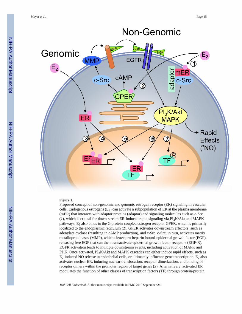
Figure 1.Proposed concept of non-genomic and genomic estrogen receptor (ER) signaling in vascularcells. Endogenous estrogens (E2) can activate a subpopulation of ER at the plasma membrane(mER) that interacts with adaptor proteins (adaptor) and signaling molecules such as c-Src(1), which is critical for down-stream ER-induced rapid signaling via PI3K/Akt and MAPKpathways. E2 also binds to the G protein-coupled estrogen receptor GPER, which is primarilylocalized to the endoplasmic reticulum (2). GPER activates downstream effectors, such asadenylate cyclase (resulting in cAMP production), and c-Src. c-Src, in turn, activates matrixmetalloproteinases (MMP), which cleave pro-heparin-bound-epidermal growth factor (EGF),releasing free EGF that can then transactivate epidermal growth factor receptors (EGF-R).EGFR activation leads to multiple downstream events, including activation of MAPK andPI3K. Once activated, PI3K/Akt and MAPK cascades can either induce rapid effects, such asE2-induced NO release in endothelial cells, or ultimately influence gene transcription. E2 alsoactivates nuclear ER, inducing nuclear translocation, receptor dimerization, and binding ofreceptor dimers within the promoter region of target genes (3). Alternatively, activated ERmodulates the function of other classes of transcription factors (TF) through protein-protein
Meyer et al. Page 15
Mol Cell Endocrinol. Author manuscript; available in PMC 2010 September 24.
NIH
-PA Author Manuscript
NIH
-PA Author Manuscript
NIH
-PA Author Manuscript
interactions (4). ER transcriptional activity may be further enhanced by phosphorylation (5),or other transcription factors may be activated that either directly interact with ER (6), or bindindependent of ER (7) within the promoter region of the target gene.
Meyer et al. Page 16
Mol Cell Endocrinol. Author manuscript; available in PMC 2010 September 24.
NIH
-PA Author Manuscript
NIH
-PA Author Manuscript
NIH
-PA Author Manuscript
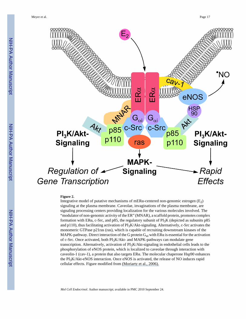
Figure 2.Integrative model of putative mechanisms of mERα-centered non-genomic estrogen (E2)signaling at the plasma membrane. Caveolae, invaginations of the plasma membrane, aresignaling processing centers providing localization for the various molecules involved. The“modulator of non-genomic activity of the ER” (MNAR), a scaffold protein, promotes complexformation with ERα, c-Src, and p85, the regulatory subunit of PI3K (depicted as subunits p85and p110), thus facilitating activation of PI3K/Akt-signaling. Alternatively, c-Src activates themonomeric GTPase p21ras (ras), which is capable of recruiting downstream kinases of theMAPK-pathway. Direct interaction of the G protein Gαi with ERα is essential for the activationof c-Src. Once activated, both PI3K/Akt- and MAPK-pathways can modulate genetranscription. Alternatively, activation of PI3K/Akt-signaling in endothelial cells leads to thephosphorylation of eNOS protein, which is localized to caveolae through interaction withcaveolin-1 (cav-1), a protein that also targets ERα. The molecular chaperone Hsp90 enhancesthe PI3K/Akt-eNOS interaction. Once eNOS is activated, the release of NO induces rapidcellular effects. Figure modified from (Moriarty et al., 2006).
Meyer et al. Page 17
Mol Cell Endocrinol. Author manuscript; available in PMC 2010 September 24.
NIH
-PA Author Manuscript
NIH
-PA Author Manuscript
NIH
-PA Author Manuscript
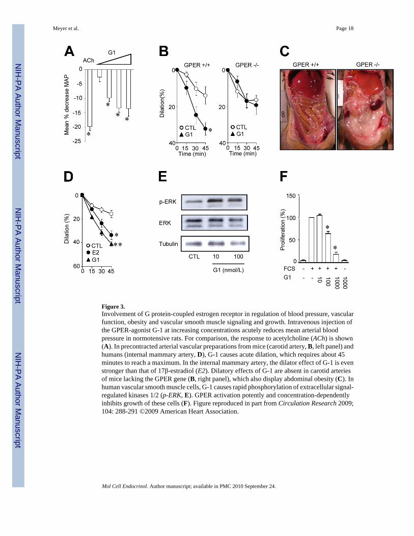
Figure 3.Involvement of G protein-coupled estrogen receptor in regulation of blood pressure, vascularfunction, obesity and vascular smooth muscle signaling and growth. Intravenous injection ofthe GPER-agonist G-1 at increasing concentrations acutely reduces mean arterial bloodpressure in normotensive rats. For comparison, the response to acetylcholine (ACh) is shown(A). In precontracted arterial vascular preparations from mice (carotid artery, B, left panel) andhumans (internal mammary artery, D), G-1 causes acute dilation, which requires about 45minutes to reach a maximum. In the internal mammary artery, the dilator effect of G-1 is evenstronger than that of 17β-estradiol (E2). Dilatory effects of G-1 are absent in carotid arteriesof mice lacking the GPER gene (B, right panel), which also display abdominal obesity (C). Inhuman vascular smooth muscle cells, G-1 causes rapid phosphorylation of extracellular signal-regulated kinases 1/2 (p-ERK, E). GPER activation potently and concentration-dependentlyinhibits growth of these cells (F). Figure reproduced in part from Circulation Research 2009;104: 288-291 ©2009 American Heart Association.
Meyer et al. Page 18
Mol Cell Endocrinol. Author manuscript; available in PMC 2010 September 24.
NIH
-PA Author Manuscript
NIH
-PA Author Manuscript
NIH
-PA Author Manuscript


