Embed Size (px)
Citation preview

GASTROENTEROLOGY 1996;111:1134–1140
RAPID COMMUNICATIONS
Elevated Cyclooxygenase-2 Levels in Min Mouse Adenomas
CHRISTOPHER S. WILLIAMS,*,‡ CINDY LUONGO,§ ARAMANDLA RADHIKA,* TONG ZHANG,x
LAURA W. LAMPS,Ø LILLIAN B. NANNEY,‡,\,Ø R. DANIEL BEAUCHAMP,‡,\
and RAYMOND N. DUBOIS*,‡,Ø
Departments of *Medicine, \Surgery, ‡Cell Biology, and ØPathology, The Vanderbilt Cancer Center, Vanderbilt University Medical Center, andØVA Medical Center, Nashville, Tennessee; and §University of Wisconsin, Madison, Wisconsin
Background & Aims: Mutations in the APC gene result C57BL/6J mice were treated with ethylnitrosourea andin an increased propensity to develop intestinal neopla- bred for transmission of germline mutations. A line wassia; however, a complete understanding of the mecha- developed that had multiple intestinal neoplasms (Min).3nisms resulting in tumor formation has remained elu- These mice exhibited a phenotype similar to FAP insive. Min mice possess a mutation in the APC gene humans. Specifically, they developed numerous intestinaland display a neoplastic phenotype similar to that ob- polyps, with a subpopulation exhibiting a neoplastic phe-served in familial adenomatous polyposis coli in hu- notype that was inherited in a fully penetrant autosomal-mans. Cyclooxygenase (COX) inhibitors decrease tu-
dominant fashion. Linkage studies revealed that germlinemor multiplicity in the Min mouse intestine. Thetransmission of a mutant allele of the APC gene waspresent study was designed to determine if there wastightly linked with the multiple intestinal neoplasia phe-an increase in COX-2 in adenomas harvested from Minnotype.4 Subsequently, it was shown that a germlinemouse intestine. Methods: COX-2 messenger RNA lev-nonsense mutation in the APC gene was strongly corre-els were determined by Northern blots and reverse-
transcription polymerase chain reactions of B6Min 1 lated with this phenotype.5 Additional studies have re-129 mouse-derived tumors. Protein levels and localiza- vealed that a second inactivating mutation in the normaltion were determined by Western blots and immunohis- allele of the APC gene results in adenoma formation.6,7
tochemical staining. Results: The Northern blots re- This is consistent with Knudson’s hypothesis, whichvealed an approximately threefold increase in the level states that at least two separate events are required forof COX-2 messenger RNA in Min mouse adenoma com- tumor formation.pared with normal mucosa. COX-2 protein levels in ade- Two cyclooxygenase (COX) isoforms have been identi-nomatous tissues were also approximately threefold
fied, which will be referred to as COX-1 and COX-2 inhigher compared with normal mucosa from the samethis report. COX-1 is expressed constitutively in a num-mouse. Immunohistochemical staining with a mono-ber of cell types, whereas COX-2 is inducible by a varietyspecific COX-2 antibody confirmed that increases inof factors including cytokines, growth factors, and tumorCOX-2 immunoreactivity were restricted to dysplasticpromoters.8,9 COX-2 was identified by many groups asand neoplastic foci within intestinal mucosa. Conclu-
sions: These data show that COX-2 levels may be in- a member of a class of genes referred to as ‘‘immediatecreased at an early stage in colorectal neoplasia during early’’ or ‘‘early growth response’’ genes.10–14 These genespolyp formation and before invasion. are rapidly and transiently induced after growth factor
or phorbol ester stimulation of quiescent cells.15–17 Mem-
F bers of the immediate early gene family are quite diverse,amilial adenomatous polyposis (FAP) is an autoso-mal-dominant disease that has been linked to germ- ranging from nuclear transcription factors to cytokines.
line mutations in the APC gene.1,2 Individuals possessing The exact role many of these play in regulation of cellularthese mutations develop numerous intestinal polyps at responses to growth stimuli and tumor promoters hasan early age. Additionally, APC mutations occur in at not been clearly defined.least 60% of spontaneous human colon cancers and arealso found in adenomatous polyps. Currently, the precise Abbreviations used in this paper: bp, base pairs; COX, cyclooxygen-
ase; FAP, familial adenomatous polyposis; Min, multiple intestinalmechanisms by which inactivation of the APC gene re-neoplasia; PCR, polymerase chain reaction; RT, reverse transcrip-sults in tumor formation are unknown; however, addi-tion; SDS, sodium dodecyl sulfate.
tional mutations are necessary for progression to a trans- q 1996 by the American Gastroenterological Association0016-5085/96/$3.00formed state.
/ 5e12$$0035 09-04-96 16:37:21 gasa WBS-Gastro

CYCLOOXYGENASE–2 LEVELS IN MIN MOUSE ADENOMAS 1135October 1996
(3 mmol/L) was used to prime a standard reverse-transcriptionBoth isoforms of COX convert arachidonic acid to pros-(RT) reaction. Polymerase chain reaction (PCR) cocktail con-taglandin endoperoxide H2, which is a substrate for asisted of 10 mmol/L Tris-HCl, 50 mmol/L KCl, 1.5 mmol/Lnumber of cell- and tissue-specific prostaglandin synthases.MgCl2 , gelatin 0.01% (wt/vol), 0.2 mmol/L each deoxynucleo-Recently COX-2 has been implicated in intestinal neopla-side triphosphate, 2.0 U AmpliTaq Polymerase (Perkin-Elmersia, because increased levels of COX-2 messenger RNACorp., Norwalk, CT), and 0.4 mmol/L each of the following(mRNA) and protein have been observed in colonic adeno-primers: COX-2 5*-GTCTGATGATGTATGCCACAATCTG,
mas and carcinoma samples from humans.18–20 Addition-3*-GATGCCAGTGATAGAGGGTGTTAAA, rat b-actin (Clon-
ally, patients with FAP receiving sulindac (a COX inhibi- tech #5506-3) 5*-TTGTAACCAACTGGGACGATATGG,tor) have a significant reduction in the size and number and 3*-GATCTTGATCTTCATGGTGCTAGG in a total vol-of adenomatous polyps.21–24 Furthermore, recent work in ume of 50 mL. Two microliters of cDNA template was addedour laboratory has revealed that cultured rat intestinal and finger-vortexed. b-Actin cDNA (positive control) and dou-epithelial cells constitutively expressing COX-2 are resis- ble-distilled water (negative control) were analyzed in paralleltant to sodium butyrate–induced apoptosis and exhibit to ensure the integrity of the reaction cocktail. Thermocycling
was performed according to the following profile: 947C for 45increased adhesion to extracellular matrix proteins, whichseconds, 607C for 45 seconds, and 727C for 2 minutes, repeatedare two characteristics likely to affect the tumorigenic35 times followed by a final extension at 727C for 7 minutes.potential of intestinal epithelial cells.25 Finally, a recentAnalysis of amplicons was performed on a 1.8% Tris acetatereport has indicated a reduction in tumor multiplicity inethylenediaminetetraacetic acid agarose gel. Eight microlitersB6Min mice treated with piroxicam, a potent COX inhibi-of each sample was mixed with 2 mL of DNA loading buffertor.26 These data strongly suggest a possible role for eicosa-and loaded onto the gel, and electrophoresis was performed at
noids in tumor formation in the Min mouse.125 V for 1.5 hours. A 100–base pair (bp) ladder (Promega,
The present study was designed to determine whether Madison, WI) was used as a size standard. The gel was stainedCOX-2 levels were altered in adenoma tissue harvested with ethidium bromide and then photographed. The ampli-from B6Min 1 129 mice (Min mice). We found increased cons generated by COX-2 and control b-actin primers wereCOX-2 mRNA and protein levels in adenomas obtained 276 and 764 bp in length, respectively.from Min mice.
Western BlottingMaterials and MethodsThe tissues were homogenized at 47C in radioimmuno-Extraction of Total RNA From Tissue
precipitation assay buffer (150 mmol/L NaCl, 1% NonidetSamples P40, and 50 mmol/L Tris, pH 8.0) containing 10 mg/mLIntestinal tissue samples were obtained from C57BL6/ aprotinin, 1 mmol/L sodium orthovanadate, and 100 mg/mL
J-Min//1 129///mice after death. Samples were harvested phenylmethylsulfonyl fluoride. Centrifuged homogenates (100and immediately snap frozen in liquid nitrogen. Tissue samples mg) were denatured and fractionated on 7.5% polyacrylamidewere divided and weighed, and an appropriate amount of re- gels containing SDS and then transferred to nitrocelluloseagent was added before homogenization. Total RNA extraction membrane using a semidry cell (Bio-Rad Laboratories, Rich-was performed as previously described.18
mond, CA). Filters were incubated overnight at room tempera-ture in blocking solution (Tris-buffered saline containing 5%Northern Blottingnonfat dried milk and 0.05% Tween 20), followed by a 4-hour
Forty micrograms of total RNA from each sample was incubation period with primary rabbit antisera25 in blockingelectrophoresed in denaturing agarose gels and transferred to solution. Filters were washed three times and incubated withnitrocellulose. Nitrocellulose blots were hybridized using stan- a horseradish peroxidase–conjugated goat anti-rabbit immu-dard conditions followed by 0.1% standard saline citrate/0.1% noglobulin as a secondary antibody (1:2000 dilution) for 1sodium dodecyl sulfate (SDS) posthybridization washes at hour. After three additional washes, filters were treated by427C.18 Blots were exposed for various lengths of time before the enhanced chemiluminescence system (Amersham Corp.,they were developed. Neoplastic intestinal mucosa samples
Arlington Heights, IL) and exposed to X-omat AR film (Ko-were evaluated and compared with results from adjacent nor-
dak, New Haven, CT). Protein levels were quantified by scan-mal mucosa. Blots were stripped and rehybridized with a 32P-
ning the autoradiographs and analyzing band density via NIHlabeled 18S ribosomal probe to ensure loading consistency.
Image 1.59 software.mRNA levels were quantified by scanning the autoradiographsand analyzing band density via NIH Image 1.59 software Histopathology and Immunohistochemistry(National Institutes of Health, Bethesda, MD).
Serial sections cut from each paraffin-embedded blockReverse-Transcription Multiplex were stained with H&E for morphological examination by aPolymerase Chain Reaction pathologist who was unaware of the results of the immunoper-
oxidase, Northern blot, or Western blot results. Each specimenFirst-strand complementary DNA (cDNA) was gener-ated using 10 mg of total RNA as template. Random hexamer was examined for the presence of adenomatous polyps, which
/ 5e12$$0035 09-04-96 16:37:21 gasa WBS-Gastro
1136 WILLIAMS ET AL. GASTROENTEROLOGY Vol. 111, No. 4
were classified as tubular, villous, or tubulovillous.27 The de-gree of cytological dysplasia within each adenomatous polypwas further classified as low grade or high grade according tothe World Health Organization criteria.28
Paraffin-embedded sections (5 mm) were adhered to glassslides, dewaxed, deparaffinized, and rehydrated in xylene,graded alcohol, and phosphate-buffered saline (PBS), respec-tively. The sections were then incubated in 1% hydrogen per-oxide for 15 minutes at room temperature to quench endoge-nous peroxidase and then digested in 0.1% trypsin for 5minutes at 377C. After the sections were blocked in 1.5%normal horse serum for 30 minutes at room temperature, theexcess serum was removed and sections were incubated with1:25 diluted mouse anti–COX-2 monospecific antibody for18 hours at 47C. Then, the biotinylated horse anti-mouse im-munoglobulin and horseradish peroxidase–conjugated antibio-tin antibody were respectively applied to the sections for 30minutes at RT. The sections were washed in PBS three timesbetween each step. Peroxidase activity was shown by applying3,3*-diaminobenzidine tetrahydrochloride containing 0.05%hydrogen peroxide for 5–10 minutes at RT. The sections werethen thoroughly rinsed in tap water and counterstained withhematoxylin. Finally, the sections were dehydrated, cleared,and mounted with coverslips. The specificity of the immuno-histochemical staining with the COX-2 antibody has beenestablished previously. Intestinal epithelial cell lines engi-neered to express COX-2 or COX-1 have been probed withthe COX-2 antibody, and specificity was shown via specificstaining of the COX-2 cell line with the absence of a signalin the COX-1 cell line (data not shown).
Figure 1. (A) Analysis of COX-2 mRNA levels in total RNA extracted fromboth adenomas and normal mucosa from selected C57BL6/J-Min// 1Results129 /// mice. Northern blotting was performed using 40 mg of total
Northern Hybridization RNA per sample. The blot was hybridized with a radiolabeled COX-2 cDNAcoding region fragment and was then stripped and rehybridized with a
Forty micrograms of total RNA from Min mice radiolabeled 18S ribosomal RNA probe to ensure loading consistencybetween samples. N, normal mucosa; T, tumor. (B) Analysis of COX-2neoplastic and normal intestinal mucosa was electropho-mRNA levels with RT multiplex PCR. Ten micrograms of total RNA ex-resed on a denaturing agarose gel and transferred to atracted from matched tumor and normal mucosal samples was used asnitrocellulose filter. Loading consistency was determined template to generate cDNA. Two microliters of cDNA was used as tem-
by direct comparison of 18S ribosomal RNA (present at plate in a 50 mL multiplex RT-PCR reaction primed with COX-2 and b-actin control primers. Eight microliters of sample reaction was mixedhigh levels in eukaryotic cells) visualized by ethidiumwith 2 mL of DNA loading buffer followed by agarose gel electrophoresis.bromide staining and through a separate hybridization107 and 108, animal identification numbers; N, normal mucosa; T,
with a 32P-labeled 18S specific probe. The murine COX- tumor; PC, positive control; b-actin, cDNA control; NC, negative control.(C) Immunoblotting of COX-2 protein from both adenomas and normal2 cDNA was radiolabeled with [32P] deoxycytidine tri-mucosa from Min mice. N, normal mucosa; T, tumor.phosphate and hybridized to the nitrocellulose filter
where the RNAs were transferred. We observed an ap-proximately threefold increase in COX-2 RNA levels in generated amplicon for COX-2 primer amplificationthe neoplastic mucosal samples compared with adjacent was 276 bp. The contamination control (water blank)normal tissue (Figure 1A). indicated that no amplification occurred in the ab-
sence of bona fide template. Comparison of the COX-RT-PCR 2 amplicons with b-actin control amplicons indicatedTen micrograms of Min mouse total RNA from the high quality of the cDNA template. Direct com-
each sample was used to generate cDNA, which was parison of the COX-2 tumor and normal PCR prod-subsequently used as template in a standard RT-PCR ucts showed an increased COX-2 in tumor, corrobo-reaction using both COX-2 and b-actin control-spe- rating the previous Northern blot observations (see
Figure 1A and B).cific primer pairs. The predicted size of the PCR-
/ 5e12$$0035 09-04-96 16:37:21 gasa WBS-Gastro

CYCLOOXYGENASE–2 LEVELS IN MIN MOUSE ADENOMAS 1137October 1996
Western Blotting factors can be rigidly controlled. Important work withmouse models of colorectal cancer has recently shownAfter determining that the COX-2 RNA levelsome of the key genetic components involved in certainwas increased, we sought to determine if there was atypes of neoplastic transformation in the human gastroin-similar increase in the amount of COX-2 protein in thetestinal tract.adenoma. We electrophoresed 100 mg of protein from
The Min mice line was originally derived from aeach sample on a SDS-polyacrylamide gel, transferred thefounding male C57BL/6J mouse that had been treatedseparated proteins onto a nitrocellulose membrane, andwith the carcinogen ethylnitrosourea and bred for trans-probed the filter with monospecific rabbit antisera tomission of germline mutations.3 On close examination,COX-2. Immunoblot analysis indicated an approxi-some of the progeny possessed numerous intestinal pol-mately threefold increase in the amount of protein pres-yps, a phenotype similar to FAP in humans. Later it wasent in the adenomatous tissue compared with samplesdiscovered that these mice shared an additional featureobtained from normal mucosa (Figure 1C).in common with FAP; they also harbored a mutation in
Histopathology the APC gene, and thus they are justifiably a model ofFAP. However, tumor distribution differs in the MinFour adenomatous polyps were identified in speci-mice because most of the tumors are localized in themens from three Min mice: two tubular, one villous, andsmall intestine and not in the colon, as in FAP. Thisone tubulovillous. Three adenomas contained low-grademurine line presents a unique opportunity to dissect thedysplasia, and the tubulovillous adenoma containedgenetic components responsible for neoplastic progres-high-grade dysplasia throughout the lesion. No invasivesion in tumors occurring as a result of mutations in thecarcinoma or carcinoma in situ were found in these sam-APC gene.ples. Sections of adjacent grossly normal intestinal epi-
We report an increased level of both COX-2 mRNAthelium were histologically unremarkable.and protein in tumors harvested from intestinal mucosa
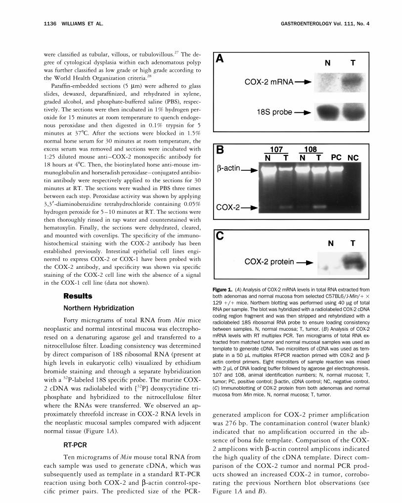
Immunohistochemical Staining of a particular strain of Min mice (B6Min 1 129). COX-2 mRNA or protein in normal mucosal samples wereRepresentative tissue sections from adenomatousbarely detectable; however, COX-2 was present in thelesions and normal epithelium were prepared and probedtumor samples. Levels of both COX-2 mRNA and pro-with a monospecific COX-2 antibody. COX-2 proteintein in the tumor samples were increased approximatelystaining was not observed in either wild-type mouse in-threefold over adjacent normal intestinal mucosa. Thesetestinal tissue sections (Figure 2A) or sections derivedassays probably underestimated the relative expression offrom Min mice–normal intestinal mucosa (Figure 2B).COX-2 in dysplastic cells because processing involvedHowever, in accordance with the previous data, highthe homogenization of grossly dissected adenomatous tis-levels of COX-2 protein were observed in subpopulationssue, which is likely to contain a variety of epithelialof adenomatous and dysplastic intestinal epithelium (Fig-phenotypes ranging from normal to neoplastic.ure 2C). Some of the sections of polypoid tumors con-
RT-PCR was performed to confirm the differentialtained normal (white arrow) and neoplastic cells (blackexpression of COX-2 in tumor samples when comparedarrow) and therefore afforded internal positive and nega-with normal mucosa. After standardization to a b-actintive controls (Figure 2D). We have also included somecontrol, a definitive increase in the level of COX-2representative immunostaining results from a human co-mRNA in the tumor samples was observed. The presencelorectal adenocarcinoma for comparison. The most strik-of a faint COX-2 amplicon in the normal mucosal sam-ing result from the human studies is that the bulk ofples is not surprising, because the sensitivity of the RT-COX-2 protein is expressed predominantly in the neo-PCR reaction and the harvesting technique we used couldplastic epithelial cells (arrows). This observation confirmsnot distinguish microscopic lesions from normal mucosa.a previous report.20
Alternatively, the up-regulation of COX-2 may occur atDiscussion a very early stage in neoplastic transformation, but such
a point would be difficult to distinguish at the grossThe precise cause of colorectal cancer is currentlylevel. However, the RT-PCR experiment confirms theunder intense investigation. Complex genetic and envi-initial Northern blotting results that indicate an increaseronmental factors contribute to the predisposition for andin COX-2 mRNA levels in neoplastic tissue.subsequent development of neoplastic transformation of
colonic epithelial cells. Animal models are extremely use- The immunohistochemical localization patterns fromthe Min mouse intestinal mucosal samples support theful for investigating components contributing to tumor
progression because both genotypic and environmental Northern and Western blotting data. Within these sec-
/ 5e12$$0035 09-04-96 16:37:21 gasa WBS-Gastro

1138 WILLIAMS ET AL. GASTROENTEROLOGY Vol. 111, No. 4
Figure 2. Immunohistochemical staining with a monospecific COX-2 antibody. (A) C57BL6/J Apc (///) wild-type mouse normal intestinalmucosa sample (original magnification 3001). (B) C57BL6/J-Min// 1 129 /// normal mucosa sample (original magnification 1301). (C)C57BL6/J-Min// 1 129 /// neoplastic mucosa sample (original magnification 3201). (D) C57BL6/J-Min// 1 129 /// mixed normal andneoplastic section (original magnification 2001). (E) Human adenocarcinoma sample indicating increased COX-2 immunohistochemical localiza-tion in neoplastic regions (original magnification 1301).
tions, multiple cell types such as lymphocytes and con- It has long been known that nonsteroidal anti-in-flammatory drugs (NSAIDs) inhibit COX enzymatic ac-nective tissue components were present, which showed
some immunoreactivity to COX-2 antiserum. Sections tivity. Several large studies have shown a positive correla-tion between continuous NSAID use and decreased riskalso included normal mucosal cells with no immunoreac-
tivity for COX-2 and dysplastic or neoplastic intestinal of colorectal cancer in humans. This suggests a possiblerole for COX-2 in tumor formation, although NSAIDsepithelial cells, which were predominantly stained posi-
tive with COX-2 antibody. Therefore, the low level of have other effects in addition to their ability to inhibitCOXs. Interestingly, COX-2 up-regulation has been ob-expression observed in the Northern and Western blot-
ting data is understandable considering the ratio of dys- served in tumor samples harvested from patients withcolorectal cancer. Additionally, our laboratory has re-plastic cells to normal cells observed after immunohisto-
chemical staining. cently reported increased levels of COX-2 mRNA and
/ 5e12$$0035 09-04-96 16:37:21 gasa WBS-Gastro

CYCLOOXYGENASE–2 LEVELS IN MIN MOUSE ADENOMAS 1139October 1996
M, White R. Identification and characterization of the familialprotein in rat colonic tumors that developed after carcin-adenomatous polyposis coli gene. Cell 1991;66:589–600.ogen treatment.29 Most interesting, perhaps, is the recent 3. Moser AR, Pitot HC, Dove WF. A dominant mutation that predis-
report that showed a decrease in tumor load in B6Minposes to mutiple intestinal neoplasia in the mouse. Science1990;247:322–325.mice treated with piroxicam, which is a COX inhibitor.26
4. Su LK, Kinzler KW, Vogelstein B, Preisinger AC, Moser AR, LuongoTo the best of our knowledge, the findings of Jacoby etC, Gould KA, Dove WF. Multiple intestinal neoplasia caused by
al.26 represent the first report demonstrating a role for a mutation in the murine homolog of the APC gene. Science1992;256:668–670.NSAIDs in tumor prevention in the Min mouse. Con-
5. Oshima M, Oshima H, Kitagawa K, Kobayashi M, Itakura C, Ta-firmation of these results has been obtained from a studyketo M. Loss of APC heterozygosity and abnormal tissue building
in which Min mice were treated with sulindac, which is in nascent intestinal polyps in mice carrying a truncated APCanother potent NSAID. These mice also had a decrease gene. Proc Natl Acad Sci USA 1995;92:4482–4486.
6. Luongo C, Moser AR, Gledhill S, Dove WF. Loss of APC/ inin tumor multiplicity as well as an increase in apoptosisintestinal adenomas from Min mice. Cancer Res 1994;54:when compared with control Min mice.30 Further re-5947–5952.
search will be necessary to determine mechanisms 7. Levy DB, Smith KJ, Beazer–Barclay Y, Hamilton SR, VogelsteinB, Kinzler KW. Inactivation of both APC alleles in human andwhereby NSAID treatment results in decreased tumormouse tumors. Cancer Res 1994;54:5953–5958.multiplicity in these mice.
8. Williams CW, DuBois RN. Prostaglandin endoperoxide synthase:COX-2 protein is undetectable in normal intestinal why two isoforms? Am J Physiol 1996;270:G393–G400.mucosa; however, at some step in the transformation of 9. Eberhart CE, DuBois RN. Eicosanoids and the gastrointestinal
tract. Gastroenterology 1995;109:285–301.normal intestinal epithelial cells to malignant cells an10. DuBois RN, Tsujii M, Bishop P, Awad JA, Makita K, Lanahan A.increase in COX-2 expression occurs. The data presented
Cloning and characterization of a growth factor-inducible cyclooxy-in this report indicate that COX-2 expression is up- genase gene from rat intestinal epithelial cells. Am J Physiol
1994;266:G822–G827.regulated at an early, preinvasive stage in Min mouse11. Farber JM. A collection of mRNA species that are inducible inadenomas. It is highly likely that additional genetic alter-
the RAW 264.7 mouse macrophage cell line by gamma interferonations such as the loss of p53 or DCC mutations are and other agents. Mol Cell Biol 1992;12:1535–1545.required for progression to a malignant state. 12. Kujubu DA, Fletcher BS, Varnum BC, Lim RW, Herschman HR.
TIS10, a phorbol ester tumor promoter-inducible mRNA fromWhereas the precise role that COX-2 plays in tumorSwiss 3T3 cells, encodes a novel prostaglandin synthase/formation remains unclear, an enlarging body of evidencecyclooxygenase homologue. J Biol Chem 1991;266:12866–
suggests that COX-2 expression is altered during the 12872.13. Simmons DL, Levy DB, Yannoni Y, Erikson RL. Identification ofdevelopment of colorectal cancer in rodent models and in
a phorbol ester-repressible v-src-inducible gene. Proc Natl Acadhumans. Our work in cultured epithelial models suggestsSci USA 1989;86:1178–1182.that the increase in COX-2 may enable the cell to resist 14. Ryseck RP, Raynoschek C, Macdonald–Bravo H, Dorfman K,
apoptosis and enhance the ability of the cell to bind Mattei MG, Bravo R. Identification of an immediate early gene,pghs-B, whose protein product has prostaglandin synthase/extracellular matrix. This report shows an increase incyclooxygenase activity. Cell Growth Differ 1992;3:443–450.COX-2 mRNA and a subsequent increase in COX-2
15. Rollins BJ, Stiles CD. Serum-inducible genes. Adv Cancer Resprotein in adenomas harvested from Min mice. The speci- 1989;53:1–32.ficity of COX-2 protein expression in transformed epithe- 16. Herschman HR. Primary response genes induced by growth fac-
tors and tumor promoters. Annu Rev Bioch 1991;60:281–319.lial cells is shown by immunohistochemical staining.17. Nathans D, Lau LF, Christy B, Hartzell S, Nakabeppu Y, Ryder K.Detailed studies are underway that will more carefully
Genomic response to growth factors. Cold Spring Harb Sympevaluate the temporal pattern of COX-2 expression in Quant Biol 1988;53:893–900.
18. Eberhart CE, Coffey RJ, Radhika A, Giardiello FM, Ferrenbach S,Min mice and in selected backcrosses that present withDuBois RN. Up-regulation of cyclooxygenase 2 gene expressionlower tumor multiplicity and, consequently, have an in-in human colorectal adenomas and adenocarcinomas. Gastroen-
creased life span enabling long-term studies. These future terology 1994;107:1183–1188.studies will include a more detailed, long-term investiga- 19. Kargman S, O’Neill G, Vickers P, Evans J, Mancini J, Jothy S.
Expression of prostaglandin G/H synthase-1 and -2 protein intion to fully analyze the role of COX-2 in tumorigenesishuman colon cancer. Cancer Res 1995;55:2556–2559.in the Min mouse.
20. Sano H, Kawahito Y, Wilder RL, Hashiramoto A, Mukai S, AsaiK, Kimura S, Kato H, Kondo M, Hla T. Expression of cyclooxygen-Referencesase-1 and -2 in human colorectal cancer. Cancer Res 1995;55:3785–3789.1. Nishisho I, Nakamura Y, Miyoshi Y, Miki Y, Ando H, Horii A,
21. Giardiello FM, Hamilton SR, Krush AJ, Piantadosi S, Hylind LM,Koyama K, Utsunomiya J, Baba S, Hedge P. Mutations of chromo-Celano P, Booker SV, Robinson CR, Offerhaus GJ. Treatment ofsome 5q21 genes in FAP and colorectal cancer patients. Sciencecolonic and rectal adenomas with sulindac in familial adenoma-1991;253:665–669.tous polyposis. N Engl J Med 1993;328:1313–1316.2. Groden J, Thliveris A, Samowitz W, Carlson M, Gelbert L, Al-
22. Waddell WR, Gasner GF, Cerise EJ, Loughry RW. Sulindac forbertsen H, Joslyn G, Stevens J, Spirio L, Robertson M, Sargeantpolyposis of the colon. Am J Surg 1989;157:175–178.L, Krapcho K, Wolff E, Burt R, Hughes JP, Warrington J, McPher-
son J, Wasmuth J, Le Paslier D, Abderrahim H, Cohen D, Leppert 23. Winde G, Gumbinger HG, Osswald H, Kemper F, Bunte H. The
/ 5e12$$0035 09-04-96 16:37:21 gasa WBS-Gastro

1140 WILLIAMS ET AL. GASTROENTEROLOGY Vol. 111, No. 4
NSAID sulindac reverses rectal adenomas in colectomized pa- Ramonetti JT, Abreu–Goris M, Newmark HL, Lipkin ML, DeCosseJJ, Bertagnolli MM. Cyclooxygenase-2 overexpression and tumortients with familial adenomatous polyposis: clinical results of a
dose-finding study on rectal sulindac administration. Int J Colo- formation are blocked by sulindac in a murine model of familialadenomatous polyposis. Cancer Res 1996;56:2556–2560.rectal Dis 1993;8:13–17.
24. Nugent KP, Farmer KC, Spigelman AD, Williams CB, Phillips RK.Randomized controlled trial of the effect of sulindac on duodenaland rectal polyposis and cell proliferation in patients with familial Received April 16, 1996. Accepted July 8, 1996.
Address requests for reprints to: Raymond N. DuBois, M.D., Ph.D.,adenomatous polyposis. Br J Surg 1993;80:1618–1619.25. Tsujii M, DuBois RN. Alterations in cellular adhesion and Department of Medicine/GI; MCN C-2104, Vanderbilt University Med-
ical Center, Nashville, Tennessee 27232-2279. Fax: (615) 343-apoptosis in epithelial cells overexpressing prostaglandin endo-peroxide synthase-2. Cell 1995;83:493–501. 6229. e-mail: [email protected].
Supported in part from United States Public Health Services grants26. Jacoby RF, Marshall DJ, Newton MA, Novakovic K, Tutsch K,Cole CE, Lubet RA, Kelloff GJ, Verma A, Moser AR, Dove WF. P30 AR41943 (to L.B.N.), GM-40439 (to L.B.N.), DK 47297 (to
R.N.D.), 5P030 ES-00267 (to R.N.D.), GM-53319 (to R.D.B.), andChemoprevention of spontaneous intestinal adenomas in theApcMin mouse model by the nonsteroidal anti-inflammatory drug CA 69457 (to R.D.B.). Dr. DuBois is a recipient of a VA Research
Associate career development award and a Boehringer Ingelheimpiroxicam. Cancer Res 1996;56:710–714.27. Kozuka S. Premalignancy of the mucosal polyp in the large intes- New Investigator Award and is an AGA Industry Research Scholar.
Support for Dr. Luongo is derived from grant R01-CA63677 (to Wil-tine. Dis Colon Rectum 1975;18:483–493.28. Jass R, Sobiu LH. Histological typing of intestinal tumors. WHO liam F. Dove), grant TG32-GM07133, and Core Grant CA07175 from
the National Cancer Institute.Intl Histological Classification of Tumors 1989;13:29–30.29. DuBois RN, Radhika A, Reddy BS, Entingh AJ. Increased cyclooxy- The authors thank Bill Dove for his kind support of this project and
critical input and M. E. John and family for their kind hospitalitygenase-2 levels in carinogen-induced rat colonic tumors. Gastro-enterology 1996;110:1259–1262. during trips to Madison, Wisconsin, which were required for tumor
collection from the Min mice.30. Boolbol SK, Dannenberg AJ, Chadburn A, Martucci C, Guo X,
/ 5e12$$0035 09-04-96 16:37:21 gasa WBS-Gastro