Embed Size (px)
Citation preview

P2
Experimental Cell Research 264, 397–407 (2001)doi:10.1006/excr.2000.5145, available online at http://www.idealibrary.com on
Enhancement of Connexin 43 Expression Increases Proliferationand Differentiation of an Osteoblast-like Cell Line
B. Gramsch,* H.-D. Gabriel,† M. Wiemann,* R. Grummer,† E. Winterhager,†D. Bingmann,*,1 and K. Schirrmacher*
*Department of Physiology and †Department of Anatomy, Medical School, University of Essen, D-45122 Essen, Germany
tsgta
bcomvsdbrinmwrpatw[tpolsla
tCn
Bone cells form a functional syncytium as they arecoupled by gap junctions composed mainly of con-nexin 43 (Cx43). To further understand the role ofCx43 in bone cell growth and differentiation, we stablytransfected Cx45-expressing UMR 106-01 cells withCx43 using an expression vector containing rat Cx43cDNA. Three stably transfected clones were analyzed,all of which showed altered expression of Cx43 and/orCx45 as was obvious from immunocytochemistry andNorthern blotting. Double whole-cell patch clampingrevealed single-channel conductances of 20 (Cx45) and60 pS (Cx43). The overexpression of Cx43 led to anincrease in dye coupling concomitant with elevatedgap-junctional conductance. The phenotype of thetransfected clones was characterized by an increasedproliferation (4- to 7-fold) compared to controls. More-over, a transfectant clone with 10- to 12-fold enhancedCx43 expression showed a significantly increased cal-cium content of the extracellular matrix and enlargedmineralization nodules, while alkaline phosphatasewas moderately increased. We conclude that enhancedgap-junctional coupling via Cx43 significantly pro-motes proliferation and differentiation of UMR cells.© 2001 Academic Press
Key Words: osteoblast-like cells; transfection; gapjunctions; electric coupling; proliferation; differenti-ation.
INTRODUCTION
Recent studies have provided evidence that directcell–cell communication via gap junctions is an impor-tant element promoting growth and differentiation invarious tissues [1]. Also physiological functions of tis-sues, such as the proper functioning of pancreatic isletcells, obviously depend on gap junction coupling [2].Gap junctions are composed of connexins, which belongto a multigene family with numerous members and are
1 To whom reprint requests should be addressed at the Institut furhysiologie, Universitat Essen, D-45122 Essen, Germany. Fax: 149
t01 723-4648. E-mail: [email protected].
397
differentially expressed in a tissue-specific manner [3].Two hemichannels of neighboring cells, each composedof six specific protein subunits (21–47 kDa) termedconnexins [1], form a pore which allows ions and smallmolecules (,1 kDa) to pass. Consequently, gap junc-ions are involved in the transmission of bioelectricalignals such as ion currents as well as second messen-ers [4–6]. They may furthermore play a role in nutri-ion of cells remote from energy sources, thus enablingmetabolic cooperation.Gap-junctional communication is important also in
one cells [7], where the channels are involved in me-hanical transmission [8–10], induction of cytokines insteoblasts by T cells [11], and coordination of hor-onal responses [12, 13]. In osteoblast-like cells in
itro connexin 43 (Cx43) is the dominant connexinubtype and probably important for normal skeletalevelopment [14–16]. Furthermore, Cx45 was found toe expressed at a low level in osteoblast-like cells de-ived from calvaria explants [14, 17, 18]. As the biolog-cal significance and the interplay of these two con-exin subtypes is currently not understood, cell cultureodels are increasingly employed to define this role. Itas recently shown that Cx43-expressing ROS cells
educed the expression of osteocalcin and alkalinehosphatase, when cells were transfected with Cx45 asn additional connexin [19]. Another transformed os-eoblastic cell line, UMR 106-01, expresses Cx45,hich has a lower channel permeability than Cx43
20–22]. In a previous study on UMR 106-1 cells [23]he additional expression of the Cx43 gene increasedroduction of osteocalcin and bone sialoproteins, bothf which are involved in the formation of the extracel-ular matrix and calcification. These studies [19, 23]uggest that the type(s) of connexin to be expressed ateast partly determines whether cells start to prolifer-te or undergo tissue differentiation.The present study was designed to further address
his problem. We investigated whether transfection ofx45-expressing UMR 106-01 cells with Cx43 altersot only dye and electric coupling but also prolifera-
ion, activity of alkaline phosphatase, and mineraliza-0014-4827/01 $35.00Copyright © 2001 by Academic Press
All rights of reproduction in any form reserved.
E
5t6
fLs
wa
Tfaw[
6trwtit
d(aaam6frowjwwpafl
ews
mf[Caia
fiSbsLs
tp
3
a
mw
t
398 GRAMSCH ET AL.
tion [24–26]. Part of this study has been published asan abstract (B. Gramsch et al., 2000, Pflugers Arch.
ur. J. Physiol. 439(Suppl.), R371).
MATERIALS AND METHODS
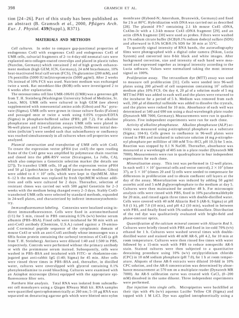
Cell cultures. In order to compare gap-junctional properties ofendogenous Cx43 with exogenous Cx43 and endogenous Cx45 ofUMR cells, calvarial fragments of 2- to 4-day-old neonatal rats wereexplanted onto collagen-coated coverslips and placed in plastic tubes(Nunclon, Germany) which contained 2 ml of high growth enhance-ment medium (high GEM; ICN, Germany), 24 mM bicarbonate, 10%heat-inactivated fetal calf serum (FCS), 1% glutamine (200 mM), and1% penicillin (5000 IU/ml)/streptomycin (5000 mg/ml). After 2 weeks% instead of 10% FCS was used. Nutrient medium was exchangedwice a week. Rat osteoblast-like (ROB) cells were investigated 2 toweeks after explantation.The osteosarcoma cell line UMR-106/01 (UMR) was a generous gift
rom T. H. Steinberg (Washington University School of Medicine, St.ouis, MO). UMR cells were cultured in high GEM (see above)upplemented with nonessential amino acids (Gibco) and Na1 pyru-
vate (Sigma). UMR cells were grown in tissue culture flasks (Falcon)and passaged once or twice a week using 0.05% trypsin/EDTA(Sigma) in phosphate-buffered saline (PBS; pH 7.2). For alkalinephosphatase and mineralization assays UMR cells were plated in96-well plates and 12-well plates, respectively. Different initial den-sities (cells/cm2) were seeded such that subconfluency or confluency
as reached simultaneously in all cultures when cell properties werenalyzed.Plasmid construction and transfection of UMR cells with Cx43.
o create the expression vector pPH4 (rat cx43) the open readingrame of the Cx43 gene was amplified by polymerase chain reactionnd cloned into the pBK-RSV vector (Stratagene, La Jolla, CA),hich also comprises a Geneticin selection marker (for details see
27]). To transfect UMR cells, 10 mg of the expression vector pPH4and 50 ml LipofectAmine in OptiMEM (serum-free medium; Gibco)were added to 4 3 106 cells, which were kept in OptiMEM. After
–12 h the medium was replaced by fresh OptiMEM without addi-ives and cells were cultured for 2 days. Thereafter, selection ofesistant clones was carried out with 500 mg/ml Geneticin for 2–3eeks with the medium being changed every 2–3 days. Stably Cx43-
ransfected UMR cells were selected by standard methods, expandedn 24-well plates, and characterized by indirect immunocytochemis-ry.
Immunofluorescence labeling. Connexins were localized using in-irect immunofluorescence. Cells were fixed in acetone/methanol1/1) for 5 min, rinsed in PBS containing 0.5% (w/v) bovine serumlbumin (PBS–BSA). Fixed cells were incubated for 90 min with annti-Cx43 antibody (Chemicon, U.S.A.) raised against a 19-amino-cid C-terminal peptide sequence of the cytoplasmic domain ofouse Cx43 or with an anti-Cx45 antibody whose immunogen was a
His fusion protein containing the carboxyl terminus of Cx45 (a giftrom T. H. Steinberg). Antisera were diluted 1:40 and 1:500 in PBS,espectively. Controls were performed without the primary antibodyr with the preimmune serum instead. Subsequently, cells wereashed in PBS–BSA and incubated with FITC- or rhodamine-con-
ugated goat anti-rabbit IgG (1:40; Sigma) for 45 min. After cellsere rinsed three times in PBS–BSA and, thereafter, in distilledater, cultures were coverslipped with glycerol containing 0.1%henylenediamine to avoid bleaching. Cultures were examined withn Axiophot microscope (Zeiss) equipped with the appropriate epi-uorescence filter sets.Northern blot analysis. Total RNA was isolated from subconflu-
nt cell monolayers using a Qiagen RNeasy Midi kit. RNA samplesere quantified spectrophotometrically at 260 nm; 5–10 mg RNA was
eparated on denaturing agarose gels which were blotted onto nylon t
embrane (Hybond-N; Amersham, Brunswick, Germany) and fixedor 2 h at 80°C. Hybridization with DNA was carried out as described28]. The plasmid F9-7 containing 2.1 kb mouse Cx45 cDNA,x43m-3c with a 1.3-kb mouse Cx43 cDNA fragment [29], and anctin cDNA fragment [30] were used as probes. Filters were washedn 1% sodium citrate buffer (SCB)/0.1% sodium dodecyl sulfate (SDS)nd afterward in 0.5% SCB/0.1% SDS for 30 min at 55°C.To quantify signal intensity of RNA bands, the autoradiography
lms were photographed with a digital color camera (Nikon, Luciaystem) and converted into 8-bit black and white images. Afterackground correction, size and intensity of each band were mea-ured and expressed together as integral intensity according to theucia software. Values were normalized taking the most intenseignal as 100%.Proliferation assay. The tetrazolium dye (MTT) assay was used
o measure cell proliferation [31]. Cells were seeded into 96-welllates using 200 ml/well of cell suspension containing 105 cells/ml
medium plus 10% FCS. On day 4, 20 ml of a solution made of 5 mgMTT/ml PBS was added to each well and the plates were returned tothe incubator for a period of 5 h. Medium was removed from eachwell, 200 ml of dimethyl sulfoxide was added to dissolve the crystals,and the plates were rocked for 10 min. Absorbance of each well wasdetermined at 540 and 690 nm using a dual-wavelength plate reader(Dynatech MR 7000, Germany). Measurements were run in quadru-plicates. Five independent experiments were run for each clone.
Activity of alkaline phosphatase. Alkaline phosphatase (ALP) ac-tivity was measured using p-nitrophenyl phosphate as a substrate(Sigma; 104-S). Cells grown to confluence in 96-well plates wererinsed in PBS and incubated in substrate solution (4 mg p-nitrophe-nyl phosphate per milliliter of 100 mM Tris–HCl, pH 10.3) for 5 min.Reaction was stopped by 0.1 N NaOH. Thereafter, absorbance wasmeasured at a wavelength of 405 nm in a plate reader (Dynatech MR7000). Measurements were run in quadruplicate in four independentexperiments for each clone.
Mineralization assay. This test was performed in 12-well plates.In each well 1 3 105 (parental cells and vector control), 2 3 104 (clone7), or 5 3 104 (clones 20 and 5) cells were seeded to compensate for
differences in proliferation and to obtain confluent cell layers at theday of the assay. Mineralization was induced by adding 50 mg/mlscorbic acid and 5 mM b-glycerophosphate to the medium at day 5.
Cultures were then maintained for another 48 h. For microscopicanalysis cells were rinsed with PBS, fixed in 10% paraformaldehydefor 20 min, and rinsed again three times with double-distilled water.Cells were covered with 40 mM Alizarin Red S (AR-S; Sigma) at pH9.0 (1 h), pH 7.0 (10 min), and pH 4.2 (10 min), washed in betweenwith PBS, and finally fixed with 5% sodium thiosulfate. Distributionof the red dye was qualitatively evaluated with bright-field andphase-contrast optics.
Quantification of the calcium mineral content with Alizarin Red S.Cultures were briefly rinsed with PBS and fixed in ice-cold 70% (v/v)ethanol for 1 h. Cultures were washed several times with double-distilled water and stained with 40 mM AR-S, pH 4.2, for 10 min atroom temperature. Cultures were then rinsed five times with waterfollowed by a 15-min wash with PBS to reduce nonspecific AR-Sstain. Stained cultures were then subjected to a quantitativedestaining procedure using 10% (w/v) cetylpyridinium chloride(CPC) in 10 mM sodium phosphate (pH 7.0), for 1 h at room temper-ature. Aliquots of these AR-S extracts were diluted 10-fold in 10%CPC solution, and the AR-S concentration was determined by absor-bance measurement at 570 nm on a multiplate reader (Dynatech MR7000). An AR-S calibration curve was created with CaCl2 (0–200
M) dissolved in the same diluent. Three independent experimentsere performed.Dye injection into single cells. Micropipettes were backfilled at
heir tips with 4% (w/v) aqueous Lucifer Yellow CH (Sigma) and
opped with 1 M LiCl. Dye was applied iontophoretically using a
a
t(
aV
399Cx43 EXPRESSION ALTERS OSTEOBLAST PHENOTYPE
20-nA hyperpolarizing current for 10 s supplied by a current source(List Electronic, Germany). Spreading of the dye from the injectedinto the adjacent cells was examined under epifluorescence illumi-nation. Three minutes after application of Lucifer yellow the numberof dye-filled coupled cells was counted in reference to all cells directlyneighboring the injected cell. The Student t test was used to deter-mine statistical differences between parental and transfected UMRcells.
Electrophysiological recordings. Pairs of cells were preparedfrom confluent cultures by trypsin treatment: ROB cells were rinsedwith PBS three times. One milliliter of trypsin solution (0.05% tryp-sin/0.02% EDTA) was added to the cells for 30–40 s. Thereafter,solution was aspirated and enzyme treatment was continued at 37°Cfor 2–3 min. Trypsinization was stopped with medium containing10% FCS (v/v) when about 90% of the cells had detached from thesubstrate. Cells were sedimented at 1800 rpm for 5 min, resus-pended in Hepes-buffered minimum essential medium (ICN), platedinto 35-mm dishes (Falcon), and allowed to settle 20–30 min beforepatching. UMR and transfected UMR cells were trypsinized thesame way but were then seeded directly for patch-clamp experimentswithout further centrifugation steps.
Total junctional conductance (g j) of selected single ROB and UMRcell pairs was determined using the double whole-cell voltage-clamptechnique [32]. Patch pipettes were made from soft-glass capillaries(Hilgenberg, Germany) in a two-step pulling procedure (P-87; Flam-ing/Brown, Sutter Instruments) and filled with a solution containing135 mM CsCl, 0.5 mM CaCl2, 1 mM MgCl2, 5 mM Na2-ATP, 10 mMEGTA, 10 mM Hepes (pH 7.2 with CsOH). Recordings were per-formed on cell pairs at room temperature in a saline composed of 160mM NaCl, 7 mM CsCl, 0.1 mM CaCl2, 1 mM MgCl2, 0.6 mM MgSO4,10 mM Hepes (pH 7.2 with NaOH). Pipette resistances were 2–3 MVs measured in the bath saline.After whole-cell conditions were achieved, transjunctional poten-
ials (V j) were evoked by stepping the holding potential of one cellV 2) from a common holding potential (V 2 5 V 1 5 0 mV, when V 1 is
the holding potential of the nonstepped cell) to a new value (V 29).Appropriate compensation for series resistance was always made.Input resistances (R i) of the cells ranged from 50 to 500 MV, whilecells with R i , 50 MV were not evaluated. An EPC9 and an EPC8mplifier (HEKA Electronics, Germany) were used to set the voltage29 in cell 1 to defined values and to record resulting junctional
currents from cell 2 and vice versa. Current signals of cell 1 and cell2 were filtered at 2.3 kHz. Data were acquired and digitized with anIBM-compatible PC-AT using PULSE software (HEKA Electronics).To measure g j, PULSE software delivered rectangular voltagepulses (210 mV, 100 ms) in one of the two cells and vice versa.Single-channel conductance (g j) was measured during exposure ofcells to 2–3 mM heptanol [33], which reduces g j to near-zero values.Under these conditions unitary current changes during continuousvoltage stimulation of 640, 650, or 660 mV were measured. Eventswere analyzed offline using the TAC program (Bruxton Corp., U.S.A.).
Statistics. We performed the unpaired Student t test for the dataobtained from studies on proliferation and alkaline phosphatase andwith the mineralization assay. Differences were considered to besignificant for P , 0.05.
RESULTS
To examine whether changes of gap-junctional inter-cellular communication influence proliferation and dif-ferentiation properties of osteoblast-like cells, we sta-bly transfected UMR 106-01 cells with an additionalCx43 gene(s) to increase Cx43 gap junction proteinexpression. This strategy was chosen since UMR cells
predominantly express Cx45, but only low amounts ofendogenous Cx43. We obtained about a hundred trans-fectant colonies, 3 of which were selected and cloned forfurther experiments as they showed stable expressionof Cx43 and Cx45 (see below). The coupling strength oftransfectants (mediated by Cx43 and Cx45) was mea-sured by double whole-cell patch clamping and com-pared with that of ROB cells and UMR cells. Results ofthese electrophysiological studies were correlated withthe transfectants’ proliferation, activity of bone-specificalkaline phosphatase, and mineralization. For all stud-ies, parental UMR cells and those transfected with thepPH4 vector lacking the Cx43 gene served as controls.
Characterization of UMR Cells Stably Transfectedwith Cx43
Immunocytochemistry. Expression of Cx45 andCx43 protein was examined in parental and trans-fected UMR cells by indirect immunocytochemistry.Both types of connexins were visible as a punctatereaction pattern at the cell boundaries (Fig. 1). Asexpected, parental UMR cells expressed endogenousCx45 (Fig. 1A) but very little amounts of Cx43 (Fig.1C). After transfection we found an increased expres-sion of Cx43 and a widely unchanged expression ofCx45. The three clones selected for this study werecharacterized as follows: clone 37 showed a 5- to 10-foldlarger number of labeled Cx43 dots (compare Figs. 1Cand 1E), while its Cx45 was widely unchanged (com-pare Figs. 1A and 1G). Thus, this clone showed thestrongest amplification of Cx43 expression. Clone 20was characterized by high amounts of Cx45 and lowamounts of Cx43 (Figs. 1M and 1O). Finally, clone 5expressed low amounts of Cx45 and also moderateamounts of Cx43 (Figs. 1I and 1K).
Northern blotting. Analysis of connexin expressionby Northern blotting showed that transfection withpPH4 resulted in increased amounts of Cx43 mRNA.The mRNA derived from the inserted Cx43 fragmentbanded at 1.8 kb, whereas the endogenous Cx43 mRNAgave rise to a 3.0-kb band, such that a Cx43-selectivecDNA probe detected two bands only in pPH4-trans-fected cells (Fig. 2). In general, findings correspondedto what was seen by immunocytochemistry: clone 37showed strongly increased Cx43 expression (11.7-foldvs control) and almost unchanged Cx45 expression(1.6-fold vs control). Clone 20 had an increased Cx45signal (3.2-fold vs control) and a weak Cx43 signal(1.3-fold vs control), composed of the endogenous andexogenous band. Clone 5 had both a weak Cx45 (2-foldvs control) and a weak Cx43 signal (1.2-fold vs control),the latter also being composed of a double band. Inter-estingly, transfection led to amplification of endoge-nous Cx43 mRNA in clone 37, which is a commonphenomenon possibly linked to the random insertion of
the transfected sequence into the genome [34].
400 GRAMSCH ET AL.
Dye coupling. Transfected UMR cells grown to nearconfluence exhibited a strong dye coupling upon Luci-fer yellow injection. Figure 3 illustrates a typical dye-transfer experiment carried out on cells transfectedwith clone 37. Within 1–3 min after dye injection into asingle cell, dye spread out into all six directly neigh-boring cells (Figs. 3A and 3B). In contrast, parentalcells showed poor dye coupling and in most cases Lu-cifer yellow was restricted to the injected cell (Figs. 3C
FIG. 1. Immunocytochemical localization of Cx43 and Cx45 gapplaques appear as an intensely stained punctate pattern mostly atamounts of Cx45 (A) and low amounts of Cx43 (C). After transfecexpression of Cx43 (E), while Cx45 was widely unchanged (G). Clone(M–P) expressed low amounts of Cx43 (M) but high amounts of Cxphase-contrast image (B, D, F, H, J, L, N, P) is shown. Bars represe
and 3D). In parental cells, 6.4 6 3.3% (8 injections) of
the directly neighboring cells were stained compared to69.5 6 5.2% in transfected cells (12 injections).
Electric coupling. Intercellular communication viagap junction channels between transfectants or con-trols was determined using the double whole-cellpatch-clamp technique [32]. We measured total junc-tional conductance in single cell pairs (Fig. 4A) as wellas single-channel openings (Fig. 4B). Results were
ction proteins in parental and transfected UMR cells. Gap junctioncell boundaries (arrows). Parental UMR cells (A–D) express highwith Cx43 clone 37 (E–H) they exhibited a drastically increased
I–L) expressed low amounts of both Cx43 (I) and Cx45 (K). Clone 20(O). At the right side of each fluorescent picture the corresponding25 mm.
junthe
tion5 (
45nt
compared with gj values measured between ROB cells

1ds(m(jwmcli
di
Co
401Cx43 EXPRESSION ALTERS OSTEOBLAST PHENOTYPE
known to express mainly Cx43. Single ROB cell pairsexhibited gj values of 42.3 6 5.3 nS (mean 6 SE; n 5
5), whereas parental UMR cells showed lower con-uctances of 20.3 6 4.7 nS (n 5 23). The clone 37howed gj values even larger than that of ROB cells50.4 6 5.8 nS; n 5 11). Clones 20 and 5 exhibitedean gj values of 31.3 6 4.7 (n 5 6) and 10.2 6 2.9 nS
n 5 7), respectively. To investigate whether Cx43 gapunction channels between transfected UMR cell pairsere operative, their single-channel conductance waseasured (Fig. 4B). In well-coupled cell pairs single-
hannel recordings were obtained by washing with sa-ine containing 2 mM heptanol. Single-channel record-
FIG. 1—
ngs from UMR cells derived from clone 37
emonstrated two step-like transitions of 20 and 60 pSn response to a transjunctional potential of 250
mV typical for Cx45 and Cx43 channels, respectively(Fig. 4B).
Adhesion and Proliferation
Cx43-transfected UMR cells exhibited altered celladhesion properties compared to parental cells (Fig. 5).Parental cells initially formed clusters of round cellswhich had no or only weak contact to the culture dishfor more than 1 day after seeding. These clusters re-mained small (diameters ,100 mm) although slow pro-
ntinued
liferation occurred 1 day after seeding (Fig. 5B). Even-

e
402 GRAMSCH ET AL.
tually, cultures grew to subconfluence after 4 days (Fig.5C). In contrast, the Cx43-transfected UMR cells werefirmly attached within 8 h after seeding and exhibited
FIG. 2. Northern blot analysis of connexin gene expression inCx43-transfected UMR 106-01 cells compared to UMR parental cells,vector control cells, and heart tissue. 5 mg of total RNA was used forach lane and probed with a Cx43-, a Cx45-, or a b-actin-specific probe.
Banding of ribosomal RNA is indicated on the right. UMR parental cellsand vector control cells express high amounts of Cx45 and low amountsof Cx43. Transfected cells show a 1.8-kb fragment characteristic of theRNA transcribed from the transfected gene (termed “exogenous”) andthe original 3.0-kb fragment (termed “endogenous”). Note that the en-dogenous Cx43 message is also increased in clone 37.
FIG. 3. Dye coupling of Cx43-transfected UMR cells vs controls. Ffrom clone 37 (A) and parental cells (C). (B and D) The correspondinextensive dye transfer to all primary neighbors occurred in cells of
parental UMR cells (C). Bars represent 20 mm.an elongated or polygonal shape. Furthermore, Cx43transfectants proliferated without delay and formedsubconfluent areas after 2–4 days in culture (Figs. 5Aand 5D).
Eight to 10 weeks after transfection we studiedwhether modification in gap-junctional intercellularcoupling affects growth of Cx43 transfectants (clones 5,20, 37) compared to controls. Using the MTT cell pro-liferation assay we found that all transfected cells grewat a significantly higher rate than parental UMR cellsor vector controls (Fig. 6A; P , 0.05). The proliferationrate of clone 37 was enhanced fourfold while clones 20and 5 grew about sevenfold faster than parental UMRcells or vector controls. In parallel experiments it wasconfirmed that the plating efficiencies (number of livecells 2 h after plating) of parental cells and all cloneswere in the same range and that the optical densitiesdelivered by the MTT procedure correlated linearlywith the cell numbers (tested for growth periodsof 4 days, data not shown). Therefore, results fromthe MTT assay of different clones were directlycomparable.
Activity of Alkaline Phosphatase
As the expression of ALP is an important marker forthe process of early differentiation of bone cells, wetested ALP activity of Cx43 transfectants. Comparisonwas based on equal cell numbers found under confluentconditions of all clones and parental cells. Figure 6B
rescence images of Lucifer yellow injected into transfected UMR cellshase-contrast pictures. Arrows point to the injected cells. Note thatne 37 (A), whereas dye coupling was absent in a confluent layer of
luog pclo

HcataicaudnU
amUsnIRocat0
403Cx43 EXPRESSION ALTERS OSTEOBLAST PHENOTYPE
shows that the activity of alkaline phosphatase of Cx43transfectants was not significantly increased vs paren-tal cells except for clone 37, which possessed the high-est expression of Cx43 and showed a slight (1.5-fold)increase in ALP activity.
Mineralization
To examine the ability of UMR cells and of transfec-tants to form mineralization nodules, cells were grownin the presence of 5 mM b-glycerophosphate and 50mg/ml ascorbic acid [11]. To quantify the degree ofmineralization we photometrically measured the cal-cium content using the Alizarin Red S assay (see Ma-terials and Methods) under conditions equal to thosechosen for the ALP assay. With this method we found
FIG. 4. Electric coupling and single-channel events in parentalnd transfected UMR cells. (A) Total junctional conductance (g j)easured in ROB cell pairs as well as in parental and transfectedMR cells. Each data point (filled circles) represents the g j of one
ingle cell pair. Open circles with error bars represent means 6 SE,umbers in parentheses give the numbers of cell pairs investigated.n parental UMR cell pairs g j was low compared to the majority ofOB cells. Transfected UMR clones showed gj values in the rankrder clone 37 . clone 20 . clone 5. (B) Double whole-cell patch-lamp recording from a Cx43-transfected UMR cell pair (clone 37)fter exposure to 2 mM heptanol revealing single-channel conduc-ances. Cell 1 was held at H 5 0 mV, while cell 2 was stepped from
to 250 mV (V j 5 250 mV). Simultaneous deflections of equalamplitude and opposite polarity show current flow through open gapjunction channels. Numerous transitions between closed (c) and open(o) channel states are visible. Single-channel conductances of 20 and60 pS were found, indicative of gap junctions composed of Cx45 andCx43, respectively.
that the calcium content was significantly higher for
the strongly Cx43-expressing clone 37 compared to pa-rental UMR or vector controls (P , 0.05, Fig. 6C).
owever, no increased calcium content was found forlones 20 and 5 (P , 0.05), both of which had compar-tively low amounts of Cx43, but differed with respecto Cx45 (Fig. 2). However, the enhanced expressionnd protein level of Cx45 found in clone 20 had nonfluence on the cell physiological properties of UMRells so far tested. Staining with Alizarin Red S waslso used to evaluate the quality of mineralization nod-les. It was obvious and in line with the quantitativeata that Cx43 transfectants of clone 37 formed largerodules covering a larger area than those of parentalMR cells (Fig. 7).
DISCUSSION
Osteoblast-like cells secrete a vast number of pro-teins for the formation of an extracellular matrix andhave the capabilities to express bone-specific alkalinephosphatase and to mineralize the extracellular os-teoid [24–26]. Recently, Lecanda and co-workers [23]reported that expression of osteocalcin and bone sialo-protein of osteoblastic osteosarcoma cells UMR 106-01was modulated by gap-junctional communication suchthat the additional expression of Cx43 was accompa-nied by an increased expression of these two bone-specific proteins [23]. Beyond this, altered connexinexpression may likewise influence the degree of con-traction in rat osteoblast-populated collagen lattices[8]. In these and similar reports on the physiologicalfunctions of gap junction channels additional nonen-dogenous gap junction proteins were experimentallyexpressed [27, 28, 35]. In our study we employed thisstrategy to investigate the role of an additional Cx43for growth and differentiation of UMR cells, which areweakly coupled, as they predominantly express Cx45and only very little endogenous Cx43. After insertingCx43, which is the main connexin in bone and is ex-pressed also in ROB cells [16], we obtained UMR sub-clones, three of which were selected for this study.Based on the analyses of these clones our main findingswere (1) that functional gap junctions composed ofCx43 were abundant after transfection, (2) that trans-fection with Cx43 led to accelerated growth and adhe-sion of all clones, and (3) that expression of ALP andmineralization was enhanced only in clone 37, whichstrongly expressed Cx43.
Characterizing the subclones’ coupling propertieswith the double whole-cell patch clamp technique [32]confirmed the increased Cx43 coupling in transfectedUMR cells. However, despite the high overall expres-sion at least in clone 37, we found a considerable vari-ation of the total junctional conductance (gj) amongselected cell pairs. This is in line with an often inho-
mogeneous connexin expression pattern [27], which
ensoiwesbpoUndc
. B
404 GRAMSCH ET AL.
was also seen in our immunocytochemical study. Inho-mogeneous gj values may also reflect different states ofconnexin phosphorylation [36]. A functional interfer-ence between Cx45 and Cx43 was unlikely becausesingle-channel conductances characteristic of Cx45and Cx43 [22, 27, 37] coexisted within transfected cells.Nevertheless, there was a clear tendency showing thatgj values were large in clone 37, which also showed astrong dye coupling due to a more than 10-fold im-proved Cx43 expression (see Figs. 1 and 2). This clone,therefore, resembled the previously characterized ROBcells [16] with respect to intercellular communicationproperties. Expression of Cx43 was less effective inUMR clones 20 and 5, which contained low levels ofexogenous Cx43 mRNA. Interestingly, clone 20 ex-pressed more Cx45 than control cells. Although we donot know whether this was indirectly related to trans-fection per se, the clone was incorporated into theanalyses as it provided an additional control and per-mitted some insight into effects of additional Cx45.
In all clones transfection with Cx43 led to increasedproliferation. As proliferation was measured by theMTT assay, which provides a measure of live cells atthe end of an experiment, and no signs of cell death orcell detachment were obvious during the period of ex-ponential growth (not shown), we conclude that theresults from the MTT assay indeed reflect the in-creased proliferation rates of clones 5, 20, and 37. It
FIG. 5. Growth and adhesion of Cx43-transfected UMR cells andappear flattened and firmly attached to the culture dish. Parental celclusters of round cells and have not reached confluence after 4 days
was a striking finding that the strongly Cx43-coupled
clone 37 exhibited the highest rate of mineralizationand ALP activity and that it grew faster than (vector)control cells. However, clones 5 and 20, the latter over-expressing Cx45, grew even faster than clone 37, whileALP and mineralization remained unchanged. Fromthis comparison we conclude that both growth anddifferentiation are favored by Cx43. There might by agene dose effect which underlies the differentiationprocess exemplified in our study by enhanced mineral-ization and/or ALP expression. However, the enhancedproliferation obvious in clones 5 and 20 seems to de-pend on the presence of Cx43. At present, this phenom-non cannot be interpreted and further studies areeeded to clarify whether this effect of Cx43 is cell typepecific or may be generalized. Increased proliferationf clone 37 was surprising as many studies suggest anmportant role for gap junctions in growth control, inhich strong coupling could down-regulate [27, 38] orven arrest [35] cell proliferation. Furthermore, ithould be taken into account that reduced proliferationy restoration of cell–cell communication has beenroven in malignant tumor cell lines, but not in benignnes [39]. Augmented proliferation of Cx43-transfectedMR cells could be due to insertion of multiple con-exin channels with larger conductance than the en-ogenous Cx45 channel, allowing for an improved ex-hange of signaling molecules (e.g., Ca21 or inositol
3-phosphate) between cells [22, 40] even if only few
trols. Cx43 transfectants after 2 (A) and 4 days (D) in culture. Cells(B) and 4 days (C) after plating. Note that parental UMR cells form
ar represents 50 mm.
conls 2
Cx43 channels are expressed as is the case with clones

vU
405Cx43 EXPRESSION ALTERS OSTEOBLAST PHENOTYPE
5 and 20. It is furthermore tempting to speculate thatlarger, hitherto unknown, molecules unable to passCx45 channels can be exchanged in transfected cellsweakly coupled by Cx43 and that these molecules in-duce proliferation. In the case of extensive Cx43 cou-pling, differentiation processes may be turned on by
FIG. 6. Proliferation, alkaline phosphatase (ALP) activity, andmineralization of Cx43-transfected UMR clones (5, 20, 37) vs paren-tal cells (UMR) and vector controls (vector). (A) Proliferation mea-sured as optical density of reduced MTT 4 days after seeding iden-tical cell numbers per well. (B) Comparison of ALP activity inconfluent cultures; ALP activity was optically measured at 405 nmusing p-nitrophenyl phosphate as a substrate. (C) Calcium mineralcontent was optically measured after dissolving bound Alizarin RedS at 570 nm. Optical values were converted into millimolar by meansof a standard curve made up with CaCl2. Error bars represent SEM;alues which are significantly different (P , 0.05) from parentalMR cells and vector controls are labeled with “*”.
FIG. 7. Mineralization nodules in parental UMR cells (A) and Cx
the sizes of nodules (arrows) and areas covered by them are much largstrongly increased intracellular signals or by increasedexchange of substrates needed, e.g., for augmentedprotein synthesis.
Increased proliferation of Cx43 transfectants may belinked as well to improved cell adhesion, which wasobserved in all three clones (see Fig. 5). Also Bowmanand co-workers [8] have described changes in cell mor-phology of osteoblastic cell lines after Cx43 transfec-tion. They reported that the majority of elongated cellswas highly coupled and responsible for a maximal col-lagen lattice contraction. Thus, Cx43 gap-junctionalcoupling might help to keep osteoblastic cells elongatedwhile a lack of functional gap junctions may favor arounded phenotype, typical of parental UMR cells (seeFig. 5). Cell shape and adhesion properties may also belinked to cell adhesion molecules, as during bone for-mation, e.g., N-cadherin seems to be associated withthe establishment of cell–cell contacts which may pre-cede the formation of Cx43-mediated gap-junctionalcoupling [41].
As formation of hard bone tissue is the main task ofbone cells, we have selected the capacity of transfectedUMR cells to mineralize the extracellular matrix [24–26] as a parameter for differentiation. Mineralizationof the extracellular matrix started after cells hadgrown to confluence [42], when b-glycerophosphateand ascorbic acid were added to the medium [24, 43].Light-microscopic investigations of the degree of min-eralization revealed pronounced nodule formation inCx43 transfectants of clone 37—but not of clones 5 and20. Again, a causal relationship between enhancedCx43 coupling and augmented mineralization, which isan important marker of differentiation, appears obvi-ous. In that particular, clone 37 bone-specific ALP wasslightly (1.5-fold, not significant) increased and this isin line with assumptions that increased ALP can belinked to increased mineralization [42, 44–46].
Many studies using various types of osteoblasts havedemonstrated a reciprocal relationship between decayin cell growth and the subsequent increase in the ex-pression of tissue-specific genes (e.g., ALP, fibronectin,
cells transfected with clone 37 (B) after 7 days in culture. Note that
43 er in Cx43-transfected UMR cells. Bar represents 50 mm.
1
1
1
1
1
406 GRAMSCH ET AL.
type I collagen, osteocalcin, and osteopontin) duringmineralization [46, 47]. In contrast to these studies ourresults show for clone 37 that an increased expressionof Cx43 apparently promoted cell proliferation and dif-ferentiation, while in clones 5 and 20 growth waswidely unchanged. The latter findings are in line withthose investigations [8, 23] which show that improvedintercellular communication favors cell differentiationin bone.
In summary, the present study shows that addi-tional Cx43 expression can enhance bone cell growthand differentiation and suggests a role for Cx43 as achannel used by bone cells for the passage of differen-tiation signals.
We thank Dr. T. H. Steinberg (Washington University School ofMedicine, St. Louis, MO) for providing the UMR 106-01 cells and theanti Cx45 antibodies. We are also indebted to O. Traub and H.Hennemann (Institut fur Genetik, Bonn, Germany) for providing theCx constructs, and P. Hellmann (Institut fur Anatomie, Essen, Ger-many) for the vector pPH4. This study was supported by an inter-disciplinary research grant (IFORES 107446-0) from the MedicalFaculty of the University of Essen to K.S. and E.W.
REFERENCES
1. Bruzzone, R., White, T. W., and Paul, D. L. (1996). Connectionswith connexins: The molecular basis of direct intercellular sig-nalling. Eur. J. Biochem. 238, 1–27.
2. Bertuzzi, F., Davalli, A. M., Nano, R., Socci, C., Codazzi, F.,Fesce, R., Di Carlo, V., Pozza, G., and Grohovaz, F. (1999).Mechanisms of coordination of Ca21 signals in pancreatic isletcells. Diabetes 48, 1971–1978.
3. Simon, A. M., and Goodenough, D. A. (1998). Diverse functionsof vertebrate gap junctions. Trends Cell. Biol. 8, 477–483.
4. Bingmann, D., Schirrmacher, K., and Jones, D. B. (1994). Sig-nalling in bone: Electrophysiological studies on cultured cellsderived from calvarial fragments of rats. Cells Mater. 4, 275–286.
5. Kolb, H.-A., and Somogyi, R. (1991). Biochemical and biophys-ical analysis of cell-to-cell channels and regulation of gap junc-tional permeability. Rev. Physiol. Biochem. Pharmacol. 118,1–47.
6. Schirrmacher, K., Lauterbach, S., and Bingmann, D. (1997).Oxygen consumption of calvarial bone cells in vitro. J. Orthop.Res. 15, 558–562.
7. Doty, S. B. (1981). Morphological evidence of gap junctionsbetween bone cells. Calcif. Tissue Int. 33, 509–512.
8. Bowman, N. N., Donahue, H. J., and Ehrlich, H. P. (1998). Gapjunctional intercellular communication contributes to the con-traction of rat osteoblast populated collagen lattices. J. BoneMiner. Res. 13, 1700–1706.
9. Donahue, H. J. (1998). Gap junctional intercellular communi-cation in bone: A cellular basis for the mechanostat set point.Calcif. Tissue Int. 62, 85–88.
10. Duncan, R. L., and Turner, C. H. (1995). Mechanotransductionand functional response of bone to mechanical strain. Calcif.Tissue Int. 57, 344–358.
11. Tanaka, Y., Morimoto, I., Nakano, Y., Okada, Y., Hirota, S.,Nomura, S., Nakamura, T., and Eto, S. (1995). Osteoblasts areregulated by the cellular adhesion through ICAM-1 and
VCAM-1. J. Bone Miner. Res. 10, 1462–1469.12. Civitelli, R., Ziambaras, K., Warlow, P. M., Lecanda, F., Nelson,T., Harley, J., Atal, N., Beyer, E. C., and Steinberg, T. H. (1998).Regulation of connexin43 expression and function by prosta-glandin E2 (PGE2) and parathyroid hormone (PTH) in osteo-blastic cells. J. Cell. Biochem. 68, 8–21.
3. Vander Molen, M. A., Rubin, C. T., McLeod, K. J., McCauley,L. K., and Donahue, H. J. (1996). Gap junctional intercellularcommunication contributes to hormonal responsiveness in os-teoblastic networks. J. Biol. Chem. 271, 12165–12171.
4. Civitelli, R., Beyer, E. C., Warlow, P. M., Robertson, A. J., Geist,S. T., and Steinberg, T. H. (1993). Connexin43 mediates directintercellular communication in human osteoblastic cell net-works. J. Clin. Invest. 91, 1888–1896.
5. Donahue, H. J., McLeod, K. J., Rubin, C. T., Andersen, J.,Grine, E. A., Hertzberg, E. L., and Brink, P. R. (1995). Cell-to-cell communication in osteoblastic networks: Cell line-depen-dent hormonal regulation of gap junction function. J. BoneMiner. Res. 10, 881–889.
6. Schirrmacher, K., Schmitz, I., Winterhager, E., Traub, O.,Brummer, F., Jones, D., and Bingmann, D. (1992). Character-ization of gap junctions between osteoblast-like cells in culture.Calcif. Tissue Int. 51, 285–290.
7. Schiller, P. C., Mehta, P. P., Roos, B. A., and Howard, G. A.(1992). Hormonal regulation of intercellular communication:Parathyroid hormone increases connexin43 gene expressionand gap-junctional communication in osteoblastic cells. Mol.Endocrinol. 6, 1433–1440.
18. Schirrmacher, K., Grummer, R., Ramanan, S. V., and Brink,P. R. (1995). Voltage sensitivity of gap junctions in osteoblast-like cells in vitro. Pflugers Arch. 431(Suppl.), R93.
19. Li, Z., Zhou, Z., Yellowley, C. E., and Donahue, H. J. (1999).Inhibiting gap junctional intercellular communication altersexpression of differentiation markers in osteoblastic cells. Bone6, 661–666.
20. Jorgensen, N. R., Geist, S. T., Civitelli, R., and Steinberg, T. H.(1997). ATP- and gap junction-dependent intercellular calciumsignaling in osteoblastic cells. J. Cell Biol. 139, 497–506.
21. Koval, M., Geist, S. T., Westphale, E. M., Kemendy, A. E.,Civitelli, R., Beyer, E. C., and Steinberg, T. H. (1995). Trans-fected connexin45 alters gap junction permeability in cells ex-pressing endogenous connexin43. J. Cell Biol. 130, 987–995.
22. Steinberg, T. H., Civitelli, R., Geist, S. T., Robertson, A. J.,Hick, E., Veenstra, R. D., Wang, H.-Z., Warlow, P. M., West-phale, E. M., Laing, J. G., and Beyer, E. C. (1994). Connexin43and connexin45 form gap junctions with different molecularpermeabilities in osteoblastic cells. EMBO J. 13, 744–750.
23. Lecanda, F., Towler, D. A., Ziambaras, K., Cheng, S. L., Koval,M., Steinberg, T. H., and Civitelli, R. (1998). Gap junctionalcommunication modulates gene expression in osteoblastic cells.Mol. Biol. Cell 9, 2249–2258.
24. Chung, C. H., Golub, E. E., Forbes, E., Tokuoka, T., and Sha-piro, I. M. (1992). Mechanism of action of b-glycerophosphate onbone cell mineralization. Calcif. Tissue Int. 51, 305–311.
25. Lee, K. L., Aubin, J. E., and Heersche, J. N. M. (1992). b-Gly-cerophosphate-induced mineralization of osteoid does not alterexpression of extracellular matrix components in fetal rat cal-varial cell cultures. J. Bone Miner. Res. 7, 1211–1219.
26. Nefussi, J. R., Ollivier, A., Oboeuf, M., and Forest, N. (1997).Rapid nodule evaluation computer-aided image analysis proce-dure for bone nodule quantification. Bone 20, 5–16.
27. Hellmann, P., Grummer, R., Schirrmacher, K., Rook, M.,Traub, O., and Winterhager, E. (1999). Transfection with dif-ferent connexin genes alters growth and differentiation of hu-
man choriocarcinoma cells. Exp. Cell. Res. 246, 480–490.
2
3
3
4
4
RRP
407Cx43 EXPRESSION ALTERS OSTEOBLAST PHENOTYPE
28. Elfgang, C., Eckert, R., Lichtenberg-Frate, H., Butterweck, A.,Traub, O., Klein, R. A., Hulser, D. F., and Willecke, K. (1995).Specific permeability and selective formation of gap junctionchannels in connexin-transfected HeLa cells. Cell Biol. 129,805–817.
9. Hennemann, H., Schwarz, H. J., and Willecke, K. (1992). Char-acterization of gap junction genes expressed in F9 embryoniccarcinoma cells: Molecular cloning of mouse connexin31 and245 cDNAs. Eur. J. Cell. Biol. 57, 51–58.
30. Moos, M., and Gallwitz, D. (1983). Structure of two humanb-actin-related processed genes one of which is located next tosimple repetitive sequence. EMBO J. 2, 757–761.
1. Muller, M. R., Lennartz, K., Boogen, C., Nowrousian, M. R.,Rajewsky, M. F., and Seeber, S. (1992). Cytotoxicity of adria-mycin, idarubicin, and vincristine in acute myeloid leukemia:Chemosensitization by verapamil in relation to P-glycoproteinexpression. Ann. Hematol. 65, 206–212.
2. Neyton, J., and Trautmann, A. (1985). Single-channel currentsof an intercellular junction. Nature (London) 317, 331–335.
33. Bastiaanse, E. M. L., Jongsma, H. J., van der Laarse, A., andTakens-Kwak, B. R. (1993). Heptanol-induced decrease in gapjunctional conductance is mediated by a decrease in the fluidityof membranous cholesterol-rich domains. J. Membr. Biol. 136,135–145.
34. King, T. J., Fukushima, L. H., Donlon, T. A., Hieber, A. D.,Shimabukuro, K. A., and Bertram, J. S. (2000). Correlationbetween growth control, neoplastic potential and endogenousconnexin43 expression in HeLa cell lines: Implications for tu-mor progression. Carcinogenesis 21, 311–315.
35. Bond, S. L., Bechberger, J. F., Khoo, N. K., and Naus, C. C.(1994). Transfection of C6 glioma cells with connexin32: Theeffects of expression of nonendogenous gap junction protein.Cell Growth Differ. 5, 179–186.
36. Bennett, M. V. L., Barrio, L. C., Bargiello, T. A., Spray, D. C.,Hertzberg, E., and Saez, J. C. (1991). Gap junctions: New tools,new answers, new questions. Neuron 6, 305–320.
37. Veenstra, R. D., Wang, H.-Z., Beyer, E. C., and Brink, P. R.(1994). Selective dye and ionic permeability of gap junction
channels formed by connexin45. Circ. Res. 75, 483–490.38. Yamasaki, H. (1996). Role of disrupted gap junctional intercel-lular communication in detection and characterization of car-cinogens. Mutat. Res. 365, 91–105.
39. Laird, D. W., Fistouris, P., Batist, G., Alpert, L., Huynth, H. T.,Carystinos, G. D., and alaouin-Jamali, M. A. (1999). Deficiencyof connexin43 gap junctions is an independent marker forbreast tumors. Cancer Res. 59, 4104–4110.
40. Gabriel, H. D., Jung, D., Butzler, C., Temme, A., Traub, O.,Winterhager, E., and Willecke, K. (1998). Transplacental up-take of glucose is decreased in embryonic lethal connexin26-deficient mice. Cell. Biol. 140, 1453–1461.
41. Rundus, V. R., Marshall, G. B., Parker, S. B., Bales, E. S.,Hertzberg, E. L., and Minkoff, R. (1998). Association of cell andsubstrate adhesion molecules with connexin43 during in-tramembranous bone formation. Histochem. J. 30, 879–896.
42. Collignon, H., Davicco, M. J., and Barlet, J. P. (1997). Isolationof cells from ovine fetal long bone and characterization of theirosteoblastic activities during in vitro mineralization. Arch.Physiol. Biochem. 105, 158–166.
43. Stanford, C. M., Jacobsen, P. A., Eanes, E. D., Lembke, L. A.,and Midura, R. J. (1995). Rapidly forming apatitic mineral inosteoblastic cell line (UMR 106-01 BSP). J. Biol. Chem. 270,9420–9428.
44. Beck, G. R., Jr., Sullivan, E. C., Moran, E., and Zerler, B. (1998).Relationship between alkaline phosphatase levels, osteopontinexpression, and mineralization in differentiating MC3T3-E1osteoblasts. J. Cell. Biochem. 68, 269–280.
45. Chak, C. W., Lee, K. M., Leung, K. S., and Fung, K. P. (1995).No change in bone-specific alkaline phosphatase activities incultured rat osteoblastic cells under L-ascorbate and beta-gly-cerophosphate-induced mineralization. Cell. Biol. Int. 19, 979–985.
6. Stein, G. S., Lian, J. B., and Owen, T. A. (1990). Relationship ofcell growth to the regulation of tissue-specific gene expressionduring osteoblast differentiation. FASEB J. 4, 3111–3123.
7. Aubin, J. E., Liu, F., Malaval, L., and Gupta, A. K. (1995).
Osteoblast and chondroblast differentiation. Bone 17, 77S–83S.eceived September 11, 2000evised version received November 27, 2000ublished online February 9, 2001