Embed Size (px)
Citation preview

RESEARCH ARTICLE
Epithelial and Stromal Cell UrokinasePlasminogen Activator Receptor ExpressionDifferentially Correlates with Survival inRectal Cancer Stages B and C PatientsSeong Beom Ahn1, Charles Chan2, Owen F. Dent3, Abidali Mohamedali1, SunYoung Kwun2, Candice Clarke2, Julie Fletcher2, Pierre H. Chapuis3, Edouard C. Nice4,Mark S. Baker1*
1 Australian School of Advanced Medicine, Faculty of Human Science, Macquarie University, North Ryde,NSW 2109, Australia, 2 Anatomical Pathology Department, Concord Hospital and Discipline of Pathology,University of Sydney, Concord, NSW 2139, Australia, 3 Department of Colorectal Surgery, Concord Hospitaland Discipline of Surgery, University of Sydney, Concord, NSW 2139, Australia, 4 Department ofBiochemistry and Molecular Biology, Monash University, Clayton, VIC 3800, Australia
AbstractUrokinase plasminogen activator receptor (uPAR) has been proposed as a potential prog-
nostic factor for colorectal cancer (CRC) patient survival. However, CRC uPAR expression
remains controversial, especially regarding cell types where uPAR is overexpressed (e.g.,
epithelium (uPARE) or stroma-associated cells (uPARS)) and associated prognostic rele-
vance. In this study, two epitope-specific anti-uPARmonoclonal antibodies (MAbs) could dis-
criminate expression of uPARE from uPARS and were used to examine this association with
survival of stages B and C rectal cancer (RC) patients. Using immunohistochemistry, MAbs
#3937 and R4 were used to discriminate uPARE from uPARS respectively in the central and
invasive frontal regions of 170 stage B and 179 stage C RC specimens. Kaplan-Meier and
Cox regression analyses were used to determine association with survival. uPAR expression
occurred in both epithelial and stromal compartments with differential expression observed
in many cases, indicating uPARE and uPARS have different cellular roles. In the central and
invasive frontal regions, uPARE was adversely associated with overall stage B survival (HR
= 1.9; p = 0.014 and HR = 1.5; p = 0.031, respectively) reproducing results from previous
studies. uPARS at the invasive front was associated with longer stage C survival (HR = 0.6; p
= 0.007), reflecting studies demonstrating that macrophage peritumoural accumulation is as-
sociated with longer survival. This study demonstrates that different uPAR epitopes should
be considered as being expressed on different cell types during tumour progression and at
different stages in RC. Understanding how uPARE and uPARS expression affects survival is
anticipated to be a useful clinical prognostic marker of stages B and C RC.
PLOSONE | DOI:10.1371/journal.pone.0117786 February 18, 2015 1 / 19
a11111
OPEN ACCESS
Citation: Ahn SB, Chan C, Dent OF, Mohamedali A,Kwun SY, Clarke C, et al. (2015) Epithelial andStromal Cell Urokinase Plasminogen ActivatorReceptor Expression Differentially Correlates withSurvival in Rectal Cancer Stages B and C Patients.PLoS ONE 10(2): e0117786. doi:10.1371/journal.pone.0117786
Academic Editor: Anthony W.I. Lo, Queen MaryHospital, HONG KONG
Received: September 15, 2014
Accepted: December 31, 2014
Published: February 18, 2015
Copyright: © 2015 Ahn et al. This is an open accessarticle distributed under the terms of the CreativeCommons Attribution License, which permitsunrestricted use, distribution, and reproduction in anymedium, provided the original author and source arecredited.
Data Availability Statement: All relevant data arewithin the paper.
Funding: This study was supported with researchproject grant funding from the NHMRC (#1010303),Cancer Council NSW (RG10-04 & RG08-16), and aMacquarie University MQSN grant; and supportedthrough the Australian School of Advanced Medicine(ASAM), Macquarie University, Concord RepatriationGeneral Hospital Diagnostic Pathology, MQ Biofocus,and Biomolecular Frontiers Research Centres. Thefunders had no role in study design, data collection

IntroductionRecent data from the World Health Organisation indicates colorectal cancer (CRC) is the thirdmost common malignancy (~1.36 million cases worldwide in 2012) with a mortality of over50% [1]. The major cause of cancer related death is metastasis. Clinico-pathological staging ofCRC demonstrates a dramatic fall in survival between stages B and C, corresponding to ab-sence versus presence of lymph node metastasis [2]. Despite its clinical relevance, the molecularmechanisms underpinning metastasis are still not fully characterised and development of newtargeted strategies to counter metastasis remain elusive.
The plasminogen activation proteolytic cascade is one of a number of pivotal biological pro-cesses implicated in cancer cell invasion and metastasis. These include extracellular matrix(ECM) degradation allowing detachment of tumour cells from the original site and penetrationof basement membrane, growth factor activation and intracellular signalling [3]. A glycosyl-phosphatidylinositol-anchored membrane protein called urokinase plasminogen activator re-ceptor (uPAR) is central to this cascade. uPAR is a tri-domain protein (i.e., D1, 2 and 3) whichforms a thick-fingered glove-like receptor providing a central pocket for the binding of its cog-nate protease ligand, urokinase plasminogen activator (uPA) [4]. Initial studies focused on theregulation of proteolysis (i.e., plasminogen and MMP activation) though uPAR. More recently,it has been shown that up to 42 proteins (9 extracellular and 33 lateral interacting partners)purportedly interact with uPAR [5]. The shape of uPAR involves a large contralateral externalsurface which is suggested to facilitate interaction/s with many of these ancillary proteins [4].This large repertoire of interactions suggests that uPAR has evolved a complex regulatorymechanism to control proteolysis, cell migration, proliferation, cell signalling and other aspectsof cell behaviour. In fact, in the last decade, extensive evidence has shown uPAR is implicatedin cell adhesion, proliferation, migration, tissue remodelling and in the regulation of signallingpathways (e.g., MAP kinase, Ras pathways) [3]. These are important features not only of ubiq-uitous developmental pathways, but also cancer metastasis.
uPAR expression in various cancers has been extensively studied over the past two decades,as reflected by>800 uPAR oncology-related publications [6]. However, uPAR expression inthe cancer microenvironment remains controversial, in particular with regard to the cell type/son which uPAR is overexpressed (e.g., uPAR expression in epithelia (uPARE) or stroma-associated cells (uPARS)) [6,7]. Association between uPAR and cancer was first recognised in1991 [8]. Since then, numerous studies have evaluated the levels of uPARE and uPARS in vari-ous cancers using an extensive range of antibodies [6,7]. However, there have been conflictingresults. Specifically in CRC, Pyke et al., found that uPAR was strongly expressed in tumour-infiltrating macrophages, neutrophils and eosinophils (using immunohistochemistry (IHC))but only weakly to moderately expressed in neoplastic tumour cells (using monoclonal anti-bodies (MAbs) against human uPAR clones R2 and R4) [9]. Later, another study reported thatuPAR expression occurred mainly in tumour epithelia rather than stroma (using the anti-uPAR MAb #3937) [10]. Despite this apparent contradiction, both studies agreed uPAR washighly expressed in the tumour microenvironment and was concentrated at the tumour inva-sive front. Further studies on uPARE and uPARS in CRC [6,7,11–17] generally agreed that highuPARE is independently and adversely related to patient survival [11,12,15]. Seetoo et al. [12]suggested that uPAR (expressed mainly in epithelia) is an independent predictor of liver metas-tasis and overall patient survival post CRC resection. In agreement, a more recent studyshowed significantly elevated uPAR in CRC tumours at infiltrating tumour margins which wasassociated with poorer survival [15]. However, data on uPARS in CRC are contradictory interms of survival association. It has been suggested that macrophages, which are a major sourceof uPAR in stroma, play a role in preventing haematogenous metastasis [16]. Additionally, an
uPAR in Rectal Cancer Stages B and C
PLOSONE | DOI:10.1371/journal.pone.0117786 February 18, 2015 2 / 19
and analysis, decision to publish, or preparation ofthe manuscript.
Competing Interests: This study received fundingfrom Cancer Council NSW. This does not alter theauthors’ adherence to PLOS ONE policies on sharingdata and materials.

inverse association between CRC liver metastasis and uPAR primary tumour stromal expres-sion was observed [18]. Whilst not directly correlating uPARS with patient survival, it is well-known that CRC patients with liver metastasis have significantly shorter survival than thosewithout. In contrast, a recent report suggested that both uPARE and uPARS were negatively as-sociated with overall CRC patient survival, as well as with disease free survival (DFS) [17].However, only uPARS was independently associated with DFS in multivariable analysis. Collec-tively, we propose that these conflicting observations are due to the use of particular MAbswith different uPAR epitope-specificity when uPAR is involved in cell-specific protein interac-tions. In fact, most uPARE or uPARS studies were performed with a single MAb and hence willonly detect specific uPAR domain/s. Previous studies have demonstrated that uPAR can bepresent in either full length, soluble and/or cleaved domain forms and these may be differen-tially expressing epitopes identified by specific MAbs [19]. Additionally, some epitopes may bemasked when uPAR interacts with ancillary proteins. Therefore, to precisely examine the prog-nostic relevance of uPAR in cancer, MAbs detecting different key epitopes expressed on partic-ular cell types should be applied to identical pathological tissue samples.
In this study, taking account of questions regarding uPAR expression on different cell types,we specifically measured uPARE and uPARS across a cohort (n = 349) of non-metastatic(stage B) and nodal-metastatic (stage C) rectal cancer (RC) specimens. Two commerciallyavailable epitope-specific anti-uPAR MAbs #3937 (for uPARE) and R4 (for uPARS) were usedto probe serial sections of RC tissue microarrays (TMA). Within each sample, uPARE anduPARS were measured in the central region, the invasive tumour front and adjacent non-neoplastic mucosa. The aim was to assess associations between uPAR measurements and path-ological characteristics of the tumour and patients’ overall survival.
Materials and Methods
Patient cohort and tumour characterisationData were drawn from a prospective registry of consecutive colorectal cancer resections whichwas initiated in 1971 at Concord Hospital, a public tertiary referral hospital in Sydney, Australiaand contains detailed clinical, operative, pathology, adjuvant therapy and follow-up informa-tion [20,21]. All resections were performed by specialist colorectal surgeons using a standard-ized technique [22]. Resections for rectal cancer between 1988 and 2001 inclusive were selectedfor analysis and all non-deceased patients were followed for a minimum of five years. Only ade-nocarcinomas were included in the registry. Where multiple tumours were present, only themost advanced-stage lesion was included. Patients were excluded if they had previous colorectalcancer, inflammatory bowel disease or familial adenomatous polyposis coli. The rectum was de-fined as including the rectosigmoid junction but excluding the anal canal. Over 90% of speci-mens were examined according to a standard protocol [2] by a single pathologist (R.C.Newland) who also reviewed the remainder. Tumour size was measured as the greatest surfacedimension and blocks were taken to demonstrate maximum direct tumour penetration of thebowel wall. Additional blocks were taken to demonstrate the relationship between tumour andany adherent structure or tissue as well as lines of resection and the free serosal surface. Venousinvasion, assessed by hematoxylin and eosin staining, was recorded as involvement of thick orthin-walled veins, either within or beyond the bowel wall. When doubt existed as to whether astructure involved was a vein, a negative finding was recorded. An apical lymph node was de-fined as the most proximal node found within 1 cm of the vessel ligation at the apex of a vascu-lar pedicle. Tumours were histologically classified as low-, average-, or high grade malignancy.Grade was assessed taking into account the degree of differentiation and anaplasia, the natureof the tumour margin (pushing or infiltrating) and the presence and prominence of vascular
uPAR in Rectal Cancer Stages B and C
PLOSONE | DOI:10.1371/journal.pone.0117786 February 18, 2015 3 / 19

invasion. In advanced stage tumours the proportion of involved lymph nodes was calculated asa percentage of the total number of nodes harvested. Before 2002 over 90% of specimens werereported on or reviewed by a single pathologist (R.C. Newland). All pathology features analysedwere looked for in every specimen and their presence or absence recorded explicitly. Therewere no missing data on any variable. Tumours were staged according to the Australian Clin-ico-Pathological Staging (ACPS) system for CRC [2]. The four main stages of this system (A, B,C, D) are directly equivalent to the main stages (I, II, III, IV) of the pTNM system [23] but, im-portantly, ACPS differs in that all lesions with macro- or microscopic tumour in any resectionmargin are coded as stage D. The CRC registry at Concord Hospital is conducted with the ap-proval of the South Western Sydney Health Area Ethics Committee (CH62/6/2011–136) withwritten consent in accordance with the requirements of the NSWHuman Tissue Act 1983 andthe NHMRC National Statement on Ethical Conduct in Human Research 2007. The study wasalso approved by the Macquarie University Human Ethics Committee (#5201100858).
TMA constructionFormalin-fixed paraffin-embedded TMA were constructed using an Advanced Tissue ArrayerATA-100 (Chemicon, Temecula, CA, USA). From original rectal cancer tissue paraffin blocks,cores (1.5mm) were taken from selected morphologically representative areas of central regionof the tumour (avoiding luminal surfaces), the invasive front of the tumour and histologicallynormal mucosa (1–2cm from the tumour margin) and arrayed into freshly made recipientparaffin blocks.
IHCTMA sections were prepared and processed simultaneously with the same batches of antibod-ies and reagents, and staining was performed in a single clinical pathology laboratory on a sin-gle run of all samples. Two epitope-specific murine anti-human uPARMAbs, #3937(American Diagnostica Inc (ADI), Greenwich, CT, USA) and R4 (Dako, Glostrup, Denmark),were used for IHC. Staining was performed with a polymer-based IHC detection system on aBond-Max Autostainer (Leica Biosystems, Melbourne, Australia) as described [24] with somemodifications. For #3937 MAb staining, no antigen retrieval was performed and 3.33μg/ml of#3937 was applied to TMA sections. For R4 MAb staining, proteinase K (Leica Microsystems)was used for antigen retrieval, the concentration of R4 being 10μg/ml. Negative control slideswere incubated with isotype IgG1 (R&D Systems, Minneapolis, USA) using the same antigenretrieval methods and concentrations as used for both primary antibodies.
IHC evaluationStaining intensities for both MAbs were evaluated independently by two assessors (SBA, CC),who were blinded to patients’ clinico-pathological status. Scoring was performed separately forthe central region, invasive tumour front and adjacent non-neoplastic mucosa: 0 = none, 1 =weak, 2 = moderate and 3 = strong staining. The concordance rate between both assessorsacross the whole group of samples was over 95% and any disagreement/s were resolved by jointre-examination of the data, discussion and re-review of scores. If staining intensity was hetero-geneous in any single tissue core, the predominant staining intensity was recorded.
Outcome variable and patient follow-upOverall survival time was measured from date of surgical resection to date of death, with timescensored for patients lost to follow-up or who remained alive at study close. Patients were
uPAR in Rectal Cancer Stages B and C
PLOSONE | DOI:10.1371/journal.pone.0117786 February 18, 2015 4 / 19
followed annually until death or up to December 31, 2011. The follow-up protocol has been de-scribed elsewhere [22].
Statistical AnalysisChi-squared χ² test or Fisher’s exact test were used to examine statistical significance of differ-ences in proportions. TheWilcoxon matched pairs signed ranks test was used to compare thefrequency distributions of uPARE and uPARS between the central region and invasive tumourfront. Comparisons of overall survival time between strata of uPAR expression and covariateswere made with the Kaplan-Meier method and log-rank test and also Cox regression andWaldtest. Continuous and multi-category covariates were dichotomised at conventional or otherwiseappropriate cutting points. As the clinico-pathological stage is the strongest known predictor ofprognosis, associations with overall survival were examined for stages B and C separately as wellas for combined stages, in order to identify any differences in effects of uPARE and uPARS be-tween stages. The level for two-tailed statistical significance was p�0.05 with confidence inter-vals at the 95% level. Analyses were performed with SPSS version 20 (IBM Australia Limited).
ResultsBetween January 1988 and December 2001 there were 782 rectal cancer resections; 206 for stageB and 251 for stage C tumour. Resection specimens for 77 (31 from stage B and 46 from stage C)did not yield sufficient or appropriate archival material for TMA construction. The remaining175 stage B and 205 stage C specimens yielded informative IHC results that varied by stage, siteand for uPARE, uPARS and uPARPT (peritumoural uPAR) expression. After excluding thosewith no informative IHC, there remained 170 patients with stage B and 179 with stage C tumourfor whom data were available on at least one of central or frontal uPARE, uPARS or uPARPT.Clinical and pathology features of these patients are shown in Table 1. For stages B and C sepa-rately and for the uPAR assessments separately we compared patients who had a TMA/IHC re-sult with those lacking a result in regard to the variables in Table 1 and found only threestatistically significant differences (data not shown), from which we concluded that the patientswith a TMA/IHC result whom we analysed for this study did not differ materially from the ex-cluded patients who had no TMA/IHC result. In subsequent tables the numbers of patients ana-lysed vary according to the number who had to be excluded because of missing IHC results.
Detection and comparison of uPARE and uPARS in RCThe main aim of the present study was to correlate uPARE and uPARS with patient survival instages B and C RC. To achieve this, we initially determined the location and expression levelsof uPARE and uPARS in RC tissues using two epitope-specific anti-uPAR MAbs #3937 and R4.These MAbs were chosen since #3937 and #3936 (both from ADI) were the most commoncommercially available MAbs used for uPARE detection, and R4 & R2 (from Dako and/or col-laborators at Finsen Laboratory) were most commonly used to differentiate uPARS [6,7,9,10].MAb #3936 was also tested to detect uPARE in our study: however we were not able to optimisea reliable antigen retrieval method for consistent detection of uPARE using this MAb. Specifi-cally MAb #3936 gave high background staining or no staining depending upon the antigen re-trieval methods used and, more importantly, an isotype control (IgG2a) also weakly stained theRC TMA (data not shown). Isotype control MAbs for #3937 and R4 (i.e., IgG1) were also testedwith relevant antigen retrieval methods and no non-specific binding was observed (data notshown). R2 was not used in this study.
IHC demonstrated that MAb #3937 primarily stained the RC epithelia and stained somestroma-associated cells weakly. Conversely, R4 was mainly confined to RC stroma-associated
uPAR in Rectal Cancer Stages B and C
PLOSONE | DOI:10.1371/journal.pone.0117786 February 18, 2015 5 / 19

Table 1. Clinical and pathology features of 170 patients with stage B tumour and 179 with stage C tumour for whom data were available foreither central or frontal uPARE, uPARS or uPARPT. Number (percent).
Variable Category Stage B n = 170 Stage C n = 179
Sex Male 113 (67) 113 (63)
Female 57 (33) 66 (37)
Age 20–74 years 122 (72) 132 (74)
� 75 years 48 (28) 47 (26)
Urgent resection Yes 1 (1) 3 (2)
No 169 (99) 176 (98)
Type of surgery Anterior resection 124 (73) 145 (81)
Abdominoperineal excision 33 (19) 25 (14)
Hartmann’s operation 13 (8) 9 (5)
Tumour distance from anal verge < 7 cm 61 (36) 50 (28)
8–12 cm 60 (35) 74 (41)
> 12 cm 49 (29) 55 (31)
Tumour maximum surface dimension < 5 cm 72 (42) 102 (57)
� 5 cm 98 (58) 77 (43)
Histological type of tumour Adenocarcinoma 164 (97) 164 (92)
Mucinous or signet ring adenocarcinoma 6 (3) 15 (8)
Direct tumour spread Confined to submucosa – 5 (3)
Not beyond muscularis propria – 24 (13)
Beyond muscularis propria 170 (100) 150 (84)
Number of lymph nodes involved None (N0) – –
1–3 (N1) 116 (65)
>3 (N2) 63 (35)
Percent of involved nodes < 40% – 141 (79)
� 40% 38 (21)
Apical node involved Yes – 9 (5)
No 170 (95)
Tumour grade Low 19 (11) 1 (1)
Average 127 (75) 111 (62)
High 24 (14) 67 (37)
Free serosal surface involved Yes 5 (3) 15 (8)
No 165 (97) 164 (92)
Venous invasion Yes 33 (19) 63 (35)
No 137 (81) 116 (65)
Adjacent organ infiltrated Yes 2 (1) 6 (3)
No 168 (99) 173 (97)
Preoperative radiotherapy with or without chemotherapy Yes 7 (4) 8 (5)
No 163 (96) 171 (95)
Postoperative radiotherapy Yes 6 (3) 3 (2)
No 164 (97) 176 (98)
Postoperative chemotherapy Yes 5 (3) 45 (25)
No 165 (97) 134 (75)
doi:10.1371/journal.pone.0117786.t001
uPAR in Rectal Cancer Stages B and C
PLOSONE | DOI:10.1371/journal.pone.0117786 February 18, 2015 6 / 19

cells with very weak epithelial staining (Fig. 1). Importantly, in many cases differential stainingpatterns of #3937 and R4 were observed using serial sections from the same TMA, indicatingthat uPARE and uPARS were differentially expressed. For instance, in some cases, uPAR wasoverexpressed in both epithelia and stroma (Fig. 1a & 1b), and in other cases uPAR was weaklyexpressed in epithelia but strongly expressed in stroma (Fig. 1c & 1d). The opposite was also ob-served (i.e., high uPARE with weak uPARS; Fig. 1e & 1f). To compare the distribution betweenuPARE and uPARS, expression levels were evaluated based on staining intensities (Fig. 2a & 2b)for both the central and invasive tumour front. In the central tumour, 33% (89/268) tissue coreshad approximately equal expression of uPARE and uPARS, 48% (128/268) had higher expres-sion of uPARE and 19% (51/268) had higher expression of uPARS. Comparable ratios were ob-served in the invasive tumour front where 31% (106/338) had similar uPARE and uPARS
expression, 41% (137/338) had higher uPARE and 28% (95/338) had higher uPARS.uPAR in RC tissues was highly expressed in the invasive tumour front compared to the cen-
tral region for both epithelial and stromal locations, consistent with other cancer types [6,7].A Wilcoxon matched pairs signed ranks test showed that staining intensity of both uPARE anduPARS was significantly higher at the invasive front compared to the central tumour location(uPARE, n = 255, p<0.001; uPARS, n = 257, p<0.001).
Patient follow-upAt the close of study (December 2011), of the 363 patients who had an informative TMA/IHCresult on uPARE or uPARS, 243 were deceased, 111 had been followed for between 103 and 246months (median 171 months) and 9 had been lost to follow-up after a median of 56 months.Of the deceased patients, 117 died of CRC, 117 of other causes and 9 from unknown causes.
uPARE and survival in stage B tumoursCentral region. For stages B and C combined, stronger uPARE staining was associated withpoorer survival (p = 0.004). However, when stratified by stage, this association persisted strong-ly for stage B (p = 0.002) but disappeared for stage C (p = 0.589). This indicates that the prog-nostic relevance of uPARE was confined to stage B patients. Therefore, only the associationbetween uPARE and survival of stage B patients was analysed further. As no significant survivaldifference was observed between the weak and moderate staining categories, these were com-bined to form a single “intermediate” category. Survival was significantly poorer in the inter-mediate group than the negative group (p = 0.035) but not significantly different between theintermediate and strong groups (p = 0.206). Thus, the latter were combined into a single posi-tive uPARE group. Kaplan Meier analysis showed that patients in the uPARE positive group ex-perienced significantly poorer overall survival than those in the uPARE negative group (Fig. 3a;p = 0.006). Multivariate analysis demonstrated that uPARE in the central tumour was an inde-pendent negative prognostic indicator of overall survival in stage B RC patients after adjust-ment for other prognostic variables (HR 1.9 [95% CI 1.1–3.1] Wald-p 0.014) (Table 2).
Invasive front. For stages B and C combined, there was no significant association betweenuPARE and overall survival (p = 0.179). Patients with stage C tumours had no association (p =0.848) but the p-value approached significance in stage B (p = 0.087). In case the latter resultwas a function of small sample numbers in some categories, the data were re-examined bycombining negative, weak and moderate staining, as their association with overall survival didnot differ significantly (p = 0.294). Kaplan Meier analysis of uPARE expression in the combinedgroup showed patients with strong expression of uPARE had a significantly poorer survival(p = 0.017) (Fig. 3b). This persisted after multivariable analysis adjusting for other prognosticvariables (HR 1.5 [95% CI 1.1–2.3] Wald-p 0.031) (Table 2).
uPAR in Rectal Cancer Stages B and C
PLOSONE | DOI:10.1371/journal.pone.0117786 February 18, 2015 7 / 19

uPARS at the invasive front and survival in stage C tumoursEvaluation of uPAR expression in RC stroma-associated cells employed two different approaches.Firstly, overall stromal staining intensity was scored as 0, 1, 2 and 3 (Fig. 2b), in the same way asuPARE. Secondarily, the presence of peritumoural accentuation (uPARPT; uPAR expression instroma but concentrated around the tumour cells) was compared with its absence (Fig. 2c).
Central region. Overall survival was not significantly related to uPARS either for stages Band C combined (p = 0.492), stage B alone (p = 0.071) or stage C alone (p = 0.436). The pres-ence or absence of uPARPT in the central tumour showed no significant association withpatient survival.
Fig 1. Expression of uPARE and uPARS in rectal cancer tissue microarray, detected by different epitope-specific MAbs: 3937 (left column) and R4(right column). Images in rows (i.e., (a) & (b) or (c) & (d) or (e) & (f)) are the same tissue cores from serial sections of the tissue microarray. (a) & (b)represent strong expression of both uPARE and uPARS. (c) & (d) exemplify partial expression of uPARE and strong uPARS, respectively. Conversely, (e) & (f)show strong uPARE and partial uPARS, respectively.
doi:10.1371/journal.pone.0117786.g001
uPAR in Rectal Cancer Stages B and C
PLOSONE | DOI:10.1371/journal.pone.0117786 February 18, 2015 8 / 19

Invasive front. uPARS was not significantly associated with overall survival for combinedstages B and C (p = 0.226) or stage B alone (p = 0.641). However, within stage C, there was anoverall tendency towards increasing survival as uPARS expression progressed from negative tostrong (p = 0.015). There was no survival significance between negative and moderate, whereasthere was a significant difference between moderate and strong (p = 0.031). Therefore negativeto moderate staining were combined into a single category and compared to strong expression.Strong uPARS expression was significantly associated with longer overall survival (Fig. 3c; p =0.009). After adjustment for other prognostic variables this difference persisted (HR 0.6 [95%CI 0.4–0.9] Wald-p = 0.007) (Table 3). Furthermore the stage C patient group with uPARPT
present in the invasive front of tumour tissues also showed a longer survival time comparedwith patients without uPARPT (Fig. 3d; p = 0.017) on multivariable analyses (HR 0.7 [95% CI0.5–0.9] Wald-p 0.016) (Table 3).
Fig 2. Staining intensities of uPARE (a; stained with anti-uPAR #3937 MAb) and uPARS (b; stained with anti-uPAR R4 MAb) were evaluated asnegative, weak, moderate or strong. Peritumoral accentuation of uPAR (i.e., uPARPT; uPAR expression in stroma-associated cells and concentratedaround the tumour epithelium) represented in (c), stained with R4.
doi:10.1371/journal.pone.0117786.g002
uPAR in Rectal Cancer Stages B and C
PLOSONE | DOI:10.1371/journal.pone.0117786 February 18, 2015 9 / 19
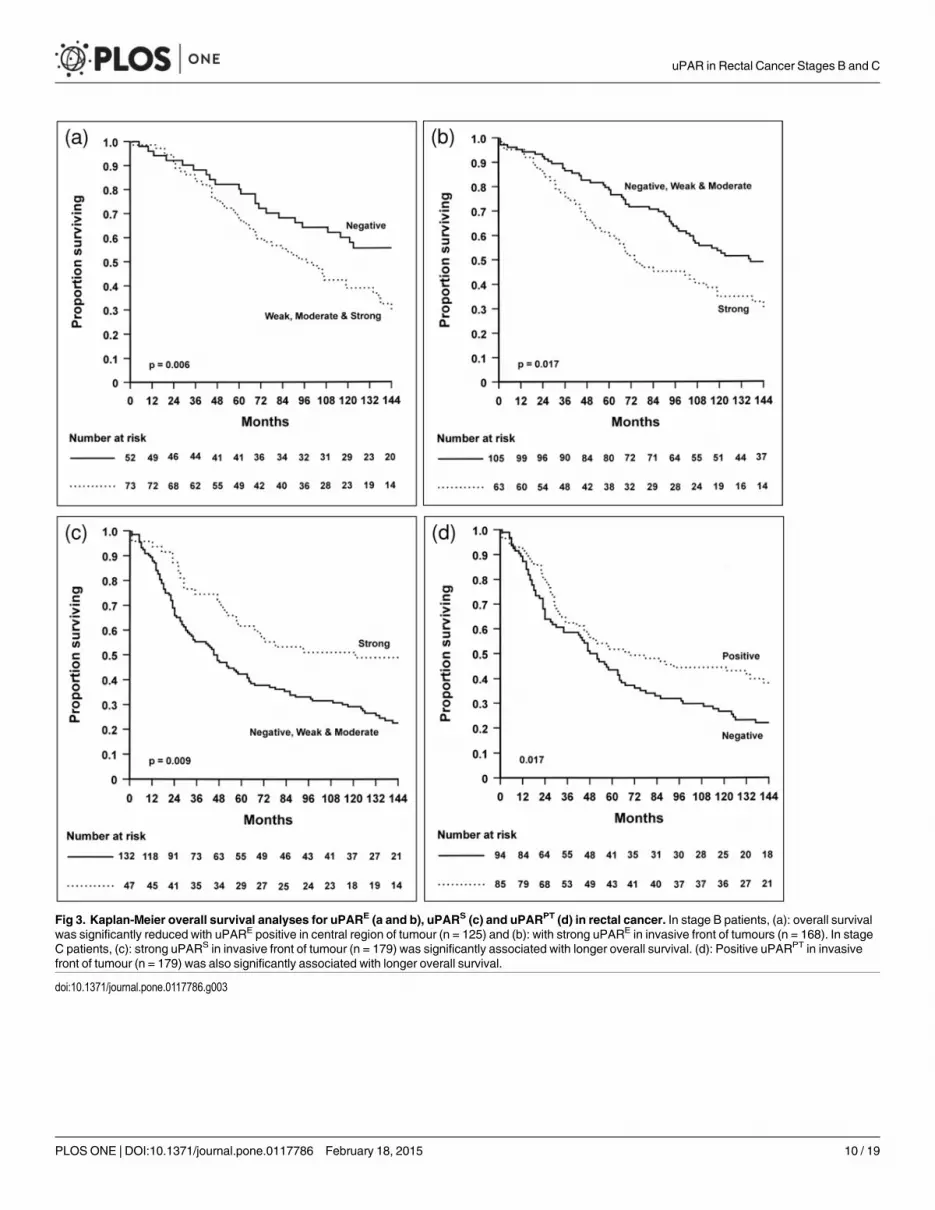
Fig 3. Kaplan-Meier overall survival analyses for uPARE (a and b), uPARS (c) and uPARPT (d) in rectal cancer. In stage B patients, (a): overall survivalwas significantly reduced with uPARE positive in central region of tumour (n = 125) and (b): with strong uPARE in invasive front of tumours (n = 168). In stageC patients, (c): strong uPARS in invasive front of tumour (n = 179) was significantly associated with longer overall survival. (d): Positive uPARPT in invasivefront of tumour (n = 179) was also significantly associated with longer overall survival.
doi:10.1371/journal.pone.0117786.g003
uPAR in Rectal Cancer Stages B and C
PLOSONE | DOI:10.1371/journal.pone.0117786 February 18, 2015 10 / 19

Table 2. Association between stage B patient overall survival and expression of uPAR in tumour epithelium (detected by #3937 MAb) in both thecentral region and invasive front of tumour tissues with adjustment for other potentially prognostic variables.
Stage B central region of tumour tissue (n =125)
Stage B invasive front of tumour tissue (n =168)
Variable Category Deaths/Total
BHR(95%CI)
Waldp
MHR(95%CI)
Waldp
Deaths/Total
BAR(95%CI)
Wald p MHR(95%CI)
Waldp
Sex Male 44/84 0.5(0.3–0.8)
0.006 – – 62/112 0.7(0.5–1.1)
0.102 – –
Female 31/41 39/56
Age � 75 years 29/37 2.3(1.4–3.6)
0.001 2.1(1.3–3.4)
0.002 38/47 2.3(1.5–3.5)
<0.001 2.0(1.3–3.1)
0.002
<75 years 46/88 63/121
Operation Hartmann 7/7 2.8(1.3–6.3)
0.007 – – 13/13 3.5(2.0–6.4)
<0.001 2.4(1.3–4.5)
0.006
AR or APE 68/118 88/155
Tumour size < 5 cm 43/71 1.1(0.7–1.8)
0.589 – – 59/98 1.0(0.7–1.5)
0.986 – –
� 5 cm 32/54 42/70
Pathological type Mucinous or signet ring 4/6 0.9(0.3–2.4)
0.771 – – 3/5 0.8(0.3–2.6)
0.744 – –
Adenocarcinoma 71/119 98/163
Grade High 7/13 1.0(0.5–2.2)
0.978 – – 16/24 1.5(0.9–2.5)
0.152 – –
Low/average 68/112 85/144
Venous invasion Present 11/18 1.4(0.7–2.7)
0.299 – – 19/33 1.0(0.6–1.7)
0.909 – –
Absent 64/107 82/135
Free serosalsurface involved
Yes 2/4 0.8(0.2–3.4)
0.806 – – 3/5 1.2(0.4–3.9)
0.717 – –
No 73/121 98/163
Adjacent organinfiltrated
Yes 75/125 – – – – 2/2 1.8(0.4–7.4)
0.393 – –
No 0/0 99/166
uPAR in tumourepithelial cells
Weak, moderate &Strong (i.e., Positive)
51/73 2.0(1.2–3.3)
0.006 1.9(1.1–3.1)
0.014 – – – – –
vs.
Negative 24/52
Strong – – – – – 43/63 1.6(1.1–2.4)
0.017 1.6(1.1–2.3)
0.031
vs.
Negative, weak &moderate
58/105
BHR: Bivariate Hazard Ratio, MHR: Multivariable Hazard Ratio, AR: Anterior resection, APE: Abdominoperineal excision
doi:10.1371/journal.pone.0117786.t002
uPAR in Rectal Cancer Stages B and C
PLOSONE | DOI:10.1371/journal.pone.0117786 February 18, 2015 11 / 19

uPAR at the adjacent non-neoplastic mucosal tissuesFor epithelia uPAR expression at the adjacent non-neoplastic mucosal tissues (n = 312), 36cases demonstrated strong uPAR expression (19 in stage B and 17 in stage C), 58 cases hadmoderate expression (28 in stage B and 30 in stage C), 81 cases had weak expression (41 instage B and 40 in stage C) and 137 cases were negative (68 in stage B and 69 in stage C). Instage B, there was no association between staining intensity and survival (p = 0.700) while instage C there was no difference in survival between negative, weak and intermediate. The onlysignificant association was longer survival for strong than for negative (p = 0.018) but, as thiswas based on only 17 cases with strong staining, it appeared anomalous and may be a Type 1error arising from this small number. uPAR expression in stroma was almost absent, with neg-ative expression in 320 cases (156 in stage B and 164 in stage C) and only moderate expression
Table 3. Association between stage C patient overall survival and expression of uPAR in stroma-associated cells (detected by R4 MAb) ininvasive front of tumour tissues with adjustment for other potentially prognostic variables.
Variable Category Deaths/Total BHR (95% CI) Wald p MHR (95% CI) Wald p
Sex Male 87/113 1.2 (0.9–1.8) 0.245 – –
Female 46/66
Age � 75 years 41/47 1.6 (1.1–2.4) 0.009 1.7 (1.2–2.5) 0.007
<75 years 92/132
Operation Hartmann 8/9 3.1 (1.5–6.4) 0.001 4.5 (2.1–9.5) <0.001
AR or APE 125/170
Tumour size < 5 cm 55/77 1.0 (0.7–1.4) 0.928 – –
� 5 cm 78/102
Pathological type Mucinous or signet ring 12/15 1.1 (0.6–2.0) 0.724 – –
Adenocarcinoma 121/164
Spread beyond muscularis propria Yes 111/150 1.3 (0.8–2.1) 0.284 – –
No 21/29
Apical node involved Yes 8/9 2.8 (1.4–5.8) 0.004 – –
No 125/170
� 4 nodes involved Yes 50/63 1.4 (1.1–2.1) 0.039 – –
No 83/116
Node ratio � 40% 32/38 1.8 (1.2–2.7) 0.003 1.8 (1.2–2.8) 0.005
<40% 101/141
Grade High 49/67 1.3 (0.9–1.9) 0.128 – –
Low/average 84/112
Venous invasion Present 53/63 1.8 (1.2–2.5) 0.001 1.5 (1.1–2.2) 0.019
Absent 80/116
Free serosal surface Yes 10/15 0.9 (0.5–1.8) 0.819 – –
involved No 123/164
Adjacent organ infiltrated Yes 6/6 3.5 (1.5–8.1) 0.002 3.0 (1.3–6.9) 0.012
No 127/173
uPAR in stroma-associated cells Strong 30/47 0.6 (0.4–0.9) 0.009 0.6 (0.4–0.9) 0.007
vs.
Negative, Weak & Moderate 103/132
uPAR accentuated in Positive 55/85 0.7 (0.5–0.9) 0.017 0.7 (0.5–0.9) 0.016
peritumour Negative 78/94
BHR: Bivariate Hazard Ratio, MM HR: Multivariable Hazard Ratio, AR: Anterior resection, APE: Abdominoperineal excision.
doi:10.1371/journal.pone.0117786.t003
uPAR in Rectal Cancer Stages B and C
PLOSONE | DOI:10.1371/journal.pone.0117786 February 18, 2015 12 / 19

in 9 cases (6 in stage B and 3 in stage C), insufficient uPAR expression specimens were availablefor a comprehensive statistical analysis.
DiscussionAlthough the prognostic relevance of uPAR in cancer has been extensively studied, significantdiscrepancies have rendered much of the work inconclusive. Two major issues remain unre-solved: firstly, the discrepancy regarding the cell types where uPAR is overexpressed (i.e.,uPARE or uPARS), and secondly, the prognostic relevance of uPAR in different cell types anddifferent stages of tumour progression. In this study we have addressed the first paradox bydemonstrating that uPAR expression in different cell types can be detected using two epitope-specific anti-human uPARMAbs #3937 and R4. These antibodies delineated between uPARE
and uPARS expression in RC tissues, showing antigen expression could be differentially de-tected in different cell types and tumour locations in the same RC tissues. Upon examinationof uPARE and uPARS from 349 stage B or C RC tissues, we were able to decipher the secondcontroversy, revealing that elevated uPARE in both the central region and invasive tumourfront adversely correlated with stage B overall survival, whereas elevated uPARS at the invasivefront favourably correlated with stage C overall survival.
The recognition of different uPAR epitopes by different antibodies is an important factor tobe considered, not only for detection in different cell types but also for determination of the po-tential clinical prognostic relevance of uPAR. There are multiple anti-uPAR polyclonal anti-bodies (PAbs) and MAbs which have been developed and studied extensively in clinicalapplications [6]. Of these, MAbs #3937 (like #3936) and R4 (like R2) are most frequently usedfor uPARE and uPARS detection respectively and stand at the centre of disparate results ob-tained by different laboratories. Several factors may explain the specificity of these different an-tibodies, the primary one being binding to different “available” epitopes reflecting potentiallydiverse roles of uPAR in each cell type. As uPAR has 42 known interacting partners [5], it isalso possible that the antibody epitopes may be masked by other uPAR interacting partners indifferent cell types. The multifunctional nature of uPAR is a function of its interactome [3,5],and therefore uPAR detected by different epitope-specific MAbs may have different interactingpartners, which may reflect divergent functions in discrete cell types. This concept is supportedby the fact that the population of soluble uPAR (suPAR) in specific cell types has shown dia-metrically altered staining patterns reflecting functional differences as a result of structural var-iations [25]. suPAR has been found in three different forms in both tissues and body fluids[26] depending upon the number of domain(s) present (e.g. D1D2D3, D2D3 or D1) [25].suPARD1D2D3 has been proposed as a uPA-scavenger, and although it has the ability to binduPA, the protease does not autocleave the linker region between D1 and D2 [25,27]. Therefore,increased suPARD1D2D3 may reduce uPA-dependant proteolysis leading to inhibition of cancermetastasis by reducing the ability of cells to leave the ECM or drive alternative uPAR-depen-dant biologies [25]. In contrast, suPARD2D3 acts primarily as a chemotactic agent promotingan immune response via the SRSRY sequence in the D1 & D2 linker-region [25]. Furthermore,other studies have demonstrated a high concentration of suPARs (i.e., suPARD1 andsuPARD1D2D3+D2D3) in CRC patient sera associated with significantly reduced overall survival[28]. Therefore, based on our evidence, uPARE and uPARS may reflect functional differencesin the biology of uPAR expressed in cell types that differently influences tumour biology, can-cer metastasis and therefore association with patient survival. To address these possibilities,survival significances for both uPARE and uPARS at both the central region and the invasive tu-mour front between stages B and C were analysed (i.e., detected by epitope-specific MAbs indifferent cell types, in different locations of tumour progression, and at different cancer stages).
uPAR in Rectal Cancer Stages B and C
PLOSONE | DOI:10.1371/journal.pone.0117786 February 18, 2015 13 / 19

Not only do our results confirm previous studies demonstrating that both uPARE and uPARS
concentrate at the invasive tumour margin in CRC, they also demonstrate that uPARE adverselycorrelates with patient survival in RC. Thus, collectively it appears that uPARE may be an inde-pendent negative prognostic indicator of survival in many types of human cancers, includingCRC [6]. This was especially apparent in a recent large CRC study (n = 811) which demonstrat-ed that uPARE was significantly associated with poor survival across all CRC stages (i.e., stagesA-D) [15]. We were particularly interested in stages B and C RC patients in our study, because,although these patients are deemed to have had a curative operation, there remains a pro-nounced stage difference in their survival [2]. Interestingly, for both central and frontal tumours,survival significance of uPARE appeared only in RC stage B tissues. The results indicate thatuPARE may be a prognostic survival indicator for pre-lymph node metastatic tumours, demon-strating an independent significance in multivariable analyses. Additionally, uPARE intensity incentral and invasive tumour fronts was differentially associated with survival. In the central re-gion, positive uPARE (strong, moderate & weak) had significantly shorter survival compared tonegative, whereas in the invasive front, strong uPARE had shorter survival compared to a com-bined group (moderate, weak and negative). We propose this observation could be due to ex-pression of uPARE in the frontal tumour region, facilitating metastasis to neighbouring tissuesor lymph nodes. Indeed, our results, and those from others, demonstrate that uPARE was con-centrated in the invasive front [6,7,9,10,29]. Furthermore, uPAR has been shown to play a cru-cial role in cancer cell invasion and metastasis involving many biological processes includingepithelial-mesenchymal transition (EMT), ECM degradation, cell migration and adhesion, andactivation of MAP kinase and Ras pathways [3], all supporting the presence of uPAR in the inva-sive tumour front. Overall, our results provide an understanding of uPARE distribution in the tu-mour microenvironment and show that uPARE is a survival indicator of non-metastatic RCtumours (i.e. stage B) and its expression should be considered in both the central region and in-vasive front of tumours in any future diagnostic, prognostic and/or therapeutic studies.
Unlike uPARE, uPARS remains controversial in terms of survival significance. The two mostrecent studies demonstrated that uPARS was negatively associated with CRC patient survival.Boonstra et al., [17] demonstrated that uPARS was adversely associated with overall survival aswell as DFS across all stages from A to D (n = 262). Illemann et al. [30] also demonstrate uPARpositive macrophages in tumour cores (stroma-associated cells in the central tumour region) werenegatively associated with overall survival in all stages (n = 244), but the significance did not ap-pear in the tumour invasive front. Conversely, our study shows that uPARS was positively associ-ated with overall survival specifically in stage C at the invasive front only, supported byindependent significance in multivariable analysis. In terms of MAb utilisation, Boonstra et al.,used ATN615 [17] whilst Illemann et al., used rabbit PAb and R2MAb (with identical stainingpatterns observed for PAb & R2MAb) [30], whereas our study utilised R4MAb. The characteris-tics of ATN615 (together with ATN658) have been extensively studied recently [17,31–34]. Specif-ically, uPAR epitope-binding sites for these MAbs were identified from the crystal structure[31,32]. For ATN615, the P189 and R192 of human uPAR D3 region were identified as critical toformation of the epitope [32]. The epitope sites for R2 and R4 (D275 and L276 (for MAb R2) andR192, D214, G217 and S269 (for MAb R4) in the D3 region of uPAR) have also been reported byother studies using surface plasmon resonance and/or Western blotting [35,36], although no crys-tal structure studies are currently available. It is likely that these MAbs are targeting the D3 regionof uPAR but through different epitopes located in this domain. Thus the different staining pat-terns of these MAbs may represent different roles of uPAR or different uPAR-interactomes, linkedto differential survival significance results. The crystal structures of both the uPAR-R4 and uPAR-R2 complexes may be an important direction to pursue in the future.
uPAR in Rectal Cancer Stages B and C
PLOSONE | DOI:10.1371/journal.pone.0117786 February 18, 2015 14 / 19

Importantly, Illemann et al., [30] showed that the association of uPARE and uPARS with sur-vival significance was independent of CRC stage, which was further confirmed in our study.These data suggest that the plasminogen activation proteolytic cascade is not only implicated intumour cell invasion/metastasis but is also related to patient survival rates at different CRCstages. However, the difference between Illemann’s study and ours is the opposite prognosticrelevance of uPARS at the different location during tumour progression (i.e., negative prognosticindicator in central region vs. positive prognostic indicator in invasive front, respectively). InCRC tumour-associated stroma, expression of uPAR has been observed in monocytes/macro-phages, fibroblasts, neutrophils, myofibroblasts and endothelial cells [9,14,17,29]. Of these, mac-rophages (also known as tumour-associated macrophages (TAMs)) are a major source of uPARexpression [29] and are the most abundant immune cells in the tumour microenvironment[37]. During tumour progression, circulating monocytes in blood vessels are recruited to the tu-mour site and differentiated into mature macrophages such as M1- andM2-polarised macro-phages [38]. M1-macrophages are known to mediate tumour elimination, whilst M2-macrophages have a rather contradictory role as acting in either a pro- or anti-tumour fashion[37]. In the tumour microenvironment, TAMs resemble M2-macrophages and induce the pro-duction of a large range of growth factors and proteolytic enzymes such as EGF, TGFβ1, VEGEandMMPs to stimulate ECM degradation, thus promoting tumour metastasis, resulting in poorcancer prognosis [37]. However other studies have demonstrated that TAM accumulation at theinvasive tumour front can also be associated positively with CRC patient survival [39–41]. Fur-thermore, supporting data has demonstrated that TAM concentrated around tumour cells areable to induce apoptosis in a Fas ligand-dependent manner, and the degree of apoptotic cancercells is inversely correlated with haematogenous metastasis, emphasising the protective role ofTAMs [42]. These results suggest that the location of TAMs in CRCs (i.e., at invasive front orperitumourally) appear to be an important factor in antitumour activation. This concept is fur-ther supported by other reports demonstrating that peritumoral macrophages are likely to haveless contact with tumour-derived cytokines, and are positioned in less hypoxic areas, indicatingthat they may display a tumouricidal rather than tumour promoting activity [37]. This modelvery closely aligns with our data that demonstrates uPARS is a positive prognostic indicator ofsurvival in the invasive front of stage C RCs. These data are further supported by the presence ofuPARPT at the same location and stage, which was associated with longer survival than whenuPARPT was absent. Collectively, it is possible that the population of M1 macrophages and/ornewly recruited monocytes (i.e., before polarisation) may represent a higher proportion of mac-rophages at the invasive front and in peritumoural regions of CRCs, and that uPARS detected byR4 might be expressed by those stroma-associated cells. In fact, a recent study demonstratedthat in an experimental model of colitis, uPAR controls the function of intestinal macrophagesby reducing inflammatory cytokines and controlling M1 andM2 polarisation [43]. For futurestudies, simultaneous IHC of monocytes, M1- andM2-polarised macrophages, and R4 (or otherMAbs such as ATN615 or R2) on serial CRC TMA sections may further clarify our understand-ing of the role/s of uPAR in stroma-associated cancer biology.
Although uPARE was expressed in a significant number of adjacent non-neoplastic mucosaltissues, we have not considered to use this as an internal standard because it may not be as rep-resentative as healthy mucosa, since the histologically normal tissues used in this study weretaken from 1–2cm from the tumour margin. We have recently demonstrated that integrinανβ6 (a potential prognostic indicator of colorectal cancer and recognised to be absence or lowexpression in normal tissues) was expressed in almost all histological normal mucosa (fromsame TMA used in this study) [44]. As the integrin ανβ6 is one of key regulators of EMT alongwith TGF beta1 [45,46], it indicates that EMT-associated changes are occurring in that tissue.This observation also supported by other types of cancer demonstrated that the expression of
uPAR in Rectal Cancer Stages B and C
PLOSONE | DOI:10.1371/journal.pone.0117786 February 18, 2015 15 / 19

EMT markers (e.g., α-smooth muscle actin & SNAIL) occur in “apparently” histologically nor-mal breast tissue that is located 1cm away from breast cancer tissue margins [47].
In conclusion, we have found that uPARE is associated with poorer RC survival in stage B (inthe central and the invasive front regions) whilst uPARS is correlated with longer survival instage C (in the invasive tumour front). This indicates that uPAR has an opposite role in differentcell types at different tumour locations across RC stages B and C. We have proposed that thesefunctional differences may potentially be related to differences in the uPAR-interactomes pres-ent in distinct cell types. In this regard, we have already unequivocally shown uPAR interactswith αvβ6 which is an epithelially-restricted integrin (i.e., one that would never occur in stromalcells) [48]. Therefore, a comprehensive study of the uPAR interactome in different cell typesand consequent reactivity of uPAR with various anti-uPARMAbs is a necessary step towardsan understanding of its roles in CRC. Indeed, MAb inhibition of the uPAR-integrin interactomehas been recently proposed as a new anti-cancer therapeutic approach and a basis to develop tu-mour imaging methodologies [31,34,49,50]. Overall, accurate prediction of patient survivalbased on uPAR expression coupled with a better understanding and targeting of specific uPARinteractomes (using human or humanised MAbs against interaction surfaces, inhibitory pep-tides or small molecule antagonists of interactomes) may lead to the development of novel, per-sonalised companion immunopathology prognostics and anti-metastasis therapeutics.
AcknowledgmentsWe acknowledge Lisa Sedger (ASAM) for kindly reviewing some aspects of our manuscript.Some of the research described herein was facilitated by access to the Australian ProteomeAnalysis Facility (APAF) and Monash University Antibody Technology Facility (MATF) bothestablished under the Australian Government’s National Collaborative Research InfrastructureStrategy (NCRIS).
Author ContributionsConceived and designed the experiments: MB EN SBA. Performed the experiments: SBA C.Chan SYK C. Clarke. Analyzed the data: OD C. Chan SBAMB. Contributed reagents/materi-als/analysis tools: PC C. Chan JF MB. Wrote the paper: SBA OD AMMB EN.
References1. Ferlay JSI, Ervik M, Dikshit R, Eser S, Mathers C, et al. (2012) GLOBOCAN 2012 v1.0, Cancer Inci-
dence and Mortality Worldwide: IARC CancerBase No. 11 [Internet]. Available: http://globocan.iarc.fr.2013 ed. Lyon, France: International Agency for Research on Cancer.
2. Davis NC, Newland RC (1983) Terminology and classification of colorectal adenocarcinoma: the Aus-tralian clinico-pathological staging system. Aust N Z J Surg 53: 211–221. PMID: 6309132
3. Smith HW, Marshall CJ (2010) Regulation of cell signalling by uPAR. Nat Rev Mol Cell Biol 11: 23–36.doi: 10.1038/nrm2821 PMID: 20027185
4. Llinas P, Le Du MH, Gardsvoll H, Dano K, Ploug M, et al. (2005) Crystal structure of the human uroki-nase plasminogen activator receptor bound to an antagonist peptide. The EMBO journal 24:1655–1663. PMID: 15861141
5. Eden G, Archinti M, Furlan F, Murphy R, Degryse B (2011) The urokinase receptor interactome. Currentpharmaceutical design 17: 1874–1889. PMID: 21711237
6. Boonstra MC, Verspaget HW, Ganesh S, Kubben FJ, Vahrmeijer AL, et al. (2011) Clinical applicationsof the urokinase receptor (uPAR) for cancer patients. Curr Pharm Des 17: 1890–1910. PMID:21711239
7. de Bock CE, Wang Y (2004) Clinical significance of urokinase-type plasminogen activator receptor(uPAR) expression in cancer. Med Res Rev 24: 13–39. PMID: 14595671
8. Ossowski L, Clunie G, Masucci MT, Blasi F (1991) In vivo paracrine interaction between urokinase andits receptor: effect on tumor cell invasion. J Cell Biol 115: 1107–1112. PMID: 1659573
uPAR in Rectal Cancer Stages B and C
PLOSONE | DOI:10.1371/journal.pone.0117786 February 18, 2015 16 / 19

9. Pyke C, Ralfkiaer E, Ronne E, Hoyer-Hansen G, Kirkeby L, et al. (1994) Immunohistochemical detec-tion of the receptor for urokinase plasminogen activator in human colon cancer. Histopathology 24:131–138. PMID: 8181805
10. Suzuki S, Hayashi Y, Wang Y, Nakamura T, Morita Y, et al. (1998) Urokinase type plasminogen activa-tor receptor expression in colorectal neoplasms. Gut 43: 798–805. PMID: 9824607
11. Yang JL, Seetoo D, Wang Y, Ranson M, Berney CR, et al. (2000) Urokinase-type plasminogen activa-tor and its receptor in colorectal cancer: independent prognostic factors of metastasis and cancer-spe-cific survival and potential therapeutic targets. Int J Cancer 89: 431–439. PMID: 11008205
12. Seetoo DQ, Crowe PJ, Russell PJ, Yang JL (2003) Quantitative expression of protein markers of plas-minogen activation system in prognosis of colorectal cancer. J Surg Oncol 82: 184–193. PMID:12619063
13. Kaneko I, Tanaka S, Oka S, Yoshida S, Hiyama T, et al. (2007) Immunohistochemical molecular mark-ers as predictors of curability of endoscopically resected submucosal colorectal cancer. World J Gas-troenterol 13: 3829–3835. PMID: 17657837
14. Illemann M, Bird N, Majeed A, LaerumOD, Lund LR, et al. (2009) Two distinct expression patterns ofurokinase, urokinase receptor and plasminogen activator inhibitor-1 in colon cancer liver metastases.Int J Cancer 124: 1860–1870. doi: 10.1002/ijc.24166 PMID: 19123477
15. Minoo P, Baker K, Baumhoer D, Terracciano L, Lugli A, et al. (2010) Urokinase-type plasminogen acti-vator is a marker of aggressive phenotype and an independent prognostic factor in mismatch repair-proficient colorectal cancer. Hum Pathol 41: 70–78. doi: 10.1016/j.humpath.2009.05.013 PMID:19740518
16. Saito K, Takeha S, Shiba K, Matsuno S, Sorsa T, et al. (2000) Clinicopathologic significance of uroki-nase receptor- and MMP-9-positive stromal cells in human colorectal cancer: functional multiplicity ofmatrix degradation on hematogenous metastasis. Int J Cancer 86: 24–29. PMID: 10728590
17. Boonstra MC, Verbeek FP, Mazar AP, Prevoo HA, Kuppen PJ, et al. (2014) Expression of uPAR intumor-associated stromal cells is associated with colorectal cancer patient prognosis: a TMA study.BMC Cancer 14: 269. doi: 10.1186/1471-2407-14-269 PMID: 24742002
18. Takeha S, Fujiyama Y, Bamba T, Sorsa T, Nagura H, et al. (1997) Stromal expression of MMP-9 andurokinase receptor is inversely associated with liver metastasis and with infiltrating growth in human co-lorectal cancer: a novel approach from immune/inflammatory aspect. Jpn J Cancer Res 88: 72–81.PMID: 9045899
19. Rasch MG, Lund IK, Almasi CE, Hoyer-Hansen G (2008) Intact and cleaved uPAR forms: diagnosticand prognostic value in cancer. Front Biosci 13: 6752–6762. PMID: 18508692
20. Newland RC, Chapuis PH, Pheils MT, MacPherson JG (1981) The relationship of survival to stagingand grading of colorectal carcinoma: a prospective study of 503 cases. Cancer 47: 1424–1429. PMID:7226068
21. Newland RC, Chapuis PH, Smyth EJ (1987) The prognostic value of substaging colorectal carcinoma.A prospective study of 1117 cases with standardized pathology. Cancer 60: 852–857. PMID: 3594403
22. Bokey EL, Ojerskog B, Chapuis PH, Dent OF, Newland RC, et al. (1999) Local recurrence after curativeexcision of the rectum for cancer without adjuvant therapy: role of total anatomical dissection. Br J Surg86: 1164–1170. PMID: 10504371
23. Fielding LP, Arsenault PA, Chapuis PH, Dent O, Gathright B, et al. (1991) Clinicopathological stagingfor colorectal cancer: an International Documentation System (IDS) and an International Comprehen-sive Anatomical Terminology (ICAT). J Gastroenterol Hepatol 6: 325–344. PMID: 1912440
24. Kho PS, Jankova L, Fung CL, Chan C, Clarke C, et al. (2012) Overexpression of protein S100A4 is in-dependently associated with overall survival in stage C colonic cancer but only in cytoplasm at the ad-vancing tumour front. Int J Colorectal Dis 27: 1409–1417. doi: 10.1007/s00384-012-1469-8 PMID:22569556
25. Thuno M, Macho B, Eugen-Olsen J (2009) suPAR: the molecular crystal ball. Dis Markers 27:157–172. doi: 10.3233/DMA-2009-0657 PMID: 19893210
26. Hoyer-Hansen G, Lund IK (2007) Urokinase receptor variants in tissue and body fluids. Adv Clin Chem44: 65–102. PMID: 17682340
27. Hoyer-Hansen G, Pessara U, Holm A, Pass J, Weidle U, et al. (2001) Urokinase-catalysed cleavage ofthe urokinase receptor requires an intact glycolipid anchor. Biochem J 358: 673–679. PMID: 11535128
28. Lomholt AF, Christensen IJ, Hoyer-Hansen G, Nielsen HJ (2010) Prognostic value of intact and cleavedforms of the urokinase plasminogen activator receptor in a retrospective study of 518 colorectal cancerpatients. Acta Oncol 49: 805–811. doi: 10.3109/0284186X.2010.491086 PMID: 20524776
uPAR in Rectal Cancer Stages B and C
PLOSONE | DOI:10.1371/journal.pone.0117786 February 18, 2015 17 / 19

29. Ohtani H, Pyke C, Dano K, Nagura H (1995) Expression of urokinase receptor in various stromal-cellpopulations in human colon cancer: immunoelectron microscopical analysis. Int J Cancer 62:691–696. PMID: 7558416
30. Illemann M, LaerumOD, Hasselby JP, Thurison T, Høyer-Hansen G, et al. (2014) Urokinase-type plas-minogen activator receptor (uPAR) on tumor-associated macrophages is a marker of poor prognosis incolorectal cancer. Cancer Med 1002: 242.
31. Xu X, Cai Y, Wei Y, Donate F, Juarez J, et al. (2014) Identification of a new epitope in uPAR as a targetfor the cancer therapeutic monoclonal antibody ATN-658, a structural homolog of the uPAR bindingintegrin CD11b (alphaM). PLoS One 9: e85349. doi: 10.1371/journal.pone.0085349 PMID: 24465541
32. Li Y, Parry G, Chen L, Callahan JA, Shaw DE, et al. (2007) An anti-urokinase plasminogen activator re-ceptor (uPAR) antibody: crystal structure and binding epitope. J Mol Biol 365: 1117–1129. PMID:17101149
33. Kenny HA, Leonhardt P, Ladanyi A, Yamada SD, Montag A, et al. (2011) Targeting the urokinase plas-minogen activator receptor inhibits ovarian cancer metastasis. Clin Cancer Res 17: 459–471. doi: 10.1158/1078-0432.CCR-10-2258 PMID: 21149615
34. Rabbani SA, Ateeq B, Arakelian A, Valentino ML, Shaw DE, et al. (2010) An anti-urokinase plasmino-gen activator receptor antibody (ATN-658) blocks prostate cancer invasion, migration, growth, and ex-perimental skeletal metastasis in vitro and in vivo. Neoplasia 12: 778–788. PMID: 20927316
35. Gardsvoll H, Jacobsen B, KriegbaumMC, Behrendt N, Engelholm L, et al. (2011) Conformational regu-lation of urokinase receptor function: impact of receptor occupancy and epitope-mapped monoclonalantibodies on lamellipodia induction. J Biol Chem 286: 33544–33556. doi: 10.1074/jbc.M111.220087PMID: 21799009
36. Ronne E, Behrendt N, Ellis V, Ploug M, Dano K, et al. (1991) Cell-induced potentiation of the plasmino-gen activation system is abolished by a monoclonal antibody that recognizes the NH2-terminal domainof the urokinase receptor. FEBS Lett 288: 233–236. PMID: 1715292
37. Erreni M, Mantovani A, Allavena P (2011) Tumor-associated Macrophages (TAM) and Inflammation inColorectal Cancer. Cancer Microenviron 4: 141–154. doi: 10.1007/s12307-010-0052-5 PMID:21909876
38. Richards DM, Hettinger J, Feuerer M (2013) Monocytes and macrophages in cancer: development andfunctions. Cancer Microenviron 6: 179–191. doi: 10.1007/s12307-012-0123-x PMID: 23179263
39. Khorana AA, Ryan CK, Cox C, Eberly S, Sahasrabudhe DM (2003) Vascular endothelial growth factor,CD68, and epidermal growth factor receptor expression and survival in patients with Stage II and StageIII colon carcinoma: a role for the host response in prognosis. Cancer 97: 960–968. PMID: 12569594
40. Funada Y, Noguchi T, Kikuchi R, Takeno S, Uchida Y, et al. (2003) Prognostic significance of CD8+ Tcell and macrophage peritumoral infiltration in colorectal cancer. Oncol Rep 10: 309–313. PMID:12579264
41. Forssell J, Oberg A, Henriksson M, Stenling R, Jung A, et al. (2007) High macrophage infiltration alongthe tumor front correlates with improved survival in colon cancer. Clin Cancer Res 13: 1472–1479.PMID: 17332291
42. Sugita J, Ohtani H, Mizoi T, Saito K, Shiiba K, et al. (2002) Close association between Fas ligand(FasL; CD95L)-positive tumor-associated macrophages and apoptotic cancer cells along invasive mar-gin of colorectal carcinoma: a proposal on tumor-host interactions. Jpn J Cancer Res 93: 320–328.PMID: 11927015
43. Genua M, D’Alessio S, Cibella J, Gandelli A, Sala E, et al. (2014) The urokinase plasminogen activatorreceptor (uPAR) controls macrophage phagocytosis in intestinal inflammation. Gut.
44. Ahn SB, Mohamedali A, Chan C, Fletcher J, Kwun SY, et al. (2014) Correlations between Integrinalphanubeta6 Expression and Clinico-Pathological Features in Stage B and Stage C Rectal Cancer.PLoS One 9: e97248. doi: 10.1371/journal.pone.0097248 PMID: 24821188
45. Bates RC (2005) Colorectal cancer progression: integrin alphavbeta6 and the epithelial-mesenchymaltransition (EMT). Cell Cycle 4: 1350–1352. PMID: 16123591
46. Bates RC, Bellovin DI, Brown C, Maynard E, Wu B, et al. (2005) Transcriptional activation of integrinbeta6 during the epithelial-mesenchymal transition defines a novel prognostic indicator of aggressivecolon carcinoma. J Clin Invest 115: 339–347. PMID: 15668738
47. Trujillo KA, Heaphy CM, Mai M, Vargas KM, Jones AC, et al. (2011) Markers of fibrosis and epithelial tomesenchymal transition demonstrate field cancerization in histologically normal tissue adjacent tobreast tumors. Int J Cancer 129: 1310–1321. doi: 10.1002/ijc.25788 PMID: 21105047
48. Saldanha RG, Molloy MP, Bdeir K, Cines DB, Song X, et al. (2007) Proteomic identification of lynchpinurokinase plasminogen activator receptor protein interactions associated with epithelial cancer malig-nancy. J Proteome Res 6: 1016–1028. PMID: 17330942
uPAR in Rectal Cancer Stages B and C
PLOSONE | DOI:10.1371/journal.pone.0117786 February 18, 2015 18 / 19

49. Lebeau AM, Sevillano N, King ML, Duriseti S, Murphy ST, et al. (2014) Imaging the urokinase plasmi-nongen activator receptor in preclinical breast cancer models of acquired drug resistance. Theranostics4: 267–279. doi: 10.7150/thno.7323 PMID: 24505235
50. Duriseti S, Goetz DH, Hostetter DR, LeBeau AM, Wei Y, et al. (2010) Antagonistic anti-urokinase plas-minogen activator receptor (uPAR) antibodies significantly inhibit uPAR-mediated cellular signalingand migration. J Biol Chem 285: 26878–26888. doi: 10.1074/jbc.M109.077677 PMID: 20501655
uPAR in Rectal Cancer Stages B and C
PLOSONE | DOI:10.1371/journal.pone.0117786 February 18, 2015 19 / 19


