Embed Size (px)
Citation preview

HEREGULIN INHIBITS PROLIFERATION VIA ERKs ANDPHOSPHATIDYL-INOSITOL 3-KINASE ACTIVATION BUT REGULATESUROKINASE PLASMINOGEN ACTIVATOR INDEPENDENTLY OF THESEPATHWAYS IN METASTATIC MAMMARY TUMOR CELLSLydia PURICELLI
2, Cecilia J. PROIETTII1, Leticia LABRIOLA
1, Mariana SALATINO1, Marıa E. BALANA
1, Julio Aguirre GHISO2, Ruth LUPU
3,Omar P. PIGNATARO
1, Eduardo H. CHARREAU1,4, Elisa BAL DE KIER JOFFE
2 and Patricia V. ELIZALDE1*
1Instituto de Biologıa y Medicina Experimental (IBYME), CONICET, Buenos Aires, Argentina2Instituto de Oncologıa Angel H. Roffo, Universidad de Buenos Aires, Argentina3Lawrence Berkeley National Laboratories, University of California Berkeley, Berkeley CA, USA4Facultad de Ciencias Exactas y Naturales, Universidad de Buenos Aires, Argentina
Heregulin (HRG) and type I receptor tyrosine kinase(RTK) expression was investigated in the highly invasive andmetastatic LM3 cell line, our previously described model ofmetastasis for mammary cancer (Bal de Kier Joffe et al.[1986] Invasion Metastasis 6:302–12; Urtreger et al. [1997]Int J Oncol 11:489–96). Although LM3 cells do not expressHRG, they exhibit high levels of ErbB-2 and ErbB-3 as well asmoderate expression of ErbB-4. Addition of exogenousHRG�1 resulted in inhibition of both proliferation and migra-tion of LM3 cells. HRG�1 was also able to decrease theactivity of urokinase-type plasminogen activator (uPA) andmatrix metalloproteinase 9 (MMP-9), 2 key enzymes in theinvasion and metastatic cascade. HRG�1 treatment of LM3cells induced tyrosine phosphorylation of ErbB-2, ErbB-3 andErbB-4 as well as the formation of ErbB-2/ErbB-3 and ErbB-2/ErbB-4 heterodimers. Assessment of the signaling path-ways involved in HRG�1 action indicated that the addition ofHRG�1 to LM3 cells resulted in activation of phosphatidyl-inositol 3- kinase (PI-3K) and in strong induction of the asso-ciation of the p85 subunit of PI-3K with ErbB-3. HRG�1 alsocaused the rapid activation of ERK1/ERK2 and Stat3 andStat5 (signal transducers and activators of transcription[STAT]). This is the first demonstration of the ability ofHRG�1 to activate STATs in mammary tumor cells. Block-age of PI-3K activity with its chemical inhibitor wortmannin,or of MEK1/ERKs activity with PD98059, resulted in suppres-sion of the ability of HRG�1 to inhibit LM3 cell growth.Notwithstanding the suppression of these 2 signaling path-ways, HRG�1 still proved capable of inhibiting uPA activity.Therefore, our results provide evidence that signaling path-ways involved in HRG�1-induced proliferation appear to bedistinct from those involved in HRG�1 regulation of uPA, aprotease that plays a pivotal role in invasion and metastasis.© 2002 Wiley-Liss, Inc.
Key words: heregulin; ErbB receptors; metastatic mammary tumors;ERKs; phosphatidylinositol 3-kinase
The high mortality rate associated with breast cancer is mainlydue to its ability to invade and metastasize distant sites. Tumorinvasion and the development of metastasis entails a complex andmultistep process in which unique properties are acquired bytumor cells. These properties include limitless growth, alterationsin cell communication, enhanced motility and the capacity todegrade basement membrane components, to invade tissues and togrow autonomously at secondary sites.1 Accumulating evidenceshows that growth factors (GFs) are able to regulate most of thesebiologic behaviors.2
The neu differentiation factor 1 (NDF1) or heregulin (HRG)family includes a series of polypeptides generated by differentialsplicing of a single primary transcript.3,4 All HRG isoforms sharean epidermal growth factor (EGF)-like motif that acts as thereceptor binding domain.3,4 The structure of this motif reveals 2major subclasses of HRG variants, � and �.5 Recently, 2 otherfamilies of heregulins have been described, NDF2 and NDF3,encoded by different genes.6,7 NDF1/HRG exerts multiple physi-
ologic actions through a unique combinatorial signaling resultingfrom dimerization of the members of the type I receptor tyrosinekinase family (RTKs).8 Type I RTKs includes 4 members: epider-mal growth factor receptor (EGFR/ErbB-1),9 ErbB-2,10 ErbB-311,12 and ErbB-4.13 HRG binds to ErbB-3 and ErbB-4.5 Theremaining RTK-I family members, ErbB-1/EGF-R and ErbB-2,act as coreceptors.8
The biologic effects of HRG exhibit variations that depend oncell type, HRG isoform and cellular complements of RTK-I mem-bers available to make up functional heterodimers. Certain HRGisoforms are reported to induce growth arrest and differentiation ofmammary epithelial cells, whereas other breast cells respond mi-togenically.14–16 Also, stimulatory17,18 and inhibitory effects19–21
of HRG on the proliferation of breast cancer cell lines, expressingdifferent levels of ErbB-2, have been reported. At the moment, aconsensus is emerging on the fact that HRG exerts a growth-inhibitory effect on ErbB-2-overexpressing breast tumor cells.19–24
HRG treatment of breast cancer cells induces activation of theErk/MAP kinases,25,26 Jnk/SAP (stress-activated protein) ki-nases,27 p70/p85 S6 kinase25 and phosphatidylinositol 3-kinase(PI-3K).26,27 However, the role that each of these signal transduc-tion cascades plays in HRG regulation of proliferation or differ-entiation remains to be elucidated.
HRG has also been found to regulate several cellular responsesassociated with breast cancer cell progression to a metastaticphenotype. Thus, HRG was found to promote motility and inva-sion and to induce cytoeskeletal reorganization of breast cancercells.24,28,29 HRG regulation of the expression and activity ofproteases capable of degrading the extracellular matrix, such asmatrix metalloproteinase-9 (MMP-9) and urokinase-type plasmin-
Grant sponsor: National Scientific Council of Argentina (CONICET);Grant numbers: PID 4188/96 and PID 4199/96; Grant sponsor: NationalAgency of Scientific Promotion of Argentina; Grant numbers: IDB 802/OC-AR PICT 0503402, IDB 1201/OC-AR PICT 0506114; Grant sponsor:Centro Argentino Brasilero de Biotecnologıa (CABBIO); Grant sponsor:University of Buenos Aires; Grant sponsor: Beca Ramon Carrillo-ArturoOnativia.
The first two authors contributed equally to this work.
*Correspondence to: Laboratory of Molecular Mechanisms of Carcino-genesis, Instituto de Biologıa y Medicina Experimental (IBYME), Ob-ligado 2490, Buenos Aires 1428 Argentina. Fax: �5411-4786-2564.E-mail: [email protected]
Received 26 July 2001; Revised 2 May 2002; Accepted 3 May 2002
DOI 10.1002/ijc.10533Published online 1 July 2002 in Wiley InterScience (www.interscience.
wiley.com).
Int. J. Cancer: 100, 642–653 (2002)© 2002 Wiley-Liss, Inc.
Publication of the International Union Against Cancer

ogen activator (uPA), has also been reported in breast cancer celllines.24,28,30,31 Varying results have been found regarding the par-ticipation of different signaling pathways in HRG regulation of themetastatic phenotype. Particularly, PI-3K and MAPK activationwere found to be involved in HRG regulation of cellular behaviorsrelated to invasion and metastasis such as adhesion, aggregation,migration and regulation of cytoskeletal reorganization.23,32,33
The aim of our study was to investigate the expression of HRGand type I RTKs, within our well-characterized model of metas-tasis for mammary cancer. The LM3 cell line derives from the M3mouse mammary adenocarcinoma. Like its parental tumor, LM3cells exhibit a highly invasive behavior and 100% incidence oflung metastasis.34 In the present study, we found that LM3 cells donot produce HRG and that they overexpress ErbB-2 and ErbB-3.Exogenous HRG-�1 inhibits LM3 cell proliferation as well as uPAand MMP-9 activities. Whereas ERKs and PI-3K signaling path-ways are involved in HRG�1 regulation of growth, they play norole in HRG�1 inhibition of uPA activation.
MATERIAL AND METHODS
Lm3 cell line and proliferation assaysThe LM3 cell line was established in our laboratory.34 Briefly,
it was obtained from primary cultures of the Balb/c transplantablemammary adenocarcinoma M3, metastatic to lung.35 Upon s.c.injection, LM3 cells produced locally invasive, poorly differenti-ated adenocarcinoma highly metastatic to the lung.34 LM3 cellswere maintained in MEM supplemented with 2.5% FCS, 2 mML-glutamine and 80 �g/ml gentamicin. For proliferation assays,cells were plated in multiwell plastic dishes at a density of 20 �103 in MEM � 2.5% FCS and allowed to attach overnight beforetreatment with recombinant human HRG�1 (Neomarkers, Free-mont, CA) at concentrations ranging from 0.02 to 40 ng/ml.
In experiments assessing the role of ErbB-4 or ErbB-3 in HRG-induced proliferation, cells were preincubated with 10 �g/ml ofeither an ErbB-4 mouse monoclonal antibody (Oncoprotein Ab-3,clone H4.72.8, Neomarkers) or an ErbB-3 mouse monoclonalantibody (Oncoprotein Ab-5, clone H3.105.5, Neomarkers) priorto HRG treatment. As control, cells were also incubated withpreimmunne mouse serum. After 24 hr of incubation, 50% ofmedia was replaced by fresh media and cells were incubated foranother 24 hr in the presence of 0.2 �Ci of 3H-thymidine (NEN,Dupont, Boston MA; specific activity: 70–90 Ci/mmol). Cellswere then trypsinized and harvested. Assays were performed inoctuplicate. The differences between control and experimentalgroups were analyzed by ANOVA followed by Tukey’s t-testbetween groups.
In previous experiments we demonstrated that thymidine uptakecorrelates with the number of cells/well. In some experiments, cellgrowth was evaluated by protein content, using the MTS assay, aspreviously described.34 We have already proved the existence of adirect correlation between cell number and protein content.34 Tostudy the effect of HRG�1 on plating efficiency (clonogenicassay), 2 � 103 monodispersed LM3 cells were seeded on 60 mmplastic dishes in MEM medium with 2.5 % FCS. At 24 hr, cellswere treated with different concentrations of HRG�1 in the pres-ence of 2.5% FCS. Medium was changed every 72 hr. After 8 daysof culture, plates were washed, fixed with 5% acetic acid inmethanol and stained with hematoxylin and the number of colonieswas counted under inverted microscope. Clonogenic ability wasdefined as the percentage of cells able to grow as colonies of morethan 10 cells.
Migration assayA wound assay was employed to study the effect of HRG�1 on
LM3 cell migration. Wounds 400 �m wide were made in confluentLM3 monolayers. Cells were treated with varying concentrationsof HRG�1 in the presence of 2.5% FCS and allowed to migrateinto the cell-free area. At 24 hr, cells were fixed and stained withGiemsa and the cell-free area was quantitated by densitometry. We
also performed migration assays using Transwell cell culturechambers (8 �m membrane pore; Corning Costar, Corning, NY).Each filter was coated with 0.1% gelatin on the lower side anddried for 3 hr and then 2 � 105 LM3 cells, in 180 �l serum-freeMEM, were seeded on the upper surface of the chamber. The wellscontained 0.5 ml MEM and 8 �g/ml human fibronectin as achemoattractant. Cells were incubated for 20 hr at 37°C in a CO2incubator in the presence or absence of 20 ng/ml of HRG.
At the end of the incubation period, only cells that had passedthrough the filter pores and attached on the lower surface of thefilter were considered to have migratory ability. Cells on the uppersurface of the filter were completely removed by wiping with acotton swab. Membranes were fixed with Carnoy’s fixative andstained with Hoescht 33258. The nucleus of those cells that mi-grated was counted under a fluorescence microscope. At least 20fields of �400 per membrane were counted and the mean valuewas employed to perform the statistical analysis using Student’st-test. Assays were done in triplicate.
Urokinase-type plasminogen activator and matrixmetalloproteinase 9 activity
The effect of HRG�1 on both uPA and MMP-9 activity wasevaluated in conditioned media. Semiconfluent monolayers ofLM3 cells cultured in 35 mm well plates were extensively washedwith PBS and then incubated in serum-free MEM in the presenceor absence of varying concentrations of HRG�1 for 24 hr. Whenthe effect of the kinase inhibitors wortmannin or PD98059 wasevaluated, cells were pretreated with these inhibitors for 90 minprior to the addition of HRG�1. Conditioned media were individ-ually harvested and the remaining monolayers were lysed to mea-sure protein content.34 Samples were stored at �70°C and usedonly once after thawing.
A radial caseinolytic method of Saksela36 was used to quantifyuPA activity. uPA activities were referred to a urokinase standardcurve ranging from 0.05 to 10 IU/ml and normalized to the originalcell culture protein content. Specificity of uPA activity was deter-mined by blocking sample activity with anticatalytic uPA antibod-ies (kindly provided by G. Hansen, Righospitalet, Copenhagen,Denmark) or with 1 �M amiloride. Plasminogen-free casein-aga-rose gels were used to test plasminogen-independent activity.MMP-9 activity was studied by measuring the collagenolytic ac-tivity secreted by LM3 cells by SDS-PAGE copolymerized with0.1% gelatin, as previously reported.37 After running, gels werewashed in 2% Triton X-100 and incubated for 72 hr in 0.25 MTris-HCl/1 M NaCl/25 mM CaCl2 (pH 7.4) buffer for specificactivity detection, or in the same solution plus 40 mM EDTA todetect nonspecific activity. Gels were fixed and stained with Coo-massie blue. Activity bands were visualized by negative staining.
RNAse protection assayThe HRG probe was the 333 bp HRG cDNA fragment cloned in
a pCRII vector (Invitrogen, La Jolla, CA) and linearized withHindIII to provide the antisense riboprobe.38 The rpL32 humancDNA clone (encoding ribosomal protein L32)39 was provided byDr. R. Rochford (The Scripps Research Institute, La Jolla, CA).This recombinant clone, constructed in a pGem 4 vector (Promega,Madison, WI), was linearized with EcoIR, providing a template forthe antisense riboprobe protecting a 76 bp mRNA fragment. An-tisense 32P-labeled RNA probes were transcribed with T7 RNApolymerase following the manufacturer’s protocol (Promega).Thirty micrograms of total RNA, isolated as described,40 werehybridized with 2 � 105 cpm of each probe in the conditionspreviously described,38 followed by digestion with RNase A(Sigma, St. Louis, MO). All RNA samples were hybridized simul-taneously with HRG and rpL32 probes (used to correct for smallvariations in the amount of RNA loaded). Samples were extractedwith phenol/chloroform/isoamyl alcohol (20:20:1) and precipitatedwith 20 �g of tRNA (Sigma) and 2 vol of absolute ethanol. Thepellets were resuspended in 5 �l of an 80% formamide loadingbuffer and run on a 7.5% polyacrylamide sequencing gel with 8 M
643HEREGULIN ROLE IN METASTATIC MAMMARY TUMORS

urea. Size markers were prepared by end labeling MspI-digestedfragments of pBR322 plasmid.
Type I RTK expression, tyrosine phosphorylation andheterodimerization
To study RTK expression, LM3 cells were lysed in buffercontaining 50 mM Tris (pH 7.4), 150 mM NaCl, 1 mM EDTA, 1mM ethylene glycol-bis (�-aminoethyl ether)-N-N-N�-N� tetraace-tic acid (EGTA), 10% glycerol, 0.5% Nonidet P-40, 1 mM ClMg21mM phenylmethylsulfonylfluoride (PMSF), 10 �g/ml leupeptin,5 �g/ml pepstatin, 5 �g/ml aprotinin, 1 mM sodium orthovana-date, 5 mM NaF, 20 mM sodium molybdate and 5 mM sodiumpyrophosphate. Lysates were centrifuged at 40,000g for 40 min at4°C and the protein content in the supernatant was determinedusing a Bio-Rad (Richmond, CA) kit. Proteins were solubilized insample buffer (60 mM Tris-HCl, pH 6.8, 2% SDS, 10% glyceroland 0.01% bromophenol blue) and subjected to SDS-PAGE on a6% gel. Proteins were electroblotted onto nitrocellulose. Mem-branes were blocked with PBS, 0.1% Tween 20 (PBST) andimmunoblotted with the following antibodies: ErbB-2 rabbit poly-clonal antibody Neu C-18 (Santa Cruz Biotechnology, Santa Cruz,CA), ErbB-3 rabbit polyclonal antibody C-17 (Santa Cruz Bio-technology), ErbB-4 rabbit polyclonal antibody C-18 (Santa CruzBiotechnology) and ErbB-4/HER-4 Oncoprotein Ab-2 rabbit poly-clonal antibody (Neomarkers). After washing, the membraneswere incubated with HRP-conjugated secondary antibody (Amer-sham International, Buckinghamshire, UK). Enhanced chemilumi-nescence (ECL) was performed according to the manufacturer’sinstructions (Amersham). To perform RTK tyrosine phosphoryla-tion analysis, lysates from LM3 cells treated or untreated withHRG�1 were prepared as described above. In experiments assess-ing the role of blocking antibodies on HRG-induced tyrosinephosphoryation, cells were preincubated with 10 �g/ml of eitheran ErbB-4 mouse monoclonal antibody (Oncoprotein Ab-3, cloneH4.72.8, Neomarkers) or am ErbB-3 mouse monoclonal antibody(Oncoprotein Ab-5, clone H3.105.5, Neomarkers) prior to HRGtreatment. As control, cells were also incubated with preimmunnemouse serum. All lysates (1 mg protein) were precleared withProtein A-Agarose (Santa Cruz Biotechnology). Two to 5 �g ofeither ErbB-2 Neu C-18, ErbB-3 C-17 (Santa Cruz Biotechnology)or ErbB-4 Ab-2 (Neomarkers) were used in each immunoprecipi-tation, which was rocked for 2 hr at 4°C. Thereafter, the immu-nocomplexes were captured by adding Protein A-Agarose androcked for an additional 2 hr. Beads were washed 3 times withlysis buffer and then boiled for 10 min in sample buffer andsubjected to SDS-PAGE on a 6% gel. Proteins were electroblottedonto nitrocellulose and filters were probed with mouse monoclonalAnti-P-Tyr PY-99 (Santa Cruz Biotechnology). Proteins were vi-sualized with HRP-conjugated secondary antibody, using ECLdetection (Amersham). Identical aliquots of each immunoprecipi-tate were subjected to immunoblot analysis with anti-ErbB-2,anti-ErbB-3 or anti-ErbB-4 antibodies to verify that nearly equalamounts of immunoprecipitated proteins were loaded. To studytype I RTK association, 1 mg protein from LM3 cell lysates wasimmunoprecipitated with the ErbB-2 antibody as described.Immnunoprecipitates were separated by SDS-PAGE and trans-ferred to nitrocellulose. Filters were probed with either the ErbB-3or the ErbB-4 antibody. ErbB-1/EGF-R receptor binding assay wasperformed as previously described.41
PI-3K activationLysates (1 mg protein) from LM3 cells treated or untreated with
HRG�1 were immunoprecipitated with 4 �g of a mouse mono-clonal anti-P-Tyr PY-99 antibody (Santa Cruz Biotechnology).Immunocomplexes were subjected to SDS-PAGE (7.5% gel) andanalyzed by Western blotting with an anti-p85 antibody (Neomar-kers, clone UB93-3). To study HRG�1 activation of PI-3K enzy-matic activity in intact LM3 cells, subconfluent cultures of LM3cells treated or untreated with HRG�1 were prelabeled for 24 hr inserum-free MEM containing 1 mg/ml albumin and 4 �Ci/ml
[2-3H]inositol (Dupont-New England Nuclear, 14.6–15.2 Ci/mmol). At the end of the prelabeling period, cells were extensivelywashed and then incubated for 10 min in serum-free MEM con-taining 1 mg/ml albumin in the presence or absence of HRG�1 (20ng/ml) or insulin-like growth factor-I (IGF-I, 50 ng/ml). Whenappropriate, cells were preincubated with wortmannin (500 mM)or LY294002 for 90 min prior to stimulation with HRG�1. At theend of the incubation with GFs, the dishes were plated on ice, themedium was aspirated and the cells were covered with cold meth-anol/concentrated HCl (100:1). Cells were scraped off the dishesand transferred to glass tubes. After washing the dishes with anadditional aliquot of the extraction solution, the extract was mixedwith an equal volume of chloroform and the phases were sepa-rated, washed and dried as described before.42 Phosphatidylinosi-tol was separated by thin-layer chromatography developed withCHCl3/CH3OH / NH4OH 9.15 M (40:40:15) as described.42 Phos-phorylated products were visualized by autoradiography. To studyPI-3K association with ErbBs, protein extracts (1 mg) from LM3cells treated with HRG�1 for the indicated periods were immuno-precipitated with anti-ErbB-3, -ErbB-2 or -ErbB-4 antibodies asdescribed above and Western blotting was performed with ananti-p85 antibody (Neomarkers). Protein extracts (100 �g totalprotein) were blotted in parallel with the anti-p85 antibody.
ERK1/2 and STATs activationLM3 cells were left untreated or treated with 20 ng/ml of
HRG-� for varying periods and 100 �g protein from cell lysateswere electrophoresed on 12% SDS gels and immunoblotted withan anti-phospho ERK1/ERK2 monoclonal antibody (E-4, SantaCruz Biotechnology). Membranes were stripped and hybridizedwith an antibody anti-total ERK1/ERK2 (C-14, Santa Cruz Bio-technology). When appropriate, LM3 cells were pretreated withPD98059 (10 �M) or with its solvent DMSO for 90 min beforestimulation with HRG�1 20 ng/ml for 10 min. To study Stat3activation, 100 �g of protein from cells treated as described abovewere electrophoresed and immunoblotted with an anti-phosphoStat3 antibody (B-7, Santa Cruz Biotechnology). The membranewas stripped and hybridized with an anti-total Stat3 (C-20). Foranalysis of Stat5, 1 mg of protein from LM3 cells, treated asdescribed above, was immunoprecipitated with an anti-total Stat5antibody (C-17), followed by Western blotting with a mousemonoclonal Anti-P-Tyr PY-99 antibody. After stripping, the mem-brane was probed with the anti-total Stat5 antibody.
To explore the role of ErbB-4 or ErbB-3 in HRG-induced STATactivation, cells were preincubated for 90 min with 10 �g/ml of anErbB-4 mouse monoclonal antibody (Oncoprotein Ab-3, cloneH4.72.8, Neomarkers) or with 10 �g/ml of an ErbB-3 mousemonoclonal antibody (Oncoprotein Ab-5, clone H3.105.5, Neo-markers) prior to HRG treatment. As control, cells were alsoincubated with preimmune mouse serum.
RESULTS
Expression of HRG and type I RTKs in LM3 cellsHRG and type I RTK expression was investigated in LM3 cells,
1 of the tumor lines of our previously described model of metas-tasis for mammary cancer.34,35 The LM3 cell line, obtained fromthe well-characterized transplantable M3 murine mammary ade-nocarcinoma,35 evidences a highly invasive behavior in vitro and100% incidence of lung metastasis when inoculated into Balb/cmice.34
To examine HRG expression at the mRNA level, we performedan RNAse protection assay using the homologous probe we pre-viously obtained in cloning the mouse HRG.38 This probe recog-nizes both HRG� and HRG� isoforms. As can be seen in Figure1, HRG message was not found in LM3 cells. As positive controlfor HRG expression, we used our well-characterized C4HD cellsderived from a murine mammary adenocarcinoma induced bymedroxyprogesterone acetate.38,43
644 PURICELLI ET AL.

We then investigated ErbB-2, ErbB-3 and ErbB-4 expres-sions in LM3 cells by Western blot analysis. As control for lowErbB-2 levels, we used the human breast cancer MCF-7 cellline,44 and for ErbB-2 overexpression, we used C4HD cells.38,43
LM3 evidenced significantly higher levels of ErbB-2 thanMCF-7 cells (3– 4-fold), comparable to those present in C4HDcells and those considered as overexpression in human breastcancer cell lines44 (Fig. 2). LM3 cells also expressed high levelsof ErbB-3, comparable to those present in C4HD cells38,43 and3–5-fold higher than those expressed by MCF-7 cells (Fig. 2).Our analysis of ErbB-4 expression, using a panel of ErbB-4-specific antibodies, indicated the presence of low ErbB-4 levelsin LM3 cells (Fig. 2), similar to those found in C4HD cells.38,43
EGF-R (ErbB-1) was also present in LM3 cells (46.4 � 5.1fmol/mg protein).
HRG�1 inhibits LM3 cell proliferationPrevious reports demonstrated that HRG growth effects were
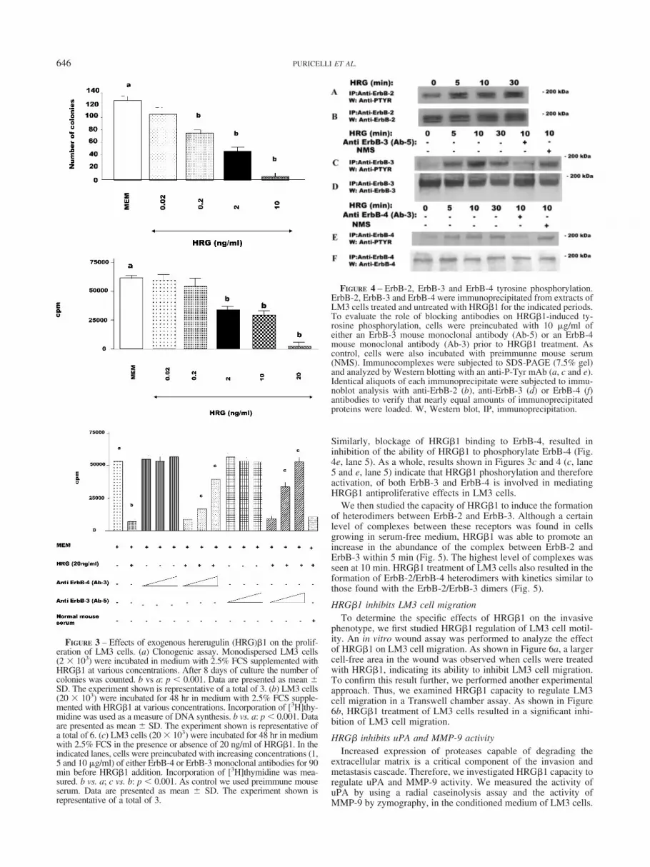
best observed on low-density cultures.22 Therefore, we first studiedthe effect of exogenous HRG�1 on the proliferation of LM3 cellsseeded at low density, using a clonogenic assay. HRG�1 signifi-cantly inhibited clonal LM3 cell growth (median effective dose[ED50]: 0.8 � 0.18 ng/ml), reducing the number of colonies withmore than 10 cells after 8 days of treatment (Fig. 3a). We thenseeded LM3 cells at higher density and treated them with HRG�1for 48 hr. Effects of HRG�1 on cell growth were evaluated byusing a [3H]thymidine incorporation assay. Figure 3b shows thatHRG�1 was still able to inhibit LM3 cell proliferation, althoughthe doses required to achieve ED50 (10.2 � 1.5 ng/ml) were higherthan those required when cells were seeded at low densities. After48 hr of HRG�1 treatment, complete inhibition of growth wasobserved with an HRG�1 concentration of 20 ng/ml (Fig. 3b).Similar results were obtained when cell growth was evaluated bycell protein content or using an MTS assay (data not shown).HRG�1 did not stimulate growth of LM3 cells at any of the dosestested (0.02–40 ng/ml).
It has recently been reported that ErbB-4 is both necessary andsufficient to trigger an antiproliferative response in human breastcancer cells.45 We therefore assessed the role of ErbB-4 in trans-mitting HRG�1 antiproliferative signals in LM3 cells. For thispurpose, we blocked HRG�1 binding to ErbB-4 by using a mousemonoclonal ErbB-4 antibody (Ab-3). As shown in Figure 3c,addition of the ErbB-4 antibody to HRG�1-treated LM3 cellsresulted in a dose-dependent inhibition of HRG�1 antiproliferativeeffects. The ErbB-4 antibody, at the highest dose tested (10 �g/ml), inhibited HRG�1 antiproliferative response by 60–70% (Fig.3c). To investigate ErbB-3 involvement in HRG�1 inhibition ofLM3 cell growth, we used the same experimental approach. Wefound that blockage of HRG�1 binding to ErbB-3, by using themonoclonal ErbB-3 antibody Ab-5, inhibited HRG�1 antiprolif-erative response in a dose-dependent fashion (Fig. 3c). It is note-worthy that the highest dose of the ErbB-3 antibody used (10�g/ml) completely abolished any HRG�1 effect on LM3 cellproliferation (Fig. 3c).
HRG� induces ErbB-2, ErbB-3 and ErbB-4 tyrosinephosphorylation and heterodimerization
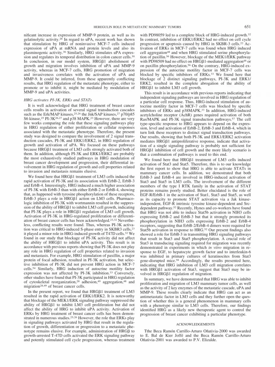
HRG�1-induced antiproliferative response in LM3 cells wasinhibited when HRG�1 binding to ErbB-4 or to ErbB-3 wasabolished, suggesting that HRG�1 activation of these receptorswas involved in the antiproliferative effects. Therefore, we exam-ined HRG�1 effects on the level of tyrosine phosphorylation ofErbB-3 and ErbB-4, as well as on ErbB-2 tyrosine phosphorylationlevels, as a marker of activation of these receptors Extracts fromLM3 cells treated with HRG�1 were immunoprecipitated withanti-ErbB-2, ErbB-3 or ErbB-4 antibodies and the phosphotyrosinecontent of these receptors was determined by performing Westernblotting with an anti-phosphotyrosine antibody. A certain degreeof ErbB-2 tyrosine phosphorylation was observed in LM3 cellsgrowing in medium without the addition of HRG�1 (Fig. 4a). Thisis in accordance with previous findings showing that ErbB-2overexpression may result in its constitutive tyrosine phosphory-lation.46,47 HRG�1 treatment of LM3 cells induced an increase inErbB-2 tyrosine phosphorylation as early as 5 min after treatment(Fig. 4a). On the other hand, it is noteworthy that HRG�1 dra-matically increased tyrosine phosphorylation of ErbB-3 (Fig. 4c).Induction of ErbB-4 tyrosine phosphorylation was also observedafter HRG treatment (Fig. 4e).
To confirm that inhibition of HRG�1 binding to ErbB-3 orErbB-4, by the use of the respective blocking antibody, resulted inthe abrogation of HRG�1 ability to phosphorylate these receptors,we assessed their degree of tyrosine phosphorylation. Therefore,LM3 cells were preincubated with anti-ErB-3 (Ab-5) or ErbB-4(Ab-3) blocking antibodies prior to HRG�1 treatment. As seen inFigure 4c (lane 5), abolishment of HRG-ErbB-3 binding resultedin the abrogation of HRG�1-induced ErbB-3 phosphorylation.
FIGURE 1 – Hereglulin (HRG) expression. HRG RNAse protectionassay. Thirty micrograms of total RNA from LM3 cells were hybrid-ized with HRG and rpL32 probes as described in Material and Meth-ods. The HRG-protected fragment is 333 bp, and the rpL32-protectedfragment is 76 bp. MWM, molecular weight markers. C4HD cells,derived from a murine mammary adenocarcinoma induced by me-droxyprogesterone acetate, were used as positive control.
FIGURE 2 – Expression of type I RTKs at the protein level. Eightymicrograms of protein from LM3 cell lysates were electrophoresedand immunoblotted for ErbB-2, ErbB-3 and ErbB-4. MCF-7 humanbreast cancer cells and C4HD murine mammary adenocarcinoma cellswere used as controls for RTK expression levels.
645HEREGULIN ROLE IN METASTATIC MAMMARY TUMORS
Similarly, blockage of HRG�1 binding to ErbB-4, resulted ininhibition of the ability of HRG�1 to phosphorylate ErbB-4 (Fig.4e, lane 5). As a whole, results shown in Figures 3c and 4 (c, lane5 and e, lane 5) indicate that HRG�1 phoshorylation and thereforeactivation, of both ErbB-3 and ErbB-4 is involved in mediatingHRG�1 antiproliferative effects in LM3 cells.
We then studied the capacity of HRG�1 to induce the formationof heterodimers between ErbB-2 and ErbB-3. Although a certainlevel of complexes between these receptors was found in cellsgrowing in serum-free medium, HRG�1 was able to promote anincrease in the abundance of the complex between ErbB-2 andErbB-3 within 5 min (Fig. 5). The highest level of complexes wasseen at 10 min. HRG�1 treatment of LM3 cells also resulted in theformation of ErbB-2/ErbB-4 heterodimers with kinetics similar tothose found with the ErbB-2/ErbB-3 dimers (Fig. 5).
HRG�1 inhibits LM3 cell migrationTo determine the specific effects of HRG�1 on the invasive
phenotype, we first studied HRG�1 regulation of LM3 cell motil-ity. An in vitro wound assay was performed to analyze the effectof HRG�1 on LM3 cell migration. As shown in Figure 6a, a largercell-free area in the wound was observed when cells were treatedwith HRG�1, indicating its ability to inhibit LM3 cell migration.To confirm this result further, we performed another experimentalapproach. Thus, we examined HRG�1 capacity to regulate LM3cell migration in a Transwell chamber assay. As shown in Figure6b, HRG�1 treatment of LM3 cells resulted in a significant inhi-bition of LM3 cell migration.
HRG� inhibits uPA and MMP-9 activityIncreased expression of proteases capable of degrading the
extracellular matrix is a critical component of the invasion andmetastasis cascade. Therefore, we investigated HRG�1 capacity toregulate uPA and MMP-9 activity. We measured the activity ofuPA by using a radial caseinolysis assay and the activity ofMMP-9 by zymography, in the conditioned medium of LM3 cells.
FIGURE 3 – Effects of exogenous hererugulin (HRG)�1 on the prolif-eration of LM3 cells. (a) Clonogenic assay. Monodispersed LM3 cells(2 � 103) were incubated in medium with 2.5% FCS supplemented withHRG�1 at various concentrations. After 8 days of culture the number ofcolonies was counted. b vs a: p 0.001. Data are presented as mean �SD. The experiment shown is representative of a total of 3. (b) LM3 cells(20 � 103) were incubated for 48 hr in medium with 2.5% FCS supple-mented with HRG�1 at various concentrations. Incorporation of [3H]thy-midine was used as a measure of DNA synthesis. b vs. a: p 0.001. Dataare presented as mean � SD. The experiment shown is representative ofa total of 6. (c) LM3 cells (20 � 103) were incubated for 48 hr in mediumwith 2.5% FCS in the presence or absence of 20 ng/ml of HRG�1. In theindicated lanes, cells were preincubated with increasing concentrations (1,5 and 10 �g/ml) of either ErbB-4 or ErbB-3 monoclonal antibodies for 90min before HRG�1 addition. Incorporation of [3H]thymidine was mea-sured. b vs. a; c vs. b: p 0.001. As control we used preimmune mouseserum. Data are presented as mean � SD. The experiment shown isrepresentative of a total of 3.
FIGURE 4 – ErbB-2, ErbB-3 and ErbB-4 tyrosine phosphorylation.ErbB-2, ErbB-3 and ErbB-4 were immunoprecipitated from extracts ofLM3 cells treated and untreated with HRG�1 for the indicated periods.To evaluate the role of blocking antibodies on HRG�1-induced ty-rosine phosphorylation, cells were preincubated with 10 �g/ml ofeither an ErbB-3 mouse monoclonal antibody (Ab-5) or an ErbB-4mouse monoclonal antibody (Ab-3) prior to HRG�1 treatment. Ascontrol, cells were also incubated with preimmunne mouse serum(NMS). Immunocomplexes were subjected to SDS-PAGE (7.5% gel)and analyzed by Western blotting with an anti-P-Tyr mAb (a, c and e).Identical aliquots of each immunoprecipitate were subjected to immu-noblot analysis with anti-ErbB-2 (b), anti-ErbB-3 (d) or ErbB-4 (f)antibodies to verify that nearly equal amounts of immunoprecipitatedproteins were loaded. W, Western blot, IP, immunoprecipitation.
646 PURICELLI ET AL.

Figure 7a and b shows that HRG�1 induced a dose-dependentinhibition of the activity of both enzymes.
Activation of PI-3K, ERK1/ERK2 and STATs by HRG�
Activation of PI-3K has emerged as a critical component in avariety of cellular functions regulated by HRG such as prolifera-tion,23 cytoskeletal reorganization,28,29 induction of cell aggrega-
tion,48 adhesion and migration.28,49 We therefore investigatedwhether PI-3K plays any role in HRG�1 signaling pathways in ourexperimental system. Stimulation of LM3 cells with HRG�1 re-sulted in a rapid recruitment of the p85 subunit of PI-3K to thephosphotyrosine-containing cellular fraction, consistent with pre-vious reports of growth factor-stimulated PI-3K activation in othercell lines (Fig. 8a).49,50 To assess the effect of HRG�1 on PI-3Kactivation in intact cells, we prelabeled LM3 cells for 24 hr inMEM containing [2-3H]inositol and then incubated them in thepresence or absence of HRG�1 (20 ng/ml). Because it has alreadybeen shown that IGF-I activates PI-3K,51 we also treated LM3cells with IGF-I as a positive control. Figure 8b shows that PI-3Kactivity was greatly increased by 10 min of HRG� stimulation ofLM3 cells, revealed by the appearance of Ptdlns-3-P, Ptdlns-3,4-P2and Ptdlns-3,4,5-P3. Wortmannin, a specific chemical inhibitor ofPI-3K, at a concentration of 500 nM, completely blocked HRG�1-induced PI-3K activation (Fig. 8b).
We then investigated the association of the p85 subunit of PI-3Kwith ErbB-2, ErbB-3 and ErbB-4. Extracts from LM3 cells treatedwith HRG�1 were immunoprecipitated with anti- ErbB-2, -ErbB-3or -ErbB-4 antibodies and Western blotting was performed with ananti-p85 antibody (Fig. 8c). HRG-� stimulation greatly inducedp85 association with ErbB-3 (Fig. 8c). A moderate induction of theassociation of p85 with ErbB-2 and ErbB-4 was also seen (Fig.8c).
HRG is also known to stimulate ERK1/ERK2.25,26 Therefore,we next examined the effect of HRG� on the activity of ERK1/ERK2, using antisera specific for the dually phosphorylated, activeform of this kinase. As shown in Figure 9a, HRG� treatmentresulted in the rapid activation of ERK1/ERK2 that began toincrease after 5 min, reached its highest levels at 10 min andremained sustained for at least 30 min. Activation of ERKs by
FIGURE 5 – ErbB-2, ErbB-3 and ErbB-4 heterodimerization. ErbB-2was immunoprecipitated from LM3 cell extracts, and immunocom-plexes were subjected to SDS-PAGE (7.5% gel) and analyzed byWestern blotting with anti-ErbB-3 (a) or -ErbB-4 (c) antibodies.Identical aliquots of each immunoprecipitate were subjected to immu-noblot analysis with anti-ErbB-2 (b and d) antibody to verify thatnearly equal amounts of immunoprecipitated protein were loaded. W,Western blot, IP, immunoprecipitation. HRG, heregulin.
FIGURE 6 – Effects of heregulin (HRG)�1 on LM3 cell migration.Wounds 400 �M wide were made in confluent LM3 cell monolayers.Cells were treated with HRG�1 at various concentrations and allowedto migrate into the cell-free area for 24 hr. The cell-free area wasmeasured by densitometry and expressed as arbitrary units. (a) p 0.01 with respect to cells growing in MEM without HRG�1 supple-mentation. Shown is a representative experiment of a total of 3 thatgave similar results. (b) Migration assay using Transwell cell culturechambers. At least 20 fields of �400 per membrane were counted. bvs. a: p 0.001. Data are presented as mean � SD. Assay was donein triplicate. The experiment shown is representative of a total of 3.
FIGURE 7 – Effect of heregulin (HRG)�1 on uPA and MMP-9 ac-tivity. Semiconfluent monolayers of LM3 cells were incubated inserum free-medium for 24 hr with and without the addition of HRG�1at various concentrations. Conditioned media were collected, and theremaining monolayers were lysed to measure protein content. (a) uPAactivity was quantified by a radial caseinolytic assay. Data are pre-sented as mean � SD. Shown is a representative experiment of a totalof 4. (b) MMP-9 activity was detected by zymography. The experi-ment shown is representative of a total of 4.
647HEREGULIN ROLE IN METASTATIC MAMMARY TUMORS

HRG� was suppressed when cells were treated with PD98059 (10�M), a specific inhibitor of MEK1 (Fig. 9b).
The involvement of the different members of the type I RTKfamily in the activation of STAT proteins remains poorly studied.We therefore sought to determine whether HRG�1 treatment ofLM3 cells induced activation of STATs. As can be seen in Figure10A (a), HRG�1 induced a strong tyrosine phosphorylation ofStat3 that was maximal after 10 min of treatment and remained atthe same high level after 30 min. To investigate the effect ofHRG�1 on Stat5 activation, we immunoprecipitated LM3 cellextracts with a polyclonal antibody that reacts with both Stat5a and5b and performed Western blot with an anti-phosphotyrosine an-tibody. Figure 10A (c) shows that HRG�1 induced an increase inStat5 phosphotyrosine content. It has been previously shown thatErbB-4 expression is required for Stat5b activation in response toHRG.52 Therefore, to explore the role of ErbB-4 in HRG�1-induced activation of Stat3 and Stat5, we blocked HRG�1 bindingto ErbB-4 by using the monoclonal antibody Ab-3. Disruption ofHRG�1 binding to ErbB-4 dramatically inhibited HRG�1-inducedphosphorylation of both Stat3 (Fig. 10A, a) and Stat5 (Fig. 10A, c).
The same experimental approach was performed to exploreErbB-3 involvement in the mechanism of HRG�1 activation ofStat3 and Stat5. We found that blockage of HRG�1 binding toErbB-3, by using the anti-ErbB-3 monoclonal antibody Ab-5,resulted in inhibition of the ability of HRG�1 phosphorylate Stat3(Fig. 10A, a) and Stat5 (Fig. 10A, c). To determine that inhibitionof HRG�1 binding to ErbB-4 resulted in the abolishment of theHRG-ErbB-4 signaling pathway, we assessed the degree of ty-rosine phosphorylation of ErbB-4 in LM3 cells in which HRG�1binding to ErbB4 had been blocked by the anti-ErbB-4 monoclonalantibody Ab-3. As seen in Figure 10B, a, abolishment of HRG-ErbB-4 binding resulted in abrogation of HRG�1-induced ErbB-4phosphorylation. Similarly, blockage of HRG�1 binding toErbB-3 by the ErbB-3 antibody Ab-5 resulted in the abrogation ofHRG�1 capacity to phosphorylate ErbB-3 (Fig. 10B, c). There-fore, our findings show that HRG�1 binding to and activation ofboth ErbB-3 and ErbB-4 are required for HRG�1-induced activa-tion of Stat3 ands Stat5 in LM3 cells.
FIGURE 8 – Heregulin (HRG)�1 induces PI-3K activation and asso-ciation with ErbBs. (a) HRG�1 induces recruitment of the p85 subunitof PI-3K to the phosphotyrosine cellular fraction. LM3 cells incubatedin the presence or absence of 20 ng/ml HRG�1 for the indicatedperiods were lysed, and phosphotyrosine-containing proteins wereimmunoprecipitated with an anti-P-Tyr (PTYR) MAb. Immunocom-plexes were subjected to SDS-PAGE (7.5% gel) and analyzed byWestern blotting with anti-p85 antibody . In the middle panel, identicalaliquots of each immunoprecipitate were subjected to immunoblotanalysis with an anti-P-Tyr MAb. In the lower panel, 100 �g proteinfrom cell lysates was subjected to SDS-PAGE and analyzed by West-ern blotting with an anti-p85 antibody to show identical levels of thep85 PI-3K subunit in lysates from cells treated and untreated withHRG�1. Shown is a representative experiment of a total of 3. IP,immunoprecipitation; W, Western blot. (b) HRG activates PI-3K en-zymatic activity in vivo. Subconfluent cultures of LM3 cells wereprelabeled for 24 hr in serum-free MEM containing 4 �Ci/ml [2-3H]ino-sitol. At the end of the prelabeling period, cells were extensivelywashed and then incubated for 10 min in serum-free MEM in presenceor absence of HRG (20 ng/ml) or insulin-like growth factor-I (IGF-I;50 ng/ml, used as positive control for PI-3K activation). In addition,cells were preincubated with wortmannin (500 mM) for 90 min priorto stimulation with HRG�1 (fourth lane). Phospholipids were ex-tracted and separated by TLC chromatography as described in Materialand Methods. The position of commercial standards (left) and D3-phosphoinositides (right) are indicated. Shown is a representativeexperiment of a total of 3. (c) HRG induces the association of the p85subunit of PI-3K with ErbB-2, ErbB-3 and ErbB-4. Extracts from LM3cells treated with HRG�1 for the indicated periods were immunopre-cipitated with anti- ErbB-3, -ErbB-2 or -ErbB-4 antibodies, and West-ern blotting was performed with an anti-p85 antibody. The lanesmarked “lysate” show LM3 cell lysates blotted in parallel with anti-p85 antibody.
648 PURICELLI ET AL.

HRG�1 effect on LM3 cell proliferation requires PI-3K andERK activation although HRG�1 effect on uPA synthesis isindependent of these 2 pathways
There are few works comparing the role that different sig-naling pathways play in HRG regulation of either proliferationor of cellular responses associated with the metastatic pheno-type. We focused here on the role of PI-3K and ERK1/ERK2signaling cascades because they are strongly activated byHRG�1 treatment of LM3 cells. In addition, although it isfirmly acknowledged that these pathways are involved in someof the biologic responses to HRG in breast cancer cells, theirdifferential involvement in HRG modulation of growth and ofproperties related to invasion and metastasis remains elusive.To understand the involvement of PI-3K in HRG�1 effects onLM3 cell proliferation, we tested whether wortmannin wouldblock HRG�1-induced inhibition of LM3 cell growth. Asshown in Figure 11a, wortmannin alone had no detectable effecton LM3 cell proliferation during 48 hr addition, but led insteadto the blockage of HRG�1 capacity to inhibit LM3 cell prolif-eration. Similar results were obtained using another PI-3Kchemical inhibitor, LY294002 (data not shown).
We then investigated whether blockage of the MEK1-ERKsignaling pathway would inhibit the ability of HRG�1 to regulateLM3 cell growth by using PD98059. Treatment of LM3 cells withPD98059 alone had no effect on their proliferation (Fig. 11a).However, HRG�1 inhibition of LM3 cell growth was blocked byPD98059 (Fig. 11a).
As a way of assessing the role of PI-3K and ERKs on HRG�1regulation of LM3 metastatic phenotype, we chose to study uPA
activity. Neither wortmannin or LY294002, nor PD98059 resultedin a significant abolishment of the ability of HRG�1 to inhibit uPAactivity (Fig. 11b).
FIGURE 9 – Activation of ERKs by heregulin HRG�1 a) LM3 cellswere treated with HRG� for the indicated periods, and 100 �g proteinfrom cell lysates was electrophoresed on 12% SDS gels and immuno-blotted with an anti-phospho ERK1/ERK2 antibody (upper panel). Themembrane was stripped and hybridized with anti-total ERK1/ERK2antibody (lower panel). (b) LM3 cells were pretreated with PD98059or its solvent DMSO for 90 min before stimulation with HRG 20 ng/mlfor 10 min. Cell lysates were electrophoresed and immunoblotted withan anti-phospho ERK1/ERK2 antibody (upper panel). The membranewas stripped and hybridized with an anti-total ERK1/ERK2 antibody(lower panel).
FIGURE 10 – Heregulin (HRG)�1 activates Stat3 and Stat5. (A) LM3cells were treated with 20 ng/ml HRG�1 for the indicated periods orwere preincubated with 10 �g/ml of mouse monoclonal ErbB-4 (Ab-3)or ErbB-3 (Ab-5) antibodies or normal mouse serum for 90 min andthen were treated with HRG for 10 min. (a) Proteins from cell lysates(100 �g) were electrophoresed and immunoblotted with an anti-phospho Stat3 antibody. (b) The membrane was stripped and hybrid-ized with an anti-total Stat3. (c) Proteins from cell lysates (1 mg) wereimmunoprecipitated with an anti-total Stat5 antibody followed byWestern blotting with an anti-phosphotyrosine antibody. (d) Afterstripping, the membrane was probed with the anti-total Stat5 antibody.(B) (a) Inhibition of HRG-mediated tyrosine phosphorylation ofErbB-4 by the ErbB-4 monoclonal antibody Ab-3. Protein (1 mg) fromlysates of LM3 cells, treated with HRG or preincubated with theErbB-4 monoclonal antibody Ab-3, or with normal mouse serum priorto HRG treatment, was immunoprecipitated with a rabbit polyclonalantibody to ErbB-4, and immunocomplexes were analyzed by Westernblotting with an anti-P-Tyr MAb. (b) Identical aliquots of each immu-noprecipitate were subjected to immunoblot analysis with the anti-ErbB-4 rabbit polyclonal antibody to verify that nearly equal amountsof immunoprecipitated proteins were loaded. (c) Inhibition of HRG-mediated tyrosine phosphorylation of ErbB-3 by the ErbB-3 monoclo-nal antibody Ab-5. Protein (1 mg) from lysates of LM3 cells, treatedwith HRG or preincubated with the ErbB-3 monoclonal antibodyAb-5, or with normal mouse serum prior to HRG treatment, wasimmunoprecipitated with a rabbit polyclonal antibody to ErbB-3, andimmunocomplexes were analyzed by Western blotting with an anti-P-Tyr MAb. (d) Identical aliquots of each immunoprecipitate weresubjected to immunoblot analysis with the anti-ErbB-3 rabbit poly-clonal antibody. W, Western blot, IP, immunoprecipitation.
649HEREGULIN ROLE IN METASTATIC MAMMARY TUMORS

DISCUSSION
HRG inhibits LM3 cell proliferation and migrationOur results showing that HRG�1 inhibited LM3 cell growth are
in line with accumulating evidence indicating that HRG exerts agrowth-inhibitory effect on ErbB-2-overexpressing breast tumorcells.19–24 Accordingly, transfection of HRG cDNA to SKBR3cells resulted in pronounced inhibition of anchorage-dependentand -independent growth.53 To date, there is consensus in that thedifferent cellular responses to HRG are very likely dependent onthe array of type I RTKs activated and the intracellular signalingengaged. However, the influence that the levels of expression ofeach of the ErbBs receptors has on the growth effects of HRGremains poorly understood. For example, cells with elevated levelsof ErbB-2 but little or no ErbB-3 and ErbB-4 are not growth-inhibited by HRG.22 Recent work by Daly et al.22,23 has confirmedthe importance of ErbB-3 in HRG inhibition of SKBR3 cellgrowth. These authors generated 3 different pools of SKBR3 cells,1 of them ectopically expressing ErbB-4 and the others expressingenhanced levels of ErbB-1 or ErbB-3. Only ErbB-3 overexpressionmarkedly enhanced growth inhibition by HRG.23 On the otherhand, it has recently been reported that expression of ErbB-4 isboth necessary and sufficient to trigger an antiproliferative re-
sponse to HRG in human breast cancer cells.45 In the present work,the importance of both ErbB-3 and ErbB-4 in HRG�1 inhibition ofLM3 cell growth was demonstrated by the finding that blockage ofHRG binding to either ErbB-3 or ErbB-4 resulted in significantabrogation of the ability of HRG�1 to inhibit LM3 cell growth. Itis noteworthy that at the highest concentration of blocking anti-bodies used, only the anti-ErbB-3 antibody resulted in a completeabolishment of HRG�1 biologic effect. In addition, HRG�1 in-duced a dramatically higher degree of tyrosine phosphorylation ofErbB-3 than those of ErbB-2 and ErbB-4. Taken together, theseresults suggest that that ErbB-3 could be a major target of HRG�1in LM3 cells.
Notably, we found here that HRG�1 growth-inhibitory effectson LM3 cells were highest when cells were plated at low density,in accordance with previous work by Daly et al.,22 who showedthat conditioned medium from high-density cultures protectedSKBR3 cells from HRG-induced growth inhibition. A likely ex-planation for the density effect may be that SKBR3 cells secreteGFs that accumulate in dense cultures and protect them from HRG.Of the GFs that Daly et al.23 examined, only IGF-I and insulinshowed a protective effect. Interestingly, we had already foundexpression of IGF-I and its receptor, namely, IGF-IR, in M3tumors and in primary cultures,54 as well as in LM3 cells (Elizaldeet al., unpublished data). In addition, we have recently demon-strated that a functional IGF-IR is required for HRG mitogenicactivity in mammary tumor cells.38 Furthermore, we have alsoalready shown the existence of a hierarchical interaction betweenIGF-IR and ErbB-2, by means of which IGF-IR directs ErbB-2phosphorylation in breast cancer cells.43 Therefore, our resultssuggest that IGF-I accumulated in dense LM3 cultures could beinterfering with HRG�1 action on LM3 cells through the ability ofIGF-I to transmodulate ErbB-2 activity.
Compatible with its antiproliferative effects on LM3 cells,HRG�1 also inhibited migration of these cells at all doses tested.Although there is no consensus on HRG role on migration, resultssimilar to ours have been reported in SKBR3 cells in which highdoses of HRG resulted in inhibition of motility and invasion.29
Conversely, HRG has been found to induce migration and inva-siveness and to regulate actin cytoskeleton in the nonmetastaticSKBR3 cells, in striking contrast to its growth-inhibitory effect onthese cells.24 HRG also promotes cell migration and regulatescytoskeletal reorganization of noninvasive MCF-7 cells.28 In ad-dition, HRG treatment of the metastatic MDA-MB-435 line ex-hibiting high levels of HRG expression also resulted in stimulationof cell adhesion and migration.49 Interestingly, the phenotypiccharacteristics of all the cell lines mentioned above, in which HRGpromotes invasion and metastasis, are different from those of LM3cells. First, whereas MCF-7 and SKBR3 cells are nonmetastaticbreast cancer cells, LM3 cells are highly metastatic. Second,MDA-MB-435 cells, which, like LM3, are highly metastatic anddo not produce HRG, express an array and level of type I RTKsdifferent from those of LM3 cells. These differences between LM3cells and the other cells described confirm the hypothesis that HRGregulation of breast cancer cell activities may depend on thecoexpression of type I RTKs as well as on the relative levels ofexpression of these receptors, which in turn might result in thedifferential activation of signaling pathways.
HRG inhibits MMP-9 and uPA activityThe role of proteases capable of degrading the extracellular
matrix in HRG regulation of motility and invasiveness also re-mains poorly explored. Ours is the first evidence to show thatHRG�1 inhibition of growth and migration of mammary tumorcells correlates with its capacity to inhibit MMP-9 and uPA activ-ity. Varying results have been reported in the literature. Thus, itwas found that HRG treatment of SKBR3 cells increased expres-sion of MMP-9, despite inducing growth inhibition.24 However,another study reported that exposure of SKBR3 and MCF-7 cellsto HRG had no effect on MMP-9 induction.55 It has recently beennoted that stimulation of MCF-7 cells with HRG leads to a sig-
FIGURE 11 – Effect of PI-3K and MEK1/ERKs inhibitors on heregu-lin (HRG)�1 regulation of LM3 cell growth and uPA activity. (a) LM3cells were incubated for 48 hr in MEM with 2.5% FCS containingvehicle DMSO (1:2,000) or in MEM (containing DMSO 1:2,000) �20 ng/ml HRG�1. To study the effects of wortmannin or PD98059,these 2 inhibitors were added 90 min before the 48 hr incubation withHRG�1. Incorporation of [3H]thymidine was used as a measure ofDNA synthesis. b vs. a: p 0.001. Data are presented as mean � SD.The experiment shown is representative of a total of 5. (b) Semicon-fluent monolayers of LM3 cells growing in serum-free MEM were leftuntreated or preincubated for 90 min with wortmannin, LY294002 orPD98059. Cells were then either treated with HRG�1 or given vehiclealone and were incubated for 24 hr. Conditioned media were collected,and uPA activity was quantified by a radial caseinolytic assay. Data arepresented as means � SD. Shown is a representative experiment of atotal of 4.
650 PURICELLI ET AL.
nificant increase in expression of MMP-9 protein, as well as itsgelatinolytic activity.28 In regard to uPA, recent work has shownthat stimulation by HRG of noninvasive MCF-7 cells inducedexpression of uPA at mRNA and protein levels and also itsplasminogenic activity.30 Similarly, HRG stimulates uPA expres-sion and regulates its temporal distribution in colon cancer cells.30
In conclusion, in our model system, HRG�1 abolishment ofgrowth and migration involves inhibition of uPA and MMP-9activity, whereas in MCF-7 cells, HRG promotion of migrationand invasiveness correlates with the activation of uPA andMMP-9. It could be inferred, from these apparently conflictingresults, that HRG regulation of the metastatic phenotype, either topromote or to inhibit it, might be mediated by modulation ofMMP-9 and uPA activities.
HRG activates PI-3K, ERKs and STATsIt is well acknowledged that HRG treatment of breast cancer
cells results in activation of several signal transduction cascadessuch as the Erk/MAP kinases,25,26 the Jnk/SAP kinases,27 p70/p85S6 kinase,25 PI-3K26,27 and p38 MAPK.23 However, there are veryfew works comparing the role that these signaling pathways playin HRG regulation of either proliferation or cellular responsesassociated with the metastatic phenotype. Therefore, the presentstudy was designed to compare the involvement of 2 signal trans-duction cascades, PI-3K and ERK1/ERK2, in HRG regulation ofgrowth and activation of uPA. We focused on these pathwaysbecause HRG�1 treatment of LM3 cells strongly activated both ofthem. In addition, although PI-3K and ERK1/ERK2 are probablythe most exhaustively studied pathways in HRG modulation ofbreast cancer development and progression, their differential in-volvement in HRG regulation of growth and cell properties relatedto invasion and metastasis remains elusive.
We found here that HRG�1 treatment of LM3 cells induced therapid activation of PI-3K and its association with ErbB-2, ErbB-3and ErbB-4. Interestingly, HRG induced a much higher associationof PI-3K with ErbB-3 than with either ErbB-2 or ErbB-4, showingthat, as happened with tyrosine phosphorylation of these receptors,ErbB-3 plays a role in HRG�1 action on LM3 cells. Pharmaco-logic inhibition of PI-3K with wortmannin resulted in the suppres-sion of the ability of HRG�1 to inhibit LM3 cell growth, indicatingthat PI-3K plays a role in HRG�1 regulation of LM3 cell growth.Activation of PI-3K in HRG-regulated proliferation or differenti-ation of breast cancer cells has been acknowledged.23,32 Neverthe-less, conflicting results were found. Thus, whereas PI-3K activa-tion was critical to HRG-induced S-phase entry in SKBR3 cells,23
it played a minor role in HRG-induced growth of T47D cells.32 Wefound in our study that blockage of PI-3K activity did not affectthe ability of HRG�1 to inhibit uPA activity. This result is inaccordance with previous reports showing that PI-3K does not playany role in HRG regulation of cell properties related to invasionand metastasis. For example, HRG stimulation of paxillin, a majorprotein of focal adhesion, resulted in PI-3K activation, but selec-tive inhibition of PI-3K did not prevent HRG action in MCF-7cells.54 Similarly, HRG induction of autocrine motility factorexpression was not affected by PI-3K inhibition.33 Conversely,other studies have found that PI-3K participated in HRG regulationof cytoskeletal reorganization,28 adhesion,49 aggregation,48 andmigration28,49 of breast cancer cells.
In the present report, we found that HRG�1 treatment of LM3resulted in the rapid activation of ERK1/ERK2. It is noteworthythat blockage of the MEK1/ERK signaling pathway suppressed theability of HRG�1 to inhibit LM3 cell proliferation but did notaffect the ability of HRG to inhibit uPA activity. Activation ofERKs by HRG treatment of breast cancer cells has been demon-strated in numerous studies.25,26 However, the role that ERKs playin signaling pathways activated by HRG that result in the regula-tion of growth, differentiation or progression to a metastatic phe-notype remains elusive. For example, administration of HRG� togrowth-arrested T-47D cells activated the ERK signaling pathwayand potently stimulated cell cycle progression, whereas treatment
with PD98059 led to a complete block of HRG-induced growth.32
In contrast, inhibition of ERK1/ERK2 had no effect on cell cycleprogression or apoptosis caused by HRG in SKBR-3 cells.22 Ac-tivation of ERKs in MCF-7 cells was found when HRG inducedcell aggregation48 and when HRG stimulated serine phosphoryla-tion of paxillin.56 However, blockage of the MEK1/ERK pathwaywith PD98509 had no effect on HRG�1-mediated aggregation48 oron paxillin phosphorylation.56 On the contrary, HRG-induced ex-pression of the autocrine motility factor in MCF-7 cells wasblocked by specific inhibitors of ERKs.33 We found here thatblockage of 2 distinct signaling pathways, PI-3K and ERK1/ERK2, resulted in the complete abrogation of the ability ofHRG�1 to inhibit LM3 cell growth.
This result is in accordance with previous reports indicating thatindependent signaling pathways are involved in HRG regulation ofa particular cell response. Thus, HRG-induced stimulation of au-tocrine motility factor in MCF-7 cells was blocked by specificinhibitors of ERKs and p38MAPK.33 In addition, HRG-inducedacetylcholine receptor (AchR) genes required activation of bothRas/MAPK and PI-3K signal transduction pathways.57 The celltype-specific response to HRG appears to depend on the expres-sion, level and activation of ErbB-2, ErbB-3 and ErbB-4, which inturn link these receptors to distinct signal transduction pathways.Our results, showing that both PI-3K and ERK1/ERK2 activationmediates HRG�1 antiproliferative response, indicate that activa-tion of a single signaling pathway is probably not sufficient forHRG�1 inhibition of cell growth and the more likely scenario isthat a combination of pathways is used to this end.
We found here that HRG�1 treatment of LM3 cells inducedactivation of Stat3 and Stat5. Therefore, this is to our knowledgethe first report to show that HRG is able to activate STATs inmammary cancer cells. In addition, we demostrated that bothErbB-3 and ErbB-4 are involved in HRG-induced activation ofStat3 and Stat5 in LM3 cells. The involvement of the differentmembers of the type I RTK family in the activation of STATproteins remains poorly studied. Better elucidated is the role ofEGF-R/ErbB-1 in the activation of Stat1, Stat3 and Stat5, as wellas its capacity to promote STAT activation via a Jak kinase-independent, EGF-R intrinsic tyrosine kinase-dependent and Src-dependent pathway.52 Recently, Hynes and co-workers have foundthat HRG was not able to induce Stat5b activation in NIH3 cellsexpressing ErbB-2 and ErbB-3 but that it strongly promoted itsphosphorylation in NIH3 cells expressing ErbB-2 and ErbB-4receptors, suggesting that ErbB-2/ErbB-4 dimers were required forStat5b activation in response to HRG.52 Our present findings alsoshowed a role for ErbB-3 in transmitting HRG signaling pathwaysthat result in Stat3 and Stat5 phosphorylation. A crucial role ofStat3 in transducing signaling required for migration was recentlydemonstrated in experiments in which in vitro migration in re-sponse to EGF, to hepatocyte growth factor and to interleukin-6was inhibited in primary cultures of keratinocites from Stat3gene-disrupted mice.58 Accordingly, the results presented here,indicating that HRG inhibition of LM3 cell migration correlateswith HRG�1 activation of Stat3, suggest that Stat3 may be in-volved in HRG�1 regulation of migration.
In summary, we have demonstrated that HRG was able to inhibitproliferation and migration of LM3 mammary tumor cells, as wellas the activity of 2 key enzymes of the metastatic cascade, uPA andMMP-9. These results clearly indicate that HRG can act as anantimetastatic factor in LM3 cells and they further open the ques-tion of whether this is a general phenomenon in mammary cellswith a phenotype similar to LM3 cells. Therefore, our findingsidentified HRG as a likely new therapeutic agent to control theprogression of breast cancer exhibiting a particular phenotype.
ACKNOWLEDGEMENTS
Tthe Beca Ramon Carrillo-Arturo Onativia-2000 was awardedto E. Bal de Kier Joffe and the Beca Ramon Carrillo-ArturoOnativia-2001 was awarded to P.V. Elizalde.
651HEREGULIN ROLE IN METASTATIC MAMMARY TUMORS

REFERENCES
1. Nicolson GL. Cancer progression and growth: relationship of para-crine and autocrine growth mechanisms to organ preference of me-tastasis. Exp Cell Res 1993;204:171–80.
2. Kerbel RS. Growth dominance of the metastatic cancer cell: cellularand molecular aspects. Adv Cancer Res 1990;55:87–132.
3. Peles E, Yarden Y. Neu and its ligands: from an oncogene to neuralfactors.
4. Wen D, Suggs SV, Karunagaran D, Liu N, Cupples RL, Luo Y,Jansen AM, Ben Barush N, Trollinger DB, Jacobsen VL, Meng T, LuHS, Hu S, Chang D, et al. M Structural and functional aspects of themultiplicity of Neu differentiation factors. Mol Cell Biol. 1994;14:1909–19.
5. Carraway KL, Cantley LC. A neu acquaintance for ErbB3 and ErbB4:a role for receptor heterodimerization in growth signaling. Cell 1994;78:5–8.
6. Chang H, Riese DJ II, Gilbert W, Stern DF, McMahan UJ. Ligands forErbB-family receptors encoded by a neuregulin–like gene. Nature1997;387:509–12.
7. Zhang DX, Sliwkowski MX, Mark M, Frantz G, Akita R, Sun Y,Hillan K, Brush J, Godowski PJ. Neuregulin 3 (NRG3) a novel neuraltissue-enriched protein that binds and activates erbB-4. Proc NatlAcad Sci USA 1997;94:9562–7.
8. Pinkas-Kramarski R, Alroy I, Yarden Y. ErbB receptors and EGF-likeligands: cell lineage determination and oncogenesis through combi-natorial signaling. J Mam Gland Biol Neoplasia 1997;2:97–107.
9. Ullrich A, Coussens L, Hayflick JS, Dull TJ, Gray A, Tam AW, LeeJ, Yarden Y, Libermann TA, Schlessinger J, Downward J, MayesELV. Human epidermal growth factor receptor cDNA. Nature 1984;309:418–25.
10. Yamamoto T, Ikawa S, Akiyama T. An erbB-related gene, c-erbB-2,encodes a possible receptor protein similar to the epidermoid growthfactor receptor. Nature 1986;319:230–4.
11. Kraus MH, Issing W, MikiT, Popescu NC, Aaronson SA. Isolationand characterization of ERBB3, a third member of the ERBB/epider-mal growth factor receptor family: evidence for overexpression in asubset of human mammary tumors. Proc Natl Acad Sci USA 1989;86:9193–7.
12. Plowman GD, Whitney GS, Neubauer MG, Green JM, McDonald VL,Todaro GJ, Shoyab M. Molecular cloning and expression of anadditional epidermal growth factor receptor-related gene. Proc NatlAcad Sci USA 1990;87:4905–9.
13. Plowman GD, Culouscou JM, Whitney GS, Green JM, Carlton GW,Foy L, Neubauer MG, Shoyab M. Ligand-specific activation ofHER4/p180erbB-4, a fourth member of the epidermal growth factorreceptor family. Proc Natl Acad Sci USA 1993;90:1746–50.
14. Yang Y, Spitzer E, Meyer D, Sachs M, Niemann C, Hartmann G,Weidner KM, Birchmeier C, Birchmeier W. Sequential requirementof hepatocyte growth factor and neuregulin in the morphogenesis anddifferentiation of the mammary gland. J Cell Biol 1995;131:215–26
15. Marte BM, Jeschke M, Graus-Porta D, Taverna D, Hofer P, Groner B,Yarden Y, Hynes N. Neu differentiation factor/heregulin modulatesgrowth and differentiation of HC11 mammary epithelial cells. MolEndocrinol 1995;9:14–23.
16. Ram TG, Kokeny KE, Dilts CA, Ethier SP. Mitogenic activity of neudifferentiation factor/heregulin mimics that of epidermal growth fac-tor and insulin-like growth factor-I in human mammary epithelialcells. J Cell Physiol 1995;163:589–96.
17. Holmes WE, Sliwkowski MX, Akita RW, Henzel WJ, Lee J, Park JW,Yansura D, Abadi N, Raab H, Lewis GD, Shepard M, Wood WI,Goeddel DV, Vandlen RL. Identification of heregulin, a specificactivator of p185erbB2. Science 1992;256:1205–10.
18. Lewis GD, Lofgren JA, McMurtrey AE, Nuijens A, Fendly BM,Bauer KD, Sliwkowski MX. Growth regulation of human breast andovarian tumor cells by heregulin: evidence for the requirement oferbB2 as a critical component in mediating heregulin responsiveness-.Cancer Res 1996;56:1457–65.
19. Peles E, Bacus SS, Koski RA, Lu HS, Wen D, Odgen SG, Ben LevyR, Yarden Y. Isolation of the neu/HER-2 stimulatory ligand: a 44 Kdglycoprotein that induces differentiation of mammary tumor cells.Cell 1992;69:205–16.
20. Bacus SS, Gudkov AV, Zelnick CR, Chin D, Stern R, Stancovski H,Peles E, Ben-Baruch N, Farbstein H, Lupu R, Wen D, Sela M, et al.Neu differentiation factor (heregulin) induces expression of intercel-lular adhesion molecule 1: implications for mammary tumors. CancerRes 1993;53:5251–61.
21. Culouscou JM, Plowman GD, Carlton GW, Green JM, Shoyab M.Characterization of a breast cancer cell differentiation factor thatspecifically activates the HER4/p180erbB4 receptor. J Biol Chem 1993;268:18407–10.
22. Daly JM, Jannot CB, Beerli RR, Graus-Porta D, Maurer FG, HynesNE. Neu differentiation factor induces ErbB2 down-regulation and
apoptosis of ErbB2-overexpressing breast tumor cells. Cancer Res1997;57:3804–11.
23. Daly JM, Olayioye MA, Wong AM-L, Neve R, Lane HA, Maurer FG,Hynes NE. NDF/heregulin-induced cell cycle changes and apoptosisin breast tumor cells: role of PI3 kinase and p38 MAP kinase path-ways. Oncogene 1999;18:3440–51.
24. Xu FJ, Stack S, Boyer C, O�Briant K, Whitaker R, Mills GB, Yu YH,Bast RC Jr. Heregulin and agonistic anti-p185(c-erbB2) antibodiesinhibit proliferation but increase invasiveness of breast cancer cellsthat overexpress p185(c-erbB2): increased invasiveness may contrib-ute to poor prognosis. Clin Cancer Res 1997;3:1629–34.
25. Marte BM, Graus-Porta D, Jeschke M, Fabbro D, Hynes NE, TavernaD. NDF/heregulin activates MAP kinase and p70/p85 S6 kinaseduring proliferation or differentiation of mammary epithelial cells.Oncogene 1995;10:167–75.
26. Sepp-Lorenzino L, Eberhard I, Ma Z, Cho C, Serve H, Liu F, RosenN, Lupu R. Signal transduction pathways induced by heregulin inMDA-MB-453 breast cancer cells. Oncogene 1996;12:1679–87.
27. Amundadottir LT, Leder P. Signal transduction pathways activatedand required for mammary carcinogenesis in response to specificoncogenes. Oncogene 1998;16:737–46.
28. Adam L, Vadlamudi R, Kondapaka SB, Chernoff J, Mendelsohn J,Kumar R. Heregulin regulates cytoskeletal reorganization and cellmigration through the p21-activated kinase-1 via phosphatidylinosi-tol-3 kinase. J Biol Chem 1998;43: 28238–46.
29. Hijazi MM, Thompson EW, Tang C, Coopman P, Torri J, Yang D,Mueller SC, Lupu R. Heregulin regulates the actin cytoskeleton andpromotes invasive properties in breast cancer cell lines. Int J Oncol2000;17:629–41.
30. Adam L, Mazumdar A, Sharma T, Jones TR, Kumar R. A three-dimensional and temporo-spatial model to study invasiveness of can-cer cells by heregulin and prostaglandin E2. Cancer Res 2001;61:81–7
31. Mazumdar A, Adam L, Boyd D, Kumar R. Heregulin regulation ofurokinase plasminogen activator and its receptor: human breast epi-thelial cell invasion. Cancer Res 2001;61:400–1.
32. Fiddes RJ, Janes PW, Sivertsen SP, Sutherland RL, Musgrove EA,Daly RJ. Inhibition of the MAP kinase cascade blocks heregulin-induced cell cycle progression in T-47D human breast cancer cells.Oncogene 1998;16:2803–13.
33. Talukder AH, Adam L, Raz A, Kumar R. Heregulin regulation ofautocrine motility factor expression in human tumor cells. Cancer Res2000;60:474–80.
34. Urtreger AJ, Ladeda VE, Puricelli L, Rivelli A, Vidal M del C,Sacerdote de Lustig E, Bal de Kier Joffe E. Modulation of fibronectinexpression and proteolytic activity associated with the invasive andmetastatic phenotype in 2 new murine mammary tumor cell lines. IntJ Oncol 1997;11:489–96.
35. Bal de Kier Joffe E, Puricelli L, Lustig ES. Modified adhesionbehavior after in vitro passage of two related murine mammaryadenocarcinomas with different metastasizing ability. Invasion Me-tastasis 1986;6:302–12.
36. Saksela O. Radial caseinolysis in agarose: a simple method for de-tection of plasminogen in the presence of inhibitory substances andserum. Anal Biochem 1981;111:276–82.
37. Pittman R. Release of plasminogen activator and calcium dependentmetalloprotease from cultured sympathic and sensor neurons. DevBiol 1985;110:91–101.
38. Balana ME, LupuR, LabriolaL, Charreau EH, Elizalde PV. Interac-tions between progestins and heregulin (HRG) signaling pathways:HRG acts as mediator of progestins proliferative effects in mousemammary adenocarcinomas. Oncogene 1999;18:6370–9.
39. Young JAT, Trowsdale J. A processed pseudogene in an intron of theHLA-DPb1 chain gene is a member of the ribosomal protein L32 genefamily. Nucleic Acids Res 1985;13:8883–91.
40. Chomczynski P, Sacchi N. Single-step method of RNA isolation byacid guanidinium thiocyanate-phenol-cloroform extraction. Anal Bio-chem 1987;162:156–9.
41. Kordon EC, Guerra F, Molinolo AA, Elizalde PV, Charreau, Pas-qualini CD, Montecchia F, Lanari C. Effect of sialoadenectomy onmedroxyprogesterone acetate-induced mammary carcinogenesis inBALB/c mice. Correlation between histology and epidermal-growth-factor receptor content. Int J Cancer 1994;59:196–203.
42. Pignataro OP, Ascoli M. Epidermal growth factor increases the la-beling of phosphatidylinositol 3,4-bisphosphate in MA-10 Leydigtumor cells. J Biol Chem 1990;265:1718–23.
43. Balana ME, Labriola L, Salatino M, Movsichoff F, Peters G, CharreauEH, Elizalde PV. Activation of ErbB-2 via a hierarchical interactionbetween ErbB-2 and type I insulin-like growth factor receptor inmammary tumor cells. Oncogene 2001;20:34–47
44. Lupu R, Lippman ME. The role of erbB-2 signal transduction path-
652 PURICELLI ET AL.

ways in human breast cancer. Breast Cancer Res Treat 1993;27:83–93.
45. Sartor CI, Zhou H, Kozlowska E, Guttridge K, Kawata E, Caskey L,Harrelson J, Hynes N, Ethier S, Calvo B, Earp HS 3rd. HER4mediates ligand-dependent antiproliferative and differentiation re-sponses in human breast cancer cells. Mol Cell Biol 2001;21:4265–75.
46. Ram TG, Dilts, CA, Dziubinski ML, Pierce LJ, Ethier SP. Insulin-likegrowth factor and epidermal growth factor independence in humanmammary carcinoma cells with c-erbB-2 gene amplification and pro-gressively elevated levels of tyrosine-phosphorylated p185erbB-2.Mol Carcinog 1996;15:227–38.
47. Woods-Ignatoski KM, Lapointe AJ, Radany EH, Ethier SP. ErbB-2overexpression in human mammary epithelial cells confers growthfactor independence. Endocrinology 1999;140:3615–22.
48. Tan M, Grijalva R, Yu D. Heregulin �1-activated phosphatidylinosi-tol 3-kinase enhances aggregation of MCF-7 breast cancer cells in-dependent of extracellular signal-regulated kinase. Cancer Res 1999;59:1620–5.
49. Adelsman MA, McCarthy JB, Shimizu Y. Stimulation of �1-integrinfunction by epidermal growth factor and heregulin-� has distinctrequirements for erbB2 but a similar dependence on phosphoinositide3-OH kinase. Mol Biol Cell 1999;10:2861–78.
50. Backer JM, Myers MG, Sun XJ, Chen DJ, Schoelson SE, MivalpeixM, White MF. Association of IRS-I with the insulin receptor andphosphatidyl inositol 3-kinase. J Biol Chem 1993;268:8204–12.
51. Carraway KL III, Soltoff SP, Diamonti AJ, Cantley LJ. Heregulinstimulates mitogenesis and phosphatidyl inositol 3-kinase in mouse
fibroblasts transfected with erbB-2/neu and erbB-3. J Biol Chem1995;270:7111–6.
52. Olayioye MA, Beuvink I, Horsh K, Daly JM, Hynes NE. ErbBreceptor-induced activation of Stat transcription factors is mediated bySrc tyrosine kinases. J Biol Chem 1999;274:17209–18.
53. Guerra-Vladusic F, Scott G, Weaver V, Vladusic EA, Tsai MS, BenzCC, Lupu R. Constitutive expression of heregulin induces apoptosis inan erbB-2 overexpressing breast cancer cell line SKBr-3. Int J Oncol1999;15:883–92.
54. Guerra FK, Eijan AM, Puricelli L, Alonso D, Bal de Kier Joffe E,Kornblihtt AR, Charreau EH, Elizalde PV. Varying patterns of ex-pression of insulin-like growth factor I and II and their receptors inmurine mammary adenocarcinomas of different metastasizing ability.Int J Cancer 1996;65:1–10.
55. Kondapaka SB, Fridman R, Reddy KB. Epidermal growth factor andamphiregulin up-regulate matrix metalloproteinase-9 (MMP-9) in hu-man breast cancer cells. Int J Cancer 1997;70:722–6
56. Vadlamudi R, Adam L, Talukder A, Mendelsohn J, Kumar R. Serinephosphorylation of paxillin by heregulin-�1. Role of p38 mitogenactivated protein kinase. Oncogene 1999;18:7253–64.
57. Tansey MG, Chu GC, Merlie JP. ARIA/HRG regulates AchR epsilonsubunit gene expression at the neuromuscular synapse via activationof phosphatidylinositol 3-kinase and Ras/MAPK pathway. J Cell Biol1996;134:465–76.
58. Sano S, Kira M, Takagi S, Yoshikawa K, Takeda J, Itami S. Twodistinct signaling pathways in hair cycle induction: Stat3-dependentand-independent pathways. Proc Natl Acad Sci USA2000;97:13824–9. Bioessays 1993;15:815–24.
653HEREGULIN ROLE IN METASTATIC MAMMARY TUMORS


