Embed Size (px)
Citation preview
ORIGINAL RESEARCH
Establishment of functional primary cultures of heart cellsfrom the clam Ruditapes decussatus
H. Hanana • H. Talarmin • J. P. Pennec •
M. Droguet • E. Gobin • P. Marcorelle •
G. Dorange
Received: 14 October 2010 / Accepted: 26 February 2011 / Published online: 20 March 2011
� Springer Science+Business Media B.V. 2011
Abstract Heart cells from the clam Ruditapes
decussatus were routinely cultured with a high level
of reproducibility in sea water based medium. Three
cell types attached to the plastic after 2 days and
could be maintained in vitro for at least 1 month:
epithelial-like cells, round cells and fibroblastic cells.
Fibroblastic cells were identified as functional
cardiomyocytes due to their spontaneous beating,
their ultrastructural characteristics and their reactivity
with antibodies against sarcomeric a-actinin, sarco-
meric tropomyosin, myosin and troponin T-C. Patch
clamp measurements allowed the identification of
ionic currents characteristic of cardiomyocytes: a
delayed potassium current (IK slow) strongly sup-
pressed (95%) by tetraethylammonium (1 mM), a fast
inactivating potassium current (IK fast) inhibited
(50%) by 4 amino-pyridine at 1 mM and, at a lower
level (34%) by TEA, a calcium dependent potassium
current (IKCa) activated by strong depolarization.
Three inward voltage activated currents were also
characterized in some cardiomyocytes: L-type
calcium current (ICa) inhibited by verapamil at
5 9 10-4 M, T-type Ca2? current, rapidly activated
and inactivated, and sodium current (INa) observed in
only a few cells after strong hyperpolarization. These
two currents did not seem to be physiologically
essential in the initiation of the beatings of cardio-
myocytes. Potassium currents were partially inhibited
by tributyltin (TBT) (1 lM) but not by okadaic acid
(two marine pollutants). DNA synthesis was also
demonstrated in few cultured cells using BrdU
(bromo-20-deoxyuridine). Observed effects of okada-
ic acid and TBT demonstrated that cultured heart
cells from clam Ruditapes decussatus can be used as
an experimental model in marine toxicology.
Keywords Clam � Ruditapes decussatus �Cardiomyocyte � Patch clamp � Ionic currents �In vitro
Introduction
Numerous attempts in culturing cells have been
performed from different marine or freshwater
bivalves (Rinkevich 2005) such as clams (Cecil
1969; Wen et al. 1993b; Chen and Wen 1999),
mussels (Chardonnet and Peres 1963; Quinn et al.
2009), scallops (Le Marrec-Croq et al. 1998, 1999;
Fritayre 2004; Talarmin et al. unpublished) and
H. Hanana (&) � H. Talarmin � J. P. Pennec �M. Droguet � G. Dorange
Faculte de medecine, EA 4326, Universite Europeenne de
Bretagne, Universite de Bretagne Occidentale, 22 Avenue
Camille Desmoulins, 29238 Brest Cedex 3, France
e-mail: [email protected]
E. Gobin � P. Marcorelle
CHU Morvan, Service d’Anatomie Pathologique,
29609 Brest, France
123
Cytotechnology (2011) 63:295–305
DOI 10.1007/s10616-011-9347-8

oysters (e.g. Le Deuff et al. 1994; Renault et al. 1995;
Buchanan et al. 1999; Chen and Wen 1999; Domart-
Coulon et al. 2000; Pennec et al. 2002, 2004; Droguet
2006; Talarmin et al. 2008). The aim of such attempts
was to establish experimental models for applications
in pathology and toxicology.
The heart of marine bivalves has been used as a
target organ in most of these cell culture attempts due
to limited risks of microbial contaminations. Such
contaminations have been reported by many authors
as one of the major sources of problems in estab-
lishing cell cultures from aquatic invertebrates.
Cardiomyocytes e.g. from oysters and scallops can
be now routinely maintained functional in culture up
to 1 month (Domart-Coulon et al. 1994; Le Marrec-
Croq et al. 1997, 1998, 1999; Pennec et al. 2002,
2004; Fritayre 2004; Droguet 2006; Talarmin et al.
2008). Primary cultures can be obtained from freshly
isolated cardiomyocytes and from cryopreserved cells
(Le Marrec-Croq et al. 1998; Cheng et al. 2001).
The usefulness of bivalves as sensitive bioindicators
of environmental changes has been established for a
long time, due to their high filtration rate, their ability
to bioconcentrate toxicants, their widespread distribu-
tion and abundance. It has been demonstrated for
example that clam, an economically important bivalve
in many countries, such as in France, Tunisia and
Portugal could be submitted to various pollutants of
anthropogenic origin (Bebianno et al. 2004). There-
fore, it was interesting to attempt to establish cell
cultures from this species. Characterization of the ionic
currents of these cardiomyocytes is also a preliminary
step before using them in electrophysiological assays.
This is the aim of the present work in order to provide a
new suitable cell culture system with particular
emphasis on its application in toxicology.
Materials and methods
Adult clams, R. decussatus, of 3–4 cm shell in length,
were collected from a local fish farm. After brushing
the shells, the animals were briefly washed with
sterile seawater (SSW) and rinsed with ethanol 70%.
Heart cell cultures
Animals were carefully opened under sterile condi-
tions by section of the adductor muscle. Tissues were
rinsed twice with SSW. After removing of the
pericardium, the heart was taken out with sterile
forceps and treated 3 times for 5 min with antibiotics
in decreasing concentrations (4X, 2X, 1X). 1X was a
solution of streptomycin (13 g L-1), gentamycin
(4 g L-1) and erythromycin (2 g L-1). The heart of
several animals was dissected into pieces of around
1 mm3 and placed overnight in pronase 0.2% (W/V)
in Hank’s balanced salt solution (osmolarity adjusted
with sodium chloride to 900 mOsm), at 4 �C. The
cell suspension was filtred through a 60 lm nylon
mesh and centrifuged (500 g for 5 min). The pellet
was washed twice in SSW and finally resuspended in
a culture medium (SSW supplemented with 20%
Leibovitz L15, 10% fetal calf serum, 10 mM HEPES
buffer and antibiotics 1X. The pH was adjusted to 7.3
and the osmolality to 900 mOsm by addition of NaCl.
Cell viability was evaluated by the trypan blue
exclusion test adapted to the marine environment.
Cells were seeded in Petri dishes at a density of
0.8 9 106 cells cm-2 and incubated at 20 �C. Micro-
scopic observations were performed daily. Cells in
culture were colored using May-Grunwald-Giemsa
(MGG) procedure after 4 and 15 days and observed
microscopically.
Beating rhythm of cardiomyocytes
Beating rate of cultured cardiomyocytes was moni-
tored on an inverted phase-contrast microscope
coupled to high speed CCD camera and computer
system. For each contractile cardiomyocyte, 5 suc-
cessive countings were realized in standardized
experimental conditions (20 �C) to determine the
average frequency of the contractions in beats per
minute (bpm). Beating rate of clam heart was also
evaluated in vivo for comparison with in vitro data.
Immunocytochemistry
After 4 days of culture, the medium was pipetted off.
Adherent cells were washed with phosphate buffered
saline (PBS) and fixed with ice-cold methanol.
Immunoreactivity of cardiomyocytes against sarco-
meric a-actinin (Sigma A7811, 1:400), sarcomeric
tropomyosin (Sigma T9283, 1:50), myosin (Sigma M
8421, 1:10) and troponin T-C (CT3) (Santa Cruz
biotechnology sc-20025, 1:200) was investigated. For
immunostaining, cells were treated by using an
296 Cytotechnology (2011) 63:295–305
123
automated immunostainer (Ventana Medical Sys-
tems) with a NiewTM DAB detection kit (Ventana,
760-091). Negative control without primary antibod-
ies were realized.
Ultrastructural study
For an ultrastructural study after 4 days of culture,
cells were fixed in situ at 4 �C with 2.5% glutaral-
dehyde in 0.1 M phosphate buffer (PBS: pH 7.2—
osmolarity adjusted to 900 mOsm by addition of
sodium chloride) for 10 min and postfixed after
washing with PBS in 1% osmium tetroxide in the
same buffer for 30 min. They were then washed,
dehydrated, and embedded in EMBed 812. Ultrathin
sections were prepared by using conventional meth-
ods for transmission electron microscopy (TEM).
They were contrasted with uranyl acetate and lead
citrate (Reynolds 1963).
Cell proliferation
A RPN 20 kit supplied by Amersham was used to
investigate DNA synthesis. A thymidine analog, the
[5-bromo-20-deoxyuridine] BrdU, was incorporated
into replicating DNA. After 3 days of culture, cells
were incubated with BrdU (1:1,000) during 24 h.
Then, they were washed twice with PBS and fixed for
5 min at 4 �C in methanol with 1.5% H2O2. After
washing 3 times with PBS for 5 min, cells were
incubated with BSA (bovine serum albumin) 2% in
PBS for 30 min. Cells were incubated in Nuclease/
anti-BrdU for 1 h. They were then treated for 30 min
with anti-mouse IgG2a antibody coupled with per-
oxydase. Mitotic cells were revealed using DAB
(diaminobenzidine). Positive nuclei were dark.
Cardiomyocytes were characterized by a double
immunostaining using antibodies against BrdU and
troponine. After saturation with BSA 2% in PBS,
cells were incubated with anti-troponin T-C (CT3)
antibody (1:200 in PBS-Tween (PBT) 0.05%-BSA
0.2%) for 1 h. Then, cells were washed twice with
PBS and incubated with a secondary antibody
coupled with peroxydase (Sigma, 1:500). After PBS
washing (3 9 5 min), detection of immunocom-
plexes was carried out using DAB. Then, cells were
treated for BrdU as described above. For negative
controls, the anti-troponin T-C (CT3) antibody was
omitted.
Electrophysiology
A macro-patch clamp technique was used to charac-
terize the main ionic currents according to the method
previously described (Pennec et al. 2004). The
diameter of opening of the used pipettes (3 ±
0.02 lm) was checked by electron microscopy. The
average resistance of pipettes filled with standard
medium (modified seawater) was 1.5 Mohm. Junc-
tion potential was corrected before the realization of
the seal. The microelectrodes were connected to
an amplifier (Axon; Geneclamp 500 B, USA) via
a current-to-voltage converter headstage (CV5-
100MU). The signal was digitized at 48 kHz by an
analog-to-digital converter (CED 1401 plus, UK). A
program (WCP v. 3.52 from Strathclyde University,
UK) was used to record the currents and to deliver
sequences of programmed voltage pulses. A classical
P/4 protocol was used to remove residual leak
current, if any, and residual capacitance artefacts
(Almers et al. 1983). The currents were analyzed off-
line by using WCP to draw the current–voltage
relationships and to calculate ionic conductances.
Experiments were carried out at room temperature
(20 ± 1 �C).
Pharmacological agents were used to selectively
block ionic currents. They were: tetraethylammonium
(TEA, Sigma: T2265), 4-aminopyridin (4-AP, Sigma,
A0152), verapamil (Sigma, V4629). The stock solu-
tions were achieved in distilled water then higher
dilutions were made in SSW to be used with the cells.
The effects of the drugs on the ionic currents were
recorded after direct addition in the cell bath.
Chemicals
Experiments were carried out to determine using
the macro-patch clamp technique the effect of a
phycotoxin, the okadaic acid (OA) (Alexis
Biochemicals, ALX-350-003-C100), and of tributyl-
tin chloride (TBT) (Sigma, T5,020-2) on the ionic
currents in clam heart cells after 7 days of culture.
Stock solutions of OA (100 lM) and TBT (10-2 M)
were prepared in dimethyl sulfoxide (DMSO 4%).
Chemicals were then diluted in the culture medium
to 1 lM. Cells were exposed to pollutants for
30 min.
Cytotechnology (2011) 63:295–305 297
123

Results
Cultured adherent cells
Ninety percent of cell viability was routinely
obtained after dissociation of clam heart with pron-
ase. 48 h after seeding, isolated cells and cells in
clusters were attached to the substrate. Cultured cells
were maintained alive for up to 1 month. Three types
of adherent cells could be observed microscopically:
fibroblastic cells, epithelial-like cells (Fig. 1) and
round cells. Some round cells have numerous cyto-
plasmic inclusions (Fig. 1). Fibroblastic cells were
organized in networks (Fig. 1) and were spontane-
ously contractile 3 days after seeding. Beatings were
observed during at least 1 month of culture. The
beating rate (Fig. 2) in vitro (15 ± 4 bpm) remained
stable throughout the first 15 days of culture and was
quite similar to what was observed in vivo
(14 ± 3 bpm).
Immunostaining performed by using the anti-tro-
ponin antibody showed positive fibroblastic cells
(Fig. 3). These cells also slightly cross reacted with
the sarcomeric a-actinin, tropomyosin and myosin
antibodies. TEM study of fibroblastic cells after 4 days
of culture showed myofilaments characteristic for
cardiomyocytes (Fig. 4a, b). The dense area connect-
ing myofibrilla give the cardiomyocyte a striated
appearance (Fig. 4a, b). Mitochondria and glycogen-
like particules were abundant in the cytoplasm.
DNA synthesis
Around 10% of cells cultured for 3 days were
positively marked (Fig. 5) after 24 h of incubation
with BrdU. Positive dark nuclei were observed in
fibroblastic cells and especially in round cells. Some
of them cross reacted with the anti-troponin T-C
(CT3) antibody. That shows that some of the cultured
cells are progressing in the cell cycle.
Electrophysiology
A slow activating, non inactivating potassium out-
ward current (IKslow) similar to the classical delayed
potassium current observed in numerous cell types
was measured (Fig. 6a). The current was triggered by
depolarizing voltage steps and increased rapidly. The
current–voltage relationship (I/V curve) showed a
strong rectification (Fig. 6b). Its maximal conduc-
tance, calculated by linear regression performed on
the linear part of the I/V relationship, was
0.057 ± 0.008 lS (n = 18). This delayed potassium
current was significantly inhibited (76%) by TEA
(10-5 M), the percentage of inhibition reached 95%
with 1 mM TEA (Fig. 7a).
Fig. 1 Heart cells in culture colored by using MGG after 4 days (a) and 15 days (b). Small clusters ( ), epithelial-like cells ( ),
fibroblastic cells ( ) and round cells (5). Scale bar 160 lm (a); 100 lm (b)
Fig. 2 Spontaneous beating rhythm of cardiomyocytes mea-
sured in vitro and in vivo. Values are means ± SD
298 Cytotechnology (2011) 63:295–305
123

A fast activating and spontaneously inactivating
outward current identified as IK fast, was elicited by
depolarizing pulses in hyperpolarized cells (holding
potential = -120 mV) (Fig. 6a). The mean value of
maximum conductance was 0.1 ± 0.006 lS
(n = 22) (Fig. 6b). This current was lightly inhib-
ited by 10-5 M TEA (about 27% of inhibition) and
a little more by 1 mM TEA (about 34% of
inhibition) (Fig. 7a). The conductance was reduced
to 0.016 ± 0.007 lS compared to 0.024 ±
0.008 lS.
IK fast was strongly inhibited (50%) by 4-AP
10-3 M (Fig. 7b) which did not modify the slow
inward current.
Fig. 3 Cardiomyocytes positively immunostained (5) with anti-sarcomeric a-actinin- (a), anti-myosin- (b), anti-sarcomeric
tropomyosin- (c), anti-troponin- (d) antibodies. Uncolored fibroblastic cells as negative controls ( ). Scale bar 40 lm
Fig. 4 Ultrastucture of clam cultured cells after 4 days.
Myofilaments ( ) in a cardiomyocyte (a). Details of a
cardiomyocyte with longitudinal myofibrilla of striated
appearance (b). Cyt cytoplasm, M mitochondria, N nucleus,
RER rough endoplasmic reticulum, glycogen-like particles
(5). Scale bars 20 lm for a and 5 lm for b
Cytotechnology (2011) 63:295–305 299
123

In most cells, a large outward current elicited by
strong depolarizing pulses was identified as a calcium
dependent potassium current (IKCa), having a large
conductance of 0.94 ± 0.05 lS (n = 8). This current
was also inhibited by preventing intracellular calcium
increase by a calcium channel inhibitor: verapamil at
5 9 10-4 M (data not shown).
In TEA treated cells, a long lasting inward current,
not inactivating throughout the 100 ms pulse dura-
tion, was observed (Fig. 8a). The maximum conduc-
tance was: 0.46 ± 0.14 lS (n = 5) (Fig. 8d). The
inversion potential could not be systematically
determined because large depolarizing pulses induced
patch rupture. This current was inhibited by
Fig. 5 DNA synthesis evidenced by using BrdU. Dark positive nuclei ( ) (a); double BrdU-troponin immunostained cells ( ) (b).
Scale bar 40 lm
Fig. 6 Example of potassium currents recorded in cultured
clam heart cells (a) showing fast potassium outward current (IK
fast) and delayed potassium current (IKslow). Lower tracevoltage step applied to the membrane. Vertical bars 1 nA
(top) and 100 mV (bottom). Horizontal bar 10 ms. Current–
voltage relationship for respectively delayed and fast potassium
(b) currents showing a strong outward rectification. Opencircles IKslow, open diamonds IK fast. Values are means ± SEM
Fig. 7 Example of current inhibition of both the delayed and
the fast inactivating current by TEA 1 mM (a). Lower tracevoltage step applied to the membrane. Vertical bars 1 nA (top)
and 100 mV (bottom). Horizontal bar 2 ms. Example of
inhibition of the fast inactivating potassium current by 4-amino
pyridine (4-AP) 10-3 M (b). The fast potassium current is
totally inhibited; only remains the delayed potassium current.
Vertical bars 0.1 nA (top) and 1 mV (bottom). Horizontal bar10 ms
300 Cytotechnology (2011) 63:295–305
123

verapamil at 5 9 10-4 M (Fig. 8b, c). As it showed
no noticeable inactivation, it was identified as a slow
inward calcium current of the L-Type. In few cells,
when strong hyperpolarisation was applied to the
membrane: holding potential of -40 to -60 mV
below the normal resting potential, a transient inward
current could also be observed (Fig. 9a) suggesting
the possibility of a fast inactivating calcium current
of the T-type in some cells.
A fast activating and inactivating inward current
identified as a voltage gated sodium current could
also be observed in few cells (2%), in the same
conditions than the calcium current (Fig. 9a). The
maximum conductance, obtained from the slope of
the linear portion of the curve was 0.056 ± 0.031 lS
(n = 3) (Fig. 9b).
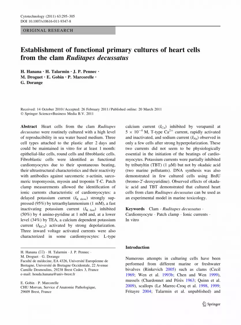
Effect of pollutants on ionic currents
Results showed that ionic currents were not signif-
icantly modulated after 30 min of exposure to OA
1 lM as compared with untreated cells (Fig. 10a).
Concerning TBT 1 lM a significant decrease of
potassium currents (Fig. 10b) and a significant
increase of L-type calcium currents were observed
after 30 min of exposure.
Discussion
The main purpose of this study was to establish
primary cultures of clam heart cells. Our experiments
showed that cells isolated from R. decussatus using
Fig. 8 Example of typical recording of the calcium inward
current alone (a very small fast potassium current is not totally
abolished at the start of recordings) (a). Inhibition of the
calcium current by verapamil 5 9 10-4 M (b). Voltage steps
imposed to the membrane (c). Vertical bars 10 nA (top) and
200 mV (bottom). Horizontal bar 2 ms. Current–voltage
relationship corresponding to the calcium current (d)
Fig. 9 Example of a transient inward calcium current follow-
ing a rapid voltage gated sodium current after inhibition of
potassium currents and application of a 40 mV hyperpolarizing
holding potential to the cell (a). Vertical bars 1 nA (top) and
0.1 V (bottom). Horizontal bar 10 ms. Current–voltage rela-
tionship of the sodium current recorded from isolated
cardiomyocytes (b); such a current was observed in only 2%
of cells
Cytotechnology (2011) 63:295–305 301
123
pronase, according to a protocol adapted from the
procedure defined by Le Marrec-Croq et al. (1997)
for scallop, allowed good cell viability and routinely
functional primary cultures. Clam cultured cells
remained viable for up to 1 month. Analogous results
were previously obtained in our laboratory for scallop
and oyster (Le Marrec-Croq et al. 1999; Fritayre
2004; Pennec et al. 2002, 2004; Droguet 2006).
To our knowledge it is the first time that pronase
has been used to dissociate clam heart. In the
literature, previous work showed that cell cultures
from cardiac tissue of the surf clam Spisula solidiss-
ima can also be obtained using trypsin-EDTA (Cecil
1969) or collagenase for Meretrix lusoria (Chen and
Wen 1999), the latter authors considering that
treatment with collagenase was better than trypsin
at dissociating mollusc tissue fragments for in vitro
culture. This is not in accordance with our results
after having tested, in preliminary experiments, the
three enzymes for R. decussatus heart. A higher
mortality was observed in cells isolated by trypsin or
collagenase in comparison with pronase.
Clam cultures are heterogenous as reported for
other marine bivalves (Chen and Wen 1999; Cheng
et al. 2001; Fritayre 2004; Droguet 2006). The round
cells are usually considered as haemocytes (Wen
et al. 1993b; Renault et al. 1995). We can hypoth-
esize that epithelial like cells have been isolated from
the external simple prismatic epithelium covering the
heart of bivalves (epicardium). Concerning fibroblas-
tic cells, some of them are fibroblasts isolated from
the connective tissue and the others are cardiomyo-
cytes. The characterisation of these fibroblastic cells
was the second aim of the present study.
To our knowledge, characterization of such cells
from the clam has not been previously documented.
In the literature; data obtained using patch clamp and/
or immunostaining, ultrastructural studies concerned
cultured cardiomyocytes from scallop, oyster and
mussel (Wen et al. 1993a; Le Deuff et al. 1994;
Curtis et al. 1999; Le Marrec-Croq et al. 1999;
Pennec et al. 2002, 2004; Droguet 2006). Chen and
Wen (1999) for oyster and Ellington (1993) for clam
concluded that cultured fibroblastic cells were cardio-
myocytes only by distinctive rhythmic contraction in
vitro. In the present work proof that almost all
adherent fibroblastic cells were cardiomyocytes was
based on (1) the spontaneous beatings of fibroblastic
cells observed in vitro with a rate similar to this
measured in vivo, (2) results of immunological
staining, showing positive reaction of these cells
after treatment with antibodies against troponin,
tropomyosin, myosin and sarcomeric actinin, (3)
their ultrastructural characteristics and (4) results of
electrophysiological analyses showing ionic channels
and regulatory mechanisms very similar to those
observed in cardiomyocytes of invertebrates and
other marine bivalves (Pennec et al. 2004; Curtis
et al. 1999). The other objective of the present work
was to carry out the electrophysiological character-
ization of the cultured cardiomyocytes.
An outward potassium current, identified as a
delayed rectifying potassium current (IKslow), and a
fast activating and inactivating potassium current (IK
fast), classically observed in cardiac cells of mamma-
lian and other marine molluscs such as oyster (Pennec
et al. 2004), mussel (Curtis et al. 1999), snail
(Yeoman and Benjamin 1999), squid (Odblom et al.
2000) and sea hare (Souza et al. 2002) were observed
in clam cells. The main role of IKslow is to initiate and
terminate cardiac repolarization (Varro and Papp
1992). It is classically inhibited by TEA which is
known to block a variety of K channel types with
possible differences in their sensitivity to this
Fig. 10 Example of currents recorded after treatment with 1 lM of OA (a) and TBT (b) for 30 min. Vertical bars 1 nA (top) and
0.1 V (bottom). Horizontal bar 20 and 10 ms, respectively for for OA and TBT
302 Cytotechnology (2011) 63:295–305
123

compound (Odblom et al. 2000). In clam, IKslow was
more sensitive to TEA, compared to other species
including bivalves such as oyster (Pennec et al. 2004)
and mussel (Curtis et al. 1999), as it was almost
totally abolished (95%) by TEA 1 mM. IK fast, which
is involved in the initiation of the early fast repolar-
ization process of cardiac action potentials and in the
rate of repolarization (Varro and Papp 1992) was less
sensitive to TEA than IKslow. However, IK fast was
more sensitive in clam than in oyster or in mussel. IK
fast was inhibited by 4-AP, in agreement with data
reported in many other species (Odblom et al. 2000;
Pennec et al. 2004; Tseng et al. 1987). Then TEA and
4-AP could be used to separate the fast and the
delayed potassium currents in clam cardiomyocytes.
The question remains for the clam of a possible role
in the modulation of cardiac frequency by IK fast.
Calcium-activated outward potassium current,
activated by large depolarizing pulses and largely
sensitive to calcium influx, was also observed in clam
cardiomyocytes as in vertebrates (Siegelbaum and
Tsien 1980; Kenyon and Sutko 1987; Giles and
Imaizumi 1988) or invertebrates (Pennec et al. 2004;
Odblom et al. 2000). This current has a role in the
regulation of cell contractility and intracellular cal-
cium regulation. It is activated by increased intracel-
lular calcium and speeds up the cell repolarization,
thus reducing the duration of calcium inward current.
The clam myocytes also showed an inward
current, rapidly activated and inhibited by verapamil,
identified as a long duration calcium current (L-type).
Such a calcium current, classically observed in
vertebrate cardiac tissue (Hagiwara et al. 1988) has
been identified in other molluscs (Curtis et al. 1999;
Yeoman et al. 1999; Odblom et al. 2000; Pennec
et al. 2004). L-type channel, in molluscs as in lower
vertebrates, is thought to be the primary pathway for
Ca2? entry for the excitation-contraction mechanism
(Ferrier et al. 2000; Bers and Perez-Reyes 1999,
Odblom et al. 2000).
In some cells, when a strong hyperpolarization was
applied to the membrane before the test pulse,
another calcium inward current (T-type) was
observed in the clam as in oyster (Pennec et al.
2004) and snail (Yeoman et al. 1999), but not in
mussel (Curtis et al. 1999). We can hypothesize, as
Pennec et al. (2004), Varro and Papp (1992), Yeoman
et al. (1999), that this current can be involved in
pacemaker function. The lack of transient calcium
current and sodium current in most clam cardiac
cells, despite of their spontaneous beatings when they
are cultured, suggests that these channels are not
essential in the automaticity of the cultured cells as
reported by Curtis et al. (1999) in mussel without
presuming the role of these currents in vivo. It was
also reported that calcium released from the sarco-
plasmic reticulum can be triggered by the T-type
current, but this mechanism is less physiologically
important in excitation–contraction coupling than the
mechanism related to the L-type calcium channel
(Bers and Perez-Reyes 1999). Moreover, the ventric-
ular cells are supposed to lack the T-type calcium
current (Bers and Perez-Reyes 1999) as most of the
cultured cells are of ventricular origin, explaining
why the transient calcium current was not frequently
observed.
A sodium current was only found in few clam cells
as evidenced in cardiac tissue of other molluscs
(Pennec et al. 2004; Curtis et al. 1999; Odblom et al.
2000). Varro and Papp (1992) showed that this
current is responsible not only for the fast depolar-
ization, but, in part also, for the maintenance of the
plateau phase of the action potential. In the clam, the
small size of this current in vitro can be explained by
the fact that the fast sodium current like the T-type
calcium current is not essential to the triggering of
automaticity. Moreover it should be mostly inacti-
vated at the normal resting potential. Thus, in clam
cultured heart cells the action potential appears to be
mostly based on the inflow of calcium ions, rather
than sodium ions as suggested elsewere (Curtis et al.
1999).
DNA synthesis in few clam heart cells in culture
was proven by BrdU uptake. Some of these cells
could be identified as cardiomyocytes due to their
double positive staining BrdU-troponin. BrdU incor-
poration was also demonstrated in cultured heart cells
of scallop (Le Marrec-Croq et al. 1999) and oyster
(Droguet 2006). In vitro mitoses were also observed
under inverted microscope in cardiac tissue from surf
clam (Cecil 1969) and hard clam (Wen et al. 1993a;
Chen and Wen 1999). We can hypothesize that there
were few unipotent stem cells in the population of
adherent cardiac cells.
To summarize, the present work shows that clam
heart cells can be maintained functional in primary
culture for up to 1 month. The characterization of
cardiomyocyte physiological properties was a
Cytotechnology (2011) 63:295–305 303
123

prerequisite before using such cells in culture as tools
for toxicological studies as illustrated by results
obtained after clam heart cell exposure to two major
marine pollutants. This work showed that primary
cultures of clam heart cell can be used as experi-
mental models in marine toxicology and the interest
of patch clamp technique as a sensitive analytical
method.
References
Almers W, Stanfield PR, Stuhmer W (1983) Slow changes in
currents through sodium channels in frog muscle mem-
brane. J Physiol 339:253–271
Bebianno MJ, Geret F, Hoarau P, Serafim MA, Coelho MR,
Gnassia-Barelli M, Romeo M (2004) Biomarkers in Ru-ditapes decussatus: a potential bioindicator species. Bio-
markers 9:305–330
Bers DM, Perez-Reyes E (1999) Ca channels in cardiac myo-
cytes: structure and function in Ca influx and intracellular
Ca release. Cardiovasc Res 42:339–360
Buchanan JT, La Peyre JF, Cooper RK, Tiersch TR (1999)
Improved attachment and spreading in primary cell cul-
tures of the eastern oyster, Crassostrea virginica. In Vitro
Cell Dev Biol Anim 35:593–598
Cecil JT (1969) Mitoses in cell cultures from cardiac tissue of
the surf clam Spisula solidissima. J Invertebr Pathol
14:407–410
Chardonnet Y, Peres G (1963) Essai de culture de cellules
provenant d’un mollusque: Mytilus gallovincialis. L C R
Soc Biol Lyon 157:1593–1595
Chen SN, Wen CM (1999) Establishment of cell lines derived
from oyster, Crassostrea gigas Thunberg and hard clam,
Meretrix lusoria Roding. Methods Cell Sci 21:183–192
Cheng TC, La Peyre JF, Buchanan JT, Tiersch TR, Cooper RK
(2001) Cryopreservation of heart cells from the eastern
oyster. In Vitro Cell Dev Biol Anim 37:237–244
Curtis TM, Depledge MH, Williamson R (1999) Voltage-
activated currents in cardiac myocytes of the blue mussel,
Mytilus edulis. Comp Biochem Physiol A 124:231–241
Domart-Coulon I, Doumenc D, Auzoux-Bordenave S, Le
Fichant Y (1994) Identification of media supplements that
improve the viability of primarily cell cultures of Cras-sostrea gigas oysters. Cytotechnology 16:109–120
Domart-Coulon I, Auzoux-Bordenave S, Doumenc D, Kha-
lanski M (2000) Cytotoxicity assessment of antibiofouling
compounds and by-products in marine bivalve cell cul-
tures. Toxicol In Vitro 14:245–251
Droguet M (2006) Etude des caracteristiques fonctionnelles des
cardiomyocytes d’huıtre en culture. These de doctorat de
biologie. Universite de Bretagne Occidentale, p 211
Ellington WR (1993) Studies of intracellular pH regulation in
cardiac myocytes from the marine bivalve mollusk,
Mercenaria campechiensis. Biol Bull 184:209–215
Ferrier GR, Redondo IM, Mason CA, Mapplebeck C, Howlett
SE (2000) Regulation of contraction and relaxation by
membrane potential in cardiac ventricular myocytes. Am J
Physiol Heart Circ Physiol 278:H1618–H1626
Fritayre P (2004) Culture des cellules atriales de coquille Saint-
Jacques, Pecten maximus: valeur et limites du modele.
Applications en toxicologie. These en Oceanologie Bio-
logique et Environnement marin. Universite de Bretagne
Occidentale, p 230
Giles WR, Imaizumi Y (1988) Comparison of potassium cur-
rents in rabbit atrial and ventricular cells. J Physiol
405:123–145
Hagiwara N, Irisawa H, Kameyama M (1988) Contribution of
two types of calcium currents to the pacemaker potentials
of rabbit sino-atrial node cells. J Physiol 395:233–253
Kenyon JL, Sutko JL (1987) Calcium- and voltage-activated
plateau currents of cardiac Purkinje fibers. J Gen Physiol
89:921–958
Le Deuff RM, Lipart C, Renault T (1994) Primary culture of
pacific oyster, Crassostrea gigas, heart cells. J Tissue Cult
Methods 16:67–72
Le Marrec-Croq F, Dorange G, Chesne C (1997) Procede de
culture de cellules d’invertebres marins et cultures obte-
nues, French Patent no 9506921; no of publication:
2735146 (1996)
Le Marrec-Croq F, Fritayre P, Chesn GuillouzoA, Dorange G
(1998) Cryopreservation of Pecten maximus heart cells.
Cryobiology 37:200–206
Le Marrec-Croq F, Glaise D, Guguen-Guillouzo C, Chesne C,
Guillouzo A, Boulo V, Dorange G (1999) Primary cultures
of heart cells from the scallop Pecten maximus (mollusca-
bivalvia). In Vitro Cell Dev Biol Anim 35:289–295
Odblom MP, Williamson R, Jones MB (2000) Ionic currents in
cardiac myocytes of squid, Alloteuthis subulata. J Comp
Physiol B 170:11–20
Pennec JP, Gallet M, Gioux M, Dorange G (2002) Cell culture
of bivalves: tool for the study of the effects of environ-
mental stressors. Cell Mol Biol (Noisy-le-grand)
48:351–358
Pennec JP, Talarmin H, Droguet M, Giroux-Metges MA,
Gioux M, Dorange G (2004) Characterization of the
voltage-activated currents in cultured atrial myocytes
isolated from the heart of the common oyster Crassostreagigas. J Exp Biol 207:3935–3944
Quinn B, Costello MJ, Dorange G, Wilson JG, Mothersill C
(2009) Development of an in vitro culture method for
cells and tissues from the zebra mussel (Dreissena poly-morpha). Cytotechnology 59:121–134
Renault T, Flaujac G, Le Deuff RM (1995) Isolation and cul-
ture of heart cells from the European flat oyster, Ostreaedulis. Methods Cell Sci 17:199–205
Reynolds ES (1963) The use of lead citrate at high pH as an
electron-opaque stain in electron microscopy. J Cell Biol
17:208–212
Rinkevich B (2005) Marine invertebrate cell cultures: new
millennium trends. Mar biotechnol (NY) 7:429–439
Siegelbaum SA, Tsien RW (1980) Calcium-activated transient
outward current in calf cardiac Purkinje fibres. J Physiol
299:485–506
Souza MM, Stucchi-Zucchi A, Cassola AC, Scemes E (2002)
Electrophysiology of cardiac myocytes of Aplysia brasi-liana. Comp Biochem Physiol A Mol Integr Physiol
133:161–168
304 Cytotechnology (2011) 63:295–305
123

Talarmin H, Droguet M, Pennec JP, Schroder HC, Muller
WEG, Gioux M, Dorange G (2008) Effects of a phyco-
toxin, okadaic acid, on oyster heart cell survival. Toxicol
Environ Chem 90:153–168
Tseng GN, Robinson RB, Hoffman BF (1987) Passive prop-
erties and membrane currents of canine ventricular myo-
cytes. J Gen Physiol 90:671–701
Varro A, Papp JG (1992) The impact of single cell voltage
clamp on the understanding of the cardiac ventricular
action potential. Cardioscience 3:131–144
Wen CM, Kou GH, Chen SN (1993a) Cultivation of cells from
the heart of the hard clam, Meretrix lusoria (RODING).
J Tissue Cult Methods 15:123–130
Wen CM, Kou GH, Chen SN (1993b) Establishment of cell
lines from the Pacific oyster. In Vitro Cell Dev Biol Anim
29A:901–903
Yeoman MS, Benjamin PR (1999) Two types of voltage-gated
K(?) currents in dissociated heart ventricular muscle cells
of the snail Lymnaea stagnalis. J Neurophysiol
82:2415–2427
Yeoman MS, Brezden BL, Benjamin PR (1999) LVA and
HVA Ca(2?) currents in ventricular muscle cells of the
Lymnaea heart. J Neurophysiol 82:2428–2440
Cytotechnology (2011) 63:295–305 305
123


