Embed Size (px)
Citation preview
JOURNAL OF CLINICAL MICROBIOLOGY,0095-1137/98/$04.0010
Jan. 1998, p. 168–178 Vol. 36, No. 1
Copyright © 1998, American Society for Microbiology
Evaluation of Four DNA Typing Techniques in EpidemiologicalInvestigations of Bovine Tuberculosis
DEBBY COUSINS,1* SUZETTE WILLIAMS,1 ERNESTO LIEBANA,2 ALICIA ARANAZ,2
ANNELIES BUNSCHOTEN,3 JAN VAN EMBDEN,3 AND TREVOR ELLIS1
Australian Reference Laboratory for Bovine Tuberculosis, Animal Health Laboratories, Agriculture Western Australia,South Perth, Western Australia 6151, Australia1; Departamento de Patologa Animal I (Sanidad Animal),
Facultad de Veterinaria, Universidad Complutense de Madrid, s/n, 28100 Madrid, Spain2; andDepartment of Bacteriology, Research Laboratory for Infectious Diseases, National Institute of Public
Health and Environmental Protection, 3720A Bilthoven, The Netherlands3
Received 22 April 1997/Returned for modification 8 July 1997/Accepted 1 October 1997
DNA fingerprinting techniques were used to type 273 isolates of Mycobacterium bovis from Australia, Canada,the Republic of Ireland, and Iran. The results of restriction fragment length polymorphism (RFLP) analysiswith DNA probes from IS6110, the direct repeat (DR), and the polymorphic GC-rich sequence (PGRS) werecompared with those of a new PCR-based method called spacer oligonucleotide typing (spoligotyping) devel-oped for the rapid typing of Mycobacterium tuberculosis (J. Kamerbeek et al., J. Clin. Microbiol. 35:907–914,1997). Eighty-five percent of the isolates harbored a single copy of IS6110, and 81.5% of these carried IS6110on the characteristic 1.9-kb restriction fragment. RFLP analysis with IS6110 identified 23 different types, RFLPanalysis with the DR probe identified 35 types, RFLP analysis with the PGRS probe identified 77 types, andthe spoligotyping method identified 35 types. By combining all results, 99 different strains could be identified.Isolate clusters were frequently associated within herds or were found between herds when epidemiologicalevidence confirmed animal movements. RFLP analysis with IS6110 was sufficiently sensitive for the typing ofisolates with more than three copies of IS6110, but RFLP analysis with the PGRS probe was the most sensitivetyping technique for strains with only a single copy of IS6110. Spoligotyping may have advantages for the rapidtyping of M. bovis, but it needs to be made more sensitive.
DNA typing techniques are now frequently used for epide-miological investigations of many infectious diseases. The in-sertion sequence (IS) IS6110 is accepted as an excellent toolfor the identification of restriction fragment length polymor-phisms (RFLPs) in Mycobacterium tuberculosis strains and isused in epidemiological studies worldwide (31). RFLP analysiswith IS6110 has been used to define disease outbreaks and totrace the spread of multidrug-resistant strains of M. tuberculo-sis (2, 7, 16, 21, 34, 37). Other repetitive elements such as thedirect repeat (DR) (13) and the GC-rich repetitive sequence(PGRS) (8, 25) now appear to be finding acceptance for use inthe characterization of M. tuberculosis strains (10, 30, 33, 38).
Some of these methods have been applied to the typing ofMycobacterium bovis. In a previous study (6), a plasmid con-taining PGRS (pTBN12) was found to be more useful as aprobe in RFLP analysis than the left-hand side (LHS) of thePvuII fragment in IS6110 (IS6110-L) in differentiating betweenAustralian M. bovis isolates. However, a similar study foundthat pTBN12 was only marginally better than the entire IS6110probe in the identification of polymorphisms in 109 isolatesfrom cattle in Northern Ireland (27). Some workers have sug-gested that DNA fingerprinting with a sequence to the right-hand side (RHS) of the PvuII site of IS6110 (IS6110-R) as aprobe may be useful in the study of M. bovis, but this appearsto be confined to cases in which M. bovis isolates harbor mul-tiple copies of IS6110 (12, 15). For example, in a study of 153M. bovis isolates, van Soolingen et al. (32) reported the occur-
rence of strains of M. bovis with multiple copies of IS6110 inassociation with zoo and exotic animals, whereas M. bovisstrains isolated from cattle in The Netherlands and Argentinagenerally harbored only a single copy of IS6110, usually carriedin a characteristic 1.9-kb PvuII restriction fragment. When asmall number of strains of M. bovis with single copies of IS6110were analyzed by using the DR probe and pTBN12, furtherdifferentiation was achieved. In a comparison of RFLP tech-niques with IS6110, DR, and PGRS conducted with 85 M. bovisisolates from Argentina, DR and PGRS were recommended asthe probes of choice for use in RFLP analysis (24). A compar-ison of the results of RFLP analyses with IS6110 and DR with79 isolates of M. bovis from Texas and Mexico indicated thatDR was a more sensitive probe than IS6110. Another compar-ison of the results of RFLP analyses with the entire IS6110,DR, and PGRS probes with 210 isolates of M. bovis, mostlyfrom Northern Ireland, indicated that the effectiveness ofRFLP analysis with each of these probes was almost the same,but when the results of RFLP analyses with all probes wereused, the number of strains that were differentiated was almostdoubled (28).
An innovative PCR-based typing method for the differenti-ation of M. tuberculosis strains has been reported recently (11).This method, termed spacer oligonucleotide typing (spoligo-typing), relies on the in vitro amplification of DNA across theunique, highly polymorphic DR locus present in the M. tuber-culosis complex chromosome. This region contains multipleshort 36-bp DRs, and nonrepetitive spacers, which are 35 to 41bp in length, are interspersed between the DRs. The PCRproduct from individual isolates is allowed to hybridize to 37spacers identified in M. tuberculosis H37Rv and 6 spacers iden-tified in M. bovis BCG P3 (14). Following hybridization anddetection, the spacers that are common to the isolate being
* Corresponding author. Mailing address: Australian ReferenceLaboratory for Bovine Tuberculosis, Agriculture Western Australia, 3Baron-Hay Ct., South Perth, Western Australia 6151, Australia.Phone: 618 9368 3429. Fax: 618 9474 1881. E-mail: [email protected].
168
on Septem
ber 17, 2016 by PE
NN
ST
AT
E U
NIV
http://jcm.asm
.org/D
ownloaded from
tested and the standard set of spacer oligonucleotides can beidentified. Only one report on the use of spoligotyping com-pared with the use of RFLP analysis with IS6110 for the dif-ferentiation of M. bovis isolates has been published (1).
This study was undertaken to comprehensively compare theusefulness of the three most commonly used RFLP techniques(RFLP analyses with IS6110-R, DR, and PGRS) and the newspoligotyping method for the differentiation of M. bovis iso-lates from Australian sources. In addition, isolates from othercountries were included to determine whether a geographicdifference could be identified by any of the markers. The use-fulness of these techniques in epidemiological studies of bo-vine tuberculosis in Australia and overseas was demonstrated.
MATERIALS AND METHODS
Source of M. bovis isolates. Two hundred seventy-three M. bovis isolates weretested. Two hundred eleven animal isolates originated in Australia from thefollowing states: Western Australia (n 5 121), the Northern Territory (n 5 46),Queensland (n 5 35), Victoria (n 5 8), and New South Wales (n 5 1). TheWestern Australian isolates came from the agricultural area (n 5 30), thenorthern pastoral area (n 5 21), the Broome area (n 5 5), the West Kimberleyarea (n 5 57), and the East Kimberley area (n 5 8). Sixty-one isolates of M. bovisobtained from overseas sources for comparison originated from Canada (n 533), Iran (n 5 10), the Republic of Ireland (n 5 14), the United Kingdom (n 53), and New Zealand (n 5 1). In addition, the reference strain of M. bovis (strainAN5) was included.
Good epidemiological information was provided for 32 animal isolates and asingle human isolate from Canada (4). The 33 Canadian isolates originated from4 outbreaks (outbreaks A to D) involving 15 premises, 10 different animalspecies, and 1 human patient. Outbreak A involved four animal species and fourrelated premises over the period from 1992 to 1994. Outbreak B was traced tofour premises from 1990 to 1994 and involved four elk, a bison, and a veterinar-ian who was diagnosed with M. bovis infection after he treated one of the elk.Outbreak C involved a large collection of exotic species comprising seven animalspecies and four premises from 1989 to 1992. Epidemiological information sug-gested there was nose-to-nose contact between elk and a Sika deer, elk and aPere David deer, and the Pere David deer and cattle. A cougar on one propertywas fed carcasses of other animals on the premises and chickens from a neigh-boring farm. Deer from New Zealand were imported to one of the previouslyinfected premises after it had been depopulated, cleaned, disinfected, and re-leased from quarantine. The deer were skin test negative before and after arrivalin quarantine. Outbreak D in 1991 involved cattle on one property and an elkfrom a neighboring national park.
DNA probes. PCR was used to amplify DNA from the right-hand side of thePvuII site in IS6110-R as described previously (31), and the IS6110-R probe wasused to determine the number of IS6110 copies in each isolate. RFLPs weredetermined for all isolates by using probes prepared from amplified DNA ofIS6110-R and from the oligonucleotides from the DR (59 GTC GTC AGA CCCAAA ACC CCG AGA GGG GAC GGA AAC 39) and PGRS (59 CCG CCGTTG CCG CCG TTG CCG CCG TTG CCG CCG 39).
RFLP methods. Cells were grown and DNA was extracted as described pre-viously (6, 31). RFLP analyses with IS6110-R and DR were performed basicallyby the standard method recommended for the DNA fingerprinting of M. tuber-culosis (31), with the exception of the electrophoresis conditions and the probelabelling and detection methods. For RFLP analyses with IS6110-R and DR,electrophoresis was performed with 1 and 1.5% agarose gels, respectively, andgels were run in TAE (Tris, acetate, EDTA) buffer for 16 h at 45 V by using arefrigerated buffer recirculation pump at a constant temperature of 14°C. RFLPanalysis with PGRS was performed as described previously (6), with the excep-tion that the gels were run longer (45 V for 28 h) in an attempt to achieve betterdiscrimination between strains by spreading the bands that hybridize with PGRSover a greater distance. All probes were labelled by using the nonradioactivedigoxigenin system (Boehringer Mannheim), and hybridization and detection ofbound probe with a chemiluminescent substrate were performed as described bythe manufacturer.
DNAs from M. tuberculosis Mt 14323 and M. bovis BCG 3 were run on all gelsin positions 1 and 30 as external standards. To allow for computer-assistedanalysis of the IS6110-R, DR, and PGRS fingerprints, an internal marker (withhigh- and low-molecular-mass standards) was added to the loading buffer witheach sample as recommended by van Embden and colleagues (31, 33) for theirstandardized method for RFLP analysis with IS6110-R for the differentiation ofM. tuberculosis isolates.
Spoligotyping. PCR of the DR locus was performed with extracted DNA (6,31) or heat-treated cell suspensions, and the spoligotyping method was per-formed as described previously (1).
Analysis of results. Analysis of the bands generated with the IS6110 and DRprobes and the spoligotypes for the set of isolates tested was performed with theaid of a computer software program by using the Dice unweighted pair group
method with arithmetic averages (UPGMA) (GelCompar, version 3.1; AppliedMaths, Kortrijk, Belgium). The profiles obtained by RFLP analysis with thePGRS probe were analyzed by using the clustering correlation and UPGMA(GelCompar).
Type nomenclature. No standard nomenclature for the naming of different M.tuberculosis complex DNA types has yet been agreed upon internationally. Forthe purposes of this study, different numbers were allocated to isolates when agenetic difference could be detected. These different DNA types were given aprefix to show the probe used; IS for IS6110-R types, DR for DR types, SP forspoligotypes, and PG for PGRS types. In addition, isolates that were identified asunique strains were given an overall DNA type based on the composite results ofthe four different typing methods. Strains that had the most common IS6110-R,DR, and SP types (IS01, DR01, and SP01) and differed only in PGRS type werecalled type A and allocated the number of the PGRS type (e.g., A061 is IS01,DR01, SP01, and PG61). The prefixes B and C were given to isolates that had themost common IS6110-R and SP types (IS01 and SP01) with DR type 02 and DRtype 04 patterns, respectively. For these isolates the number following the prefixwas the number of the PGRS type. When strains had a common prefix, they wereconsidered to be genetically more closely related to each other than to otherstrains that did not have the same prefix. Other strains were allocated a numberonly. Numbers were allocated as the isolates were analyzed and did not neces-sarily bear any relationship to specific patterns. For example, strains 001, 002,and 003 may not be any more closely related to each other than they may be tostrain 023 or 091.
RESULTS
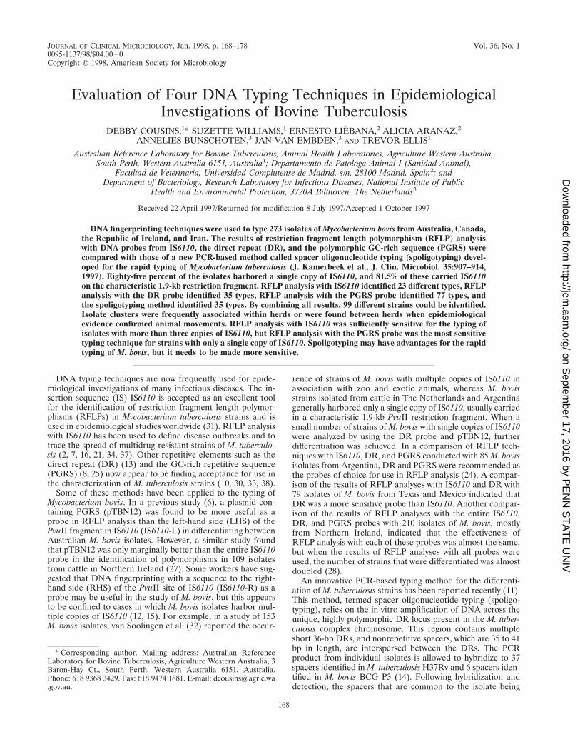
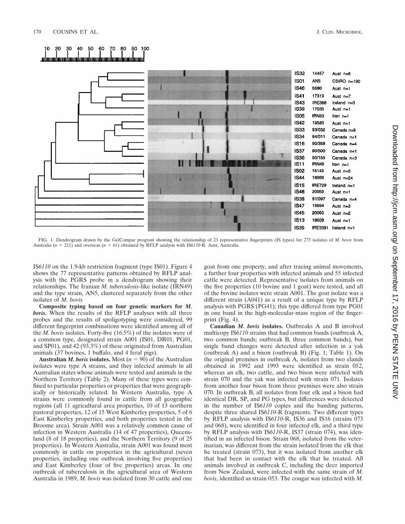
RFLP analysis with IS6110-R. Two hundred thirty-three(85.4%) of the 273 isolates of M. bovis examined in this studyharbored a single copy of IS6110. Of the 233 isolates with asingle copy of IS6110, 190 (81.5%) carried the IS on a 1.9-kbrestriction fragment, and this IS type was designated IS01. Thismeans that almost 70% of all M. bovis isolates could not bedifferentiated by RFLP analysis with IS6110-R. Furthermore,194 (91.9%) of the 211 Australian isolates had a single copy ofIS6110, and 158 (81.4%) of these were type IS01. Figure 1shows the 23 representative patterns (types) obtained by RFLPanalysis with IS6110-R in a dendrogram indicating the rela-tionships between the types. The Canadian isolates containedone (n 5 15), two (n 5 9), three (n 5 5), or four (n 5 4) copiesof IS6110, and overall, nine different strains were identifiedamong the Canadian isolates. Different strains were implicatedin each of the four outbreaks (Table 1).
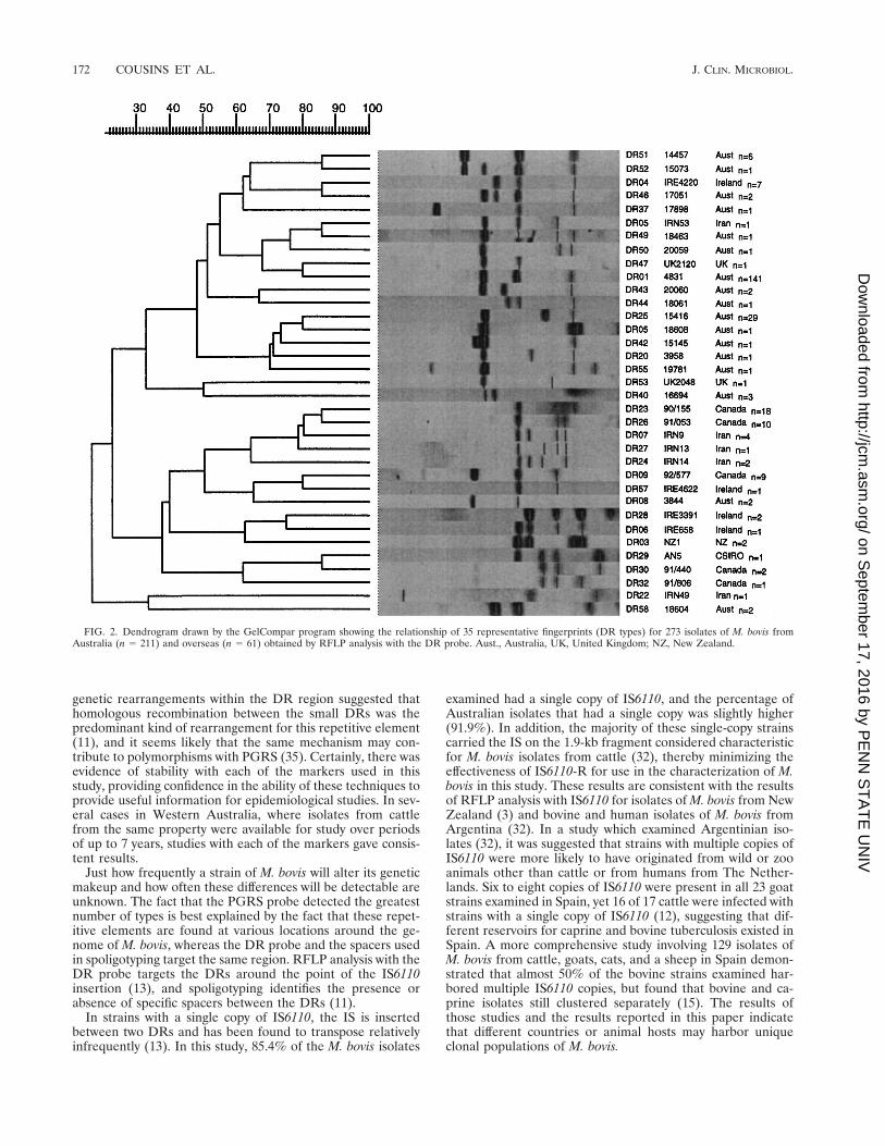
RFLP analysis with the DR probe. RFLP analysis with theDR probe identified 35 different types among the 273 M. bovisisolates. By RFLP analysis with DR, 141 (51.6%) of the iso-lates had a common fingerprint, designated DR01. Many of theCanadian and Iranian strains had DR patterns that appearedto be unique to the country of origin, and these were notidentified among the Australian isolates. Figure 2 shows the 35representative fingerprints obtained by RFLP analysis with theDR probe in a dendrogram showing the relationship betweenthe types.
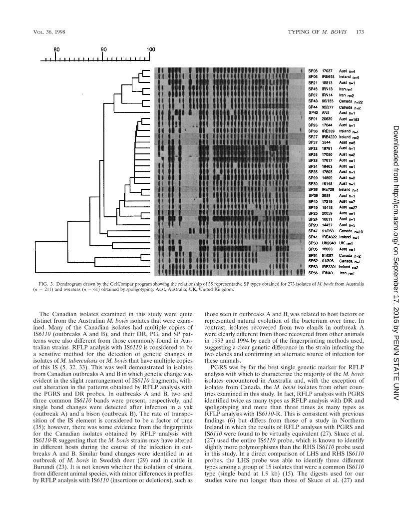
Spoligotyping. The spoligotyping method identified 35 dif-ferent types among the 273 M. bovis isolates. One hundredfifty-three (56%) of the isolates had a common spoligotype(designated SP01) and therefore could not be differentiated byspoligotyping. Figure 3 shows the 35 representative spoligo-types in a dendrogram showing their relationships. With oneexception (Iranian isolate IRN49), all of the isolates lacked thefive spacers at the 39 end of the DR locus. When the Iranianisolate was reexamined biochemically, its identity was moreconsistent with M. tuberculosis.
RFLP analysis with the PGRS probe. Use of the PGRSprobe differentiated the M. bovis isolates into the most types,with 77 different types being identified among the 273 M. bovisisolates. Eighty (29.3%) of the isolates, including 77 (36.5%) ofthe Australian isolates, were identified as being of a commontype, designated PG01. All of the isolates identified as PG01harbored a single copy of IS6110, and 50 of the PG01 isolates,including 47 of the Australian isolates, carried a single copy of
VOL. 36, 1998 TYPING OF M. BOVIS 169
on Septem
ber 17, 2016 by PE
NN
ST
AT
E U
NIV
http://jcm.asm
.org/D
ownloaded from
IS6110 on the 1.9-kb restriction fragment (type IS01). Figure 4shows the 77 representative patterns obtained by RFLP anal-ysis with the PGRS probe in a dendrogram showing theirrelationships. The Iranian M. tuberculosis-like isolate (IRN49)and the type strain, AN5, clustered separately from the otherisolates of M. bovis.
Composite typing based on four genetic markers for M.bovis. When the results of the RFLP analyses with all threeprobes and the results of spoligotyping were considered, 99different fingerprint combinations were identified among all ofthe M. bovis isolates. Forty-five (16.5%) of the isolates were ofa common type, designated strain A001 (IS01, DR01, PG01,and SP01), and 42 (93.3%) of these originated from Australiananimals (37 bovines, 1 buffalo, and 4 feral pigs).
Australian M. bovis isolates. Most (n 5 90) of the Australianisolates were type A strains, and they infected animals in allAustralian states whose animals were tested and animals in theNorthern Territory (Table 2). Many of these types were con-fined to particular properties or properties that were geograph-ically or historically related. In Western Australia, type Astrains were commonly found in cattle from all geographicregions (all 11 agricultural area properties, 10 of 13 northernpastoral properties, 12 of 15 West Kimberley properties, 5 of 6East Kimberley properties, and both properties tested in theBroome area). Strain A001 was a relatively common cause ofinfection in Western Australia (14 of 47 properties), Queens-land (8 of 18 properties), and the Northern Territory (9 of 25properties). In Western Australia, strain A001 was found mostcommonly in cattle on properties in the agricultural (sevenproperties, including one outbreak involving five properties)and East Kimberley (four of five properties) areas. In oneoutbreak of tuberculosis in the agricultural area of WesternAustralia in 1989, M. bovis was isolated from 30 cattle and one
goat from one property, and after tracing animal movements,a further four properties with infected animals and 55 infectedcattle were detected. Representative isolates from animals onthe five properties (10 bovine and 1 goat) were tested, and allof the bovine isolates were strain A001. The goat isolate was adifferent strain (A041) as a result of a unique type by RFLPanalysis with PGRS (PG41); this type differed from type PG01in one band in the high-molecular-mass region of the finger-print (Fig. 4).
Canadian M. bovis isolates. Outbreaks A and B involvedmulticopy IS6110 strains that had common bands (outbreak A,two common bands; outbreak B, three common bands), butsingle band changes were detected after infection in a yak(outbreak A) and a bison (outbreak B) (Fig. 1; Table 1). Onthe original premises in outbreak A, isolates from two elandsobtained in 1992 and 1993 were identified as strain 052,whereas an elk, two cattle, and two bison were infected withstrain 070 and the yak was infected with strain 071. Isolatesfrom another four bison from three premises were also strain070. In outbreak B, all isolates from four elk and a bison hadidentical DR, SP, and PG types, but differences were detectedin the number of IS6110 copies and the banding patterns,despite three shared IS6110-R fragments. Two different typesby RFLP analysis with IS6110-R, IS36 and IS16 (strains 073and 068), were identified in four infected elk, and a third typeby RFLP analysis with IS6110-R, IS37 (strain 074), was iden-tified in an infected bison. Strain 068, isolated from the veter-inarian, was different from the strain isolated from the elk thathe treated (strain 073), but it was isolated from another elkthat had been in contact with the elk that he treated. Allanimals involved in outbreak C, including the deer importedfrom New Zealand, were infected with the same strain of M.bovis, identified as strain 053. The cougar was infected with M.
FIG. 1. Dendrogram drawn by the GelCompar program showing the relationship of 23 representative fingerprints (IS types) for 273 isolates of M. bovis fromAustralia (n 5 211) and overseas (n 5 61) obtained by RFLP analysis with IS6110-R. Aust, Australia.
170 COUSINS ET AL. J. CLIN. MICROBIOL.
on Septem
ber 17, 2016 by PE
NN
ST
AT
E U
NIV
http://jcm.asm
.org/D
ownloaded from
bovis and Mycobacterium avium. In outbreak D, the straininfecting cattle on one property (strain 076) had DR, PG, andSP types different from those of the strain isolated from an elk(strain 077) from the national park on the border of the pre-mises from which strain 076 was isolated, suggesting that in-fection had originated from different sources.
Effectiveness of GelCompar in the analysis of genetic types.GelCompar was a very effective means of analyzing the differ-ent fingerprints obtained by the RFLP analyses with IS6110-Rand DR and by the spoligotyping method. However, in ana-lyzing the fingerprints obtained by RFLP analysis with PGRS,which were much more complex because of the number ofbands that hybridized with the PGRS probe, the program wasless effective. Because of this, all isolates that had similarpatterns by RFLP analysis with PGRS had to be checkedmanually.
DISCUSSION
The M. bovis isolates examined in this study could be char-acterized to various degrees by up to four genotyping proce-dures. Each of the procedures was able to differentiate strainsof M. bovis and could be used for epidemiological studies of M.
bovis isolates from animals. However, the combined use of thefour procedures resulted in superior differentiation of strainsfor a detailed epidemiological investigation. The finding of agenetic difference between two strains by the use of any par-ticular marker would imply infection with different strains,presumably originating from different sources. Confidence inthis assumption would be increased if more than one typingsystem detected a difference, since three of the genetic markersdetect changes or mutations within different parts of the ge-nome. One example of this was a case in which strain 008(IS42, DR44, PG59, and SP01) was identified from an animalin 1984 and strain 009 (IS32, DR52, PG46, and SP01) wasisolated from animals on the same property in 1991. Apartfrom the spoligotyping method, each of the markers identifieda difference between the two isolates, suggesting that they werenot closely related genetically and that infection was intro-duced from different sources. It must be remembered, how-ever, that some genetic change or drift must occur to accountfor the development of different strains, and one method mightidentify an initial change earlier than another technique. TheDNA polymorphism driven by insertion elements, such asIS6110, is due to their inherent capacity to move about thegenome with little target specificity. Studies on the nature of
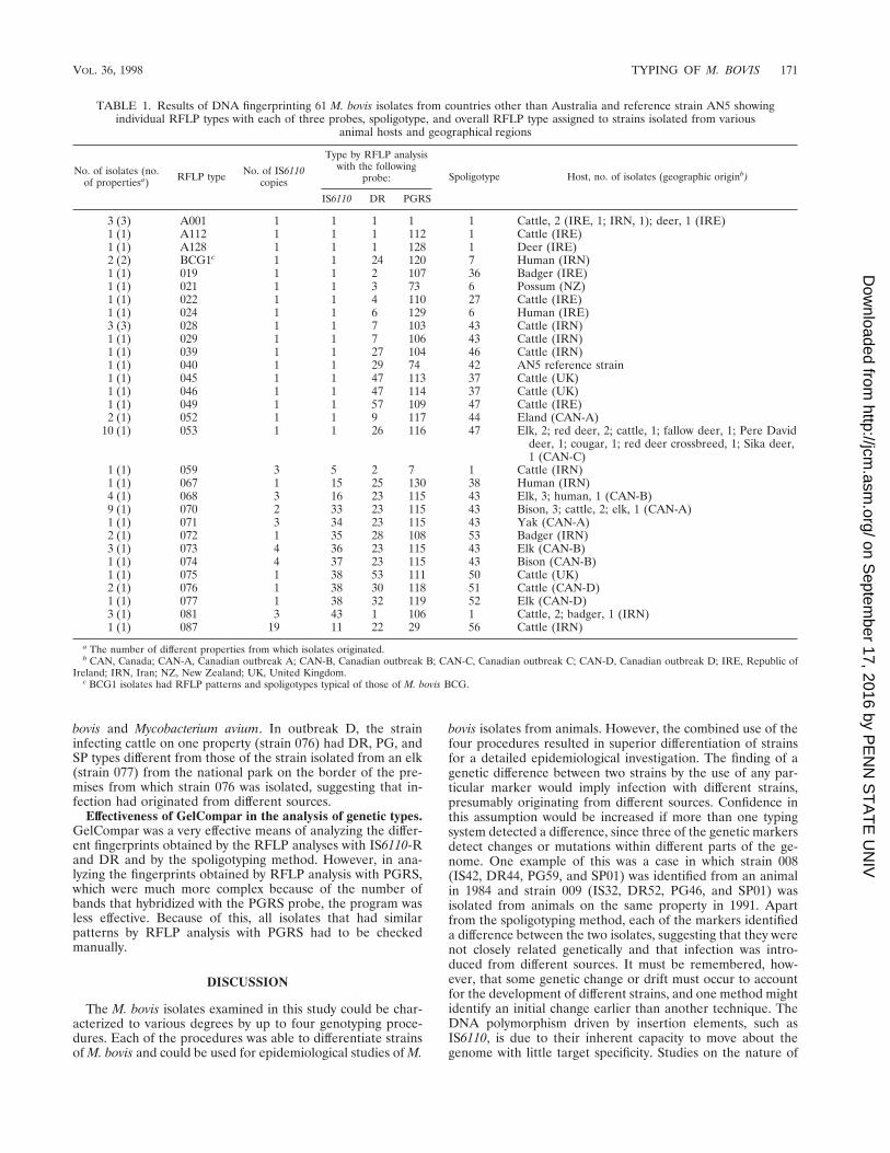
TABLE 1. Results of DNA fingerprinting 61 M. bovis isolates from countries other than Australia and reference strain AN5 showingindividual RFLP types with each of three probes, spoligotype, and overall RFLP type assigned to strains isolated from various
animal hosts and geographical regions
No. of isolates (no.of propertiesa) RFLP type No. of IS6110
copies
Type by RFLP analysiswith the following
probe: Spoligotype Host, no. of isolates (geographic originb)
IS6110 DR PGRS
3 (3) A001 1 1 1 1 1 Cattle, 2 (IRE, 1; IRN, 1); deer, 1 (IRE)1 (1) A112 1 1 1 112 1 Cattle (IRE)1 (1) A128 1 1 1 128 1 Deer (IRE)2 (2) BCG1c 1 1 24 120 7 Human (IRN)1 (1) 019 1 1 2 107 36 Badger (IRE)1 (1) 021 1 1 3 73 6 Possum (NZ)1 (1) 022 1 1 4 110 27 Cattle (IRE)1 (1) 024 1 1 6 129 6 Human (IRE)3 (3) 028 1 1 7 103 43 Cattle (IRN)1 (1) 029 1 1 7 106 43 Cattle (IRN)1 (1) 039 1 1 27 104 46 Cattle (IRN)1 (1) 040 1 1 29 74 42 AN5 reference strain1 (1) 045 1 1 47 113 37 Cattle (UK)1 (1) 046 1 1 47 114 37 Cattle (UK)1 (1) 049 1 1 57 109 47 Cattle (IRE)2 (1) 052 1 1 9 117 44 Eland (CAN-A)
10 (1) 053 1 1 26 116 47 Elk, 2; red deer, 2; cattle, 1; fallow deer, 1; Pere Daviddeer, 1; cougar, 1; red deer crossbreed, 1; Sika deer,1 (CAN-C)
1 (1) 059 3 5 2 7 1 Cattle (IRN)1 (1) 067 1 15 25 130 38 Human (IRN)4 (1) 068 3 16 23 115 43 Elk, 3; human, 1 (CAN-B)9 (1) 070 2 33 23 115 43 Bison, 3; cattle, 2; elk, 1 (CAN-A)1 (1) 071 3 34 23 115 43 Yak (CAN-A)2 (1) 072 1 35 28 108 53 Badger (IRN)3 (1) 073 4 36 23 115 43 Elk (CAN-B)1 (1) 074 4 37 23 115 43 Bison (CAN-B)1 (1) 075 1 38 53 111 50 Cattle (UK)2 (1) 076 1 38 30 118 51 Cattle (CAN-D)1 (1) 077 1 38 32 119 52 Elk (CAN-D)3 (1) 081 3 43 1 106 1 Cattle, 2; badger, 1 (IRN)1 (1) 087 19 11 22 29 56 Cattle (IRN)
a The number of different properties from which isolates originated.b CAN, Canada; CAN-A, Canadian outbreak A; CAN-B, Canadian outbreak B; CAN-C, Canadian outbreak C; CAN-D, Canadian outbreak D; IRE, Republic of
Ireland; IRN, Iran; NZ, New Zealand; UK, United Kingdom.c BCG1 isolates had RFLP patterns and spoligotypes typical of those of M. bovis BCG.
VOL. 36, 1998 TYPING OF M. BOVIS 171
on Septem
ber 17, 2016 by PE
NN
ST
AT
E U
NIV
http://jcm.asm
.org/D
ownloaded from
genetic rearrangements within the DR region suggested thathomologous recombination between the small DRs was thepredominant kind of rearrangement for this repetitive element(11), and it seems likely that the same mechanism may con-tribute to polymorphisms with PGRS (35). Certainly, there wasevidence of stability with each of the markers used in thisstudy, providing confidence in the ability of these techniques toprovide useful information for epidemiological studies. In sev-eral cases in Western Australia, where isolates from cattlefrom the same property were available for study over periodsof up to 7 years, studies with each of the markers gave consis-tent results.
Just how frequently a strain of M. bovis will alter its geneticmakeup and how often these differences will be detectable areunknown. The fact that the PGRS probe detected the greatestnumber of types is best explained by the fact that these repet-itive elements are found at various locations around the ge-nome of M. bovis, whereas the DR probe and the spacers usedin spoligotyping target the same region. RFLP analysis with theDR probe targets the DRs around the point of the IS6110insertion (13), and spoligotyping identifies the presence orabsence of specific spacers between the DRs (11).
In strains with a single copy of IS6110, the IS is insertedbetween two DRs and has been found to transpose relativelyinfrequently (13). In this study, 85.4% of the M. bovis isolates
examined had a single copy of IS6110, and the percentage ofAustralian isolates that had a single copy was slightly higher(91.9%). In addition, the majority of these single-copy strainscarried the IS on the 1.9-kb fragment considered characteristicfor M. bovis isolates from cattle (32), thereby minimizing theeffectiveness of IS6110-R for use in the characterization of M.bovis in this study. These results are consistent with the resultsof RFLP analysis with IS6110 for isolates of M. bovis from NewZealand (3) and bovine and human isolates of M. bovis fromArgentina (32). In a study which examined Argentinian iso-lates (32), it was suggested that strains with multiple copies ofIS6110 were more likely to have originated from wild or zooanimals other than cattle or from humans from The Nether-lands. Six to eight copies of IS6110 were present in all 23 goatstrains examined in Spain, yet 16 of 17 cattle were infected withstrains with a single copy of IS6110 (12), suggesting that dif-ferent reservoirs for caprine and bovine tuberculosis existed inSpain. A more comprehensive study involving 129 isolates ofM. bovis from cattle, goats, cats, and a sheep in Spain demon-strated that almost 50% of the bovine strains examined har-bored multiple IS6110 copies, but found that bovine and ca-prine isolates still clustered separately (15). The results ofthose studies and the results reported in this paper indicatethat different countries or animal hosts may harbor uniqueclonal populations of M. bovis.
FIG. 2. Dendrogram drawn by the GelCompar program showing the relationship of 35 representative fingerprints (DR types) for 273 isolates of M. bovis fromAustralia (n 5 211) and overseas (n 5 61) obtained by RFLP analysis with the DR probe. Aust., Australia, UK, United Kingdom; NZ, New Zealand.
172 COUSINS ET AL. J. CLIN. MICROBIOL.
on Septem
ber 17, 2016 by PE
NN
ST
AT
E U
NIV
http://jcm.asm
.org/D
ownloaded from
The Canadian isolates examined in this study were quitedistinct from the Australian M. bovis isolates that were exam-ined. Many of the Canadian isolates had multiple copies ofIS6110 (outbreaks A and B), and their DR, PG, and SP pat-terns were also different from those commonly found in Aus-tralian strains. RFLP analysis with IS6110 is considered to bea sensitive method for the detection of genetic changes inisolates of M. tuberculosis or M. bovis that have multiple copiesof this IS (5, 32, 33). This was well demonstrated in isolatesfrom Canadian outbreaks A and B in which genetic change wasevident in the slight rearrangement of IS6110 fragments, with-out alteration in the patterns obtained by RFLP analysis withthe PGRS and DR probes. In outbreaks A and B, two andthree common IS6110 bands were present, respectively, andsingle band changes were detected after infection in a yak(outbreak A) and a bison (outbreak B). The rate of transpo-sition of the IS element is considered to be a factor of time(35); however, there was some evidence from the fingerprintsfor the Canadian isolates obtained by RFLP analysis withIS6110-R suggesting that the M. bovis strains may have alteredin different hosts during the course of the infection in out-breaks A and B. Similar band changes were identified in anoutbreak of M. bovis in Swedish deer (29) and in cattle inBurundi (23). It is not known whether the isolation of strains,from different animal species, with minor differences in profilesby RFLP analysis with IS6110 (insertions or deletions), such as
those seen in outbreaks A and B, was related to host factors orrepresented natural evolution of the bacterium over time. Incontrast, isolates recovered from two elands in outbreak Awere clearly different from those recovered from other animalsin 1993 and 1994 by each of the fingerprinting methods used,suggesting a clear genetic difference in the strain infecting thetwo elands and confirming an alternate source of infection forthese animals.
PGRS was by far the best single genetic marker for RFLPanalysis with which to characterize the majority of the M. bovisisolates encountered in Australia and, with the exception ofisolates from Canada, the M. bovis isolates from other coun-tries examined in this study. In fact, RFLP analysis with PGRSidentified twice as many types as RFLP analysis with DR andspoligotyping and more than three times as many types asRFLP analysis with IS6110-R. This is consistent with previousfindings (6) but differs from those of a study in NorthernIreland in which the results of RFLP analyses with PGRS andIS6110 were found to be virtually equivalent (27). Skuce et al.(27) used the entire IS6110 probe, which is known to identifyslightly more polymorphisms than the RHS IS6110 probe usedin this study. In a direct comparison of LHS and RHS IS6110probes, the LHS probe was able to identify three differenttypes among a group of 15 isolates that were a common IS6110type (single band at 1.9 kb) (15). The digests used for ourstudies were run longer than those of Skuce et al. (27) and
FIG. 3. Dendrogram drawn by the GelCompar program showing the relationship of 35 representative SP types obtained for 273 isolates of M. bovis from Australia(n 5 211) and overseas (n 5 61) obtained by spoligotyping. Aust, Australia; UK, United Kingdom.
VOL. 36, 1998 TYPING OF M. BOVIS 173
on Septem
ber 17, 2016 by PE
NN
ST
AT
E U
NIV
http://jcm.asm
.org/D
ownloaded from

FIG. 4. Dendrogram drawn by the GelCompar program showing the relationship of 77 representative fingerprints (PG types) for 273 isolates of M. bovis fromAustralia (n 5 211) and overseas (n 5 61) obtained by RFLP analysis with the PGRS probe. UK, United Kingdom; Aust, Australia; NZ, New Zealand.
174 COUSINS ET AL. J. CLIN. MICROBIOL.
on Septem
ber 17, 2016 by PE
NN
ST
AT
E U
NIV
http://jcm.asm
.org/D
ownloaded from
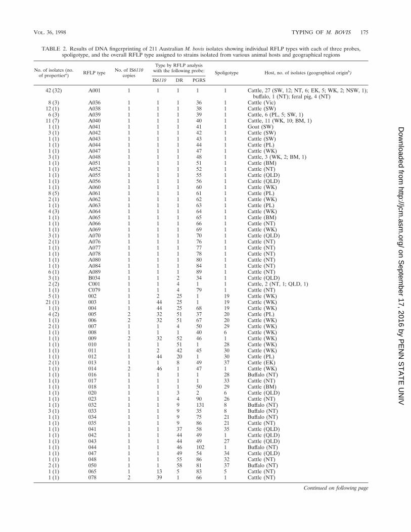
TABLE 2. Results of DNA fingerprinting of 211 Australian M. bovis isolates showing individual RFLP types with each of three probes,spoligotype, and the overall RFLP type assigned to strains isolated from various animal hosts and geographical regions
No. of isolates (no.of propertiesa) RFLP type No. of IS6110
copies
Type by RFLP analysiswith the following probe: Spoligotype Host, no. of isolates (geographical originb)
IS6110 DR PGRS
42 (32) A001 1 1 1 1 1 Cattle, 27 (SW, 12; NT, 6; EK, 5; WK, 2; NSW, 1);buffalo, 1 (NT); feral pig, 4 (NT)
8 (3) A036 1 1 1 36 1 Cattle (Vic)12 (1) A038 1 1 1 38 1 Cattle (SW)6 (3) A039 1 1 1 39 1 Cattle, 6 (PL, 5; SW, 1)
11 (7) A040 1 1 1 40 1 Cattle, 11 (WK, 10; BM, 1)1 (1) A041 1 1 1 41 1 Goat (SW)3 (1) A042 1 1 1 42 1 Cattle (SW)1 (1) A043 1 1 1 43 1 Cattle (SW)1 (1) A044 1 1 1 44 1 Cattle (PL)1 (1) A047 1 1 1 47 1 Cattle (WK)3 (1) A048 1 1 1 48 1 Cattle, 3 (WK, 2; BM, 1)1 (1) A051 1 1 1 51 1 Cattle (BM)1 (1) A052 1 1 1 52 1 Cattle (NT)1 (1) A055 1 1 1 55 1 Cattle (QLD)1 (1) A056 1 1 1 56 1 Cattle (QLD)1 (1) A060 1 1 1 60 1 Cattle (WK)8 (5) A061 1 1 1 61 1 Cattle (PL)2 (1) A062 1 1 1 62 1 Cattle (WK)1 (1) A063 1 1 1 63 1 Cattle (PL)4 (3) A064 1 1 1 64 1 Cattle (WK)1 (1) A065 1 1 1 65 1 Cattle (BM)1 (1) A066 1 1 1 66 1 Cattle (NT)1 (1) A069 1 1 1 69 1 Cattle (WK)3 (1) A070 1 1 1 70 1 Cattle (QLD)2 (1) A076 1 1 1 76 1 Cattle (NT)1 (1) A077 1 1 1 77 1 Cattle (NT)1 (1) A078 1 1 1 78 1 Cattle (NT)1 (1) A080 1 1 1 80 1 Cattle (NT)1 (1) A084 1 1 1 84 1 Cattle (NT)6 (1) A089 1 1 1 89 1 Cattle (NT)3 (1) B034 1 1 2 34 1 Cattle (QLD)2 (2) C001 1 1 4 1 1 Cattle, 2 (NT, 1; QLD, 1)1 (1) C079 1 1 4 79 1 Cattle (NT)5 (1) 002 1 2 25 1 19 Cattle (WK)
21 (1) 003 1 44 25 1 19 Cattle (WK)1 (1) 004 1 44 25 68 19 Cattle (WK)4 (2) 005 2 32 51 37 20 Cattle (PL)1 (1) 006 2 32 51 67 20 Cattle (WK)2 (1) 007 1 1 4 50 29 Cattle (WK)1 (1) 008 1 1 1 40 6 Cattle (WK)1 (1) 009 2 32 52 46 1 Cattle (WK)1 (1) 010 1 1 51 1 28 Cattle (WK)1 (1) 011 1 2 42 45 30 Cattle (WK)1 (1) 012 1 44 20 1 30 Cattle (PL)2 (1) 013 1 1 8 49 37 Cattle (EK)1 (1) 014 2 46 1 47 1 Cattle (WK)1 (1) 016 1 1 1 1 28 Buffalo (NT)1 (1) 017 1 1 1 1 33 Cattle (NT)1 (1) 018 1 1 1 50 29 Cattle (BM)1 (1) 020 1 1 3 2 6 Cattle (QLD)1 (1) 023 1 1 4 90 26 Cattle (NT)1 (1) 032 1 1 9 131 8 Buffalo (NT)3 (1) 033 1 1 9 35 8 Buffalo (NT)1 (1) 034 1 1 9 75 21 Buffalo (NT)1 (1) 035 1 1 9 86 21 Cattle (NT)1 (1) 041 1 1 37 58 35 Cattle (QLD)1 (1) 042 1 1 44 49 1 Cattle (QLD)1 (1) 043 1 1 44 49 27 Cattle (QLD)1 (1) 044 1 1 46 102 1 Buffalo (NT)1 (1) 047 1 1 49 54 34 Cattle (QLD)1 (1) 048 1 1 55 86 32 Cattle (NT)2 (1) 050 1 1 58 81 37 Buffalo (NT)1 (1) 065 1 13 5 83 5 Cattle (NT)1 (1) 078 2 39 1 66 1 Cattle (NT)
Continued on following page
VOL. 36, 1998 TYPING OF M. BOVIS 175
on Septem
ber 17, 2016 by PE
NN
ST
AT
E U
NIV
http://jcm.asm
.org/D
ownloaded from
those reported previously (6) in an attempt to improve theresolution of bands. In addition, we included RFLP bands of.1.3 kb in the analysis. In Northern Ireland, more than 40% of109 isolates were identified as having a common pattern byusing a combination of PGRS, IS6110, and IS1081 probes, andalmost 50% of the isolates had a common pattern when thePGRS probe was used (27). By comparison, 29.3% of theisolates were identified in this study as a common type byRFLP analysis with PGRS, PG01, and 36.5% of Australianisolates were identified as PG01. The use of other geneticmarkers resulted in further characterization of 45% of theseisolates with the common PG01 type, so that 19.9% of Aus-tralian isolates were identified as the most common strain,designated A001. In a more recent study in Northern Ireland(28), bands greater than 2.26 kb were included in the analysis,but the sensitivity of RFLP analysis with PGRS was not mark-edly improved. Additional studies may determine whether thecommon strain found in Northern Ireland can be further dif-ferentiated and whether it is the same as any of the morecommon strains found in Australia.
The finding of a common strain distributed throughout Aus-tralia suggests that animals on many properties were infectedfrom the same source. This may have occurred over time asinfected cattle moved from one area to another throughout thecountry and spread infection caused by a single clone. It isgenerally accepted that bovine tuberculosis was introducedinto Australia with the introduction of infected cattle duringthe early settlement period (26). The number of introductionswould have been limited, and this may account for some of thegenetic similarity of strains seen in Australia. The finding of afurther 22.7% of isolates with common IS, DR, and SP typesbut different PG types (type A strains other than type A001)suggests the clonal expansion of strain A001. The question ofhost influence versus genetic drift over time was also raisedwith the finding of type A041 in a single goat in a mixed cattleand goat herd in Western Australia in which cattle were in-fected with strain A001. Four other cattle herds that wereepidemiologically linked to this herd were also infected withstrain A001. The goats on the affected property were in poorcondition and were considered to be highly stressed because ofthe excessively high stocking rate. It is possible that the hostresponse to infection in this goat may have caused the geneticmakeup of the organism to alter.
RFLP analysis with the DR probe produced results similarto those obtained by the spoligotyping method, a finding that isnot surprising, considering that both target the same chromo-somal locus. In one previous small-scale comparison of IS01isolates, analysis with PGRS allowed for a somewhat betterdifferentiation than analysis with DR (32), but another study
suggested that the results of analyses with the two markerswere equivalent (12).
The advantages of spoligotyping lie in the speed and low costof the technique and in the ease of analysis. The spoligotypingmethod was originally designed for epidemiological studies ofM. tuberculosis, and as such, 37 of the 43 spacers presently usedon the membranes originate from M. tuberculosis H37Rv and 6spacers were derived from M. bovis BCG (11). Better discrim-ination of M. bovis strains may be achieved by this technique inthe future by the identification and use of spacer sequencesthat are specific for M. bovis strains. The spoligotyping tech-nique has the added advantage that it can be used directly withisolates and tissue specimens for the rapid screening of M.bovis isolates. It would appear to be sensible to further char-acterize isolates with common spoligotypes at least with thePGRS probe. RFLP analyses with IS6110-R and DR could beused if full characterization was needed. The lack of the fivespacers at the 39 end of the DR locus detected by spoligotypingis considered to be consistent with M. bovis (14). All but 1 ofthe 273 isolates of M. bovis tested in this study lacked thesespacers. The one isolate (IRN49) that was found to containfour of the five 39 end spacers was biochemically more consis-tent with M. tuberculosis, and use of each of the probe methodsproduced an unusual pattern for M. bovis. These findings con-firmed the lack of the 39 DR spacers as being consistent withthe identification of M. bovis isolates.
Spread of infection with M. bovis between animals is gener-ally considered to be via aerosols, although ingestion of infec-tious materials is also accepted as a route of transmission (20,22). Contact with infected nasal mucus was identified as animportant source of infection in a series of studies with natu-rally and experimentally infected cattle in Northern Ireland(17–19). In the present study, DNA fingerprinting results forisolates from Canadian outbreak C were able to support theproposal that nose-to-nose contact between elk and deer andbetween deer and cattle on opposite sides of a fence hadspread the infection. In addition, DNA fingerprinting providedconfirmatory evidence that a cougar on one property had be-come infected with M. bovis and M. avium after being fedtuberculosis-affected carcasses of other animals on the pre-mises and chickens from a neighboring farm, respectively.
Although early studies suggested that M. bovis could survivein cow feces for between 2 and 5 months, depending on theseason, and in soil for 2 years, more recent evidence suggeststhat survival in soil or feces exposed to natural conditions ofsunlight is limited (36). Under Australian conditions, M. bovissurvived for 4 weeks in soil in 80% shade or more, but noisolation from dry or moist soils exposed to sunlight or fromfeces held in any conditions was made at 4 weeks (9). In
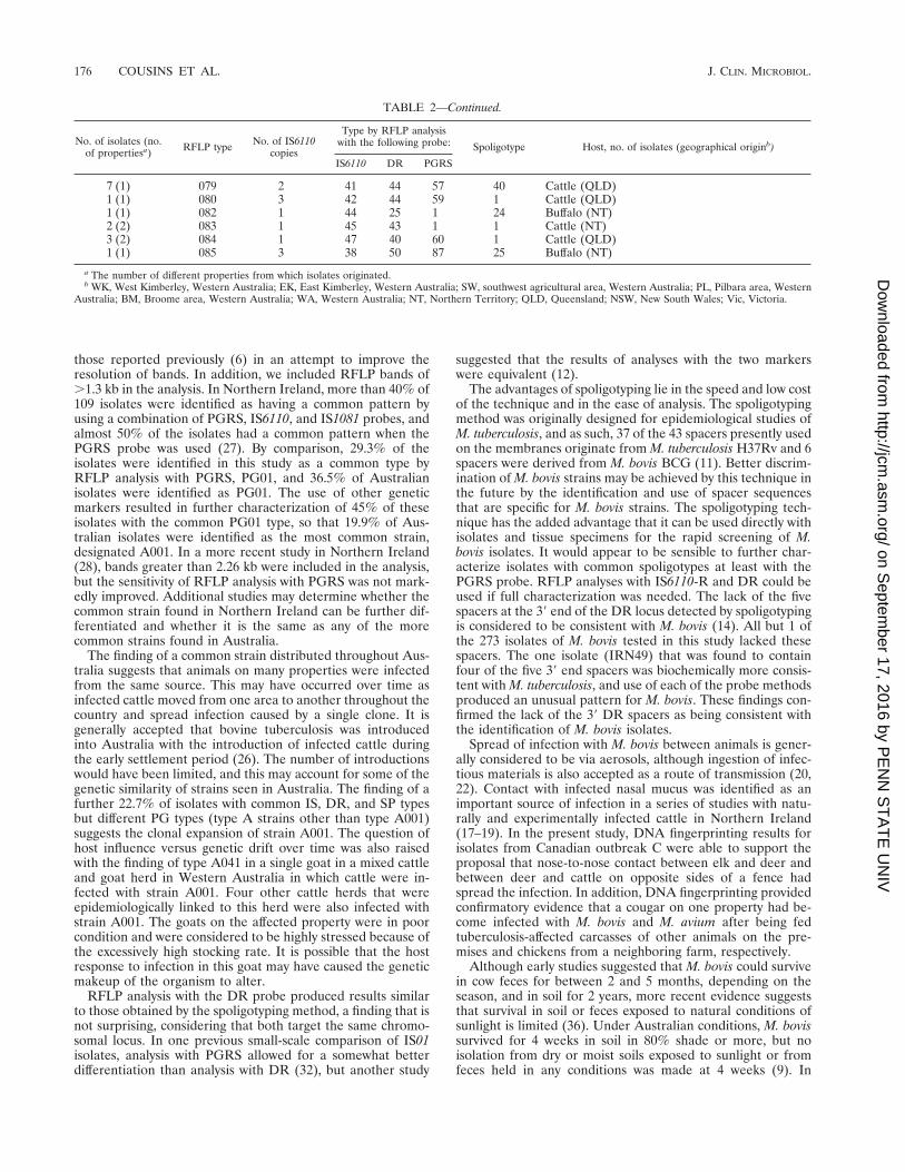
TABLE 2—Continued.
No. of isolates (no.of propertiesa) RFLP type No. of IS6110
copies
Type by RFLP analysiswith the following probe: Spoligotype Host, no. of isolates (geographical originb)
IS6110 DR PGRS
7 (1) 079 2 41 44 57 40 Cattle (QLD)1 (1) 080 3 42 44 59 1 Cattle (QLD)1 (1) 082 1 44 25 1 24 Buffalo (NT)2 (2) 083 1 45 43 1 1 Cattle (NT)3 (2) 084 1 47 40 60 1 Cattle (QLD)1 (1) 085 3 38 50 87 25 Buffalo (NT)
a The number of different properties from which isolates originated.b WK, West Kimberley, Western Australia; EK, East Kimberley, Western Australia; SW, southwest agricultural area, Western Australia; PL, Pilbara area, Western
Australia; BM, Broome area, Western Australia; WA, Western Australia; NT, Northern Territory; QLD, Queensland; NSW, New South Wales; Vic, Victoria.
176 COUSINS ET AL. J. CLIN. MICROBIOL.
on Septem
ber 17, 2016 by PE
NN
ST
AT
E U
NIV
http://jcm.asm
.org/D
ownloaded from
Australia, when infected properties are destocked, the owner isadvised to wait a minimum of 30 days before restocking. Thisprecaution is taken to minimize the risk of reinfection whennew stock are brought in. In the case of outbreak C in Canada,one property that had undergone cleaning and disinfection andthat had been released from quarantine was restocked withdeer from New Zealand that were skin test negative. Thefinding in three imported New Zealand deer of a strain of M.bovis identical to the strain previously identified on the prop-erty confirmed infection with the same strain in the importedanimals. It is possible that the M. bovis organisms on theproperty remained infectious, despite thorough decontamina-tion procedures. Another possible explanation is that the NewZealand animals, which had passed pre- and postquarantinetesting and isolation, were infected, coincidentally, with anidentical strain before they arrived at the premises. Alterna-tively, a worker in contact with both groups of animals mayhave acted as a vector in passing on infection to the newlyarrived animals. Typing of strains from the herd or region oforigin in New Zealand may assist in clarifying this situation.
It was expected that some similarities would be found be-tween isolates from Australia and those from the United King-dom and the Republic of Ireland since M. bovis was believed tohave been originally imported with infected cattle from thesecountries. In fact, only a few similarities were seen betweensome of the Australian strains and a small number of isolatesfrom Ireland and one isolate from Iran. This similarity wasseen by the identification of some type A strains from Ireland,suggesting a common clonal origin for these strains in Austra-lia and Ireland. The majority of isolates from other countriesappeared to be unique. Certainly, the isolates from Canadawere clearly different from the “Australian” strains, and this isconsistent with observations that geographically distinct strainsexist in The Netherlands and Argentina (32), regions whereclones of organisms may have spread within countries withlittle opportunity for genetic exchange. The finding of geo-graphically distinct populations of M. bovis may be useful forconfirming the source of infection in any imported animals thatare subsequently diagnosed with bovine tuberculosis.
ACKNOWLEDGMENTS
This work was supported by the Australian Brucellosis and Tuber-culosis Eradication Campaign.
We gratefully acknowledge the many collaborators who providedisolates or reference strains for study, in particular, Elizabeth Rohon-czy and Claude Turcotte, Animal Disease Research Institute, Nepean,Ontario, Canada; Anne Fanning, TB Services, Alberta Health, Ed-monton, Alberta, Canada; Louis O’Reilly, Veterinary Research Lab-oratory, Abbottstown, Castleknock, Ireland; Mohammad Feizabadi,courtesy of the Razi Institute, Tehran, Iran; and Jeremy Dale, SurreyUniversity, Guildford, United Kingdom. In addition, we acknowledgeBarry Francis for excellent technical assistance and colleagues in Aus-tralian State, Northern Territory, and CSIRO Animal Health labora-tories for supplying Australian isolates for typing.
REFERENCES
1. Aranaz, A., E. Liebana, A. Mateos, L. Dominguez, D. Vidal, M. Domingo, O.Gonzales, E. F. Rodrigues-Ferri, A. Bunshoten, J. van Embden, and D.Cousins. 1996. Spoligotyping of Mycobacterium bovis strains from cattle andother animals: a tool for epidemiology of tuberculosis. J. Clin. Microbiol.34:2734–2740.
2. Beck-Sague, C., S. W. Dooley, M. D. Hutton, J. Otten, A. Breeden, J. T.Crawford, A. E. Pitchenik, C. Woodley, G. Cauthen, and W. R. Jarvis. 1992.Hospital outbreak of multidrug-resistant Mycobacterium tuberculosis infec-tions. JAMA 268:1280–1286.
3. Collins, D. M., S. K. Erasmuson, D. M. Stephens, G. F. Yates, and G. W. deLisle. 1993. DNA fingerprinting of Mycobacterium bovis strains by restrictionfragment analysis and hybridization with insertion elements IS1081 andIS6110. J. Clin. Microbiol. 31:1143–1147.
4. Cousins, D. V. 1996. Molecular epidemiology and diagnosis of Mycobacte-rium bovis and M. bovis-like organisms causing tuberculosis. Ph.D. thesis.University of Western Australia, Perth, Australia.
5. Cousins, D. V., R. A. Skuce, R. R. Kazwala, and J. D. A. van Embden.Towards a standardized approach to DNA fingerprinting of Mycobacteriumbovis. Int. J. Tuberc. Lung Dis., in press.
6. Cousins, D. V., S. N. Williams, B. C. Ross, and T. M. Ellis. 1993. Use of arepetitive element isolated from Mycobacterium tuberculosis in hybridizationstudies with Mycobacterium bovis: a new tool for epidemiological studies ofbovine tuberculosis. Vet. Microbiol. 37:1–17.
7. Daley, C. L., P. M. Small, G. F. Schecter, G. K. Schoolnik, R. A. McAdam,W. R. Jacobs, Jr., and P. C. Hopewell. 1992. An outbreak of tuberculosis withaccelerated progression among persons infected with human immunodefi-ciency virus: an analysis using restriction fragment length polymorphisms.N. Engl. J. Med. 326:231–235.
8. Doran, T. J., A. L. M. Hodgson, J. K. Davies, and A. J. Radford. 1993.Characterisation of a highly repeated DNA sequence from Mycobacteriumbovis. FEMS Microbiol. Lett. 111:147–152.
9. Duffield, B. J., and D. A. Young. 1985. Survival of Mycobacterium bovis indefined environmental conditions. Vet. Microbiol. 10:193–197.
10. Dwyer, B., K. Jackson, K. Raios, A. Sievers, E. Wilshire, and B. Ross. 1993.DNA restriction fragment analysis to define an extended cluster of tubercu-losis in homeless men and their associates. J. Infect. Dis. 167:490–494.
11. Groenen, P. M. A., A. E. Bunschoten, D. van Soolingen, and J. D. A. vanEmbden. 1993. Nature of DNA polymorphism in the direct repeat cluster ofMycobacterium tuberculosis; application of strain differentiation by a noveltyping method. Mol. Microbiol. 10:1057–1065.
12. Gutierrez, M., S. Samper, J. A. Gavigan, J. F. Garcıa-Marın, and C. Martın.1995. Diffentiation by molecular typing of Mycobacterium bovis strains caus-ing tuberculosis in cattle and goats. J. Clin. Microbiol. 33:2953–2956.
13. Hermans, P. W. M., D. van Soolingen, E. M. Bik, P. E. W. de Haas, J. W.Dale, and J. D. A. van Embden. 1991. The insertion element IS987 fromMycobacterium bovis BCG is located in a hot-spot integration region forinsertion elements in Mycobacterium tuberculosis complex strains. Infect.Immun. 59:2695–2705.
14. Kamerbeek, J., L. Schouls, M. van Agterveld, A. Kolk, S. Kuijper, D. vanSoolingen, P. de Haas, A. Bunschoten, and J. van Embden. 1997. Simulta-neous strain detection and differentiation of Mycobacterium tuberculosis fordiagnosis and epidemiology. J. Clin. Microbiol. 35:907–914.
15. Liebana, E., A. Aranaz, L. Dominguez, A. Mateos, O. Gonzales-Llamazares,E. F. Rodrigues-Ferri, M. Domingo, D. Vidal, and D. Cousins. 1997. Theinsertion element IS6110 is a useful tool for DNA fingerprinting of Myco-bacterium bovis isolates from cattle and goats in Spain. Vet. Microbiol.54:223–233.
16. Mazurek, G. H., M. D. Cave, K. D. Eisenach, R. J. J. Wallace, J. H. Bates,and J. T. Crawford. 1991. Chromosomal DNA fingerprint patterns producedwith IS6110 as strain-specific markers for epidemiologic study of tuberculo-sis. J. Clin. Microbiol. 29:2030–2033.
17. McIlroy, S. G., S. D. Neill, and R. M. McCracken. 1986. Pulmonary lesionsand Mycobacterium bovis excretion from the respiratory tract of tuberculinreacting cattle. Vet. Rec. 118:718–721.
18. Neill, S. D., J. Hanna, D. P. Mackie, and T. G. D. Bryson. 1992. Isolation ofMycobacterium bovis from the respiratory tract of skin test-negative cattle.Vet. Rec. 131:45–47.
19. Neill, S. D., J. Hanna, J. J. O’Brien, and R. M. McCracken. 1989. Trans-mission of tuberculosis from experimentally infected cattle to in-contactcalves. Vet. Rec. 124:269–271.
20. Neill, S. D., J. M. Pollock, D. B. Bryson, and J. Hanna. 1994. Pathogenesisof Mycobacterium bovis infection in cattle. Vet. Microbiol. 40:41–52.
21. Otal, I., C. Martın, V. Vincent-Levy-Frebault, D. Thierry, and B. Gicquel.1991. Restriction fragment length polymorphism analysis using IS6110 as anepidemiological marker in tuberculosis. J. Clin. Microbiol. 29:1252–1254.
22. Pritchard, D. G. 1988. A century of bovine tuberculosis 1888–1988: conquestand controversy. J. Comp. Pathol. 99:357–399.
23. Rigouts, L., B. Maregeya, H. Traore, J. P. Collart, K. Fissette, and F.Portaels. 1996. Use of DNA restriction fragment typing in the differentiationof Mycobacterium tuberculosis complex isolates from animals and humans inBurundi. Tubercle Lung Dis. 77:264–268.
24. Romano, M. I., A. Alito, J. C. Fisanotti, F. Bigi, I. Kantor, M. E. Cicuta, andA. Cataldi. 1996. Comparison of different genetic markers for molecularepidemiology of bovine tuberculosis. Vet. Microbiol. 50:59–71.
25. Ross, B. C., K. Raios, K. Jackson, and B. Dwyer. 1992. Molecular cloning ofa highly repeated DNA element from Mycobacterium tuberculosis and its useas an epidemiologic tool. J. Clin. Microbiol. 30:942–946.
26. Seddon, H. R., and H. E. Albiston. 1965. Bacterial diseases, vol. 1. Com-monwealth Department of Health, Canberra, Australia.
27. Skuce, R. A., D. Brittain, M. S. Hughes, L.-A. Beck, and S. D. Neill. 1994.Genomic fingerprinting of Mycobacterium bovis from cattle by restrictionfragment length analysis. J. Clin. Microbiol. 32:2387–2392.
28. Skuce, R. A., D. Brittain, M. S. Hughes, and S. D. Neill. 1996. Differentiationof Mycobacterium bovis isolates from animals by DNA typing. J. Clin. Mi-crobiol. 34:2469–2474.
VOL. 36, 1998 TYPING OF M. BOVIS 177
on Septem
ber 17, 2016 by PE
NN
ST
AT
E U
NIV
http://jcm.asm
.org/D
ownloaded from
29. Szewzyk, R., S. B. Svenson, S. E. Hoffner, G. Bolske, H. Wahlstrom, L.Englund, A. Engvall, and G. Kallenius. 1995. Molecular epidemiologicalstudies of Mycobacterium bovis infections in humans and animals in Sweden.J. Clin. Microbiol. 33:3183–3185.
30. Torrea, G., G. Levee, P. Grimont, C. Martin, S. Chanteau, and B. Gicquel.1995. Chromosomal DNA fingerprinting analysis using the insertion se-quence IS6110 and the repetitive element DR as strain-specific markers forepidemiolical study of tuberculosis in French polynesia. J. Clin. Microbiol.33:1890–1895.
31. van Embden, J. D. A., D. Cave, J. T. Crawford, J. W. Dale, K. D. Eisenach,B. Gicquel, P. Hermans, C. Martin, R. McAdam, T. M. Shinnick, and P. M.Small. 1993. Strain identification of Mycobacterium tuberculosis by DNAfingerprinting: recommendations for a standardized methodology. J. Clin.Microbiol. 31:406–409.
32. van Soolingen, D., P. E. W. de Haas, J. Haagsma, T. Eger, P. W. M.Hermans, V. Ritacco, A. Alito, and J. D. A. van Embden. 1994. Use of variousgenetic markers in differentiation of Mycobacterium bovis strains from ani-mals and humans and for studying epidemiology of bovine tuberculosis.J. Clin. Microbiol. 32:2425–2433.
33. van Soolingen, D., P. E. W. de Haas, P. W. M. Hermans, P. M. A. Groenen,and J. D. A. van Embden. 1993. Comparison of various repetitive DNA
elements as genetic markers for strain differentiation and epidemiology ofMycobacterium tuberculosis. J. Clin. Microbiol. 31:1987–1995.
34. van Soolingen, D., P. W. M. Hermans, P. E. W. de Haas, D. R. Soll, andJ. D. A. van Emden. 1991. Occurrence and stability of insertion sequences inMycobacterium tuberculosis complex strains: evaluation of an insertion se-quence-dependent DNA polymorphism as a tool in the epidemiology oftuberculosis. J. Clin. Microbiol. 29:2578–2586.
35. van Soolingen, D., L. Qian, P. E. W. de Haas, J. T. Douglas, H. Traore, F.Portaels, H. Xi Qing, D. Enkhsaikan, P. Nymadawa, and J. D. A. vanEmbden. 1995. Predominance of a single genotype of Mycobacterium tuber-culosis in countries of Asia. J. Clin. Microbiol. 33:3234–3238.
36. Wray, C. 1975. Survival and spread of pathogenic bacteria of veterinaryimportance within the environment. Vet. Bull. 45:543–550.
37. Yang, Z. H., P. E. W. de Haas, D. van Soolingen, J. D. A. van Embden, andÅ. B. Anderson. 1994. Restriction fragment length polymorphism of Myco-bacterium tuberculosis strains isolated from Greenland during 1992: evidenceof tuberculosis transmission between Greenland and Denmark. J. Clin. Mi-crobiol. 32:3018–3025.
38. Yuen, L. K. W., B. C. Ross, K. M. Jackson, and B. Dwyer. 1993. Character-ization of Mycobacterium tuberculosis strains from Vietnamese patients bySouthern blot hybridization. J. Clin. Microbiol. 31:1615–1618.
178 COUSINS ET AL. J. CLIN. MICROBIOL.
on Septem
ber 17, 2016 by PE
NN
ST
AT
E U
NIV
http://jcm.asm
.org/D
ownloaded from


