Embed Size (px)
Citation preview
Evidence for the aldo-keto reductase pathway ofpolycyclic aromatic trans-dihydrodiol activation inhuman lung A549 cellsJong-Heum Park*, Dipti Mangal†, Kirk A. Tacka*, Amy M. Quinn*, Ronald G. Harvey‡, Ian A. Blair*†,and Trevor M. Penning*†§
*Center of Excellence in Environmental Toxicology and †Center for Cancer Pharmacology, Department of Pharmacology, University of Pennsylvania Schoolof Medicine, Philadelphia, PA 19104-6084; and ‡The Ben May Institute for Cancer Research, University of Chicago, Chicago, IL 60637
Communicated by William F. DeGrado, University of Pennsylvania School of Medicine, Philadelphia, PA, March 20, 2008 (received for reviewOctober 30, 2007)
Polycyclic aromatic hydrocarbons (PAHs) are tobacco carcinogensimplicated in the causation of human lung cancer. Metabolicactivation is a key prerequisite for PAHs to cause their deleteriouseffects. Using human lung adenocarcinoma (A549) cells, we pro-vide evidence for the metabolic activation of (�)-trans-7,8-dihydroxy-7,8-dihydrobenzo[a]pyrene (B[a]P-7,8-trans-dihydrodiol)by aldo-keto reductases (AKRs) to yield benzo[a]pyrene-7,8-dione(B[a]P-7,8-dione), a redox-active o-quinone. We show that B[a]P-7,8-trans-dihydrodiol (AKR substrate) and B[a]P-7,8-dione (AKR product)lead to the production of intracellular reactive oxygen species (ROS)(measured as an increase in dichlorofluorescin diacetate fluores-cence) and that similar changes were not observed with the regio-isomer (�)-trans-4,5-dihydroxy-4,5-dihydrobenzo[a]pyrene or thediol-epoxide, (�)-anti-7,8-dihydroxy-9�,10�-epoxy-7,8,9,10-tetrahy-dro-B[a]P. B[a]P-7,8-trans-dihydrodiol and B[a]P-7,8-dione also causeda decrease in glutathione levels and an increase in NADP�/NADPHratios, with a concomitant increase in single-strand breaks (as mea-sured by the comet assay) and 7,8-dihydro-8-oxo-2�-deoxyguanosine(8-oxo-dGuo). The specificity of the comet assay was validated bycoupling it to human 8-oxo-guanine glycosylase (hOGG1), whichexcises 8-oxo-Gua to yield single-strand breaks. The levels of 8-oxo-dGuo observed were confirmed by an immunoaffinity purificationstable isotope dilution ([15N5]-8-oxo-dGuo) liquid chromatography-electrospray ionization/multiple reaction monitoring/mass spectrom-etry (LC-ESI/MRM/MS) assay. B[a]P-7,8-trans-dihydrodiol producedDNA strand breaks in the hOGG1-coupled comet assay as well as8-oxo-dGuo (as measured by LC-ESI/MRM/MS) and was enhanced bya catechol O-methyl transferase (COMT) inhibitor, suggesting thatCOMT protects against o-quinone-mediated redox cycling. We con-clude that activation of PAH-trans-dihydrodiols by AKRs in lungcells leads to ROS-mediated genotoxicity and contributes to lungcarcinogenesis.
8-oxo-dGuo � DNA strand breaks � tobacco carcinogens �reactive oxygen species
Polycyclic aromatic hydrocarbons (PAHs) are ubiquitous en-vironmental pollutants, which are produced as a result of
fossil-fuel combustion and are found in car exhaust and char-broiled and smoked foods (1, 2). They are also present asmixtures in tobacco smoke and are implicated in the causationof human lung cancer (3). To exert their carcinogenic effects,PAHs must be metabolically activated to DNA-damaging agentsthat will result in the signature mutations in lung cancer. Thesemutations are G-to-T transversions that either activate the K-rasprotooncogene at the 12th and 61st codon (4) or inactivate thep53 tumor suppressor gene at hot spots in its DNA bindingdomain (5).
Using benzo[a]pyrene (B[a]P) as a representative PAH, threepathways of activation have been proposed that lead to thesemutations. The first pathway involves the formation of (�)-anti-7�,8�-dihydroxy-9�,10�-epoxy-7,8,9,10-tetrahydroB[a]P {(�)-
anti-B[a]PDE}. In this pathway there is sequential monoxygen-ation catalyzed by cytochrome P450 (P450) 1A1/1B1 andhydration to form 7�,8�-dihydroxy-7,8-dihydroxy-B[a]P, whichundergoes a secondary monoxygenation to form (�)-anti-B[a]PDE (6). This diol-epoxide forms stable (�)-anti-trans-B[a]PDE-N2-2�-deoxyguanosine (dGuo) adducts, which viatrans-lesional bypass DNA polymerases, yield G-to-T transver-sions (7).
The second pathway involves metabolic activation by P450peroxidases to yield radical cations (8), which can form depuri-nating adducts that lead to abasic sites. Apurinic/apyrimdinic(AP) sites, if not repaired, can give rise to G-to-T transversions(9). However, it is unlikely that radical cations are sufficientlylong-lived to damage DNA in intact cells.
The third pathway of PAH activation is the NAD(P�)-dependent oxidation of PAH-trans-dihydrodiols to PAH o-quinones catalyzed by dihydrodiol dehydrogenase members ofthe aldo-keto reductase (AKR) superfamily (10). AKRs divertPAH trans-dihydrodiols to form ketols that spontaneously rear-range to catechols (Scheme 1). The catechols undergo twoone-electron oxidation events to produce the correspondingredox-active and electrophilic o-quinones. PAH o-quinones canform stable and depurinating DNA adducts in vitro (11, 12), andthese adducts may provide a route to G-to-T transversionmutations.
In the presence of NAD(P)H, PAH o-quinones also undergononenzymatic reduction back to catechols. This event establishesfutile redox cycles, which amplify the generation of reactiveoxygen species (ROS) at the expense of NADPH and may leadto a prooxidant cellular state. Because a prooxidant state hasbeen associated with tumor initiation and promotion (13), theAKR pathway of PAH activation is attractive in that it couldexplain how PAHs act as complete carcinogens. In addition,ROS may cause oxidative DNA damage such as 7,8-dihydro-8-oxo-2�-deoxyguanosine (8-oxo-dGuo) lesions, which can lead toG-to-T transversions (14). Amplification of ROS by catechol-o-quinone interconversion has also been proposed as a cause ofestrogen carcinogenesis (15).
Using a yeast gap repair assay to detect p53 mutations, Yu etal. (16) showed that PAH o-quinones were more potent asmutagens than (�)-anti-7,8-dihydroxy-9�,10�-epoxy-7,8,9,10-
Author contributions: J.-H.P. and T.M.P. designed research; J.-H.P., D.M., K.A.T., and A.M.Q.performed research; R.G.H. and I.A.B. contributed new reagents/analytic tools; J.-H.P.,D.M., K.A.T., and A.M.Q. analyzed data; and J.-H.P. and T.M.P. wrote the paper.
The authors declare no conflict of interest.
Freely available online through the PNAS open access option.
§To whom correspondence should be addressed. E-mail: [email protected].
This article contains supporting information online at www.pnas.org/cgi/content/full/0802776105/DCSupplemental.
© 2008 by The National Academy of Sciences of the USA
6846–6851 � PNAS � May 13, 2008 � vol. 105 � no. 19 www.pnas.org�cgi�doi�10.1073�pnas.0802776105
tetrahydroB[a]P (anti-B[a]PDE) provided that the o-quinoneswere allowed to redox cycle. Furthermore, the mutation patternobserved was dominated by G-to-T transversions. These muta-tions were suppressed by ROS attenuators. Recent HPLC-electrochemical detection (ECD) analysis showed that there wasa direct linear correlation between 8-oxo-dGuo formation andmutagenic frequency in p53 observed with PAH o-quinones (17).
Five human AKR isoforms including aldehyde reductase(AKR1A1) and hydroxysteroid dehydrogenases AKR1C1–AKR1C4 have been implicated in PAH activation (10). Jianget al. (18, 19) showed that (�)-trans-7,8-dihydroxy-7,8-dihydroB[a]P (B[a]P-7,8-trans-dihydrodiol) is activated to the corre-sponding B[a]P-7,8-dione in H358 human lung bronchoalveo-lar cell lysates and intact cells stably transfected withAKR1A1. Palackal et al. (20) also demonstrated that 7,12-dimethylbenz[a]athracene-3,4-dihydrodiol is activated to7,12-dimethylbenz[a]athracene-3,4-dione in A549 lung adeno-carcinoma cell extracts that constitutively overexpressAKR1C1–AKR1C3 isoforms. Clinical and epidemiologicalstudies have suggested that AKR expression is positively
correlated with the development of lung cancer. AKRs areup-regulated in nonsmall cell lung carcinoma (NSCLC) andthe bronchial epithelium of smokers (21). AKR1C1 andAKR1B10 were also two of seven genes among 30,000 genesmost overexpressed in smokers with NSCLC (22). However,despite this evidence, the hypothesis that PAH activation byAKRs results in ROS amplification, a prooxidant state, andoxidative DNA damage has not yet been formally tested inhuman lung cells.
We show that B[a]P-7,8-trans-dihydrodiol (AKR substrate)and B[a]P-7,8-dione (AKR product) generate ROS and create aprooxidant cellular state (i.e., change in cellular redox status) inA549 lung adenocarcinoma cells. We also show that conversionof B[a]P-7,8-trans-dihydrodiol to B[a]P-7,8-dione by the AKRpathway causes DNA strand breaks and 8-oxo-dGuo formation.These findings support a role for the AKR pathway in humanlung carcinogenesis.
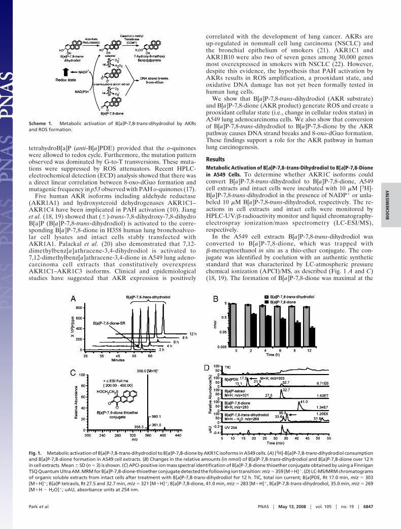
ResultsMetabolic Activation of B[a]P-7,8-trans-Dihydrodiol to B[a]P-7,8-Dionein A549 Cells. To determine whether AKR1C isoforms couldconvert B[a]P-7,8-trans-dihydrodiol to B[a]P-7,8-dione, A549cell extracts and intact cells were incubated with 10 �M [3H]-B[a]P-7,8-trans-dihydrodiol in the presence of NADP� or unla-beled 10 �M B[a]P-7,8-trans-dihydrodiol, respectively. The re-actions in cell extracts and intact cells were monitored byHPLC-UV/�-radioactivity monitor and liquid chromatography-electrospray ionization/mass spectrometry (LC-ESI/MS),respectively.
In the A549 cell extracts B[a]P-7,8-trans-dihydrodiol wasconverted to B[a]P-7,8-dione, which was trapped with�-mercaptoethanol in situ as a thio-ether conjugate. The con-jugate was identified by coelution with an authentic syntheticstandard that was characterized by LC-atmospheric pressurechemical ionization (APCI)/MS, as described (Fig. 1 A and C)(18, 19). The formation of B[a]P-7,8-dione was maximal at the
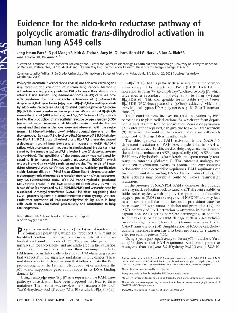
Scheme 1. Metabolic activation of B[a]P-7,8-trans-dihydrodiol by AKRsand ROS formation.
Fig. 1. Metabolic activation of B[a]P-7,8-trans-dihydrodiol to B[a]P-7,8-dione by AKR1C isoforms in A549 cells. (A) [3H]-B[a]P-7,8-trans-dihydrodiol consumptionand B[a]P-7,8-dione formation in A549 cell extracts. (B) Changes in the relative amounts (in nmol) of B[a]P-7,8-trans-dihydrodiol and B[a]P-7,8-dione over 12 hin cell extracts. Mean � SD (n � 3) is shown. (C) APCI-positive ion mass spectral identification of B[a]P-7,8-dione thioether conjugate obtained by using a FinniganTSQ Quantum Ultra AM. MRM for B[a]P-7,8-dione-thioether conjugate detected the following ion transition: m/z � 359 [M�H]�. (D) LC-MS/MRM chromatogramsof organic soluble extracts from intact cells after treatment with B[a]P-7,8-trans-dihydrodiol for 12 h. TIC, total ion current; B[a]PDE, Rt 17.0 min, m/z � 303[M�H]�; B[a]P-tetraols, Rt 27.5 and 32.7 min, m/z � 321 [M�H]�; B[a]P-7,8-dione, 41.0 min, m/z � 283 [M�H]�, B[a]P-7,8-trans-dihydrodiol, 35.0 min, m/z � 269[M�H � H2O]�; uAU, absorbance units at 254 nm.
Park et al. PNAS � May 13, 2008 � vol. 105 � no. 19 � 6847
BIO
CHEM
ISTR
Y
earliest time point taken (2 h) at which point only 15% of theB[a]P-7,8-trans-dihydrodiol was metabolized (Fig. 1 A and B).Both B[a]P-7,8-trans-dihydrodiol and B[a]P-7,8-dione were con-sumed so that by 12 h no organic metabolites were detectedbecause of the formation of water-soluble metabolites.
In intact A549 cells, conversion of 10 �M B[a]P-7,8-trans-dihydrodiol to B[a]P-7,8-dione was observed (Fig. 1D). B[a]P-7,8-trans-dihydrodiol was gradually depleted at 12 h, and the concurrentformation of B[a]P-7,8-dione (M�H m/z � 283) and B[a]P-tetraol-1 (hydrolyzed product of anti-B[a]PDE; M�H m/z � 321) wasobserved, suggesting that P450s were also involved in the metabolicactivation of B[a]P-7,8-trans-dihydrodiol in A549 cells.
These results agree with our previous work in which 7,12-dimethylbenz[a]athracene-3,4-dihydrodiol was converted to 7,12-dimethylbenz[a]athracene-3,4-dione in A549 lung cell extracts (20),and B[a]P-7,8-trans-dihydrodiol was converted to B[a]P-7,8-dionein H358 cells stably transfected with AKR1A1 (19).
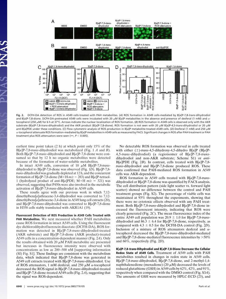
Fluorescent Detection of ROS Production in A549 Cells Treated withPAH Metabolites. We next measured whether PAH metabolitescause ROS formation in intact A549 cells. Using the fluorescentdye dichlorodihydrofluorescin diacetate (DCFH-DA), ROS for-mation was detected in B[a]P-7,8-trans-dihydrodiol-treated(AKR substrate) and B[a]P-7,8-dione (AKR product)-treatedA549 cells in a concentration-dependent manner (Fig. 2A). Onlythe results obtained with 20 �M PAH metabolite are presentedbut increases in fluorescence intensity were observed withconcentrations as low as 200–500 nM [supporting information(SI) Fig. S1]. These findings are consistent with the metabolismdata, which indicated that B[a]P-7,8-dione was generated inA549 cell extracts treated with B[a]P-7,8-trans-dihydrodiol. Useof ROS attenuators, 1 mM desferal, and 250 �M �-tocopheroldecreased the ROS signal in B[a]P-7,8-trans-dihydrodiol-treatedand B[a]P-7,8-dione-treated A549 cells (Fig. 2 A), suggesting thatthe signal was ROS-dependent.
No detectable ROS formation was observed in cells treatedwith either (�)-trans-4,5-dihydroxy-4,5-dihydro B[a]P (B[a]P-4,5-trans-dihydrodiol) (a regioisomer of B[a]P-7,8-trans-dihydrodiol and non-AKR substrate; Scheme S1) or anti-B[a]PDE (Fig. 2B). In contrast, cells treated with B[a]P-7,8-trans-dihydrodiol and B[a]P-7,8-dione produced ROS. Thesedata confirmed that PAH-mediated ROS formation in A549cells was AKR-dependent.
ROS formation in A549 cells treated with B[a]P-7,8-trans-dihydrodiol or B[a]P-7,8-dione was quantified by FACS analysis.The cell distribution pattern (side light scatter vs. forward lightscatter) showed no difference between the control and PAHtreatment groups (Fig. S2). The percentage of viable cells wasmaintained at 93% throughout the experiments, showing thatthere were no cytotoxic effects observed with any PAH treat-ment. Both B[a]P-7,8-trans-dihydrodiol and B[a]P-7,8-dione in-creased the fluorescent intensity, indicating that ROS wereclearly generated (Fig. 2C). The mean fluorescence index of theentire A549 cell population was 20.9 � 1.0 for B[a]P-7,8-trans-dihydrodiol and 86.3 � 6.4 for B[a]P-7,8-dione, respectively, ascompared with 4.3 � 0.5 for the DCFH-DA control (Fig. 2D).Inclusion of a mixture of ROS attenuators desferal and �-tocopherol decreased the B[a]P-7,8-trans-dihydrodiol-mediatedand B[a]P-7,8-dione-mediated fluorescence intensities by �50%and 66%, respectively (Fig. 2D).
B[a]P-7,8-trans-Dihydrodiol and B[a]P-7,8-Dione Decrease the CellularRedox State of A549 Cells. Treatment of A549 cells with PAHmetabolites resulted in changes in redox state in A549 cells.B[a]P-7,8-trans-dihydrodiol, B[a]P-7,8-dione, and 2-methyl-1,4-naphthalenedione (menadione) (20 �M) decreased the levels ofreduced glutathione (GSH) in A549 cells by 62%, 42%, and 91%,respectively when compared with the DMSO control (Fig. S3A).The amounts of GSH were measured by HPLC-ECD (23), and
Fig. 2. DCFH-DA detection of ROS in A549 cells-treated with PAH metabolites. (A) ROS formation in A549 cells-mediated by B[a]P-7,8-trans-dihydrodioland B[a]P-7,8-dione. DCFH-DA-pretreated A549 cells were incubated with 20 �M B[a]P metabolites in the absence and presence of desferal (1 mM) and �-tocopherol (250 �M) for 6 h at 37°C. Arrows indicate the nuclear localization of ROS formation. (B) ROS formation in A549 cells is observed only with the AKRsubstrate (B[a]P-7,8-trans-dihydrodiol) and the AKR product (B[a]P-7,8-dione). ROS formation is not seen with 20 �M B[a]P-4,5-trans-dihydrodiol or 20 �Manti-B[a]PDE under these conditions. (C) Flow cytometric analysis of ROS production in B[a]P metabolite-treated A549 cells. (D) Desferal (1 mM) and 250 �M�-tocopherol attenuate ROS formation-mediated by B[a]P metabolites in A549 cells as measured by FACS. Significant changes in ROS after PAH treatment or PAHtreatment plus ROS attenuators were seen (**, P � 0.005).
6848 � www.pnas.org�cgi�doi�10.1073�pnas.0802776105 Park et al.
the assays were validated by spiking with known amounts of theanalyte, where recovery was �95%.
Identical treatments with B[a]P-7,8-trans-dihydrodiol, B[a]P-7,8-dione, and menadione increased in the intracellular NADP�/NADPH ratio in A549 cells. B[a]P-7,8-trans-dihydrodiol, B[a]P-7,8-dione, and menadione increased the NADP�/NADPH ratio by3-, 3-, and 8-fold, respectively when compared with the DMSOcontrol (Fig. S3B). The amounts of NADP� and NADPH weremeasured by enzymatic cycling after destruction of the oxidized orreduced pyridine nucleotide (24). The assays were validated byspiking known amounts of the analyte where recovery was �95%.Thus, prolonged exposure with both B[a]P-7,8-trans-dihydrodioland B[a]P-7,8-dione resulted in a prooxidant state in A549 cells.
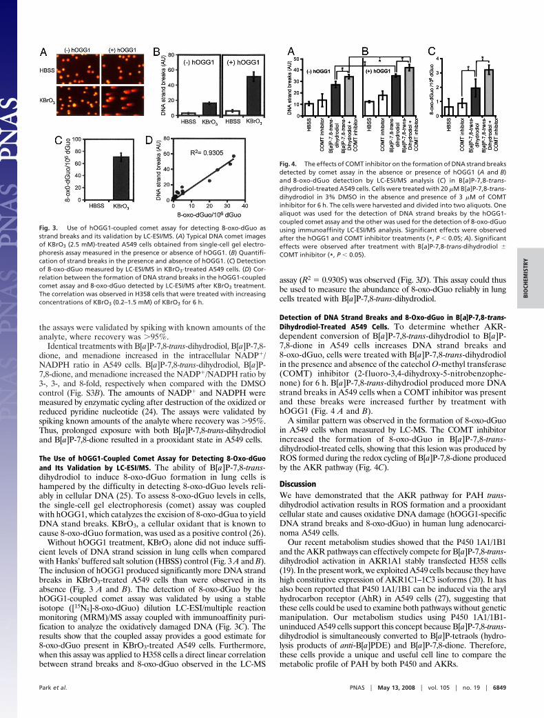
The Use of hOGG1-Coupled Comet Assay for Detecting 8-Oxo-dGuoand Its Validation by LC-ESI/MS. The ability of B[a]P-7,8-trans-dihydrodiol to induce 8-oxo-dGuo formation in lung cells ishampered by the difficulty in detecting 8-oxo-dGuo levels reli-ably in cellular DNA (25). To assess 8-oxo-dGuo levels in cells,the single-cell gel electrophoresis (comet) assay was coupledwith hOGG1, which catalyzes the excision of 8-oxo-dGua to yieldDNA stand breaks. KBrO3, a cellular oxidant that is known tocause 8-oxo-dGuo formation, was used as a positive control (26).
Without hOGG1 treatment, KBrO3 alone did not induce suffi-cient levels of DNA strand scission in lung cells when comparedwith Hanks’ buffered salt solution (HBSS) control (Fig. 3 A and B).The inclusion of hOGG1 produced significantly more DNA strandbreaks in KBrO3-treated A549 cells than were observed in itsabsence (Fig. 3 A and B). The detection of 8-oxo-dGuo by thehOGG1-coupled comet assay was validated by using a stableisotope ([15N5]-8-oxo-dGuo) dilution LC-ESI/multiple reactionmonitoring (MRM)/MS assay coupled with immunoaffinity puri-fication to analyze the oxidatively damaged DNA (Fig. 3C). Theresults show that the coupled assay provides a good estimate for8-oxo-dGuo present in KBrO3-treated A549 cells. Furthermore,when this assay was applied to H358 cells a direct linear correlationbetween strand breaks and 8-oxo-dGuo observed in the LC-MS
assay (R2 � 0.9305) was observed (Fig. 3D). This assay could thusbe used to measure the abundance of 8-oxo-dGuo reliably in lungcells treated with B[a]P-7,8-trans-dihydrodiol.
Detection of DNA Strand Breaks and 8-Oxo-dGuo in B[a]P-7,8-trans-Dihydrodiol-Treated A549 Cells. To determine whether AKR-dependent conversion of B[a]P-7,8-trans-dihydrodiol to B[a]P-7,8-dione in A549 cells increases DNA strand breaks and8-oxo-dGuo, cells were treated with B[a]P-7,8-trans-dihydrodiolin the presence and absence of the catechol O-methyl transferase(COMT) inhibitor (2-f luoro-3,4-dihydroxy-5-nitrobenzophe-none) for 6 h. B[a]P-7,8-trans-dihydrodiol produced more DNAstrand breaks in A549 cells when a COMT inhibitor was presentand these breaks were increased further by treatment withhOGG1 (Fig. 4 A and B).
A similar pattern was observed in the formation of 8-oxo-dGuoin A549 cells when measured by LC-MS. The COMT inhibitorincreased the formation of 8-oxo-dGuo in B[a]P-7,8-trans-dihydrodiol-treated cells, showing that this lesion was produced byROS formed during the redox cycling of B[a]P-7,8-dione producedby the AKR pathway (Fig. 4C).
DiscussionWe have demonstrated that the AKR pathway for PAH trans-dihydrodiol activation results in ROS formation and a prooxidantcellular state and causes oxidative DNA damage (hOGG1-specificDNA strand breaks and 8-oxo-dGuo) in human lung adenocarci-noma A549 cells.
Our recent metabolism studies showed that the P450 1A1/1B1and the AKR pathways can effectively compete for B[a]P-7,8-trans-dihydrodiol activation in AKR1A1 stably transfected H358 cells(19). In the present work, we exploited A549 cells because they havehigh constitutive expression of AKR1C1–1C3 isoforms (20). It hasalso been reported that P450 1A1/1B1 can be induced via the arylhydrocarbon receptor (AhR) in A549 cells (27), suggesting thatthese cells could be used to examine both pathways without geneticmanipulation. Our metabolism studies using P450 1A1/1B1-uninduced A549 cells support this concept because B[a]P-7,8-trans-dihydrodiol is simultaneously converted to B[a]P-tetraols (hydro-lysis products of anti-B[a]PDE) and B[a]P-7,8-dione. Therefore,these cells provide a unique and useful cell line to compare themetabolic profile of PAH by both P450 and AKRs.
Fig. 3. Use of hOGG1-coupled comet assay for detecting 8-oxo-dGuo asstrand breaks and its validation by LC-ESI/MS. (A) Typical DNA comet imagesof KBrO3 (2.5 mM)-treated A549 cells obtained from single-cell gel electro-phoresis assay measured in the presence or absence of hOGG1. (B) Quantifi-cation of strand breaks in the presence and absence of hOGG1. (C) Detectionof 8-oxo-dGuo measured by LC-ESI/MS in KBrO3-treated A549 cells. (D) Cor-relation between the formation of DNA strand breaks in the hOGG1-coupledcomet assay and 8-oxo-dGuo detected by LC-ESI/MS after KBrO3 treatment.The correlation was observed in H358 cells that were treated with increasingconcentrations of KBrO3 (0.2–1.5 mM) of KBrO3 for 6 h.
Fig. 4. The effects of COMT inhibitor on the formation of DNA strand breaksdetected by comet assay in the absence or presence of hOGG1 (A and B)and 8-oxo-dGuo detection by LC-ESI/MS analysis (C) in B[a]P-7,8-trans-dihydrodiol-treated A549 cells. Cells were treated with 20 �M B[a]P-7,8-trans-dihydrodiol in 3% DMSO in the absence and presence of 3 �M of COMTinhibitor for 6 h. The cells were harvested and divided into two aliquots. Onealiquot was used for the detection of DNA strand breaks by the hOGG1-coupled comet assay and the other was used for the detection of 8-oxo-dGuousing immunoaffinity LC-ESI/MS analysis. Significant effects were observedafter the hOGG1 and COMT inhibitor treatments (*, P � 0.05; A). Significanteffects were observed after treatment with B[a]P-7,8-trans-dihydrodiol �COMT inhibitor (*, P � 0.05).
Park et al. PNAS � May 13, 2008 � vol. 105 � no. 19 � 6849
BIO
CHEM
ISTR
Y
We find that B[a]P-7,8-trans-dihydrodiol (AKR substrate) andB[a]P-7,8-dione (AKR product) generate ROS in situ in A549 cellspretreated with DCFH-DA, and this effect is suppressed by ROSattenuators (Fig. 2A). Because ROS formation is not observed inA549 cells treated with either a regioisomer of B[a]P-7,8-trans-dihydrodiol (B[a]P-4,5-trans-dihydrodiol) or a product of furtherP450 metabolism (anti-B[a]PDE), PAH-metabolite induced ROSformation is solely AKR-dependent (Fig. 2B). The fluorescentsignal was also nuclear in localization, indicative of a transportmechanism for B[a]P-7,8-dione (Fig. 2 A and B and Fig. S1, arrow).We have reported that this PAH o-quinone acts as a ligand for theAhR (28). These findings suggest that ROS-mediated DNA dam-age by PAH o-quinones may depend on transport by the AhR.
Treatment of A549 cells with B[a]P-7,8-trans-dihydrodiol orB[a]P-7,8-dione caused a decrease in GSH levels with a con-comitant increase in the intracellular NADP�/NADPH ratio(Fig. S3). Interestingly, the bolus addition of B[a]P-7,8-dionealso caused a decrease in oxidized glutathione, which likelyreflects GSH-conjugate formation. The B[a]P-7,8-dione-GSHconjugate is redox-active in its own right (T.M.P., unpublishedobservation). Thus the changes in the NADP�/NADPH ratio arelikely associated with PAH metabolite-mediated ROS forma-tion, the elimination of peroxides by the GSH peroxidase system,and depletion of NADPH. Similar changes in redox state havebeen observed in V79 cells after treatment with p-quinones (29).
PAH o-quinones produced by AKRs cause oxidative DNAdamage in the form of 8-oxo-dGuo in vitro (30). However,whether 8-oxo-dGuo could be detected reliably as a result ofPAH activation by AKRs in intact cells was unknown. To test thispossibility, hOGG1 was coupled to the comet assay technique(26). hOGG1 is a base excision repair enzyme, which excises8-oxo-Gua from DNA (31). This reaction results in the forma-tion of AP sites that through a �-elimination process subse-quently causes overt strand breaks in the DNA. The hOGG1-coupled comet assay made it possible to detect 8-oxo-dGuo asDNA strand breaks in human lung A549 cells after PAHtreatment. However, because hOGG1 can recognize 2,6-diamino-4-hydroxy-5-formamidopyrimidine (Fapy-Gua) thatcan be formed by ROS, caution was required in measuring8-oxo-dGuo, especially when using the hOGG1-coupled cometassay alone. Our LC-MS data provides confidence that thehOGG1-coupled comet assay is a semiquantitative method todetect 8-oxo-dGuo in lung cells (Fig. 3).
The hOGG1-coupled comet assay showed that B[a]P-7,8-trans-dihydrodiol generated overt strand breaks in A549 cell DNA.Furthermore, the fact that the COMT inhibitor amplified B[a]P-7,8-trans-dihydrodiol-mediated DNA strand breaks and 8-oxo-dGuo formation in the cellular DNA indicates that these eventsoccur as result of AKR-dependent conversion of B[a]P-7,8-trans-dihydrodiol to B[a]P-7,8-dione and its subsequent redox cyclingback to the catechol (Fig. 4). These findings suggest that AKR-dependent PAH activation can induce ROS and the ROS formedcauses oxidative DNA damage in the form of 8-oxo-dGuo in humanlung cells.
Although the formation of covalent PAH-DNA adducts has beenextensively studied as a known mechanism for PAH carcinogenesis(2, 3, 6), our study suggests that formation of PAH-mediatedoxidative DNA damage may also contribute to carcinogenesis.PAHs have previously been reported to cause oxidative DNAdamage in vitro and in vivo when using methods of varying sensi-tivity and specificity (32, 33). Base modifications such as thymineglycol, etheno adducts, and 8-oxo-dGuo all have been detectedupon PAH exposure (34). However, the mechanism by which PAHcan cause oxidative DNA damage has been uncertain. Our resultsshow that AKR-dependent PAH activation can account for theoxidative DNA damage caused by parent PAHs and PAH trans-dihydrodiols. In addition, our in vitro p53 mutagenesis studiesshowed that PAH o-quinones produced by AKRs generate 8-oxo-
dGuo and cause G-to-T transversions in p53 cDNA, and that theseeffects were abolished by ROS scavengers (16, 17), suggesting adirect relationship between oxidative DNA damage and the ob-served mutational pattern in p53.
Interestingly, the loss of heterozygosity of hOGG1 has beenassociated with the development of lung cancer (35). Polymor-phism in the hOGG1 gene locus and loss of heterozygosity bothincrease lung cancer susceptibility (36). Patients exhibiting lossof heterozygosity of hOGG1 gene had high levels of 8-oxo-dGuoin their DNA (37). In addition, hOGG1 activity was decreasedin peripheral blood monocytes of patients with NSCLC (38), and8-oxo-dGuo levels were significantly higher in leucocytes of lungcancer patients and healthy smokers when compared withhealthy nonsmokers (39). This finding suggests that during lungcancer development, cells may have an increased mutationalload because of the inability to repair 8-oxo-dGuo.
Clinical and epidemiological studies support the concept thatAKR isoforms, which lead to oxidative DNA damage, can contrib-ute to the initiation of lung cancer (21, 22). Moreover, AKRexpression in human oral squamous cell carcinoma has beenobserved after areca-quid chewing in combination with smoking(40). Exposure of human buccal cells to 1-hydroxychavicol (a majoringredient of areca-quid) induced AKR1C1, and subsequent treat-ment with B[a]P caused a decrease in bulky stable adducts asmeasured by [32P]-postlabeling together with a concomitant in-crease in 8-oxo-dGuo as measured by HPLC-ECD. Unfortunately,the analytical methods used in these studies had questionablespecificity.
In summary, our data show that oxidation of B[a]P-7,8-trans-dihydrodiol to B[a]P-7,8-dione by AKRs results in ROS generation,a prooxidant cellular state, and oxidative DNA damage in humanlung A549 adenocarcinoma cells. These results provide strongevidence that AKR-dependent PAH activation and the resultantROS formation could contribute to PAH-mediated lung mutagen-esis and carcinogenesis.
Materials and MethodsPAH Metabolites. (�)[1,3-3H]-B[a]P-7,8-trans-dihydrodiol (specific activity1,170 cpm/nmol, � 98% pure by HPLC), (�)B[a]P-7,8-trans-dihydrodiol,(�)B[a]P-4,5-trans-dihydrodiol, (�)B[a]P-7,8-dione, and (�)anti-B[a]PDE werepurchased from the National Cancer Institute Chemical Carcinogen StandardReference Repository (Midwest Research Institute, Kansas City) or synthesizedaccording to published methods (41).
Cells and PAH Treatment. A549 human lung adenocarcinoma cells were ob-tained from the American Type Culture Collection (ATCC no. CCL-185) andcultured as recommended. The cells were treated with PAH metabolites asfollows. Ninety to 100% confluent cells were washed with HBSS buffer con-taining Mg2� and Ca2� and treated with the same HBSS buffer containing0–20 �M of B[a]P-7,8-trans-dihydrodiol, B[a]P-7.8-dione, B[a]P-4,5-trans-dihydrodiol, or anti-B[a]PDE in 2% DMSO.
Oxidation of B[a]P-7,8-trans-Dihydrodiol to B[a]P-7,8-Dione by AKR1C Isoformsin A549 Cell Extracts and Intact Cells. Chromatographic analysis, separation,and quantification of PAH metabolites in vitro and in vivo were achieved asdescribed (18, 19). A detailed description of the PAH metabolism experimentsis found in SI Text.
Detection of Intracellular ROS Produced by PAH Metabolites. Formation ofintracellular ROS in PAH-treated A549 cells was measured with DCFH-DA dye.Formation of intracellular ROS was also measured by a fluorescence-activatedcell sorter (FACS-calibur; Becton-Dickinson) equipped with an argon laser,yielding a 488-nm primary emission line. Detailed descriptions of these meth-ods can be found in SI Text.
Measurement of Intracellular GSH. A549 cells (1 � 107 cells) were homogenizedin 200 mM methane sulfonic acid containing 5 mM diethylenetriamine penta-acetic acid by sonication. Intracellular GSH was measured in the homogenates
6850 � www.pnas.org�cgi�doi�10.1073�pnas.0802776105 Park et al.
by HPLC-ECD assay with an ESA Coularray detector (23). A detailed descriptionof the measurement of reduced GSH can be found in SI Text.
Measurement of Intracellular Reduced and Oxidized Pyridine Nucleotides[NADP(H)]. Intracellular amounts of NADPH and NADP� were measured in celllysates spectrophotometrically after destruction of either the contaminatingoxidized or reduced cofactors, respectively, followed by enzymatic cycling(24). Detailed descriptions for the quantification of the oxidized and reducedpyridine nucleotide cofactors can be found in SI Text.
Measurement of DNA Strand Breaks in PAH-Treated A549 Cells. DNA strandbreaks in PAH-treated A549 cells were measured by using a modified cometassay technique as described (26). Detailed descriptions of this assay and thequantification of DNA strand breaks can be found in SI Text.
Quantification of 8-Oxo-dGuo by LC-ESI/MRM/MS in PAH-Treated A549 Cells.Genomic DNA was extracted from PAH-treated cells by using DNAzol BD(Invitrogen). The DNA was quantitatively digested by the addition of DNase I,phosphodiesterase I (from Crotalus adamanteus venom), and shrimp alkalinephosphatase and spiked with an internal standard [15N5]-8-oxo-dGuo. TheDNA samples were divided into two aliquots. One aliquot was used for theisolation and quantification of 8-oxo-dGuo via immunoaffinity purificationstable isotope dilution LC-MRM/MS analysis. The other aliquot was used forDNA base analysis. Detailed descriptions of 8-oxo-dGuo detection and baseanalysis can be found in SI Text.
ACKNOWLEDGMENTS. This work was supported by National Institutes ofHealth Grants R01CA-39504, R01ES015857, and P30ES013508 (to T.M.P.) andR25CA101871 and R01CA130038 (to I.A.B.).
1. Grimmer G, Bohnke H (1975) Polycyclic aromatic hydrocarbon profile analysis ofhigh-protein foods, oils, and fats by gas chromatography. J Assoc Off Anal Chem58:725–733.
2. Xue W, Warshawsky D (2005) Metabolic activation of polycyclic and heterocyclic aromatichydrocarbons and DNA damage: A review. Toxicol Appl Pharmacol 206:73–93.
3. Hecht SS (1999) Tobacco smoke carcinogens and lung cancer. J Natl Cancer Inst91:1194–1210.
4. Marshall CJ, Vousden KH, Phillips DH (1984) Activation of c-Ha-ras-1 protooncogene byin vitro modification with a chemical carcinogen, benzo(a)pyrene diol-epoxide. Nature310:586–589.
5. Denissenko MF, Pao A, Tang M, Pfeifer GP (1996) Preferential formation of benzo-[a]pyrene adducts at lung cancer mutational hotspots in P53. Science 274:430–432.
6. Conney AH (1982) Induction of microsomal enzymes by foreign chemicals and carci-nogenesis by polycyclic aromatic hydrocarbons: G. H. A. Clowes Memorial Lecture.Cancer Res 42:4875–4917.
7. Zhang Y, et al. (2000) Error-prone lesion bypass by human DNA polymerase eta. NucleicAcids Res 28:4717–4724.
8. Cavalieri EL, Rogan EG (1995) Central role of radical cations in metabolic activation ofpolycyclic aromatic hydrocarbons. Xenobiotica 25:677–688.
9. Sagher D, Strauss B (1985) Abasic sites from cytosine as termination signals for DNAsynthesis. Nucleic Acids Res 13:4285–4298.
10. Penning TM, et al. (1999) Dihydrodiol dehydrogenases and polycyclic aromatic hydro-carbon activation: Generation of reactive and redox active o-quinones. Chem ResToxicol 12:1–18.
11. Balu N, et al. (2004) Identification and characterization of novel stable deoxyguanosineand deoxyadenosine adducts of benzo[a]pyrene-7,8-quinone from reactions at phys-iological pH. Chem Res Toxicol 17:827–838.
12. McCoull KD, Rindgen D, Blair IA, Penning TM (1999) Synthesis and characterization ofpolycyclic aromatic hydrocarbon o-quinone depurinating N7-guanine adducts. ChemRes Toxicol 12:237–246.
13. Cerutti PA (1985) Prooxidant states and tumor promotion. Science 227:375–381.14. Cheng KC, et al. (1992) 8-Hydroxyguanine, an abundant form of oxidative DNA
damage, causes G-T and A-C substitutions. J Biol Chem 267:166–172.15. Bolton JL, et al. (2000) Role of quinones in toxicology. Chem Res Toxicol 13:135–160.16. Yu D, Berlin JA, Penning TM, Field J (2002) Reactive oxygen species generated by PAH
o-quinones cause change-in-function mutations in p53. Chem Res Toxicol 15:832–842.17. Park J-H, et al. (2008) The pattern of P53 mutation caused by PAH o-quinones is driven
by 8-oxo-dGuo formation while the spectrum of mutations is determined by biologicalselection for dominance. Chem Res Toxicol 21, in press.
18. Jiang H, Shen YM, Quinn AM, Penning TM (2005) Competing roles of cytochrome P4501A1/1B1 and aldo-keto reductase 1A1 in the metabolic activation of (�/�)-7,8-dihydroxy-7,8-dihydro-benzopyrene in human bronchoalveolar cell extracts. Chem ResToxicol 18:365–374.
19. Jiang H, Vudathala DK, Blair IA, Penning TM (2006) Competing roles of aldo-ketoreductase 1A1 and cytochrome P4501B1 in benzo[a]pyrene-7,8-diol activation in hu-man bronchoalveolar H358 cells: Role of AKRs in P4501B1 induction. Chem Res Toxicol19:68–78.
20. Palackal NT, et al. (2002) Activation of polycyclic aromatic hydrocarbon trans-dihydrodiol proximate carcinogens by human aldo-keto reductase (AKR1C) enzymesand their functional overexpression in human lung carcinoma (A549) cells. J Biol Chem277:24799–24808.
21. Woenckhaus M, et al. (2006) Smoking and cancer-related gene expression in bronchialepithelium and non-small-cell lung cancers. J Pathol 210:192–204.
22. Fukumoto S, et al. (2005) Overexpression of the aldo-keto reductase family proteinAKR1B10 is highly correlated with smokers’ non-small cell lung carcinomas. Clin CancerRes 11:1776–1785.
23. Lakritz J, Plopper CG, Buckpitt AR (1997) Validated high-performance liquid chroma-tography-electrochemical method for determination of glutathione and glutathionedisulfide in small tissue samples. Anal Biochem 247:63–68.
24. Lowry OH, Passoneau JV, Rock MK (1961) The measurement of pyridine nucleotides byenzymatic cycling. J Biol Chem 236:2756–2759.
25. European Standards Committee on Oxidative DNA Damage, Gedik CM, Collins A (2005)Establishing the background level of base oxidation in human lymphocyte DNA:Results of an interlaboratory validation study. FASEB J 19:82–84.
26. Smith CC, O’Donovan MR, Martin EA (2006) hOGG1 recognizes oxidative damageusing the comet assay with greater specificity than FPG or ENDOIII. Mutagenesis21:185–190.
27. Foster KA, et al. (1998) Characterization of the A549 cell line as a type II pulmonaryepithelial cell model for drug metabolism. Exp Cell Res 243:359–366.
28. Burczynski ME, Penning TM (2000) Genotoxic polycyclic aromatic hydrocarbon ortho-quinones generated by aldo-keto reductases induce CYP1A1 via nuclear translocationof the aryl hydrocarbon receptor. Cancer Res 60:908–915.
29. Ludewig G, Dogra S, Glatt H (1989) Genotoxicity of 1,4-benzoquinone and 1,4-naphthoquinone in relation to effects on glutathione and NAD(P)H levels in V79 cells.Environ Health Perspect 82:223–228.
30. Park J-H, et al. (2005) Formation of 8-oxo-7,8-dihydro-2�-deoxyguanosine (8-oxo-dGuo) by PAH o-quinones: Involvement of reactive oxygen species and copper(II)/copper(I) redox cycling. Chem Res Toxicol 18:1026–1037.
31. Bjørås M, et al. (1997) Opposite base-dependent reactions of a human base excisionrepair enzyme on DNA containing 7,8-dihydro-8-oxoguanine and abasic sites. EMBO J16:6314–6322.
32. Leadon SA, Stampfer MR, Bartley J (1988) Production of oxidative DNA damage duringthe metabolic activation of benzo[a]pyrene in human mammary epithelial cells cor-relates with cell killing. Proc Natl Acad Sci USA 85:4365–4368.
33. Leadon SA, Sumerel J, Minton TA, Tischler A (1995) Coal tar residues produce both DNAadducts and oxidative DNA damage in human mammary epithelial cells. Carcinogen-esis 16:3021–3026.
34. Kim KB, Lee BM (1997) Oxidative stress to DNA, protein, and antioxidant enzymes(superoxide dismutase and catalase) in rats treated with benzo(a)pyrene. Cancer Lett113:205–212.
35. Chevillard S, et al. (1998) Mutations in OGG1, a gene involved in the repair ofoxidative DNA damage, are found in human lung and kidney tumors. Oncogene16:3083–3086.
36. Wikman H, et al. (2000) hOGG1 polymorphism and loss of heterozygosity (LOH):Significance for lung cancer susceptibility in a Caucasian population. Int J Cancer88:932–937.
37. Hardie LJ, et al. (2000) The effect of hOGG1 and glutathione peroxidase I genotypesand 3p chromosomal loss on 8-hydroxydeoxyguanosine levels in lung cancer. Carcino-genesis 21:167–172.
38. Paz-Elizur T, et al. (2003) DNA repair activity for oxidative damage and risk of lungcancer. J Natl Cancer Inst 95:1312–1319.
39. Gackowski D, et al. (2003) Products of oxidative DNA damage and repair as possiblebiomarkers of susceptibility to lung cancer. Cancer Res 63:4899–4902.
40. Tang DW, Chang KW, Chi CW, Liu TY (2004) Hydroxychavicol modulates benzo-[a]pyrene-induced genotoxicity through induction of dihydrodiol dehydrogenase.Toxicol Lett 152:235–243.
41. Harvey RG, Dai Q, Ran C, Penning TM (2004) Synthesis of the o-quinones and otheroxidized metabolites of polycyclic aromatic hydrocarbons implicated in carcinogenesis.J Org Chem 69:2024–2032.
Park et al. PNAS � May 13, 2008 � vol. 105 � no. 19 � 6851
BIO
CHEM
ISTR
Y


![Keto-enol tautomers and distonic ions: The chemistry of [CnH2nO] radical cations. Part I](https://img.pdfslide.net/doc/110x75/6349002a031992cdcf025401/keto-enol-tautomers-and-distonic-ions-the-chemistry-of-cnh2no-radical-cations.jpg)


