Embed Size (px)
Citation preview
595.754:575.854
MEDEDELINGEN LANDBOUWHOGESCHOOL WAGENINGEN • NEDERLAND • 78-5(1978)
EVOLUTIONARY TRENDS IN HETEROPTERA
PART II. MOUTHPART-STRUCTURES AND FEEDING STRATEGIES
R. H.COBBEN
Laboratory of Entomology, Agricultural University, Wageningen, The Netherlands
(Received 5-V-1977)
H. VEENMAN & ZONEN B V . - W A G E N I N G E N - 1978
Dedicated to Wies and children who obligingly let me prepare most of this work in the family living room.
CONTENTS
INTRODUCTION 5
MATERIALS AND METHODS 6
1. OBSERVATIONS ON STYLET STRUCTURE AND FUNCTION 13 1.1 Gcrromorpha 13 1.2 Nepomorpha 27 1.3 Reduvioidea 39 1.4 Leptopodomorpha 45 1.5 Cimicomorpha s.str 49 1.6 Pentatomomorpha 57 1.7 Thaumastocoroidea 64 1.8 Dipsocoromorpha 64 1.9 Enicocephalomorpha 64
2. OTHER STRUCTURES ASSOCIATED WITH FEEDING 65 2.1 Cross-sections through rostrum and stylets 65 2.2 Rostral structural specializations and function 69 2.3 Internal head structures 75 2.4 Sensory apparatus 87
3. EVOLUTIONARY TRENDS IN STRUCTURES OTHER THAN THOSE ASSOCIATED WITH FEEDING 96 3.1 Egg-systems 96 3.2 Eye of the first larval instar 102 3.3. Pretarsal structures I l l 3.4 Other postembryonic changes 126 3.5 Metasternal scent glands 146 3.6 Trichobothria and other cuticular structures of unknown function 159 3.7 The male intromittent organ 165
4. DISCUSSION 187 4.1 Phylogenetic procedure 187 4.2 Feeding structures and feeding 191 4.3 Food specialization 201 4.4 A tentative, evolutionary scheme for Heteroptera 219 4.5 The relationship between Heteroptera and Homoptera 234
5- SUMMARY AND CONCLUSIONS 244
ACKNOWLEDGEMENTS 255
REFERENCES 257
LEGENDS OF THE PLATES 282
PHOTOGRAPHS 296
AUTHOR INDEX 396
SUBJECT INDEX 401

INTRODUCTION
An explanation is needed for the title and content of this paper, since they are not in accord with an assertion made in my 1968 book. It was then indicated that Part II would deal with the reproduction of Heteroptera, and Part III with miscellaneous evolutionary subjects and a final synthesis. Indeed quite a bit of preparation for the next parts already was done before 1968. However, rapidly changing academic circumstances have made it impossible for me to cope with the original design within a reasonably short time. Furthermore, several investigators in different parts of the world are presently engaged in detailed studies on reproduction on various groups of Heteroptera, dealing in particular with the evolutionary aspects touched in the first part of this series. Therefore, it seems advisable to postpone my publication on reproduction, originally planned as Part II.
There are a number of recent studies on the feeding behaviour and mouthpart structure of phytophagous Hemiptera. Most of these studies have been prompted by the rôle of these insects in economic entomology and epidemic virology, and often sophisticated research techniques have been utilized. However, some authors have failed to consider information previously published in a wide variety of papers on comparative morphology and systematics. The consequence of this failure becomes most apparent in the comments made regarding the evolution of the various structures and functions related to feeding phenomena in the order Hemiptera. Another source of confusion results from the implied or explicitly stated belief that the Homoptera are more generalized (or more symplesiomorphous) than the Heteroptera.
Some authors (SWEET, 1963;GOODCHILD, 1966;SCHLEE, 1969; MILES, 1972) recently have expressed the opinion that the ancestors of the Heteroptera were phytophagous and formed a salivary sheath. In 1968 (p. 376) I expressed the view 'that the archetypical hemipteran was a predominantly carnivorous insect'. That belief was based on the results of studies which were not devoted directly to the feeding phenomenon. The compilation and interpretation of my data on the egg systems resulted in an evolutionary scheme (1968, Fig. 316), which was presented as an hypothesis for future work. The same scheme is reproduced here as Fig. 71 for reference of the reader. The intention of the present study of some restricted aspects of feeding biology, was to see whether new facts, not known to me in 1968, would contribute to or detract from this hypothesis.
In contrast to the level of knowledge regarding phytophagous and haema-tophagous Hemiptera, we are rather poorly informed about the fine structure and function of the stylets of predatory forms. Stress is plac^jn this paper on a remarkable type of food intake, which is characteristic of many, but not all predatory Heteroptera. The evolutionary coherence of the various types encountered will be discussed, and special attention will be given to the bearing °f such information on the ancestral feeding habits of the Hemiptera.
Meded. Landbouwhogeschool Wageningen 78-5 (1978) 5
MATERIALS AND METHODS
A list of taxa of which stylets and associated structures were studied, is given below. The addition SEM, TEM means that structures also were studied with the stereoscanning and/or transmission electron-microscope. Those species for which the origin of the material has not been indicated, were collected in the Netherlands. Since mouthparts are less variable within each family than are the eggs (COBBEN, 1968a), I have studied fewer representatives. The many species, of which structures other than those associated with feeding were studied (Chapter 3), are not listed here.
HETEROPTERA
A. GERROMORPHA*
HEBRIDAE
MESOVELIIDAE
HYDROMETRIDAE
VELIIDAE
Hebrus pusillus Fall. Hebrus rufweps Ths. (SEM, TEM). Mesoveliafurcata Muls. & Rey. Mesovelia mulsanti White (origin Curaçao, Antilles) (SEM). Hydrometra stagnorum L. (SEM, TEM) Bacillometra woytkowskii Hungerf. (origin Peru). Heterocleptes hoberlandti China et al. (origin Angola). Limnobatodes paradoxus Hussey (origin Brazil). including some aberrant genera. Microvelia reticulata Burm. (SEM). Velia caprai Tam. (SEM, TEM). Troclwpus plumbeus Uhler (origin Curaçao, Antilles) (SEM). Veloidea reposita Dr. & Hott. (Honduras) (SEM), Hebrovelia sp. (origin Ivory Coast) (SEM). Macrovelia horni Uhl. (origin Colorado and Oregon, USA) (SEM). Oravelia pege Dr. & Chapm. (origin California, USA) (SEM). Gerris, several spp. (SEM). Aquarius najas De G. (SEM). Cylindrostethus hungerfordi Dr. & C. (origin Surinam).
* STYS & KERZHNER (1975) proposed a consistent nomenclatorial system for the major subdivisions of Heteroptera. I welcome this endeavour and follow their terminology except for a few groups, which I tentatively indicate with the superfamily name: Reduvioidea and Thaumastocoroidea (see discussion on pages 226, 230).
6 Meded. Landbouwhogeschool Wageningen 78-5 (1978)
GERRIDAE
B. NEPOMORPHA
OCHTERIDAE
GELASTOCORIDAE
BELOSTOMATIDAE
NEPIDAE
NAUCORIDAE
POTAMOCORIDAE*
NOTONECTIDAE
PLEIDAE
HELOTREPHIDAE
CORIXIDAE
Ptilomera agriodes Schm. (origin India) (SEM). Potametra berezovskii Bianchi (origin China) (SEM). Halobates princeps White (origin Indonesia).
Ochterus marginatus Latr. (origin Ivory Coast). Ochterus perbosci Guér. (origin Antilles) (SEM). Gelastocoris nebulosus Guér.-Men. (origin Argentina) (SEM). Nerihra laticollis Guér.-Men. (origin New Guinea) Nerthra colaticollis Todd (origin New Guinea). Lethocerus niloticus Stâl (origin Madagascar) (SEM). Ne pa rubra L. Ilyocoris cimicoides L. Aphelocheirus aestivalis F. (origin USSR). Coleopterocoris kleerekoperi Hungf. (origin Brazil). Notonecta glauca L. (TEM). Notonecta obliqua Fall. (SEM). Plea atomaria Pal. (SEM). Idiotrephes chinai Lundbl. (origin Vietnam) (SEM). Micronecta meridionalis Cost. Diaprepocoris zealandiae Hale (origin New Zealand). Cymatia coleoptrata Fabr. Cymatia bonsdorffl Sahlb. Corixa panzeri Fieb. (SEM, TEM). Sigara fossarum Lch. (TEM).
C. REDUVIOIDEA
REDUVIIDAE
Emesinae
Saicinae Holoptilinae Stenopodinae Piratinae
Empicoris vagabundus L. Empicoris culiciformis De G. Schidium callipygum Wyg. (origin Ivory Coast). Gardena pipara McAtee& Malloch (origin Brazil). Emesaya brevipennis Say (origin Antilles) (SEM). Oncerotrachelus acuminatus Say (origin USA). Holoptilus melanospilus Walk, (origin India). Stenopoda wygodzinskyi Giacchi (origin Antilles) (SEM). Rasahus hamatus F. (origin Antilles) (SEM). Pirates hybridus Scop, (origin France) (TEM).
In 1968,1 argued that the genera Potamocoris and Coleopterocoris are too deviant to be retained as a naucorid subfamily. Since then having studied more characters of this group, I am convinced that these taxa should be considered to belong in a separate family.
Meded. Landbouwhogeschool Wageningen 78-5 (1978) 7
Harpactorinae
Sphaeridopinae Triatominae Raphidosominae Ectrichodiinae
Cor anus subapterus De G. Sinea diadema Fabr. (origin USA). Sphaeridops amoenus Amyot & Serville (origin Brazil). Triatoma maculata Erichs, (origin Antilles) (SEM). Raphidosoma sp. (origin Ethiopia) (SEM). Brontostoma discus Burm. (origin Brasil).
D. LEPTOPODOMORPHA
LEPTOPODIDAE Leptopus marmoratus Gz. (origin Italy). Erianotus lanosus Duf. (origin USSR). Valleriola assouanensis Costa (origin Sudan).
LEOTiCHHDAE Leoüchius speluncarum China (origin Malaya). OMANiiDAE Corallocoris marksae Woodw. (origin Australia).
Omania coleoptrata Horv. (origin Red Sea). SALDIDAE Aepophilus bonnairei Sign, (origin France) (SEM).
Chiloxanthus pilosus Fall. Pentacora signoreti Guer. (origin Antilles). Saldula, several spp. (SEM, TEM). Salda lit (oralis L. Salda buenoi McDunn (origin USA). Salda lugubris Say (origin USA) (SEM).
E. CIMICOMORPHA s.str.
MICROPHYSIDAE
PLOKIOPHILIDAE
JOPPEICIDAE
NABIDAE
ANTHOCORIDAE
VELOCIPEDIDAE
PACHYNOMIDAE
CIMICIDAE
MIRIDAE
Loricula pselaphiformis Curt. Loricula elegantula Baerenspr. (SEM). Myrmedobia coleoptrata Fall. Embiophila myersi China (origin Trinidad). Joppeicus paradoxus Puton (laboratory stock from University of Connecticut, origin Egypt) (SEM). Nabis rugosus L. (TEM). Himacerus myrmecoides Costa. Himacerus apterus Fabr. (SEM). Alloeorhynchus chinai Harris (origin West Irian). Arachnocoris trinitatis Berg, (origin Trinidad). Anthocoris nemoralis Fab. (TEM). Orius minutus L. Scotomedes alienus Dist. (origin Sikkim) (SEM). Pachynomus picipes Klug (origin Soedan) (SEM). Cimex lectularius L. (SEM). Isometopus intrusus H.-S. (SEM). Bryocoris pteridis Fall. Fulvius oxycarenoides Reut, (origin USSR). Notostira elongata Geoff. (TEM).
Meded. Landbouwhogeschool Wageningen 78-5 (1978)
TINGIDAE
Lygus pabulinus L. Exolygus rugulipennis Popp. (TEM). Dicyphuspallicomis M.-D. (TEM). Deraeocoris ruber L. (SEM). Deraeocoris olivaceus Fabr. (SEM). Pantilius tunicatus Fabr. Dictyla symphyti Vall. Acalypta carinata Panz.
F. PENTATOMOMORPHA
ARADIDAE
IDIOSTOUDAE THAUMASTELLIDAE PIESMATIDAE BERYTINIDAE
LYGAEIDAE
PYRRHOCORIDAE
COI-OBATHRISTIDAE
COREIDAE
ALYDIDAE
RHOPALIDAE
CYDNIDAE
ACANTHOSOMATIDAE UROSTYLIDAE
PHLOEIDAE
SCUTELLERIDAE
DINIDORIDAE
TESSAROTOMIDAE PENTATOMIDAE
Aradus cinnamomeus Panz. Dysodius lunatus Fabr. (origin S. America). Aneurus laevis F. Trisecus pictus Berg. (origin Australia). Thaumastella aradoides Horv. (origin Sudan) (SEM). Piesma c'mereum C. (origin Antilles). Neides tipularius L. Metatropisrufescens H.S. Geocoris punctipes Say (origin Antilles) (SEM). Henestaris laticeps Curt, (origin France). Scolopostethusdecoratus Hahn (TEM). Oncopeltus fasciatus Dall. (laboratory stock). Spilostethus pandurus Scop, (origin Ethiopia) (SEM). Pyrrhocoris apterus L. Phaenacantha saccharicida Karsch. (origin Indonesia) (SEM). Acantliocoris sp. (origin Ethiopia) (TEM). Coreus marginatus L. Spathocera batatas F. (origin Antilles). Alydus calcaratus L. Myrmus miriformis Fall. Sehirus biguttatus L. Prolobodes giganteus Burm. (origin Paraguay). Macroscytus javanus Mayr. (origin Indonesia). Elasmostethus interstinctus L. Urochela luleovaria Dist. (origin Japan) (SEM). Urostflis woodwardi Scott, (origin Japan). Phloea spec, third larval instar (origin Venezuela) (SEM). Hotea curculionoides H.-S. (origin Indonesia). Poecilocoris latus Dall. (origin Indonesia) (SEM). Coridius brunneus Thunb. (origin Indonesia). Tessarotoma javanica Thunb. (origin Indonesia). Graphosoma linea tum L. Dohcoris baccarum L.
Meded. Landbouwhogeschool Wapeningen 78-5 (1978)
Perillus bioculatus Fab. (laboratory stock) (SEM, TEM). PLATASPIDAE Coptosoma coleoptrata (origin France).
Libyaspis haglundi Mont, (origin Madagascar) (SEM).
G. THAUMASTOCOROIDEA
THAUMASTOCORIDAE Xylastodoris luteolus Barb, (origin Florida, USA) (SEM).
H. DlPSOCOROMORPHA**
CERATOCOMBIDAE
DIPSOCORIDAE
HYPSIPTERYGIDAE
SCHIZOPTER1DAE
Ceratocombus coleoptratus Zett. Trichotoncmnus dundo Wyg. (origin Soedan). Pachycoleus waltli Fieb. (SEM). Hypsipteryx machadoi Drake (origin Angola). Schizoplera slricklandi China (origin Trinidad) (SEM). Hypselosoma hiroshimai Esaki & Miyam. (origin Japan).
I. ENICOCEPHALOMORPHA
ENICOCEPHALIDAE
J. COLEORRHYNCHA
PEl.ORIDIIDAE
Oncylocotis curculio Karsh (origin Ethiopia) (SEM). Embolorrhinus tuberculatus Bgr. (origin Sudan).
Hemiodoecus veitchi Hack, (origin Australia). Peloridium hammoniorum Bredd. (origin Chile) (SEM). Xenophytes cascus Bergr. (origin Chile).
K . HOMOPTERA AUCHENORRHYNCHA
TETTIGOMETRIDAE
CIXIIDAE
DELPHACIDAE
CERCOPIDAE
MEMBRACIDAE
LEDRIDAE
CICADIDAE
Tettigometra virescens Panz. (origin France) (SEM). Cixius nervosus L. (SEM). Muellerianella fairmairei Perris (SEM). Philaenus spumarius L. Cercopis vulnerata Rossi Aphrophora alnx Fall. (SEM). Gargara genistae Fabr. (SEM). Ledra aurita L. (SEM). Quesadagigas Oliv, (origin Brazil) (SEM).
L . HOMOPTERA STERNORRHYNCHA
APHIDIDAE Myzus persicae Sulz.
** According to EMSLEY (1969), Cryptostemmatidae should have priority over the commonly used name: Dipsocoridae. His reasoning was refuted by STYS ( 1970C) whose new family subdivision is followed here.
10 Meckel. Landbouwhogeschool Wageningen 78-5 (1978)
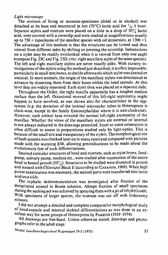
Light microscopy The rostrum of living or museum-specimens (dried or in alcohol) was
detached at its base and macerated in hot (70°C) lactic acid for 1l2-\ hour. Separate stylets and rostrum were placed on a slide in a drop of 50% lactic acid, were covered with a coverslip and were studied at magnifications usually up to 750 x (specimens of the smallest species with oil immersion, at 1200 x). The advantage of this medium is that the structures can be turned and thus viewed from different sides by shifting or pressing the coverslip. Indentations on a stylet may be readily overlooked when it is viewed from only one angle (compare Fig. 23C and Fig. 23D, rmx : right maxillary stylet of the same species). The left and right maxillary stylets are never exactly alike. With cursory investigations of the stylets using the method just described, it is often impossible, particularly in small specimens, to decide afterwards which stylet was dextral or sinistral. In most animals, the origin of the maxillary stylets was determined in advance by dissecting them from their bases within the head capsule. At this level they are widely separated. Each stylet then was placed on a separate slide.
Throughout the Order, the right maxilla apparently has a rougher median surface than the left. Occasional reversal of this left-right asymmetry might happen to have involved, as was shown also for characteristics in the egg-system (e.g. the deviation of the internal micropylar tubes in Heteroptera is clock-wise, except in the family Enicocephalidae where it is anti-clockwise). However, such critical taxa revealed the normal left-right asymmetry of the maxillae. Whether the views of the maxillary stylets are external or internal is not always indicated in the drawings presented. Inner or outer orientation is often difficult to assess in preparations studied only by light-optics. This is because of the small size and transparency of the stylets. The morphological site of barb systems was checked later on in many cases and compared with pictures made with the scanning EM, allowing generalizations to be made about the evolutionary fate of such differentiations.
Internal cuticular structures of head and rostrum, such as stylet levers, food-pump, salivary pump, tendons etc., were studied after maceration of the entire head in heated potash (10'%). Structures to be studied were dissected in potash and stained with Chlorazol Black E (according to CARA YON, 1969). When high power examination was necessary, the stained parts were transferred into lactic acid on a slide.
The cephalic skeletomusculature was investigated after fixation of the decapitated animal in Bouin solution. Abrupt fixation of small specimens during the sucking act was achieved by spraying them with a jet of ethylchloride. With specimens of larger species, the rostrum was cut off with iridectomy scissors.
I did not attempt a detailed and complete comparative morphological study of head-capsule and internal skeletal differentiations as was done in an excellent way for some groups of Heteroptera by PARSONS (1959-1974).
AH drawings are free-hand. Unless otherwise stated, drawings and photographs refer to the adult stage.
Metled. Landbouwhogeschool U'ageningen 78-5 (1978) 11
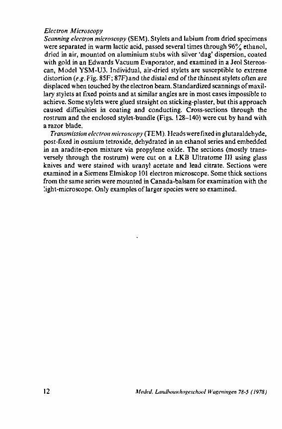
Electron Microscopy Scanning electron microscopy (SEM). Stylets and labium from dried specimens were separated in warm lactic acid, passed several times through 96% ethanol, dried in air, mounted on aluminium stubs with silver 'dag' dispersion, coated with gold in an Edwards Vacuum Evaporator, and examined in a Jeol Stereos-can, Model YSM-U3. Individual, air-dried stylets are susceptible to extreme distortion (e.g. Fig. 85F ; 87F) and the distal end of the thinnest stylets often are displaced when touched by the electron beam. Standardized scannings of maxillary stylets at fixed points and at similar angles are in most cases impossible to achieve. Some stylets were glued straight on sticking-plaster, but this approach caused difficulties in coating and conducting. Cross-sections through the rostrum and the enclosed stylet-bundle (Figs. 128-140) were cut by hand with a razor blade.
Transmission electron microscopy (TEM ). Heads were fixed in glutaraldehyde, post-fixed in osmium tetroxide, dehydrated in an ethanol series and embedded in an aradite-epon mixture via propylene oxide. The sections (mostly transversely through the rostrum) were cut on a LKB Ultratome III using glass knives and were stained with uranyl acetate and lead citrate. Sections were examined in a Siemens Elmiskop 101 electron microscope. Some thick sections from the same series were mounted in Canada-balsam for examination with the !ight-microscope. Only examples of larger species were so examined.
12 Mecled. Landbouwhogeschool Wageningen 78-5 (1978)
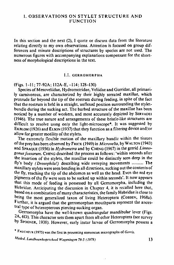
1. OBSERVATIONS ON STYLET STRUCTURE AND FUNCTION
In this section and the next (2), I quote or discuss data from the literature relating directly to my own observations. Attention is focused on group differences and minute descriptions of structures by species are not used. The numerous figures with accompanying explanations compensate for the shortness of morphological descriptions in the text.
1.1. GERROMORPHA
(Figs. 1-11; 77-92A; 112A-E.-114; 128-130) Species of Mesoveliidae, Hydrometridae, Veliidae and Gerridae, all primari
ly carnivorous, are characterized by their highly serrated maxillae, which protrude far beyond the tip of the rostrum during feeding, in spite of the fact that the rostrum is held in a straight, unflexed position surrounding the stylet-bundle during the sucking act. The barbed structure of the maxillae has been noticed by a number of workers, and most accurately depicted by SERVADEI (1946). The true nature and arrangements of these bristle-like structures are difficult to resolve using only the light-microscope*. It was suggested by EKBLOM (1926) and ELSON (1937) that they function as a filtering device and/or allow for greater motility of the stylets.
The extremely flexible motion of the maxillary bundle within the tissues of the prey has been observed by FRICK (1949) in Microvelia, by WALTON (1943) and SPRAQUE (1956) in Hydrometra and by CHENG (1967) in the gerrid Limno-gonus fossarum. CHENG described the process as follows: 'within seconds after the insertion of the stylets, the maxillae could be distinctly seen deep in the fy's body (Drosophila!) describing wide sweeping movements The maxillary stylets were seen bending in all directions, sucking out the contents of the fly, reaching the tip of the abdomen as well as the head. Even the red eye pigments of the fly were seen to be sucked up within seconds'. It now appears that this mode of feeding is possessed by all Gerromorpha, including the Hebridae. Anticipating the discussion in Chapter 4, it is recalled here that, based on a combination of many characteristics, the family Hebridae is close to being the most generalized taxon of living Heteroptera (COBBEN, 1968a). Further, it is argued that the gerromorphan mouthparts represent the ancestral type of heteropterous piercing-sucking organ.
Gerromorpha have the well-known quadrangular mandibular lever (Figs. 2A, 6D). This character sets them apart from all other Heteroptera (see survey by SPOONER, 1938). However, early instar larvae of Gerromorpha possess a
FAUCHEUX (1975) was the first in presenting stereoscan micrographs of Gt-rris.
Meckel. Landbouwhogeschool Wapeningen 78-5 (1978) 13
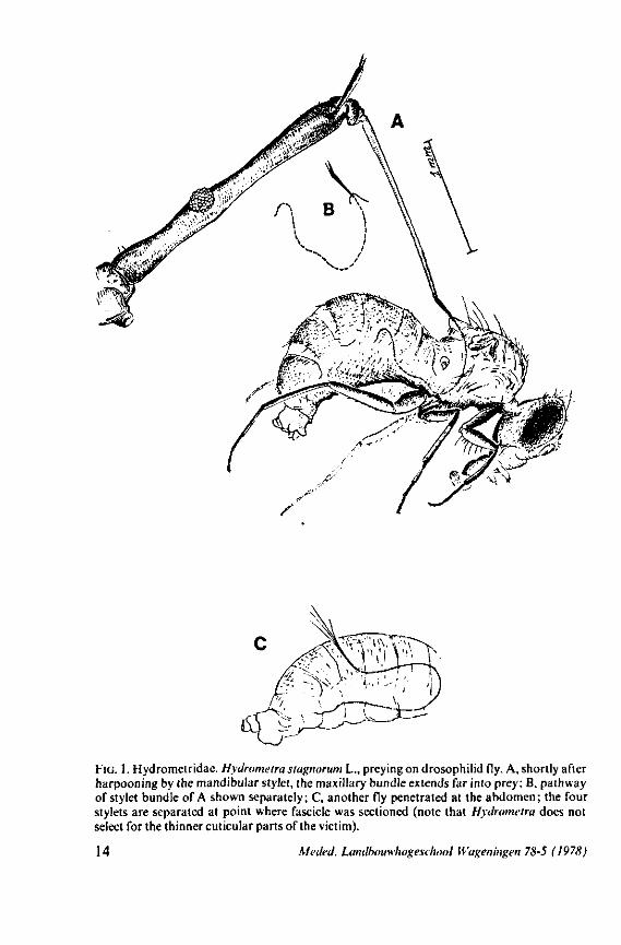
FIG. 1. Hydrometridae. Hydrometra stagnorum L., preying on drosophilid fly. A, shortly after harpooning by the mandibular stylet, the maxillary bundle extends far into prey; B, pathway of stylet bundle of A shown separately; C, another fly penetrated at the abdomen; the four stylets are separated at point where fascicle was sectioned (note that Hydrometra does not select for the thinner cuticular parts of the victim).
14 Meded. Landbouwhogeschool Wageningen 78-5 (1978)
triangular-shaped lever (Figs. 6A, 8B), whereas the maxillary stylets of first instar larvae in this group are similarly barbed as in later larval stages and in the adult. Thus, these ontogenetic specializations of the mandibular lever mask somewhat the primitive status of the sucking mouthparts.
Careful observations on feeding behaviour of Hebrus, Hydrometra, and Microvelia, supplemented by data obtained from transmission and scanning electron microscopy, strongly suggest that the gerromorphan maxillary stylets allow rapid rasping of prey tissue by a non-discriminatory, drilling-filing process. The sharp barbs of the mandibles (Figs. 7C, 82E) are held just beneath the cuticle of the prey, and the maxillary tube is forced with great speed, deep into the host (Fig. 1). There is no indication of salivation external to the host's cuticle and there is no salivary sheath formed inside. Immediately after mandibular harpooning, there must be considerable discharge and extensive spreading of toxic saliva, judging from the rapid paralysis of the prey. Hungry Hydrometra adults immobilize living, short-winged Drosophila flies within a few seconds, irrespective of whether the victim is attacked at the tip of its abdomen or its proboscis; Musca flies (wings and legs removed) are paralyzed often within 10 seconds. Abundant fluid is extruded from the maxillae when gerromorphous bugs struggle in ethylacetate vapour or when they are held roughly by tweezers. There is a possibility that this is a regurgitation from the gut, but since the drops are entirely clear, we assume that the fluid represents saliva. Most of the fluid is expelled along the subapical region, not just from the tip of the maxillary tube. The drops are usually largest proximally, where the gratings of the stylet bundle become obvious, and diminish in size more distally. What is thought to be saliva, hardens very quickly in open air. It frequently glues the rostral tip to the gular region, showing that external salivation is unlikely under natural conditions.
Hebrus, Mesovelia and Hydrometra (Fig. 3B, C) possess cephalic glands. These acinous, bilobed glands consist of numerous cells which discharge along an elaborate ramification of short secondary efferent ducts leading without reservoirs to a single common duct ; a cuticular lining of this duct could not be detected after chlorazol-treatment. In all these aspects the cephalic glands are quite different from the salivary glands. Cephalic glands were known from Gerris (CRANSTON & SPRAGUE, 1961), but not from Mesovelia and Hydrometra (EKBi.oM, 1926, 1930; SPRAGUE, 1956). The location of the outlet of the cephalic glands of Hydrometra in the stylet groove of the gular region (Fig. 4B) suggests that their products most probably serve as a lubricant for the stylets*.
* I consider the presence of cephalic glands as a plesiomorphous condition. These glands will not be further discussed in the present publication. They are now known from representatives of most major groups. The reader is referred to BENWITZ (1956), LINDER, 1956, w 'th comprehensive survey), NEISWANDER (1926), PARSONS (1958), POISSON (1924), Popov vWl), PTJCHKOVA (1965). SLATER & CARAYON (1963), SWEET (1964). The exact rôle of the secretion of these glands is not known and many speculations have been made (excretion,
ncation of stylets, defence, grooming substance, pheromone).
Meded. Landbouwhogeschool Hagen ingen 78-5 (1978) 15
The functional mouth in Gerromorpha is not a single pore near the apex of the maxillary cylinder. Scanning electron-micrographs of maxillary architecture (Figs. 77E, F; 80; 83) suggest that the fluid contents of the prey could enter through the gratings of the joined maxillae. Transverse thin-sections, seen with the transmission EM (Figs. 144A, B, F; 147E, F; 150E, F), reveal that the ventral seams in Hebrus, Hydrometra and Velia have open connections with the voluminous food-canal of the inner stylets; the dorsal serrations, however, are separated from the central lumen of the stylet-bundle by an interlocking tongue-in-groove. From my observations (of both the feeding act and micrographs) I conclude that the projecting irregularities along both ventral and dorsal edges of the maxillae lacerate the host's tissues with their continuous filing actions. There are minute drilling movements to and fro (visible as vibrations) superimposed on the gross gliding route of the inner stylets. This latter tube flexes in all directions within the host with amazing speed. The position of the stylets as shown in Fig. IA, C, can be completely switched to the opposite side of the fly host within a few seconds. This is accomplished by a rapid partial retraction of the maxillae and an equally rapid thrusting forwards along radially different pathways. The file in Hydrometra* adults extends over one third (that is about 0.5 mm) of the maximum extension of the maxillae from the rostrum. It thus represents a long and apparently effective apparatus for injuring cells of tissues and walls of organs. One can only guess at the actual mechanical effects brought about by such an apparatus, and may even doubt its filing potency in such a soft substrate as the interior of an insect. Semi-fluid substances and particulate matter, which result from the lacerating effects of the spines extending from the maxillae, must be filtered through the underlying baleen-like structures. It is possible that the baleens also have a triturating function, because independent forces of the left and right stylet (the stylets can not slide along each other!) would alter the spaces between their flexible lamellar interfaces. The minute vibrations of the maxillary stylets observed, and the frequent erratic curvatures made by the bundle, suggest that such triturating actions do occur. Filing actions likely occur when the rough dorsal and ventral seams of the stylet bundle brush against cells and tissues connected to the inner surface of the host's cuticle and against other more solid inner structures such as the muscles, intestine and reproductive organs.
Salivation might occur through the entire length of the ventral file during the rasping process, but unfortunately it is not known if salivation continues after the first injections. Also, the enzymatic properties of the gerromorphan saliva are unknown. Starved Hydrometra tried to feed on adult house flies, which were dried out completely. They did not succeed in continuous feeding although they attempted to pierce and suck for more than two hours. This suggests only limited enzymatic properties for the saliva. Living larvae and adults of Droso-phila are completely emptied by Hydrometra as a result of mandibular piercing,
* The barbs of other, deviant hydrometrid genera, such as Bacillometra, Heterocleptes and Limnobatodes have a similar arrangement as in Hydrometra, but the length of the files is proportionally shorter.
16 Kleded. Landbouwhogeschool Wageningen 78-5 (1978)
maxillary filing, sucking, and probably salivation; the main tracheal trunks being the only internal structures of the victims which remain intact.
Prey recorded for Hydrometra are plankton animals, adult midges, mosquito wigglers, collembolans, blood worms, cladocerans and ostracods (SPRA-GUE, 1956); and undoubtedly many other tiny, soft skinned animals can serve as prey. My own experiments suggest that the presence of prey cuticle, preferably moving in the water-film, is a prerequisite for the initiation and continuation of the feeding act in surface bugs. Fly maggots are preferably attacked through the intact cuticle, even when large artificial wounds were made allowing free access to the haemocoel. Predatory behaviour is similar in all larval instars. First instar larvae of Hydrometra were able to kill Musca maggots considerably larger than themselves. Polyphagy is not complete however since Leptinotarsa larvae and eggs were rejected instantly after piercing.
Neither Hydrometra nor Hebrus will feed on the conventional artificial diets prepared for Musca flies, their maggots and for Drosophila, nor on honey-like substrates. Phytophagous tendencies are apparently lacking altogether since the maxillary stylets of these bugs are not adapted for piercing plant tissues. Hydrometra adults, starved for three days, inspected with their rostra different kinds of plants offered to them, such as Glyceria grass, and soft stems or leaves of various dicotyledons. The animals found these plant parts only by accident and never pierced them with their stylets. Pieces of onion bulb stripped of their cuticles, were probed for longer periods. In these cases, it appeared that the mandibles did not operate and that the maxillary bundle only sucked from the fluid contained in the externally opened cells. The maxillary fascicle was sometimes maximally protruded, but never penetrated the cell walls. It functioned as a tongue, dipping into the free, watery plant-sap, and gliding with worm-like movements along the onion surface. Deviations in the movements of the tongue were determined by the obstacles formed by the cellular walls of the plant tissue. A drinking-posture often seen, is drawn in Fig. 11C, lower left. The stylet bundle first bent upon the bottom of a cell wall, and reflected upwards along the margin of the wall, so that the distal end of the bundle extended in the air. When the stylet bundle was in this position, the cell-contents were swallowed completely within a few seconds. This is proof that fluid can pass through the proximal gratings of the stylet fascicle, as was shown above for the saliva in the reverse way. Slices of apple pulp, wiped off with filter-paper, were shallowly rasped by the maxillary stylets, but never penetrated.
The organization of the maxillae of different Gerromorpha varies in detail (Figs. 77-90), but the left-right asymmetry is always apparent and the right maxillary stylet always bears the most pronounced differentiations. The barbed maxillae operate as one unit; the two halves are incapable of gliding independently one ahead of the other. The large quantities of saliva injected in the absence (Fig. 147A-C, Hydrometra) or the presence of a scarcily functional salivary canal (Fig. 143A, Hebrus and other Gerromorpha), most probably means that the route of ejection of saliva generally occurs along the voluminous central canal.
Meded. Landbouwhogeschool Wageningen 78-5 (1978) 17
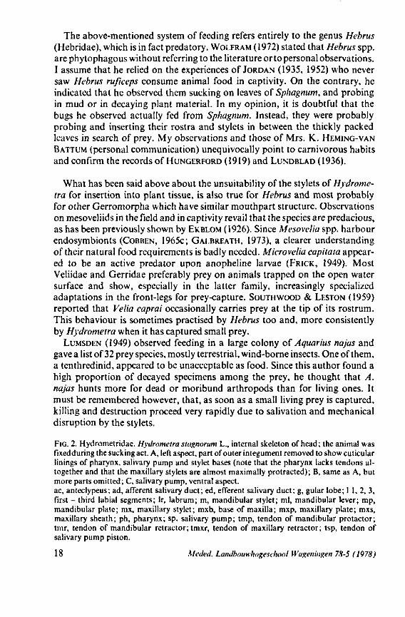
The above-mentioned system of feeding refers entirely to the genus Hebrus (Hebridae), which is in fact predatory. WOLFRAM (1972) stated that Hebrus spp. are phytophagous without referring to the literature or to personal observations. I assume that he relied on the experiences of JORDAN (1935, 1952) who never saw Hebrus ruficeps consume animal food in captivity. On the contrary, he indicated that he observed them sucking on leaves of Sphagnum, and probing in mud or in decaying plant material. In my opinion, it is doubtful that the bugs he observed actually fed from Sphagnum. Instead, they were probably probing and inserting their rostra and stylets in between the thickly packed leaves in search of prey. My observations and those of Mrs. K. HEMING-VAN
BATTUM (personal communication) unequivocally point to carnivorous habits and confirm the records of HUNGERFORD (1919) and LUNDBLAD (1936).
What has been said above about the unsuitability of the stylets of Hydrome-tra for insertion into plant tissue, is also true for Hebrus and most probably for other Gerromorpha which have similar mouthpart structure. Observations on mesoveliids in the field and in captivity revail that the species are predacious, as has been previously shown by EKBLOM (1926). Since Mesovelia spp. harbour endosymbionts (COBBEN, 1965c; GAI.BREATH, 1973), a clearer understanding of their natural food requirements is badly needed. Microvelia capitata appeared to be an active predator upon anopheline larvae (FRICK, 1949). Most Veliidae and Gerridae preferably prey on animals trapped on the open water surface and show, especially in the latter family, increasingly specialized adaptations in the front-legs for prey-capture. SOUTH WOOD & LESTON (1959) reported that Velia caprai occasionally carries prey at the tip of its rostrum. This behaviour is sometimes practised by Hebrus too and, more consistently by Hydrometra when it has captured small prey.
LUMSDEN (1949) observed feeding in a large colony of Aquarius najas and gave a list of 32 prey species, mostly terrestrial, wind-borne insects. One of them, a tenthredinid, appeared to be unacceptable as food. Since this author found a high proportion of decayed specimens among the prey, he thought that A. najas hunts more for dead or moribund arthropods than for living ones. It must be remembered however, that, as soon as a small living prey is captured, killing and destruction proceed very rapidly due to salivation and mechanical disruption by the stylets.
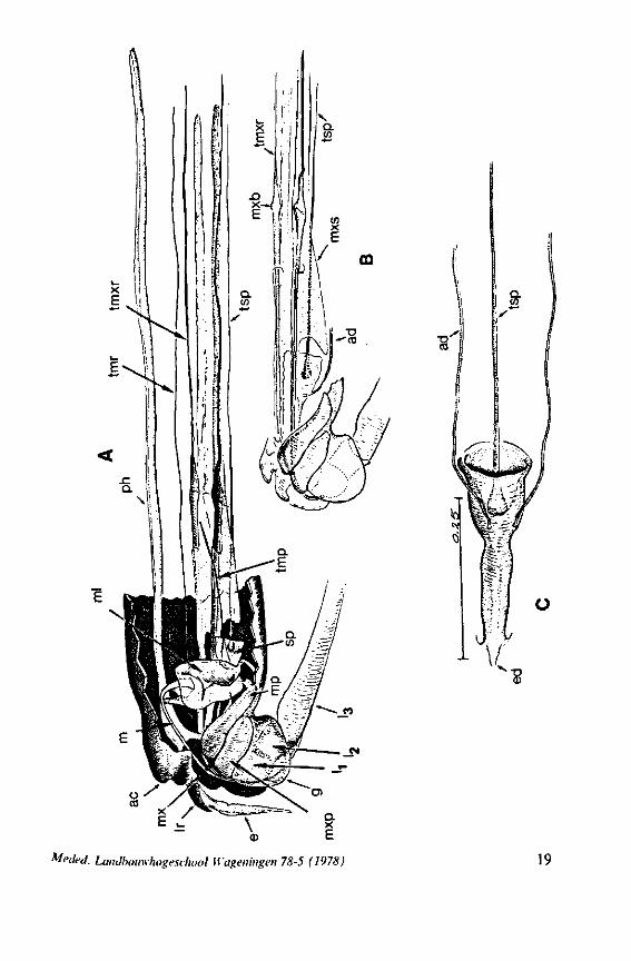
FIG. 2. Hydrometridae. Hydrometra stagnorum L., internal skeleton of head; the animal was fixedduring the sucking act. A, left aspect, part of outer integument removed to showcuticular linings of pharynx, salivary pump and stylet bases (note that the pharynx lacks tendons altogether and that the maxillary stylets are almost maximally protracted); B, same as A, but more parts omitted; C, salivary pump, ventral aspect. ac, anteclypeus; ad, afferent salivary duct; ed, efferent salivary duct; g, gular lobe; I 1, 2, 3, first - third labial segments; lr, labrum; m, mandibular stylet; ml, mandibular lever; mp, mandibular plate; mx, maxillary stylet; mxb, base of maxilla; mxp, maxillary plate; mxs, maxillary sheath; ph, pharynx; sp, salivary pump; tmp, tendon of mandibular protactor; tmr, tendon of mandibular retractor; tmxr, tendon of maxillary retractor; tsp, tendon of salivary pump piston.
18 Meded. Landbouwhogeschool Wageningen 78-5 (1978)
feà2'
toedeel. Landbouwhogeschool Wageningen 78-5 (1978) 19
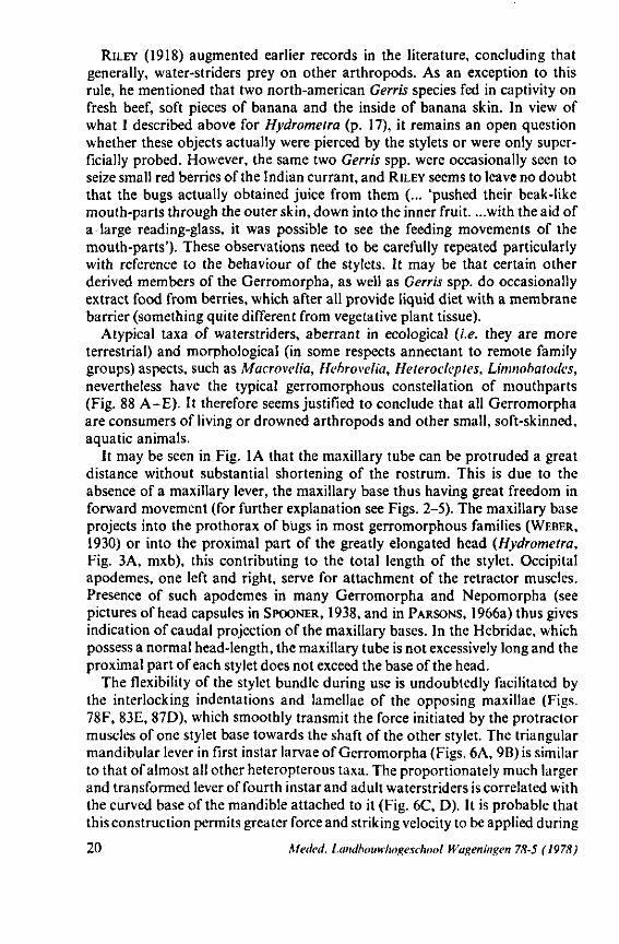
RILEY (1918) augmented earlier records in the literature, concluding that generally, water-striders prey on other arthropods. As an exception to this rule, he mentioned that two north-american Gerris species fed in captivity on fresh beef, soft pieces of banana and the inside of banana skin. In view of what I described above for Hydrometra (p. 17), it remains an open question whether these objects actually were pierced by the stylets or were only superficially probed. However, the same two Gerris spp. were occasionally seen to seize small red berries of the Indian currant, and RILEY seems to leave no doubt that the bugs actually obtained juice from them (... 'pushed their beak-like mouth-parts through the outer skin, down into the inner fruit. ...with the aid of a large reading-glass, it was possible to see the feeding movements of the mouth-parts'). These observations need to be carefully repeated particularly with reference to the behaviour of the stylets. It may be that certain other derived members of the Gerromorpha, as well as Gerris spp. do occasionally extract food from berries, which after all provide liquid diet with a membrane barrier (something quite different from vegetative plant tissue).
Atypical taxa of waterstriders, aberrant in ecological (i.e. they are more terrestrial) and morphological (in some respects annectant to remote family groups) aspects, such as Macrovelia, Hehrovelia, Heterocleptes, Limnobatodes, nevertheless have the typical gerromorphous constellation of mouthparts (Fig. 88 A-E). It therefore seems justified to conclude that all Gerromorpha are consumers of living or drowned arthropods and other small, soft-skinned, aquatic animals.
It may be seen in Fig. 1A that the maxillary tube can be protruded a great distance without substantial shortening of the rostrum. This is due to the absence of a maxillary lever, the maxillary base thus having great freedom in forward movement (for further explanation see Figs. 2-5). The maxillary base projects into the prothorax of bugs in most gerromorphous families (WEBER, 1930) or into the proximal part of the greatly elongated head (Hydrometra, Fig. 3A, mxb), this contributing to the total length of the stylet. Occipital apodemes, one left and right, serve for attachment of the retractor muscles. Presence of such apodemes in many Gerromorpha and Nepomorpha (see pictures of head capsules in SPOONER, 1938, and in PARSONS, 1966a) thus gives indication of caudal projection of the maxillary bases. In the Hebridae, which possess a normal head-length, the maxillary tube is not excessively long and the proximal part of each stylet does not exceed the base of the head.
The flexibility of the stylet bundle during use is undoubtedly facilitated by the interlocking indentations and lamellae of the opposing maxillae (Figs. 78F, 83E, 87D), which smoothly transmit the force initiated by the protractor muscles of one stylet base towards the shaft of the other stylet. The triangular mandibular lever in first instar larvae of Gerromorpha (Figs. 6A, 9B) is similar to that of almost all other heteropterous taxa. The proportionately much larger and transformed lever of fourth instar and adult waterstriders is correlated with the curved base of the mandible attached to it (Fig. 6C, D). It is probable that this construction permits greater force and striking velocity to be applied during
20 Kfeded. Landbouwhogeschool Wageningen 78-5 (1978)
ms_
mpV
mxb
mr 'K
. phm mr
mxp
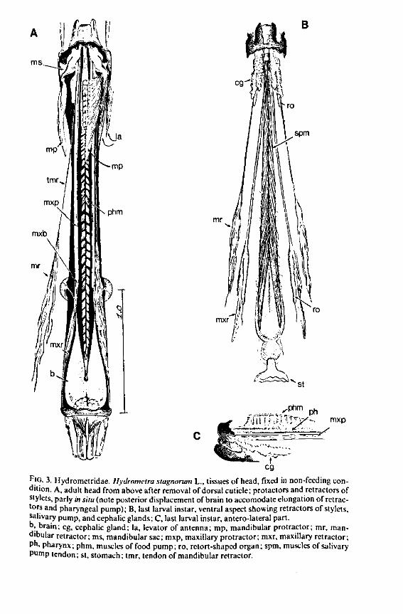
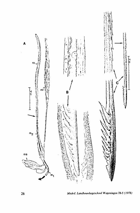
FIG. 3. Hydrometridae. Hydrometra stagnoruin L., tissues of head, fixed in non-feeding condition. A, adult head from above after removal of dorsal cuticle; protactors and retractors of stylets, parly in situ (note posterior displacement of brain to accomodate elongation of retractors and pharyngeal pump); B, last larval instar, ventral aspect showing retractors of stylets, salivary pump, and cephalic glands; C, last larval instar, antero-lateral part, b, brain; eg, cephalic gland; la, levator of antenna; mp, mandibular protractor; mr, mandibular retractor; ms, mandibular sac; mxp, maxillary protractor; mxr, maxillary retractor; Ph, pharynx; phm, muscles of food pump; ro, retort-shaped organ; spm, muscles of salivary pump tendon; st, stomach; tmr, tendon of mandibular retractor.
mx
mxpr —
tsp
mxr —
M KV, 'nmt
3 ö
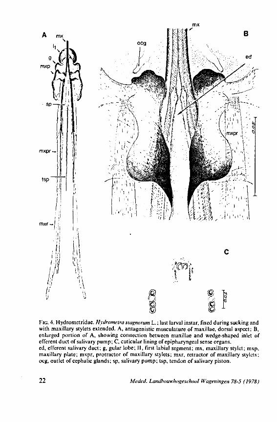
FIG. 4. Hydrometridae. HyJrometra stagnorum L. ; last larval instar, fixed during sucking and with maxillary stylets extended. A, antagonistic musculature of maxillae, dorsal aspect; B, enlarged portion of A, showing connection between maxillae and wedge-shaped inlet of efferent duct of salivary pump; C, cuticular lining of epipharyngeal sense organs, ed, efferent salivary duct; g, gular lobe; 11, first labial segment; mx, maxillary stylet; mxp, maxillary plate; mxpr, protractor of maxillary stylets; mxr, retractor of maxillary stylets; ocg, outlet of cephalic glands; sp, salivary pump; tsp, tendon of salivary piston.
22 MeJeJ. Landbouwhogeschool Wageningen 78-5 (1978)
B
) _ mxr
n •T.
i.-i'
H
mx
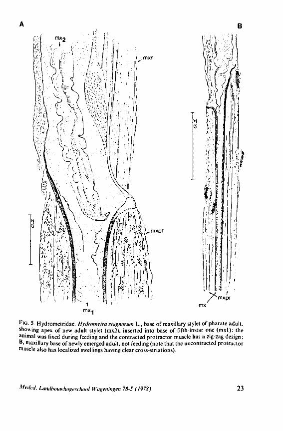
f'G- 5. Hydrometridae. HyJromeira siagnorum L., base of maxillary stylet of pharate adult, showing apex of new adult stylet (mx2), inserted into base of fifth-instar one (mxl); the animal was fixed during feeding and the contracted protractor muscle has a zig-zag design; B. maxillary base of newly emerged adult, not feeding (note that the uncontracted protractor muscle also has localized swellings having clear cross-striations).
Meded. Landbouwhogeschool Wagenmgen 78-5 (1978) 23
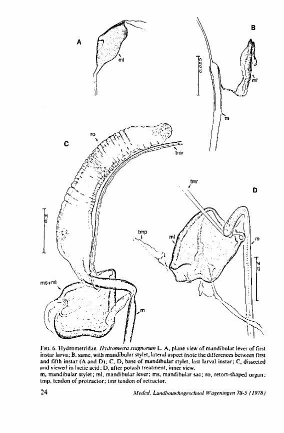
ms+ml
FIG. 6. Hydrometridae. Hydromeira siagnorum L. A, plane view of mandibular lever of first instar larva; B, same, with mandibular stylet, lateral aspect (note the differences between first and fifth instar (A and D); C, D, base of mandibular stylet, last larval instar; C, dissected and viewed in lactic acid ; D, after potash treatment, inner view. m, mandibular stylet; ml, mandibular lever; ms, mandibular sac; ro, retort-shaped organ: tmp, tendon of protractor; tmr tendon of retractor.
24 Meded. Landbouwhogeschool Wageningen 78-5 (1978)
harpooning of the prey. It is also feasible that this type of lever allows torsion of the mandibles to occur; the interaction between the two sets of protractors thus turning the vertical position of the lever into an oblique one. That the mandibles actually rotate to some degree during use is suggested by the following observation: when disturbed while sucking on a drosophilid fly, Hydrometra can easily rid itself of the speared prey. When the bug is abruptly killed during feeding and one tries to remove the fly from the stylets by gently pulling with tweezers, a patch of the host's cuticle remains attached to the mandibular stylets. The living bug must thus have some control over this rotation and the ability to withdraw the mandibular harpoon (Fig. 7C, 82E) out of the victim with exactly the same amount of twisting as occurred during piercing. Recurved indentations occur along the median margin of the mandibular shaft (Fig. 7C, 82F, Hydrometra, Hebrus; also present in Velia, Gerris (EKBLOM, 1926)). Because of friction by these hooks upon the maxillary bundle, it is possible that mandibular rotation by means of the dorsal rotator of the lever causes simultaneous torsion of the maxillae although the sequence of events may occur in the reverse way. Drosophila flies pierced by the stylets of adults and older larvae of Hydrometra and held above the substrate, rotate slightly with short jerks, often at regular intervals of 5-10 seconds, these movements suggesting brief rotations of the mandibles.
It was mentioned on p. 16, that the maxillary bundle whips in all radial directions during the rasping-sucking act. Unequal muscular forces applied to the two maxillary stylets and torsion of the maxillary bundle certainly are responsible for this universal motility. The angle of deviation of the stylets during protraction is determined furthermore by the articulation existing between the last two labial segments. In Hebrus, the terminal rostral segment is directed to the left and to the right at a regular, alternating rate (Fig. 11B). When Hydrometra has noticed the presence of a prey animal, its distal rostral segment is often seen undergoing a circular movement, whereas the other segments remain motionless and straight. The joint between the last two segments of the rostrum has a great intrinsic flexibility, and is protected dorsally by a flap-like structure (Fig. 79D, 113C, D, is). This is a characteristic feature of Gerromorpha and may serve to control the extent of stylet deviation*. Lateral deviation of the stylet-bundle is also facilitated by the tripartite ending of the rostrum (Figs. 79D, U2D, 11). Finally, alterations in the directional pathway of the central stylets may be influenced by the incurved apices of both maxillae, in particular t ne right maxilla (Figs. 77E, 85D, 88D), which always extends slightly ahead of the left maxillary stylet (Figs. 83D, 84F, 85B).
More data on rostral differentiations are given in Chapter 2.
Meded. Landbouwhogeschool Wageningen 78-5 (1978) 25
m0 _a
26 Meded. Landbouwhogeschool Wageningen 78-5 (1978)
1.2. NEPOMORPHA
(Figs. 12-14; 92B-F.-98; 116-120; 131-132) Taxa having barbed maxillary stylets The strongly indented interfaces of the maxillae as described above for Ger-romorpha, are also found in a modified form in the better known carnivorous families of Nepomorpha: Belostomatidae (Figs. 12, 92F, 95, 96A), Nepidae (Fig. 96B-E), Naucoridae (Fig. 13A), and Notonectidae. The barbed nature of the stylets has been noted by several authors (HAMILTON, 1931 ; ELSON, 1937 ; QUADRI, 1951), but accurate descriptions are lacking. The same toothed differentiations are shared by the littoral nepomorphan families Gelastocoridae (PARSONS, 1959) and Ochteridae (Fig. 92B-E), which are predators. PARSONS was the first to describe (using the optical microscope) the exact insertion and arrangement of the rows of bristles in Gelastocoris oculatus. Her description and diagrammatic figure 18 (redrawn by me in Fig. 11F) are presented here, since the architecture of these fairly stout stylets gives a clear picture of the sites from which the barbed system arises. This system is thus more easily surveyed than that of Gerromorpha (and also in Ochteridae), where the differentiations are more elaborate in number and in variety and therefore much more difficult to analyse three-dimensionally.
A passage of PARSONS (1959, p. 27) reads as follows: 'Along the medial surfaces of the tips of each stylet are longitudinal rows of anteriorly-directed bristles. Examination of the maxillae of two individuals revealed that the tip of the right maxilla differs from that of the left. The right maxilla bears four bristle rows, as shown in Figure 18B (= my lower Fig. 11F); the two outer ones, located on the dorsal and ventral margins, consist of rather fine hairs Projecting outwards, while the two inner ones are composed of suffer bristles which extend medially. The latter are located along the ridge dividing the food and salivary canals and along the dorsal margin of the dorsal groove. The left maxilla bears a tuft of fine hairs on the dorsal margin of the dorsal groove, along with only two rows of bristles, one on the ventral margin and one on the separating ridge (Fig. 18A = my upper Fig. 1 IF). These rows are shorter than those of the right maxilla. The bristles of the more dorsal row are stiffened and Project medially. Longitudinal sections through the stylet bundle reveal that the bristles of the opposed separating ridges on the two stylets interlock thus holding the right and left maxillae together.'
F'G. 7. Hydrometridae. Hydrometra stagnorum L., last larval instar. A, B, development of adult mandibular stylet within retort-shaped organ; A, total aspect (note the extremely elongated stylet forming organ in contrast to the non-functional one in Fig. 3A); B, apex of "ew adult mandibular stylet (below) and mandibular part at level of arrow in A (note that differentiation of the developing stylet starts at its distal end; only proximad, cuticular components of the enveloping tissue are seen at this stage of cuticle secretion); C, the functional mandibular stylet, seen with the optical microscope (note the irregularities of the inner side ° ' the mandibular trunk ; compare with the scannings Fig. 182E, F).
Meded. Landbouwhogeschool Wageningen 78-5 (1978) 27
8
B 'o. àiF*
28 Meded. Landbouwhogeschool Wageningen 78-5 (1978)
SEM pictures of the same species (Figs. 93,94,97A, B) allow a more detailed spatial representation of these correct observations to be made. The ventral seam of both right (Fig. 93 A) and left (Fig. 94A) maxillary stylet bears a row of teeth which extend at different angles off the stylet shaft. This is not an artefact of preparation but represents a natural situation. The spines project outwards and cross each other in the united stylet bundle in situ (Fig. 117A, B). It is clear that they function in rupturing the tissues of the host, and in mixing the saliva which is simultaneously ejected along this ventral grating. Filtering and trituration of food fragments can take place subsequently within the internal food canal.
Because the external rows of spines interweave, independent longitudinal gliding of the stylets, one on the other, is probably limited. Direct observations are lacking, but one dried specimen had the right stylet projecting 0.4 mm in front of the left. Thus, some independent gliding of the maxillae may occur during feeding, a method which is actually practised by some other waterbugs (described below). Such a displacement has never been observed in Gerro-morpha, and is probably also impossible in Ochteridae, whose stylets are more gerromophan like (Fig. 92D, E).
The ventral spines of the right stylet of Gelastocoris are implanted into wrincled cuticle; some have a socket or posses a peculiar, twisted and incised base (Fig. 93B-E). This suggests great flexibility and a passive movement of the spines, when unequal muscular force is applied to the individual stylets. Therefore these spines probably do not break off when rubbed by the opposing stylet. The dorsal file of spines, which could cause damage to the host's tissue, is short and restricted entirely to the right stylet (Fig. 93A). Another difference existing between Gelastocoris and Gerromorpha, Ochteridae (Nepomorpha) is that the maxillae of the former taxon are straight, and lack incurved tips.
The internal filter system of the food-canal is very complex and is confined predominantly to the right stylet (Fig. 93B-E). It consists of a ventral row of sharp spines and a dorsal row of broad, flat, truncate projections which are regularly bent inwards underneath the ventral series of spines (resembling conditions in some Gerromorpha, Fig. 83E). The median surface of the left maxillary stylet appears as in Fig. 94C, in which the ventral side, bordering the salivary canal, faces towards the right of the photographs. One of the most striking aspects of this stylet, obvious only at high magnifications and in three dimensions, is that the food-canal is not simply a gutter which faces a similar concavity in the opposed stylet. The food-canal of the left stylet forms a nearly
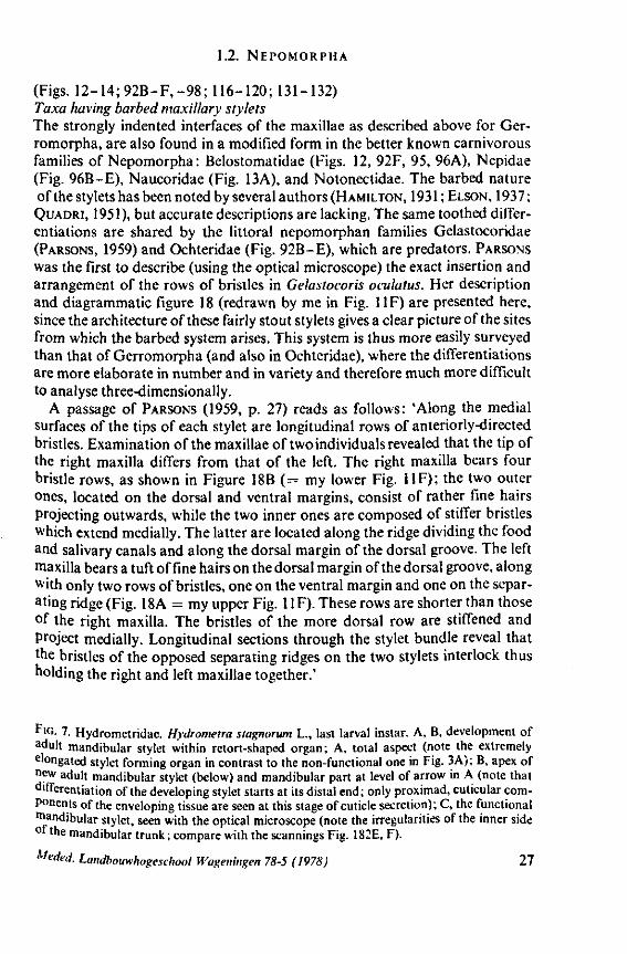
FIG. 8, 9. Hydrometridae. Hydromelra stagnorum L. 8, first larval instar. A, ventral aspect of aPex of labium; B, mandibular stylet; 9, second larval instar (except A", first instar); A, epipharynx and foodpump, ventral view; A', epipharyngeal organ, sensilla located in the •dorsal wall of foregut at sites indicated by arrows (note that conditions of the paired sensilles are relatively unchanged between first (A") and fifth instar (4C); B, plane view of mandibular 'ever; C, the same lever hinged to the exoskeleton (the mandibular stylet is shown out of natural position), ap, apical plate; ep, epipharynx; 1, labrum; m, mandibular stylet; ml, mandibular lever.
Meded. Landbouwhogeschool Wageningen 78-5 (1978) 29
~ 3
- 4
^ i^S^SiÄi f i^^^Ä ~ÖTZ—I
30 Meded. Landbouwhogeschool Wageningen 78-5 (1978)
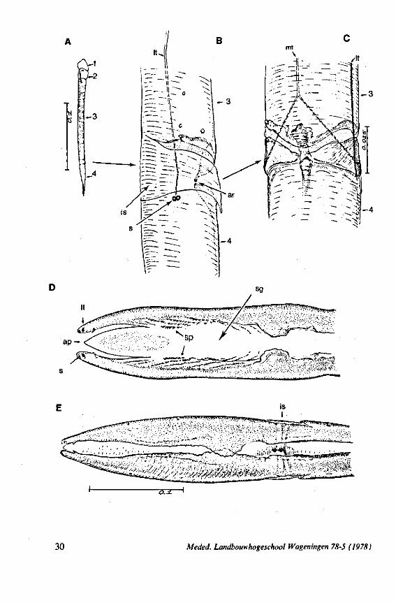
complete cylinder in itself. This cylinder extends proximally for some distance from the stylet apex (the ventral seam is marked by a white line in Fig. 94C). Fig. 97A and Fig. 97B show the inner surface of the right and left maxillary stylets, respectively, approximately at the level where the rasping-triturating devices begin. The left and right photographs considered together in the mind's eye, create the picture of the united maxillae in situ. Thus, it appears that the food canal must consist of a nearly double-walled cylinder, a constellation which never has been figured in published cross-sections of any hemipteron. Figures 97A and B demonstrate that the maxillary pair is closely bound together, not by the marginal bristles, but by the smooth interlocking ridge-groove system which extends dorsally much further distad than ventrally.
The family Gelastocoridae contains only two genera, Gelastocoris and Nerthra, respectively placed in the subfamilies Gelastocorinae and Nerthrinae. The structure of the maxillary stylets of Nerthra (laticollis and colaticollis studied) deviates considerably from that of Gelastocoris. The right stylet has an apical inward curvature and the rows of spines extend over a much greater length (0.5 mm). These spines do not project outwards obliquely or perpendicularly from the body of the stylet. The spatial conditions of the stylets of these bugs have not been elucidated in detail, but I gained the impression that two rows of combs are situated only within the food canal. The left stylet lacks spines and other irregularities altogether.
The right maxillary stylet of members of the only other littoral nepomorphan family, the Ochteridae (like Gelastocoridae represented only by two recent genera) has a very complex vestiture of bristles (Fig. 92E, 131, 132). In the new-world Ochterus perbosci, their configuration and the curvature of the stylet's tip resembles conditions in the Gerromorpha, except that in addition the food canal has a wash-board texture consisting of two layers of adpressed scale-like barbs (Fig. 92C, D). In the old world Ochterus marginatus, the ventral rasping system consists entirely of externally projecting bristles arising from both stylets. They occupy 0.3 mm of the length of the right and 0.15 mm of the left maxillary apex. The whole system, as seen light-optically, is too elaborate to describe verbally, but the bristle-combs of both stylets are directed towards the 'eft and the spines of various other combs in slightly different directions. The dorsal file is much finer and extends over a much greater distance. The right
FIG. 10. Hydrometridae. Hydrometra stagnorum L., labial structures. A-C, first instar; D, E. adult. A, entire rostrum, left lateral aspect, showing the four labial segments (note that alternating areas of sclerotized and non-sclerotized parts are less numerous than in later instars, cf. Figs. 79C, D; 113E); B, elements of junction (is) between third and fourth segment (note that the long tendons of the muscles flexing the distal segment extend proximally to the base of segment 3 and that their insertions contact two sense organs (which are probably Proprioceptors); C, same as B, ventral aspect; D, E, dorsal aspect of last rostral segment; the lateral walls in D have been flexed out artificially to show more clearly the tripartite ending and crenations of the stylet groove. aP. apical plate; ar, articulating point; is, intercalary sclerite; 11, lateral lobe; It, lateral tendon; mt, median tendon; s, sensilla; sg, stylet groove; sp, fine spines; t, tendon.
Meded. Landbouwhogeschool Wageningen 78-5 (1978) 31
32 Meded. Landbouwhogeschool Wageningen 78-5 (1978)

stylet has top curvature, the left one not. The ground-plan of the acute left stylet bears some resemblance to that of other nepomorphan families; the dorsal margin having a preapical overfold with an irregular outline (more or less as in Fig. 95B, for Lethocerus). Its oblique row of strong teeth resembles that of Notonecta and Ilyocoris (Fig. 13A), although it runs over a longer trajectory. Staggering of the stylet tips during feeding is probably limited (in one mounted specimen there was a displacement of 0.07 mm).
Feeding behaviour was studied in Notonectidae (Notonecta glauca) and Naucoridae (Ilyocoris cimicoides), and proved to be quite different from that of Gerromorpha. Notonecta may be taken as an example. During feeding, the mandibles secure the prey's cuticle, and the stiff maxillary bundle is pushed forward in a straight line. Directional change in the axis of the bundle is accomplished wholly by lateral deviation of the last rostral segment. These movements of the rostrum are accompanied by pressure upon the cuticle of the host, which is clearly seen to be deformed. This discontinuous pressure causes intense mixing of the host's contents. There is also a regularly alternating forward and backward movement of the maxillae. The forward thrust of the right maxillary stylet is usually distinctly further than that of the left and is accompanied by a displacement of the maxillary apices. The main mechanical effect on the prey's tissue may be similar to that caused by a pair of clippers. The sawing action is performed by both maxillae, but the right stylet has a much longer stroke. It is clear from Fig. 13A (Ilyocoris, similar to Notonecta) that the opposing rows of teeth glide along one another in a longitudinal direction. The maxillae are firmly interlocked preapically along a straight line both ventrally and dorsally (in contrast to those of the Gerromorpha). The long files of barbs of the right stylet extend proximad for a considerable distance, and lie entirely within the food canal. These barbs have the same forward-backward movements as the entire stylet and help in further grinding particulate food matter.
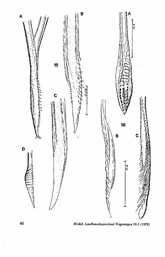
It was mentioned above that Nerthra (Gelastocoridae) has the left maxillary stylet entirely smooth. This is nearly also the case in the atypical naucorid Aphelocheirus aestivalis; the right maxillary stylet of this species resembles that of the reduviid Sphaeridops amoenus (see Fig. 16C), and bears a sublateral row of pegs in addition to the lateral rows of long teeth.
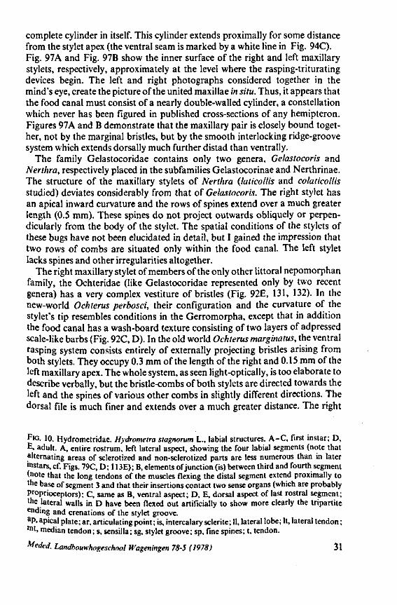
The strategy of the maxillae of Belostomatidae and Nepidae during feeding was not observed, but I assume that they have less freedom of independent FIG. 11. A-C, Gerromorpha. A, B, Hebrus; C, Hydrometra, dislocation of labial segments during sucking (note that the bend between segment 3 and 4 is mostly directed backwards (Fig. A) ; this position and the lateral turning of the distal segment can also be achieved actively when the bug is not feeding (Fig. C, at right); Fig. C, upper left, shows the beak of the bug sucking from a drop of water, and, lower left, from the sap of a slice of onion. D. Ceratocombidae. Ceratocombus coleoptratus Zett., mandibular (left) and both maxillary stylets. E. Enicocephalidae. Embolorrhinus tuberculatum Dgr. (origin: Sudan); both maxillary stylets (left) and mandibular stylets (right) (the file of the maxillary stylet extends 0.4 mm proximad). F. Gelastocoridae. Gelastocoris oculalus Fabr. ; medial surface of left (above) and right maxil-'ary stylets, redrawn from Parsons (1959). fc, food canal; sc, salivary canal.
Meded. Landbouwhogeschool Wageningen 78-5 (1978) 33
T 'f ff » V V
34 Meded. Landbouwhogeschool Wageningen 78-5 (1978)
movement than those of Notonecta and Ilyocoris, because of their more elaborate complement of bristles (Figs. 12A, C; 95A, B; 96B-E). These families more likely form a link in their mouthpart functioning between Ochteridae and Gelastocoridae on one hand, and Naucoridae and Notonectidae on the other; at least it seems so from the species studied. The structure of the rostrum and the great length of the retracted maxillary stylets of belostomatid and nepid bugs, further suggest more penetrating capacity, than Notonecta. As NEIS-
WANDER (1926) concluded from a study otRanatra: 'The length of the extensor muscle would seem to permit a great extension of the maxilla in probing about through the body of an insect for blood'. The tip of the right maxillary stylet of Lethocerus is incurved (Fig. 95A), that oîNepa not (Fig. 95D)*.
For all the nepomorphan groups mentioned above (except perhaps for Ochterus) it is significant that the maxillary barbs do not serve primarily for holding the stylets closely together, as was thought by HAMPTON (1931) and QUADRI (1951).
Taxa having greatly reduced maxillary barbs Stylets of the derived subfamily Corixinae of the Corixidae have long been
known to be atypical for Heteroptera (EKBLOM, 1930; HUNGERFORD, 1919). BENWITZ (1956) characterized the right maxillary stylet of Corixa punctata as a sharpened spoon, the left one as a gouge. The right stylet has a row of spines and the action of both stylets, whose muscles may permit some rotation, is described as a smashing of the substrate. Independent protrusion of the maxillae has also been observed. The shape of both stylets is shown in the scanning micrographs Fig. 98A-F , and in cross-sections in Fig. 152H. Corixinae are commonly believed to be detritus feeders specializing on algae and other botanical substances (HUNGERFORD, 1919; SUTTON, 1947; WA..TON, 1943). ZWART (1965) showed experimentally for several corixine species that survival of both larvae and adults is high when they are fed only on living animal food. The mortality curves for bugs fed on plant material alone were similar to those of unfed bugs. Recent observations (JANSSON & SCUDDER, 1972; PAJUNEN, 1970; PETERS &
. * The feeding act of Nepa could be studied recently. The behaviour of the maxillary stylets is more or less like in Notonecta and Ilvocoris, but they protrude much further indeed. Nevertheless, their shafts remain straight when moving within the host. The unequal sawing action ° ' the individual stylets to and fro is present, but not to that extent as in Notonecta. The maxillary apices diverge slightly from each other during the snapping actions, thus causing a wider entrance towards the stylets mouth (slightly more open than shown for Ilyocoris "iFig.HA). 3
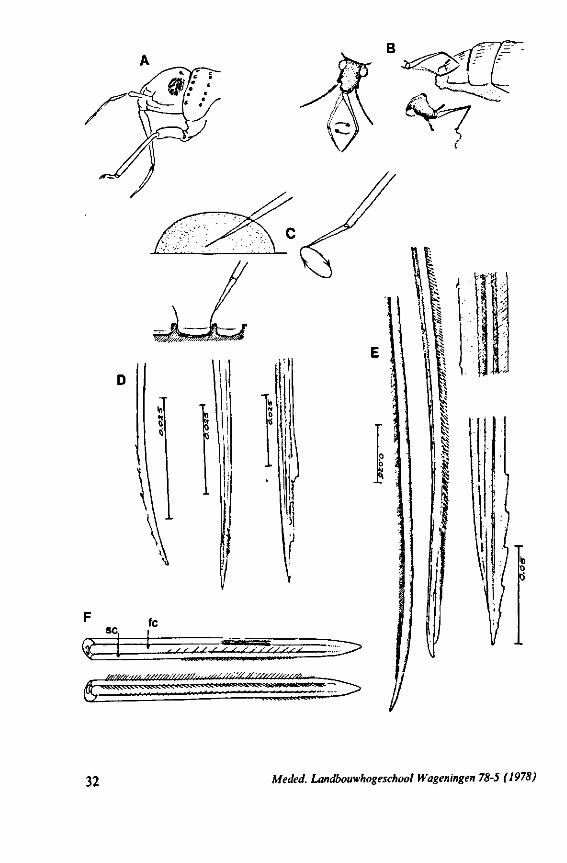
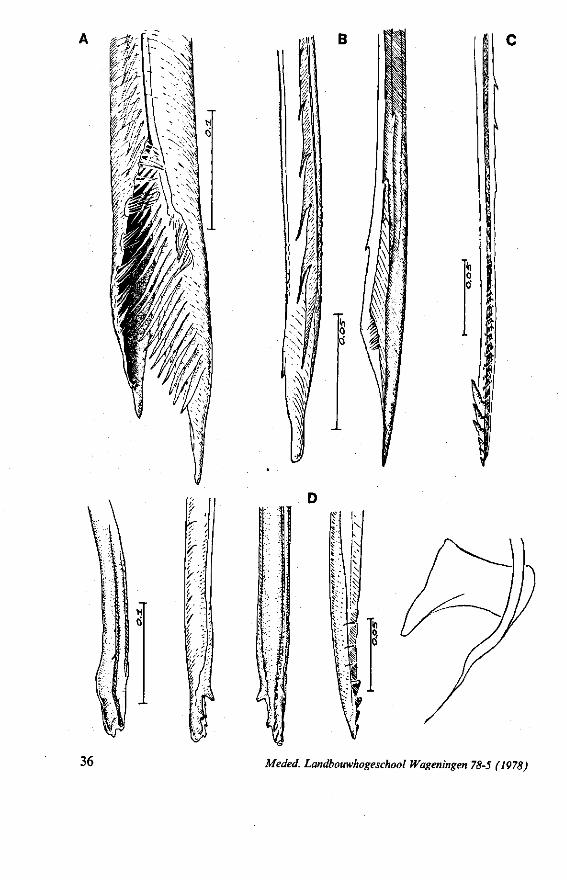
FIG. 12. Belostomatidae. Lethocerus niloticus Stâl (origin: Madagascar). A, left maxillary «yiet, inner view; barbs extend over a long range; A' is taken at a distance of 1.6 mm from ">e Up of the stylet; A' , external face of same stylet; B, apex of mandibular stylet; B', the same about halfway along its length; C, right maxillary stylet; D, same, viewed from the tner side; there is a pit near the top (asterisk), which might mark a nerve ending (see also
tannings Fig. 95).
Meded. Landbouwhogeschool Wageningen 78-5 (1978) 35
^
B
Mil
W'i
36 Meded. Landbouwhogeschool Wageningen 78-5 (1978)
ULBRICH, 1973; SOKOL'SKAYA & ZHITENEVA, 1973) reveal that predacious feeding habits are more general in Corixinae than suggested in the past.
Members of the less derived corixid subfamilies Micronectinae, Diapre-pocorinae and Cymatiinae are believed to be predatory because of the structure of their front legs and from limited observation of living animals. Cymatia species are very destructive predators, killing far more animals than they need (WALTON, 1943). The maxillary stylets of representatives of these three subfamilies do not deviate much from the corixine ground-plan. The maxillary stylets of Micronecta are rather similar to those of Cymatia (Fig. 13D). WALTON
(1943) observed that Cymatia sp. could protrude its stylets farther than other Corixidae. These stylets 'are also quite flexible and therefore effective in reaching the juices of their victims'. The internal stylets of Diaprepocoris zealandiae (subfamily restricted to New Zealand) are both devoid of spines.
The maxillary bundle in representatives of three other families was discovered to lack a distinct barb system. The stylets of the remarkable south-american genus Coleopterocoris (Potamocoridae, see footnote on p. 7) are shown in Fig. 13B. They are structurally entirely different from the typical naucorid stylets illustrated in Fig. 13A. In the families Pleidae and Helotre-phidae (both containing only a few species), the maxillary stylet-bundle is so constructed that it probably functions as a unit. The two halves fit closely together apically, leaving only a narrow slit for uptake of liquid material (Fig. 97C, D). Tooth-like or other lacerating structures are absent according examinations by SEM. The outward aspect of the stylet bundle thus resembles that of some terrestrial groups as for example the higher Saldidae (Fig. 99A) or Lygaeidae (Fig. 109C, D). In Plea, however, there are within the food-canal about 17 large, stout spines with broad bases projecting distad. In the related family Helotrephidae, conditions are about the same, but fewer (±8) internal spines are present. The mandibles in both families have, in addition to the normal complement of apical notches a preapical collar of sharp recurved projections, disposed in a row oblique to the longitudinal axis of the stylet (Fig. 97E, F). The mandibular trunk is notched for a considerable length along both its median and external edges, and the lateral margins are flattened out as ribbons.
Of the three families considered here (Potamocoridae, Pleidae, and Helotrephidae), only Pleidae have been observed to be predators of small arthropods (e.g. HUNGERFORD, 1919; WEFELSCHEID, 1912). The latter author supposed that Plea atomaria also on occasion takes plant-sap, but convincing evidence for this is lacking.
FIG. 13. A, Naucoridae. Ilyocoris cimicoides L., ventral view of maxillary bundle. B- Potamocoridae. Potamocoris spec, (origin : Ecuador), maxillary stylets. C. Ochteridae. Ochterus perbosci Guer. (origin: Curaçao, Antilles), mandibular stylet. D, Corixidae, Cymatia bonsdorffl Sahib., from left to right: left maxillary stylet, right maxillary stylet (lateral), right maxillary stylet (inner side), mandibular stylet, mandibular lever (see also Fig. 98).
Meded. Landbouwhogeschool Wageningen 78-5 (1978) 37
iOT
38 Meded. Landbouwhogeschool Wageningen 78-5 (1978)
1.3. REDUVIOIDEA
(Figs. 15-221, A-I; 101-104; 133-134) Since this superfamily has an evolutionary origin close to that of the Nepo-
morpha (COBBEN, 1968a), it is treated here. The stylet structures of Reduviidae reveal a surprisingly wide range of evolutionary progression. I have studied the stylets of 16 species representing 10 subfamilies. Phylogenetic relationships within this family are much debated and several recent important studies (e.g. DAVIS, 1969) suggest that subfamily extent and placement remain complex matters that have yet to be resolved. The species selected for examination in the present work represent a very small sample of the diversity present, too small to allow systematic speculation. I have the feeling that each valid subfamily has been subject to a wider range of parallel evolution in stylet structure than is shown here. The species are thus treated here 'sec' in a sequence showing progressive loss of maxillary barbs, without reference to subfamily divisions.
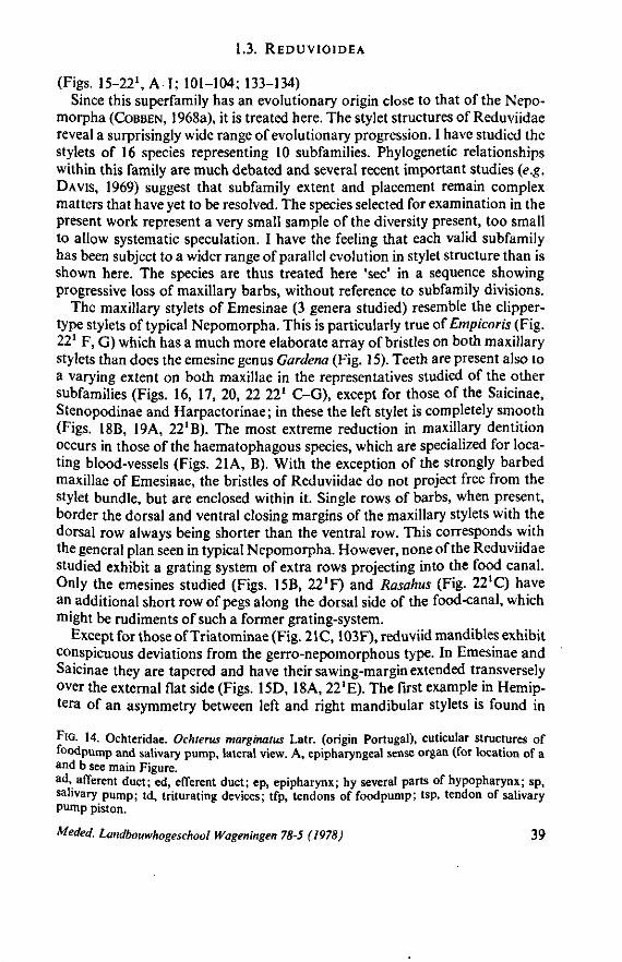
The maxillary stylets of Emesinae (3 genera studied) resemble the clipper-type stylets of typical Nepomorpha. This is particularly true of Empicoris (Fig. 221 F, G) which has a much more elaborate array of bristles on both maxillary stylets than does the emesine genus Gardena (Fig. 15). Teeth are present also to a varying extent on both maxillae in the representatives studied of the other subfamilies (Figs. 16, 17, 20, 22 221 C-G), except for those of the Saicinae, Stenopodinae and Harpactorinae ; in these the left stylet is completely smooth (Figs. 18B, 19A, 22'B). The most extreme reduction in maxillary dentition occurs in those of the haematophagous species, which are specialized for locating blood-vessels (Figs. 21A, B). With the exception of the strongly barbed maxillae of Emesinae, the bristles of Reduviidae do not project free from the stylet bundle, but are enclosed within it. Single rows of barbs, when present, border the dorsal and ventral closing margins of the maxillary stylets with the dorsal row always being shorter than the ventral row. This corresponds with the general plan seen in typical Nepomorpha. However, none of the Reduviidae studied exhibit a grating system of extra rows projecting into the food canal. Only the emesines studied (Figs. 15B, 221F) and Rasahus (Fig. 22'C) have an additional short row of pegs along the dorsal side of the food-canal, which might be rudiments of such a former grating-system.
Except for those of Triatominae (Fig. 21C, 103F), reduviid mandibles exhibit conspicuous deviations from the gerro-nepomorphous type. In Emesinae and Saicinae they are tapered and have their sawing-margin extended transversely over the external flat side (Figs. 15D, 18A, 22'E). The first example in Hemip-tera of an asymmetry between left and right mandibular stylets is found in
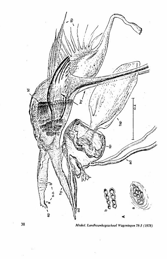
FIG. 14. Ochteridae. Ochterus marginatus Latr. (origin Portugal), cuticular structures of foodpump and salivary pump, lateral view. A, epipharyngeal sense organ (for location of a and b see main Figure. ad, afferent duct; ed, efferent duct; ep, epipharynx; hy several parts of hypopharynx; sp, salivary pump; td, triturating devices; tfp, tendons of foodpump; tsp, tendon of salivary pump piston.
Meded. Landbouwhogeschool Wageningen 78-5 (1978) 39
ir
i r t i ^
II «
16
B
l
'
40 Meded. Landbouwhogeschool Wageningen 78-5 (1978)
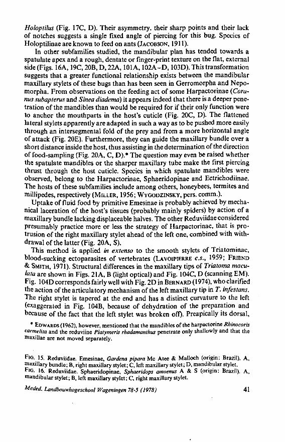
Holoptilus (Fig. 17C, D). Their asymmetry, their sharp points and their lack of notches suggests a single fixed angle of piercing for this bug. Species of Holoptilinae are known to feed on ants (JACOBSON, 1911).
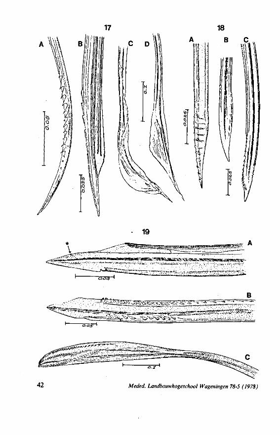
In other subfamilies studied, the mandibular plan has tended towards a spatulate apex and a rough, dentate or finger-print texture on the flat, external side (Figs. 16A, 19C,20B,D,22A, 101 A, 102A-D, 103D). This transformation suggests that a greater functional relationship exists between the mandibular maxillary stylets of these bugs than has been seen in Gerromorpha and Nepo-morpha. From observations on the feeding act of some Harpactorinae (Commis subapterus and Sinea diadema) it appears indeed that there is a deeper penetration of the mandibles than would be required for if their only function were to anchor the mouthparts in the host's cuticle (Fig. 20C, D). The flattened lateral stylets apparently are adapted in such a way as to be pushed more easily through an intersegmental fold of the prey and from a more horizontal angle of attack (Fig. 20E). Furthermore, they can guide the maxillary bundle over a short distance inside the host, thus assisting in the determination of the direction of food-sampling (Fig. 20A, C, D).* The question may even be raised whether the spatulate mandibles or the sharper maxillary tube make the first piercing thrust through the host cuticle. Species in which spatulate mandibles were observed, belong to the Harpactorinae, Sphaeridopinae and Ectrichodiinae. The hosts of these subfamilies include among others, honeybees, termites and millipedes, respectively (MILLER, 1956; WYGODZINSKY, pers. comm.).
Uptake of fluid food by primitive Emesinae is probably achieved by mechanical laceration of the host's tissues (probably mainly spiders) by action of a maxillary bundle lacking displaceable halves. The other Reduviidae considered presumably practice more or less the strategy of Harpactorinae, that is protrusion of the right maxillary stylet ahead of the left one, combined with withdrawal of the latter (Fig. 20A, S).
This method is applied in extenso to the smooth stylets of Triatominae, blood-sucking ectoparasites of vertebrates (LAVOIPIERRE C.S., 1959; FRIEND & SMITH, 1971). Structural differences in the maxillary tips of Triatoma macula ta are shown in Figs. 21 A, B (light optical) and Fig. 104C, D (scanning EM). Fig. 104D corresponds fairly well with Fig. 2D in BERNARD (1974), who clarified the action of the articulatory mechanism of the left maxillary tip in T. infestons. The right stylet is tapered at the end and has a distinct curvature to the left (exaggerated in Fig. 104B, because of dehydration of the preparation and because of the fact that the left stylet was broken off). Preapically its dorsal,
* EDWARDS (1962), however, mentioned that the mandibles of the harpactorine Rhinocoris carmelita and the reduviine Platymeris rhadamanthus penetrate only shallowly and that the maxillae are not moved separately.
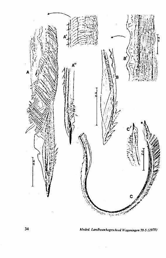
FIG. 15. Reduviidae. Emesinae, Gardena pipara Mc Atee & Malloch (origin: Brazil). A, maxillary bundle; B, right maxillary stylet; C, left maxillary stylet; D, mandibular stylet. F'G. 16. Reduviidae. Sphaeridopinae, Sphaeridops amoenus A & S (origin: Brazil). A, mandibular stylet; B, left maxillary stylet; C, right maxillary stylet.
Meded. Landbouwhogeschool Wageningen 78-5 (1978) 41
17
19
Q C 5 "
? f t ^ ^ ^ s ^ c -B
Meded. Landbouwhogeschool Wageningen 78-5 (1978)
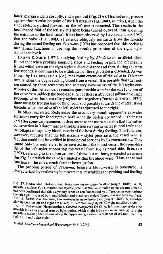
inner, margin widens abruptly, and is grooved (Fig. 21 A). This widening presses against the articulation point of the left maxilla (Fig. 104D, asterisk), when the right stylet is pushed forward, or the left one is retracted. This results in the heel-shaped fold of the left stylet's apex being turned outward, thus widening the entrance to the food canal. It has been observed by LAVOIPIERRE C.S. 1959, that the valve (Fig. 104D, v) extends obliquely outwards from the fascicle during the actual feeding act. BERNARD (1974) has proposed that this rocking-mechanism functions in opening the mouth; protrusion of the right stylet should achieve it.
FRIEND & SMITH (1971), studying feeding by Rhodnius on artificial diets, found that when probing sampling stops and feeding begins, the left maxilla is first withdrawn on the right stylet a short distance and then, during the next few seconds, it continues to be withdrawn or the right stylet is extended. As was shown by LAVOIPIERRE C.S. (I.e.), maximum extension of the valve in Triatoma occurs when the bug is sucking blood from a vessel. It is possible that the friction caused by short retractory and rotatory movements of the left stylet contributes of this behaviour. It remains questionable whether the sole function of the valve is to unblock the food-canal. Since there is abundant salivation during probing, when both maxillary stylets are together (FRIEND & SMITH, 1971), there must be free passage of fluid from and possibly towards the central stylet fascicle, when the valve of the left stylet is adpressed to the right.
In other, unrelated Reduviidae the secondary mouth apparently provides sufficient entry for food uptake both when the stylets are united at their tips and after some displacement. It thus seems to me more plausible that the valve-construction in Triatominae is an adaptation serving to increase local resistance to collapse of capillary blood-vessels of the host during feeding. This function, however, requires that the left maxillary stylet penetrates the vessel wall, a fact that could not be verified in histological sections by LAVOIPIERRE C.S. They found only the right stylet to be inserted into the blood vessel, the spine-like tip of the left stylet supporting the vessel from the external side. BERNARD (1974), referring to the observations of these last authors, presented a scheme (his Fig. 3) in which the valve is situated within the blood vessel. Thus, the actual function of the valve, needs further investigation.
The probing period of Triatoma, before a blood-vessel is punctured, is characterized by restless stylet movements, resembling the probing and feeding
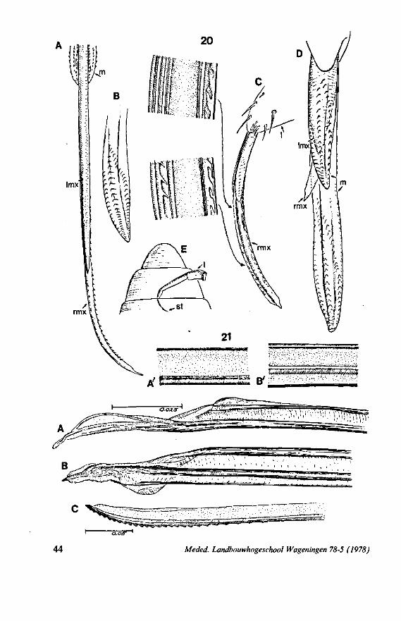
F]G. 17. Reduviidae. Holoptilinae, Holoptilus melanospilus Walker (origin: India). A, B, maxillary stylets; C, D, mandibular stylets (note that the mandibular stylets are not alike; it has been confirmed that this assymetry is not an artefact caused by differences in orientation ; the left-right origin of both mandibular and maxillary stylets figured has not been verified). F'G. 18. Reduviidae. Saicinae, Oncerotrachelus acuminatus Say. (origin: USA). A, mandibular stylets (the left and right are alike!); B, left maxillary stylet; C, right maxillary stylet. *"1G. 19. Reduviidae. Harpactorinae, Coranus subapterus De G. A, left maxillary stylet (the asterisk indicates a canal seen by light-optics, which might indicate a nerve ending); B, right maxillary stylet (indentations along the upper margin extend a distance of 0.9 mm from the llP); C, mandibular stylet.
Meded. Landbouwhogeschool Wageningen 78-5 (1978) 43
Imx
* y < / t f ; / / < / f . ^
/ fewTJJ,?;."Mr '*''f\ÏI^Ifnwffr''."v,'• -5 Q / - - •» •
44 Meded. Landbouwhogeschool Wageningen 78-5 (1978)
movements of Gerromorpha (see pages 15,16). LAVOIPIERRE C.S. (1959, p. 242, 243) described this activity of the stylet bundle as whip-like. Their observations are cited here : 'Entry of the stylets into the host's skin is very rapid ; it is initiated by rapid alternating movements of the mandibles which, having penetrated the tissues, remain still. Immediately after the introduction of the mandibles into the skin, the maxillae are projected well beyond the mandibles, as a single bundle. The maxillae are remarkably flexible (unlike the mandibles, which appear to be rigid structures) and the maxillary bundle is thrust forward first in one direction and then in another. So flexible, indeed, are the maxillae that occasionally they may be seen to bend to an angle of well over 90°, as if they had come into contact with dense tissues. A striking feature of the movement of the maxillae is a twisting action, which is particularly well shown at the point where they enter the skin of the host, and if observations are extended to the intralabial portion of the fascicle an active buckling movement can be seen'.
1.4. LEPTOPODOMORPHA
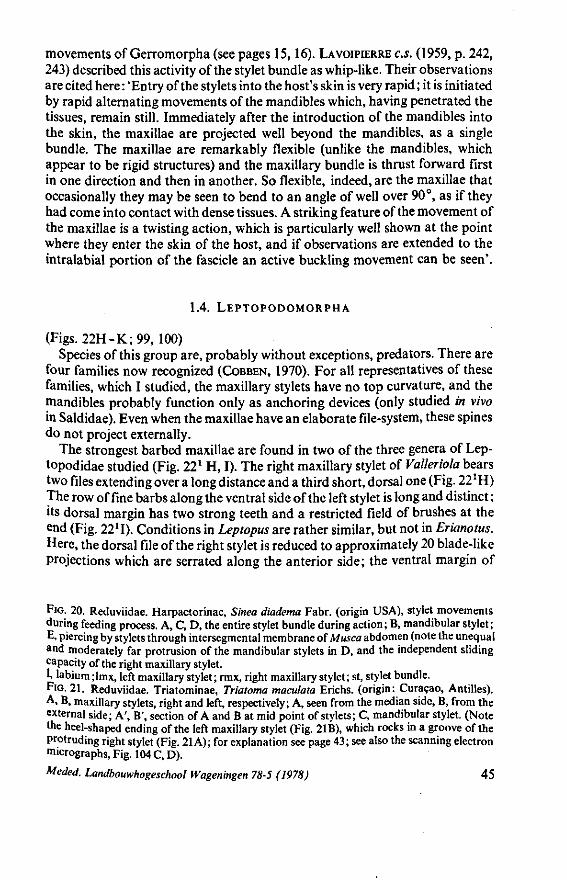
(Figs. 22H-K;99, 100) Species of this group are, probably without exceptions, predators. There are
four families now recognized (COBBEN, 1970). For all representatives of these families, which I studied, the maxillary stylets have no top curvature, and the mandibles probably function only as anchoring devices (only studied in vivo in Saldidae). Even when the maxillae have an elaborate file-system, these spines do not project externally.
The strongest barbed maxillae are found in two of the three genera of Lep-topodidae studied (Fig. 22l H, I). The right maxillary stylet of Valleriola bears two files extending over a long distance and a third short, dorsal one (Fig. 221 H) The row of fine barbs along the ventral side of the left stylet is long and distinct ; its dorsal margin has two strong teeth and a restricted field of brushes at the end (Fig. 2211). Conditions in Leptopus are rather similar, but not in Erianotus. Here, the dorsal file of the right stylet is reduced to approximately 20 blade-like projections which are serrated along the anterior side; the ventral margin of
FIG. 20. Reduviidae. Harpactorinae, Sinea diadema Fabr. (origin USA), stylet movements during feeding process. A, C, D, the entire stylet bundle during action; B, mandibular stylet; E, piercing by stylets through intersegmental membrane of Musca abdomen (note the unequal and moderately far protrusion of the mandibular stylets in D, and the independent sliding capacity of the right maxillary stylet. 1, labium ; lmx, left maxillary stylet ; rmx, right maxillary stylet ; st, stylet bundle. FIG. 21. Reduviidae. Triatominae, Triatoma maculata Erichs, (origin: Curaçao, Antilles). A, B, maxillary stylets, right and left, respectively; A, seen from the median side, B, from the external side; A', B', section of A and B at mid point of stylets; C, mandibular stylet. (Note the heel-shaped ending of the left maxillary stylet (Fig. 2IB), which rocks in a groove of the Protruding right stylet (Fig. 21 A); for explanation see page 43; see also the scanning electron micrographs, Fig. 104 C, D).
Meded. Landbouwhogeschool Wageningen 78-5 (1978) 45
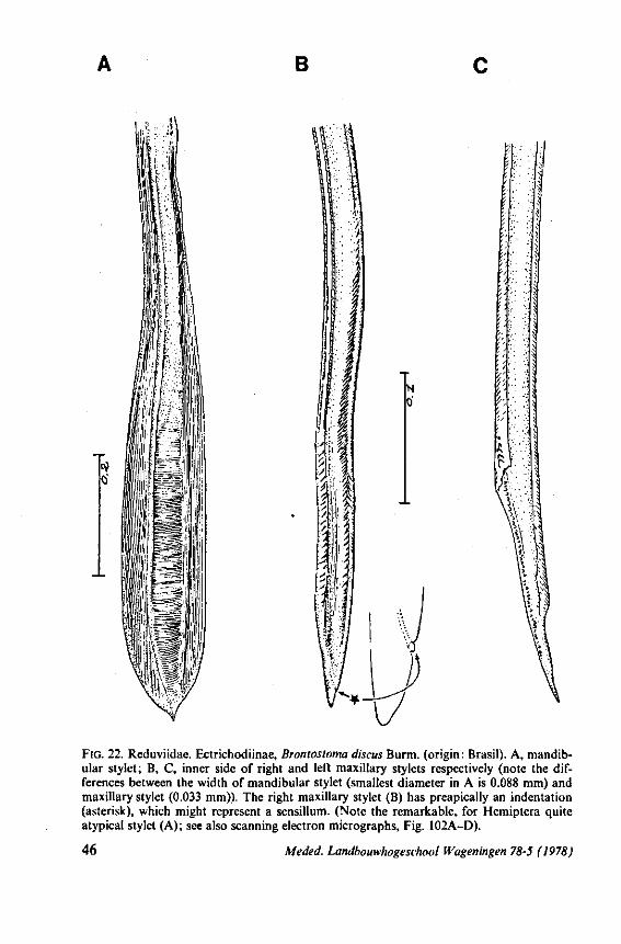
B
Ä
4:
FIG. 22. Reduviidae. Ectrichodiinae, Brontosioma discus Burm. (origin : Brasil). A, mandibular stylet; B, C, inner side of right and left maxillary stylets respectively (note the differences between the width of mandibular stylet (smallest diameter in A is 0.088 mm) and maxillary stylet (0.033 mm)). The right maxillary stylet (B) has preapically an indentation (asterisk), which might represent a sensillum. (Note the remarkable, for Hemiptera quite atypical stylet (A); see also scanning electron micrographs, Fig. 102A-D).
46 Meded. Landbouwhogeschool Wageningen 78-5 (1978)
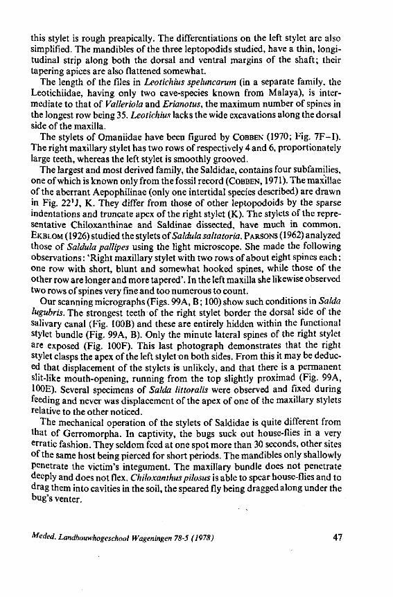
this stylet is rough preapically. The differentiations on the left stylet are also simplified. The mandibles of the three leptopodids studied, have a thin, longitudinal strip along both the dorsal and ventral margins of the shaft; their tapering apices are also flattened somewhat.
The length of the files in Leotichius speluncarum (in a separate family, the Leotichiidae, having only two cave-species known from Malaya), is intermediate to that of Valleriola and Erianotus, the maximum number of spines in the longest row being 35. Leotichius lacks the wide excavations along the dorsal side of the maxilla.
The stylets of Omaniidae have been figured by COBBEN (1970; Fig. 7F-I). The right maxillary stylet has two rows of respectively 4 and 6, proportionately large teeth, whereas the left stylet is smoothly grooved.
The largest and most derived family, the Saldidae, contains four subfamilies, one of which is known only from the fossil record (COBBEN, 1971). The maxillae of the aberrant Aepophilinae (only one intertidal species described) are drawn in Fig. 22'J, K. They differ from those of other leptopodoids by the sparse indentations and truncate apex of the right stylet (K). The stylets of the representative Chiloxanthinae and Saldinae dissected, have much in common. EKBLOM (1926) studied the stylets of Saldula saltatoria. PARSONS (1962) analyzed those of Saldula pallipes using the light microscope. She made the following observations : 'Right maxillary stylet with two rows of about eight spines each ; one row with short, blunt and somewhat hooked spines, while those of the other row are longer and more tapered'. In the left maxilla she likewise observed two rows of spines very fine and too numerous to count.
Our scanning micrographs (Figs. 99A, B ; 100) show such conditions in Salda lugubris. The strongest teeth of the right stylet border the dorsal side of the salivary canal (Fig. 100B) and these are entirely hidden within the functional stylet bundle (Fig. 99A, B). Only the minute lateral spines of the right stylet are exposed (Fig. 100F). This last photograph demonstrates that the right stylet clasps the apex of the left stylet on both sides. From this it may be deduced that displacement of the stylets is unlikely, and that there is a permanent slit-like mouth-opening, running from the top slightly proximad (Fig. 99A, 100E). Several specimens of Salda littoralis were observed and fixed during feeding and never was displacement of the apex of one of the maxillary stylets relative to the other noticed.
The mechanical operation of the stylets of Saldidae is quite different from that of Gerromorpha. In captivity, the bugs suck out house-flies in a very erratic fashion. They seldom feed at one spot more than 30 seconds, other sites of the same host being pierced for short periods. The mandibles only shallowly penetrate the victim's integument. The maxillary bundle does not penetrate deeply and does not flex. Chiloxanthus pilosus is able to spear house-flies and to drag them into cavities in the soil, the speared fly being dragged along under the bug's venter.
Meded. Landbouwhogeschool Wageningen 78-5 (1978) 47
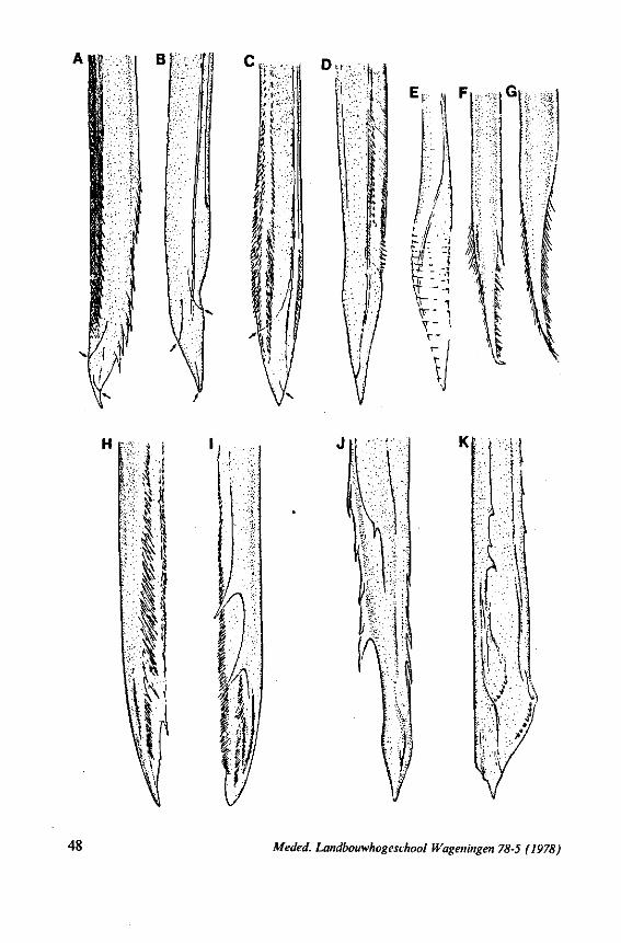
m mi
V\ < / - ^
48 Meded. Landbouwhogeschool Wageningen 78-5 (1978)
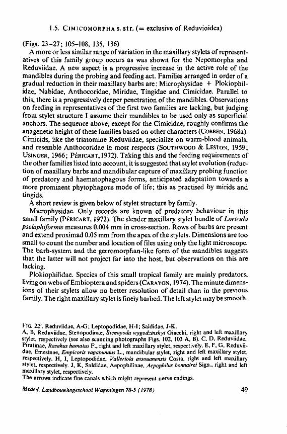
1.5. CiMicoMORPHA s. str. (= exclusive of Reduvioidea)
(Figs. 23-27; 105-108, 135, 136) A more or less similar range of variation in the maxillary stylets of represent
atives of this family group occurs as was shown for the Nepomorpha and Reduviidae. A new aspect is a progressive increase in the active role of the mandibles during the probing and feeding act. Families arranged in order of a gradual reduction in their maxillary barbs are: Microphysidae + Plokiophil-idae, Nabidae, Anthocoridae, Miridae, Tingidae and Cimicidae. Parallel to this, there is a progressively deeper penetration of the mandibles. Observations on feeding in representatives of the first two families are lacking, but judging from stylet structure I assume their mandibles to be used only as superficial anchors. The sequence above, except for the Cimicidae, roughly confirms the anagenetic height of these families based on other characters (COBBEN, 1968a). Cimicids, like the triatomine Reduviidae, specialize on warm-blood animals, and resemble Anthocoridae in most respects (SOUTHWOOD & LESTON, 1959; USINGER, 1966; PÉRICART, 1972). Taking this and the feeding requirements of the other families listed into account, it is suggested that stylet evolution (reduction of maxillary barbs and mandibular capture of maxillary probing function of predatory and haematophagous forms, anticipated adaptation towards a more prominent phytophagous mode of life ; this as practised by mirids and tingids.
A short review is given below of stylet structure by family. Microphysidae. Only records are known of predatory behaviour in this
small family (PÉRICART, 1972). The slender maxillary stylet bundle of Loricula pselaphiformis measures 0.004 mm in cross-section. Rows of barbs are present and extend proximad 0.05 mm from the apex of the stylets. Dimensions are too small to count the number and location of files using only the light microscope. The barb-system and the gerromorphan-like form of the mandibles suggests that the latter will not project far into the host, but observations on this are lacking.
Plokiophilidae. Species of this small tropical family are mainly predators, living on webs of Embioptera and spiders (CARAYON, 1974). The minute dimensions of their stylets allow no better resolution of detail than in the previous family. The right maxillary stylet is finely barbed. The left stylet may be smooth.
FIG. 22'. Reduviidae, A-G; Leptopodidae, H-I; Saldidae, J-K. A, B, Reduviidae, Stenopodinae, Stenopoda wygodzinskyi Giacchi, right and left maxillary stylet, respectively (see also scanning photographs Figs. 102, 103 A, B). C, D, Reduviidae, Piratinae, Rasahus hamatus F., right and left maxillary stylet, respectively. E, F, G, Reduviidae, Emesinae, Empicoris vagabundus L., mandibular stylet, right and left maxillary stylet, respectively. H, I, Leptopodidae, Valleriola assouanensis Costa, right and left maxillary stylet, respectively. J, K, Saldidae, Aepophilinae, Aepophilus bonnairei Sign., right and left maxillary stylet, respectively. The arrows indicate fine canals which might represent nerve endings.
Meded. Landbouwhogeschool Wageningen 78-5 (1978) 49
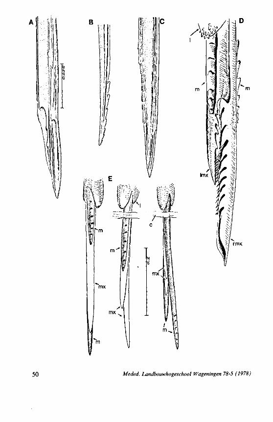
50 Meded. Landbouwhogeschool Wageningen 78-5 (1978)
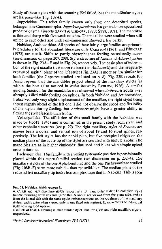
Study of these stylets with the scanning EM failed, but the mandibular stylets are harpoon-like (Fig. 108A).
Joppeicidae. This relict family known only from one described species, belongs in the Cimicomorpha. Joppeicusparadoxus is a general, non-specialized predator of small insects (DAVIS & USINGER, 1970; STYS, 1971). The mandible is thin and sharp with five weak notches. The maxillae were studied when still united to each other and under oil-immersion showed a few barbs.
Nabidae, Anthocoridae. All species of these fairly large families are primarily predatory (of the abundant literature only CARA YON (1961) and PÉRICART
(1972) are cited). Shifts to partly phytophagous feeding occasionally occur (see discussion on pages 207,208). Stylet structure of Nabis and AUoeorhynchus is shown in Fig. 23A-E and in Fig. 24, respectively. The basic plan of indentation of the right maxilla (it is more elaborate in Anthocoris) and the irregularly excavated sagittal plane of the left stylet (Fig. 23A) is more or less similar for both families (the 7 species studied are listed on p. 8). Fig. 23E reveals for Nabis rugosus that the mandibles project ahead of the maxillary stylets far within the host (also noticed in Nabis brevis by EKBLOM, 1926). A similar guiding function for the mandibles was observed when Anthocoris adults were abruptly killed while feeding on aphids. In both Nabidae and Anthocoridae, I observed only very slight displacement of the maxillae, the right stylet being thrust slightly ahead of the left one. I did not observe the speed and flexibility of the stylets during feeding, but Anthocoris might have a greater ability in flexing the stylet fascicle than Nabis.
Velocipedidae. The affiliation of this small family with the Nabidae, was made by BLÖTE (1945) and is confirmed in the present study from stylet and other cephalic structures (see p. 79). The right maxillary stylet of Scotomedes alienus bears a dorsal and ventral row of about 19 and 16 stout spines, respectively. The left stylet has the nabid plan, but five preapical ridges on the median plane of the acute tip of the stylet are serrated with minute knobs. The mandibles are as in higher cimicoids : flattened and blunt with simple apical cross-striations.
Pachynomidae. This family with a vexing systematic position is provisionally placed within this supra-familial section (see discussion on p. 232^) . The maxillary stylets of the one Aphelonotinae and the one Pachynominae studied (Fig. 108B-F) seem more nabid - than reduviid-like. The median plane of the reduviid left maxillary tip looks less complex than that in Nabidae. This is seen
FIG. 23. Nabidae. Nabis rugosus L. A, C, left and right maxillary stylets respectively; B, mandibular stylet; D, complete stylet bundle extruding from rostrum (note that A and C are viewed from the plane side, and D from the lateral side with the same optics; misconceptions on the roughness of the maxillary stylets readily arise when viewed only in one fixed orientation); E, movements of individual stylets during food uptake. c, cuticle of host; 1. labium; m, mandibular stylet; lmx, rmx, left and right maxillary stylets, respectively.
Meded. Landbouwhogeschool Wageningen 78-5 (1978) 51
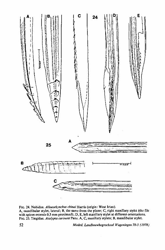
24 \
lm
%
25
B d f f l
Y^Y^-^^F^
FIG. 24. Nabidae. Alloeorhynchus chinai Harris (origin: West Irian). A, mandibular stylet, lateral; B, the same from the plane; C, right maxillary stylet (the file with spines extends 0,5 mm proximad); D, E, left maxillary stylet at different orientations. FIG. 25. Tingidae. Acalypta carinata Panz. A, C, maxillary stylets; B, mandibular stylet.
52 Meded. Landbouwhogeschool Wageningen 78-5 (1978)
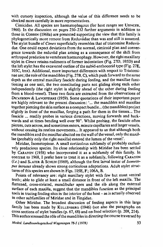
with cursory inspection, although the value of this difference needs to be checked more carefully in more representatives.
Cimicidae. All species are haematophagous (for host ranges see USINGER, 1966). In the discussion on pages 230-232 further arguments in addition to those in COBBEN (1968a) are presented supporting the view that this family is phylogenetically more remote from Reduviidae than was and still is assumed. The stylet bundle of Cimex superficially resembles that of triatomine Reduviidae. One could expect deviations from the normal, cimicoid plan and convergence towards the reduviid plan arising as a consequence of the shift from arthropod prédation to vertebrate haematophagy. However, the right maxillary stylet in Cimex retains rudiments of former indentation (Fig. 27D, 105D) and the left stylet has the excavated outline of the nabid-anthocorid type (Fig. 27E, 105C, lmx). Additional, more important differences with respect to Triatomi-nae are ; the role of the mandibles (Fig. 27B, C), which push forward to the same depth as the central maxillary fascicle during feeding, and the maxillae functioning as one unit, the two constituting parts not gliding along each other independently (the right stylet is slightly ahead of the other during feeding from a blood-vessel). These two facts are extracted from the observations of DICKERSON & LAVOIPIERRE (1959). Some quotations of their important paper are highly relevant to the present discussion: '... the mandibles and maxillae together piercing the skin surface as a compact bundle... (the mandibles) project slightly in front of the maxillae, forging a path for them in the tissues... The fascicle ... readily probes in various directions, moving forwards and backwards and at times bending well over 90°. Whilst probing, the fascicle often pierces, cuts across, and sometimes enters, minute capillaries and larger vessels, without ceasing its restless movements... It appeared to us that although both the mandibles and the maxillae abutted on the wall of the vessel, only the maxillae (probably only the right maxilla) entered the lumen of the vessel'.
Miridae, Isometopinae. A small corticolous subfamily of probably exclusively predacious species. Its close relationship with Miridae has been settled by CARAYON (1958) who incorporated it as a subfamily of this family. In contrast to 1968,1 prefer here to treat it as a subfamily, following CARAYON (I.e.) and SLATER & SCHUH (1969), although the first larval instar of Isometo-pus intrusus already shows strong corticolous adaptations. Adult stylet structures of this species are shown in Figs. 105E, F; 106A, B.
Points of relevancy are : right maxillary stylet with few but stout ventral teeth; able to glide at least a small distance in front of the left maxilla. The flattened, cross-striated, mandibular apex and the rib along the external surface of each maxilla, suggest that the mandibles function as the principal tools in tracing feeding sites in the interior of the host - as is actually observed in other subfamilies of Miridae and in Tingidae.
Other Miridae. The broadest discussion of feeding aspects in this large family has been made by KULLENBERG (1946) (see also the paragraphs on cross sections of stylet bundles (p. 67, 68) and on food selection (p. 208, 214). This authorstressed the rôle of the mandibles in directing the course traversed by
Meded. Landbouwhogeschool Wageningen 78-5 (1978) 53
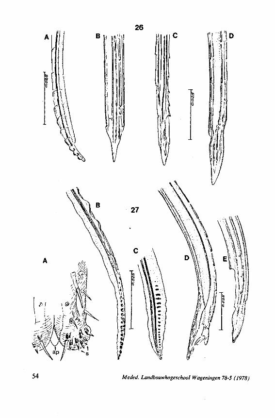
54 Meded. Landbouwhogeschool Wageningen 78-5 (1978)
the central stylet bundle. This is done by means of top curvature and by a groove interlocking with a longitudinal ridge on each of the maxillary stylets (demonstrated in 16 species with phytophagous and/or carnivorous habits). KULLENBERG fixed the stylets during sucking in some phytophagous species and found that the mandibles penetrate alternately to the same depth as the maxillae. There is only slight displacement of the maxillary stylets during feeding, the right stylet being more roughly armoured than the left one. Structural characteristics of the stylets are illustrated here as seen light-optically in Fig. 26 (Lygus pabulinus) and with the scanning EM (Deraeocoris spp.) in Figs. 106C-F, 107). Worth mentioning is the peculiar median plane of the left maxillary stylet (Fig. 107B, C). This does not seem to exhibit a simple longitudinal excavation fitting the groove of the opposed stylet to form the food canal as is usually illustrated. It seems instead that the left stylet contains a nearly complete hollow cylinder formed by a convex cuticular extension of the dorsal margin of the food canal, folding over to the ventral margin. Cross-sections of the stylet-bundle of 4 specimens of Deraeocoris, taken through the last rostral segment, did not, however, reveal such a cuticular extension within the food canal.
Mirid mandibular tips have a moderate number of lateral notches which extend somewhat transversely. The trunk of these stylets is ribbon-like and the longitudinal median groove, fitting to the rim of the adjacent maxilla can be seen by light-optics (Fig. 24B, gg). KULLENBERG (1946) thought that stylet insertion by phytophagous mirids is cautious and slow, but FLEMION (1958) recorded in Exolygus lineolaris rapid and extremely flexible penetration of the fascicle without following a particular path (The stylets travel in the tissue by short rapid thrusts and bend in any direction such as turning back or making a right angle'). SMITH (1926) attributed the purely mechanical damage of plant tissue by Lygus and Calocoris to the serrate edges of the mandibular stylets and to the frequent withdrawing and plunging of the stylet bundle. None of the many mirids studied or fixed during feeding was found to produce a stylet sheath.
Tingidae. A moderately large family containing only phytosuccivorous species. The stylets of two species were studied by light optics, one of them being depicted in Fig. 25A-C. The maxillary stylets closely resemble each other in gross shape and in finer differentiations, distinct spininess being absent. Rudimentary indentations are more apparent in the stylet of Fig. 25C, and I assume that this is the dextral one. The predominantly intracellular pathway through the plant-tissue is made equally by both pairs of stylets (JOHNSON, 1937; POLLARD, 1959; LIVINGSTONE, 1969). The stylet bundle fixed
FIG. 26. Miridae. Lygus pabulinus L. A, mandibular stylet; B. left maxillary stylet; C, D, right maxillary stylet. FIG. 27. Cimicidae. Cimex lectularius L. A, rostral tip, ventral aspect; B, mandibular stylet, lateral aspect; C, mandibular stylet, plane aspect; D, right maxillary stylet; E, left maxillary stylet (see also scanning electron micrographs, Fig. 105 A-D). ap, apical plate; s, sensilla.
Meded. Landbouwhogeschool Wageningen 78-5 (1978) 55
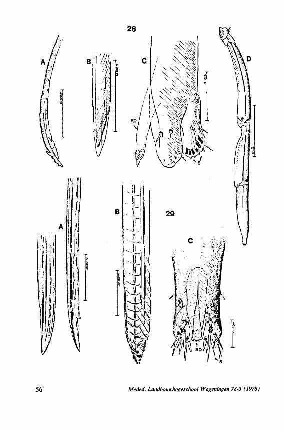
56 Meded. Landbouwhogeschool Wageningen 78-5 (1978)
in situ gives indications of bending capacities (POLLARD, 1959), but observations on the speed and exact behaviour of the individual maxillary stylets (probably both functioning as a single unit) are lacking. Although Tingidae is the only fairly large family of Heteroptera having plant-feeding species only, a capacity for stylet sheath formation has not been shown in species of the several genera so far studied.
1.6. PENTATOMOMORPHA
(Figs. 28-33, 109-111, 137) This major taxon is predominantly phytophagous. Some comments on
carnivorous species will be given at the end of this section. Stylet structures
Study of the stylets of representatives of most families (list on p. 9, 10) confirm more or less the data from the literature based on typical representatives (e.g. BONNEMAISON, 1952, Eurydema; BUGNION & POPOV, 1911, several spp.; COBBEN, 1953. Pyrrhocoris; EKBLOM, 1926, Rhyparochromus, 1930, Myrmus). The mandibles have acquired a complex ribbed texture over much of their outer and inner surfaces and their apices are knotted with irregular prominences (Figs. 29B, 30A, 31 A, 32B, 33A).
Only Aradidae (Aradus and Dysodius studied) have a long, terminal, file consisting of more than 20 regular, transverse, parallel grooves on the external side of each mandibular stylet. Details of the mandibles differ from taxon to taxon and may be used for taxonomie purposes after standardized comparative studies have been made. Of particular interest is the squamous texture of the inner face of each mandible, which scanning micrographs reveal to be different to the left and right of the longitudinal groove (Fig. 109A, B; 110A-D). This groove accomodates the longitudinal external maxillary rib. The orientation of this pavement is such that the forward thrust of one mandible will cause considerable friction against the outer surface of the adjacent maxillary stylet contributing to its inward deviation.
Both mandibles together with the maxillary bundle function as a single Plunging instrument (Fig. 31 A, B; 32A). The maxillae are either rather slender and sharply pointed as in Neididae (Fig. 29A), or broad and blunt as in more highly evolved taxa (Fig. 30B, C; 33B ; 11 IB).
Their inner surfaces show traces of small, widely spaced notches arranged in longitudinal strips (Figs. 29A, 33B). Some SEM photographs (Fig. 109F)
FIG. 28. Thaumastellidae. Thaumastella aradoides Horv. (origin: Sudan). A, mandibular stylet; B, united maxillary stylets; C, apex of rostrum, obliquely ventral (apical plate (ap) somewhat out of natural position); D, rostrum, dextro-lateral. FIG. 29. Berytinidae. Neides tipularius L.A, maxillary stylets; B, mandibular stylet; C, apex of rostrum, ventral. ap, apical plate; s, sensilla.
Meded. Landbouwhogeschool Wageningen 78-5 (1978) 57
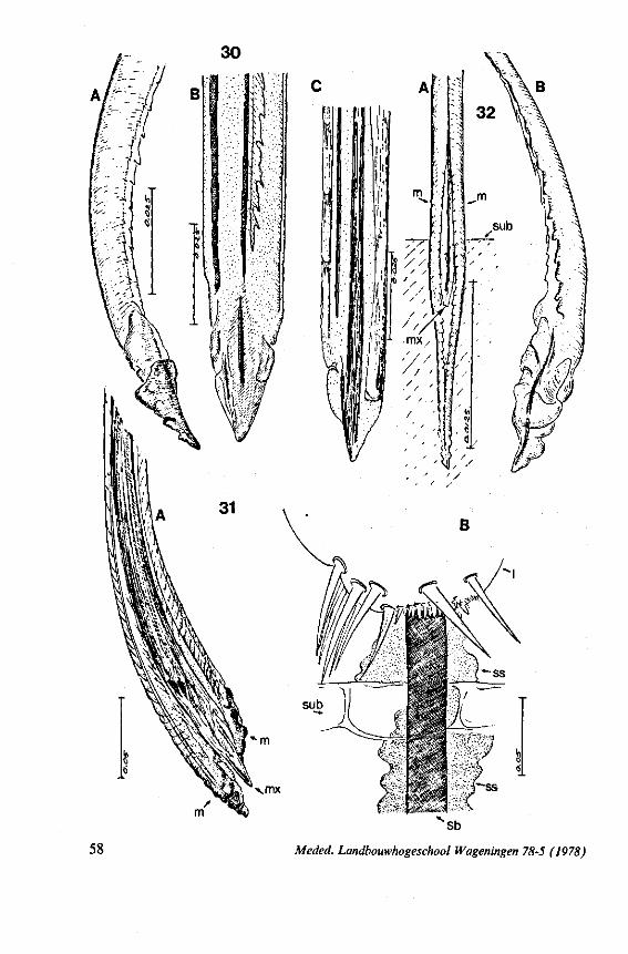
58 Meded. Landbouwhogeschool Wageningen 78-5 (1978)
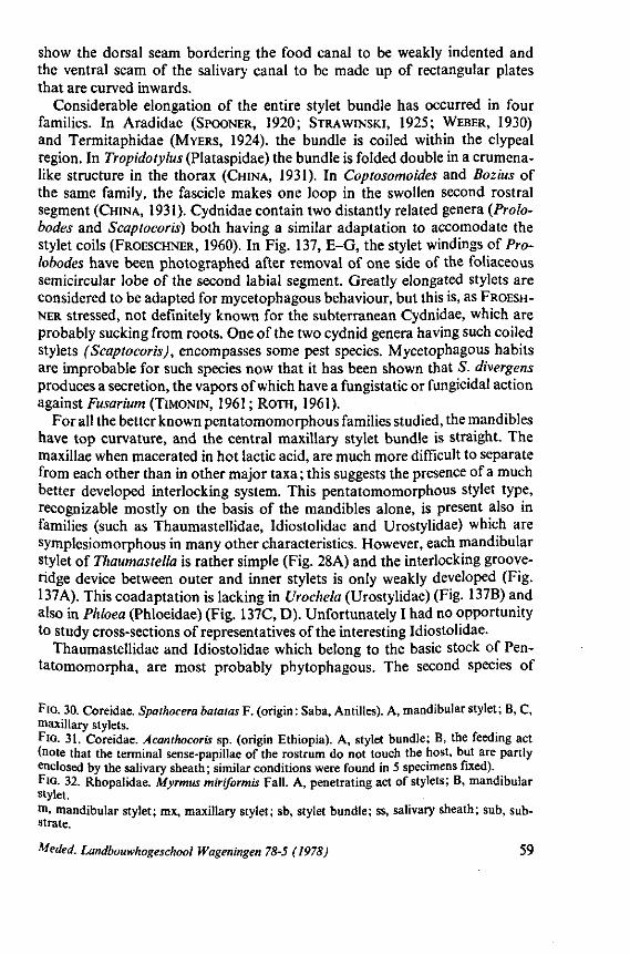
show the dorsal seam bordering the food canal to be weakly indented and the ventral seam of the salivary canal to be made up of rectangular plates that are curved inwards.
Considerable elongation of the entire stylet bundle has occurred in four families. In Aradidae (SPOONER, 1920; STRAWINSKI, 1925; WEBER, 1930) and Termitaphidae (MYERS, 1924). the bundle is coiled within the clypeal region. In Tropidotylus (Plataspidae) the bundle is folded double in a crumena-like structure in the thorax (CHINA, 1931). In Coptosomoides and Bozius of the same family, the fascicle makes one loop in the swollen second rostral segment (CHINA, 1931). Cydnidae contain two distantly related genera (Prolo-bodes and Scaptocoris) both having a similar adaptation to accomodate the stylet coils (FROESCHNER, 1960). In Fig. 137, E-G, the stylet windings of Pro-lobodes have been photographed after removal of one side of the foliaceous semicircular lobe of the second labial segment. Greatly elongated stylets are considered to be adapted for mycetophagous behaviour, but this is, as FROESH-
NER stressed, not definitely known for the subterranean Cydnidae, which are probably sucking from roots. One of the two cydnid genera having such coiled stylets (Scaptocoris), encompasses some pest species. Mycetophagous habits are improbable for such species now that it has been shown that S. divergens produces a secretion, the vapors of which have a fungistatic or fungicidal action against Fusarium (TIMONIN, 1961 ; ROTH, 1961).
For all the better known pentatomomorphous families studied, the mandibles have top curvature, and the central maxillary stylet bundle is straight. The maxillae when macerated in hot lactic acid, are much more difficult to separate from each other than in other major taxa; this suggests the presence of a much better developed interlocking system. This pentatomomorphous stylet type, recognizable mostly on the basis of the mandibles alone, is present also in families (such as Thaumastellidae, Idiostolidae and Urostylidae) which are symplesiomorphous in many other characteristics. However, each mandibular stylet of Thaumastella is rather simple (Fig. 28A) and the interlocking groove-ridge device between outer and inner stylets is only weakly developed (Fig. 137A). This coadaptation is lacking in Urochela (Urostylidae) (Fig. 137B) and also in Phloea (Phloeidae) (Fig. 137C, D). Unfortunately I had no opportunity to study cross-sections of representatives of the interesting Idiostolidae.
Thaumastellidae and Idiostolidae which belong to the basic stock of Pen-tatomomorpha, are most probably phytophagous. The second species of
FIG. 30. Coreidae. Spaihocera batatas F. (origin : Saba, Antilles). A, mandibular stylet; B, C, maxillary stylets. FIG. 31. Coreidae. Acanthocoris sp. (origin Ethiopia). A, stylet bundle; B, the feeding act (note that the terminal sense-papillae of the rostrum do not touch the host, but are partly enclosed by the salivary sheath; similar conditions were found in 5 specimens fixed). FIG. 32. Rhopalidae. Myrmus miriformis Fall. A, penetrating act of stylets; B, mandibular stylet. m, mandibular stylet; mx, maxillary stylet; sb, stylet bundle; ss, salivary sheath; sub, substrate.
Meded. Landbouwhogeschool Wageningen 78-5 (1978) 59
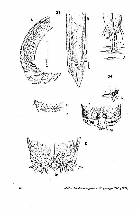
B
34
60 Meded. Landbouwhogeschool Wageningen 78-5 (1978)
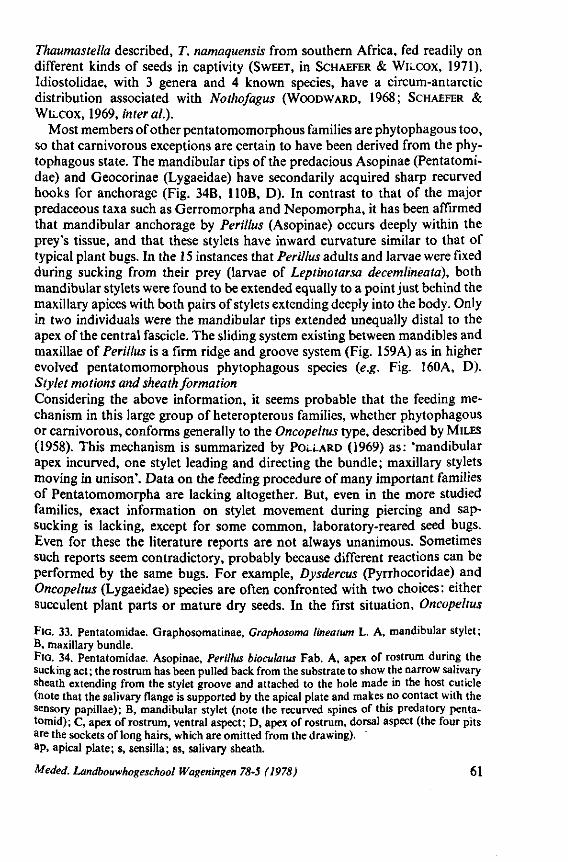
Thaumastella described, T. namaquensis from southern Africa, fed readily on different kinds of seeds in captivity (SWEET, in SCHAEFER & WILCOX, 1971). Idiostolidae, with 3 genera and 4 known species, have a circum-antarctic distribution associated with Nothofagus (WOODWARD, 1968; SCHAEFER & WILCOX, 1969, inter al).
Most members of other pentatomomorphous families are phytophagous too, so that carnivorous exceptions are certain to have been derived from the phytophagous state. The mandibular tips of the predacious Asopinae (Pentatomi-dae) and Geocorinae (Lygaeidae) have secondarily acquired sharp recurved hooks for anchorage (Fig. 34B, HOB, D). In contrast to that of the major predaceous taxa such as Gerromorpha and Nepomorpha, it has been affirmed that mandibular anchorage by Perillus (Asopinae) occurs deeply within the prey's tissue, and that these stylets have inward curvature similar to that of typical plant bugs. In the 15 instances that Perillus adults and larvae were fixed during sucking from their prey (larvae of Leptinotarsa decemlineata), both mandibular stylets were found to be extended equally to a point just behind the maxillary apices with both pairs of stylets extending deeply into the body. Only in two individuals were the mandibular tips extended unequally distal to the apex of the central fascicle. The sliding system existing between mandibles and maxillae of Perillus is a firm ridge and groove system (Fig. 159A) as in higher evolved pentatomomorphous phytophagous species {e.g. Fig. 160A, D). Stylet motions and sheath formation Considering the above information, it seems probable that the feeding mechanism in this large group of heteropterous families, whether phytophagous or carnivorous, conforms generally to the Oncopeltus type, described by MILES (1958). This mechanism is summarized by POLLARD (1969) as: 'mandibular apex incurved, one stylet leading and directing the bundle; maxillary stylets moving in unison'. Data on the feeding procedure of many important families of Pentatomomorpha are lacking altogether. But, even in the more studied families, exact information on stylet movement during piercing and sap-sucking is lacking, except for some common, laboratory-reared seed bugs. Even for these the literature reports are not always unanimous. Sometimes such reports seem contradictory, probably because different reactions can be performed by the same bugs. For example, Dysdercus (Pyrrhocoridae) and Oncopeltus (Lygaeidae) species are often confronted with two choices: either succulent plant parts or mature dry seeds. In the first situation, Oncopeltus
FIG. 33. Pentatomidae. Graphosomatinae, Graphosoma lineatwn L. A, mandibular stylet ; B, maxillary bundle. FIG. 34. Pentatomidae. Asopinae, Perillus bioculatus Fab. A, apex of rostrum during the sucking act; the rostrum has been pulled back from the substrate to show the narrow salivary sheath extending from the stylet groove and attached to the hole made in the host cuticle (note that the salivary flange is supported by the apical plate and makes no contact with the sensory papillae); B, mandibular stylet (note the recurved spines of this predatory penta-tomid); C, apex of rostrum, ventral aspect; D, apex of rostrum, dorsal aspect (the four pits are the sockets of long hairs, which are omitted from the drawing), ap, apical plate; s, sensilla; ss, salivary sheath.
Meded. Landbouwhogeschool Wageningen 78-5 (1978) 61
tends to suck from phloem by intracellular piercing accompanied by an external and internal deposition of salivary sheath material (MILES, 1959, 1967; BON-GERS, 1969b). The internal sheath may be absent as in Dysdercus koenigii (SAXENA, 1963) or present as in D.fasciatus (KHAN, 1972). When feeding on seeds, only a surface flange of sheath material is secreted. When these bugs suck from cut surfaces of seed kernels or from freely exposed liquid, they do not produce sheath material at all. Therefore, piercing through a non-soluble, hard substrate stimulates the formation of a salivary flange (SAXENA, 1963). SWEET (1964) suggested that direct access to digestible food or water inhibits the inherent behavior of these bugs to produce a salivary cone. HORI (1968) found that the cabbage bug Eurydema rugosa (Pentatomidae) produced a flange and a sheath when feeding on phloem sap, but not when sucking from meso-phyl cells. MILES (1972) suggested that such predetermination of stylet activity depends on the physiological state of the individual bug. Piesma quadrata (Piesmatidae), a vector of circulative virus of beet, is a typical phloem-feeder, which almost always pierces intercellularly. Both flange and continuous, internal, salivary sheath are secreted. White spots on the leafs are caused when parenchym cells are incidentally touched and sucked out (EHRHARDT & SMUT-TERER, 1965).
I found copious sheath material, both external and internal at 8 feeding sites fed on by the coreid Acanthocoris sp. (Fig. 3IB, ss). A photograph of v. DER VECHT (1933) of a feeding track of the rhopalid Dasynus piperis in pepper-fruit is very suggestive of an internal stylet sheath. SWEET (1964) found, as he called it 'salivary sheath fluid' in many lygaeids studied by him, including predacious Geocorinae, predacious Pentatomidae (Podisus, Perillus) and in species of Alydidae and Rhopalidae, which he reared on sunflower seeds. I assume that he had only the external salivary cone in mind in this survey. I could not trace internal sheath tracks in prey of Perillus, but an external flange is usually present. Fig. 34A shows that this external sheath extends into the stylet groove and does not surround the rostral tip (see also page 201).
The stylet behaviour of Perillus within its prey (Colorado beetle larvae) is much the same as that practised by Lygaeidae and Pyrrhocoridae when they are emptying seed contents. I observed that the manner in which Dysdercus, Oncopeltus and Spilostethus spp. dilute and suck the contents of sunflower seeds, is as described by SAXENA (1963) for Dysdercus. The stylets rapidly extend and withdraw from several zones of the substrate for varying distances. MILES (1958) recorded for Oncopeltus that regular cyclus of forward and backward movements of the stylet bundle occurred about every quarter second, irrespective of the type of artificial substrate. Indeed, the restless speed with which the stylets swish through the substrate is such that the share of the individual stylets is difficult to analyse, even when the course of the whole bundle is easily traced as when thin slices of kernel are provided. The mandibles are alternately forced fairly deeply into the substrate, then they stop for a short while serving as a quiver for the to and fro glidings of the maxillary bundle. To the eel-like undulations made by the distal end of the fascicle, only the two
62 Meded. Landbouwhogeschool Wageningen 78-5 (1978)
central stylets seem to contribute. The maxillary bundle repeatedly traverses and extends exposed from the opposite side of the kernel-slices, at which time delivery of watery saliva can often be observed.
Spilostethus pandurus (Lygaeidae), when sucking on strips of onion bulb, behaves in the same way, although the areas explored by its maxillary bundle are more compartmentalized ; considerable bending in the proximal portions of the free maxillary fascicle is prevented by cell-walls. To overcome this limitation, the mandibular quiver is often be retracted and guided in other directions. The maxillae seem to function more or less as a unit, but their tips slide longitudinally and nearly imperceptibly over one another at a rapid rate. The apex of the right maxilla is usually ahead of the opposite one during these snapping motions. As mentioned above, the apex of the maxillary bundle bends apparently without the assistance of the mandibles. This curvature may be explained by the twisting line of closure between the two maxillae (Fig. 109D). This photograph also shows that the maxillae, when not displaced, have a preapical slit-like opening. It is possible that only this functional mouth operates when seed-bugs take phloem-sap or water. Indeed, accurate observations on how the stylets behave and with what speed they operate in intact plant tissue during ploem searching are lacking. Published observations generally are on stylets fixed in situ or on salivary tracks.
Stylet behavior of Perillus when sucking from Leptinotarsa larvae is essentially the same as described above for phytophagous bugs sucking from seeds. The pronounced stylet swinging was noticed in other Asopinae by GULDE (1919). In fact, the motility, speed and bending capacity of the stylets, resemble closely those of Gerromorpha. However, the mandibles penetrate much more deeply into the prey or seed and the maxillary bundle does not perform the minute rasping vibrations characterizing the surface bugs.
It has been repeatedly suggested that Asopinae kill their prey by injecting toxic saliva (references in SCHUMACHER (1910) and in OETTING & YONKE, 1971b). These and other authors noticed thrashing reactions on the part of the prey when it was attacked by the predator. Struggling of the prey ended 3-10 minutes after being pierced by the stylets. With such delayed reactions on the part of the prey it is difficult to discriminate between toxic and feeding effects, because the bugs immediately start to suck when their stylets are inserted. My observations on Perillus bioculatus do not support the presence of a strong paralyzing role for the saliva. On the contrary, older prey larvae were often seen to continue leaf-eating in a quite normal way, when they were speared simultaneously by two to four older Perillus larvae. Young bug larvae behave more like ectoparasites than predators. Attached by their stylets to a beetle •arva, they let themselves be transported like ticks, often upside down and with their legs motionless and spread.
In future experiments on this topic, age and size of both prey and predator must be noted, since these can both influence reaction patterns and salivation effects.
Meded. Landbouwhogeschool Wageningen 78-5 (1978) 63
1.7. THAUMASTOCOROIDEA
(Fig. 138B, C) The phylogenetic position of the phytophagous Thaumastocoridae, a family of few species and with a restricted distribution, is not well-understood. Although cimicoid (DRAKE & SLATER, 1957; KUMAR, 1964) or reduvioid (STYS, 1962) affinities have been suggested, I showed in 1968 that egg-structures might be more pentatomomorphous. Embryological data are badly needed. The mandible has distinct top curvature and its ribbed structure and irregular apex reminds one of pentatomoid mandibles. There is a rim-groove connection between maxillary and mandibular stylets (Fig. 138B, C).
1.8. DIPSOCOROMORPHA
(Fig. 11D) Species in this taxon are cryptic, small-sized and live in damp places in the
litter zone of mostly tropical regions. The Schizopteridae, particularly, show considerable morphological diversification (EMSLEV, 1969) and most living species are not yet described. Feeding habits remain very obscure, although it is generally believed that these bugs are predators. I studied six species representing four families (listed on p. 10). A superficial analysis of stylet morphology (specimens only 1 -2 mm) suggest that they are carnivorous, because their maxillary stylets have fringes of fine spines. Both maxillae of Hypsipteryx and Hypselosoma have at least two long rows of barbs. Ceratocombus coleoptratus (Fig. 11D) and Schizoptera stricklandi have one of the maxillae barbed, the opposed stylet being nearly devoid of teeth, at least appearing so when observed light-optically. The mandibles are rather flattened, sharply pointed and have a few apical notches in S. stricklandi or more than 10 hooks in the Hypsipteryx and Hypselosoma species studied.
1.9. ENICOCEPHALOMORPHA
(Figs. HE, 138E, F) Species of this isolated group belong to a single family and are ground-
dwellers in warm regions. Beak structure and special adaptations of the front legs strengthen the general opinion that all species are predators (STYS, 1970a, inter ai). Structures of mandibular and maxillary stylets (Fig. 1 IE) expressively point to carnivorous habits, since strongly barbed maxillae were shown in the present paper to be used for triturating the contents of arthropod prey. Stereos-can electron micrographs reveal a system of barbs in oblique cross-sections (Fig. 138E, F), which entirely matches conditions existing in Gerromorpha.
64 Meded. Landbouwhogeschool Wageningen 78-5 (1978)
2. O T H E R S T R U C T U R E S A S S O C I A T E D W I T H F E E D I N G
2.1. CROSS-SECTIONS T H R O U G H ROSTRUM AND STYLETS
Drawings of cross-sections of the stylet-bundle, based on light-optical observations, have been presented by a number of authors :
Gerromorpha: Mesovelia (EKBLOM 1930), Aquarius (SERVADEI 1946), Gerris (CRANSTON & SPRAGUE 1961), Limnogonus (CHENG 1967), Velia (EKBLOM
1926; SERVADEI 1946), and Hydrometra (EKBLOM 1926; SERVADEI 1946; SPRAGUE 1956).
Nepomorpha: Gelastocoris (PARSONS 1959), Nepa (GEISE 1883), Notonecta (GEISE 1883; WEDDE 1885; BUTT 1943); Sigara (WEDDE 1885; EKBLOM 1930), and Corixa (Benwitz 1956).
Reduvioidea; Platymeris (EDWARDS 1960), Rhodnius (LAVOIPIERRE C.S.
1959), and Triatoma (BARTH 1953). Leptopodomorpha: Saldula (EKBLOM 1926; PARSONS 1962). Cimicomorpha: Cimex (WEDDE 1885; PATTON & CRAGG 1913; SNODGRASS
1944), Nabis (EKBLOM 1926), Lygus(Aw\Ti 1914), 17 species of Miridae (KUL-
LENBERG 1946), Tingis (LIVINGSTONE 1969), and Urentius (POLLARD 1959). Pentatomomorpha : Pyrrhocoris (WEDDE 1885), Dysdercus (MACGILL 1947;
KHAN 1972), Oncopeltus (BUTT 1943), Rhyparochromus (EKBLOM 1926), Gra-phosoma (BUGNION & POPOV 1911), Myrmus (EKBLOM 1930) and Anasa (TOWER 1914).
Since the species treated in this paper represent all major groups, reasonable generalizations, based on the cross-sections, can be made.
Sec t ion t h r o u g h s ty le t b und l e in loto Because of structural irregularities in the stylet apices, only sections made
far proximal to the bundle apex may be compared. The width of the bundle in most Heteroptera, inclusive of Enicocephalidae (Fig. 138E), Dipsocoridae and Thaumastocoridae (Fig. 138B), is distinctly greater than the height, when mandibular and maxillary stylets are adjacent (in the enicocephalid studied, the width of the bundle is four times the height). In the better known families of Pentatomomorpha, dorsoventral and lateral axes are similar in length (Figs. 158E, 159A, 160D). This is true also of the more evolutionarily isolated pen-tatomomorphous families such as Thaumastellidae (Fig. 137A), Urostylidae (Fig. 137B) and Phloeidae (Fig. 137D). No clear relation exists between these two types of stylet bundles (dorso-ventrally or laterally compressed) and feeding habits. Phytophagous mirids and tingids are similar to other, but carnivorous, cimicoids, and the stylet bundle of the carnivorous Perillus closely resembles that of other, phytophagous, pentatomomorphous bugs.
Mandibular stylets The mandibles of generalized Gerromorpha are irregularly circular in
Meded. Landbouwhogeschool Wageningen 78-5 (1978) 65
outline and are positioned mid-laterally to the maxillary stylets (Fig. 143A, C; 147A, C). Furthermore, these lateral stylets have no obvious interlocking with the central stylets so that rotation of the maxillary stylets, independent of the mandibular stylets, is possible. In higher Gerromorpha (Veliidae, Gerri-dae), the diameter of the proximal parts of the mandibular stylets is enlarged and faces a greater surface of the central bundle (Fig. 129B, F).
A closer association of both pairs of stylets is seen to varying degrees in all other Heteroptera. In Saldidae (Leptopodomorpha), Nepomorpha and lower Cimicomorpha (inclusive of Loriculidae and Joppeicidae), the centre of the mandibular stylet is situated somewhat below the mid-transverse axis of the maxillary bundle (Figs. 99D, 131D, 135B, C; 155A). Except for Saldidae, the mandibular stylets in these taxa and also in Reduviidae, encompass the central bundle dorsally and ventrally with foliaceous cuticular extensions (Figs. 133,135D, E). The lateral surfaces of the mandibular stylets are provided with fine, longitudinal ribs (Figs. 99F, 13ID, 133E). Rotation of the mandibular stylets independent of the maxillary stylets is supposedly largely reduced by this relationship. Such an independent rotation seems impossible in Miridae, Tingidae and most Pentatomomorpha, in which the inner, dorsal edge of the mandibular stylet has a groove into which a longitudinal rib of the maxillary stylet fits (Figs. 105F, r; 110A, 154F, 157A, C; 159A, 160A, D, E). Such interlocking devices, by which differential protrusion of the mandibular stylets guide the path of the maxillary stylets, have been known for a long time to students of these family groups. WEBER (1930) based his theory of stylet movement in Hemiptera on this type of relationship between the four stylets. The same interlocking device occurs also in the primarily carnivorous Isometopus (Miridae, Fig. 105F, r), Xylastodoris (Thaumastocoridae, Fig. 138B, gr), and in Ochteridae (Fig. 131D, int ; the only example so far discovered in Nepomorpha). Phloea (Phloeidae) is the only example so far found in Pentatomomorpha to lack this mandibular-maxillary coadaptation (Fig. 137D), although in Thaumastellidae it is only weakly developed (Fig. 137A).
For all Heteroptera studied, it is significant that the left and right mandibular stylets are mirror images of each other. In Gerromorpha, this symmetry is not very clear, because of irregular, cuticular extensions of the surface and because of rotation of the stylets.
Maxillary stylets The cross-sections made of the stylets of Gerromorpha (Figs. 128-130,
143-145, 147, 149-152A-E), some Nepomorpha (Figs. 131, 132) and of Enicocephalidae (Fig. 138E) disclose the extremely intricate pattern of interweaving combs knitting the left and right stylets together. They are more or less square in section except in the enicocephalid studied. However, the dorsal surface of the bundle is notably wider than the ventral surface in the proximal sections of the gerromorphous Hebrus (Fig. 143D) and in Hydrometra (Fig. 147C). This preponderance of cuticular material in the dorsal half of the stylet is generally present in adults of other heteropteran categories, inclusive of
66 Meded. Landbouwhogeschool Wageningen 78-5 (1978)
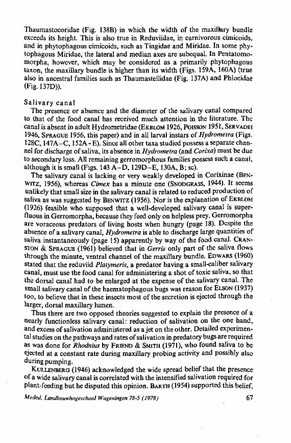
Thaumastocoridae (Fig. 138B) in which the width of the maxiltery bundle exceeds its height. This is also true in Reduviidae, in carnivorous cimicoids, and in phytophagous cimicoids, such as Tingidae and Miridae. In some phytophagous Miridae, the lateral and median axes are subequal. In Pentatomo-morpha, however, which may be considered as a primarily phytophagous taxon, the maxillary bundle is higher than its width (Figs. 159A, 160A) (true also in ancestral families such as Thaumastellidae (Fig. 137A) and Phloeidae (Fig. 137D)).
Sa l iva ry c ana l The presence or absence and the diameter of the salivary canal compared
to that of the food canal has received much attention in the literature. The canal is absent in adult Hydrometridae (EKBLOM 1926, POISSON 1951, SERVADEI
1946, SPRAGUE 1956, this paper) and in all larval instars of Hydrometra (Figs. 128C, 147A-C, 152A-E). Since all other taxa studied possess a separate channel for discharge of saliva, its absence in Hydrometra (and Corixa) must be due to secondary loss. All remaining gerromorphous families possess such a canal, although it is small (Figs. 143 A - D , 129D-E, 130A, B; sc).
The salivary canal is lacking or very weakly developed in Corixinae (BEN-WITZ, 1956), whereas Cimex has a minute one (SNODGRASS, 1944). It seems unlikely that small size in the salivary canal is related to reduced production of saliva as was suggested by BENWITZ (1956). Nor is the explanation of EKBLOM
(1926) feasible who supposed that a well-developed salivary canal is superfluous in Gerromorpha, because they feed only on helpless prey. Gerromorpha are voraceous predators of living hosts when hungry (page 18). Despite the absence of a salivary canal, Hydrometra is able to discharge large quantities of saliva instantaneously (page 15) apparently by way of the food canal. CRAN
STON & SPRAGUE (1961) believed that in Gerris only part of the saliva flows through the minute, ventral channel of the maxillary bundle. ED WARS (1960) stated that the reduviid Platymeris, a predator having a small-caliber salivary canal, must use the food canal for administering a shot of toxic saliva, so that the dorsal canal had to be enlarged at the expense of the salivary canal. The small salivary canal of the haematophagous bugs was reason for ELSON (1937) too, to believe that in these insects most of the secretion is ejected through the larger, dorsal maxillary lumen.
Thus there are two opposed theories suggested to explain the presence of a nearly functionless salivary canal: reduction of salivation on the one hand, and excess of salivation administered as a jet on the other. Detailed experimental studies on the pathways and rates of salivation in predatory bugs are required as was done for Rhodnius by FRIEND & SMITH (1971), who found saliva to be ejected at a constant rate during maxillary probing activity and possibly also during pumping.
KULLENBERG (1946) acknowledged the wide spread belief that the presence of a wide salivary canal is correlated with the intensified salivation required for plant-feeding but he disputed this opinion. BARTH (1954) supported this belief,
Meded. Landbouwhogeschool Wageningen 78-5 (1978) 67
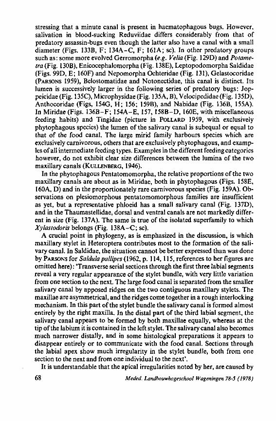
stressing that a minute canal is present in haematophagous bugs. However, salivation in blood-sucking Reduviidae differs considerably from that of predatory assassin-bugs even though the latter also have a canal with a small diameter (Figs. 133B, F; 134A-C, F; 161A; sc). In other predatory groups such as: some more evolved Gerromorpha (e.g. Velia (Fig. 129D) and Potame-tra (Fig. 130B), Enicocephalomorpha (Fig. 138E), Leptopodomorpha Saldidae (Figs. 99D, E; 160F) and Nepomorpha Ochteridae (Fig. 131), Gelastocoridae (PARSONS 1959), Belostomatidae and Notonectidae, this canal is distinct. Its lumen is successively larger in the following series of predatory bugs: Jop-peicidae (Fig. 135C), Microphysidae (Fig. 135A, B), Velocipedidae (Fig. 135D), Anthocoridae (Figs, 154G, H; 156; 159B), and Nabidae (Fig. 136B, 155A). In Miridae (Figs. 136B-F; 154A-E, 157, 158B-D, 160E, with miscellaneous feeding habits) and Tingidae (picture in POLLARD 1959, with exclusively phytophagous species) the lumen of the salivary canal is subequal or equal to that of the food canal. The large mirid family harbours species which are exclusively carnivorous, others that are exclusively phytophagous, and examples of all intermediate feeding types. Examples in the different feeding categories however, do not exhibit clear size differences between the lumina of the two maxillary canals (KULLENBERG, 1946).
In the phytophagous Pentatomomorpha, the relative proportions of the two maxillary canals are about as in Miridae, both in phytophagous (Figs. 158E, 160A, D) and in the proportionately rare carnivorous species (Fig. 159A). Observations on plesiomorphous pentatomomorphous families are insufficient as yet, but a representative phloeid has a small salivary canal (Fig. 137D), and in the Thaumastellidae, dorsal and ventral canals are not markedly different in size (Fig. 137A). The same is true of the isolated superfamily to which Xylastodoris belongs (Fig. 138A-C; sc).
A crucial point in phylogeny, as is emphasized in the discussion, is which maxillary stylet in Heteroptera contributes most to the formation of the salivary canal. In Saldidae, the situation cannot be better expressed than was done by PARSONS for Saldula pallipes (1962, p. 114,115, references to her figures are omitted here) : 'Transverse serial sections through the first three labial segments reveal a very regular appearance of the stylet bundle, with very little variation from one section to the next. The large food canal is separated from the smaller salivary canal by apposed ridges on the two contiguous maxillary stylets. The maxillae are asymmetrical, and the ridges come together in a rough interlocking mechanism. In this part of the stylet bundle the salivary canal is formed almost entirely by the right maxilla. In the distal part of the third labial segment, the salivary canal appears to be formed by both maxillae equally, whereas at the tip of the labium it is contained in the left stylet. The salivary canal also becomes much narrower distally, and in some histological preparations it appears to disappear entirely or to communicate with the food canal. Sections through the labial apex show much irregularity in the stylet bundle, both from one section to the next and from one individual to the next'.
It is understandable that the apical irregularities noted by her, are caused by
68 Meded. Landbouwhogeschool Wageningen 78-5 (1978)
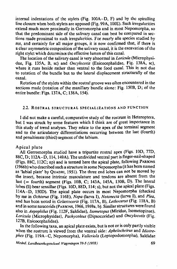
internal indentations of the stylets (Fig. 100A-D, F) and by the spiralling line closure when both stylets are apposed (Fig. 99A, 100E). Such irregularities extend much more proximally in Gerromorpha and in most Nepomorpha, so that the predominant side of the salivary canal can best be compared in sections made proximal to such irregularities. For nearly alle species studied by me, and certainly for all major groups, it is now confirmed that, if there is a clear asymmetric composition of the salivary canal, it is the excavation of the right stylet which determines the effective lumen of this canal.
The location of the salivary canal is very abnormal in Loricula (Microphysi-dae, Fig. 135A, B, sc) and Oncylocotis (Enicocephalidae, Fig. 138A, sc), where it runs beside rather than ventral to the food canal. This in not due to rotation of the bundle but to the lateral displacement structurally of the canal.
Rotation of the stylets within the rostral groove was often encountered in the sections made (rotation of the maxillary bundle alone: Fig. 150B, D; of the entire bundle: Figs. 137A, C; 138A, 154).
2.2. ROSTRAL STRUCTURAL SPECIALIZATIONS AND FUNCTION
I did not make a careful, comparative study of the rostrum in Heteroptera, but I was struck by some features which I think are of great importance in this study of trend analyses. They relate to the apex of the terminal segment and to the articulatory differentiations occurring between the last (fourth) and penultimate (third) segment of the labium.
Apical plate All Gerromorpha studied have a tripartite rostral apex (Figs. 10D, 77D,
88C, D; 112A-D, 114,149A). The undivided ventral part is finger-nail-shaped (Figs. 88C, 112C; ap) and is termed here the apical plate, following PARSONS (1966b) who described such a structure in some Nepomorpha (it has been named as 'labial plate' by QUADRI, 1951). The three end lobes can not be moved by the insect, because intrinsic musculature and tendons are absent from the last (= fourth) segment (Figs. 10B, C; 143A, 145A, 150B, D). The lateral lobes (11) bear sensillae (Figs. 10D, 88D, 114; s), but not the apical plate (Figs. 114A-D, 150D). The apical plate occurs in most Nepomorpha (checked by me in Ochterus (Fig. 116E), Nepa (larva I), Notonecta (larva I), and Plea, and has been noted in Gelastocoris (Fig. 117A, B), Lethocerus (Fig. 118A, B) and in some naucorids (PARSONS, 1966,1969a, b). Similar structures were found also in Aepophilus (Fig. 112F, Saldidae), Isometopus (Miridae, Isometopinae), Loricula (Microphysidae), Pachycoleus (Dipsocoridae) and Oncylocotis (Fig. 127B, Enicocephalidae).
In the following taxa, an apical plate exists, but is not or is only partly visible when the rostrum is viewed from the ventral side: Aphelocheirus and Idiotre-phes (Fig. 119A-C, Nepomorpha), Valleriola (Leptopodomorpha), Saldidae
Meded. Landbouwhogeschool Wageningen 78-5 (1978) 69
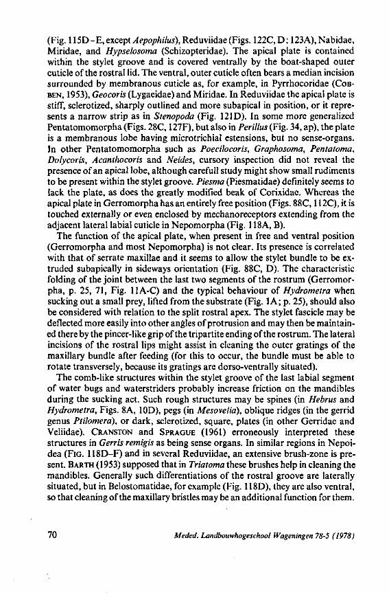
(Fig. 115D-E, except Aepophilus), Reduviidae (Figs. 122C,D; 123A),Nabidae, Miridae, and Hypselosoma (Schizopteridae). The apical plate is contained within the stylet groove and is covered ventrally by the boat-shaped outer cuticle of the rostral lid. The ventral, outer cuticle often bears a median incision surrounded by membranous cuticle as, for example, in Pyrrhocoridae (COB-BEN, 1953), Geocoris (Lygaeidae) and Miridae. In Reduviidae the apical plate is stiff, sclerotized, sharply outlined and more subapical in position, or it represents a narrow strip as in Stenopoda (Fig. 121D). In some more generalized Pentatomomorpha (Figs. 28C, 127F), but also in Perillus (Fig. 34, ap), the plate is a membranous lobe having microtrichial estensions, but no sense-organs. In other Pentatomomorpha such as Poecilocoris, Graphosoma, Pentatoma, Dolycoris, Acanthocoris and Neides, cursory inspection did not reveal the presence of an apical lobe, although carefull study might show small rudiments to be present within the stylet groove. Piesma (Piesmatidae) definitely seems to lack the plate, as does the greatly modified beak of Corixidae. Whereas the apical plate in Gerromorpha has an entirely free position (Figs. 88C, 112C), it is touched externally or even enclosed by mechanoreceptors extending from the adjacent lateral labial cuticle in Nepomorpha (Fig. 118A, B).
The function of the apical plate, when present in free and ventral position (Gerromorpha and most Nepomorpha) is not clear. Its presence is correlated with that of serrate maxillae and it seems to allow the stylet bundle to be extruded subapically in sideways orientation (Fig. 88C, D). The characteristic folding of the joint between the last two segments of the rostrum (Gerromorpha, p. 25, 71, Fig. 11A-C) and the typical behaviour of Hydrometra when sucking out a small prey, lifted from the substrate (Fig. 1 A; p. 25), should also be considered with relation to the split rostral apex. The stylet fascicle may be deflected more easily into other angles of protrusion and may then be maintained there by the pincer-like grip of the tripartite ending of the rostrum. The lateral incisions of the rostral lips might assist in cleaning the outer gratings of the maxillary bundle after feeding (for this to occur, the bundle must be able to rotate transversely, because its gratings are dorso-ventrally situated).
The comb-like structures within the stylet groove of the last labial segment of water bugs and waterstriders probably increase friction on the mandibles during the sucking act. Such rough structures may be spines (in Hebrus and Hydrometra, Figs. 8A, 10D), pegs (in Mesovelia), oblique ridges (in the gerrid genus Ptilomera), or dark, sclerotized, square, plates (in other Gerridae and Veliidae). CRANSTON and SPRAGUE (1961) erroneously interpreted these structures in Gerris remigis as being sense organs. In similar regions in Nepoi-dea (FIG. 118D-F) and in several Reduviidae, an extensive brush-zone is present. BARTH (1953) supposed that in Triatoma these brushes help in cleaning the mandibles. Generally such differentiations of the rostral groove are laterally situated, but in Belostomatidae, for example (Fig. 118D), they are also ventral, so that cleaning of the maxillary bristles may be an additional function for them.
70 Meded. Landbouwhogeschool Wageningen 78-5 (1978)
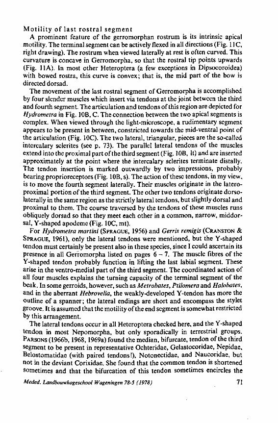
Motility of last rostral segment A prominent feature of the gerromorphan rostrum is its intrinsic apical
motility. The terminal segment can be actively flexed in all directions (Fig. 11C, right drawing). The rostrum when viewed laterally at rest is often curved. This curvature is concave in Gerromorpha, so that the rostral tip points upwards (Fig. IIA). In most other Heteroptera (a few exceptions in Dipsocoroidea) with bowed rostra, this curve is convex ; that is, the mid part of the bow is directed dorsad.
The movement of the last rostral segment of Gerromorpha is accomplished by four slender muscles which insert via tendons at the joint between the third and fourth segment. The articulation and tendons of this region are depicted for Hydrometra in Fig. 10B, C. The connection between the two apical segments is complex. When viewed through the light-microscope, a rudimentary segment appears to be present in between, constricted towards the mid-ventral point of the articulation (Fig. IOC). The two lateral, triangular, pieces are the so-called intercalary sclerites (see p. 73). The parallel lateral tendons of the muscles extend into the proximal part of the third segment (Fig. 10B, It) and are inserted approximately at the point where the intercalary sclerites terminate distally. The tendon insertion is marked outwardly by two impressions, probably bearing proprioreceptors (Fig. 10B, s). The action of these tendons, in my view, is to move the fourth segment laterally. Their muscles originate in the latero-proximal portion of the third segment. The other two tendons originate dorsolateral^ in the same region as the strictly lateral tendons, but slightly dorsal and proximal to them. The course traversed by the tendons of these muscles runs obliquely dorsad so that they meet each other in a common, narrow, middor-sal, Y-shaped apodeme (Fig. IOC, mt).
For Hydrometra martini (SPRAGUE, 1956) and Gerris remigis (CRANSTON & SPRAGUE, 1961), only the lateral tendons were mentioned, but the Y-shaped tendon must certainly be present also in these species, since I could ascertain its presence in all Gerromorpha listed on pages 6 - 7 . The muscle fibres of the Y-shaped tendon probably function in lifting the last labial segment. These arise in the ventro-medial part of the third segment. The coordinated action of all four muscles explains the turning capacity of the terminal segment of the beak. In some gerroids, however, such as Metrobates, Ptilomera and Halobates, and in the aberrant Hebrovelia, the weakly-developed Y-tendon has more the outline of a spanner ; the lateral endings are short and encompass the stylet groove. It is assumed that the motility of the end segment is somewhat restricted by this arrangement.
The lateral tendons occur in all Heteroptera checked here, and the Y-shaped tendon in most Nepomorpha, but only sporadically in terrestrial groups. PARSONS (1966b, 1968,1969a) found the median, bifurcate, tendon of the third segment to be present in representative Ochteridae, Gelastocoridae, Nepidae, Belostomatidae (with paired tendons!), Notonectidae, and Naucoridae, but not in the deviant Corixidae. She found that the common tendon is shortened sometimes and that the bifurcation of this tendon sometimes encircles the
Meded. Landbouwhogeschool Wageningen 78-5 (1978) 71
stylet groove, as is the case in some gerrids mentioned above. Its function was interpreted by her as deforming the cross-sectional shape of the stylet groove, thus restricting movement of the stylet bundle.
As well as being present in primarily hygrophilous bugs, I found the median apodeme in the third labial part only of some reduviid larvae (probably Reduvii-nae) and (weakly developed) in Loricula (Microphysidae). It appeared to be absent in representatives of all other major groups studied including the nepomorphan helotrephid genus Idiotrephes. The lateral tendons are present in all taxa, whether carnivorous or phytophagous. It may be that some reduviids still have the ability to flex the last rostral segment in the sagittal plane, but this is not the case in Triatoma (BARTH, 1953). It is assumed that in the bulk of terrestrial Heteroptera, including Dipsocoroidea and Enicocephaloidea, the lateral retractor muscles usually function together providing rigid support to the rostral tip. Alternate contraction and relaxation of these muscles allows some lateral movement, as is the case in Triatoma (according to BARTH, 1953).
PARSONS (1962) described the lateral muscles of the third rostral segment of Saldula (Saldidae) as functioning in retracting the last segment or in moving it laterally. I found representatives of this group to pierce straight forward only, eventual lateral deviation of the terminal segment was achied passively by the bugs through pressure of their heads. Passive lifting of this segment during the sucking act will be restored by two, backward-pointing, dorsal pegs which touch the adjacent borderline of the penultimate segment (Fig. 115A, F,pr).
The intrinsic motility of the rostral tip must be studied in live members of additional family groups. Species of the schizopterid genus Nannocoris have a concave rostrum (EMSLEY, 1969), as in Gerromorpha, with the distal segment curved upwards. I had no opportunity to study the tendon arrangement of these bugs, but other dipsocoroids studied lacked the median tendon. This is also the case in Hypsipteryx which seemingly also has a concave beak. In Hypsip-teryx, however, this concavity is produced by the long slender third labial segment only ; the last segment being very short and blunt.
Close in front of the insertion of the lateral tendons are two pits situated one beside the other in Gerromorpha (Fig. 10B, s). These pits probably register the lateral bending of the rostral joint. In some Nepomorpha, only one peg was found, its axis pointing proximally to the borderline of the third segment. In Aepophilus, a primitive member of Saldidae, two pits are also present, but they are more separated from each other. The most dorsal pit in other Saldidae extends as a peg. Such arrangements occur also more or less variably in terrestrial groups.
Intercalary sclerites This term, used for the two dorsal plates at the base of the fourth rostral
segment of Nepomorpha (PARSONS, 1959, 1966b, 1968, 1969a), is not quite appropriate for most Gerromorpha (Hebridae, Mesoveliidae, Hydrometridae). Here, they form a thin, cuticular layer around the whole rostral circumference,
72 Meded. Landbouwhogeschool Wageningen 78-5 (1978)
separated into two dorsal and two ventral flaps. Their distal margins are extended very thinly and irregularly and are adpressed close to the segment (Fig. 113C, D, asterisks). It should be remembered that these bugs have the greatest ability to revolve the rostral apex. In connection with this, these flaps apparently function as a support, much like of a stretch-bandage. The dorsal flaps are folded over each other (Fig. 113A, F), thus completely covering the stylet groove, and preventing the stylet bundle from escaping from the groove when the rostral apex is sharply bent upwards.
In higher Gerromorpha, such as some Veliidae (Oravelia, Fig. 113B) and Gerridae (Aquarius, Metrobates, Ptilomera, Halobates, reported also for other Gerridae by MATSUDA, 1960) only the two dorsal coverings exist and these resemble the intercalary sclerites of Nepomorpha. They are thicker, more clearly defined and squamous. In these evolved waterstriders, the median tendon, which flexes the apical joint (see p. 71), is a short, sclerotized strip (Fig. 9IE, t), incised at its apex where it contacts the base of the intercalary sclerites left and right of the stylet groove (also in Nepomorpha). In the less-derived Gerromorpha (p. 71) this tendon was described as being long and delicate, its arms diverging widely towards the lateral sides of the joint between the two terminal segments (Fig. IOC, mt ). The strong reduction of this tendon and its close association with the intercalary sclerites and the stylet groove is most apparent in Ptilomera, where it is fused with the bottom of the stylet groove (Fig. 9IE, ce). This configuration must limit the lifting potential of the last rostral segment and probably provides better control over the sliding of the stylet bundle.
A sclerotized ring in the stylet groove, where the tendon inserts, was not observed in Gerromorpha. Such a ring occurs in Nepomorpha and PARSONS (1969a) suggested that a pull on the tendon would flatten this ring, causing the intercalary sclerites to come together and thus restrict movements of the stylet bundle. The same author examined labial structures in representatives of Ochteridae, Gelastocoridae, Nepidae, Belostomatidae, Notonectidae and Naucoridae (PARSONS, 1959,1966b, 1968,1969a). She stated (1969, p. 298) that in all Nepomorpha thus far examined, a holdfast mechanism was present similar to that in Aphelocheirus, but that some possess only the muscle and sclerotized ring and not the intercalary sclerites. However, in the same paper (p. 304), she mentioned that in the naucorid Ambrysus, the holdfast mechanism is present, but not the muscle associated with it. On the other hand, I could not find the median tendon in the first larval instar of Notonecta, nor in the adults of the naucorid Ilyocoris and the helotrephid Idiotrephes (the semicircular reinforcement of the stylet groove is present also in these taxa).
Intercalary sclerites occur in all 6 families of water bugs studied by PARSONS, but are absent in some naucorid genera and weakly developed in others. It is very unlikely that the intercalary sclerites have evolved independently and recently as was thought by PARSONS (1969a) (see discussion section 4). The sclerites are missing also in Coleopterocoris (Potamocoridae), Plea (Pleidae) and Idiotrephes (Helotrephidae). Outside the major semiaquatic and aquatic
Meded. Landbouwhogeschool Wageningen 78-5 (1978) 73
groups, differentiations resembling intercalary sclerites were found only in Embiophila (Plokiophilidae, Fig. 123D, is), in several Reduviidae (Figs. 121 A, ris; 122E) and in some Pachynomidae (Fig. 123C).
F o l d i ng s of the r o s t r a l s egmen t s Differentiations of the proximal rostral segments and their musculature
will be omitted here, although they certainly harbour features important in phylogeny. Anatomies of these parts have been presented for representatives of a few families, e.g. Gerridae (CRANSTON & SPRAGUE, 1961), Saldidae (PAR
SONS, 1962), families of Nepomorpha (PARSONS 1969a, and others), Pyrrhoco-ridâe (KHAN, 1972), Dinidoridae (BANERJI, 1960) and Tingidae (LIVINGSTONE,
1969). Two structures not dealt whith in the literature are a pair of laterolongitudinal
sutures which, left and right of the stylet-groove, delimit a dorsal (dorsal when rostrum is extended), cuticular strip separate from the lateral walls of the rostrum. Such sutures do not occur in the principal predatory groups. They do exist in all labial segments of the Pentatomomorpha examined. In the first segment, the sutures are often indistinct and in the last they sometimes reach only halfway to its tip (Fig. 28D, Thaumastella, Thaumastellidae). In the cimicoid families, such sutures are only distinctly present in Tingidae and, to a lesser degree, in Miridae, but are absent in Isometopus (Isometopinae). In Cimicidae, all lateral areas of the rostrum are membranous. Embiophila (Plokiophilidae), Nabis and Himacerus (Nabidae) have only the distal half of the third segment longitudinally lined by a membranous strip (Fig. 126A, s), whereas Orius (Anthocoridae) has only a very faint line of desclerotization.
Considering these facts in relation to feeding behaviour, it is here suggested that the longitudinal sutures allow the rostral segments to be compressed dorso-ventrally when they are 'elbow' folded. The stylets are lifted from the labial groove by the consequent shortening of the rostrum, thus making deeper penetration of the stylets within the substrate possible. Such bending actions of the rostrum are practised by representative Pentatomomorpha (e.g. BUGNION & POPOV, 1911; SCHUBERT, 1927; WEBER, 1930; BONNEMAISON, 1952; HORI,
1968), and by Cimicidae (HASE, 1917; KEMPER, 1932; DICKERSON & LAVOIPIER-
RE, 1959). Miridae (KULLENBERG, 1946) and Tingidae (LIVINGSTONE, 1969). The rostral tip does not penetrate through a membrane covering a source
of food*. The observation of LIVINGSTONE (1969) that the terminal segment of Tingis partly penetrates plant tissue, needs re-affirmation. The stylet bundle of Pentatomomorpha is sometimes entirely free from the rostrum when the latter is being placed back in the repose position under the body (Acanthaso-matidae (PENDERGRAST, 1952), Lygaeidae (BONGERS, 1968; WOLFRAM, 1972),
* Saldidae and Ochteridae insert their slender, but stiff and unflexible rostrum in between soil-particles, presumably probing for insect larvae, enchytraeids etc., or for capillary water. The beak of Anthocoridae is similarly constructed, and it needs to be checked whether anthocorids utilize only the stylets or, less likely, the rostral apex in penetrating the plant cuticle in case of feeding on leafminer-larvae (see obervation, p. 207).
74 Meded. Landbouwhogeschool Wageningen 78-5 (1978)
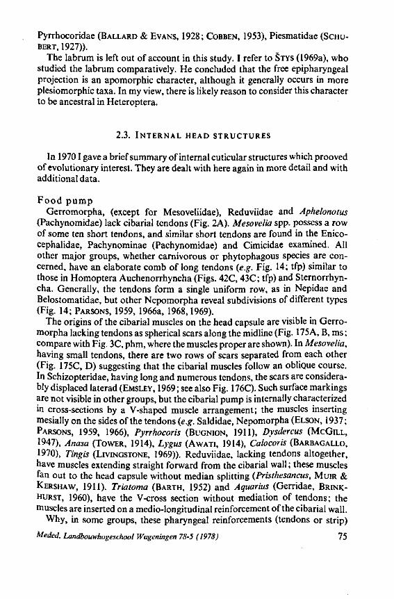
Pyrrhocoridae (BALLARD & EVANS, 1928; COBBEN, 1953), Piesmatidae (SCHUBERT, 1927)).
The labrum is left out of account in this study. I refer to STYS (1969a), who studied the labrum comparatively. He concluded that the free epipharyngeal projection is an apomorphic character, although it generally occurs in more plesiomorphic taxa. In my view, there is likely reason to consider this character to be ancestral in Heteroptera.
2.3. INTERNAL HEAD STRUCTURES
In 19701 gave a brief summary of internal cuticular structures which prooved of evolutionary interest. They are dealt with here again in more detail and with additional data.
Food pump Gerromorpha, (except for Mesoveliidae), Reduviidae and Aphelonotus
(Pachynomidae) lack cibarial tendons (Fig. 2A). Mesovelia spp. possess a row of some ten short tendons, and similar short tendons are found in the Enico-cephalidae, Pachynominae (Pachynomidae) and Cimicidae examined. All other major groups, whether carnivorous or phytophagous species are concerned, have an elaborate comb of long tendons (e.g. Fig. 14; tfp) similar to those in Homoptera Auchenorrhyncha (Figs. 42C, 43C; tfp) and Sternorrhyn-cha. Generally, the tendons form a single uniform row, as in Nepidae and Belostomatidae, but other Nepomorpha reveal subdivisions of different types (Fig. 14; PARSONS, 1959, 1966a, 1968,1969).
The origins of the cibarial muscles on the head capsule are visible in Gerromorpha lacking tendons as spherical scars along the midline (Fig. 175A, B, ms; compare with Fig. 3C, phm, where the muscles proper are shown). In Mesovelia, having small tendons, there are two rows of scars separated from each other (Fig. 175C, D) suggesting that the cibarial muscles follow an obliQue course. In Schizopteridae, having long and numerous tendons, the scars are considerably displaced laterad (EMSLEY, 1969 ; see also Fig. 176C). Such surface markings are not visible in other groups, but the cibarial pump is internally characterized in cross-sections by a V-shaped muscle arrangement; the muscles inserting mesially on the sides of the tendons (e.g. Saldidae, Nepomorpha (ELSON, 1937; PARSONS, 1959, 1966), Pyrrhocoris (BUGNION, 1911), Dysdercus (MCGILL, 1947), Anasa (TOWER, 1914), Lygus (AWATI, 1914), Calocoris (BARBAGALLO, 1970), Tingis (LIVINGSTONE, 1969)). Reduviidae, lacking tendons altogether, have muscles extending straight forward from the cibarial wall ; these muscles fan out to the head capsule without median splitting (Pristhesancus, MUIR & KERSHAW, 1911). Triatoma (BARTH, 1952) and Aquarius (Gerridae, BRINK-HURST, 1960), have the V-cross section without mediation of tendons; the muscles are inserted on a medio-longitudinal reinforcement of the cibarial wall.
Why, in some groups, these pharyngeal reinforcements (tendons or strip)
Meded. Landbouwhogeschool Wageningen 78-5 (1978) 75
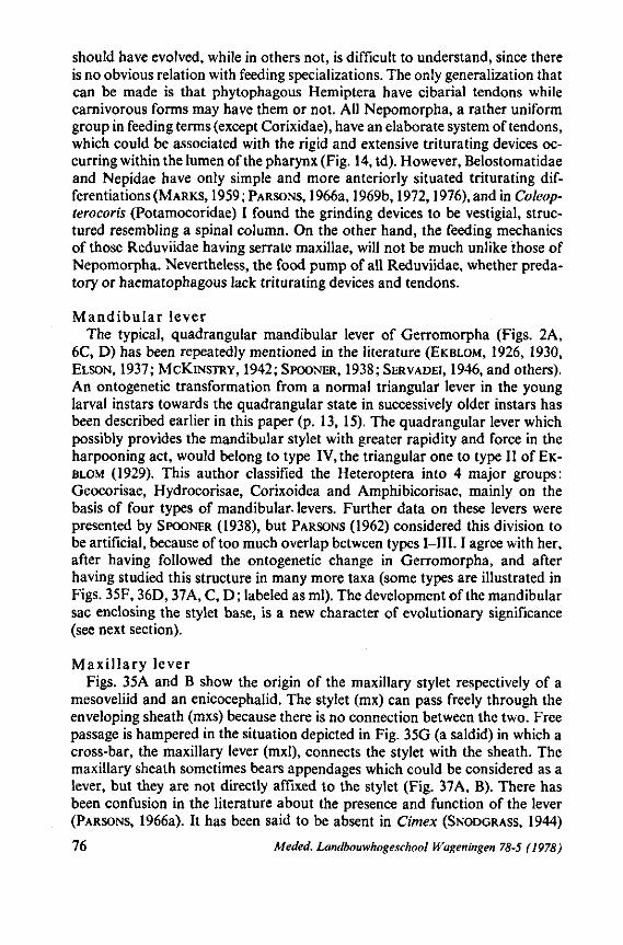
should have evolved, while in others not, is difficult to understand, since there is no obvious relation with feeding specializations. The only generalization that can be made is that phytophagous Hemiptera have cibarial tendons while carnivorous forms may have them or not. All Nepomorpha, a rather uniform group in feeding terms (except Corixidae), have an elaborate system of tendons, which could be associated with the rigid and extensive triturating devices occurring within the lumen of the pharynx (Fig. 14, td). However, Belostomatidae and Nepidae have only simple and more anteriorly situated triturating differentiations (MARKS, 1959; PARSONS, 1966a, 1969b, 1972,1976), and in Coleop-terocoris (Potamocoridae) I found the grinding devices to be vestigial, structured resembling a spinal column. On the other hand, the feeding mechanics of those Reduviidae having serrate maxillae, will not be much unlike those of Nepomorpha. Nevertheless, the food pump of all Reduviidae, whether predatory or haematophagous lack triturating devices and tendons.
Mandibular lever The typical, quadrangular mandibular lever of Gerromorpha (Figs. 2A,
6C, D) has been repeatedly mentioned in the literature (EKBLOM, 1926, 1930, ELSON, 1937; MCKINSTRY, 1942; SPOONER, 1938; SERVADEI, 1946, and others). An ontogenetic transformation from a normal triangular lever in the young larval instars towards the quadrangular state in successively older instars has been described earlier in this paper (p. 13, 15). The quadrangular lever which possibly provides the mandibular stylet with greater rapidity and force in the harpooning act, would belong to type IV, the triangular one to type II of EKBLOM (1929). This author classified the Heteroptera into 4 major groups: Geocorisae, Hydrocorisae, Corixoidea and Amphibicorisae, mainly on the basis of four types of mandibular, levers. Further data on these levers were presented by SPOONER (1938), but PARSONS (1962) considered this division to be artificial, because of too much overlap between types I—III. I agree with her, after having followed the ontogenetic change in Gerromorpha, and after having studied this structure in many more taxa (some types are illustrated in Figs. 35F, 36D, 37A, C, D; labeled as ml). The development of the mandibular sac enclosing the stylet base, is a new character of evolutionary significance (see next section).
Maxillary lever Figs. 35A and B show the origin of the maxillary stylet respectively of a
mesoveliid and an enicocephalid. The stylet (mx) can pass freely through the enveloping sheath (mxs) because there is no connection between the two. Free passage is hampered in the situation depicted in Fig. 35G (a saldid) in which a cross-bar, the maxillary lever (mxl), connects the stylet with the sheath. The maxillary sheath sometimes bears appendages which could be considered as a lever, but they are not directly affixed to the stylet (Fig. 37A, B). There has been confusion in the literature about the presence and function of the lever (PARSONS, 1966a). It has been said to be absent in Cimex (SNODGRASS, 1944)
76 Meded. Landbouwhogeschool Wageningen 78-5 (1978)
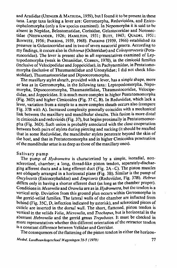
and Aradidae (USINGER & MATSUDA, 1959), but I found it to be present in these taxa. Large taxa lacking a lever are: Gerromorpha, Reduvioidea, and Enico-cephalomorpha (only a few species examined). In Nepomorpha it is said to be absent in Nepidae, Belostomatidae, Corixidae, Gelastocoridae and Notonec-tidae (NEISWANDER, 1926; HAMILTON, 1931; Burr, 1943; QUADRI, 1951; BENWITZ, 1956; PARSONS, 1959, 1968). PARSONS (1959, 1966) established its presence in Gelastocoridae and in two of seven naucorid genera. According to my findings, it coeurs also in Ochterus (Ochteridae) and Coleopterocoris (Pota-mocoridae). The lever is present also in all representatives examined of Lep-topodomorpha (weak in Omaniidae, COBBEN, 1970), in the cimicoid families (inclusive of Velocipedidae and Joppeicidae), in Pachynomidae, in Pentatomo-morpha (inclusive of Thaumastellidae and Urostylidae; I did not check Idio-stolidae), Thaumastocoridae and Dipsocoromorpha.
The maxillary stylet sheath, provided with a lever, has a simple shape, more or less as in Gerromorpha, in the following taxa: Leptopodomorpha, Nepomorpha, Dipsocoromorpha, Thaumastellidae, Thaumastocoridae, Velocipedidae, and Joppeicidae. It is much more complex in higher Pentatomomorpha (Fig. 36D) and higher Cimicoidea (Fig. 37 C, B). In Reduviidae, which lack a lever, variation from a simple to a more complex sheath occurs also (compare Fig. 37B with A). Increased complexity generally coincides with a mechanical link between the maxillary and mandibular sheaths. This fusion is more distal in cimicoids and reduvioids (Fig. 37), but begins proximally in Pentatomomorpha (Fig. 36D). Such union is probably associated with the close cooperation between both pairs of stylets during piercing and sucking (it should be recalled that in some Reduviidae, the mandibular stylets penetrate beyond the skin of the host, and that in Pentatomomorpha and in higher Cimicoidea penetration of the mandibular setae is as deep as those of the maxillary ones).
Salivary pump The pump of Hydrometra is characterized by a simple, isoradial, non-
sclerotized, chamber; a long, thread-like piston tendon, separately-discharging afferent ducts and a long efferent duct (Fig. 2A-C). The piston muscles are obliquely arranged in a horizontal plane (Fig. 3B). Similar is the pump of Oncylocotis (Enicocephalidae) and Empicoris (Reduviidae, Fig. 37B). Hebrus differs only in having a shorter efferent duct (as long as the chamber proper). Conditions in Mesovelia and Oravelia are as in Hydrometra, but the tendon is a vertical strip. Deviation from this ground plan occurs within Gerromorpha in the gerrid-veliid families. The lateral walls of the chamber are inflected from behind (Fig. 35C, D, inflection indicated by asterisk), and sclerotized pieces of cuticle are inserted in the dorsal wall. The short, flattened, piston tendon is vertical in the veliids Velia, Microvelia, and Trochopus, but is horizontal in the aberrant Hebrovelia and the gerrid genus Trepobates. It must be checked in more representatives whether this different orientation of the retractor tendon is a constant difference between Veliidae and Gerridae.
The consequences of the flattening of the piston tendon in either the horizon-
Meded. Landbouwhogeschool Wageningen 78-5 (1978) 77
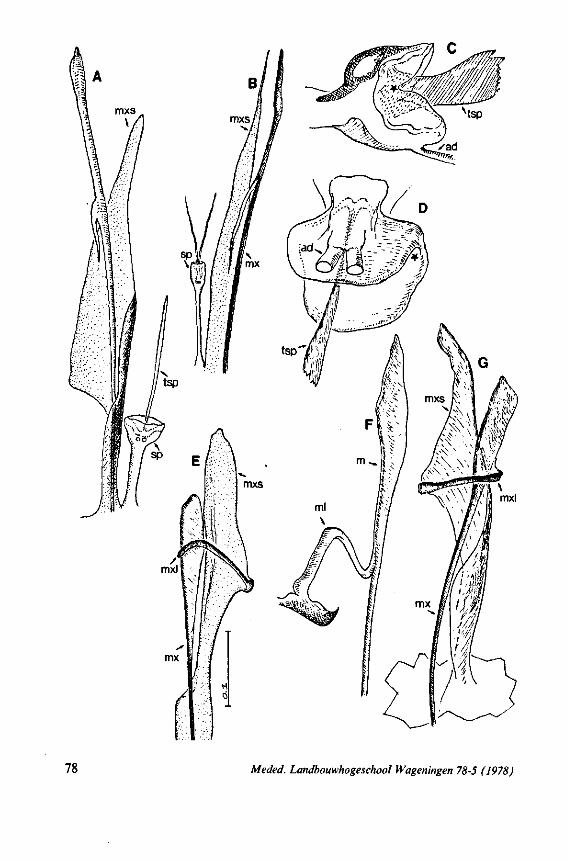
78 Meded. Landbouwhogeschool Wageningen 78-5 (1978)
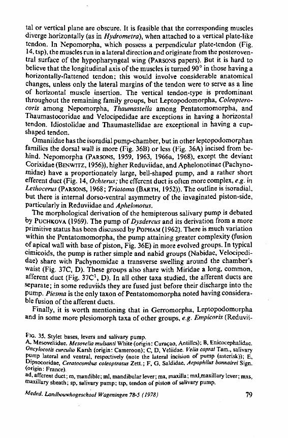
tal or vertical plane are obscure. It is feasible that the corresponding muscles diverge horizontally (as in Hydrometra), when attached to a vertical plate-like tendon. In Nepomorpha, which possess a perpendicular plate-tendon (Fig. 14, tsp), the muscles run in a lateral direction and originate from the posteroventral surface of the hypopharyngeal wing (PARSONS papers). But it is hard to believe that the longitudinal axis of the muscles is turned 90° in those having a horizontally-flattened tendon; this would involve considerable anatomical changes, unless only the lateral margins of the tendon were to serve as a line of horizontal muscle insertion. The vertical tendon-type is predominant throughout the remaining family groups, but Leptopodomorpha, Coleoptero-coris among Nepomorpha, Thaumastella among Pentatomomorpha, and Thaumastocoridae and Velocipedidae are exceptions in having a horizontal tendon. Idiostolidae and Thaumastellidae are exceptional in having a cup-shaped tendon.
Omaniidae has the isoradial pump-chamber, but in other leptopodomorphan families the dorsal wall is more (Fig. 36B) or less (Fig. 36A) incised from behind. Nepomorpha (PARSONS, 1959, 1963, 1966a, 1968), except the deviant Corixidae (BENWITZ, 1956)), higher Reduviidae, and Aphelonotinae (Pachyno-midae) have a proportionately large, bell-shaped pump, and a rather short efferent duct (Fig. 14, Ochterus; the efferent duct is often more complex, e.g. in Lethocerus (PARSONS, 1968; Triatoma (BARTH, 1952)). The outline is isoradial, but there is internal dorso-ventral asymmetry of the invaginated piston-side, particularly in Reduviidae and Aphelonotus.
The morphological derivation of the hemipterous salivary pump is debated by PUCHKOVA (1969). The pump of Dysdercus and its derivation from a more primitive status has been discussed by POPHAM (1962). There is much variation within the Pentatomomorpha, the pump attaining greater complexity (fusion of apical wall with base of piston, Fig. 36E) in more evolved groups. In typical cimicoids, the pump is rather simple and nabid groups (Nabidae, Velocipedidae) share with Pachynomidae a transverse swelling around the chamber's waist (Fig. 37C, D). These groups also share with Miridae a long, common, afferent duct (Fig. 37C\ D). In all other taxa studied, the afferent ducts are separate; in some reduviids they are fused just before their discharge into the pump. Piesma is the only taxon of Pentatomomorpha noted having considerable fusion of the afferent ducts.
Finally, it is worth mentioning that in Gerromorpha, Leptopodomorpha and in some more plesiomorph taxa of other groups, e.g. Empicoris (Reduvii-
FIG. 35. Stylet bases, levers and salivary pump. A, Mesoveliidae. Mesovelia mulsanti White (origin: Curaçao, Antilles); B, Enicocephalidae. Oncyhcotis curculio Karsh (origin: Cameroon); C, D, Veliidae. Velia capraiTam., salivary pump lateral and ventral, respectively (note the lateral incision of pump (asterisk)); E, Dipsocoridae, Ceratocombus coleoptratus Zett. ; F, G, Saldidae, Aepophilus bonnairei Sign. (origin: France). ad, afferent duct; m, mandible; ml, mandibular lever; mx, maxilla; mxl,maxillary lever; mxs, maxillary sheath; sp, salivary pump; tsp, tendon of piston of salivary pump.
Meded. Landbouwhogeschool Wageningen 78-5 (1978) 79
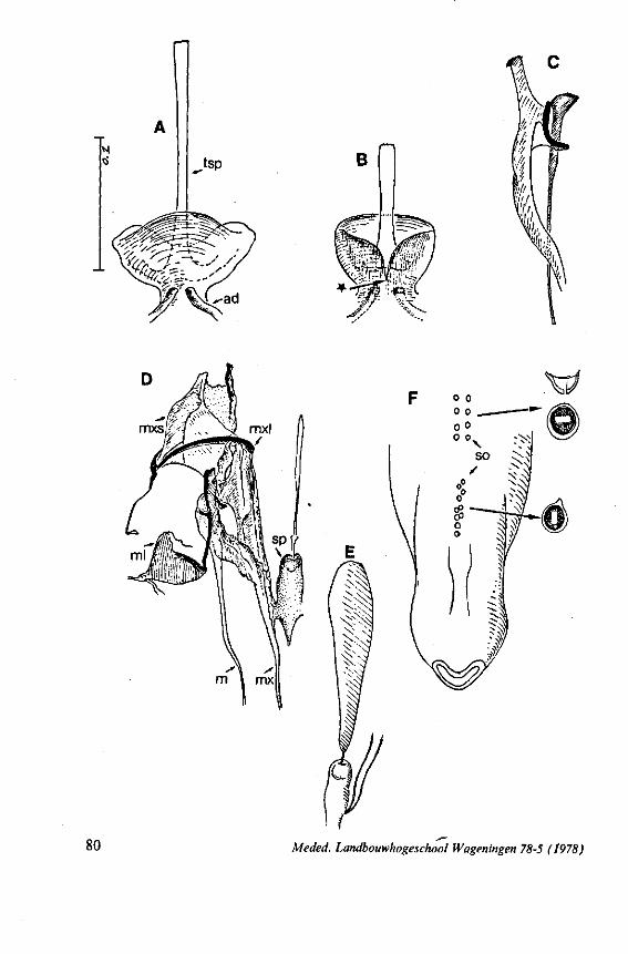
n ^.tsp
mxs
80 Meded. Landbouwhogeschool Wageningen 78-5 (f978)
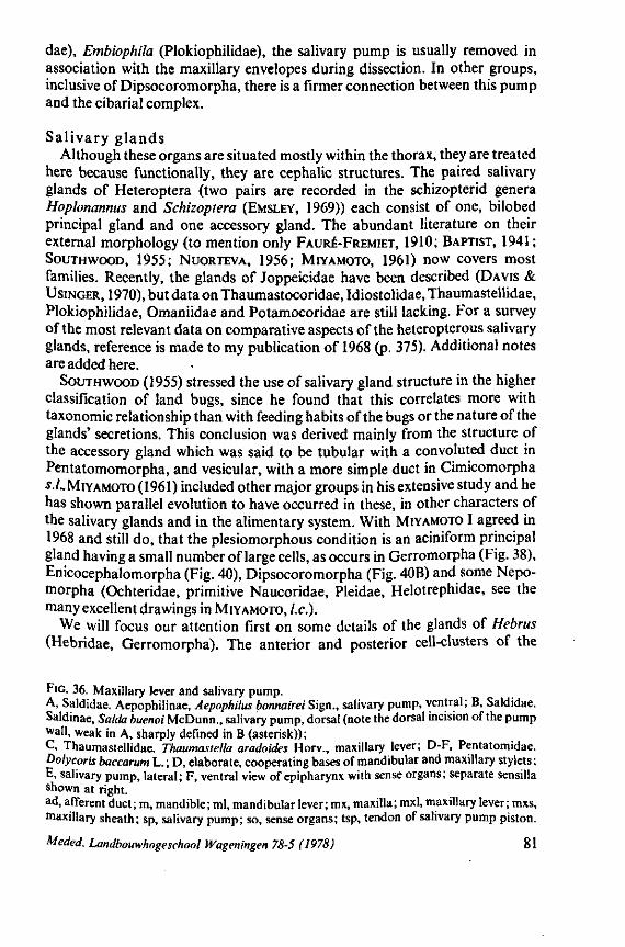
dae), Embiophila (Plokiophilidae), the salivary pump is usually removed in association with the maxillary envelopes during dissection. In other groups, inclusive of Dipsocoromorpha, there is a firmer connection between this pump and the cibarial complex.
Salivary glands Although these organs are situated mostly within the thorax, they are treated
here because functionally, they are cephalic structures. The paired salivary glands of Heteroptera (two pairs are recorded in the schizopterid genera Hoplonanmts and Schizoptera (EMSLEY, 1969)) each consist of one, bilobed principal gland and one accessory gland. The abundant literature on their external morphology (to mention only FAURE-FREMIET, 1910; BAPTIST, 1941; SOUTHWOOD, 1955; NUORTEVA, 1956; MIYAMOTO, 1961) now covers most families. Recently, the glands of Joppeicidae have been described (DAVIS & USINGER, 1970), but data on Thaumastocoridae, Idiostolidae, Thaumastellidae, Plokiophilidae, Omaniidae and Potamocoridae are still lacking. For a survey of the most relevant data on comparative aspects of the heteropterous salivary glands, reference is made to my publication of 1968 (p. 375). Additional notes are added here.
SOUTHWOOD (1955) stressed the use of salivary gland structure in the higher classification of land bugs, since he found that this correlates more with taxonomie relationship than with feeding habits of the bugs or the nature of the glands' secretions. This conclusion was derived mainly from the structure of the accessory gland which was said to be tubular with a convoluted duct in Pentatomomorpha, and vesicular, with a more simple duct in Cimicomorpha •s./.. MIYAMOTO (1961) included other major groups in his extensive study and he has shown parallel evolution to have occurred in these, in other characters of the salivary glands and in the alimentary system. With MIYAMOTO I agreed in 1968 and still do, that the plesiomorphous condition is an aciniform principal gland having a small number of large cells, as occurs in Gerromorpha (Fig. 38), Enicocephalomorpha (Fig. 40), Dipsocoromorpha (Fig. 40B) and some Nepo-morpha (Ochteridae, primitive Naucoridae, Pleidae, Helotrephidae, see the many excellent drawings in MIYAMOTO, I.e.).
We will focus our attention first on some details of the glands of Hebrus (Hebridae, Gerromorpha). The anterior and posterior cell-clusters of the
FIG. 36. Maxillary lever and salivary pump. A, Saldidae. Aepophilinae, Aepophilus bonnairei Sign., salivary pump, ventral; B, Saldidae. Saldinae, Salda buenoi McDunn., salivary pump, dorsal (note the dorsal incision of the pump wall, weak in A, sharply defined in B (asterisk)); C, Thaumastellidae. Thaumastella aradoides Horv., maxillary lever; D-F, Pentatomidae. Dolycoris baccarum L. ; D, elaborate, cooperating bases of mandibular and maxillary sty lets ; E, salivary pump, lateral; F, ventral view of epipharynx with sense organs; separate sensilla shown at right. ad, afferent duct ; m, mandible ; ml, mandibular lever ; rax, maxilla ; mxl, maxillary lever ; mxs, maxillary sheath; sp, salivary pump; so, sense organs; tsp, tendon of salivary pump piston.
Meded. Landbouwhogeschool Wageningen 78-5 (1978) 81
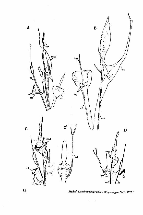
82 Kieded. Landbouwhogeschool H'ageningen 78-5 (1978)
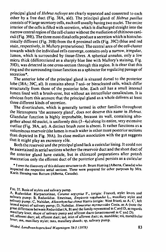
principal gland of Hebrus rußceps are clearly separated and connected to each other by a free duct (Fig. 38A, aid). The principal gland of Hebrus pusillus consists of 9 large secretory cells, each cell usually having two nuclei. The entire interior of the cells is filled with secretion, which is discharged straight into the narrow central region of the cell-cluster without the mediation of chitinous cani-culi (Fig. 39E). The three most distal cells produce a secretion which is histoche-mically different (Fig. 39B) from the 6 proximal cells (Fig. 39C) (blue and red stain, respectively, in Mallory preparations). The central area of the cell-cluster towards which the individual cells converge, contains only a narrow, irregular, lumen, which is surrounded by tissue-fibres. A spherical annulus, only a few micra thick (differentiated as a sharply blue line with Mallory's staining. Fig. 39D), was detected in one cross-section through this region. It is clear that this ring and the surrounding tissue function as a sphincter for regulating passage of secretion*.
The anterior lobe of the principal gland is situated dorsal to the posterior lobe (38A; 39C, al). It contains about 7 uni- or binucleated cells, which differ structurally from those of the posterior lobe. Each cell has a small internal lumen lined with a brush-zone, but without an intracellular canaliculum. It is obvious from this account that the principal gland as a whole delivers at least three different kinds of secretion.
The diverticulum, which is generally termed in other families throughout the Heteroptera 'the accessory gland', does not deserve this name in Hebrus. Glandular function is highly improbable, because its wall, containing altogether about 60 nuciei, is uniformly thin (3-4//) along its entire, very extensive outline (Fig. 39A, ar). A distinct brush zone is absent. It rather functions as a voluminous reservoir (the lumen is much wider in other more posterior sections than depicted in Fig. 39A). Its close median association with the gut suggests that it might play an excretory rôle.
Both the reservoir and the principal gland lack a cuticular lining. It could not be ascertained in serial sections whether the reservoir duct and the short duct of the anterior gland have cuticle, but in chlorazol preparations after potash maceration only the efferent duct of the posterior gland persists as a cuticular
* I owe the discovery of this delicate structure to dr. Bruce Heming (Alberta, Canada) who inspected the respective serial sections. These were prepared for other purposes by Mrs. Karin Heming-van Battum (Alberta, Canada).
F'G- 37. Basis of stylets and salivary pump. A, Reduviidae. Harpactorinae, Coranus aegyptius F., (origin: France), stylet levers and salivary pump; B, Reduviidae. Emesinae, Émpicoris vagabundus L., maxillary stylet and salivary pump; C, Nabidae, Alloeorhvnchus chinai Harris (origin: West Irian), as A; C', left lateral aspect of salivary pump; D, Nabidae. Himacerus myrmecoides Costa, as A (note the great differences between Reduviidae (A, B) and the family represented in C and D, as regards maxillary lever, shape of salivary pump and afferent ducts (anastomosed in C and D). ad, afferent duct; ed, efferent duct; iad, inlet of afferent duct; m, mandible; ml, mandibular lever; mx, maxillary stylet; mxs, maxillary sheath; sp, salivary pump.
Meded. Landbouwhogeschool Wageningen 78-5 (1978) 83
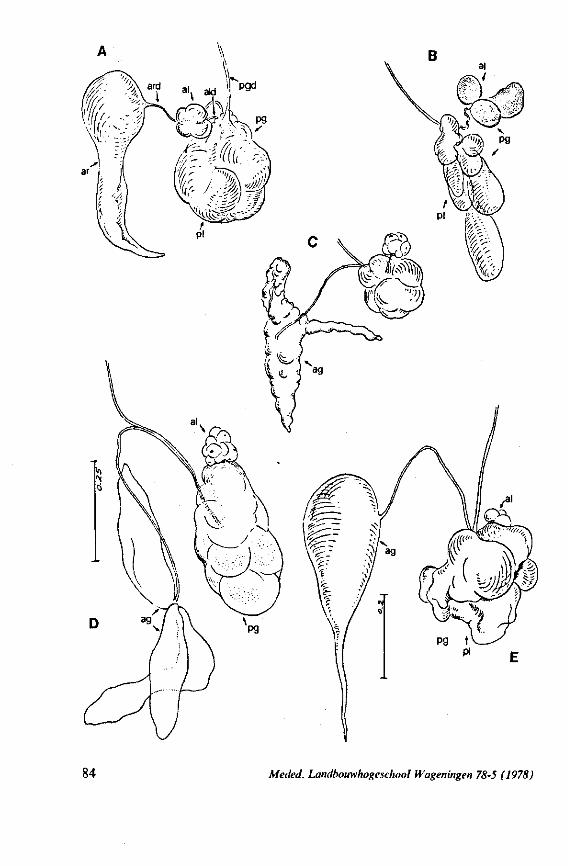
84 Meded. Landbouwhogeschool Wageningen 78-5 (1978)
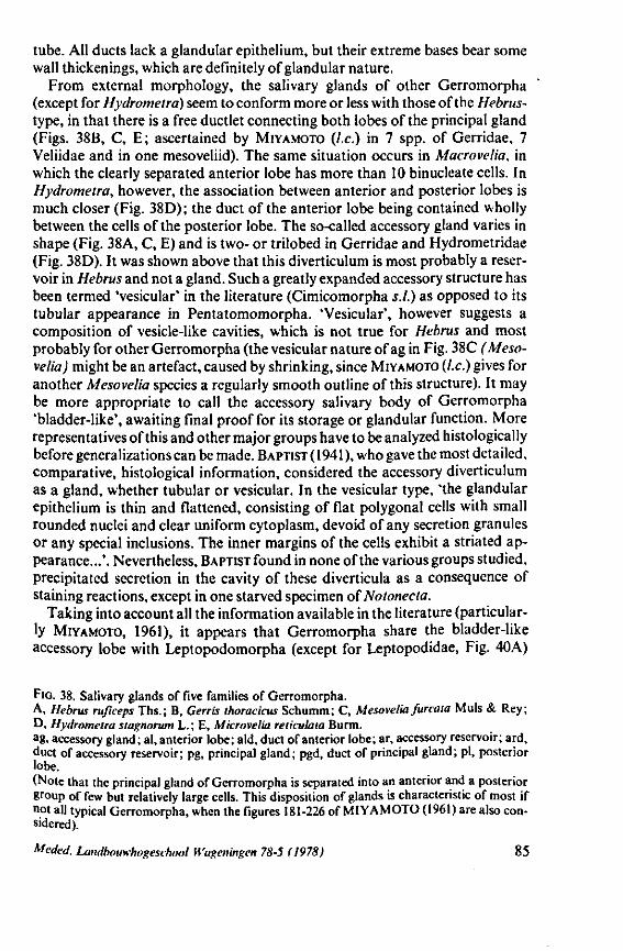
tube. All ducts lack a glandular epithelium, but their extreme bases bear some wall thickenings, which are definitely of glandular nature.
From external morphology, the salivary glands of other Gerromorpha (except for Hydrometra) seem to conform more or less with those of the Hebrus-type, in that there is a free ductlet connecting both lobes of the principal gland (Figs. 38B, C, E; ascertained by MIYAMOTO (I.e.) in 7 spp. of Gerridae, 7 Veliidae and in one mesoveliid). The same situation occurs in Macrovelia, in which the clearly separated anterior lobe has more than 10 binucleate cells. In Hydrometra, however, the association between anterior and posterior lobes is much closer (Fig. 38D); the duct of the anterior lobe being contained wholly between the cells of the posterior lobe. The so-called accessory gland varies in shape (Fig. 38A, C, E) and is two- or trilobed in Gerridae and Hydrometridae (Fig. 38D). It was shown above that this diverticulum is most probably a reservoir in Hebrus and not a gland. Such a greatly expanded accessory structure has been termed 'vesicular' in the literature (Cimicomorpha s.l.) as opposed to its tubular appearance in Pentatomomorpha. 'Vesicular', however suggests a composition of vesicle-like cavities, which is not true for Hebrus and most probably for other Gerromorpha (the vesicular nature of ag in Fig. 38C (Meso-velia) might be an artefact, caused by shrinking, since MIYAMOTO (I.e.) gives for another Mesovelia species a regularly smooth outline of this structure). It may be more appropriate to call the accessory salivary body of Gerromorpha 'bladder-like', awaiting final proof for its storage or glandular function. More representatives of this and other major groups have to be analyzed histological ly before generalizations can be made. BAPTIST (1941 ), who gave the most detailed, comparative, histological information, considered the accessory diverticulum as a gland, whether tubular or vesicular. In the vesicular type, 'the glandular epithelium is thin and flattened, consisting of flat polygonal cells with small rounded nuclei and clear uniform cytoplasm, devoid of any secretion granules or any special inclusions. The inner margins of the cells exhibit a striated appearance...'. Nevertheless, BAPTIST found in none of the various groups studied, precipitated secretion in the cavity of these diverticula as a consequence of staining reactions, except in one starved specimen of Notonecta.
Taking into account all the information available in the literature (particularly MIYAMOTO, 1961), it appears that Gerromorpha share the bladder-like accessory lobe with Leptopodomorpha (except for Leptopodidae, Fig. 40A)
FIG. 38. Salivary glands of five families of Gerromorpha. A, Hebrus ruficeps Ths.; B, Cerris thoracicus Schumm; C, Mesovelia furcata Muls & Rey; D, Hydrometra stagnorum L.; E, Microvelia reticulata Burm. ag, accessory gland; ai, anterior lobe; aid, duct of anterior lobe; ar, accessory reservoir; ard, duct of accessory reservoir; pg, principal gland; pgd, duct of principal gland; pi, posterior lobe. (Note that the principal gland of Gerromorpha is separated into an anterior and a posterior group of few but relatively large cells. This disposition of glands is characteristic of most if not all typical Gerromorpha, when the figures 181-226 of MIYAMOTO (1961) are also considered).
Meded. Landbouwhogeschool Wageningen 78-5 (1978) 85
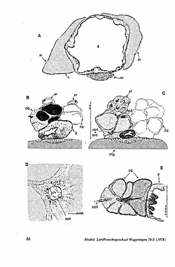
sph
86 Meded. Landbouwhogeschool Wugeningen 78-5 (1978)
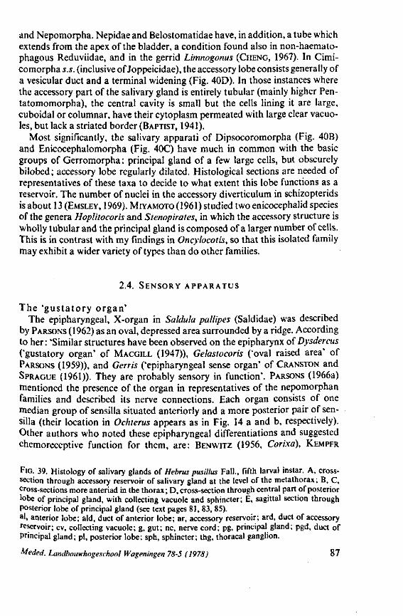
and Nepomorpha. Nepidae and Belostomatidae have, in addition, a tube which extends from the apex of the bladder, a condition found also in non-haemato-phagous Reduviidae, and in the gerrid Limnogonus (CHENG, 1967). In Cimi-comorpha s.s. (inclusive of Joppeicidae), the accessory lobe consists generally of a vesicular duct and a terminal widening (Fig. 40D). In those instances where the accessory part of the salivary gland is entirely tubular (mainly higher Pen-tatomomorpha), the central cavity is small but the cells lining it are large, cuboidal or columnar, have their cytoplasm permeated with large clear vacuoles, but lack a striated border (BAPTIST, 1941).
Most significantly, the salivary apparati of Dipsocoromorpha (Fig. 40B) and Enicocephalomorpha (Fig. 40C) have much in common with the basic groups of Gerromorpha : principal gland of a few large cells, but obscurely bilobed; accessory lobe regularly dilated. Histological sections are needed of representatives of these taxa to decide to what extent this lobe functions as a reservoir. The number of nuclei in the accessory diverticulum in schizopterids is about 13 (EMSLEY, 1969). MIYAMOTO (1961) studied two enicocephalid species of the genera Hoplitocoris and Stenopirates, in which the accessory structure is wholly tubular and the principal gland is composed of a larger number of cells. This is in contrast with my findings in Oncylocotis, so that this isolated family may exhibit a wider variety of types than do other families.
2.4. SENSORY APPARATUS
The 'gustatory organ' The epipharyngeal, X-organ in Saldula pallipes (Saldidae) was described
by PARSONS (1962) as an oval, depressed area surrounded by a ridge. According to her : 'Similar structures have been observed on the epipharynx of Dysdercus ('gustatory organ' of MACGIIX (1947)), Gelastocoris ('oval raised area' of PARSONS (1959)), and Gerris ('epipharyngeal sense organ' of CRANSTON and SPRAGUE (1961)). They are probably sensory in function'. PARSONS (1966a) mentioned the presence of the organ in representatives of the nepomorphan families and described its nerve connections. Each organ consists of one median group of sensilla situated anteriorly and a more posterior pair of sen-silla (their location in Ochterus appears as in Fig. 14 a and b, respectively). Other authors who noted these epipharyngeal differentiations and suggested chemoreceptive function for them, are: BENWITZ (1956, Corixa), KEMPER
FIG. 39. Histology of salivary glands of Hebrus pusillus Fall., fifth larval instar. A, cross-section through accessory reservoir of salivary gland at the level of the metathorax ; B, C, cross-sections more anteriad in the thorax; D, cross-section through central part of posterior lobe of principal gland, with collecting vacuole and sphincter; E, sagittal section through posterior lobe of principal gland (see text pages 81, 83, 85). a ' , anterior lobe; aid, duct of anterior lobe; ar, accessory reservoir; ard, duct of accessory reservoir; cv, collecting vacuole; g, gut; nc, nerve cord; pg, principal gland; pgd, duct of Principal gland; pi, posterior lobe; sph, sphincter; thg, thoracal ganglion.
Meded. Landbouwhogeschool Wageningen 78-5 (1978) 87
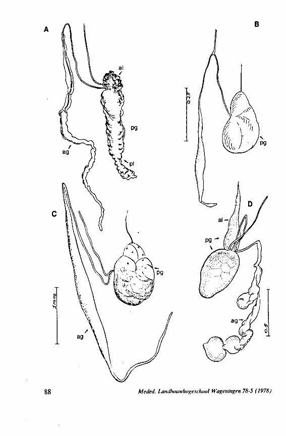
Meded. Landbouwhogeschool Wageningen 78-5 (1978)
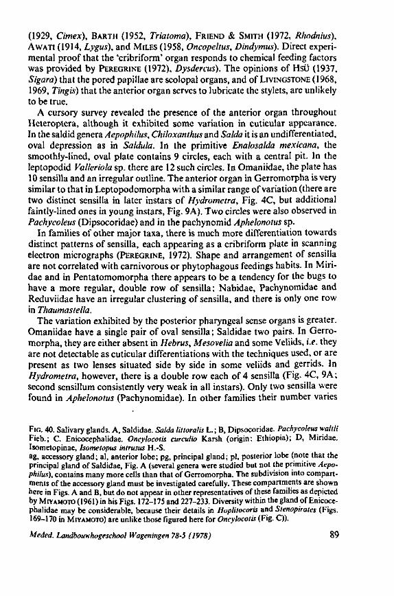
(1929, Cimex), BARTH (1952, Triatomd), FRIEND & SMITH (1972, Rliodnius), AWATI (1914, Lygus), and MILES (1958, Oncopeltus, Dindymus). Direct experimental proof that the 'cribriform' organ responds to chemical feeding factors was provided by PEREGRINE (1972), Dysdercus). The opinions of Hsü (1937, Sigara) that the pored papillae are scolopal organs, and of LIVINGSTONE (1968, 1969, Tingis) that the anterior organ serves to lubricate the stylets, are unlikely to be true.
A cursory survey revealed the presence of the anterior organ throughout Heteroptera, although it exhibited some variation in cuticular appearance. In the saldid genera Aepophilus, Chiloxanthus and Salda it is an undifferentiated, oval depression as in Saldula. In the primitive Enalosalda mexicana, the smoothly-lined, oval plate contains 9 circles, each with a central pit. In the leptopodid Valleriola sp. there are 12 such circles. In Omaniidae, the plate has 10 sensilla and an irregular outline. The anterior organ in Gerromorpha is very similar to that in Leptopodomorpha with a similar range of variation (there are two distinct sensilla in later instars of Hydrometra, Fig. 4C, but additional faintly-lined ones in young instars, Fig. 9A). Two circles were also observed in Pachycoleus (Dipsocoridae) and in the pachynomid Aphelonotus sp.
In families of other major taxa, there is much more differentiation towards distinct patterns of sensilla, each appearing as a cribriform plate in scanning electron micrographs (PEREGRINE, 1972). Shape and arrangement of sensilla are not correlated with carnivorous or phytophagous feedings habits. In Miri-dae and in Pentatomomorpha there appears to be a tendency for the bugs to have a more regular, double row of sensilla; Nabidae, Pachynomidae and Reduviidae have an irregular clustering of sensilla, and there is only one row in Thaumastella.
The variation exhibited by the posterior pharyngeal sense organs is greater. Omaniidae have a single pair of oval sensilla ; Saldidae two pairs. In Gerromorpha, they are either absent in Hebrus, Mesovelia and some Veliids, i.e. they are not detectable as cuticular differentiations with the techniques used, or are present as two lenses situated side by side in some veliids and gerrids. In Hydrometra, however, there is a double row each of 4 sensilla (Fig. 4C, 9A; second sensillum consistently very weak in all instars). Only two sensilla were found in Aphelonotus (Pachynomidae). In other families their number varies
FIG. 40. Salivary glands. A, Saldidae. Salda littoralis L.; B, Dipsocoridae. Pachycoleus waltli Fieb.; C. Enicocephalidae. Oncylocotis curculio Karsh (origin: Ethiopia); D, Miridae. Isometopinae, Isometopus inirusus H.-S. ag, accessory gland; al, anterior lobe; pg, principal gland; pi, posterior lobe (note that the principal gland of Saldidae, Fig. A (several genera were studied but not the primitive Aepophilus), contains many more cells than that of Gerromorpha. The subdivision into compartments of the accessory gland must be investigated carefully. These compartments are shown here in Figs. A and B, but do not appear in other representatives of these families as depicted by MIYAMOTO (1961) in his Figs. 172-175 and 227-233. Diversity within the gland of Enicocephalidae may be considerable, because their details in Hoplilocoris and Stenopirates (Figs. 169-170 in MIYAMOTO) are unlike those figured here for Oncylocotis (Fig. C)).
Meded. Landbouwhogeschool Wageningen 78-5 (1978) 89
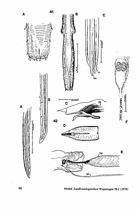
41
w B
' / / / / • • ' • / > , • / /.
90 Meded. Landbouwhogeschool Wageningen 78-5 (1978)
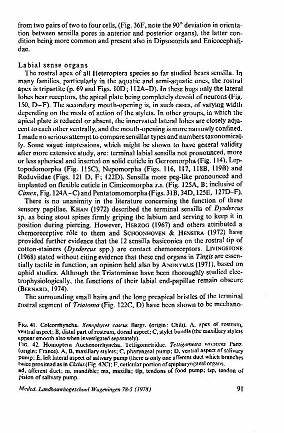
from two pairs of two to four cells, (Fig. 36F, note the 90° deviation in orientation between sensilla pores in anterior and posterior organs), the latter condition being more common and present also in Dipsocorids and Enicocephali-dae.
Labial sense organs The rostral apex of all Heteroptera species so far studied bears sensilla. In
many families, particularly in the aquatic and semi-aquatic ones, the rostral apex is tripartite (p. 69 and Figs. 10D; 112A-D). In these bugs only the lateral lobes bear receptors, the apical plate being completely devoid of neurons (Fig. 150, D-F). The secondary mouth-opening is, in such cases, of varying width depending on the mode of action of the stylets. In other groups, in which the apical plate is reduced or absent, the innervated lateral lobes are closely adjacent to each other ventrally, and the mouth-opening is more narrowly confined. I made no serious attempt to compare sensillar types and numbers taxonomical-ly. Some vague impressions, which might be shown to have general validity after more extensive study, are: terminal labial sensilla not pronounced, more or less spherical and inserted on solid cuticle in Gerromorpha (Fig. 114), Lep-topodomorpha (Fig. 115C), Nepomorpha (Figs. 116, 117, 118B, 119B) and Reduviidae (Figs. 121 D, F; 122D). Sensilla more peg-like pronounced and implanted on flexible cuticle in Cimicomorpha s.s. (Fig. 125A, B; inclusive of Cimex, Fig. 124A-C) and Pentatomomorpha (Figs. 31B, 34D, 125E, 127D-F).
There is no unanimity in the literature concerning the function of these sensory papillae. KHAN (1972) described the terminal sensilla of Dysdercus sp. as being stout spines firmly griping the labium and serving to keep it in position during piercing. However, HERZOG (1967) and others attributed a chemoreceptive rôle to them and SCHOONHOVEN & HENSTRA (1972) have provided further evidence that the 12 sensilla basiconica on the rostral tip of cotton-stainers {Dysdercus spp.) are contact chemoreceptors. LIVINGSTONE (1968) stated without citing evidence that these end organs in Tingis are essentially tactile in function, an opinion held also by ANONYMUS (1971), based on aphid studies. Although the Triatominae have been thoroughly studied elec-trophysiologically, the functions of their labial end-papillae remain obscure (BERNARD, 1974).
The surrounding small hairs and the long preapical bristles of the terminal rostral segment of Triatoma (Fig. 122C, D) have been shown to be mechano-
FIG. 41. Coleorrhyncha. Xenophytes cascus Bergr. (origin: Chili). A, apex of rostrum, ventral aspect; B, distal part of rostrum, dorsal aspect; C, stylet bundle (the maxillary stylets appear smooth also when investigated separately). FIG. 42. Homoptera Auchenorrhyncha, Tettigometridae. Tettigometra virescens Panz. (origin: France). A, B, maxillary stylets; C, pharyngeal pump; D, ventral aspect of salivary pump; E, left lateral aspect of salivary pump (there is only one afferent duct which branches twice proximad as in Cixius (Fig. 42C) ; F, cuticular portion of epipharyngaeal organs, ad, afferent duct; m, mandible; mx, maxilla; tfp, tendons of food pump; tsp, tendon of piston of salivary pump.
Meded. Landbouwhogeschool Wageningen 78-5 (1978) 91
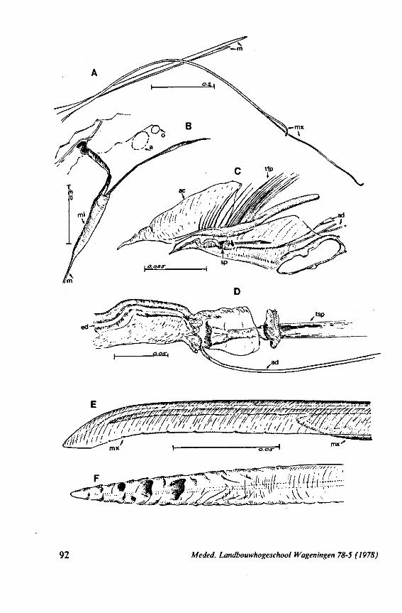
WW!/ - - • * — — **—• ' • *•• i ' : • • , • / - / - Sit //vy- •"'•••Lii -*.
w77f/•////?//'/',•-/'/'/ f ' '
92 Meded. Landbouwhogeschool Wageningen 78-5 (1978)
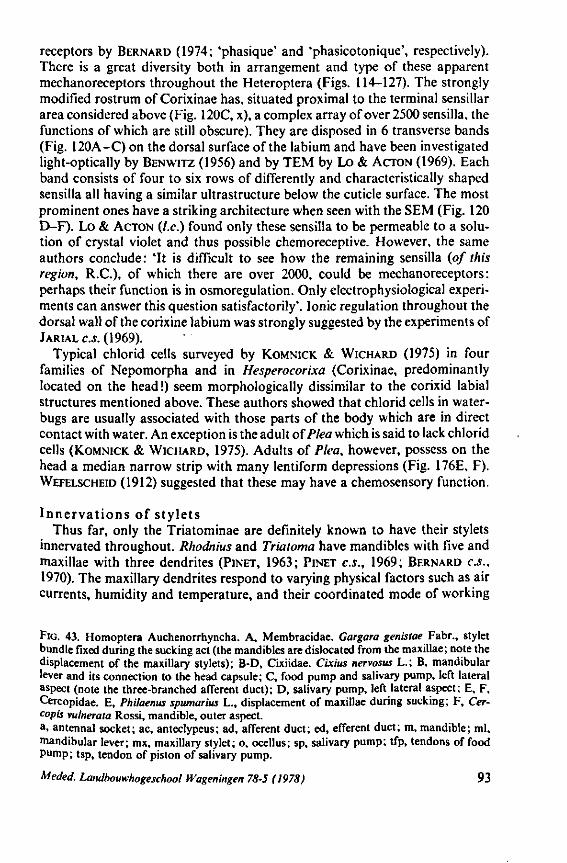
receptors by BERNARD (1974; 'phasique' and 'phasicotonique', respectively). There is a great diversity both in arrangement and type of these apparent mechanoreceptors throughout the Heteroptera (Figs. 114-127). The strongly modified rostrum of Corixinae has, situated proximal to the terminal sensillar area considered above (Fig. 120C, x), a complex array of over 2500 sensilla, the functions of which are still obscure). They are disposed in 6 transverse bands (Fig. 120A-C) on the dorsal surface of the labium and have been investigated light-optically by BENWITZ (1956) and by TEM by Lo & ACTON (1969). Each band consists of four to six rows of differently and characteristically shaped sensilla all having a similar ultrastructure below the cuticle surface. The most prominent ones have a striking architecture when seen with the SEM (Fig. 120 D-F). Lo & ACTON (I.e.) found only these sensilla to be permeable to a solution of crystal violet and thus possible chemoreceptive. However, the same authors conclude: 'It is difficult to see how the remaining sensilla (of this region, R.C.), of which there are over 2000, could be mechanoreceptors: perhaps their function is in osmoregulation. Only electrophysiological experiments can answer this question satisfactorily'. Ionic regulation throughout the dorsal wall of the corixine labium was strongly suggested by the experiments of JARIAL c.j. (1969).
Typical chlorid cells surveyed by KOMNICK & WICHARD (1975) in four families of Nepomorpha and in Hesperocorixa (Corixinae, predominantly located on the head!) seem morphologically dissimilar to the corixid labial structures mentioned above. These authors showed that chlorid cells in water-bugs are usually associated with those parts of the body which are in direct contact with water. An exception is the adult oïPlea which is said to lack chlorid cells (KOMNICK & WICHARD, 1975). Adults of Plea, however, possess on the head a median narrow strip with many lentiform depressions (Fig. 176E, F). WEFELSCHEID (1912) suggested that these may have a chemosensory function.
Innervations of stylets Thus far, only the Triatominae are definitely known to have their stylets
innervated throughout. Rhodnius and Trialoma have mandibles with five and maxillae with three dendrites (PINET, 1963; PINET C.S., 1969; BERNARD C.S., 1970). The maxillary dendrites respond to varying physical factors such as air currents, humidity and temperature, and their coordinated mode of working
FIG. 43. Homoptera Auchenorrhyncha. A, Membracidae. Gargara genistae Fabr., stylet bundle fixed during the sucking act (the mandibles are dislocated from the maxillae; note the displacement of the maxillary stylets); B-D, Cixiidae. Cixius nervosus L.; B, mandibular lever and its connection to the head capsule; C, food pump and salivary pump, left lateral aspect (note the three-branched afferent duct); D, salivary pump, left lateral aspect; E, F, Cercopidae. E, Philaenus spumarius L., displacement of maxillae during sucking; F, Cer-copis vulneraia Rossi, mandible, outer aspect. a, antennal socket; ac, anteclypeus; ad, afferent duct; ed, efferent duct; m, mandible; ml, mandibular lever; mx, maxillary stylet; o, ocellus; sp, salivary pump; tfp, tendons of food pump; tsp, tendon of piston of salivary pump.
Meded. Landbouwhogeschool Wageningen 78-5 (1978) 93
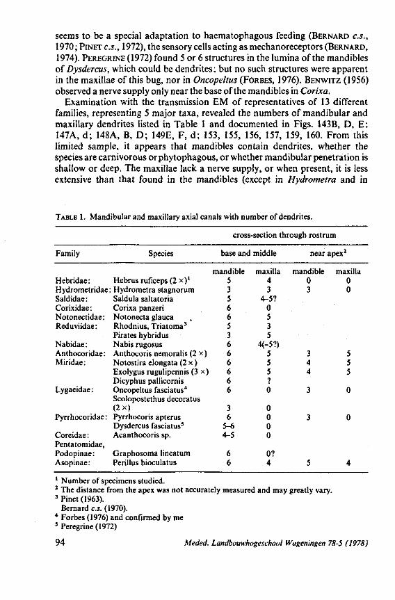
seems to be a special adaptation to haematophagous feeding (BERNARD C.S., 1970; PINET c.s., 1972), the sensory cells acting as mechanoreceptors (BERNARD, 1974). PEREGRINE (1972) found 5 or 6 structures in the lumina of the mandibles of Dysdercus, which could be dendrites ; but no such structures were apparent in the maxillae of this bug, nor in Oncopeltus (FORBES, 1976). BENWITZ (1956) observed a nerve supply only near the base of the mandibles in Corixa.
Examination with the transmission EM of representatives of 13 different families, representing 5 major taxa, revealed the numbers of mandibular and maxillary dendrites listed in Table 1 and documented in Figs. 143B, D, E: 147A, d; 148A, B, D; 149E, F, d; 153, 155, 156, 157, 159, 160. From this limited sample, it appears that mandibles contain dendrites, whether the species are carnivorous or phytophagous, or whether mandibular penetration is shallow or deep. The maxillae lack a nerve supply, or when present, it is less extensive than that found in the mandibles (except in Hydrometra and in
TABLE 1. Mandibular and maxillary axial canals with number of dendrites.
cross-section through rostrum
Family
Hebridae :
Species
Hebrus ruficeps (2 x)1
Hydrometridae: Hydrometra stagnorum Saldidae: Corixidae : Notonectidae: Reduviidae :
Nabidae : Anthocoridae: Miridae:
Lygaeidae:
Pyrrhocoridae :
Coreidae : Pentatomidae, Podopinae: Asopinae:
Saldula saltatoria Corixa panzeri Notonecta glauca Rhodnius, Triatoma3
Pirates hybridus Nabis rugosus Anthocoris nemoralis (2 x) Notostira elongata (2 x ) Exolygus rugulipennis (3 x) Dicyphus pallicornis Oncopeltus fasciatus4
Scolopostethus decoratus (2x) Pyrrhocoris apterus Dysdercus fasciatus5
Acanthocoris sp.
Graphosoma lineatum Perillus bioculatus
base and middle
mandible 5 3 5 6 6 5 3 6 6 6 6 6 6
3 6
5-6 4-5
6 6
maxilla 4 3
4-5? 0 5 3 5
4(-5?) 5 5 5 ? 0
0 0 0 0
0? 4
near
mandible 0 3
3 4 4
3
3
5
apex2
maxilla 0 0
5 5 5
0
0
4
1 Number of specimens studied. 2 The distance from the apex was not accurately measured and may greatly vary. 3 Pinet (1963).
Bernard c.s. (1970). 4 Forbes (1976) and confirmed by me 5 Peregrine (1972)
94 Meded. Landbouwhogeschool Wageningen 78-5 (1978)
Pirates. Flexibility of the nervous system is demonstrated in the predatory Asopinae, which have maxillary neurons; these are apparently secondarily re-attained, since the phytophagous Pentatomomorpha studied lack maxillary nerve supply.
It may be that careful standardized studies with light optics of methylene blue stylet-preparations in toto of fairly large species, will unravel the exact pathways followed by the dendrites. Cursory inspection revealed some differentiations suggestive of this (see Figs. 22B, 22'A-C). Further discussion is given on p. 200, 201.
Meded. Landbouwhogeschool Wageningen 78-5 (1978) 95
EVOLUTIONARY TRENDS IN STRUCTURES OTHER THAN THOSE ASSOCIATED WITH FEEDING
In this section, I review conclusions from my 1968 publication, which may aid in determining ancestral and derived character states within the principal subdivisions of the Heteroptera. Supplementary data are also added, but the enormous number of specific features of taxa of lower ranks are not treated. Arguments against my views, brought forward by some of my colleagues are also considered.
The survey is given in a series of schemes with frequent reference to some of the topics, previewed in 1968 (a) (pages 363-378) in words only. In my presentation, I often omit adjectives such as: supposedly, probably, in my view, etc., so that the conclusions may sound more apodictic than the evidence justifies. Hypotheses they nevertheless remain albeit logically derived ones.
3.1. EGG-SYSTEMS
Plesiomorphous species are usually small, with a small number of ovarioles, in which a few, relatively large, eggs ripen simultaneously. Apomorphous species are larger, and have more ovarioles with smaller follicle cells and eggs (COBBEN, 1968a, Figs. 273,274). These mature synchronously and are deposited in batches. Superficial egg-laying most probably anticipated embedding of the egg within a substrate (COBBEN, 1968a, Fig. 281).
A thin, entirely solid chorion, 'marked with a spacious, hexagonal pattern from the follicular cells, is considered plesiomorphous. The so-called aerostatic, chorionic inner-layer has evolved several times independently along different pathways. The general trend of evolution of the micropylar system involved the multiplication and displacement of the micropyles, starting from a single, simple micropyle in the centre of the cephalic pole (Fig. 268-270, 1968). The structure and orientation of the micropyle(s) have evolved in several directions within each branch ; one simple, perpendicular micropylar pore being considered as ancestral to externally or inwardly directed chorionic projections (Fig. 269, 1968). Those taxa having many, displaced micropyles in their eggs have the inner projections of these oriented in a clock-wise direction when observed anteriorly from the outside. Significantly, these run anti-clock-wise in the eggs of the dipsocorid Ceratocombus and the enicocephalid Oncylocotis.
The original, irregular, longitudinal dehiscence of the shell has evolved independently, and along different pathways, into a cap in most major groups (schemes in Figs. 270-272,1968).
The embryogénie patterns of Heteroptera are highly diverse, but show distinct trends. Since transspecific differences in embryogenesis are regarded as of great phylogenetic significance, Fig. 276 (1968) is reprinted here, slightly
96 Meded. Landbouwhogeschool Wageningen 78-5 (1978)
modified, as Fig. 44. Higher category nomenclature has been modified and the Dipsocoridae are now placed at about the same level as e (Hebridae). In the scheme published in 1968, Dipsocoridae were placed as j ' above the Gerro-morpha (= Amphibicorisae) because of spatial limitations, but the embryology of one of the genera studied is close to that of the archetype while that of the other is similar but possible lacks longitudinal rotation of the embryo. This absence needs to be verified in more eggs and in other species. The embryology of the Schizopteridae, the second family of the Dipsocoromorpha, is unknown. I have since studied the embryogenesis of a representative of the Enicocephalo-morpha, the genus Oncylocotis. Its main features are of the dipsocorid type. I am not sure if embryonic rotation occurs in Oncylocotis, because its eggs are isoradial.
Family groups are distinguished by different types of embryogenesis based on the following, varying characteristics: degree of visible development of the 'pregerm' ; location of the blastopore; growth, orientation, change in shape and displacement (mostly clockwise rotation) of the germ-band, embryo and pro-larva. A wide range in progressive evolution of embryonic events has occurred within most major phyletic lines.
The embryonic archetype invaginates along the longitudinal median axis of the yolk column (morphologically at the caudo-ventral surface of the egg) without loss of contact between head lobes and serosa, and rotates longitudinally 180° before revolution. This type of embryogenesis is more or less characteristic of the species studied of Hebridae, Nabidae, some Miridae, Dipsocoridae and Enicocephalidae (180° rotation still to be checked). Corresponding data on Microphysidae are still incomplete and for Plokiophilidae, Joppeicidae, Velo-pedidae and Pachynomidae are lacking.
Pentatomomorpha exhibit gradual loss of embryonic rotation, while Nepo-morpha retain it, sometimes with both germ-band and prolarval rotations. Both groups show a transition (anagenetic intra se, cladogenetic inter se) from the immersed to the superficial type of embryogenesis. The superficial condition of the hydrocorisous type prevails in Reduviidae and they have complete loss of rotations. This progression in embryonic evolution has reached a high level in Harpactorinae, with many species performing semi-invagination, and with species of Coranus (3 species studied) entirely omitting the invagination stage (and hence, blastokinesis). Loss of blastokinesis is associated with early differentiation of the prospective germ-band in the blastoderm stage. Similar, early development occurs in some Hydrocorisae and, through cladistic divergence, in highly derived taxa of the Pentatomomorpha. The presence of a serosal hydropyle at the caudal pole of the egg is assumed to be ancestral to its absence, although serosal hydropyles may have evolved later in other regions of the egg.
Four different methods of rupturing the chorionic and subchorionic layers are practised by Heteroptera. Their evolutionary relationships are shown in Fig. 45 (after Fig. 278 in COBBEN, 1968a). A transverse, paired, ruptor ovi in the embryonic cuticle delimiting the anteclypeus from the postclypeus is considered
Meded. Landbouwhogeschool Wageningen 78-5 (1978) 97
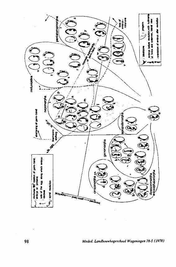
98 M e Jed. Landbouwhogeschool Wageningen 78-5 (1978)
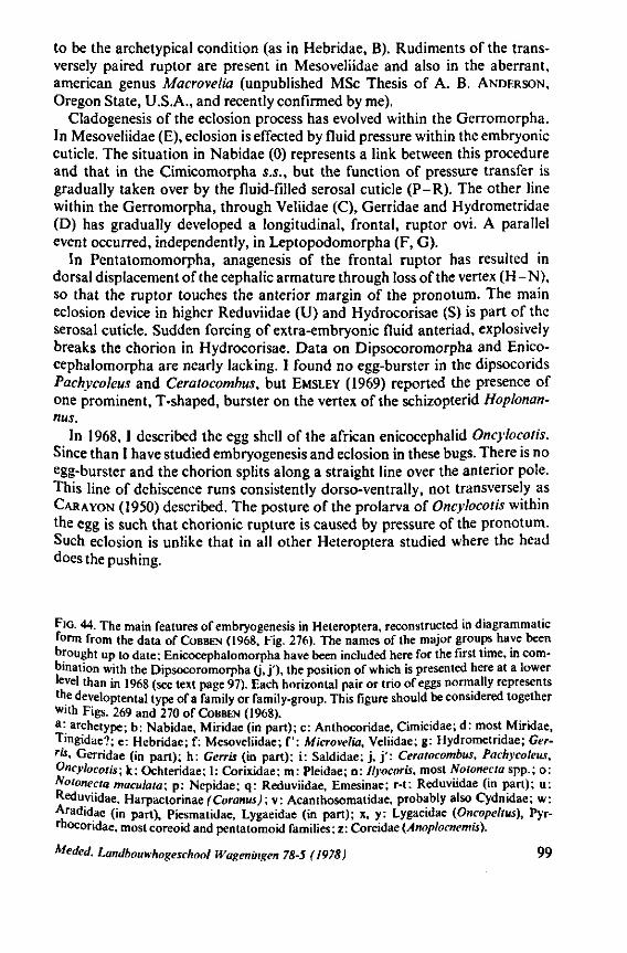
to be the archetypical condition (as in Hebridae, B). Rudiments of the transversely paired ruptor are present in Mesoveliidae and also in the aberrant, american genus Macrovelia (unpublished MSc Thesis of A. B. ANDERSON, Oregon State, U.S.A., and recently confirmed by me).
Cladogenesis of the eclosion process has evolved within the Gerromorpha. In Mesoveliidae (E), eclosion is effected by fluid pressure within the embryonic cuticle. The situation in Nabidae (0) represents a link between this procedure and that in the Cimicomorpha s.S., but the function of pressure transfer is gradually taken over by the fluid-filled serosal cuticle (P-R). The other line within the Gerromorpha, through Veliidae (C), Gerridae and Hydrometridae (D) has gradually developed a longitudinal, frontal, ruptor ovi. A parallel event occurred, independently, in Leptopodomorpha (F, G).
In Pentatomomorpha, anagenesis of the frontal ruptor has resulted in dorsal displacement of the cephalic armature through loss of the vertex (H-N), so that the ruptor touches the anterior margin of the pronotum. The main eclosion device in higher Reduviidae (U) and Hydrocorisae (S) is part of the serosal cuticle. Sudden forcing of extra-embryonic fluid anteriad, explosively breaks the chorion in Hydrocorisae. Data on Dipsocoromorpha and Enico-cephalomorpha are nearly lacking. I found no egg-burster in the dipsocorids Pachycoleus and Ceratocombus, but EMSLEY (1969) reported the presence of one prominent, T-shaped, burster on the vertex of the schizopterid Hoplonan-nus.
In 1968,1 described the egg shell of the african enicocephalid Oncylocotis. Since than I have studied embryogenesis and eclosion in these bugs. There is no egg-burster and the chorion splits along a straight line over the anterior pole. This line of dehiscence runs consistently dorso-ventrally, not transversely as CARAYON (1950) described. The posture of the prolarva of Oncylocotis within the egg is such that chorionic rupture is caused by pressure of the pronotum. Such eclosion is unlike that in all other Heteroptera studied where the head does the pushing.
FIG. 44. The main features of embryogenesis in Heteroptera, reconstructed in diagrammatic form from the data of COBBEN (1968, Fig. 276). The names of the major groups have been brought up to date; Enicocephalomorpha have been included here for the first time, in combination with the Dipsocoromorpha 0. j ' ) . the position of which is presented here at a lower level than in 1968 (see text page 97). Each horizontal pair or trio of eggs normally represents the developtental type of a family or family-group. This figure should be considered together with Figs. 269 and 270 of COBBEN (1968). a: archetype; b: Nabidae, Miridae (in part); c: Anthocoridae, Cimicidae; d: most Miridae, Tingidae?; e: Hebridae; f: Mesoveliidae; f: Microvetia, Veliidae; g: Hydrometridae; Gerrit, Gerridae (in part); h: Gerris (in part); i: Saldidae; j , j ' : Ceratocombus, Pachycoleus, Oncylocotis; k: Ochteridae; 1: Corixidae; m: Pleidae; n: Ilyocoris, most Notonecta spp.; o: Notonecta maculata; p : Nepidae; q: Reduviidae, Emesinae; r-t: Reduviidae (in part); u: Reduviidae, Harpactorinae (Coranus);v: Acanthosomatidae, probably also Cydnidae; w: Aradidae (in part), Piesmatidae, Lygaeidae (in part); x, y: Lygaeidae (Oncopeltus), Pyr-rhocoridae, most coreoid and pentatomoid families; z: Coreidae (Anoplocnemis).
Meded. Landbouwhogeschool Wageningen 78-5 (1978) 99
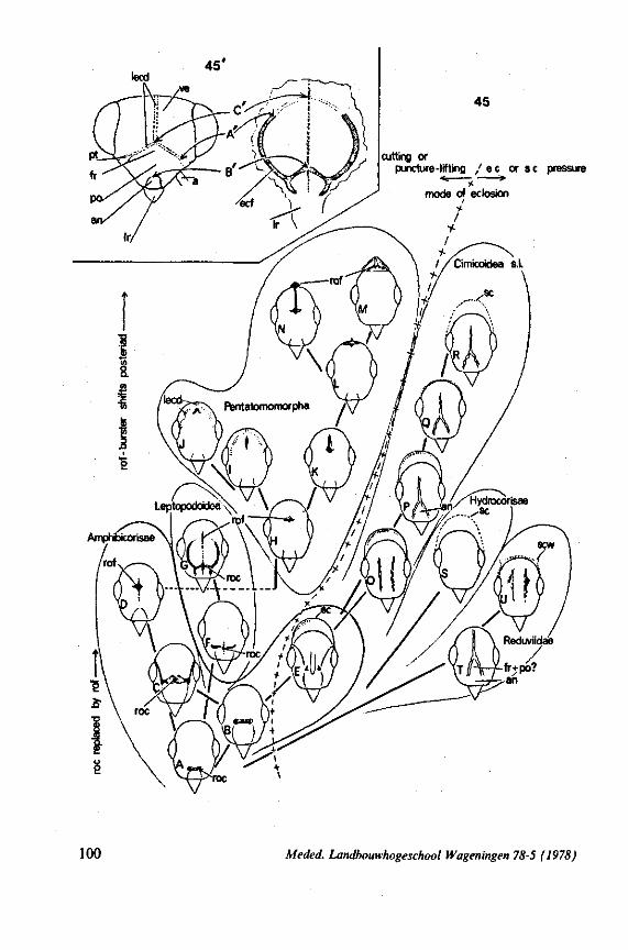
cutting or puncture-lifting / e e or sc pressure
100 Meded. Landbouwhogeschool Wageningen 78-5 (1978)
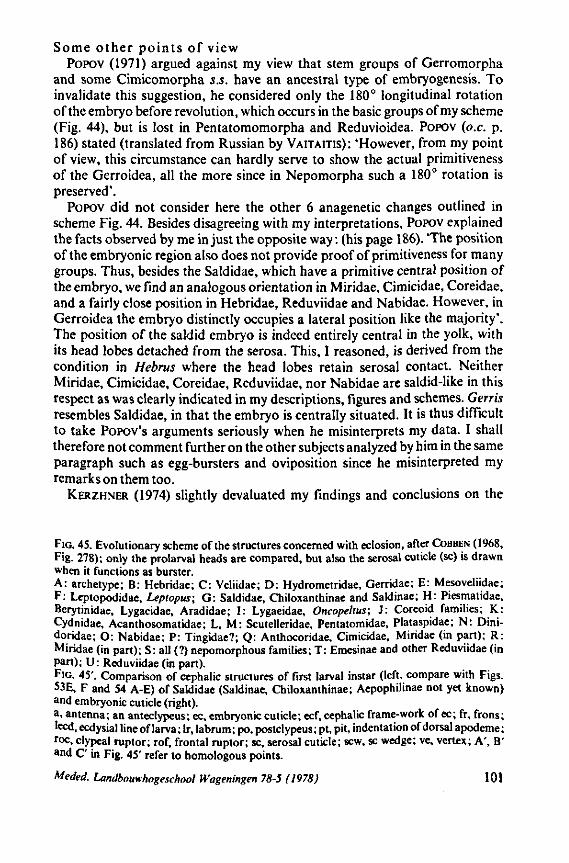
Some other points of view POPOV (1971) argued against my view that stem groups of Gerromorpha
and some Cimicomorpha s.S. have an ancestral type of embryogenesis. To invalidate this suggestion, he considered only the 180° longitudinal rotation of the embryo before revolution, which occurs in the basic groups of my scheme (Fig. 44), but is lost in Pentatomomorpha and Reduvioidea. POPOV (o.e. p. 186) stated (translated from Russian by VAITAITIS): 'However, from my point of view, this circumstance can hardly serve to show the actual primitiveness of the Gerroidea, all the more since in Nepomorpha such a 180° rotation is preserved'.
POPOV did not consider here the other 6 anagenetic changes outlined in scheme Fig. 44. Besides disagreeing with my interpretations, POPOV explained the facts observed by me in just the opposite way : (his page 186). The position of the embryonic region also does not provide proof of primitiveness for many groups. Thus, besides the Saldidae, which have a primitive central position of the embryo, we find an analogous orientation in Miridae, Cimicidae, Coreidae, and a fairly close position in Hebridae, Reduviidae and Nabidae. However, in Gerroidea the embryo distinctly occupies a lateral position like the majority'. The position of the saldid embryo is indeed entirely central in the yolk, with its head lobes detached from the serosa. This, I reasoned, is derived from the condition in Hebrus where the head lobes retain serosal contact. Neither Miridae, Cimicidae, Coreidae, Reduviidae, nor Nabidae are saldid-like in this respect as was clearly indicated in my descriptions, figures and schemes. Gerris resembles Saldidae, in that the embryo is centrally situated. It is thus difficult to take POPOV'S arguments seriously when he misinterprets my data. I shall therefore not comment further on the other subjects analyzed by him in the same paragraph such as egg-bursters and oviposition since he misinterpreted my remarks on them too.
KERZHNER (1974) slightly devaluated my findings and conclusions on the
FIG. 45. Evolutionary scheme of the structures concerned with eclosion, after COBBEN (1968, Fig. 278); only the prolarval heads are compared, but also the serosal cuticle (sc) is drawn when it functions as burster. A: archetype; B: Hebridae; C: Veliidae; D: Hydrometridae, Gerridae; E: Mesoveliidae; F: Leptopódidae, Leptopus; G: Saldidae, Chiloxanthinae and Saldinae; H: Piesmatidae, Berytinidae, Lygaeidae, Aradidae; I: Lygaeidae, Oncopeltus; J: Coreoid families; K: Cydnidae, Acanthosomatidae; L, M: Scutelleridae, Pentatomidae, Plataspidae; N: Dini-doridae; O: Nabidae; P: Tingidae?; Q: Anthocoridae, Cimicidae, Miridae (in part); R: Miridae (in part); S: all (?) nepomorphous families; T: Emesinae and other Reduviidae (in Part); U: Reduviidae (in part). FIG. 45'. Comparison of cephalic structures of first larval instar (left, compare with Figs. 53E, F and 54 A-E) of Saldidae (Saldinae, Chiloxanthinae; Aepophilinae not yet known) and embryonic cuticle (right). a, antenna; an anteclypeus; ec, embryonic cuticle; ecf, cephalic frame-work of ec; fr, frons; leed, ecdysial line of larva; lr, labrum; po, postclypeus; pt, pit, indentation of dorsal apodeme; r°c, clypeal ruptor; rof, frontal raptor; sc, serosal cuticle; sew, sc wedge; ve, vertex; A', B' and C' in Fig. 45' refer to homologous points.
Meded. Landbouwhogeschool Wageningen 78-5 (1978) 101
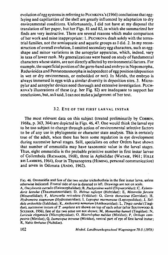
evolution of egg systems in referring to PUCHKOVA'S ( 1966) conclusions that egg-laying and capillarities of the shell are greatly influenced by adaptation to dry environmental conditions. Unfortunately, I did not have at my disposal the translation of her paper, but her Figs. 81 and 82, in which she summarized her finds are very instructive. There are several reasons which make comparison of her work and mine inappropriate : 1. PUCHKOVA dealt solely with the terrestrial families, not the semiaquatic and aquatic groups as I did. 2. In my reconstruction of overall evolution, I omitted secondary egg characters, such as egg-shape and minor variations in the aeropylar apparatus, which, indeed, vary in taxa of lower rank. My generalizations were based on study of fundamental characters whose states, are not directly affected by environmental factors. For example, the superficial position of the germ-band and embryo in Nepomorpha, Reduvioidea and Pentatomomorpha is independent of egg position (deposition in wet or dry environments, or embedded or not). In Mirids, the embryo is always immersed in eggs with a similar diversity in deposition sites. 3. Micro-pylar and aeropylar devices need thorough and extensive investigation. PUCHKOVA'S illustrations of these (e.g. her Fig. 82) are inadequate to support her conclusions, but, as I said, I can not make a judgement of her text.
3.2. E Y E OF T H E F I R S T L A R V A L I N S T A R
The most relevant data on this subject (treated preliminarily by COBBEN, 1968a, p. 363, 364) are depicted in Figs. 46,47. One would think the larval eye to be too subject to change through action of environmental selective factors to be of any use in phylogenetic or character state analysis. This is certainly true of the adult, when there has been some regulation in eye development during successive larval stages. Still, specialists on other Orders have shown that number of ommatidia may have taxonomie value in the larval stages. Thus, eight ommatidia is the probable primitive number in first instar larvae of Collembola (RICHARDS, 1968), three in Aphididae (WATASE, 1961; HILLE RIS LAMBERS, 1964), four in Thysanoptera (HEMING, personal communication) and seven in Odonata (ANDO, 1962).
FIG. 46. Ommatidia and fate of the two ocular trichobothria in the first instar larva, unless otherwise indicated. Frontal side of eye is directed to left. Drawings are not on scale. A, Oncylocotis curculio (Enicocephahdae); B, Pachycoleus tva/r/;'(Dipsocoridae); C. Xylasto-doris luteolus (Thaumastocoridae); D, Hebrus ruficeps (Hebridae); E, Mesovelia furcata (Mesoveliidae); F, Microvelia reticulata (Veliidae); G, Gerris thoracicus (Gerridae); H, Hydrometra stagnorum (Hydrometridae); I, Leptopus marmoratus (Leptopodidae); J, Sal-dula orthochila (Saldidae); K, Anthocoris nemorum (Anthocoridae); L, Tingis cardui (Tingi-dae), successive instars of T. ampliata are shown on top of each other (after SOUTHWOOD & SCUDDER, 1956), fate of the two setae are not shown; M, Monanthia humuli (Tingidae); N, Loricula elegantula (Microphysidae); O, Macrolophus nubilus (Miridae); P, Orthops cam-pestris (Miridae); Q, Isometopus intrusus (Miridae), ventral part of eye of first larval instar; R, Nabis limbatus (Nabidae).
102 Meded. Landbouwhogeschool Wageningen 78-5 (1978)
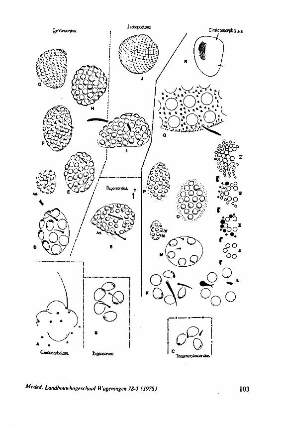
Qerromorpbo. Leptopodonj.
Cimicomorpbü *.».
f?CC C <-- v'*- ^1
tnicocqihüW Dipsoeororo. Tnoiimastocondut,
Meded. Landbouwhogeschool Wageningen 78-5 (1978) 103
Five ommatidia is most probably the original number in Heteroptera. This condition, occurs in Dipsocoridae (Fig. 46B, Pachycoleus, Ceratocombus), Anthocoridae (K, checked in several genera), Cimicidae (Cimex), Tingidae (L, M, checked in many genera), and most probably in Microphysidae (5-6 in second instar, PÉRICART, 1972). In the cimicoid family Miridae, numbers exceed 5 in the limited sample of taxa studied : Termatophylidea 9 (V. DOES-BURG, 1964). Hallodapus 10, Bryocoris 12, Macrolophus 16 (Fig. 46, 0), Liocoris 27, Orthops 30 (Fig. 46P), Phytocoris ±50, and Psallus60-70. The isometopine Isometopus intrusus has 35. Nabis (Fig. 46R) has a large facet-eye in the first instar having more than 150 lenses, but variations within this diversified family should be studied in more representatives.
The drawings K-R (Fig. 46) are arranged such that anagenetic increase of ommatidial number is suggested. Evidence for this will seem weak, when data are compared within the cimicoid groups alone, unless relict families such as Joppeicidae, Plokiophilidae and primitive nabid members are also shown to have a low number. Evidence for anagenetic increase is stronger in other major groups. Study of the cimicoids shows that number of ommatidia is not related to feeding habits. Anthocorids, cimicids and tingids, all sharing the same, primitive number of 5 ommitidia, are, respectively carnivorous, haematopha-gous and phytophagous. Increase in number of facets seems to be associated with increased body size (in the series of species reviewed above, Nabis is one of the largest), but such a relation certainly does not appear in the other major series surveyed below.
Progressive increase in ommatidial number in Gerromorpha is shown by the following series (Fig. 46, D-H): Hebrus (10), Microvelia (28),Macrovelia (35), Hydrometra (38), Mesovelia (55), fiebrovelia (about 70), and Gerris (about 130). The corneal lenses are spherical and isolated from each other even in the large eye of Gerris. The sequence given matches, more or less, the course of evolution extant within this major taxon, in which Hebridae is the ancestral and Gerridae the most derived family (this course being based on study of numerous other, fundamental, structural characters). However, within this trend, species of the respective families have also become more actively moving predators, and one could argue that this relates to their larger, more efficient, eyes. Even
FIG. 47. Ommatidia and fate of the two ocular trichobothria in the first instar larva, unless otherwise indicated. Frontal side of eye is directed to left. Drawings are not on scale. A, Aneurus laevis (Aradidae); B, Aradus cinnamomeus (Aradidae); C, Sehirus biguttaius (Cydnidae); D, Podops inuncla (Pentatomidae); E, Arvelius atropunctatus (Pentatomidae); F, Odontotarsus purpureolineatus (Scutelleridae); G, Eurygaster testudinarius (Scutelleridae); H, unidentified representative of Rhopalidae; I, Plinthisus brevipennis (Lygaeidae); J, Sco-lopostethus thomsoni (Lygaeidae); K, Trapezonotus arenarius (Lygaeidae); L, Peritrechus geniculatus (Lygaeidae); M, Acompus ruficeps (Lygaeidae); N, Oncopeltus fasciatus (Lygaeidae); O, Geocoris punclipes (Lygaeidae); P, Pyrrhocoris apierus (Lygaeidae); Q, Empicoris culiciformis, Metapterus banksii, Schidium callipygum (Reduviidae); R, Reduvius personatus (Reduviidae); S, Triatoma maculata (Reduviidae); T, Coranus aegyplius, Rhinocoris spp. (Reduviidae).
104 Meded. Landbouwhogeschool Wageningen 78-5 (1978)
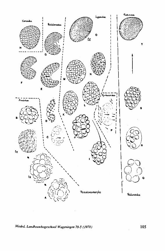
^.lAixviuioc.
Cortoidca
^ Ttntalomoraorpha
l&iiuvioickü.
V.V
Meded. Landbouwhogeschool iVageningen 78-5 (1978) 105
so, MATSUDA (1960, p. 52) concluded that within the Gerridae, eyes appear to have become larger in the more advanced subfamilies.
In the next major group, the Leptopodomorpha, the Leptopodidae have distinctly fewer ommatidia (Fig. 46, i) than the more derived Saldidae (Fig. 46, J), even though species of both families have similar searching and predatory behaviour in comparable habitats. Fig. J. shows the eyes of a number of species in two subfamilies of Saldidae, the Chiloxanthinae and Saldinae. A well-developed eye of more than 150 ommatidia is present, whether the species live in very dense vegetation or not. The multi-lensed eyes of the more generalized Chiloxanthinae (Chiloxanthus) studied are not as spherical as those of the more evolved Saldinae, and may have a smaller number of ommatidia. The first instar larvae of Aepophilus bonnairei, the only existing representative of the most generalized subfamily, the Aepophilinae (living in the intertidal Fucus-zone) is unknown. Each adult eye has 35 facts, and the third larval instar many fewer. That the dominant course of evolution within Saldidae has been towards ommatidial multiplication seems evident when accepted synapomorphies in other, independently evolved, character-sets are considered. Increase in ommatidial number through Aepophilinae, Chiloxanthinae, and Saldinae is related with decrease in size of the single larval scent-gland (Fig. 53C-54A-E), appearance and elaboration of eversible pregenital adult glands (COBBEN, 1961), progressively increasing complexity of the male intromittent organ (COBBEN, 1965a) and anagenesis of female genital ectodermalia (COBBEN. 1970) (see also this paper, pages 173-185).
The paucity of ommatidia in Aepophilus could be due to the cryptic situation in which this taxon lives, but this argument should hold true also for the lep-topodoid family Omaniidae, containing very minute species, which inhabit intertidal crevices in coral or volcanic sediments. Omaniidae, nevertheless, have huge eyes with innummerable lenses in the first larval instar (COBBEN, 1970). This last example also stresses the fact, that body size and eye size are not necessarily related.
Very large eyes are characteristic for the Nepomorpha (most common carnivorous genera of european families studied ; but also for the mixed feeding Corixinae Corixidae). Ochterus has slightly more than 50 ommatidia, Plea more than 100 (lenses small and widely separated!), and Ilyocoris, Notonecta and Corixa several hundred. Helotrephidae were not studied, and more members have to be investigated, particularly in diversified families such as Naucoridae. Within the Nepomorpha the Potamocoridae has a more embryonic eye pattern. Since the adult of Coleopterocoris has only 38 ommatidia (Fig. 46S), its first larval instar must have eyes differing greatly from those of the other waterbugs.
Another exclusively carnivorous group whose embryology has indicated close affinity with the Nepomorpha, is the Reduvioidea. The range in eye structure so far found in this taxon is depicted in Fig. 47 Q-T. The three genera of Emesinae investigated (Empicoris, Metapterus, and Schidium) have 6, large ommatidia (Q) in each first instar eye. Representatives of other subfamilies
106 Meded. Landbouwhogeschool Wageningen 78-5 (1978)
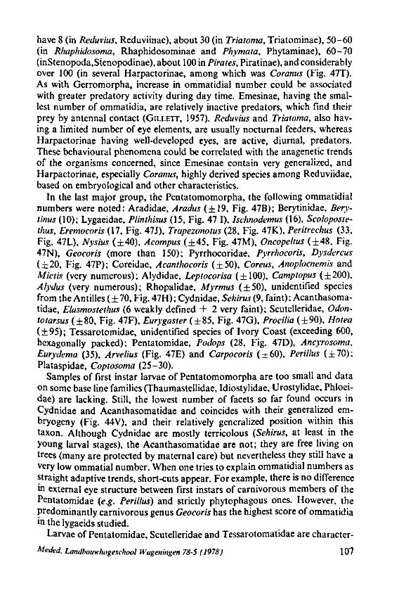
have 8 (in Reduvius, Reduviinae), about 30 (in Triatoma, Triatominae), 50-60 (in Rhaphidosoma, Rhaphidosominae and Phymata, Phytaminae), 60-70 (inStenopoda,Stenopodinae), about 100 in Pirates, Piratinae), and considerably over 100 (in several Harpactorinae, among which was Coranus (Fig. 47T). As with Gerromorpha, increase in ommatidial number could be associated with greater predatory activity during day time. Emesinae, having the smallest number of ommatidia, are relatively inactive predators, which find their prey by antennal contact (GILLETT, 1957). Reduvius and Triatoma, also having a limited number of eye elements, are usually nocturnal feeders, whereas Harpactorinae having well-developed eyes, are active, diurnal, predators. These behavioural phenomena could be correlated with the anagenetic trends of the organisms concerned, since Emesinae contain very generalized, and Harpactorinae, especially Coranus, highly derived species among Reduviidae, based on embryological and other characteristics.
In the last major group, the Pentatomomorpha, the following ommatidial numbers were noted: Aradidae, Aradus (±19, Fig. 47B); Berytinidae, Bery-ïmus (10); Lygaeidae, Plinthisus (15, Fig. 47 I), Ischnodemus (16), Scoloposte-thus, Eremocoris (17, Fig. 47J), Trapezonotus (28, Fig. 47K), Peritrechus (33, Fig. 47L), Nysius (±40), Acompus (±45, Fig. 47M), Oncopeltus (±48, Fig. 47N), Geocoris (more than 150); Pyrrhocoridae, Pyrrhocoris, Dysdercus (±20, Fig. 47P); Coreidae, Acanthocoris (±50), Coreus, Anoplocnemis and Mictis (very numerous); Alydidae, Leptocorisa (±100), Camptopus (±200), Alydus (very numerous); Rhopalidae, Myrmus (±50), unidentified species from the Antilles (±70, Fig. 47H); Cydnidae, Sehirus (9, faint); Acanthasoma-tidae, Elasmostethus (6 weakly defined + 2 very faint); Scutelleridae, Odon-totarsus (±80, Fig. 47F), Eurygaster (±85, Fig. 47G), Procilia (±90), Hotea (±95); Tessarotomidae, unidentified species of Ivory Coast (exceeding 600, hexagonally packed); Pentatomidae, Podops (28, Fig. 47D), Ancyrosoma, Eurydema (35), Arvelius (Fig. 47E) and Carpocoris (±60), Perillus (±70); Plataspidae, Coptosoma (25-30).
Samples of first instar larvae of Pentatomomorpha are too small and data on some base line families (Thaumastellidae, Idiostylidae, Urostylidae, Phloei-dae) are lacking. Still, the lowest number of facets so far found occurs in Cydnidae and Acanthasomatidae and coincides with their generalized em-bryogeny (Fig. 44V), and their relatively generalized position within this taxon. Although Cydnidae are mostly terricolous (Sehirus, at least in the young larval stages), the Acanthasomatidae are not; they are free living on trees (many are protected by maternal care) but nevertheless they still have a very low ommatial number. When one tries to explain ommatidial numbers as straight adaptive trends, short-cuts appear. For example, there is no difference in external eye structure between first instars of carnivorous members of the Pentatomidae (e.g. Perillus) and strictly phytophagous ones. However, the predominantly carnivorous genus Geocoris has the highest score of ommatidia in the lygaeids studied.
Larvae of Pentatomidae, Scutelleridae and Tessarotomatidae are character-
Meded. Landbouwhogeschool Wageningen 78-5 (1978) 107
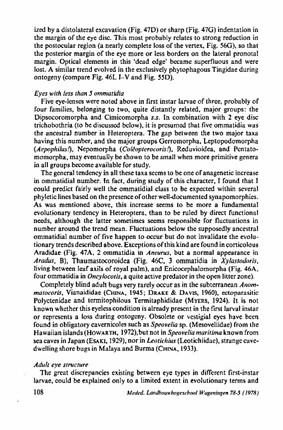
ized by a distolateral excavation (Fig. 47D) or sharp (Fig. 47G) indentation in the margin of the eye disc. This most probably relates to strong reduction in the postocular region (a nearly complete loss of the vertex, Fig. 56G), so that the posterior margin of the eye more or less borders on the lateral pronotal margin. Optical elements in this 'dead edge' became superfluous and were lost. A similar trend evolved in the exclusively phytophagous Tingidae during ontogeny (compare Fig. 46L I-V and Fig. 55D).
Eyes with less than 5 ommatidia Five eye-lenses were noted above in first instar larvae of three, probably of
four families, belonging to two, quite distantly related, major groups: the Dipsocoromorpha and Cimicomorpha s.S. In combination with 2 eye disc trichobothria (to be discussed below), it is presumed that five ommatidia was the ancestral number in Heteroptera. The gap between the two major taxa having this number, and the major groups Gerromorpha, Leptopodomorpha (Aepophilusf), Nepomorpha (Cotâopterocoris!), Reduvioidea, and Pentato-momorpha, may eventually be shown to be small when more primitive genera in all groups become available for study.
The general tendency in all these taxa seems to be one of anagenetic increase in ommatidial number. In fact, during study of this character, I found that I could predict fairly well the ommatidial class to be expected within several phyletic lines based on the presence of other well-documented synapomorphies. As was mentioned above, this increase seems to be more a fundamental evolutionary tendency in Heteroptera, than to be ruled by direct functional needs, although the latter sometimes seems responsible for fluctuations in number around the trend mean. Fluctuations below the supposedly ancestral ommatidial number of five happen to occur but do not invalidate the evolutionary trends described above. Exceptions of this kind are found in corticolous Aradidae (Fig. 47A, 2 ommatidia in Aneurus, but a normal appearance in Aradus, B), Thaumastocoroidea (Fig. 46C, 3 ommatidia in Xylastodoris, living between leaf axils of royal palm), and Enicocephalomorpha (Fig. 46A, four ommatidia in Oncylocotis, a quite active predator in the open litter zone).
Completely blind adult bugs very rarely occur as in the subterranean Anom-matocoris, Vianaididae (CHINA, 1945; DRAKE & DAVIS, 1960), ectoparasitic Polyctenidae and termitophilous Termitaphididae (MYERS, 1924). It is not known whether this eyeless condition is already present in the first larval instar or represents a loss during ontogeny. Obsolete or vestigial eyes have been found in obligatory cavernicoles such as Speovelia sp. (Mesoveliidae) from the Hawaiian islands (HOWARTH, 1972),but not in Speovelia maritima known from sea caves in Japan (ESAKI, 1929), nor in Leotkhius (Leotichiidae), strange cave-dwelling shore bugs in Malaya and Burma (CHINA, 1933).
Adult eye structure The great discrepancies existing between eye types in different first-instar
larvae, could be explained only to a limited extent in evolutionary terms and
108 Mede J. Landbouwhogeschool Wageningen 78-5 (1978)
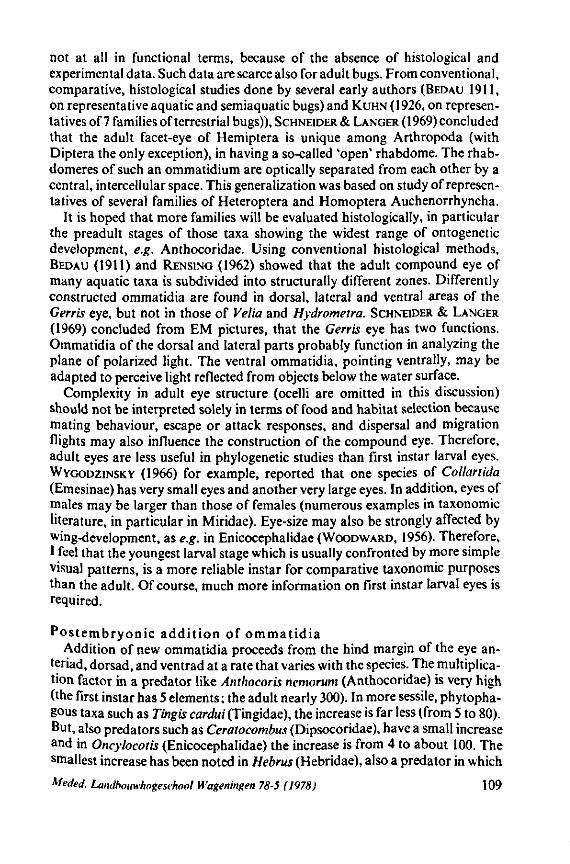
not at all in functional terms, because of the absence of histological and experimental data. Such data are scarce also for adult bugs. From conventional, comparative, histological studies done by several early authors (BEDAU 1911, on representative aquatic and semiaquatic bugs) and KUHN (1926, on representatives of 7 families of terrestrial bugs)), SCHNEIDER & LANGER (1969) concluded that the adult facet-eye of Hemiptera is unique among Arthropoda (with Diptera the only exception), in having a so-called 'open' rhabdome. The rhab-domeres of such an ommatidium are optically separated from each other by a central, intercellular space. This generalization was based on study of representatives of several families of Heteroptera and Homoptera Auchenorrhyncha.
It is hoped that more families will be evaluated histologically, in particular the preadult stages of those taxa showing the widest range of ontogenetic development, e.g. Anthocoridae. Using conventional histological methods, BEDAU (1911) and RENSING (1962) showed that the adult compound eye of many aquatic taxa is subdivided into structurally different zones. Differently constructed ommatidia are found in dorsal, lateral and ventral areas of the Gents eye, but not in those of Velia and Hydrometra. SCHNEIDER & LANGER
(1969) concluded from EM pictures, that the Gerris eye has two functions. Ommatidia of the dorsal and lateral parts probably function in analyzing the plane of polarized light. The ventral ommatidia, pointing ventrally, may be adapted to perceive light reflected from objects below the water surface.
Complexity in adult eye structure (ocelli are omitted in this discussion) should not be interpreted solely in terms of food and habitat selection because mating behaviour, escape or attack responses, and dispersal and migration flights may also influence the construction of the compound eye. Therefore, adult eyes are less useful in phylogenetic studies than first instar larval eyes. WYGODZINSKY (1966) for example, reported that one species of Collartida (Emesinae) has very small eyes and another very large eyes. In addition, eyes of males may be larger than those of females (numerous examples in taxonomie literature, in particular in Miridae). Eye-size may also be strongly affected by wing-development, as e.g. in Enicocephalidae (WOODWARD, 1956). Therefore, I feel that the youngest larval stage which is usually confronted by more simple visual patterns, is a more reliable instar for comparative taxonomie purposes than the adult. Of course, much more information on first instar larval eyes is required.
P o s t e m b r y o n i c a d d i t i o n of o m m a t i d i a Addition of new ommatidia proceeds from the hind margin of the eye an-
teriad, dorsad, and ventrad at a rate that varies with the species. The multiplication factor in a predator like Anthocoris nemorum (Anthocoridae) is very high (the first instar has 5 elements; the adult nearly 300). In more sessile, phytophagous taxa such as Tingis cardui (Tingidae), the increase is far less (from 5 to 80). But, also predators such as Ceratocombus (Dipsocoridae), have a small increase and in Oncylocotis (Enicocephalidae) the increase is from 4 to about 100. The smallest increase has been noted in Hebrus (Hebridae), also a predator in which
Meded. Landbouwhogeschool Wageningen 78-5 (1978) 109
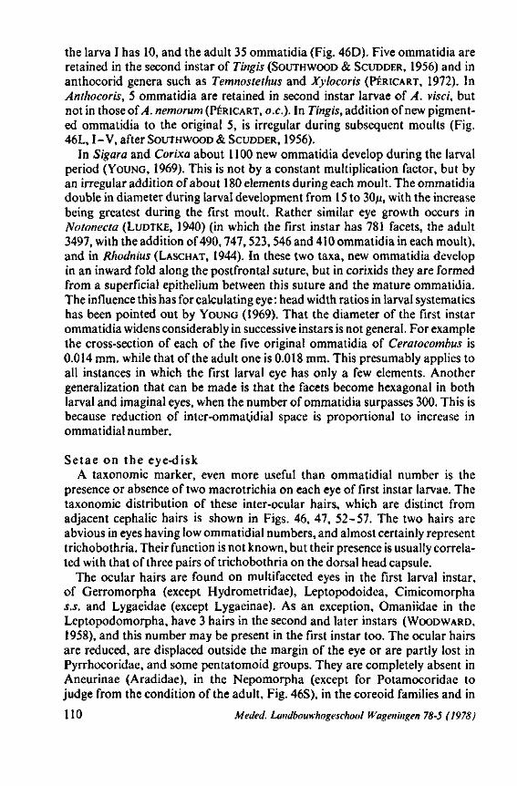
the larva I has 10, and the adult 35 ommatidia (Fig. 46D). Five ommatidia are retained in the second instar of Tingis (SOUTHWOOD & SCUDDER, 1956) and in anthocorid genera such as Temnostethus and Xylocoris (PÉRICART, 1972). In Anthocoris, 5 ommatidia are retained in second instar larvae of A. visci, but not in those of A. nemorum (PÉRICART, o.e.). In Tingis, addition of new pigmented ommatidia to the original 5, is irregular during subsequent moults (Fig. 46L, I-V, after SOUTHWOOD & SCUDDER, 1956).
In Sigara and Corixa about 1100 new ommatidia develop during the larval period (YOUNG, 1969). This is not by a constant multiplication factor, but by an irregular addition of about 180 elements during each moult. The ommatidia double in diameter during larval development from 15 to 30/x, with the increase being greatest during the first moult. Rather similar eye growth occurs in Notonecta (LUDTKE, 1940) (in which the first instar has 781 facets, the adult 3497, with the addition of 490,747,523,546 and 410 ommatidia in each moult), and in Rhodnius (LASCHAT, 1944). In these two taxa, new ommatidia develop in an inward fold along the postfrontal suture, but in corixids they are formed from a superficial epithelium between this suture and the mature ommatidia. The influence this has for calculating eye : head width ratios in larval systematics has been pointed out by YOUNG (1969). That the diameter of the first instar ommatidia widens considerably in successive instars is not general. For example the cross-section of each of the five original ommatidia of Ceratocombus is 0.014 mm. while that of the adult one is 0.018 mm. This presumably applies to all instances in which the first larval eye has only a few elements. Another generalization that can be made is that the facets become hexagonal in both larval and imaginai eyes, when the number of ommatidia surpasses 300. This is because reduction of inter-ommatjdial space is proportional to increase in ommatidial number.
Setae on the eye-disk A taxonomie marker, even more useful than ommatidial number is the
presence or absence of two macrotrichia on each eye of first instar larvae. The taxonomie distribution of these inter-ocular hairs, which are distinct from adjacent cephalic hairs is shown in Figs. 46, 47, 52-57. The two hairs are abvious in eyes having low ommatidial numbers, and almost certainly represent trichobothria. Their function is not known, but their presence is usually correlated with that of three pairs of trichobothria on the dorsal head capsule.
The ocular hairs are found on multifaceted eyes in the first larval instar, of Gerromorpha (except Hydrometridae), Leptopodoidea, Cimicomorpha s.s. and Lygaeidae (except Lygaeinae). As an exception, Omaniidae in the Leptopodomorpha, have 3 hairs in the second and later instars (WOODWARD, 1958), and this number may be present in the first instar too. The ocular hairs are reduced, are displaced outside the margin of the eye or are partly lost in Pyrrhocoridae, and some pentatomoid groups. They are completely absent in Aneurinae (Aradidae), in the Nepomorpha (except for Potamocoridae to judge from the condition of the adult, Fig. 46S), in the coreoid families and in
110 Meded. Landbouwhogeschool Wageningen 78-5 (1978)
all subfamilies of Reduviidae studied. In Oncylocotis (Enicocephalidae), having only 4 ommatidia (Fig. 46A), there are many hairs, not differing from normal, cephalic hairs.
The gradual disappearance of these hairs in the Pentatomomorpha may indicate an anagenetic trend. Their overall absence in Reduviidae, together with the basic six-faceted eyes of this taxon, pin-point an obvious discontinuity with all other groups and is in great contrast to the Cimicomorpha s.S.
The fate of the oculair hairs throughout postembryogenesis has not been followed in as many taxa as were studied in the first instar. They persist at least in Potamocoridae, Hebridae, many Gerridae, Schizopteridae, and in Thau-mastocoridae (Xylastodoris). Complete loss during ontogeny is the usual situation.
Secondary, interocular, micro- and macrotrichial patterns usually appear in later stages of an individual's development, as for example in the adult of Tingis (one papilla between each of three adjacent ommatidia) or Anthocoris (about 15 small interocular spines). Often a dense long hair vestiture develops. Such diversified, innervated, cuticular extensions have been described in other orders of insects (HINTON, 1970), but their function remains unknown except for those of the honeybee (NEESE, 1965; measuring sun-angle, control of sidewind deviation). The wide spaces between the facets of first instar larvae is always smooth ; the sole exception noted being Isometopus (Fig. 46Q).
3.3. PRETARSAL STRUCTURES
The conclusion made in 1968 (p. 365), that pretarsal organization in Heterop-tera shows perplexing plasticity, is elaborated further here with the aid of illustrations (Figs. 48-51). The range in pretarsal structure is best illustrated with reference to two, closely related, families. The most simple pretarsus is found in Tingidae and consists of two simple ungues (claws) and an unguitrac-tor bearing two, very short parempodia. In the related family Miridae there is great variation in plantular emergentia and claw types. The question of which of these types is considered to be ancestral, and whether one or both are evolutionary extremes, will be answered at the end of this section.
Confusion in the literature concerning homologies of pretarsal constituents in insects and in Heteroptera in particular, has been reviewed by COBBEN (1968a) and GOEL & SCHAEFER (1970), and is not repeated here. The following, still incomplete, data are surveyed with reference to each major group, with stress being placed on interesting points which hopefully will be substantiated by future research.
Gerromorpha. This group is characterized by the presence of strange arolial appendages. Two arolia, laterally flattened, inserted in a vertical plane, and one curved upwards and the other downwards, is the usual condition (Fig. 48, A-C, G)*. These transparent (sclerotized in Microvelia) slips originate
* TULLGREN (1918) was the first in having recognized the two arolia in Hebrus ruficeps, and BORNER (1934) emphasized the importance of this condition in Gerromorpha.
Meded. Landbouwhogeschool Wageningen 78-5 (1978) 111
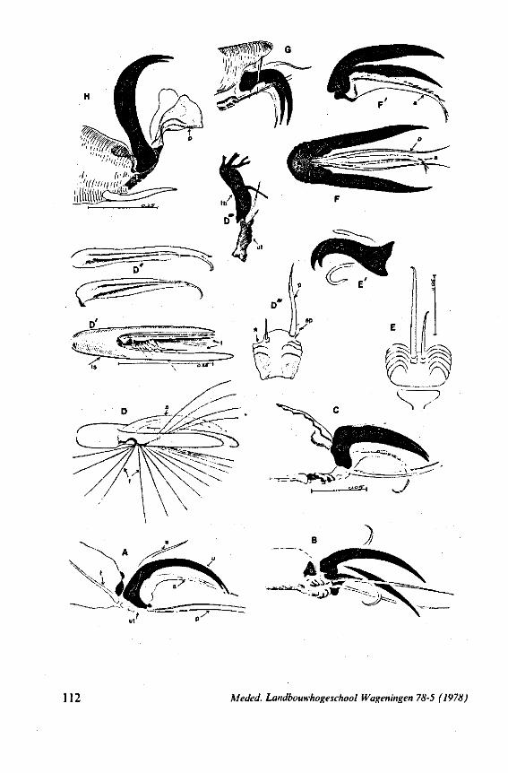
112 Meded. Landbouwhogeschool Wageningen 78-5 (1978)
from the membrane between the claws dorsal to the unguitractor. They are present in all legs of common taxa of Hebridae, Mesoveliidae, Hydrometridae, Veliidae and Gerridae, in both larvae and adults. Similar structures are found in some aberrant taxa such as Ocellovelia and Hebrovelia.
Preapical shift of the pretarsus does not affect the presence of arolia. For example, in the rhagioveliin Trochopus, they lie between the displaced claws of fore and hind legs (Fig. 7 in COBBEN, I960). The mid legs in members of this veliid subfamily exhibit the most extreme pretarsal modification found in Heteroptera: complete loss of the arolia and with drastic change of the two claws, originating at the base of a deep cleft in the disti-tarsus (Fig. 48D'). The claws became asymmetrical, transparent slips (Fig. 48D, u; D")*. The stem of the fan of feathery hairs, which characterizes the subfamily Rhagiove-liinae (Fig. 48D, D' ; f ; the 'wealthy plumage' is omitted but is partly seen in the scanning micrograph Fig. 166E), arises unilaterally from the parempodia (Fig. 48D", place of attachment marked with*). Its flat plane is perpendicular to that of the unguitractor plate (Fig. 48D""). The parempodium adjacent to the fan is strongly reduced and the counterpart of the fan is visible at the other distal angle of the unguitractor as a lobular extension dorsolateral to the neighbouring parempodial bristle. Since rudiments of such an accessory empodial pair occasionally occur in other taxa, I propose the term 'accessory parempodia'' for these secondary extensions. The primary parempodia, which certainly belong to the original equipment of the unguitractor, are mostly straight, spinous structure« inserted into a distinct pit ('alveolus', GOEL, 1972).
Fig. 48D'" shows a bilateral, structural flexibility to exist in the unguitractor which did not emerge from the studies of DASHMAN (1953) and GOEL (I.e.); papers especially devoted to unguitractor variation in Heteroptera. Asymmetric parempodia occur also in other Gerromorpha (e.g. Fig. 48E, associated with asymmetry of the claws, Fig. 48 E'). The gerromorphan unguitractor is
* In 1968 (p. 365), I thought that these structures represented the arolia, but after correspondence with dr. N. M0LLER ANDERSEN (Copenhagen) and having studied other representatives of the Rhagioveliinae, 1 now believe that the two filaments are indeed the transformed claws.
FIG. 48. Pretarsal structures of Gerromorpha; not on scale; claws are drawn black; the origin of the leg (anterior, mid or posterior) is only mentioned when structures differ by leg; A, B of first larval instar, C-H of adult. A, Hebrus ruficeps; B, Mesoveliafurcata; C, Speovelia acta; D, Trochopusplumbeus, mid leg; D', ventral view (see also Fig. 166E); D' , arolia; D", unguitractor plate, asterisk points to the attachment site of the swimming fan; D " , side view of unguitractor with basis of fan; E, Cylindrosteihus sp., unguitractor and parempodia; E', side view of claws and arolia; F, Chepuvelia usingeri, ventral view of pretarsus; F', side view; G, Limnobatodes paradoxus, note the vertical tarsal spines enclosing the pretarsus. a, arolium; ap, accessory parempodium; f. plumose fan, side-branches omitted (see also Fig- 166E); fb, base of fan; p, parempodium; t, tendon of unguitractor; ts, tarsal segment; u, unguus; ut, unguitractor.
Meded. Landbouwhogeschool Wageningen 78-5 (1978) 113
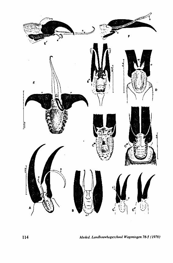
114 Meded. Landbouwhogeschool Wageningen 78-5 (1978)
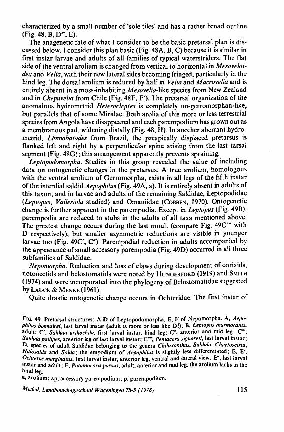
characterized by a small number of 'sole tiles' and has a rather broad outline (Fig. 48, B, D"\ E).
The anagenetic fate of what I consider to be the basic pretarsal plan is discussed below. I consider this plan basic (Fig. 48A, B, C) because it is similar in first instar larvae and adults of all families of typical waterstriders. The flat side of the ventral arolium is changed from vertical to horizontal in Mesoveloi-dea and Velia, with their new lateral sides becoming fringed, particularly in the hind leg. The dorsal arolium is reduced by half in Velia and Macrovelia and is entirely absent in a moss-inhabiting Mesovelia-Yike species from New Zealand and in Chepuvelia from Chile (Fig. 48F, F'). The pretarsal organization of the anomalous hydrometrid Heterocleptes is completely un-gerromorphan-like, but parallels that of some Miridae. Both arolia of this more or less terrestrial species from Angola have disappeared and each parempodium has grown out as a membranous pad, widening distally (Fig. 48, H). In another aberrant hydrometrid, Limnobatodes from Brazil, the preapically displaced pretarsus is flanked left and right by a perpendicular spine arising from the last tarsal segment (Fig. 48G); this arrangement apparently prevents spraining.
Leptopodomorpha. Studies in this group revealed the value of including data on ontogenetic changes in the pretarsus. A true arolium, homologous with the ventral arolium of Gerromorpha, exists in all legs of the fifth instar of the interdial saldid Aepophilus (Fig. 49A, a). It is entirely absent in adults of this taxon, and in larvae and adults of the remaining Saldidae, Leptopodidae {Leptopus, Valleriola studied) and Omaniidae (COBBEN, 1970). Ontogenetic change is further apparent in the parempodia. Except in Leptopus (Fig. 49B), parempodia are reduced to stubs in the adults of all taxa mentioned above. The greatest change occurs during the last moult (compare Fig. 4 9C" with D respectively), but smaller asymmetric reductions are visible in younger larvae too (Fig. 49C', C ) . Parempodial reduction in adults accompanied by the appearance of small accessory parempodia (Fig. 49D) occurred in all three subfamilies of Saldidae.
Nepomorpha. Reduction and loss of claws during development of corixids, notonectids and belostomatids were noted by HUNGERFORD (1919) and SMITH
(1974) and were incorporated into the phylogeny of Belostomatidae suggested byLAUCK&MENKE(1961).
Quite drastic ontogenetic change occurs in Ochteridae. The first instar of
FIG. 49. Pretarsal structures; A-D of Leptopodomorpha, E, F of Nepomorpha. A, Aepophilus bonnairei, last larval instar (adult is more or less like D!); B, Leptopus marmoratus. adult; C', SalJula orthochila, first larval instar, hind leg; C', anterior and mid leg; C", SalJula ptitlipes, anterior leg of last larval instar; C" , Pentacora signoreii, last larval instar; D, species of adult Saldidae belonging to the genera Chiloxanthus, SalJula, Chartoscirta, Halosalda and Salda; the empodium of Aepophilus is slightly less differentiated; E, E', Ochierus marginatus, first larval instar, anterior leg, ventral and lateral view; E*, last larval instar and adult; F, Potamocorisparvus, adult, anterior and mid leg, the arolium lacks in the hind leg. a, arolium; ap, accessory parempodium; p, parempodium.
Meded. Landbouwhogeschool Wageningen 78-5 (1978) 115
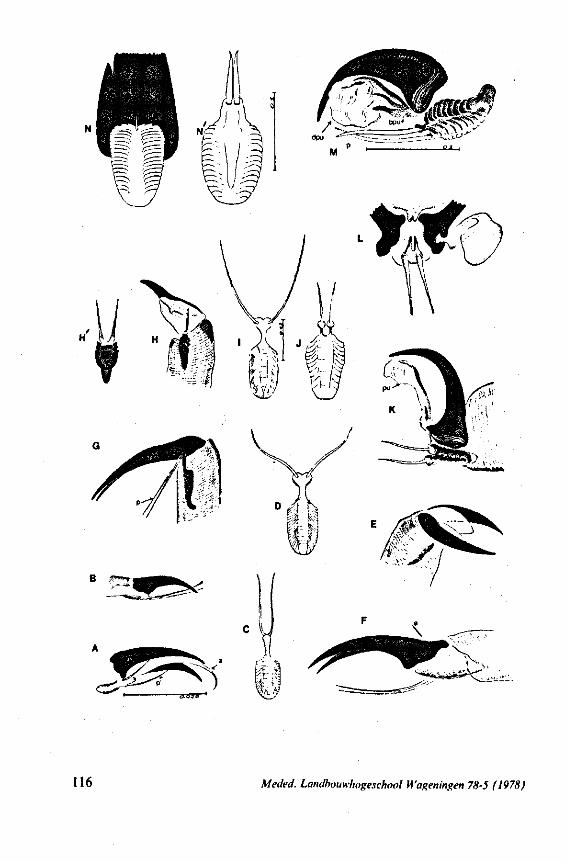
B ; f *1r - i
c u i
116 Meded. Landbouwhogeschool Wageningen 78-5 (1978)
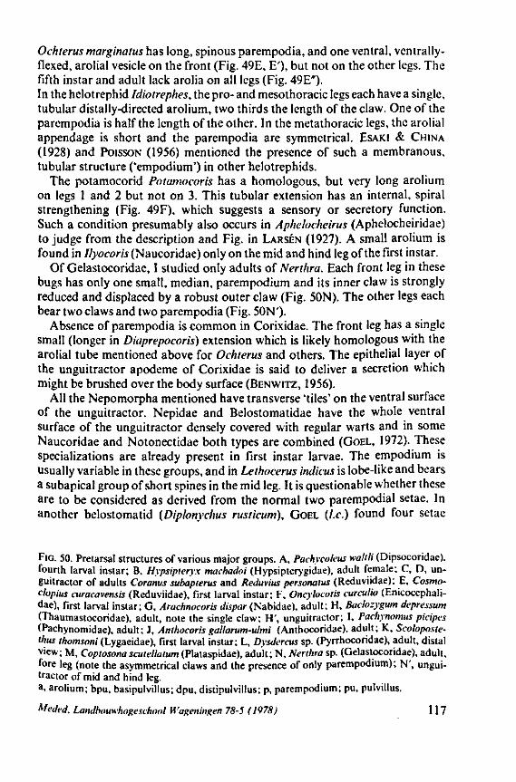
Ochterus marginatus has long, spinous parempodia, and one ventral, ventrally-flexed, arolial vesicle on the front (Fig. 49E, E'), but not on the other legs. The fifth instar and adult lack arolia on all legs (Fig. 49E"). In the helotrephid Idiotrephes, the pro- and mesothoracic legs each have a single, tubular distally-directed arolium, two thirds the length of the claw. One of the parempodia is half the length of the other. In the metathoracic legs, the arolial appendage is short and the parempodia are symmetrical. ESAKI & CHINA (1928) and POISSON (1956) mentioned the presence of such a membranous, tubular structure ('empodium') in other helotrephids.
The potamocorid Potamocoris has a homologous, but very long arolium on legs 1 and 2 but not on 3. This tubular extension has an internal, spiral strengthening (Fig. 49F), which suggests a sensory or secretory function. Such a condition presumably also occurs in Aphelocheirus (Aphelocheiridae) to judge from the description and Fig. in LARSÉN (1927). A small arolium is found in Ilyocoris (Naucoridae) only on the mid and hind leg of the first instar.
Of Gelastocoridae, I studied only adults of Nerthra. Each front leg in these bugs has only one small, median, parempodium and its inner claw is strongly reduced and displaced by a robust outer claw (Fig. 50N). The other legs each bear two claws and two parempodia (Fig. 50N')-
Absence of parempodia is common in Corixidae. The front leg has a single small (longer in Diaprepocoris) extension which is likely homologous with the arolial tube mentioned above for Ochterus and others. The epithelial layer of the unguitractor apodeme of Corixidae is said to deliver a secretion which might be brushed over the body surface (BENWITZ, 1956).
All the Nepomorpha mentioned have transverse 'tiles' on the ventral surface of the unguitractor. Nepidae and Belostomatidae have the whole ventral surface of the unguitractor densely covered with regular warts and in some Naucoridae and Notonectidae both types are combined (GOEL, 1972). These specializations are already present in first instar larvae. The empodium is usually variable in these groups, and in Lethocerus indicus is lobe-like and bears a subapical group of short spines in the mid leg. It is questionable whether these are to be considered as derived from the normal two parempodial setae. In another belostomatid (Diplonychus rusticum), GOEL {I.e.) found four setae
FIG. 50. Pretarsal structures of various major groups. A, Pachycoleus waltli (Dipsocoridae). fourth larval instar; B, Hypsipieryx machadoi (Hypsipterygidae), adult female; C, D, unguitractor of adults Coranus subapterus and Reduvius personatus (Reduviidae); E, Cosmo-clopius curacavensis (Reduviidae), first larval instar; F, Oncylocotis curculio (Enicocephali-dae), first larval instar; G, Arachnocoris dispar (Nabidae), adult; H, Baclozygum depressum (Thaumastocoridae), adult, note the single claw; H', unguitractor; I, Pachynomus picipes (Pachynomidae), adult; J, Anthocoris gallarum-ulmi (Anthocoridae), adult; K, Scoloposte-thus thomsoni (Lygaeidae), first larval instar; L, Dysdercus sp. (Pyrrhocoridae), adult, distal view; M, Copiosona scutellatum (Plataspidae), adult; N, Nerthra sp. (Gelastocoridae), adult, fore leg (note the asymmetrical claws and the presence of only parempodium); N', unguitractor of mid and hind leg. a, arolium; bpu, basipulvillus; dpu, distipulvillus; p, parempodium; pu, pulvillus.
Meded. Landbouwhogeschool Wageningen 78-5 (1978J 117
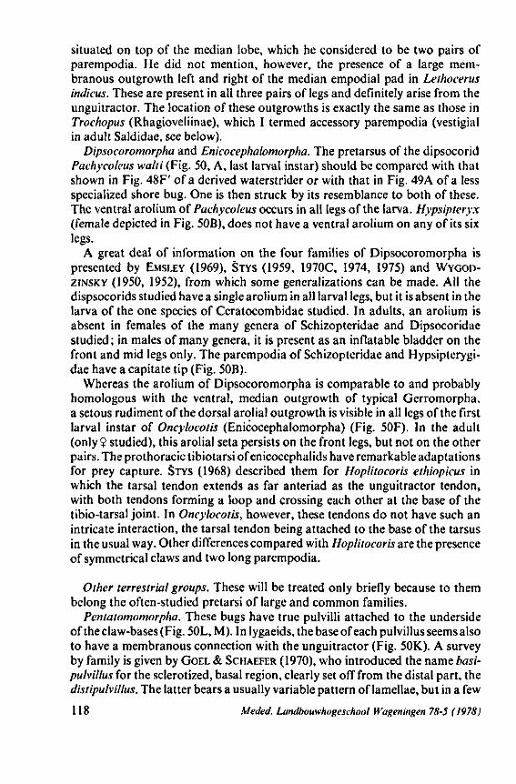
situated on top of the median lobe, which he considered to be two pairs of parempodia. He did not mention, however, the presence of a large membranous outgrowth left and right of the median empodial pad in Lethocerus indicus. These are present in all three pairs of legs and definitely arise from the unguitractor. The location of these outgrowths is exactly the same as those in Trochopus (Rhagioveliinae), which I termed accessory parempodia (vestigial in adult Saldidae, see below).
Dipsocoromorpha and Enkocephalomorpha. The pretarsus of the dipsocorid Pachycoleus walti (Fig. 50, A, last larval instar) should be compared with that shown in Fig. 48F' of a derived waterstrider or with that in Fig. 49A of a less specialized shore bug. One is then struck by its resemblance to both of these. The ventral arolium of Pachycoleus occurs in all legs of the larva. Hypsipteryx (female depicted in Fig. 50B), does not have a ventral arolium on any of its six legs.
A great deal of information on the four families of Dipsocoromorpha is presented by EMSLEY (1969), STYS (1959, 1970C, 1974, 1975) and WYGOD-ZINSKY (1950, 1952), from which some generalizations can be made. All the dispsocorids studied have a single arolium in all larval legs, but it is absent in the larva of the one species of Ceratocombidae studied. In adults, an arolium is absent in females of the many genera of Schizopteridae and Dipsocoridae studied; in males of many genera, it is present as an inflatable bladder on the front and mid legs only. The parempodia of Schizopteridae and Hypsipterygi-dae have a capitate tip (Fig. 50B).
Whereas the arolium of Dipsocoromorpha is comparable to and probably homologous with the ventral, median outgrowth of typical Gerromorpha, a setous rudiment of the dorsal arolial outgrowth is visible in all legs of the first larval instar of Oncylocotis (Enicocephalomorpha) (Fig. 50F). In the adult (only? studied), this arolial seta persists on the front legs, but not on the other pairs. The prothoracic tibiotarsi of enicocephalids have remarkable adaptations for prey capture. STYS (1968) described them for Hoplitocoris ethiopicus in which the tarsal tendon extends as far anteriad as the unguitractor tendon, with both tendons forming a loop and crossing each other at the base of the tibio-tarsal joint. In Oncylocotis, however, these tendons do not have such an intricate interaction, the tarsal tendon being attached to the base of the tarsus in the usual way. Other differences compared with Hoplitocoris are the presence of symmetrical claws and two long parempodia.
Other terrestrial groups. These will be treated only briefly because to them belong the often-studied pretarsi of large and common families.
Pentatomomorpha. These bugs have true pulvilli attached to the underside of the claw-bases (Fig. 50L, M). In lygaeids, the base of each pulvillus seems also to have a membranous connection with the unguitractor (Fig. 50K). A survey by family is given by GOEL & SCHAEFER (1970), who introduced the name basi-pulvillus for the sclerotized, basal region, clearly set off from the distal part, the distipuhillus. The latter bears a usually variable pattern of lamellae, but in a few
118 Meded. Landbouwhogeschool Wageningen 78-5 (1978)
taxa of Lygaeidae (e.g. Plinthisus) these are few and weak, and in the cydnids (4 spp.) and aradids (1 sp.) studied they are absent. I checked first instar larvae of the aradid genus Aneurus and found no distinction present between basi-and distipulvillus, contrasting with another aradid (Glochocoris sp.) studied by GOEL & SCHAEFER (I.e.). The statement of these authors on p. 312, that 'the absence of lamellae in the pulvillus appears to ally the Aradidae with the Pen-tatomoidea, if the 1 aradid species studied is representative' contradicts their own findings that pentatomoids indeed have lamellae. Special attention should be paid in future to the pretarsus of Aradidae, since some divisions lack pul-villi and some genera have median projections (USINGER & MATSUDA, 1959) which might be arolia.
Thaumastocoroidea. The phylogenetic position of the single small family contained in this superfamily is still not agreed upon, although several recent publications have been devoted to these bugs. The two subfamilies have been characterized by their different pretarsal configuration : pulvillus and parem-podia absent in Thaumastocorinae, and present in Xylastodorinae (DRAKE & SLATER, 1957). SCHAEFER (1969) concluded, after studying Xylaslodoris, that the basal part of each pulvillus is absent, and that the distal part is thin and leaflike with only a few lamellae. My experience is that this structure is hardly comparable with the pentatomomorphan pulvillus, but is rather an accessory parempodium (p. 113). Its main stem originates from the unguitractor, lateral to the primary parempodial setae. The situation is much the same as that found in the mirid genus Helopeltis (Fig. 51C, C')- Baclozygum depressum, a representative of the other subfamily Thaumastocorinae, should have no parempodium and pulvillus (DRAKE & SLATER, 1957). However, it has two parempodia (Fig. 50H') and one claw, bearing a membranous lobe (Fig. 50H), whose origin was difficult to define.
Reduvioidea. Reduviid bugs generally have a simple pretarsus, consisting of paired claws and two spinous parempodia (Fig. 50C, D). Further reductions of both claws and parempodia occur. A reliable evolutionary picture is presented by WYGODZINSKY (1966, pp. 21-24) in his world revision of the Emesinae. He showed that trends exist towards secondary asymmetry of the anterior claws, culminating in loss of the inner claw. The under surface of each claw may bear a membranous lamella of varying size and shape. Sometimes this lamella is crowned by a series of toothlike projections. WYGODZINSKY considered this and other modifications of the legs to be adaptations to life in spider webs. SNODDY e.a. (1976) observed that legs and tarsi of the emesine Stenolemus lanipes are commonly covered with strands and bits of spider web ; they believe that this material acts as an antiadhesive agent to facilitate locomotion about the spider web. Variations, if any, existing in other subfamilies are poorly known. I found first instar larvae of Harpactorinae and Reduviinae to have hyaline, pulvillus-like structures (Fig. 50E), which, in the adult, appear as sclerotized basal projections of each claw. Reduviidae generally have tibial Pads, varying in extent and fine structure; these being gripping instruments adapted to the cuticular characteristics of their prey (EDWARDS, 1962).
Meded. Landbouwhogeschool H'ageningen 78-5 (1978) 119
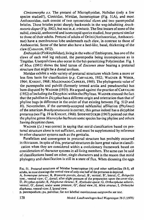
Cimicomorpha s.s. The pretarsi of Microphysidae, Nabidae (only a few species studied!), Cimicidae, Miridae, Isometopinae (Fig. 51 A), and most Anthocoridae, each consist of two symmetrical claws and two parempodial bristles. These bristles point sharply backwards in the weg-inhabiting Arach-nocoris dispar (Fig. 50G), but not in A. trinitatis. The first instars of some of the nabid, cimicid, anthocorid and isometopid species studied, bear pretarsi similar to those of their adults. Pretarsi of adults of Oriini (Anthocoridae, Anthocori-nae) have a membranous lobe underneath each claw, in contrast to those of Anthocorini. Some of the latter also have a heel-like, basal, thickening of the claw (CARAYON, 1972).
Embiophila (Plokiophilidae), living in the webs of Embioptera, has one of the claws of each mid leg reduced, the parempodia being inconspicuous as in Tingidae. Unequal claws also occur in the bat-parasitizing Polyctenidae. Fig. 1 of MAA (1961) shows the hind tarsus of Eoctenes sinae bearing a pretarsal structure that might be a dorsal arolium.
Miridae exhibit a wide variety of pretarsal structures which form a more or less firm basis for classification (e.g. CARVALHO, 1952; WAGNER & WEBER, 1964; KNIGHT, 1968; MALDONADO CAPRILES, 1969). The phylogenetic value of the parempodia and pulvilli (formerly termed arolia and pseudoarolia) has been disputed by WAGNER (1955). He argued against the practice of CARVALHO (1952) of including the Dicyphini within the Phylinae. WAGNER stressed the fact that the pulvillus in Dicyphus has a different origin and orientation than that of phyline bugs (a difference in the order ofthat existing between Fig. 51D and H). Nevertheless, if the currently-accepted subfamilial affiliation (Phylinae) of the american Brachyceratocoris is correct, this genus indeed has a dicyphine pretarsus (see Fig. 19 in KNIGHT, 1968). SEIDENSTÜCKER (1967) pointed out that the phyline genus Macrotylus harbours some species having phyline and others having dicyphine claws.
WAGNER (I.e.) was correct in saying that mirid classification based on pretarsal structure alone is not sufficient, and must be supplemented by reference to other character systems such as the genitalia.
Parallelism and convergence in pretarsal structure has probably occurred in this taxon. In spite of this, pretarsal structures do have great value in classification when they are considered within a evolutionary framework based on
consideration of character systems in all living members. The same can be said of classifications based on other, single characters and is the reason that mirid phylogeny and classification is still in a state of flux. When dicussing the eggs
FIG. 51. Pretarsal structures of Miridae Isometopinae (A) and other subfamilies (B-I), all adults, in most drawings the ventral view of only one half of the pretarsus is depicted. A, Isometopus intrusus; B, Bryocoris pteridis, dorsal; B', ventral; B", lateral; C, Helopellis spec., ventral view; C', dorsal, after slight pressure of the preparation upon the cover-slip; D, Dicyphus epilobii; E, Pilophorus clavatus; F, Hyaliodes opaca; G, Orthocephalus saltaior, ventral; G', dorsal, under some pressure; G", distal view; H, M iris striaius; I, Orthotylus diaphanus, ventral view; J, lateral view. p, parempodium; pu, pulvillus; for not labelled membranous outgrowths see text.
120 Meded. Landbouwhogeschool Wageningen 78-5 (1978)
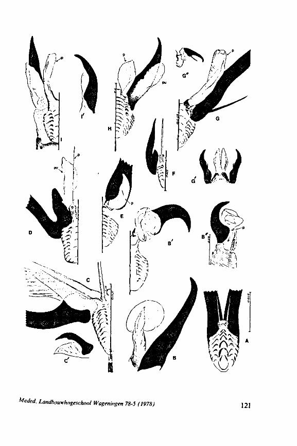
Medea. Landbouwhogeschool Wageningen 78-5 (1978) 121
of Miridae, I concluded in 1968 (p. 356) that they 'seem to have evolved explosively in morphological types, but exploratory studies of more species may throw new light on subfamilial limits and interrelations'.
A small selection of different pretarsal types in Miridae is presented in Fig. 51. The transformation of the parempodial spines into hyaline tubes or into leaf-like structures and the membranous supports of the claws are probably secondary. Thus a pretarsus having parempodial bristles but lacking pulvilli should be ancestral. This conclusion cannot be derived from study of the phylogenetic trees of WAGNER (1955) and LESTON (1961). Bryocorinae are placed by these and other authors at the base of the mirid system, and the pulvilli in this subfamily are said to arise from the fore-side of the claws in stead of ventrally as in other subfamilies. Like chorionic structures (COBBEN. 1968a), the pretarsus of Bryocorinae is atypical for mirids, but in a way other than that thought by WAGNER. Fig. 5IB, B', B" shows that the paired, bladderlike pretarsal outgrowths are neither true pulvilli nor parempodia. The weakly-sclerotized stem of the bladder arises from membrane dorsal to the unguitrac-tor and ventromedial of the claw base. The parempodia are vestigial (Fig. 51B"). More properly they should be termed 'arolia', but it is clear that these structures are not entirely homologous with the arolia of other groups. The pest genus Helopeltis and associated genera, are currently considered to be Bryocorinae. Egg characters of these insects, however, are quite different from those of typical Bryocorinae (COBBEN, 1968a), and SCHMITZ (1968) showed that the subfamily contains a mixture of diversified taxa. The pretarsus of Helopeltis is also not as in Bryocoris or Monalocoris. Helopeltis and allies bear spinous parempodia and paired, membranous, often leaf-like, outgrowths (SCHMITZ, 1968), which are termed pulvilli by GOEL & SCHAEFER (1970). Connection of each of these 'pulvilli' with the claw is weak, its greater part originating from the unguitractor and plantular membrane (Fig. 50C, C')*.
Some concluding remarks The previous survey has shown overwhelming diversity to exist in adult
pretarsal structure, and has pointed to several striking ontogenetic changes. The plesiomorphous pretarsus. Modern students of heteropteran phylo-
geny seem to agree that ancestral character states should be looked for in Gerromorpha, in Leptopodomorpha, in Nepomorpha or in Dipsocoromorpha and Enicocephalomorpha. Most members of these five taxa share the presence of arolia and setous parempodia. Typical Gerromorpha possess a ventral and a dorsal arolium in all ontogenetic stages and on all 6 legs. Within the Leptopodomorpha, a ventral arolium occurs only in larvae of Aepophilinae. Most nepomorphan families have a ventral arolium, either in all ontogenetic stages or in the larvae only. A ventral arolium occurs also in all larval instars of some families of Dipsocoromorpha; when present in adults, this persists only
* The pretarsal structures of Cimicomorpha have now been covered more extensively by SCHUH (1976). He presented many superb stereoscan micrographs and analyzed in great detail the phylogenetic implications of pretarsal and other structural features in the Miridae.
122 Meded. Landbouwhogeschool Wageningen 78-5 (1978)

on the anterior and mid legs of males. In the single enicocephalid species studied, a dorsal arolial seta was detected in both the youngest larva and the adult.
It thus seems that setous parempodia and arolia are plesiomorphous in Heteroptera. The question of whether the ancestral number of arolia was one or two (ventral and dorsal) is difficult to answer when considering the pretarsus alone. I consider that the best argument is for two arolia, the condition in Gerromorpha. For, two arolia occur in all families of waterstriders and in exactly the same extent in all developmental stages of these bugs (Fig. 48A, B). The dorsal arolium shows some reduction and, rarely, complete loss in a few aberrant forms. Absence of both arolia in the peculiar Heterocleptes is associated with the presence of membranous expansion of the parempodia (Fig. 48H). Delicate arolia occur in all typical water striders, whether they live on open water bodies (Gerridae, most Veliidae), on water with more or less covered dense vegetation (Mesoveliidae), on shores or in moss (Hydrometridae, Hebri-dae). The typical gerromorphan pretarsus is thus as well suited for locomotion on the water film as on soil and horizontal leaf surfaces (these bugs cannot climb up a dry, vertical, glass wall). I found a large colony of Hebrovelia in the Ivory Coast, actively running around in a completely dry rivulet bed. Macrovelia aestivates under similar conditions in the USA (LATTIN, personal communication, see also CHINA & USINGER, 1949). Both taxa have typical gerromorphan foot structure.
Experimental evidence on how these arolia are used during locomotion, is lacking. One could postulate the following 4 possibilities: 1) increasing frictional resistance when running on the water, 2) locating prey in picking up vibrations carried through the surface film, 3) sensing submerged prey or water quality, and 4) a mean of escaping from the water film into a solid substrate.
Possibility 1 is doubtful because of the small size of these structures, which are smaller than the claws. The latter do penetrate the surface film, but in Gerris and Velia only those of the anterior and posterior legs; those of the mid legs, which give the locomotory stroke (BAUDOIN, 1961), do not. If a pretarsal structure in Gerromorpha is indeed adapted for rowing under the water surface, as is done by the fan of feathery hairs in Rhagoveliinae (COOKER C.S., 1936), this structure originates from the unguitractor (see p. 113 and Fig. 48D of the present paper). The structures drawn in Fig. 48D", were rightly considered by these authors and by BUENO (1907) and GOULD (1931) to be modified claws. The ventral claw has been observed by COOKER C.S. (I.e.) to extend 'outward alongside the fan, and almost at right angles to the axis of the tarsus, while the other remains largely concealed within the cleft' (of the tarsus, R.C.). This suggest that only the ventral claw projects under water.
Possibility 2: locating prey, is not supported by the results of amputation experiments. These revealed that the ability to perceive vibrations leading to location of prey, is situated in the most distal portions of the tibia or in the proximal tarsal segment (MURPHEY, 1971).
Possibility 3: Any sensory rôle for the pretarsus, is open to question, since
Meded. Landbouwhogeschool Wageningen 78-5 (1978) 123
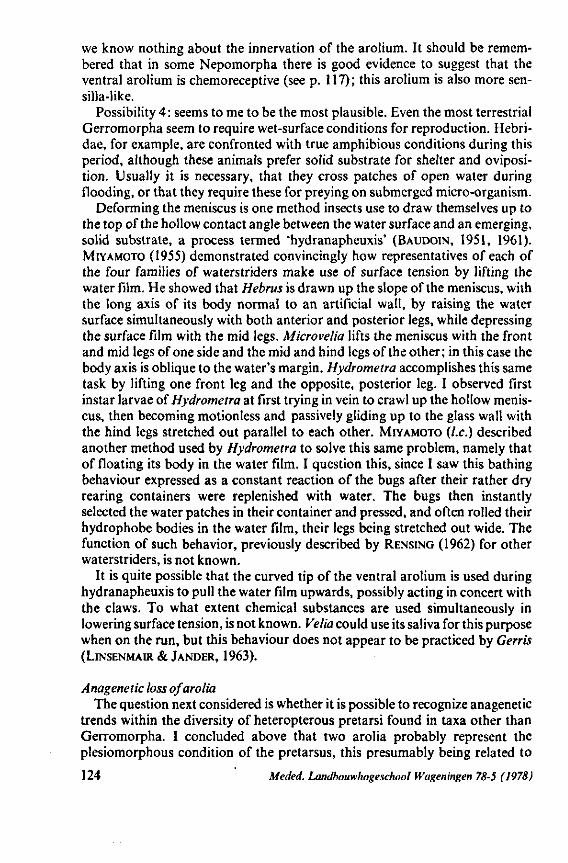
we know nothing about the innervation of the arolium. It should be remembered that in some Nepomorpha there is good evidence to suggest that the ventral arolium is chemoreceptive (see p. 117); this arolium is also more sen-silla-like.
Possibility 4: seems to me to be the most plausible. Even the most terrestrial Gerromorpha seem to require wet-surface conditions for reproduction. Hebri-dae, for example, are confronted with true amphibious conditions during this period, although these animals prefer solid substrate for shelter and oviposi-tion. Usually it is necessary, that they cross patches of open water during flooding, or that they require these for preying on submerged micro-organism.
Deforming the meniscus is one method insects use to draw themselves up to the top of the hollow contact angle between the water surface and an emerging, solid substrate, a process termed 'hydranapheuxis' (BAUDOIN, 1951, 1961). MIYAMOTO (1955) demonstrated convincingly how representatives of each of the four families of waterstriders make use of surface tension by lifting the water film. He showed that Hebrus is drawn up the slope of the meniscus, with the long axis of its body normal to an artificial wall, by raising the water surface simultaneously with both anterior and posterior legs, while depressing the surface film with the mid legs. Microvelia lifts the meniscus with the front and mid legs of one side and the mid and hind legs of the other; in this case the body axis is oblique to the water's margin. Hydrometra accomplishes this same task by lifting one front leg and the opposite, posterior leg. I observed first instar larvae of Hydrometra at first trying in vein to crawl up the hollow meniscus, then becoming motionless and passively gliding up to the glass wall with the hind legs stretched out parallel to each other. MIYAMOTO (I.e.) described another method used by Hydrometra to solve this same problem, namely that of floating its body in the water film. I question this, since I saw this bathing behaviour expressed as a constant reaction of the bugs after their rather dry rearing containers were replenished with water. The bugs then instantly selected the water patches in their container and pressed, and often rolled their hydrophobe bodies in the water film, their legs being stretched out wide. The function of such behavior, previously described by RENSING (1962) for other waterstriders, is not known.
It is quite possible that the curved tip of the ventral arolium is used during hydranapheuxis to pull the water film upwards, possibly acting in concert with the claws. To what extent chemical substances are used simultaneously in lowering surface tension, is not known. Velia could use its saliva for this purpose when on the run, but this behaviour does not appear to be practiced by Gerris (LINSENMAIR & JANDER, 1963).
Anagenetic loss ofarolia The question next considered is whether it is possible to recognize anagenetic
trends within the diversity of heteropterous pretarsi found in taxa other than Gerromorpha. I concluded above that two arolia probably represent the plesiomorphous condition of the pretarsus, this presumably being related to
124 Meded. Landbouwhogeschool Wageningen 78-5 (1978)
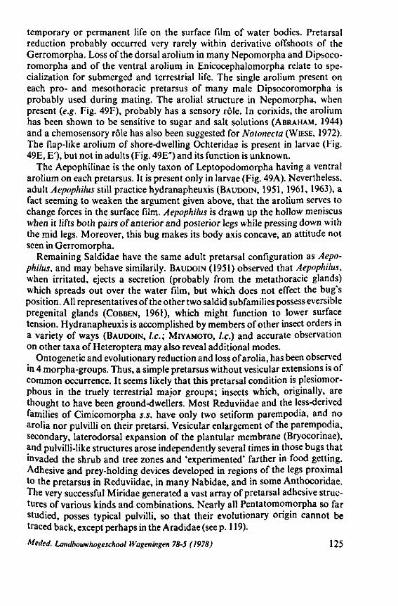
temporary or permanent life on the surface film of water bodies. Pretarsal reduction probably occurred very rarely within derivative offshoots of the Gerromorpha. Loss of the dorsal arolium in many Nepomorpha and Dipsoco-romorpha and of the ventral arolium in Enicocephalomorpha relate to specialization for submerged and terrestrial life. The single arolium present on each pro- and mesothoracic pretarsus of many male Dipsocoromorpha is probably used during mating. The arolial structure in Nepomorpha, when present (e.g. Fig. 49F), probably has a sensory rôle. In corixids, the arolium has been shown to be sensitive to sugar and salt solutions (ABRAHAM, 1944) and a chemosensory rôle has also been suggested for Notonecta (WIESE, 1972). The flap-like arolium of shore-dwelling Ochteridae is present in larvae (Fig. 49E, E'), but not in adults (Fig. 49E") and its function is unknown.
The Aepophilinae is the only taxon of Leptopodomorpha having a ventral arolium on each pretarsus. It is present only in larvae (Fig. 49A). Nevertheless, adult Aepophilus still practice hydranapheuxis (BAUDOIN, 1951, 1961, 1963), a fact seeming to weaken the argument given above, that the arolium serves to change forces in the surface film. Aepophilus is drawn up the hollow meniscus when it lifts both pairs of anterior and posterior legs while pressing down with the mid legs. Moreover, this bug makes its body axis concave, an attitude not seen in Gerromorpha.
Remaining Saldidae have the same adult pretarsal configuration as Aepophilus, and may behave similarily. BAUDOIN (1951) observed that Aepophilus, when irritated, ejects a secretion (probably from the metathoracic glands) which spreads out over the water film, but which does not effect the bug's position. All representatives of the other two saldid subfamilies possess eversible pregenital glands (COBBEN, 1961), which might function to lower surface tension. Hydranapheuxis is accomplished by members of other insect orders in a variety of ways (BAUDOIN, I.e.; MIYAMOTO, I.e.) and accurate observation on other taxa of Heteroptera may also reveal additional modes.
Ontogenetic and evolutionary reduction and loss of arolia, has been observed in 4 morpha-groups. Thus, a simple pretarsus without vesicular extensions is of common occurrence. It seems likely that this pretarsal condition is plesiomor-phous in the truely terrestrial major groups; insects which, originally, are thought to have been ground-dwellers. Most Reduviidae and the less-derived families of Cimicomorpha s.S. have only two setiform parempodia, and no arolia nor pulvilli on their pretarsi. Vesicular enlargement of the parempodia, secondary, laterodorsal expansion of the plantular membrane (Bryocorinae), and pulvilli-like structures arose independently several times in those bugs that invaded the shrub and tree zones and 'experimented' farther in food getting. Adhesive and prey-holding devices developed in regions of the legs proximal to the pretarsus in Reduviidae, in many Nabidae, and in some Anthocoridae. The very successful Miridae generated a vast array of pretarsal adhesive structures of various kinds and combinations. Nearly all Pentatomomorpha so far studied, posses typical pulvilli, so that their evolutionary origin cannot be traced back, except perhaps in the Aradidae (see p. 119).
Meded. Landbouwhogeschool Wageningen 78-5 (1978) 125
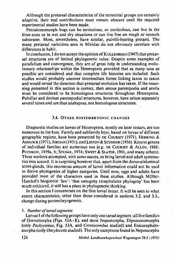
Although the pretarsal characteristics of the terrestrial groups are certainly adaptive, their real contributions must remain obscure until the required experimental studies have been made.
Pentatomomorph bugs can be terricolous, or corticolous, can live in the litter-zone or in wet and dry situations or can live free on rough or smooth substrates. Most, nevertheless, have similar, pulvilli-bearing pretarsi. The many pretarsal variations seen in Miridae do not obviously correlate with differences in habit.
In conclusion, I do not accept the opinion of KULLENBERG (1947) that pretarsal structures are of limited phylogenetic value. Despite some examples of parallelism and convergence, they are of great help in understanding evolutionary relationships within the Heteroptera provided that as many taxa as possible are considered and that complete life histories are included. Such studies would probably uncover intermediate forms linking taxon to taxon and would reveal the direction that pretarsal evolution has taken. If the reasoning presented in this section is correct, then setous parempodia and arolia must be considered to be homologous structures throughout Heteroptera. Pulvillar and deviant parempodial structures, however, have arisen separately several times and are thus analogous, not homologous structures.
3.4. OTHER POSTEMBRYONIC CHANGES
Diagnostic studies on larvae of Heteroptera, mostly on later instars, are too numerous to list here. Family and subfamily keys, based on larvae of different geographic regions, have been presented by DE COURSEY (1971), HERRING & ASHLOCK ( 1971 ), JORDAN (1951), and LESTON & SCUDDER ( 1956). Keys to genera of individual families are numerous too (e.g. DE COURSEY & ALLEN, 1968; PUCHKOV, 1959a, b; STUSAK, 1974; SWEET & SLATER, 1961, and many others). These workers attempted, with some succes, to bring larval and adult systema-tics into accord. It is surprising however that, apart from the dorso-abdominal scent-glands, this enormous amount of larval information could not be used to derive phylogenies of higher categories. Until now, eggs and adults have provided most of the characters used in these studies. Although Müller-Haeckel's biogenetic 'law' : 'that ontogeny recapitulates phylogeny' has been much criticized, it still has a place in phylogenetic thinking.
In this section I concentrate on the first larval instar. It will be seen to what extent characteristics, other than those considered in sections 3.2. and 3.3., change during postembryogenesis.
1. Number of tarsal segments Larvae I of the following groups have only one tarsal segment : all five families
of Gerromorpha (Figs. 52A-E), and most Nepomorpha, Dipsocoromorpha (only Pachycoleus, Fig. 53A, and Ceratocombus studied) and Enicocephalo-morpha (only Oncylocotis studied). The only exceptions found in Nepomorpha
126 Meded. Landbouwhogeschool Wageningen 78-5 (1978)
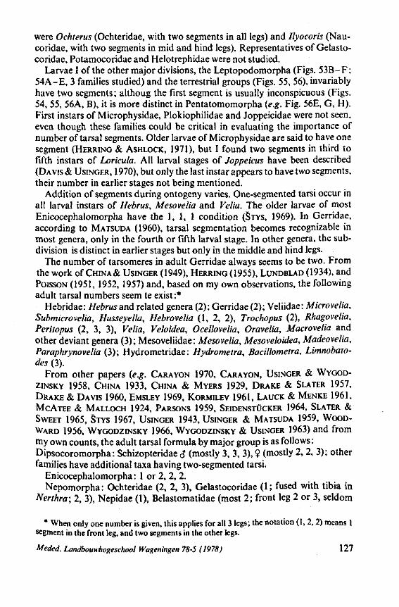
were Ochterus (Ochteridae, with two segments in all legs) and Ilyocoris (Nau-coridae, with two segments in mid and hind legs). Representatives of Gelasto-coridae, Potamocoridae and Helotrephidae were not studied.
Larvae I of the other major divisions, the Leptopodomorpha (Figs. 53B-F; 54A-E, 3 families studied) and the terrestrial groups (Figs. 55, 56), invariably have two segments ; althoug the first segment is usually inconspicuous (Figs. 54, 55, 56A, B), it is more distinct in Pentatomomorpha (e.g. Fig. 56E, G, H). First instars of Microphysidae, Plokiophilidae and Joppeicidae were not seen, even though these families could be critical in evaluating the importance of number of tarsal segments. Older larvae of Microphysidae are said to have one segment (HERRING & ASHLOCK, 1971), but I found two segments in third to fifth instars of Loricula. All larval stages of Joppeicus have been described (DAVIS & USINGER, 1970), but only the last instar appears to have two segments, their number in earlier stages not being mentioned.
Addition of segments during ontogeny varies. One-segmented tarsi occur in all larval instars of Hebrus, Mesovelia and Velia. The older larvae of most Enicocephalomorpha have the 1, 1, 1 condition (STYS, 1969). In Gerridae, according to MATSUDA (1960), tarsal segmentation becomes recognizable in most genera, only in the fourth or fifth larval stage. In other genera, the subdivision is distinct in earlier stages but only in the middle and hind legs.
The number of tarsomeres in adult Gerridae always seems to be two. From the work of CHINAÄ USINGER (1949), HERRING (1955), LUNDBLAD (1934), and POISSON (1951, 1952, 1957) and, based on my own observations, the following adult tarsal numbers seem te exist :*
Hebridae: Hebrus and related genera (2); Gerridae (2); Veliidae: Microvelia, Submicrovelia, Husseyella, Hebrovelia (1, 2, 2), Trochopus (2), Rhagovelia, Peritopus (2, 3, 3), Velia, Veloidea, Ocellovelia, Oravelia, Macrovelia and other deviant genera (3); Mesoveliidae: Mesovelia, Mesoveloidea, Madeovelia, Paraphrynovelia (3); Hydrometridae : Hydrometra, Bacillometra, Limnobato-des (3).
From other papers (e.g. CARA YON 1970, CARA YON, USINGER & WYGOD-
ZINSKY 1958, CHINA 1933, CHINA & MYERS 1929, DRAKE & SLATER 1957,
DRAKE & DAVIS 1960, EMSLEY 1969, KORMILEV 1961, LAUCK & MENKE 1961,
MCATEE & MALLOCH 1924, PARSONS 1959, SEIDENSTÜCKER 1964, SLATER &
SWEET 1965, STYS 1967, USINGER 1943, USINGER & MATSUDA 1959, WOOD
WARD 1956, WYGODZINSKY 1966, WYGODZINSKY & USINGER 1963) and from my own counts, the adult tarsal formula by major group is as follows : Dipsocoromorpha: Schizopteridae J (mostly 3, 3, 3), ? (mostly 2, 2, 3); other families have additional taxa having two-segmented tarsi.
Enicocephalomorpha: 1 or 2, 2, 2. Nepomorpha: Ochteridae (2, 2, 3), Gelastocoridae (1 ; fused with tibia in
Nerthra; 2, 3), Nepidae (1), Belastomatidae (most 2 ; front leg 2 or 3, seldom
* When only one number is given, this applies for all 3 legs; the notation (1, 2, 2) means 1 segment in the front leg, and two segments in the other legs.
Meded. Landbouwhogeschool Wageningen 78-5 (1978) 127
1), Naucoridae, Potamocoridae (1,2, 2), Pleidae (2), Notonectidae (front leg with 2 or 3, seldom 1 ; 2, 2), Corixidae (1, 1 (Diaprepocoris 2), 1).
Leptopodomorpha : Leotichiidae (1,2, 2), Saldidae, Omaniidae, Leptopo-didae (3).
Reduviidae: mostly 3; 2 in Elasmocorinae, Holoptilinae and occasional species of Reduviinae and Emesinae ; reduced numbers in front legs of Emesinae and Phymatinae.
Pachynomidae : 3 Cimicomorpha s.S.: Joppeicidae, Microphysidae, Plokiophilidae (with exception of Lipokophila (3)), Tingidae (2); Nabidae (inclusive of Medocostinae), Cimicidae, Anthocoridae, Miridae (some Cylapinae excepted) (3).
Pentatomomorpha: Aradidae, Acanthosomatidae (2); all other families, including Thaumastellidae, Hyocephalidae, Malcidae, and Urostylidae (3); in some Cydnidae reduction in hind legs. Psamminae is the only subfamily of Lygaeidae having two tarsal segments.
When one scans all these data, it appears that all first instar larvae of Ger-romorpha and most Nepomorpha and those few representatives studied of Dipsocoromorpha and Enicocephalomorpha, have no tarsal segment. All other major taxa so far investigated have two segments in the first or older larval stages.
In adults, zero- or one-segmented tarsi are very rare and occur mostly in specialized legs (raptorial or fossorial). Nepidae is the only family in which all adult legs have a single tarsal segment. Two-segmented tarsi occur in all three pairs of legs in adult Hebridae, and Gerridae, and in a number of plesiomor-phous families of Cimicomorpha, in some subdivisions of Dipsocoromorpha and Reduviidae, and in Thaumastocoridae.
Schizopteridae exhibit sexual differences in tarsal formula, these mostly being associated with the presence in the male of a median arolium. The family Veliidae is variable and contains taxa with a 1, 2, 2 formula upto those with 3, 3, 3. The latter condition is uniformly present in Mesoveliidae, and Hydro-metridae, in most reduvioids, and cimicoids and in nearly all Pentatomomorpha.
The ancestral number of tarsal segments in adult Heteroptera is often discussed. Most authors agree that the primitive number is three, a figure reached during the adult moult when another segment is added to the two-segmented larval tarsus (REUTER 1910, CHINA & MYERS 1929, USINGER 1943). CHINA (1955a) considered the 2:2:2 state in adult surface bugs to be derived from the three-segmented tarsus of shore- dwelling and terrestrial bugs. DRAKE & SLATER (1957) were less dogmatic when they considered the 2-segmented tarsi of Thaumastocoridae: 'In view of the other marked specializations of the family this too probably represents a specialized condition. However, it may be pertinent to note that since heteropterous nymphs generally possess two-segmented tarsi, such a condition does not necessarily indicate specialization and may in many cases simply represent a neotenic phenomenon'. Regarding a similar situation in Psamminae, SLATER & SWEET (1965) remarked: 'The
128 Meded. Landbouwhogeschool Wageningen 78-5 (1978)
two-segmented tarsi can be looked on as primitive ( ), a reduction phenomenon, or a neotenic condition, and must be evaluated against a character complex'.
The data presented here throw new light on the question, since a one-segmented larval tarsus appears to be present in all surface bugs, water bugs (except 1 family), Dipsocoromorpha and Enicocephalomorpha; taxa usually considered to be plesiomorphous. In my opinion, there is now very strong evidence to suggest that the 1 :1:1 condition in larvae and the 2:2:2 condition in adults represent the ancestral state in Heteroptera. This implies that there was mostly an addition of segments, not a decrease or fusion during evolution of the Order. Ontogenetic change in tarsal number varies from no change at all (e.g. Nepidae, one segment in both larvae and adults) to an addition of two in some Gerromorpha and Dipsocoromorpha. Further evidence for an evolutionary increase in segment number is provided by the many terrestrial taxa that bear two segments in the adult. As the review on p. 128 shows, such taxa hold a proportionately low anagenetic position within their respective superfamily groups. Secondary reduction may have occurred in those legs having auxiliary functions beyond locomotion.
2. Coxal types SCHIÖDTE (1870) placed great phylogenetic weight on the two different meta-
coxal types existing in Heteroptera : the cardinate and rotatory. Members of the cimicoid families and some of the Nepomorpha were supposed to have the first type, which is characterized by an inward elongation of the lateral base of the coxa, which articulates near the upper edge of the pleuron. Pentatomomorpha, reduviids and the remaining Nepomorpha supposedly had a shorter, cylindrical rotatory coxa, which allows freeer movement of the leg in all directions. More recent literature data shows the rotatory type to be present in the pentatomo-morphous Hyocephalidae (STYS, 1964a), and Malcidae (STYS, 1967b), and the cardinate type in the cimicomorphous Microphysiade (PÉRICART, 1972) and Plokiophilidae (STYS, 1967C).
The validity of this division has often been disputed and is usually discarded as of no cladistic significance. In this study, where an analysis of anagenetic changes is a principal goal, the question of which coxal type represents the original condition, is of interest. KIRKALDY (1906) considered this to be the rotatory type; REUTER (1910) the cardinate type. DRAKE & DAVIS (1960) agreed with REUTER and suggested that the rotatory type could have evolved indepen-dentlyseveraltimes. Intermediate types were recorded too. DRAKE & DAVIS (I.e.) also pointed out that there is less fundamental difference between the two types than was indicated by SCHIÖDTE. They stated that the articulation in typical pagiopodous heteropterons is, in fact, monocondylic as in bugs having the rotatory type. Because of the perpendicular insertion of the coxa a broader arc of rotation is possible in the latter.
Although I have not studied the functional morphology of the basal articulation in detail comparatively, I consider the development of the trochantin to be
Meded. Landbouwhogeschool Wageningen 78-5 (1978) 129
a better criterion for evaluating the evolutionary state of the leg base. Profiles of the coxae of first instar larvae are shown in Fig. 57. A -D are the nominate forms of four gerromorphan families. Hebrus has homonomous coxae, pleural sclerites and pleural clefts (A). Mesovelia has the hind coxa proportionately larger and more obliquely inserted than those of the mid and hind legs (B). The youngest Gerris larva (C) exhibits and enlargement in the mesothorax and in the mid coxa (the rowing leg!). In Hydrometra larvae (Fig. 57D), the front and mid legs are inserted into the thorax, a greater distance from each other than are the mid and hind legs; and the insertion of the two posterior pairs is directed backwards. Except for Hebrus, the posterior pleurites are not visibly cleft. At the time when these drawings (Fig. 57) were made, some 15 years ago, I paid no attention to the trochantin. But, the drawings (E) (saldid), (F) (mirid), (G) (nabid) and (K) (lygaeid) reveal a structure, which must represent the trochantin. This sclerite is not seen in the gerromorpha depicted, and may be absent in the first larval instar. A study of adults of Hebrus and Mesovelia revealed that they too lack a trochantin. A minute, precoxal differentiation in the hind leg of Mesovelia could be this sclerite. GUPTA (1963) reported a trochantin to be present in the first leg of Mesovelia, but confusion with one of the two coxal apodemes seems a more probable explanation. Adults of Velia and Gerris have trochantins but in the anterior legs these are very inconspicuous, because they are not sclerotized. In the other two pairs of legs, the trochantin is distinct, particularly in the rowing leg of Gerridae, and an apodeme for the insertion of the protractor muscles arises from its centre. Nevertheless, this coxa is mono-condylic, the trochantin extending from the pleural condyle only half-way to the inward process of the xoca. Since it does not reach the rim of the coxal base the latter has only one point of articulation with the thorax.
A dicondylic articulation is présent in Oncopeltus, where I found two protractor, one retractor and one rotator apodeme, apparently administering limited movement of the coxa along the restricted hinge axis between the two condyles. The muscle of the rotator apodeme originates from the trochantin close to the pleural condyle (see also GOVIND & DANDY, 1970). Fox & Fox (1964) considered this dicondylic condition mediated by the trochantin, to be more specialized than the pleural monocondylic one. I did not make a representative check of this mechanism in other family groups but it may be that its anagenesis in Heteroptera is such that a trochantin was originally absent in Gerromorpha and arose gradually as a reinforcement for the antero- lateral protractor apodeme.
The shape of the coxae in those forms such as Hebrus that lack a trochantin, is rotatory, both in larvae and adults. In these bugs, the coxal mechanism is reduced to an anterior protractor and a posterior retractor with a rotator in between. In adult Veliidae and Gerridae, the mid leg is more distinctly of the cardinate type than is the anterior or posterior leg. It is feasible that the original type was rotatory, lacked a trochantin, and evolved separately on several occasions into the cardinate type, irrespective of the presence or absence of a trochantin. The origin of the trochantin is thus probably not correlated with
130 Meded. Landbouwhogeschool Wageningen 78-5 (1978)
the transition from the rotatory into the cardinate type. Data in the literature on the occurrence of the trochantin in Heteroptera,
are insufficient to make generalizations. LARSÉN (1945) and MATSUDA (1970) stated it to be present in all three legs, although in the front leg it is often small and never firmly articulated with the coxal margin. BRINKHURST ( 1960) reported two trochantinal sclerites to be present in each coxal base of Aquarius (Gerri-dae) : one at the proximal end of the coxa, the second medial or lateral to the coxal rim; this second sclerite bearing a muscle. LARSÉN (1945), showed two trochantins to occur also in Velia. Only the uppermost sclerite is homologous to the trochantin of other groups. Hydrometra (Hydrometridae) supposedly has a trochantin (SPRAGUE, 1956), but I could not trace them. The trochantinal situation in Leptopodomorpha has been worked out for Saldula (Saldidae) by PARSONS (1963). This sclerite is present in all legs and, has its medial end articulating with the coxal rim ; its upper end extending to the coxal process in the prothorax and mesothorax, but not in the metathorax.
Nepomorpha generally have a trochantin, but it is said to be absent in Helotrephidae, Pleidae and in Tenagobia of the Corixidae (POPOV, 1971). The prothoracic leg of Gelastocoris (Gelastocoridae), Lethocerus (Belostomati-dae) and Ilyocoris (Naucoridae) is monocondylic and is provided with a small trochantin. The legs of the pterothoracic segments in these bugs have more restricted movement because of a large trochantin which creates a dicondylic condition (RAWAT, 1939; PARSONS, 1959,1965). EMSLEY (1969) found a distinct trochantin to be present in only two of the many genera of Schizopteridae studied by him, and this only in the prothoracic leg. It is significant that these two genera appear to be fairly advanced in his evolutionary scheme.
Bugs in the true terrestrial groups generally bear a trochantin. Reduviids, 'ike the Nepidae, possess rotatory coxae, and lack a distinct trochantin (representatives of Emesinae, Piratinae and Harpactorinae studied). I found a trochantin to be present in all legs of the Tingidae. It is well-developed in the anterior and mid legs, even though the coxae are definitely of the rotatory type (DRAKE & DAVIS, 1960).
The conclusion of this section is that the splitting of the Heteroptera into the Pagiopoda and Trochalopoda based on coxal type (SCHIÖDTE, 1870) is of historical interest only. Instead, attention must be paid to the actual presence and development of the trochantin in studies of heteropteran anagenesis. Absence of a trochantin is plesiomorphous in some Gerromorpha, Nepomor-Pha and Dipsocoromorpha and in the Reduviidae, and is usually associated with the presence of rotatory coxae. In other groups, a trochantin may occur, whether the coxae are of the rotatory or the cardinate type.
3- Primary larval chaetotaxy Comparison of hair patterns in first instar larvae (Figs. 52-56) of Heterop
tera does not permit any conclusion to be made about their plesiomorphous or aPomorphous state. In Gerromorpha, chaetotaxy is sparse in Hebrus (Fig. 52A), and increases in density through Mesovelia (B), Hydrometra (C), Micro-
Meded. Landbouwhogeschool Wageningen 78-5 (1978) 131
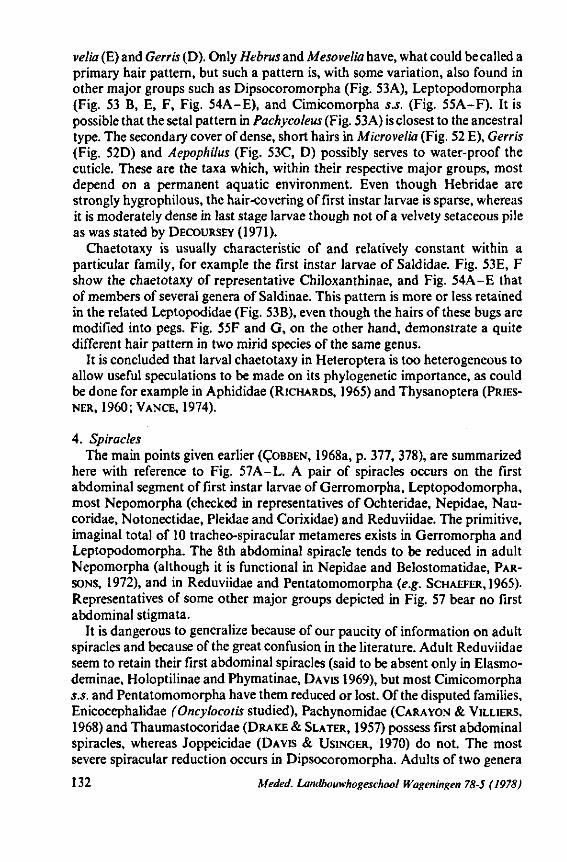
velia (E) and Gerris (D). Only Hebrus and Mesovelia have, what could be called a primary hair pattern, but such a pattern is, with some variation, also found in other major groups such as Dipsocoromorpha (Fig. 53A), Leptopodomorpha (Fig. 53 B, E, F, Fig. 54A-E), and Cimicomorpha s.S. (Fig. 55A-F). It is possible that the setal pattern in Pachycoleus (Fig. 53 A) is closest to the ancestral type. The secondary cover of dense, short hairs in Microvelia (Fig. 52 E), Gerris (Fig. 52D) and Aepophilus (Fig. 53C, D) possibly serves to water-proof the cuticle. These are the taxa which, within their respective major groups, most depend on a permanent aquatic environment. Even though Hebridae are strongly hygrophilous, the hair-covering of first instar larvae is sparse, whereas it is moderately dense in last stage larvae though not of a velvety setaceous pile as was stated by DECOURSEY (1971).
Chaetotaxy is usually characteristic of and relatively constant within a particular family, for example the first instar larvae of Saldidae. Fig. 53E, F show the chaetotaxy of representative Chiloxanthinae, and Fig. 54A-E that of members of several genera of Saldinae. This pattern is more or less retained in the related Leptopodidae (Fig. 53 B), even though the hairs of these bugs are modified into pegs. Fig. 55F and G, on the other hand, demonstrate a quite different hair pattern in two mirid species of the same genus.
It is concluded that larval chaetotaxy in Heteroptera is too heterogeneous to allow useful speculations to be made on its phylogenetic importance, as could be done for example in Aphididae (RICHARDS, 1965) and Thysanoptera (PRIES-NER, 1960; VANCE, 1974).
4. Spiracles The main points given earlier (ÇOBBEN, 1968a, p. 377, 378), are summarized
here with reference to Fig. 57A-L. A pair of spiracles occurs on the first abdominal segment of first instar larvae of Gerromorpha, Leptopodomorpha, most Nepomorpha (checked in representatives of Ochteridae, Nepidae, Nau-coridae, Notonectidae, Pleidae and Corixidae) and Reduviidae. The primitive, imaginai total of 10 tracheo-spiracular metameres exists in Gerromorpha and Leptopodomorpha. The 8th abdominal spiracle tends to be reduced in adult Nepomorpha (although it is functional in Nepidae and Belostomatidae, PARSONS, 1972), and in Reduviidae and Pentatomomorpha (e.g. SCHAEFER,1965). Representatives of some other major groups depicted in Fig. 57 bear no first abdominal stigmata.
It is dangerous to generalize because of our paucity of information on adult spiracles and because of the great confusion in the literature. Adult Reduviidae seem to retain their first abdominal spiracles (said to be absent only in Elasmo-deminae, Holoptilinae and Phymatinae, DAVIS 1969), but most Cimicomorpha s.s. and Pentatomomorpha have them reduced or lost. Of the disputed families, Enicocephalidae (Oncylocotis studied), Pachynomidae (CARAYON & VILLIERS, 1968) and Thaumastocoridae (DRAKE & SLATER, 1957) possess first abdominal spiracles, whereas Joppeicidae (DAVIS & USINGER, 1970) do not. The most severe spiracular reduction occurs in Dipsocoromorpha. Adults of two genera
132 Meded. Landbouwhogeschool Wageningen 78-5 (1978)
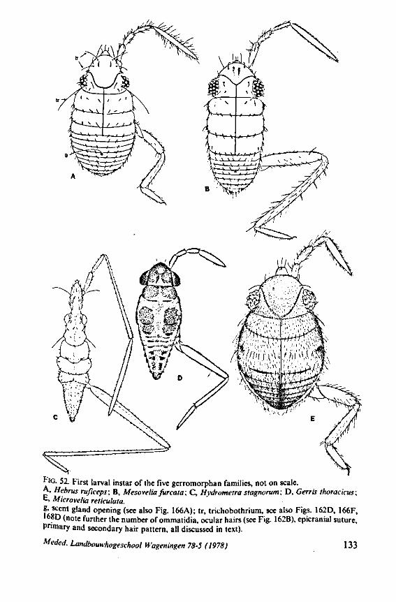
FIG. 52. First larval instar of the five gerromorphan families, not on scale. A, Hebrus ruficeps; B, Mesovelia furcata; C, Hydrometra stagnorum; D, Gerris thoracicus; E, Microvelia reticulata. g, scent gland opening (see also Fig. 166A); tr, trichobothrium, see also Figs. 162D, 166F, •68D (note further the number of ommatidia, ocular hairs (see Fig. 162B), epicranial suture. Primary and secondary hair pattern, all discussed in text).
Meded. Landbouwhogeschool Wageningen 78-5 (1978) 133
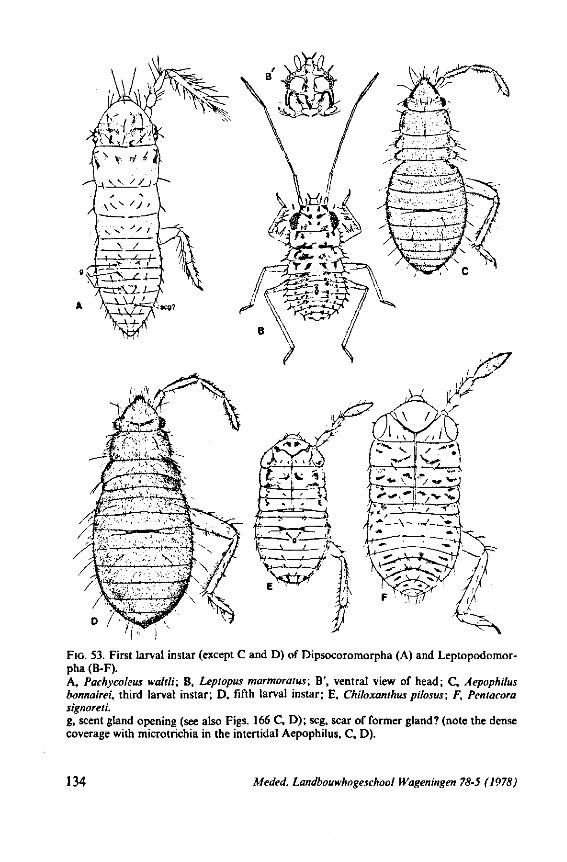
FIG. 53. First larval instar (except C and D) of Dipsocoromorpha (A) and Leptopodomor-pha (B-F). A, Pachycoleus waltli; B, Leptopus marmoratus; B', ventral view of head; C, Aepophilus bonnairei, third larval instar; D, fifth larval instar; E, Chiloxanthus pilosus; F, Pentacora signoreli. g, scent gland opening (see also Figs. 166 C, D); scg, scar of former gland? (note the dense coverage with microtrichia in the intertidal Aepophilus, C, D).
134 Meded. Landbouwhogeschool Wageningen 78-5 (1978)
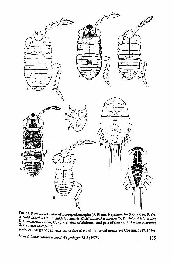
F>G. 54. First larval instar of Leptopodomorpha (A-E) and Nepomorpha (Corixidae, F, G). A. Saldula orthochila; B, Saldula palustris; C, Micracanlhia marginalis; D, Halosalda lateralis;
. Chartoscirta cincta, E', ventral view of abdomen and part of thorax; F, Corixa punctata; *->> Cymatia coleoptrata. 8. abdominal gland; go, external orifice of gland; lo, larval organ (see COBBEN, 1957, 1959).
Meded. Landbouwhogeschool Wageningen 78-5 (1978) 135
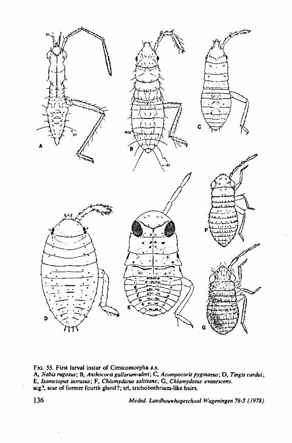
FIG. 55. First larval instar of Cimicomorpha s.s. A, Nabis rugosus; B, Anthocoris gallarum-ulmi; C, Acompocorispygmaeus; D, Tingis cardui; E, Isometopus intrusus; F, Chlamydalus saltitans; G, Chlamydatus evanescens. scg?, scar of former fourth gland?; tri, trichobothrium-like hairs.
136 Meded. Landbouwhogeschool Wageningen 78-5 (1978)
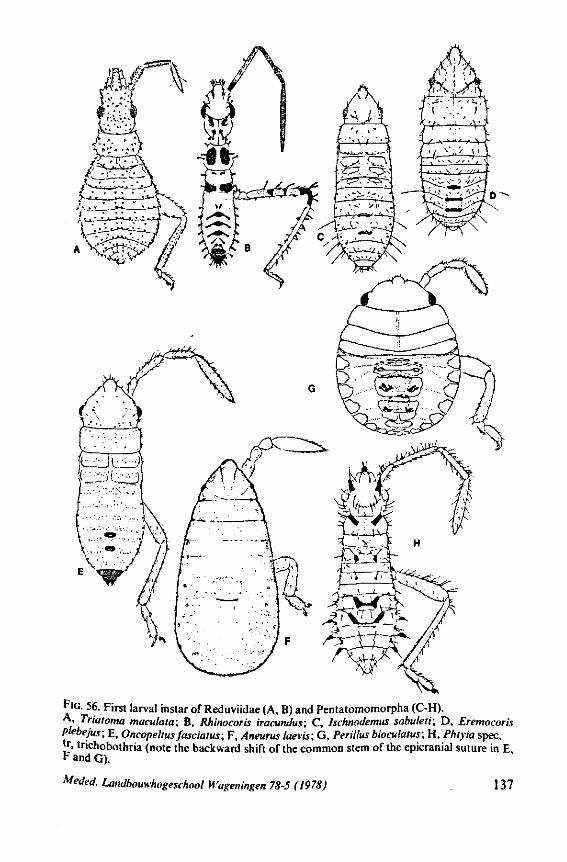
FIG. 56. First larval instar of Reduviidae (A, B) and Pentatomomorpha (C-H). A . Triatoma maculata; B, Rhinocoris iracundus; C, lschnodemus sabuleti; D, Eremocorb Plebejus; E, Oncopeltusfasciatus; F, Aneurus laevis; G, Perillus bioculaius; H, Phtyia spec. fr> trichobothria (note the backward shift of the common stem of the epicranial suture in E, F and G).
Meded. Landbouwhogeschool H'ageningen 78-5 (1978) 137
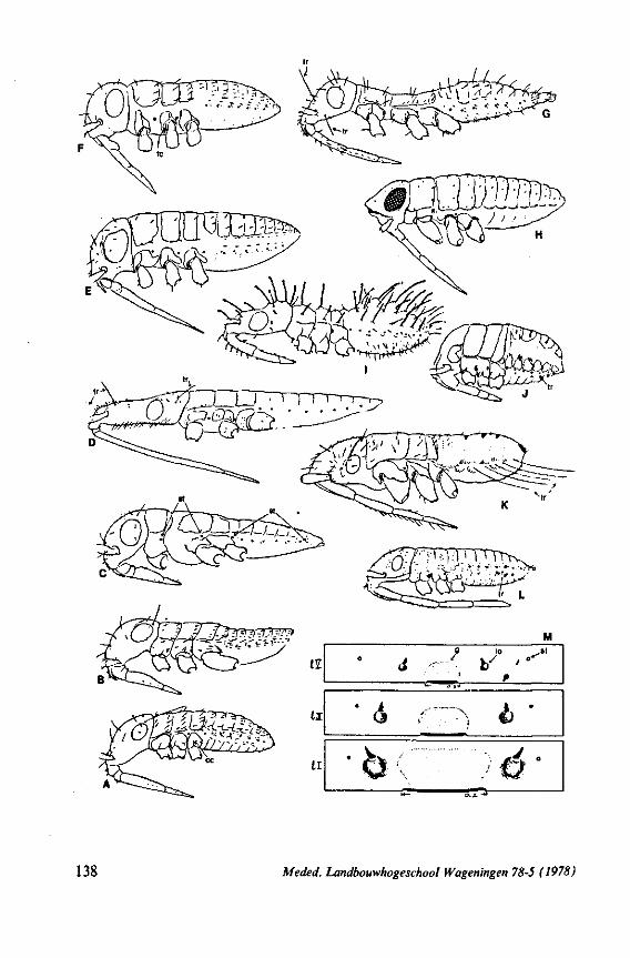
I l
t l
' •* '
' d r
%»*'
a ..y
" A
10 -SI
1 •
> « $ •
138 Meded. Landbouwhogeschool Wageningen 78-5 (1978)
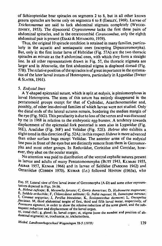
of Schizopteridae bear spiracles on segments 2 to 8, but in all other known genera spiracles are borne only on segments 6 to 8 (EMSLEY, 1969). Larvae of Trichotonannus are said to lack abdominal stigmata completely (WYGOD-ZINSKY, 1953). The dipsocorid Cryptostemma lacks the first three pairs of abdominal spiracles, and in the ceratocombid Ceratocombus, only the eighth abdominal pair is present (ESAKI & MIYAMOTO, 1959).
Thus, the original 10 spiracle condition is retained in many families, particularly in the aquatic and semiaquatic ones (excepting Dipsocoromorpha). But, only in the first instar larva of Hebridae (Fig. 57A) are the two thoracic spiracles as minute as the 8 abdominal ones, with which they form a straight line. In all other representatives drawn in Fig. 57, the thoracic stigmata are larger and in Mesovelia, the first abdominal stigma is displaced dorsad (Fig. 57B). The relative position of the spiracles is of great importance in the systema-tics of the later larval instars of Heteroptera, particularly in Lygaeidae (SWEET & SLATER, 1961).
5. Ecdysial lines A Y-shaped epicranial suture, which is split at ecdysis, is plesiomorphous in
larval Heteroptera. The stem of this suture has entirely disappeared in the pentatomoid groups except for that of Cydnidae, Acanthosomatidae and, possibly, of other less-derived families of which larvae were not studied. Only the distal ends of the lateral sutures remain, bordering the median margin of the eye (Fig. 56G). This peculiarity is due to loss of the vertex and was discussed by me in 1968 in relation to the embryonic egg-burster. A tendency towards displacement of the epicranial fork posteriad is seen also in Lygaeidae (Fig. 56E), Aradidae (Fig. 56F) and Veliidae (Fig. 52E). Hebrus also exhibits a slight trend in this direction (Fig. 52A) ; in this respect Hebrus is more advanced than other surface bugs except Veliidae. The anterior arms of the ecdysial line pass in front of the eyes but are distinctly remote from them in Gerromor-pha and most other groups. In Reduviidae, Corixidae and Coreidae, however, they abut on the ocular margin.
No attention was paid to distribution of the ventral cephalic sutures present in larvae and adults of many Pentatomomorpha (BUTT 1943, KUMARI 1955, AKBAR 1957, KUMAR 1966) and in larvae of Saldidae (PARSONS 1962) and Omaniidae (COBBEN 1970). KUMAR (I.e.) followed HINTON (1963a), who
FIG. 57. Lateral view of first larval instar of Gerromorpha (A-D) and some other representatives depicted in Figs. 54-56. A, Hebrus ruficeps; B, Mesovelia furcata; C, Gerris thoracicus; D, Hydrometra stagnorum; E, Saldula orthochila; F, Chlamydatus saltitans; G, Nabis rugosus; H, hometopus intrusus; I, Coranus aegypiius; J, Perillus bioculatus; K, Scolopostethus thomsoni; L, Oncopeltus fasciatus. M, third abdominal tergite of first, third and fifth larval instar, respectively, of Pentacora signoreti, in order to show the relative reduction of the scent gland, and the subsequent reduction and displacement of the larval organ. cc, coxal cleft; g, gland; lo, larval organ; st, stigma (note the number and position of abdominal stigmata); tc, trochantin; tr, trichobothria.
Meded. Landbouwhogeschool Wageningen 78-5 (1978) 139
studied the so-called ventral ecdysial lines in endopterygote larvae, in suggesting that such lines function as hinges during ecdysis for the outward folding of both dorsal halves of the head capsule.
6. Abdominal scent glands Table 2 gives a compilation of the distribution of the serially arranged dorso-
abdominal glands. This survey is based on data from the literature and my own work (see also Figs. 52-56). Older data are reviewed by DUPUIS (1947a, b), GULDE (1902) and HENRICI (1938). A selection of more recent papers comprises: CARA YON 1962, CARA YON e.a. 1958, COBBEN & WYGODZINSKY
1975, KUMAR 1969, LENT& JURBERG 1966, MCDONALD 1963,1969, PUCHKOVA
& PUCHKOV 1958, PUCHKOV 1958, SCHAEFER 1966, SCHAEFER & WILCOX 1971,
STYS 1964, but there are many others. As was concluded by earlier workers, it seems certain that the glands were originally median and unpaired (discussion in DUPUIS, 1947; POLIVANOVA, 1960, argued fora paired origin in Pentatomoi-dea) and that the general course of evolution throughout the suborder must be interpreted in terms of reduction processes.
The original number should be four, the glands occurring as invaginations between the tergites 3-4, 4-5, 5-6 and 6-7. The last gland underwent rapid loss*. Its remnants (as intersegmental cuticular thickenings) are seen in two families of Dipsocoromorpha and in some stem groups of cimicoids. Rudiments of what could have been this last gland are also stated to occur in some penta-tomoids. The gland between terigtes 6 and 7 is indeed functional as the sole gland in Schizopteridae and, in combination with other glands, in Joppeicidae. Although families or subfamilies are fairly well characterized by a consistent configuration of the glands, the gland formula is not usable in higher rank phylogeny because of the many parallelisms concerning their reduction.
A few discrepancies are worth mentioning. Corixidae have a unique position within the Nepomorpha because of the presence of three glands. All other waterbugs, surface bugs and shore bugs have only the first gland present or non. The pentatomoid families exhibit within the Pentatomomorpha the strongest permanence of the three glands, with a tendency of the anterior gland to become paired and reduced successively. In the Cimicomorpha s.s. the first gland persisted longest. In Reduvioidea, however, just the first gland is liable to earlier disappearance than the other glands. The state in the Pachy-nomidae is not known. The taxonomie position of this family, either in Cimici-morpha s.S. or in Reduvioidea, is questioned. I assume that Pachynomidae have no larval glands, since I found no traces of glands in adult representatives of both subfamilies Pachynominae and Aphelonotinae. Traces of reasonably well-developed glands usually persist in the adults as cuticular invaginations or ostiolar scars. There is no behavioral proof that they actually function during adult life, but judgement from their histology or full cuticular complement
* Trace of invagination occurs in the embryo of Palomena, but not in the larva (HEYMONS, 1899).
140 Meded. Landbouwhogeschool Wageningen 78-5 (1978)

TABLE 2. Distribution of the larval abdominal glands.
glands
between segments
1
3-4
2
4-5
3
5-6
4
6-7
Gerromorpha. Hebridae, Mesoveliidae, some aberrant Veliidae Veliidae, Gerridae, Hydrometridae
Leptopodomorpha. Omaniidae, Leptopodidae Saldidae
Nepomorpha. Corixidae Nepidae, Belostomatidae Naucoridae, Aphelocheiridae Pleidae, Helotrephidae, Potamocoridae* Notonectidae, Ochteridae, Gelastocoridae
Dipsocoromorpha. Dipsocoridae Ceratocombidae Schizopteridae
Enicocephalomorpha. Enicocephalidae Cimicomorpha s.s. Anthocoridae, Cimicidae, Nabidae
Plokiophilidae Joppeicidae Tingidae Vianaididae Miridae
Pachynomoidea. Pachynomidae1
Reduvioidea. Bactrodinae, Harpactorinae, Reduviinae, Piratinae Ectrichodiinae, Phymatidae Stenopodinae Saicinae, Emesinae, Triatominae
Thaumastocoroidea. Thaumastocoridae Pentatomomorpha. Aradidae
or Termitaphididae Idiostolidae Malcidae Piesmatidae, Berytinidae
Lygaeidae
Thaumastellidae2
Thaumastellidae3
Colobathristidae, Pyrrhocoridae, Largidae Hyocephalidae, Stenocephalidae Alydidae, Rhopalidae, Coreidae Dinidoridae Tessarotomatidae* Phloeidae5
Cydnidae, Plataspidae6
Acanthosomatidae, Scutelleridae, Pentatomidae6
or
+ + ++• _ +/ + — + + _ +• + + + + — + •-_?
+• _ + _ + + + _ •
+ + + + + + ? _ •?
+•
• + + + -+ + + +
__
+ + — --— + + _ _ + + + +-+ —
_ ?
+ • + + — + + + -— + + + — + + + + ? _7
+• + + + + + + +
—
+ + — -— — + + — — + + -— -— _?
+ •
+ -— — + + -— + + — — + — + + ? _?
+ + + + + + + +
-— -— — • • + , -
- * — + — -— —
_ — -— — -— — — -— — — — — — — — — — — • —
—
Meded. Landbouwhogeschool Wageningen 78-5 (1978) 141
Symbols used: + , gland large; + , gland small (the decision on the gland size was chosen very arbitrarily; /, gland divided; ., gland reduced and/or displaced posteriad within the tergite; *, no gland, only cuticular scar left; —, gland and scar lacking. 1 Deduced from state in adult. 2 Deduced from adult state of T. aradoides (STYS, 1964b). 3 Status in adult of T. namaquensis (SCHAEFER & WILCOX, 1971). * Mum (1907) states presence of 4 glands in Tessarotoma papulosa. I found only a very weak scar between segment 6 and 7 in the larva of an unidentified species from the Ivory Coast. 5 LENT & JURBERG (1966) depicted for Phloeophana only two glands. I found in the larva of an unidentified species from Venezuela paired ostioles of the anterior gland. 6 The anterior abdominal gland in the pentatomoid families is on the way of reduction. Secretion was almost absent in the cydnid Scaptocoris (ROTH, 1961), and there appeared to be no secretion at all in the last larval instar of the pentatomid Apodiphus (E VERTON et ai, 1974).
suggests that they function in the adults, at least in Enicocephalidae (CARAYON, 1950b), Joppeicidae (DAVIS & USINGER, 1970), Plokiophilidae (CARAVON, 1974) and in Phymatidae Themocorinae (CARAYON, USINGER & WYGODZINS-KY, 1958). Other examples have been recorded for lygaeids, coreids, pentato-mids, nabids, corixids (compilation in DUPUIS, 1947), but the brachypterous Cimex adult shows no rudiments of the glands (HENRICI, 1938).
I found the single gland well-developed present in adults of the waterbugs Potamocoridae and Pleidae, and PARSONS (1969) reported its presence in adults of Aphelocheirus. The two anterior larval glands of Dysdercus perhaps delivering the aggregating pheromone, retain their function in the adult (CALAM & SCOTT, 1969). The posterior gland employed in defence, however, becomes functionless in the adult.
Considering the fate of the glands in smaller taxonomie 'trajectories', it appears here too that progressive evolution is accompanied by reduction. Ontogenetic reduction has been reported in Lygaeidae Cyminae (LEDVINKA, 1970) and other subfamilies (PUCHKOV, 1958; SLATER & SWEET, 1961). The first author discovered that in some species of Cymus loss of the posterior gland occurs after the moult from third to fourth instar. In the first instar of all Cymus species should the exoskeleton above the anterior gland be closed, but open in the successive instars. Whether the gland outlet is indeed blocked should be verified in sections. Phylogenic and ontogenic reduction is discernable in Saldidae. The anterior gland has considerable size in Aepophilinae (Fig. 53C, D) and most Chiloxanthinae (Fig. 53E, F) but is rudimentary in Saldinae (Fig. 54A-E). Although the gland absolutely enlarges from one larval stage to the other, it decreases in size relative to the body dimensions (Fig. 57M). The sequence of reduction through subfamilies matches the whole set of other anagenetic changes in this family (COBBEN, 1959,1965). Similar phylogenic and ontogenic reduction of all three glands, but in particular of the anterior gland, is seen in Corixidae (POISSON, 1924; BETTEN, 1943; COBBEN & MOLLER PiLLOT, 1960; but not confirmed by BRINDLEY 1929) The large genus Sigara can be subdivided in two groups on account of the colour of the glands: one group of species with dark brownish or blackish, sharply defined glands, the
142 Meded. Landbouwhogeschool Wageningen 78-5 (1978)
other group with orange- or reddish coloured glands, which are less marked (COBBEN & MOLLER PlLLOT, l.C.)
Gerromorpha lack all glands, exept for the first gland in the plesiomorphous taxa Hebridae, some Veliidae, but also in Mesoveliidae. The paucity of glands in Gerromorpha seems in contradiction concerning my belief that the stem groups of Gerromorpha represent the most generalized recent bugs. Therefore I gave in 1968 (p. 367) as an alternative explanation that the archetype heterop-teron had only the first gland, which persisted in some forms and was secondarily lost in others. The whole picture suggests that these dorso-abdominal glands have not been of great advantage for those bugs with semi-aquatic or aquatic habits (exept for Corixidae), and certainly primitive bugs were inhabitants of very damp situations. For bugs adapting to drier terrestrial circumstances, new serially homologous glands in addition to the anterior gland could have been more profitable. As was also pointed out in 1968, the four-gland condition in part of the Dipsocoromorpha, which also are hygrophilous, renders this hypothesis improbable.
This dilemma could be better approached, when we know more of the actual function of the glands in the surface bugs. Suppose, for discussion, that the single, anterior gland present in the more plesiomorphous Gerromorpha had and has a function not related to defence against arthropod predators or parasites, but supplies an antiseptic secretion which is smeared over the body with the help of the tarsi. Such a function and behavior is now known for adult Plea (MASCHWITZ, 1971), a rather primitive waterbug. The vapors of both the larval and the imaginai secretion of the cydnid Scaptocoris has been shown to have fungistatic and fungicidal action against Fusarium (TIMONIN, 1957, 1961 ; ROTH, 1961).
The single scent gland of Hebrus and Mesovelia is displaced from the intersegmental region posteriad and opens mid-dorsally in the fourth tergite (Fig. 52A, B). There are no surrounding cuticular specializations (Fig. 166A) for keeping the exuding secretion within evaporative limits or for protecting the bearer against its own poisons. Such special cuticular sculptures occur in larvae of families with abundant scretion (ALDRICH, YONKE & OETTING 1972: FILSHIE & WATERHOUSE 1969; GLUUD 1968; HEPBURN & YONKE 1971 ; REMOLD 1962; STEIN 1971). Nevertheless, the small gland in Hebrus must be functional according to the evidence of serial sections through it.
Like the cells of the salivary glands and the follicle epithelium, the gland cells of the dorso-abdominal gland of Hebrus are few in number (not exceeding 20) and proportionately large. The large vesicle (some 15/i in cross-section) has a coarse radial striation concentrating to a very narrow lumen. The efferent ductule starts from here without a distinct end apparatus, but it has a proximal swelling; it discharges into the common central reservoir which is lined with cuticle. No muscular coat surrounds the reservoir, and a muscular opening device could not be detected. With ordinary light optics only one type of gland cell could be seen, not two secretory cells as was described for Dysdercus by EM studies (STEIN, 1969).
Meded. Landbouwhogeschool Wageningen 78-5 (1978) 143
There is another family, the Miridae, which possesses only a small and often displaced anterior gland as in Hebrus. ARYEETEY & KUMAR (1973) found that this gland in the 'cocoa-capsids' is histologically complex but does not function in the defence of the bugs against a group of actual predators tested. This multicellular mirid gland is bilobed and quite differently constructed from that in Hebrus. REMOLD (1962) too could not induce reactions of this gland in the larvae of ten mirids and a function other than gross defence seems likely. However, concerning the data on Cimicomorpha s.S. in Table and the generally accepted view that the Miridae is the most derived family in this section, the single gland condition of this family is certainly the evolutionary result of loss of the mid and posterior gland. The condition in Miridae is, possibly, not be to compared with that in Hebrus, if an originally antiseptic function is postulated for an originally solely present anterior gland.
A water proofing function is also possible concerning the Hebrus gland. It may be observed that Hebrus, Mesovelia and Macrovelia, all possessing the anterior gland*, have the sparse primary hair-pattern (Fig. 52A, B), whereas Velia, Microvelia and Gerris lack the gland, but have a velvet hair-pile (Fig. 52D, E). MATSUDA (1960) found a gland in Hermatobates. This taxon was previously considered as a gerrid, but MATSUDA excluded Hermatobatinae from the Gerridae. No hair-pile and anterior gland is found in the young larvae of Hydrometra (the peculiar hydrometrid Heterocleptes has the gland rudiments in the adult !), but the stilt-legged condition in this bug may render the presence of the gland superfluous. An aggregating pheromonal function concerning the anterior gland appears unlikely for primitively carnivorous bugs, although such a function is strongly suggested for Aepophilus because of its gregarious habits (BAUDOIN, 1955; MORTON, 1954).
All the possibilities suggested above, remain to be tested. For the present, I will now adhere to the assumption that the possession of four, perhaps five, dorso-abdominal glands was an attribute of the proto-heteropterons. We await the chemical and functional analyses of the glands of the critical stem groups.
Although a great deal is now known concerning the chemical composition of the secretion from the scent glands of Heteroptera, its value for taxonomie purposes is not at present clear. This is in contrast to the work done in other groups of insects, as the Carabidae, for example. MOORE & WALLBANK (1968) investigated the carabid defensive secretions (pygidial glands of adults) of members of 15 subfamilies and found considerable, as they interpreted it, parallel evolution of chemical characters at the subfamily level. At the genus level, however, chemical composition is said to be very useful in diagnosing phyletic lines. CALAM & YOUDEOWEI (1968) tabulated all analyses made in 13 families of Heteroptera by that time by different authors and discuss their own findings on the larval glands of Dysdercus. However, most of the work concerns advanced groups of Pentatomomorpha, and from their table no distinction can
* Rudiments of this gland are also present in the adults of the aberrant veliid genera Oravelia and Chepuvelia, but larvae of these taxa were not available for study.
144 Meded. Landbouwhogeschool Wageningen 78-5 (1978)
be made between the data bearing on larval or on adult glands, which are not at all homologous organs; very few analyses are of larvae in fact.
One cannot be but largely speculative in attempting to deduce the function of the larval glands solely from peculiarities in the biology of the bugs. Loss of the glands could be the result of changes in selection pressures or of the development of other protective devices of higher value, e.g. cryptic coulors, shape, inconspicuous behavior patterns. The disadvantage of the entire disappearance of the glands in the shore-inhabiting Ochteridae and Gelastocoridae is perhaps more than compensated for by the habit of the larva of camouflaging its dorsum with debris. Many Reduviinae larvae camouflage themselves with all kinds of particles, inclusive of insect corpses, according to the species (VIL-LIERS 1948 ; MILLER 1956; ODHIAMBO 1959; Louis 1974). There are three dorso-abdominal glands in this subfamily, but they are not conspicuous and to some extent reduced. Species with and without camouflaging habits should be compared as to whether there is any correlation in gland development. Long ago, when dislodging carefully the thick pile of ant-corpses from the dorsum of an unidentified reduviid larva from Indonesia, I thought that the sticking elastic strand, which bound the ant corpses together had its source in the dorso-abdominal glands. But, Louis (1974) claims to have confirmed the observations of earlier authors that the secretion helping to anchor the camouflage material exudes through hairs on the dorsum. Each hair is provided with a glubular gland, and Louis described the regular arrangement of such secretory hairs in Acanthaspis vitticollis. If nevertheless the normal dorso-abdominal glands are present and not involved in camouflaging (Louis I.e. indicated the glandular orifices in Fig. 2 of Acanthaspis which, species camouflages itself), they might function during the vulnerable moulting period.
Cryptic adaptations combined with stilt legs could have made the dorso-abdominal glands superfluous. Such situations occur in three phylogenetically far removed taxa : Hydrometridae and Reduviidae Emesinae having no glands, and Berytinidae having only a small anterior gland. The Cyminae which have more than one gland and no stilt legs, are very close to or belong to the Berytinidae (SOUTHWOOD & LESTON, 1959). In this respect, Cyminae may be considered as the precursors of the typical berytinid stilt-bugs.
The secretion from the dorso-abdominal glands must be profitable also for larvae living under water, witness their full presence in Corixidae. However, except for the occasional occurrence of the anterior gland, the majority of the larvae of waterbugs are glandless. The main organs of defence against predators in these bugs are perhaps the cephalic glands and the stylets. Such species are generally large, but this explanation of loss of the glands can hardly apply to the youngest larval instars.
Functional differentiation between the glands of one larval individual is met with in some gregarious Pentatomomorpha. A secretion with an alarm-pheromonal action is produced by Dysdercus, Pyrrhocoridae (CALAM & YOUDEOWEI, 1968), Eurydema, Pentatomidae (ISHIWATARI, 1974) and Acan-thocoris, Coreidae (FUJISAKI, 1975). Glandular substances of young Acantho-
Meded. Landbouwhogeschool Wageningen 78-5 (1978) 145
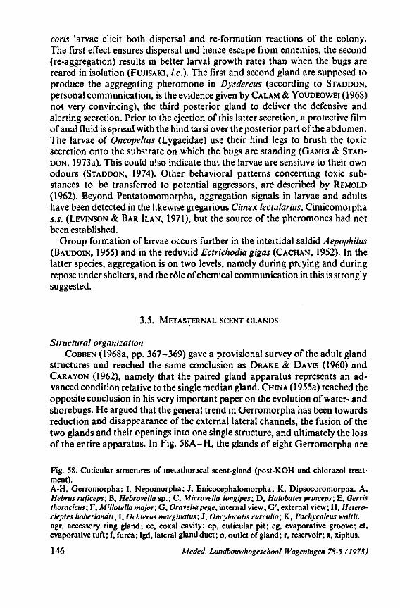
coris larvae elicit both dispersal and re-formation reactions of the colony. The first effect ensures dispersal and hence escape from ennemies, the second (re-aggregation) results in better larval growth rates than when the bugs are reared in isolation (FUJISAKI, I.e.). The first and second gland are supposed to produce the aggregating pheromone in Dysdercus (according to STADDON, personal communication, is the evidence given by CALAM & YOUDEOWEI (1968) not very convincing), the third posterior gland to deliver the defensive and alerting secretion. Prior to the ejection of this latter secretion, a protective film of anal fluid is spread with the hind tarsi over the posterior part of the abdomen. The larvae of Oncopeltus (Lygaeidae) use their hind legs to brush the toxic secretion onto the substrate on which the bugs are standing (GAMES & STADDON, 1973a). This could also indicate that the larvae are sensitive to their own odours (STADDON, 1974). Other behavioral patterns concerning toxic substances to be transferred to potential aggressors, are described by REMOLD (1962). Beyond Pentatomomorpha, aggregation signals in larvae and adults have been detected in the likewise gregarious Cimex lectularius, Cimicomorpha s.s. (LEVINSON & BAR ILAN, 1971), but the source of the pheromones had not been established.
Group formation of larvae occurs further in the intertidal saldid Aepophilus (BAUDOIN, 1955) and in the reduviid Ectrichodia gigas (CACHAN, 1952). In the latter species, aggregation is on two levels, namely during preying and during repose under shelters, and the rôle of chemical communication in this is strongly suggested.
3.5. METASTERNAL SCENT GLANDS
Structural organization COBBEN (1968a, pp. 367-369) gave a provisional survey of the adult gland
structures and reached the same conclusion as DRAKE & DAVIS (I960) and CARA YON (1962), namely that the paired gland apparatus represents an advanced condition relative to the single median gland. CHINA (1955a) reached the opposite conclusion in his very important paper on the evolution of water- and shorebugs. He argued that the general trend in Gerromorpha has been towards reduction and disappearance of the external lateral channels, the fusion of the two glands and their openings into one single structure, and ultimately the loss of the entire apparatus. In Fig. 58A-H, the glands of eight Gerromorpha are
Fig. 58. Cuticular structures of metathoracal scent-gland (post-KOH and chlorazol treatment). A-H, Gerromorpha; I, Nepomorpha; J, Enicocephalomorpha; K, Dipsocoromorpha. A, Hebrus ruficeps; B, Hebrovelia sp.; C, Microvelia longipes; D, Halobatesprinceps; E, Gerris thoracicus; F, Millotella major; G, Oraveliapege, internal view; G', external view; H, Hetero-cleptes hoberlandti; I, Ochterus marginatus; J, Oncylocotis curculio; K, Pachycoleus waltli. agr, accessory ring gland; cc, coxal cavity; cp, cuticular pit; eg, evaporative groove; et, evaporative tuft; f, furca; lgd, lateral gland duct; o, outlet of gland; r, reservoir; x, xiphus.
146 Meded. Landbouwhogeschool Wageningen 78-5 (1978)
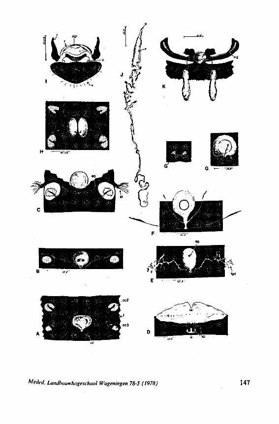
toeded. Landbouwhogeschool Wageningen 78-5 (1978) 147
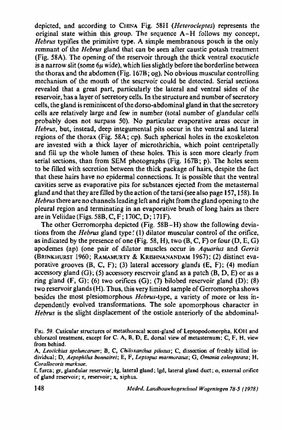
depicted, and according to CHINA Fig. 58H (Heterocleptes) represents the original state within this group. The sequence A-H follows my concept, Hebrus typifies the primitive type. A simple membranous pouch is the only remnant of the Hebrus gland that can be seen after caustic potash treatment (Fig. 58A). The opening of the reservoir through the thick ventral exocuticle is a narrow slit (some 6/i wide), which lies slightly before the borderline between the thorax and the abdomen (Fig. 167B; og). No obvious muscular controlling mechanism of the mouth of the seservoir could be detected. Serial sections revealed that a great part, particularly the lateral and ventral sides of the reservoir, has a layer of secretory cells. In the structure and number of secretory cells, the gland is reminiscent of the dorso-abdominal gland in that the secretory cells are relatively large and few in number (total number of glandular cells probably does not surpass 50). No particular evaporative areas occur in Hebrus, but, instead, deep integumental pits occur in the ventral and lateral regions of the thorax (Fig. 58A; cp). Such spherical holes in the exoskeleton are invested with a thick layer of microthrichia, which point centripetally and fill up the whole lumen of these holes. This is seen more clearly from serial sections, than from SEM photographs (Fig. 167B; p). The holes seem to be filled with secretion between the thick package of hairs, despite the fact that these hairs have no epidermal connections. It is possible that the ventral cavities serve as evaporative pits for substances ejected from the metasternal gland and that they are filled by the action of the tarsi (see also page 157,158). In Hebrus there are no channels leading left and right from the gland opening to the pleural region and terminating in an evaporative brush of long hairs as there are in Veliidae (Figs. 58B, C, F ; 170C, D ; 171 F).
The other Gerromorpha depicted (Fig. 58B-H) show the following deviations from the Hebrus gland type:* (1) dilator muscular control of the orifice, as indicated by the presence of one (Fig. 58, H), two (B, C, F) or four (D, E, G) apodemes (ap) (one pair of dilator muscles occur in Aquarius and Gerris (BRINKHURST 1960; RAMAMURTY & KRISHNANANDAM 1967); (2) distinct evaporative grooves (B, C, F); (3) lateral accessory glands (E, F); (4) median accessory gland (G); (5) accessory reservoir gland as a patch (B, D, E) or as a ring gland (F, G); (6) two orifices (G); (7) bilobed reservoir gland (D): (8) two reservoir glands (H). Thus, this very limited sample of Gerromorpha shows besides the most plesiomorphous Hebrus-type, a variety of more or less independently evolved transformations. The sole apomorphous character in Hebrus is the slight displacement of the ostiole anteriorly of the abdominal-
FIG. 59. Cuticular structures of metathoracal scent-gland of Leptopodomorpha, KOH and chlorazol treatment, except for C. A, B, D, E, dorsal view of metasternum; C, F, H, view from behind. A, Leotichius speluncarum; B, C, Chiloxanthus pilosus; C, dissection of freshly killed individual; D, Aepophilus bonnairei; E, F, Leptopus marmoratus; G, Omania coleoptrata; H, Corallocoris marksae. f, furca; gr, glandular reservoir; Ig, lateral gland; lgd, lateral gland duct; o, external orifice of gland reservoir; r, reservoir; x, xiphus.
148 Meded. Landbouwhogeschool Wageningen 78-5 (1978)
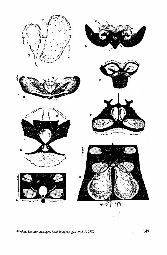
--7Î f
Meded. Landbouwhogeschool Wageningen 78-5 (1978) 149
thoracic borderline (Fig. 167B). This is the ninth trend contributing to the great cuticular variability of the metasternum.
Independent loss of the whole glandular apparatus has also occurred, for example, in many Gerridae and in Hydrometridae (Hydrometra, but not in Heterocleptes, Fig. 58H). MATSUDA (1960), considered the lack of lateral grooves in the primitive gerrid Eotrechus to be a secondary loss, but it is more likely a primary absence, in my view. Parallel events happened in other major groups, but starting from or reaching higher anagenetic levels.
CARAYON (1971) has outlined the probable course of evolution from the single median gland type ('omphalium') towards the paired gland type. His scheme is depicted in Fig. 61. The primitive 'type omphalien' is characterized by a single median aperture (A, B) or by two apertures closely side by side (C). The arrows show two different tendencies. A-+B reveals the transformation which in my drawings (Fig. 58) is indicated as the difference between A (Hebrus) and F (Millotella) or J (Pachycoleus). The median sac-like invagination became a reservoir, whereas the glandular function has been taken over by a small patch of glandular cells (ag) in the roof of the reservoir and by secondary lateral glands (lgd). The condition in Oravelia (Fig. 58G) suggests that the two lateral accessory glands might have arisen from a single median accessory gland. A->C (Fig. 61) exhibits a simple bipartition of the median gland as it occurs in Heterocleptes (Fig. 58H) on cuticular features (the distribution of secretory cells around the vesicles has not heen studied). If two openings seem to be present when the skeleton is viewed from the outside, care must be taken to detect whether the gland reservoir indeed has two ostioles. This need not necessarily be the case. The reservoir of Oravelia, for example, has one median aperture (Fig. 58G), but two external outlets (Fig. 58G'). The median internal ostiole is broadly overgrown by integument, so that the secretion has to flow left and right through two short channels, thus exhibiting the first step towards the acquisition of lateral evaporative grooves, that became fully developed in 'typical' Veliidae (Fig. 58B, C). On the other hand, the single reservoir may have two outlets (Fig. 59A, Leotichius) or paired reservoirs may discharge into a single skeletal opening (Fig. 59H, Omaniidae). Paired contiguous outer openings have been recorded in Chepuvelia and Ocellovelia, but only in the latter genus are lateral channels present (CHINA, 1955a).
It was shown above within the Gerromorpha, even when one considers only the basic evolutionary events in the metathoracic gland system (Fig. 61A-C), that 9 independent variables can be distinguished. Probably more combinations than the five depicted in Fig. 58A-H, will be found when more surface bugs are studied. In the scheme of CARAYON (1971), the body of the gland apparatus is oriented caudad, but my data indicates that an original anteriad orientation is more likely. The reservoir was found to occur in the abdomen only in Vianaididae (CARAYON, 1962), Oncylocotis (Fig. 58J), Aepo-philus (Fig. 59D) and Thaumastella (Fig. 60B). Additional material of the latter two taxa revealed a normal position in the thorax.
As indicated by CARAYON (1971) and confirmed by other authors and by
150 Meded. Landbouwhogeschool Wageningen 78-5 (1978)
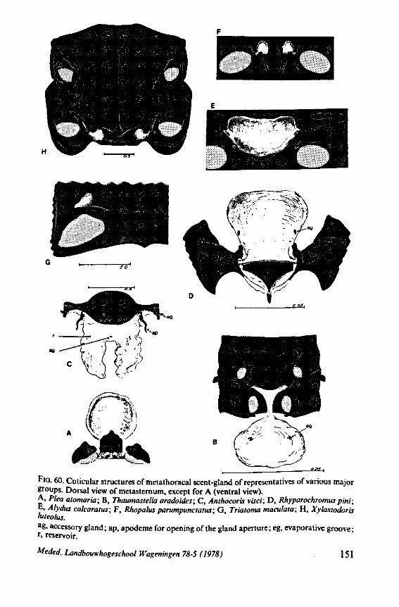
FIG. 60. Cuticular structures of metathoracal scent-gland of representatives of various major groups. Dorsal view of metasternum, except for A (ventral view). A. Plea atomaria; B, Thaumastella aradoides; C, Anthocoris visci; D, Rhyparochromus pini; E» Alydus calcaratus; F, Rhopalus parumpunctatus; G, Triatoma maculata; H, Xylastodoris weolus. ag, accessory gland; ap, apodeme for opening of the gland aperture; eg, evaporative groove; r, reservoir.
Meded. Landbouwhogeschool Wageningen 78-5 (1978) 151
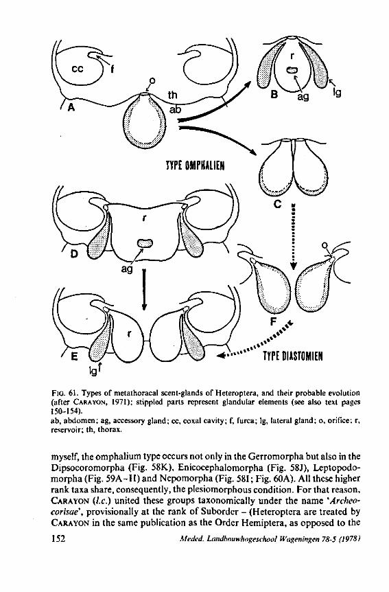
TYPE DIASTOMIEN
FIG. 61. Types of metathoracal scent-glands of Heteroptera, and their probable evolution (after CARAYON, 1971); stippled parts represent glandular elements (see also text pages 150-154). ab, abdomen; ag, accessory gland; cc, coxal cavity; f, furca; lg, lateral gland; o, orifice; r, reservoir; th, thorax.
myself, the omphalium type occurs not only in the Gerromorpha but also in the Dipsocoromorpha (Fig. 58K), Enicocephalomorpha (Fig. 58J), Leptopodo-morpha (Fig. 59A-H) and Nepomorpha (Fig. 581 ; Fig. 60A). All these higher rank taxa share, consequently, the plesiomorphous condition. For that reason, CARAYON (I.e.) united these groups taxonomically under the name 'Archeo-corisae\ provisionally at the rank of Suborder - (Heteroptera are treated by CARAYON in the same publication as the Order Hemiptera, as opposed to the
152 Meded. Landbouwhogeschool Wagen'mgen 78-5 (1978)
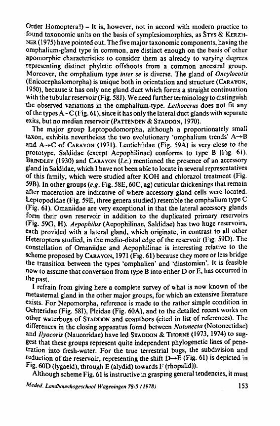
Order Homoptera !) - It is, however, not in accord with modern practice to found taxonomie units on the basis of symplesiomorphies, as STYS & KERZH-NER (1975) have pointed out. The five major taxonomie components, having the omphalium-gland type in common, are distinct enough on the basis of other apomorphic characteristics to consider them as already to varying degrees representing distinct phyletic offshoots from a common ancestral group. Moreover, the omphalium type inter se is diverse. The gland of Oncylocotis (Enicocephalomorpha) is unique both in orientation and structure (CARAVON, 1950), because it has only one gland duct which forms a straight continuation with the tubular reservoir (Fig. 58J). We need further terminology to distinguish the observed variations in the omphalium-type. Lethocerus does not fit any of the types A-C (Fig. 61), since it has only the lateral duct glands with separate exits, but no median reservoir (PATTENDEN & STADDON, 1970).
The major group Leptopodomorpha, although a proportionately small taxon, exhibits nevertheless the two evolutionary 'omphalium trends' A-*B and A-*C of CARAYON (1971). Leotichiidae (Fig. 59A) is very close to the prototype. Saldidae (except Aepophilinae) conforms to type B (Fig. 61). BRINDLEY (1930) and CARAYON (I.e.) mentioned the presence of an accessory gland in Saldidae, which I have not been able to locate in several representatives of this family, which were studied after KOH and chlorazol treatment (Fig. 59B). In other groups (e.g. Fig. 58E, 60C, ag) cuticular thickenings that remain after maceration are indicative of where accessory gland cells were located. Leptopodidae (Fig. 59E, three genera studied) resemble the omphalium type C (Fig. 61). Omaniidae are very exceptional in that the lateral accessory glands form their own reservoir in addition to the duplicated primary reservoirs (Fig. 59G, H). Aepophilus (Aepophilinae, Saldidae) has two huge reservoirs, each provided with a lateral gland, which originate, in contrast to all other Heteroptera studied, in the medio-distal edge of the reservoir (Fig. 59D). The constellation of Omaniidae and Aepophilinae is interesting relative to the scheme proposed by CARAYON, 1971 (Fig. 61) because they more or less bridge the transition between the types 'omphalien' and 'diastomien'. It is feasible now to assume that conversion from type B into either D or E, has occurred in the past.
I refrain from giving here a complete survey of what is now known of the metasternal gland in the other major groups, for which an extensive literature exists. For Nepomorpha, reference is made to the rather simple condition in Ochteridae (Fig. 581), Pleidae (Fig. 60A), and to the detailed recent works on other waterbugs of STADDON and coauthors (cited in list of references). The differences in the closing apparatus found between Notonecta (Notonectidae) and Ilyocoris (Naucoridae) have led STADDON & THORNE (1973, 1974) to suggest that these groups represent quite independent phylogenetic lines of penetration into fresh-water. For the true terrestrial bugs, the subdivision and reduction of the reservoir, representing the shift D-*E (Fig. 61) is depicted in Fig. 60D (lygaeid), through E (alydid) towards F (rhopalid)).
Although scheme Fig. 61 is instructive in grasping general tendencies, it must
Meded. Landbouwhogeschool Wageningen 78-5 (1978) 153
be kept in mind that there is considerably more variation than this in gross types and in the degree of elaboration and details of the finer structures. The whole set of metathoracic differentiations is of great help in lower rank taxonomy, when associated structures such as those that concern the closing system, efferent apparatus and evaporative area are taken into account. The exocuti-cular, so-called 'mushroom' sculpture is mostly species-specific (e.g. CARA YON, 1971; HEPBURN & YONKE, 1971) and it may be used in identification keys (DETHIER, 1974). The rôle of the epidermis in the formation of the mushroomlike projections has been described by FILSHIE & WATERHOUSE, 1969. Similar cuticular areas occur near the opening of the adult abdominal glands of Redu-viidae, the so-called 'Brindley's gland' (KÄLIN & BARRETT, 1975). Because of the many parallelisms concerned, the use of the entire gland system in the phy-logeny of higher groups is not much profitable, unless studied in very great detail (e.g. CARAYON, 1962; STADDON & THORNE, 1973), and at all taxonomie levels.
The whole scene is one of a complex evolutionary event, in which many contributing parts have developed, or reduced independently and at different rates. This applies also to the outer skeleton. In higher Gerromorpha, evaporation of the gland substances is enhanced by terminal hair tufts around the external and lateral ostiolar peritreme (Figs. 58B, C, F ; 170D). Evaporative microsculpture has arisen, in my view independently, in some Dipsocoromor-pha (Fig. 58K; eg; scanning photograph Fig. 7 in CARAYON, 1971), some Nepo-morpha (Notonecta, STADDON & THORNE, 1974) and in Pentatomomorpha and in Cimicomorpha (REMOLD, 1962). In the last group, the efferent system is very poorly developed in the primitive Plokiophilidae and Microphysidae (CARAYON, 1971).
Functions This section would be incomplete if it did not say a few words about the
possible functions of the thoracic scent gland. The reader is referred to the review of CARAYON (1971), who also gave a concise review of what is known about the chemistry of the gland secretions. Recent contributions on this subject are accumulating rapidly, and only a few remarks are given here. A difficulty in interpreting data from literature is often that authors giving analyses of the secretion do not specify whether the source has been from larval or adult glands (e.g. TSUYUKI C.S. 1965 ; Table 2 in BAGGINI C.S. 1966, summarizing new and old data; MCCULLOUGH, 1971, 1973, 1974). SWEET (1964) suggested that adults and larvae of some lygaeids have different odors. Differences in the composition between the scents of larval and adult glands were observed in seven coreoid species (ALDRICH & YONKE, 1975), and in the single pyrrhocorid, lygaeid, coreid and tessarotomatid species studied, but no differences were found in the pentatomid Apodiphus amygdali (EVERTON et al. 1974).
Secretions acting as allomone and kairomone. The better-known families of the terrestrial groups seem to eject secretions (various carbonyl compounds and aldehydes), which have a toxic or repelling effect on other arthropods,
154 Meded. Landbouwhogeschool fVageningen 78-5 (1978)
in particular ants (REMOLD 1962, WATERHOUSE c.s. 1961, TSUYUKI C.S. 1965). They are generally not effective against vertebrate predators such as birds (HEIKERTINGER 1922, KULLENBERG 1946, DETHIER 1974).
Nevertheless, Heteroptera have many effective arthropod predators and parasites. MILLER (1953) mentioned that the gland substances of Reduviidae have no deterrent effect on spiders, to which these bugs often fall victim. Mirid bugs are not preferred hosts for spiders, yet spiders and predatory bugs still are among their main enemies (KULLENBERG, 1946). CARA YON (1971) suggested that such predators and parasites are indeed sensitive to the toxic effects of the secretions, but that by their behavior they avoid making contact with the defensive secretions. He referred to the observations of DIETRICK & VAN DEN
BOSCH (1957) which showed that the phasiine parasite of Anasa trist is suffered great mortality when it was placed in a rearing cage with the host. Very interestingly, MITCHELL & MAU (1971) have given some experimental evidence indicating that the above dipterous parasite responds to the scent of Nezara viridula, and thus utilizes the volatile chemical substance of the bug as an attractant. The scent of Nezara should then be a kairomone with respect to its parasite. The glandular source of the scent has not been verified. MITCHELL
& MAU (I.e.) assumed that a male sex pheromone of Nezara attracts the parasite, since significantly more male bugs were parasitized. It can also be that in this or other cases where the hosts produce volatile secretions, the parasite has adapted to this substance which it uses as a token stimulus for attack (an evolutionary parallel to the rôle of secondary plant substances in host plant selection by phytophagous insects). It is to be pointed out that the phasiine fly mentioned, has been recorded as a parasite of bugs from at least two families (Pentatomidae, Coreidae), so that the rôle of a specific sex-pheromone of the host as the only attractant is questionable.
Kairomonal relations such as suggested above for the parasite of Nezara, may occur more generally. LARICHI & VOEGELÉ (1975) have shown that scelion-id and encyrtid egg-parasites of the pentatomid Aelia germari are attracted by the scent of the adult bugs.
It has been suggested that some Heteroptera use allomones as a kind of aggressive mimicry. The scent of species of Salyavatinae (Reduviidae) smells like that of ripe bananas. Louis (1974) supposed that this scent may have a defence function. It might also serve as a lure, that attracts Drosophila etc. to the reduviid predator. Field observations on the prey caught by one male of Apiomerus (Reduviidae) led WEAVER C.S. (1975) to surmise that attractant volatile chemicals from the bug caused stingless bees to fall its victim. A very remarkable case of resistance to the contents of the metathoracic gland is shown by a special group of tarsonemid mites of the genus Coreitarsonemus. FAIN (1970) described six new species, of which eggs, larvae and adults were found within the exoskeletal vestibules of the metasternal scent gland of different species of Coreinae (Coreidae). The mites were found in dried material but also in hosts, which were stored in alcohol, so that they must have been associated with the living bugs. The nutrition requirements of these mites is not known.
Meded. Landbouwhogeschool Wageningen 78-5 (1978) 155
SCHAEFER (1972a) demonstrated some tendencies in trichophorous families, which, according to him, indicate a defensive function for the gland. The glands are generally better developed in ground-inhabiting species than in those living on plants. The glands are relatively more poorly developed in aposematic species than in closely related cryptic species. Toxic substances in aposematic species may have their origin elsewheie in the body. Many genera of the Lygaeinae contain species which sequester cardiac glycosides from their host plants. Some genera concentrate these cardenolides in a series of dorsolateral 'glands'. Others lack such secretory openings, but may store cardiac glycosides in the secretions of the normal middorsal larval glands and the metathoracic gland of the adult (DUFFEY, 1970; DUFFEY & SCUDDER, 1972). Oncopeltus, maintained on a diet of hulled sunflower seeds, appeared to manufacture a toxic substance, possibly histamine, which appeared in the larval rectal fluid and haemolymph, and in the dorsolateral 'glands' (the toxin containing fluid is ejected through points of weakness in the body wall) and haemolymph of the adult (GRAHAM & STADDON, 1974).
In other major groups of Heteroptera also other gland systems may act as auxiliaries of the metathoracic gland. In Reduviidae and Tingidae, the distinctly paired metathoracic glands tend to be strongly reduced and, in some species, they are lost. But members of both families possess other sets of glands including paired glands at the base of the abdomen (also present in Thaumastel-lidae!, pers. observ,). The so-called Brindley's glands in Reduviidae contain chemicals that are presumably of a defensive nature (PATTENDEN & STADDON, 1972; KÄLIN & BARRETT, 1975; GAMES et al., 1974). Four different gland types occur in the reduviid Themonocoris (CARAYON, USINGER & WYGODZINSKY, 1958). Most reduviids can moreover defend themselves against larger-sized predators by the stinging action of the stylet bundle or by spitting toxic saliva, as is the case in Platymeris (EDWARDS, 1960). The huge subrectal gland present in many Reduviidae is thought by DAVIS (1969) to function in defence, but more likely it produces substances for sticking the eggs together in the batch (COB-BEN & WYGODZrNSKY, 1975).
No chemical analyses have been made of the gland secretions of the shore-inhabiting Leptopodomorpha. The paired and thus apomorphous condition in Leptopodidae (Fig. 59E), Omaniidae (Fig. 59H) and Aepophilinae (Fig. 59D), and the unpaired state in other Saldidae (Fig. 59B, C) conflicts with general morphology as a basis for deciding the evolutionary level of these taxa (Saldidae, except Aepophilinae, show derived characters in most respects). Saldidae, except Aepophilinae, have acquired extrusible pregenital glands (COBBEN, 1961) which might have arrested the evolution of the metasternal apparatus. Whether the pregenital glands are repugnatory, or/and have a propelling function by lowering the surface tension of water, only future experiments will decide. This latter passive locomotory mechanism is in fact practised by Velia, probably by the release of saliva (LINSENMAIR & JANDER, 1963) and by Microvelia and Rhagovelia (ANDERSON, in press). The secretion of the metasternal gland of Aquarius (= Gerris) najas when artificially deposited
156 Meded. Landbouwhogeschool Wageningen 78-5 (1978)
on a water surface, also lowers the surface tension (STADDON, 1972). There is very little data on the function of the adult gland system in Nepomor-
pha and all views on this matter remain conjectural ; i.e. observations have not been supplemented by experiments to test supposed functions. MASCHWITZ (1971) observed that adults of Plea (Pleidae) rub their body when the bugs are above the water level. He observed that during this 'grooming' process, a secretion which contained hydrogen peroxide, most probably from the metatho-racic gland, was delivered onto the body surface. This constituent is highly suggestive of an antiseptic function against micro-organisms. Other substances in Ilyocoris (Naucoridae) and Notonecta (Notonectidae) may have a similar function (STADDON & WEATHERSTON, 1967; PATTENDEN & STADDON, 1968). The scent-gland secretory products of Gelastocoris (Gelastocoridae) (STADDON, 1973) and Sigara (Corixidae) (PINDER & STADDON, 1965; STADDON, 1973) point more to a defence function. Observations on predators of corixids seem to oppose this view (STADDON & GRIFFITHS, 1967), however, which led STADDON (I.e.) to consider the possibility that the aquatic corixids have another use for their gland secretion rather than the original anti-predator chemical weapon, one of their terrestrial fore-runners.
Although it is very difficult to do, the actual function of the glandular organs has to be studied under situations that approximate those of the complex environments in which the bugs actually live. The complete loss of the gland system in whole taxonomie divisions, is to be seen as an intriguing chapter in evolutionary biology ; to unravel the functions of the glands in those taxa where they apparently play a vital rôle at present is a great challenge. In Coleoptero-coris (Potamocoridae), I could not find an internal cuticular intima indicative of the presence of a metasternal gland. Nor could STADDON (pers. comm.) or I find it in Nerthra sp. (Gelastocoridae, Nerthrinae), whereas representatives of the other subfamily, the Gelastocorinae, possess a well-developed gland (PARSONS, 1959; STADDON, 1973). In other groups of waterbugs similar striking plus-minus situations prevail. The glands are absent in the few nepid taxa studied. In the related family Belostomatidae, they are present in the Lethoceri-nae, but not in Belostomatinae (STADDON, 1971). In the Notonectidae, the gland was found in three genera of Notonectinae, but was absent in the two genera of Anisopinae studied (STADDON & THORNE, 1974). It must be remembered that, except for Corixidae, waterbugs are able to use their stylets in defence, and the cephalic glands also have been considered to function in defence (seep. 15).
Unfortunately, the rôle of the metasternal gland in Gerromorpha is at present obscure. As was concluded on page 148, the gland ofHebrus appears to be the most simple. On the basis of the apparent absence of a muscular opening apparatus and the presence of strange external pits in the integument, it was suggested that the secretion might be removed from the orifice of the gland with the tarsi and subsequently smeared over the pits. The brush-zone of the pits would then function as evaporative areas. The use of the secretion as an antiseptic over larger parts of the body could also be considered as possibly the
Meded. Landbouwhogeschool Wageningen 78-5 (1978) 157
original function of the gland in such primitive hygrophilous bugs. Still, this idea may conflict with the prerequisites for respiration. A thin film of atmospheric air is retained in the velvety hair-pile of Hebrus when the insect is submerged. The air-film surrounds the whole anterior half of the body, but not the pleural clefts each of which remains visible as a sharp dark line (Fig. 163 A-D, pc). The zone of contact between the air-layer and the tracheal system was not evident, but the size and density of the microtrichia (Fig. 163D) and the permanency of the air-film could suggest a plastron function as in the intertidal shore-bug Aepophilus (BAUDOIN, 1955) and in Gerridae (CHENG, 1973). A water soluble antiseptic in the secretory substance of the metathoracic gland would have the opposite effect in wetting the hair-pile. The area around the gland-orifice of Hebrus between the hind coxae, but not touching the coxal rim, becomes wet when the bug is under water as is the greater part of ventral surface of the abdomen. The abdomen became wet in some bugs at the beginning of submersion, in others the air-layer disappeared entirely within a minute. The hair-pile of the head and thorax, and consequently the resistant air-film, extends to just beyond the base of the abdomen; the remaining part of the abdomen lacks a continuous microtrichial cover (Figs. 163F, 165B). Thus, the area around the gland orifice and the abdominal venter is suited to the reception of the ejected secretion. The other suggestion made on page 148, namely that the ventral thoracic pits would serve for storage of the gland secretion, becomes problematic since they are covered with air when flooded. It may be that the gland secretion maintains the hydrofuge properties of the hair pile, as BRINKHURST (1960) suggested for Aquarius (Gerridae). STADDON (1972), however, found no support in his experiments with this species for a waterproofing function.
Finally, one can speculate about an aggregating pheromonal action for the scent of Gerromorpha. Gerrids and veliids often aggregate on open water in dense colonies (e.g. Aquarius najas and Trochopus plumbeus, respectively). Optical and tactile signals seem to be the more likely stimuli for such bugs, which are provided with good vision and vibrator receptors. Chemical assembling signals might be used in more terrestrial species, such as Hebrovelia and Macrovelia, which are found in the field aestivating in groups. This distribution pattern is, however, more likely evoked to be associated with responses to microclimatological factors.
Function as pheromone What is known of the rôle of the larval scent gland in promoting aggregation
has been briefly touched on in page 146. The adults of the human bed bug (Cimicidae) produce assembling signals, the olfactory receptors being located on the distal part of the terminal antennal segment (LEVINSON & BAR ILAN, 1971). More of such examples certainly will come to light when the behavior of other gregarious species is analyzed in simple olfactory choice tests. Aggregation before and during migration in the case of Eurygaster integriceps (Scutel-leridae) perhaps has a chemical basis (FEDOTOV, 1947; VODJDANI, 1954).
158 Meded. Landbouwhogeschool Wageningen 78-5 (1978)
The supposedly sexual function of the metathoracic gland as deduced from the sexual dimorphism of the glands has been reviewed by CARA YON (1971), who included many of his own observations. The common duct of the tubular lateral glands of Oncopeltus is distended in the sexually mature males much more than it is in the females (JOHANSSON, 1957). GAMES & STADDON (1973b) found the liquid stored in this duct to contain specific acetates the sexual function, if any, of which is not yet known.
The often quoted observation of BUTENANDT (1955) that the <$ of Lethocerus indicus (Belostomatidae) has a larger gland than the 9, was established for other Lethocerinae by PATTENDEN & STADDON (1970). But, the suggestion made by BUTENANDT and other authors that this disparity serves a sexual function is not supported by the chemical analyses of PATTENDEN & STADDON.
They concluded for L. cordofanus that the secretions are probably neither sex-nor species-specific in chemical constitution.
Coreoid males possess a ventral abdominal gland, the secretion of which may act as a long-range attractant of females (ALDRICH C.S., 1976).
PARK & SUTHERLAND (1962) concluded from their analyses of steam distillates that the secretion of the pentatomid Musgraevia contained a sex pheromone. This was not supported by the experiments of MACLEOD C.S. (1975) and Dr. STADDON has informed me that the procedure used by PARK & SUTHER
LAND is questionable. Experimental studies have now demonstrated with more or less reliability
the presence of sex-pheromones only in the following groups: Pentatomidae: Nezara (MITCHELL & MAU, 1971; çj produces pheromone highly attractive to the Ç).
Pyrrhocoridae: Dysdercus (OSMANI & NAIDU, 1966); Pyrrhocoris (ZDÂREK,
1970; probably produced by $). Miridae. Lygus (SCALES, 1968; attraction of ç? by Ç) (STRONG C.S. 1970:
source of $ pheromone probably located in the reproductive system). Reduviidae. Rhodnius (BALDWIN, KNIGHT & LYNN, 1971, volatile substances
of mating pairs attracts and sexually stimulates solitary males). It must be stressed that this survey refers only to more or less higher evolved
groups, that some data seem contradictory and, finally, that in none of these observations has the source of the volatile substances, been demonstrated unequivocally to belong to the metathoracic gland. For short range attraction, the widely distributed singular epidermal glands deserve consideration (see next section). So-called socket-glands occur in the male of Dysdercus fasciatus and might be involved in such attraction (LAWRENCE & STADDON, 75).
3.6 TRICHOBOTHRIA AND OTHER CUTICULAR STRUCTURES OF UNKNOWN
FUNCTION
1- Trichobothria* Three pairs of setae are differentiated from the remaining cephalic hair-
Meded. Landbouwhogeschool Wageningen 78-5 (1978) 159
vestiture of first instar larva of Gerromorpha and Leptopodomorpha (Figs. 52-54, 57A-E). In Gerromorpha, except part of Gerridae, they are very long, in particular the posterior pair near the anterior margin of the pronotum, and arise from a distinct, deep pit (Figs. 162D, 168D). They very obviously are trichobothria, most likely mechanoreceptors, but their actual function has not been proven experimentally. Their structure closely resembles the abdominal trichobothria of many trichophorous Pentatomomorpha (e.g. Fig. 57K; tr). In Saldidae (excepting Aepophilus, Fig. 53C, D), the trichobothria, if they may be termed here as such, are stout and erect, but otherwise not much different from the other setae on the dorsal head surface (Fig. 166F). In Leptopodidae, the original trichobothria are recognizable as pegs (Fig. 53B), but Patapius thaiensis has one pair of trichobothria near the inner margin of the eye (COBBEN, 1968b).
CHINA (1955a) included the six cephalic trichobothria in the attributes possessed by the ancestral surface bugs. Perhaps they belonged to the general sensory equipment of the archetypical Heteroptera, since comparable conspicuous setae, in larvae and adults, are present also in representatives of other families such as Lygaeidae (Fig. 57K), Cydnidae, Plokiophilidae, Micro-physidae, Anthocoridae (Fig. 55B), Nabidae (Fig. 55A) and Dipsocoridae (Fig. 53A). Three pairs of cephalic trichobothria are found in both subfamilies of Pachynomidae (not mentioned by CARAYON & VILLIERS, 1968) and moreover a pair of lateroventral cephalic bristles, which I have detected so far only, other than in the Pachynomidae, in Nabis (Nabidae, Fig. 57G; tr, first larval instar). I have not checked the first instar larva of Enicocephalidae on this character, but in the third instar of Oncylocotis no special hairs could be detected between the dense cover of macrochaetae. The head of the first instar larva of Laccotrephes (Nepidae) bears four pairs of composite trichobothria, arising from a conspicuous, likewise composite alveolus. Other waterbug families seem to lack head trichobothria, but young larvae of Potamocoridae may have them, since the ocular trichobothria which mostly occur in association with cephalic sensory hairs, are retained in the adult.
SCHAEFER (1975) gave a well-documented review of the abdominal trichobothria of Pentatomomorpha, which are of taxonomie value only at the higher levels. His discussion on some aspects of phylogeny and ontogeny are relevant for the present paper. Setae of great length are generally associated with ground-living taxa (as is the case with the cephalic setae), whereas taxa living on plants have short trichobothria. The absolute length of the abdominal trichobothria remains about the same in all instars, for which reason SCHAEFER assumed an unchanged function, probably vibration reception. DRÂSLAR
* The reader is referred to the important publication of SCHUH (1975) on trichobothria of the legs of Miridae. This paper, which came to my attention too late to incorporate it in my text, also contains a general survey of trichobothria in the Heteroptera.
160 Meded. Landbouwhogeschool H'ageningen 78-5 (1978)
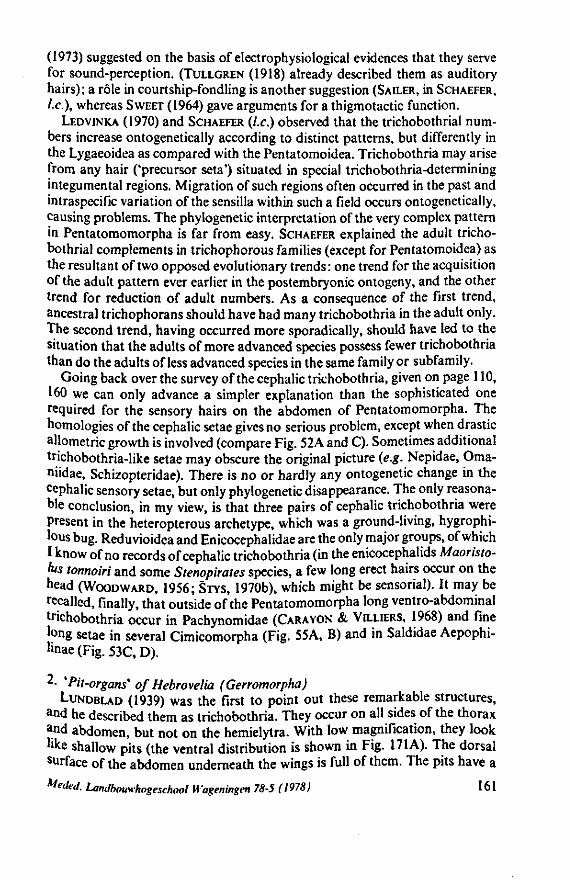
(1973) suggested on the basis of electrophysiological evidences that they serve for sound-perception. (TULLGREN (1918) already described them as auditory hairs); a rôle in courtship-fondling is another suggestion (SAILER, in SCHAEFER, I.e.), whereas SWEET (1964) gave arguments for a thigmotactic function.
LEDVINKA (1970) and SCHAEFER (I.e.) observed that the trichobothrial numbers increase ontogenetically according to distinct patterns, but differently in the Lygaeoidea as compared with the Pentatomoidea. Trichobothria may arise from any hair ('precursor seta') situated in special trichobothria-determining integumental regions. Migration of such regions often occurred in the past and intraspecific variation of the sensilla within such a field occurs ontogenetically, causing problems. The phylogenetic interpretation of the very complex pattern in Pentatomomorpha is far from easy. SCHAEFER explained the adult trichobothrial complements in trichophorous families (except for Pentatomoidea) as the resultant of two opposed evolutionary trends: one trend for the acquisition of the adult pattern ever earlier in the postembryonic ontogeny, and the other trend for reduction of adult numbers. As a consequence of the first trend, ancestral trichophorans should have had many trichobothria in the adult only. The second trend, having occurred more sporadically, should have led to the situation that the adults of more advanced species possess fewer trichobothria than do the adults of less advanced species in the same family or subfamily.
Going back over the survey of the cephalic trichobothria, given on page 110, 160 we can only advance a simpler explanation than the sophisticated one required for the sensory hairs on the abdomen of Pentatomomorpha. The homologies of the cephalic setae gives no serious problem, except when drastic allometric growth is involved (compare Fig. 52A and C). Sometimes additional trichobothria-like setae may obscure the original picture (e.g. Nepidae, Oma-niidae, Schizopteridae). There is no or hardly any ontogenetic change in the cephalic sensory setae, but only phylogenetic disappearance. The only reasonable conclusion, in my view, is that three pairs of cephalic trichobothria were Present in the heteropterous archetype, which was a ground-living, hygrophi-lous bug. Reduvioidea and Enicocephalidae are the only major groups, of which I know of no records of cephalic trichobothria (in the enicocephalids Maoristo-lus tonnoiri and some Stenopirates species, a few long erect hairs occur on the head (WOODWARD, 1956; STYS, 1970b), which might be sensorial). It may be recalled, finally, that outside of the Pentatomomorpha long ventro-abdominal trichobothria occur in Pachynomidae (CARAYON & VILLIERS, 1968) and fine long setae in several Cimicomorpha (Fig. 55A, B) and in Saldidae Aepophi-»nae (Fig. 53C, D).
2- 'Pit-organs' of Hebrovelia (Gerromorpha) LUNDBLAD (1939) was the first to point out these remarkable structures,
and he described them as trichobothria. They occur on all sides of the thorax and abdomen, but not on the hemielytra. With low magnification, they look •"ke shallow pits (the ventral distribution is shown in Fig. 171 A). The dorsal surface of the abdomen underneath the wings is full of them. The pits have a
Meded. Landbouwhogeschool Wageningen 78-5 (1978) 161
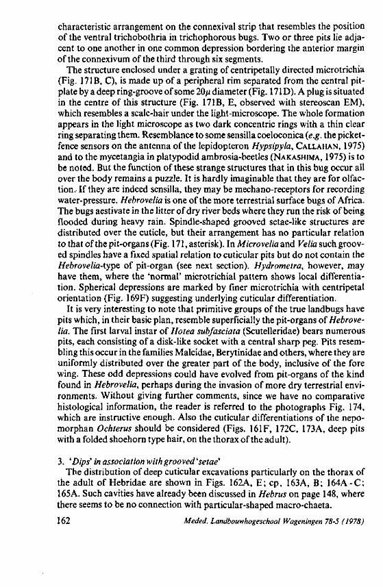
characteristic arrangement on the connexival strip that resembles the position of the ventral trichobothria in trichophorous bugs. Two or three pits lie adjacent to one another in one common depression bordering the anterior margin of the connexivum of the third through six segments.
The structure enclosed under a grating of centripetally directed microtrichia (Fig. 171B, C), is made up of a peripheral rim separated from the central pit-plate by a deep ring-groove of some 20/x diameter (Fig. 171D). A plug is situated in the centre of this structure (Fig. 171B, E, observed with stereoscan EM), which resembles a scale-hair under the light-microscope. The whole formation appears in the light microscope as two dark concentric rings with a thin clear ring separating them. Resemblance to some sensilla coeloconica (e.g. the picket-fence sensors on the antenna of the lepidopteron Hypsipyla, CALLAHAN, 1975) and to the mycetangia in platypodid ambrosia-beetles (NAKASHIMA, 1975) is to be noted. But the function of these strange structures that in this bug occur all over the body remains a puzzle. It is hardly imaginable that they are for olfaction. If they are indeed sensilla, they may be mechano-receptors for recording water-pressure. Hebrovelia is one of the more terrestrial surface bugs of Africa. The bugs aestivate in the litter of dry river beds where they run the risk of being flooded during heavy rain. Spindle-shaped grooved setae-like structures are distributed over the cuticle, but their arrangement has no particular relation to that of the pit-organs (Fig. 171, asterisk). In Microvelia and Velia such grooved spindles have a fixed spatial relation to cuticular pits but do not contain the Hebrovelia-typ& of pit-organ (see next section). Hydrometra, however, may have them, where the 'normal' microtrichial pattern shows local differentiation. Spherical depressions are marked by finer microtrichia with centripetal orientation (Fig. 169F) suggesting underlying cuticular differentiation.
It is very interesting to note that primitive groups of the true landbugs have pits which, in their basic plan, resemble superficially the pit-organs of Hebrovelia. The first larval instar of Hotea subfasciata (Scutelleridae) bears numerous pits, each consisting of a disk-like socket with a central sharp peg. Pits resembling this occur in the families Malcidae, Berytinidae and others, where they are uniformly distributed over the greater part of the body, inclusive of the fore wing. These odd depressions could have evolved from pit-organs of the kind found in Hebrovelia, perhaps during the invasion of more dry terrestrial environments. Without giving further comments, since we have no comparative histological information, the reader is referred to the photographs Fig. 174, which are instructive enough. Also the cuticular differentiations of the nepo-morphan Ochterus should be considered (Figs. 161F, 172C, 173A, deep pits with a folded shoehorn type hair, on the thorax of the adult).
3. 'Dips' in association with grooved 'setae' The distribution of deep cuticular excavations particularly on the thorax of
the adult of Hebridae are shown in Figs. 162A, E; cp, 163A, B; 164A-C; 165A. Such cavities have already been discussed in Hebrus on page 148, where there seems to be no connection with particular-shaped macro-chaeta.
162 Meded. Landbouwhogeschool Wageningen 78-5 (1978)
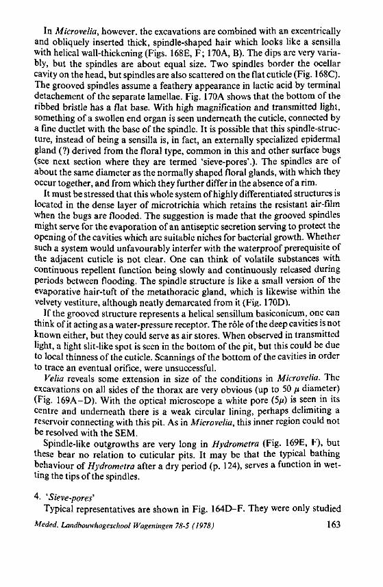
In Microvelia, however, the excavations are combined with an excentrically and obliquely inserted thick, spindle-shaped hair which looks like a sensilla with helical wall-thickening (Figs. 168E, F; 170A, B). The dips are very variably, but the spindles are about equal size. Two spindles border the ocellar cavity on the head, but spindles are also scattered on the flat cuticle (Fig. 168C). The grooved spindles assume a feathery appearance in lactic acid by terminal detachement of the separate lamellae. Fig. 170A shows that the bottom of the ribbed bristle has a flat base. With high magnification and transmitted light, something of a swollen end organ is seen underneath the cuticle, connected by a fine ductlet with the base of the spindle. It is possible that this spindle-structure, instead of being a sensilla is, in fact, an externally specialized epidermal gland (?) derived from the floral type, common in this and other surface bugs (see next section where they are termed 'sieve-pores'.). The spindles are of about the same diameter as the normally shaped floral glands, with which they occur together, and from which they further differ in the absence of a rim.
It must be stressed that this whole system of highly differentiated structures is located in the dense layer of microtrichia which retains the resistant air-film when the bugs are flooded. The suggestion is made that the grooved spindles might serve for the evaporation of an antiseptic secretion serving to protect the opening of the cavities which are suitable niches for bacterial growth. Whether such a system would unfavourably interfer with the waterproof prerequisite of the adjacent cuticle is not clear. One can think of volatile substances with continuous repellent function being slowly and continuously released during periods between flooding. The spindle structure is like a small version of the evaporative hair-tuft of the metathoracic gland, which is likewise within the velvety vestiture, although neatly demarcated from it (Fig. 170D).
If the grooved structure represents a helical sensillum basiconicum, one can think of it acting as a water-pressure receptor. The rôle of the deep cavities is not known either, but they could serve as air stores. When observed in transmitted light, a light slit-like spot is seen in the bottom of the pit, but this could be due to local thinness of the cuticle. Scannings of the bottom of the cavities in order to trace an eventual orifice, were unsuccessful.
Velia reveals some extension in size of the conditions in Microvelia. The excavations on all sides of the thorax are very obvious (up to 50 fx diameter) (Fig. 169A-D). With the optical microscope a white pore (5/x) is seen in its centre and underneath there is a weak circular lining, perhaps delimiting a reservoir connecting with this pit. As in Microvelia, this inner region could not be resolved with the SEM.
Spindle-like outgrowths are very long in Hydrometra (Fig. 169E, F), but these bear no relation to cuticular pits. It may be that the typical bathing behaviour of Hydrometra after a dry period (p. 124), serves a function in wetting the tips of the spindles.
4. 'Sieve-pores'1
Typical representatives are shown in Fig. 164D-F. They were only studied
Meded. Landbouwhogeschool Wageningen 78-5 (1978) 163
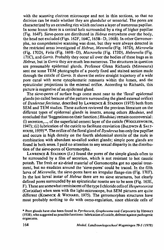
with the scanning electron microscope and not in thin sections, so that no decision can be made whether they are glandular or sensorial. The pores are characterized by an extending rim which encloses a layer of numerous papillae. In some lenses there is a central hole surrounded by a ring of higher papillae (Fig. 164F). Sieve-pores are distributed in Hebrus everywhere over the body, the head not excluded (Figs. 162F, 164C, 165B-D, 166B). In other Gerromor-pha, no comprehensive scanning was done, but they were always detected in the restricted areas investigated of Hebrus, Mesovelia (Fig. 167D), Microvelia (Fig. 170D), Velia (Fig. 169B-D), Macrovelia (Fig. 173D), Hebrovelia (Fig. 171C), and Gerris. Probably they occur all over the bodies of these bugs as in Hebrus, but in Gerris they are much less numerous. The structures in question are presumably epidermal glands. Professor Glenn Richards (Minnesota) sent me some TEM photographs of a poorly fixed and stained section made through the cuticle of Gerris. It shows the entire straight trajectory of a wide pore canal with some cytoplasmatic remnants within the lumen, and the epicuticular projections in the external orifice. According to Richards, this picture is suggestive of an epidermal gland.
The sieve-pores of surface bugs come most near to the 'floral' epidermal glands (so called because of the pattern surrounding the gland external orifice) of Dysdercus fasciatus, described by LAWRENCE & STADDON (1975) both from SEM and TEM studies. These authors reviewed the previous literature on the different types of epidermal glands in insects, among others Rhodnius, and concluded that 'Suggestions on their function (Rhodnius) remain controversial : (i) secretion,..., of the superficial cement layer of the cuticle (WIGGLESWORTH, 1947); (ii) lubrication of the cuticle to facilitate exuviation (BALDWIN & SALT-HOUSE, 1959)'*. The orifice of the floral gland of Dysdercus has only few papillae and occurs in high density on the fourth abdominal sternite of the male in combination with abundant so-called socket glands; simple pore glands are found in both sexes. I paid no attention to any sexual disparity in the distribution of the sieve-pores of Gerromorpha.
LAWRENCE & STADDON (I.e.) found the pores of the simple glands often to be surrounded by a film of secretion, which is not resistant to hot caustic potash. The fresh or air-dried material of Gerromorpha got no special treatment, but no exudates around the 'sieve-pores' could be traced. In the fifth larva of Microvelia, the sieve-pores have an irregular flange-rim (Fig. 170F). In the last larval instar of Hebrus there are no sieve structures, but clearly defined pores surrounded by an epicuticular rosette are to be seen (Fig. 165E, F). These are somewhat reminiscent of the type I chloride cells of Hesperocorixa (Corixidae) when seen with the light-microscope, but SEM pictures are quite different (KOMNICK & WICHARD, 1975). The gerromorphan structures have most probably nothing to do with osmo-regulation, since chloride cells of
* Pore glands have also been found in Pyrrhocoris, Graphosoma and Carpocoris by HENRICI ( 1938), who suggested as possible functions : lubrication of cuticle, defence against pathogenic organisms.
164 Meded. Landbouwhogeschool Wageningen 78-5 (1978)
waterbugs are characterized externally by larger size (some 7/i diameter) and occur only at those sites of the body which are in direct contact with water.
In surface bugs, the sieve-pores occur in the hydrophobic microtrichial hair-pile (Fig. 164C), but also on the wettable abdomen oîHebrus (Fig. 165C). KOMNICK & WICHARD (I.e.) found chloride cells in all stages of the representatives studied of Corixidae (see also p. 93), Nepidae, Naucoridae and Notonec-tidae. In Pleidae, however, such cells occur only in the larval stage (see also p. 93 and Fig. 176D-F). These authors showed definitely the absence of chloride cells in the larvae and adults of Hydrometra. This latter taxon, however, does possess pores, which are simple (Fig. 169E) and unlike the sieve-type structure common in other Gerromorpha. Saldidae (Chiloxanthus and Saldula studied) lack sieve-pores altogether. Few simple pores occur on the dorsal side of the larval thorax and ventrally on the abdomen of the adult.
Finally, the presence of sieve-pores in Ochterus adults must be stressed. They have exactly the same shape as in Gerromorpha, but they are ten times larger (some 80/i) (Figs. 172E, F; 173A, B). They are extremely numerous on the ventral side and the dorsal base of the head, the thorax and the hemielytra (except for the membrane). The abdomen does not have them, nor the last larval instar, but in stead only scattered simple pores of varying diameter (Fig. 173C). The sieve-pores are alternated on the pronotum by calyx-structures with a small central pit (Fig. 173B). Ochteridae are shore-inhabiting nepomor-pha as are the Gelastocoridae, but this latter family was not studied on these cuticular differentiations.
From the foregoing it appears that sieve-pores and socket-pores occur in Gerromorpha, Nepomorpha (Ochteridae) and Dipsocoromorpha (only Schi-zoptera studied). No pore-canals could be traced in Oncylocotis (Enicocephalo-morpha, note the peculiar rugose sculpture of the cuticle, Fig. 161E), nor in the few Pentatomomorpha (adults of Cymus, Malcus, Piesma) and Cimicomorpha (Anthocoris adult) investigated. It is interesting to note that several of the remarkable structures listed here, bear strong resemblance with structures recorded from insect antennae, where they definitely are considered to be sensilla (CALLAHAN, 1975). For example, folded shoehorn type (sensillum auricillicum) (Fig. 161F), cavity type pit organs (sensilla ampulacea or flasks) (Fig. 172F, 173C), picket fence type (sensilla coeloconica) (Fig. 169F), setae with helical wall sculpturing (sensilla basiconica) (Fig. 170B).
3.7. THE MALE INTROMITTENT ORGAN
POPOV (1971, p. 185; p. 113 in the translation of Vaitaitis) considered the male genitalia to be of use only at the species and genus level. According to him, this apparatus provides only one kind of information, and is too variable to be useful in phylogenetic interpretation. POPOV thought the Leptopodomor-pha to be more plesiomorphous than the Gerromorpha, but he did not consider COBBEN'S (1965, 1968a) data on genitalic evolution of these groups in his
Meded. Landbouwhogeschool Wageningen 78-5 (1978) 165
interpretation. In my view, it is a mistake to underestimate the importance of genitalic structures to the extent done by POPOV.
I consider ectodermal structures of the reproductive system to be of extreme value. In my view, they provide the strongest evidence for understanding macroevolution within the suborder, but only if they are properly studied in great detail in taxa of all levels within each major group. Some of their main traits are previewed here again; these are substantiated with drawings and schemes. The terminology used is after COBBEN (1965, 1968) and deviates in some respects from DUPUIS (in TUXEN, 1970). A full account of this subject with due credit to the voluminous literature, will be presented in a later volume of the present series.
The parts of the male intromittent organ having the most use are located in its base. Secondary, intra- and periphallic differentiations, which are used in lower level taxonomy, are manifold, and often arise through parellel evolution. These obscure the basic evolutionary scheme.
Type I. The gerromorphous type. Gerromorpha (Figs. 62, 63, 64C, C') have the most simple phallic apparatus in Heteroptera. The articulatory apparatus (= phallus base, phallobase, Fig. 64C1, phb) is a simple, U-shaped sclerite suspended in the wall of the genital chamber, the pygophore-diaphragm. It is open to the haemocoel both anteriorly and dorsally in typical surface bugs. Inflation of the long, endosomal membrane during insemination (Fig. 64C-+C ') is achieved solely by blood pressure. Complete expansion of this invaginated sac can be achieved in living bugs by applying careful manual pressure on the abdomen. The muscles which assist in pivotting the phallus base insert directly into its anterior margin ; there being no special apodemes for the protractor and retractor muscles (Fig. 64C, r.pr). The parameres are attached directly to the anterodorsal edge of the phallus base (Fig. 62D, p).
The sperm-duct (Fig. 62D, E, ds) is very thin and membranous and runs entirely free through the basal apparatus, phallotheca and endosoma. There is no structural difference between ductus seminis and ductus ejaculatorius. although a demarcation between ectodermal and mesodermal portions may appear histologically. The ductus ejaculatorius has no ejaculatory bulb. The
FIG. 62. Inflated phalli of Gerromorpha (post K.OH and chlorazol treatment). A, Hebrovelia sp., ventral view of pygophore (note that the phallus deviates to the dextral side); A', lateral view of distal part of endosoma; B, Microvelia reticulata, view from behind (note that the angle of deviation of the phallus is just opposite to that in A); B', phallus seen from a slightly different angle of B (note that the 90° deviation of the inflated phallus is caused by the joint between phallobase and phallotheca); C, Mesovelia furcata, dextral side; C', distal part of endosoma; D, Velia captai, dextral side; E, Gerris thoracicus, left side (note that D and E are viewed from opposite sides, but that the secondary gonopore (sg) points in the same direction). co, conjunctivum; cop, conjunctival process; de, ductus ejaculatorius; ds, ductus seminis; p, paramere; phb, phallobase; phth, phallotheca; pro, proctiger; py, pygophore; sg, secondary gonopore; vs, vesica; vsp, vesical process; vss, vesical sclerite.
166 Meded. Landbouwhogeschool Wageningen 78-5 (1978)
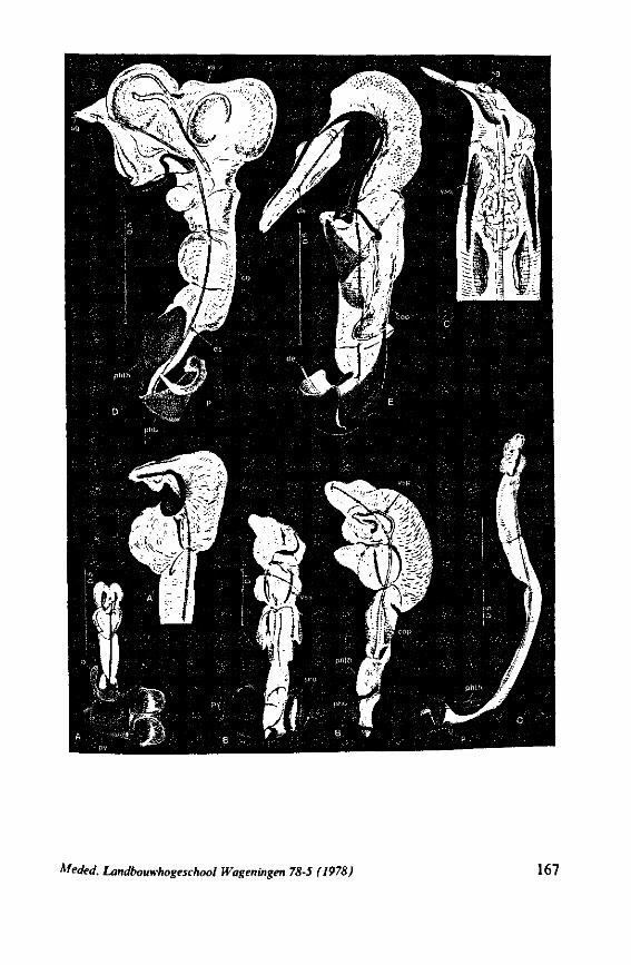
Meded. Landbouwhogeschool Wageningen 78-5 (1978) 167
phallotheca (Fig. 62, D, phth) is distinct, but, primitively, weakly sclerotized. The endosoma is divided into a proximal conjunctiva and a distal vesica (Fig. 64, C', co, vs). Two lateral sclerites within the wall of the vesica (Fig. 62B, C', D, vss) keep the vesical bladder expanded.
The gerromorphan picture given above applies equally to the male apparatus of Hebrus, The inflated phallus of Hebrus is bilaterally symmetrical and consists of a short, weakly-defined phallotheca, a tubular conjunctivum without processes and two and a half fold longer than the phallotheca, and a vesica bearing three pairs of swellings. The sperm duct terminates at the apex of the vesica and is nowhere attached to the endosomal envelope or guided by internal rods. A full account of Hebrus reproductive organs and ectodermalia will be presented elsewhere (K. HEMING-V. BATTUM, in preparation).
The endosoma of Hebrus is the most simple so far met with in surface bugs. It is complicated by the presence of extensive swellings, processes and internal sclerites in Hebrovelia (Fig. 62A, A'), Microvelia (Fig. 62B, B', note the bilateral asymmetric composition), Velia (Fig. 62D), Macrovelia and Gerris (Fig. 62E). During inflation, the phallus of Hebrus everts with its longitudinal axis in line with that of its body. It deviates 90° to the right in Hebrovelia (Fig. 62A) and 90° to the left in Microvelia (Fig. 62B). In others, e.g. Hydrometra (Fig. 63 A) and Gerris (Fig. 62E), a weak torsion of the phallic body in relation to the basal apparatus becomes discernable. The inflated phallus of Mesovelia (Fig. 62C) is seemingly undifferentiated. Its great length is correlated with that of the well-developed, laciniate, ovipositor, of the female. As the male organ penetrates the apex of the ovipositor, a greater length and tubular shape of the endosoma is required to reach the female gynatrium. (In other groups in which the female has a laciniate ovipositor, e.g. Saldidae and Nabidae, penetration of the phallus occurs at the base of the ovipositor). As reasoned earlier (COBBEN, 1968a), the laciniate ovipositor is considered to be derived from plate-shaped female terminalia. Apical reinforcements present within the endosoma of Mesovelia and its asymmetrical apex (Fig. 62C') suggest that it is more advanced than that
FIG. 63. Phallic structures of aberrant Gerromorpha. A, Hydrometra stagnorum, natural inflation, dorsal view; A', left view (note that the phallo-base makes a slight torsion with the rest of the phallus; there isa strong reduction of endosomal inflation as compared with the representatives of Fig. 62); B, Mesoveloidea sp. (note the widening of the ductus ejaculatorius prior to entering the phallobase); C, Macrovelia horni, phallus incompletely inflated, dextral side; C', basal part of phallus (note the attachment of the sperm duct to processi of the phallobase and the phallotheca, and the difference in structure between de and ds); C", phallus in repose seen caudad; D, 'Mesovelia' sp. from New Zealand, non-inflated phallus in left view (note the widening of the ductus ejaculatorius, which probably "represents the bulbus ejaculatorius); E, Ocellovelia germari, phallus in repose, left side (note that the phallobase forms a circumferentially closed structure to which the term 'basal aula' could be already applied). ba, basal aula; be, bulbus ejaculatorius; dd, dorsal diaphragma; de, ductus ejaculatorius; ds, ductus seminis; en, endosoma; p, paramere; phb, phallobase; phth, phallotheca; pr, apodeme for the protractor muscles; pro, proctiger; py, pygophore; r, apodeme for the retractor.
168 Meded. Landbouwhogeschool Wageningen 78-5 (1978)
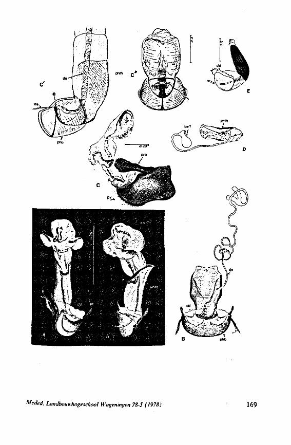
Meded. Landbouwhogeschool Wageningen 78-5 (1978) 169
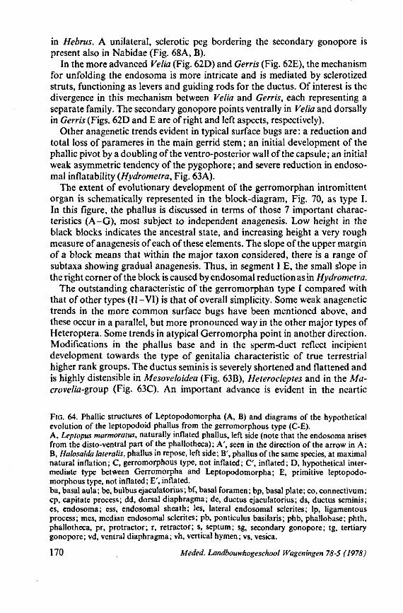
in Hebrus. A unilateral, sclerotic peg bordering the secondary gonopore is present also in Nabidae (Fig. 68A, B).
In the more advanced Velia (Fig. 62D) and Gerris (Fig. 62E), the mechanism for unfolding the endosoma is more intricate and is mediated by sclerotized struts, functioning as levers and guiding rods for the ductus. Of interest is the divergence in this mechanism between Velia and Gerris, each representing a separate family. The secondary gonopore points ventrally in Velia and dorsally in Gerris (Figs. 62D and E are of right and left aspects, respectively).
Other anagenetic trends evident in typical surface bugs are : a reduction and total loss of parameres in the main gerrid stem ; an initial development of the phallic pivot by a doubling of the ventro-posterior wall of the capsule ; an initial weak asymmetric tendency of the pygophore ; and severe reduction in endoso-mal inflatability (Hydrometra, Fig. 63A).
The extent of evolutionary development of the gerromorphan intromittent organ is schematically represented in the block-diagram, Fig. 70, as type I. In this figure, the phallus is discussed in terms of those 7 important characteristics (A-G), most subject to independent anagenesis. Low height in the black blocks indicates the ancestral state, and increasing height a very rough measure of anagenesis of each of these elements. The slope of the upper margin of a block means that within the major taxon considered, there is a range of subtaxa showing gradual anagenesis. Thus, in segment I E, the small slope in the right corner of the block is caused by endosomal reduction as in Hydrometra.
The outstanding characteristic of the gerromorphan type I compared with that of other types (II-VI) is that of overall simplicity. Some weak anagenetic trends in the more common surface bugs have been mentioned above, and these occur in a parallel, but more pronounced way in the other major types of Heteroptera. Some trends in atypical Gerromorpha point in another direction. Modifications in the phallus base and in the sperm-duct reflect incipient development towards the type of genitalia characteristic of true terrestrial higher rank groups. The ductus seminis is severely shortened and flattened and is highly distensible in Mesoveloidea (Fig. 63B), Heterocleptes and in the Ma-crovelia-group (Fig. 63C). An important advance is evident in the neartic
FIG. 64. Phallic structures of Leptopodomorpha (A, B) and diagrams of the hypothetical evolution of the leptopodoid phallus from the gerromorphous type (C-E). A, Leptopus marmoratus, naturally inflated phallus, left side (note that the endosoma arises from the disto-ventral part of the phallotheca); A', seen in the direction of the arrow in A; B, Halosalda lateralis, phallus in repose, left side; B', phallus of the same species, at maximal natural inflation; C, gerromorphous type, not inflated; C', inflated; D, hypothetical intermediate type between Gerromorpha and Leptopodomorpha; E, primitive leptopodo-morphous type, not inflated; E', inflated. ba, basal aula; be, bulbus ejaculatorius; bf, basal foramen; bp, basal plate; co, connectivum; cp, capitate process; dd, dorsal diaphragma; de, ductus ejaculatorius; ds, ductus seminis; es, endosoma; ess, endosomal sheath; les, lateral endosomal sclerites; Ip, ligamentous process; mes, median endosomal sclerites; pb, ponticulus basilaris; phb, phallobase; phth, phallotheca, pr, protractor; r, retractor; s, septum; sg, secondary gonopore; tg, tertiary gonopore; vd, ventral diaphragma; vh, vertical hymen; vs, vesica.
170 Meded. Landbouwhogeschool Wageningen 78-5 (1978)
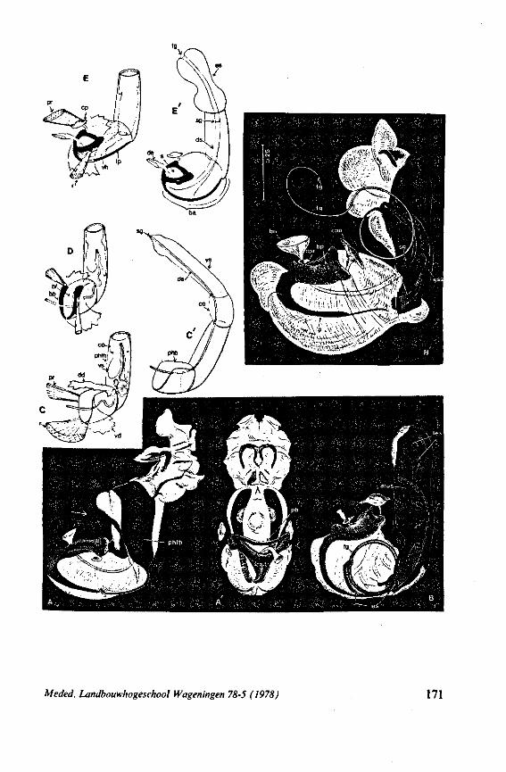
Meded. Landbouwhogeschool Wageningen 78-5 (1978) 171
genera Macrovelia, Oravelia and Chepuvelia, in which the duct has been attached to a medio-ventral extension of the base of the phallotheca (Fig. 63, C', C \ asterisk). Whereas none of the Gerromorpha studied have an ejaculatory bulb, a moss-inhabiting Mesovelia-Mke species from New Zealand seems to have one, as judged from the cuticular dilatation of the duct (Fig. 63D, be?). In Gerromorpha, one can hardly speak of a 'basal aula' (COBBEN, 1965), because the U-shaped articulatory plate is open anteriorly and dorsally. In Ocellovelia, however, the dorsal diaphragm is tightly fused with the lateral walls of this plate forming a complete cylinder (Fig. 63E).
Type II in the diagram (Fig. 70) delinates the situation found in Dipsocoro-morpha. Conditions are predominantly plesiomorphous except for the presence of exorbitant, endosomal proliferations. Fig. 68D of the dipsocorid Pachyoleus serves as an example. The picture is of a speciment fixed during copulation, but the same effect can be achieved by applying gentle pressure to the abdomen of a living, but sexually inactive male. This is possible because the entrance into the basal apparatus is broadly open to the haemocoel as in Gerromorpha. There is, however, a bulbus ejaculatorius present, which is primitively lacking in surface bugs. It is probably incorrect to speak of an inflation process in Dipsocoromorpha, since the phallus at rest already has its ultimate, though coiled, length. Only a few, membranous parts actually swell during erection. The structure of the distal part of the phallus in Pachyco-leus is so complicated and so utterly different from the general heteropterous type, that it is, at present, impossible to describe it in comparative terms. Secondary fusion of these parts simplify the structure of the phallus in other members of this taxon. The striking asymmetries and secondary specializations present in the pregenital segments of other Dipsocoromorpha are additional apomorphic features providing excellent taxonomie characters at the species level. An intensive search should be made for living representatives of taxa bridging the wide gap in genital structure between this group and the Gerromorpha.
Detailed information on the evolution of the male organ of Enicocephalo-morpha is greatly needed, because of the central place that this group, along with Gerromorpha and Dipsocoromorpha, occupies in discussions on the heteropterous archetype. These bugs are characterized by great reduction in complexity of the male genitalia. I await with great interest the results of analysis of the most primitive members of this group having non-atrophied genitalia.
Types III, IV and Kof the diagram (Fig. 70) relate to the phallic constitution of Pentatomomorpha, Reduvioidea + Nepomorpha, and Cimicomorpha s.s. Comparing the successive units (Fig. 70 A-G) of these types with the same units in Gerromorpha, shows that they are all apomorphic.
The basal aula of taxa of Nepomorpha and Reduvioidea provides more or less ready access to haemolymph as it does in Type I and II, but the articulatory apparatus is, nevertheless, rather well developed. Here I refer to a few examples of inflated phalli of representatives of types III—V (Figs. 68A-C,
172 Meded. Landbouwhogeschool Wageningen 78-5 (1978)
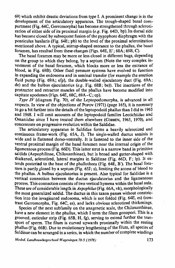
69) which exhibit drastic deviations from type I. A prominent change is in the development of the articulatory apparatus. The trough-shaped basal compartment (Fig. 64C, Gerromorpha) has become strengthened through scleroti-zation of either side of its proximal margin (e.g. Fig. 64D, bp). Its dorsal side has become closed by subsequent fusion of the pygophore diaphragm with the ponticulus basilaris (Fig. 64E ; pb) to the level of the proximal sclerotization mentioned above. A typical, stirrup-shaped entrance to the phallus, the basal foramen, has resulted from these changes (Figs. 64E, E' ; 68A ; 69B, C).
The basal foramen may be more or less closed in different bugs, depending on the group to which they belong, by a septum (Note the very complex investment of the basal foramen, which blocks more or less the entrance of blood, in Fig. 69B). Other fluid pressure systems have evolved to function in expanding the endosoma and in seminal transfer (for example the erection fluid pump (Fig. 69A; efp), the double-walled ejaculatory duct (Fig. 69A; de) and the bulbus ejaculatorius (e.g. Fig. 68B; be)). The insertions of the protractor and retractor muscles of the phallus have become modified into capitate apodemes (Figs. 64E, 68C, 69A-C; cp).
Type IV (diagram Fig. 70), of the Leptopodomorpha, is advanced in all respects. In view of the objections of POPOV (1971) (page 165), it is necessary to go a bit further into the details of the leptopodoid phallus than I did in 1965 and 1968. I will omit accounts of the leptopodoid families Leotichiidae and Omaniidae since I have treated them elsewhere (COBBEN, 1965, 1970), and concentrate on progressive evolution within the Saldidae.
The articulatory apparatus in Saldidae forms a heavily sclerotized and continuous frame-work (Fig. 65A, J). The single-walled ductus seminis is wide and is flattened dorso-ventrally. It is fastened to the inner side of the ventral proximal margin of the basal foramen near the internal origin of the ligamentous process (Fig. 66D). This latter strut is a narrow band in primitive saldids (Aepophilinae, Chiloxanthinae), but is broad and gutter-shaped with thickened, sclerotized, lateral margins in Saldinae (Fig. 66D, F; lp). It extends posteriad to the base of the phallotheca (Fig. 64E, B'). The basal foramen is partly „closed by a septum (Fig. 65 J; s), limiting the access of blood to the phallus. A bulbus ejaculatorius is present. Also typical for Saldidae is a vertical connection between the ductus ejaculatorius and the ligamentous process. This connection consists of two vertical hymena within the basal aula. These are of considerable length in Aepophilus (Fig. 66A, vh), morphologically the most generalized saldid. The ductus in this taxon passes without constriction into the invaginated endosoma, which is not folded (Fig. 64E, es) (contrast Gerromorpha, Fig. 64C, es), and lacks obvious sclerotized thickenings.
Species of the next subfamily on the anagenetic scale, the Chiloxanthinae, have a new element in the phallus, which I term the filum gonopori. This is a grooved, cuticular strip (Fig. 65B, H, fg), serving to extend further the transport of sperm. The filum is curved upwards proximally within the resting phallus (Fig. 66B). Due to evolutionary lengthening of the filum, all species of Saldinae can be arranged in a series, in which the number of complete windings
Meded. Landbouwhogeschool Wageningen 78-5 (1978) 173
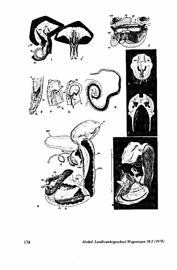
174 Meded. Landbouwhogeschool Wageningen 78-5 (1978)
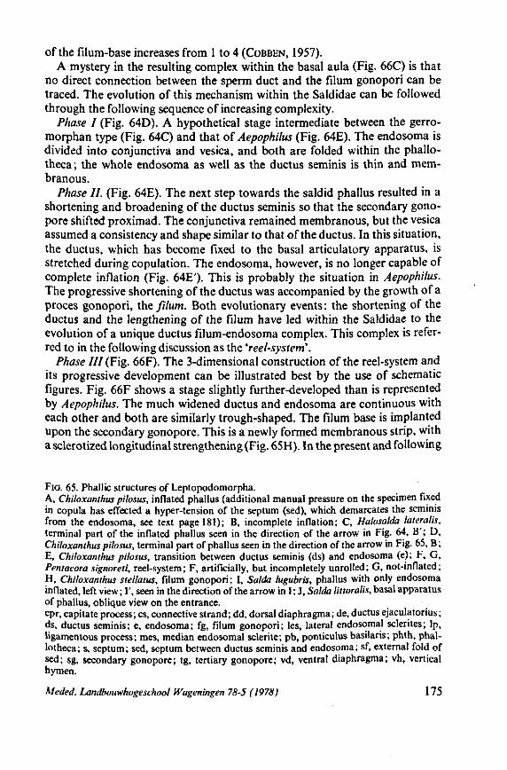
of the filum-base increases from 1 to 4 (COBBEN, 1957). A mystery in the resulting complex within the basal aula (Fig. 66C) is that
no direct connection between the sperm duct and the filum gonopori can be traced. The evolution of this mechanism within the Saldidae can be followed through the following sequence of increasing complexity.
Phase I (Fig. 64D). A hypothetical stage intermediate between the gerro-morphan type (Fig. (AC) and that of Aepophilus (Fig. 64E). The endosoma is divided into conjunctiva and vesica, and both are folded within the phallo-theca; the whole endosoma as well as the ductus seminis is thin and membranous.
Phase II. (Fig. 64E). The next step towards the saldid phallus resulted in a shortening and broadening of the ductus seminis so that the secondary gono-pore shifted proximad. The conjunctiva remained membranous, but the vesica assumed a consistency and shape similar to that of the ductus. In this situation, the ductus, which has become fixed to the basal articulatory apparatus, is stretched during copulation. The endosoma, however, is no longer capable of complete inflation (Fig. 64E'). This is probably the situation in Aepophilus. The progressive shortening of the ductus was accompanied by the growth of a proces gonopori, the filum. Both evolutionary events: the shortening of the ductus and the lengthening of the filum have led within the Saldidae to the evolution of a unique ductus filum-endosoma complex. This complex is referred to in the following discussion as the 'reel-system'.
Phase HI (Fig. 66F). The 3-dimensional construction of the reel-system and its progressive development can be illustrated best by the use of schematic figures. Fig. 66F shows a stage slightly further-developed than is represented by Aepophilus. The much widened ductus and endosoma are continuous with each other and both are similarly trough-shaped. The filum base is implanted upon the secondary gonopore. This is a newly formed membranous strip, with a sclerotized longitudinal strengthening (Fig. 65H). In the present and following
FIG. 65. Phallic structures of Leptopodomorpha. A, Chiloxanthus pilosus, inflated phallus (additional manual pressure on the specimen fixed in copula has effected a hyper-tension of the septum (sed), which demarcates the seminis from the endosoma, see text page 181); B, incomplete inflation; C, Halosalda lateralis, terminal part of the inflated phallus seen in the direction of the arrow in Fig. 64, B'; D, Chiloxanthus pilosus, terminal part of phallus seen in the direction of the arrow in Fig. 65, B; E, Chiloxanthus pilosus, transition between ductus seminis (ds) and endosoma (e); F, G, Pentacora signoreti, reel-system; F, artificially, but incompletely unrolled; G, not-inflated; H, Chiloxanthus stellatus, filum gonopori; I, Salda lugubris, phallus with only endosoma inflated, left view; I', seen in the direction of the arrow in I; J, Salda littoralis, basal apparatus of phallus, oblique view on the entrance. cpr, capitate process; cs, connective strand; dd, dorsal diaphragma; de, ductus ejaculatorius; ds, ductus seminis; e, endosoma; fg, filum gonopori; les, lateral endosomal sclerites; lp, ligamentous process; mes, median endosomal sclerite; pb, ponticulus basilaris; phth, phal-lotheca; s, septum; sed, septum between ductus seminis and endosoma; sf, external fold of sed; sg, secondary gonopore; tg, tertiary gonopore; vd, ventral diaphragma; vh, vertical hymen.
Meded. Landbouwhogeschool Wageningen 78-5 (1978) 175
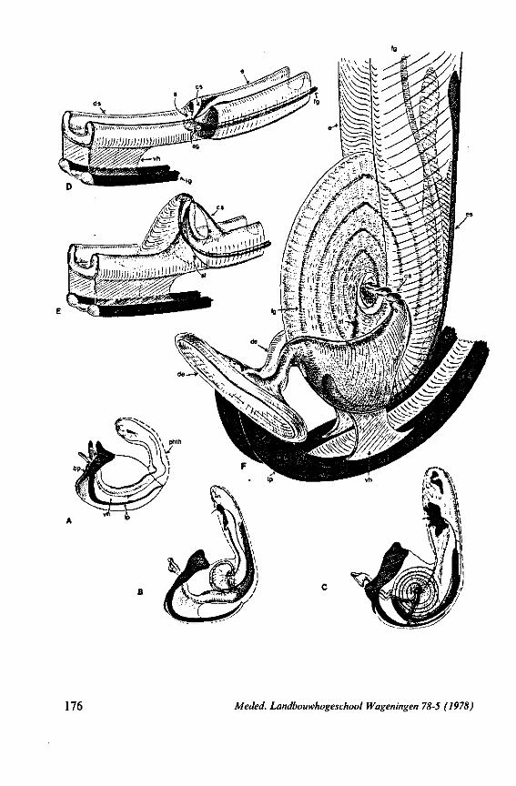
176 Meded. Landbouwhogeschool Wageningen 78-5 (1978)
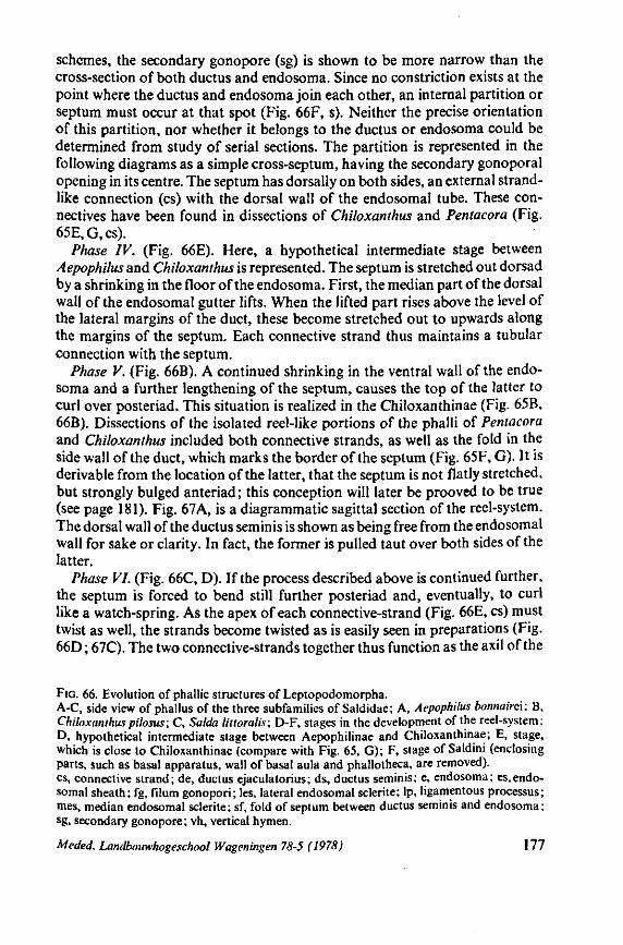
schemes, the secondary gonopore (sg) is shown to be more narrow than the cross-section of both ductus and endosoma. Since no constriction exists at the point where the ductus and endosoma join each other, an internal partition or septum must occur at that spot (Fig. 66F, s). Neither the precise orientation of this partition, nor whether it belongs to the ductus or endosoma could be determined from study of serial sections. The partition is represented in the following diagrams as a simple cross-septum, having the secondary gonoporal opening in its centre. The septum has dorsally on both sides, an external strandlike connection (cs) with the dorsal wall of the endosomal tube. These connectives have been found in dissections of Chiloxanthus and Pentacora (Fig. 65E,G,cs).
Phase IV. (Fig. 66E). Here, a hypothetical intermediate stage between Aepophilus and Chiloxanthus is represented. The septum is stretched out dorsad by a shrinking in the floor of the endosoma. First, the median part of the dorsal wall of the endosomal gutter lifts. When the lifted part rises above the level of the lateral margins of the duct, these become stretched out to upwards along the margins of the septum. Each connective strand thus maintains a tubular connection with the septum.
Phase V. (Fig. 66B). A continued shrinking in the ventral wall of the endosoma and a further lengthening of the septum, causes the top of the latter to curl over posteriad. This situation is realized in the Chiloxanthinae (Fig. 65B, 66B). Dissections of the isolated reel-like portions of the phalli of Pentacora and Chiloxanthus included both connective strands, as well as the fold in the side wall of the duct, which marks the border of the septum (Fig. 65F, G). It is derivable from the location of the latter, that the septum is not flatly stretched, but strongly bulged anteriad ; this conception will later be prooved to be true (see page 181). Fig. 67A, is a diagrammatic sagittal section of the reel-system. The dorsal wall of the ductus seminis is shown as being free from the endosomal wall for sake or clarity. In fact, the former is pulled taut over both sides of the latter.
Phase VI. (Fig. 66C, D). If the process described above is continued further, the septum is forced to bend still further posteriad and, eventually, to curl like a watch-spring. As the apex of each connective-strand (Fig. 66E, cs) must twist as well, the strands become twisted as is easily seen in preparations (Fig. 66D ; 67C). The two connective-strands together thus function as the axil of the
FIG. 66. Evolution of phallic structures of Leptopodomorpha. A-C, side view of phallus of the three subfamilies of Saldidae; A, Aepophilus bonnairei: B, Chiloxanthus pilosus; C, Salda littoralis; D-F, stages in the development of the reel-system; D, hypothetical intermediate stage between Aepophilinae and Chiloxanthinae; E, stage, which is close to Chiloxanthinae (compare with Fig. 65, G); F, stage of Saldini (enclosing parts, such as basal apparatus, wall of basal aula and phallotheca, are removed), cs, connective strand; de, ductus ejaculatorius; ds, ductus seminis; e, endosoma; es,endosomal sheath; fg, filum gonopori; les, lateral endosomal sclerite; lp, ligamentous processus; mes, median endosomal sclerite; sf, fold of septum between ductus seminis and endosoma; sg, secondary gonopore; vh, vertical hymen.
Meded. Landbouwhogeschool Wageningen 78-5 (1978) 177
reel-system. As the coiling occurs, the septum, the endosoma, the filum and the dorsal wall of the ductus become coiled too. Finally we get a complicated structure, which reaches its extreme development in the genus Salda (diagrammatic sagittal section in Fig. 67B). The reel (Fig. 66C) is comparable to a wheel, the sides of which are tightened by a membrane, namely the stretched roof of the ductus seminis. This membrane has been included in all successive windings about the connective strands. In Salda, the membrane is coiled within itself four times, giving it a lamellated appearance (Fig. 66D; note the lamelles within the 'eye' of the reel). Even so, the fold which marks the lateral margin of the septum in Chiloxanthus and Pentacora (Fig. 65G; 66E, sf) is still recognizable in species of the genus Salda (Fig. 66D ; 67C, sf).
The successive phase, outlined above, represent a rectilinear, evolutionary progression, which is revealed, with all intermediary types, in all recent Sal-didae. Each species, moreover, must, ontogenetically, pass through all preceding phases. The connective-strands, which become twisted into cables, furnish proof of this.
It follows from the previous account, that the greatest part of the exteriror suface of the reel is formed by the dorsal wall of the ductus seminis. The ductus shortens as a result of the coiling-process, and the reel comes to lie close to the base of the articulatory apparatus. The vertical hymena, which connect the ductus with the ligamentous process, thus come to extend relatively more posteriad (Fig. 66B-D, vh). In species in which the hymena come to lie at the level of the connective strands, a connection between the former and the latter is formed (Fig. 66D). When this fusion has become complete, as occurs in Salda a cup-shaped receptacle is formed. This receptacle consists of the connective strands and of the ventral and lateral walls of the endosoma and ductus. It rests on the ligamentous process on three places, proximally on the greatly shortened ductus, which is firmly attached to the base of the ligamentous process, and ventrally on the two vertical hymena. Thus a remarkable base of support has evolved to deal with the forces, which accompany the unwinding of the reel-system.
Unwinding of the reel-system The phallus has no intrinsic muscles. As indicated above, it has been assum
ed that the roof of the ductus seminis is spread tightly around the reel-structure. Thus, it may be imagined that, in sagittal section (Fig. 67A, B), this external dorsal membrane is contiguous with the underlying wall. This would imply that the sperm-fluid strikes upon a seemingly closed cross-partition and that the fluid is directed into the corner indicated in Fig. 67 with an asterisk. As the ontogenetic development of the reel-mechanism was related to a shrinking in the ventral wall of the sperm-conduit, positive pressure in the sperm fluid causes the reel to unwind. The septum and with it, the whole reel-complex, is rotated in a direction opposite to that which it had when the complex developed. Under continuous pressure and assisted by the twisted connective-strands, the reel can unroll completely, although I never succeeded in unrolling the reel-
178 Meded. Landbouwhogeschool Wageningen 78-5 (1978)
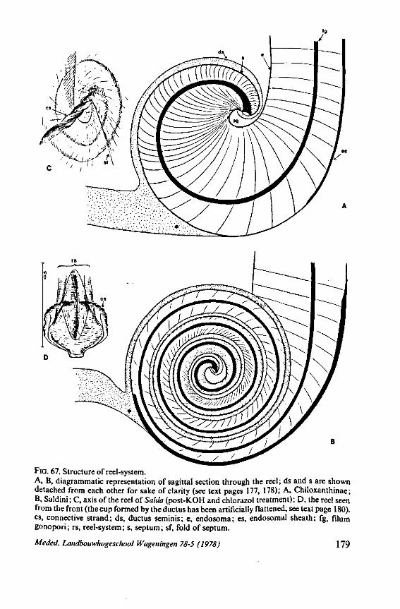
FIG. 67. Structure of reel-system. A, B, diagrammatic representation of sagittal section through the reel; ds and s are shown detached from each other for sake of clarity (see text pages 177, 178); A, Chiloxanthinae; B, Saldini; C, axis of the reel of Salda (post-KOH and chlorazol treatment); D, the reel seen from the front (the cup formed by the ductus has been artificially flattened, see text page 180). cs, connective strand; ds, ductus seminis; e, endosoma; es, endosomal sheath; fg, filum gonopori; rs, reel-system; s, septum; sf, fold of septum.
Meded. Landbouwhogeschool Wageningen 78-5 (1978) 179
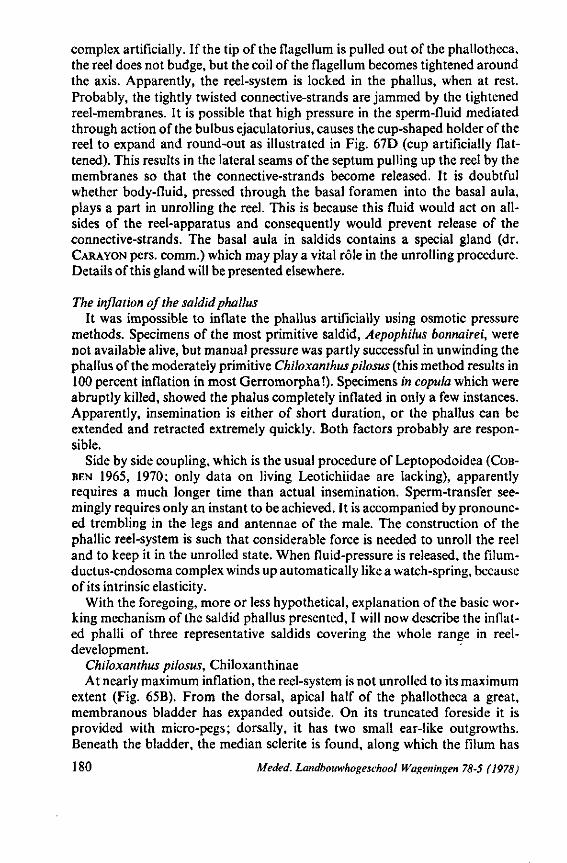
complex artificially. If the tip of the flagellum is pulled out of the phallotheca, the reel does not budge, but the coil of the flagellum becomes tightened around the axis. Apparently, the reel-system is locked in the phallus, when at rest. Probably, the tightly twisted connective-strands are jammed by the tightened reel-membranes. It is possible that high pressure in the sperm-fluid mediated through action of the bulbus ejaculatorius, causes the cup-shaped holder of the reel to expand and round-out as illustrated in Fig. 67D (cup artificially flattened). This results in the lateral seams of the septum pulling up the reel by the membranes so that the connective-strands become released. It is doubtful whether body-fluid, pressed through the basal foramen into the basal aula, plays a part in unrolling the reel. This is because this fluid would act on all-sides of the reel-apparatus and consequently would prevent release of the connective-strands. The basal aula in saldids contains a special gland (dr. CARAYON pers. comm.) which may play a vital rôle in the unrolling procedure. Details of this gland will be presented elsewhere.
The inflation of the saldid phallus It was impossible to inflate the phallus artificially using osmotic pressure
methods. Specimens of the most primitive saldid, Aepophilus bonnairei, were not available alive, but manual pressure was partly successful in unwinding the phallus of the moderately primitive Chiloxanthus pilosus (this method results in 100 percent inflation in most Gerromorpha !). Specimens in copula which were abruptly killed, showed the phalus completely inflated in only a few instances. Apparently, insemination is either of short duration, or the phallus can be extended and retracted extremely quickly. Both factors probably are responsible.
Side by side coupling, which is the usual procedure of Leptopodoidea (COB-BEN 1965, 1970; only data on living Leotichiidae are lacking), apparently requires a much longer time than actual insemination. Sperm-transfer seemingly requires only an instant to be achieved. It is accompanied by pronounced trembling in the legs and antennae of the male. The construction of the phallic reel-system is such that considerable force is needed to unroll the reel and to keep it in the unrolled state. When fluid-pressure is released, the filum-ductus-endosoma complex winds up automatically like a watch-spring, because of its intrinsic elasticity.
With the foregoing, more or less hypothetical, explanation of the basic working mechanism of the saldid phallus presented, I will now describe the inflated phalli of three representative saldids covering the whole range in reel-development.
Chiloxanthus pilosus, Chiloxanthinae At nearly maximum inflation, the reel-system is not unrolled to its maximum
extent (Fig. 65B). From the dorsal, apical half of the phallotheca a great, membranous bladder has expanded outside. On its truncated foreside it is provided with micro-pegs; dorsally, it has two small ear-like outgrowths. Beneath the bladder, the median sclerite is found, along which the filum has
180 Meded. Landbouwhogeschool Wageningen 78-5 (1978)
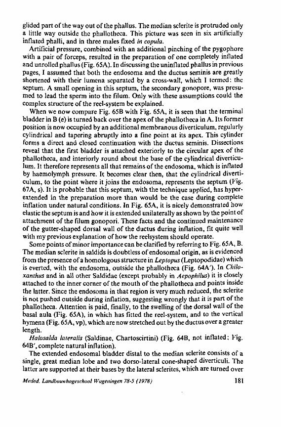
glided part of the way out of the phallus. The median sclerite is protruded only a little way outside the phallotheca. This picture was seen in six artificially inflated phalli, and in three males fixed in copula.
Artificial pressure, combined with an additional pinching of the pygophore with a pair of forceps, resulted in the preparation of one completely inflated and unrolled phallus (Fig. 65A). In discussing the uninflated phallus in previous pages, I assumed that both the endosoma and the ductus seminis are greatly shortened with their lumena separated by a cross-wall, which I termed: the septum. A small opening in this septum, the secondary gonopore, was presumed to lead the sperm into the filum. Only with these assumptions could the complex structure of the reel-system be explained.
When we now compare Fig. 65B with Fig. 65A, it is seen that the terminal bladder in B (e) is turned back over the apex of the phallotheca in A. Its former position is now occupied by an additional membranous diverticulum, regularly cylindrical and tapering abruptly into a fine point at its apex. This cylinder forms a direct and closed continuation with the ductus seminis. Dissections reveal that the first bladder is attached exteriorly to the circular apex of the phallotheca, and interiorly round about the base of the cylindrical diverticulum. It therefore represents all that remains of the endosoma, which is inflated by haemolymph pressure. It becomes clear then, that the cylindrical diverticulum, to the point where it joins the endosoma, represents the septum (Fig. 67A, s). It is probable that this septum, with the technique applied, has hyper-extended in the preparation more than would be the case during complete inflation under natural conditions. In Fig. 65A, it is nicely demonstrated how elastic the septum is and how it is extended unilaterally as shown by the point of attachment of the filum gonopori. These facts and the continued maintenance of the gutter-shaped dorsal wall of the ductus during inflation, fit quite well with my previous explanation of how the reelsystem should operate.
Some points of minor importance can be clarified by referring to Fig. 65A, B. The median sclerite in saldids is doubtless of endosomal origin, as is evidenced from the presence of a homologous structure in Leptopus (Leptopodidae) which is everted, with the endosoma, outside the phallotheca (Fig. 64A')- In Chilo-xanthus and in all other Saldidae (except probably in Aepophilus) it is closely attached to the inner corner of the mouth of the phallotheca and points inside the latter. Since the endosoma in that region is very much reduced, the sclerite is not pushed outside during inflation, suggesting wrongly that it is part of the phallotheca. Attention is paid, finally, to the swelling of the dorsal wall of the basal aula (Fig. 65A), in which has fitted the reel-system, and to the vertical hymena (Fig. 65A, vp), which are now stretched out by the ductus over a greater length.
Halosalda lateralis (Saldinae, Chartoscirtini) (Fig. 64B, not inflated; Fig. 64B', complete natural inflation).
The extended endosomal bladder distal to the median sclerite consists of a single, great median lobe and two dorso-lateral cone-shaped diverticuli. The latter are supported at their bases by the lateral sclerites, which are turned over
Meded. Landbouwhogeschool Wageningen 78-5 (1978) 181
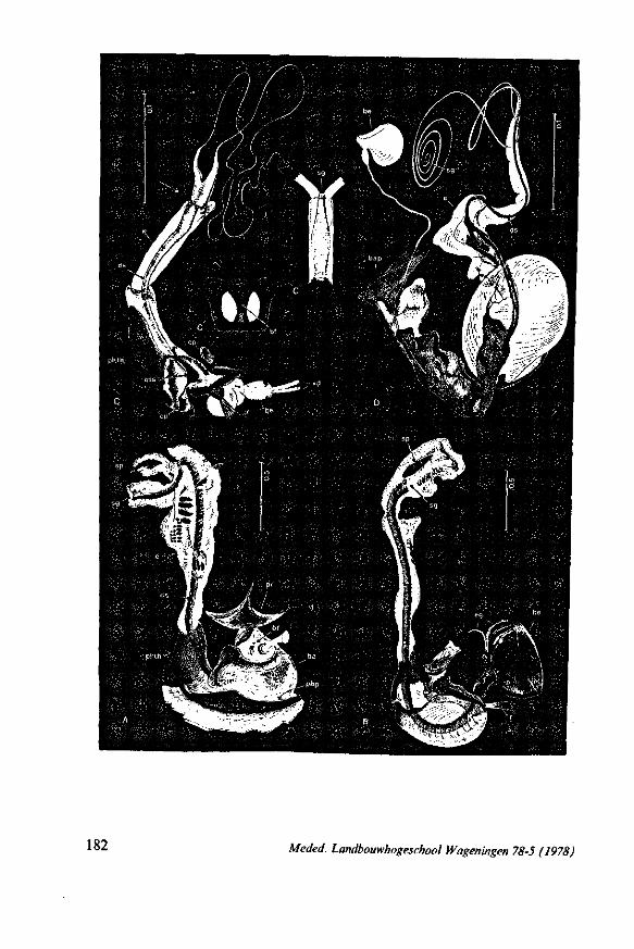
182 Meded. Landbouwhogeschool Wageningen 78-5 (1978)
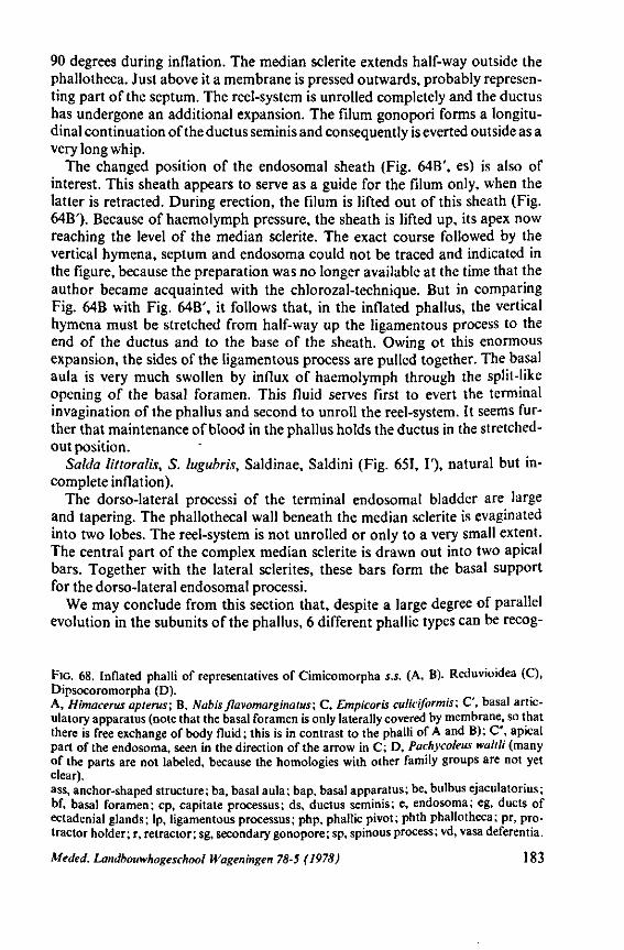
90 degrees during inflation. The median sclerite extends half-way outside the phallotheca. Just above it a membrane is pressed outwards, probably representing part of the septum. The reel-system is unrolled completely and the ductus has undergone an additional expansion. The filum gonopori forms a longitudinal continuation of the ductus seminis and consequently is everted outside as a very long whip.
The changed position of the endosomal sheath (Fig. 64B', es) is also of interest. This sheath appears to serve as a guide for the filum only, when the latter is retracted. During erection, the filum is lifted out of this sheath (Fig. 64B'). Because of haemolymph pressure, the sheath is lifted up, its apex now reaching the level of the median sclerite. The exact course followed by the vertical hymena, septum and endosoma could not be traced and indicated in the figure, because the preparation was no longer available at the time that the author became acquainted with the chlorozal-technique. But in comparing Fig. 64B with Fig. 64B', it follows that, in the inflated phallus, the vertical hymena must be stretched from half-way up the ligamentous process to the end of the ductus and to the base of the sheath. Owing ot this enormous expansion, the sides of the ligamentous process are pulled together. The basal aula is very much swollen by influx of haemolymph through the split-like opening of the basal foramen. This fluid serves first to evert the terminal invagination of the phallus and second to unroll the reel-system. It seems further that maintenance of blood in the phallus holds the ductus in the stretched-out position.
Salda littoralis, S. lugubris, Saldinae, Saldini (Fig. 651, T), natural but incomplete inflation).
The dorso-lateral processi of the terminal endosomal bladder are large and tapering. The phallothecal wall beneath the median sclerite is evaginated into two lobes. The reel-system is not unrolled or only to a very small extent. The central part of the complex median sclerite is drawn out into two apical bars. Together with the lateral sclerites, these bars form the basal support for the dorso-lateral endosomal processi.
We may conclude from this section that, despite a large degree of parallel evolution in the subunits of the phallus, 6 different phallic types can be recog-
FIG. 68. Inflated phalli of representatives of Cimicomorpha s.s. (A, B). Reduvioidea (C), Dipsocoromorpha (D). A, Himacerus apterus; B, Nabis flavomarginatus; C, Empkoris culiciformis; C', basal artic-ulatory apparatus (note that the basal foramen is only laterally covered by membrane, so that there is free exchange of body fluid; this is in contrast to the phalli of A and B); C", apical part of the endosoma, seen in the direction of the arrow in C; D, Pachycoleus waitli (many of the parts are not labeled, because the homologies with other family groups are not yet clear). ass, anchor-shaped structure; ba, basal aula; bap, basal apparatus; be, bulbus ejaculatorius; bf, basal foramen; cp, capitate processus; ds, ductus seminis; e, endosoma; eg, ducts of ectadenial glands; lp, ligamentous processus; php, phallic pivot; phth phallotheca; pr, protractor holder; r, retractor; sg, secondary gonopore; sp, spinous process; vd, vasa deferentia.
Meded. Landbouwhogeschool Wageningen 78-5 (1978) 183
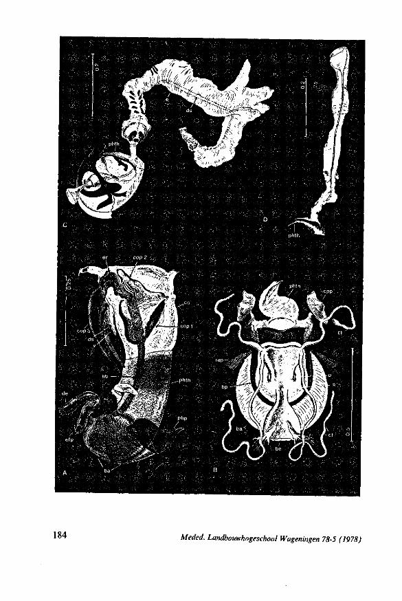
kiadU
184 Meded. Landbouwhogeschool Wapeningen 78-5 (1978)
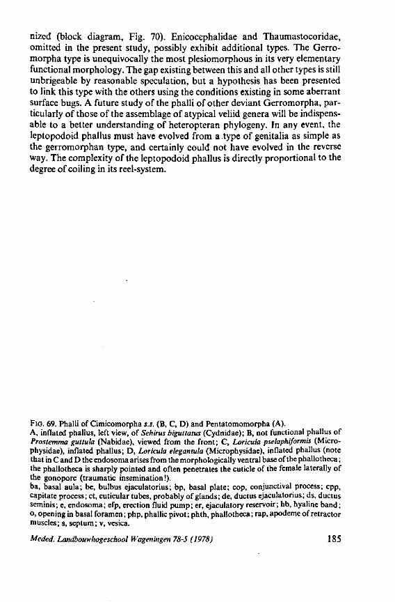
nized (block diagram, Fig. 70). Enicocephalidae and Thaumastocoridae, omitted in the present study, possibly exhibit additional types. The Gerro-morpha type is unequivocally the most plesiomorphous in its very elementary functional morphology. The gap existing between this and all other types is still unbrigeable by reasonable speculation, but a hypothesis has been presented to link this type with the others using the conditions existing in some aberrant surface bugs. A future study of the phalli of other deviant Gerromorpha, particularly of those of the assemblage of atypical veliid genera will be indispensable to a better understanding of heteropteran phylogeny. In any event, the leptopodoid phallus must have evolved from a type of genitalia as simple as the gerromorphan type, and certainly could not have evolved in the reverse way. The complexity of the leptopodoid phallus is directly proportional to the degree of coiling in its reel-system.
FIG. 69. Phalli of Cimicomorpha s.s. (B, C, D) and Pentatomomorpha (A). A, inflated phallus, left view, of Sehirus biguttatus (Cydnidae); B, not functional phallus of Prostemma gut tula (Nabidae), viewed from the front; C, Loricula pselaphiformis (Micro-physidae), inflated phallus; D, Loricula elegantula (Microphysidae), inflated phallus (note that in C and D the endosoma arises from the morphologically ventral base of the phallotheca ; the phallotheca is sharply pointed and often penetrates the cuticle of the female laterally of the gonopore (traumatic insemination!). ba, basal aula; be, bulbus ejaculatorius; bp, basal plate; cop, conjunctival process; cpp, capitate process; ct, cuticular tubes, probably of glands; de, ductus ejaculatorius; ds, ductus seminis; e, endosoma; efp, erection fluid pump; er, ejaculatory reservoir; hb, hyaline band; o, opening in basal foramen ; php, phallic pivot ; phth, phallotheca ; rap, apodeme of retractor muscles; s, septum; v, vesica.
Meded. Landbouwhogeschool Wageningen 78-5 (1978) 185
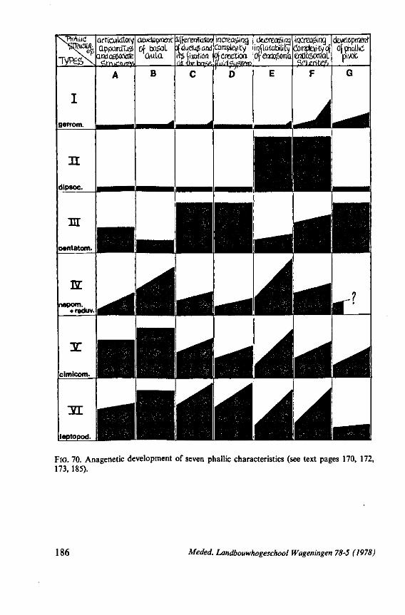
arfïcutôîory apparaTu?
andaèjotae
devetoprrKrct lifferenfatoo Of bagal tfductujand
Quia its (ixafion. fit, tiv hny.
increapirx) Complexity of erection
uirifyfiffrn
outKapm ircflatatxlity Of axlo£oroa
irXnza inç) Complexity of OTloSorcal
development Ofchdlid
piVOt
B
FIG . 70. Anagenetic development of seven phallic characteristics (see text pages 170, 172, 173, 185).
186 Meded. Landbouwhogeschool Wageningen 78-5 (1978)
4. DISCUSSION
This discussion will be rather brief, since the characters reviewed in section 3 were there evaluated and analyzed in terms of their ancestral or derived states. Furthermore, feeding structures do not vary within the 'morpha' groups to the extent that egg structures do.
4.1. PHYLOGENETIC PROCEDURE
Throughout this study, I have compared higher rank categories (the treatment of Reduviidae, separate from Cimicomorpha will be discussed on pages 230-234). The fundamental question is whether these - 'morpha' taxa can be arranged in a cladistic scheme based on the characters evaluated. My answer for the present is no! The results of my 1968 study and of the present one suggest that the higher rank taxa of Heteroptera (comprising some 70 families and 30-40 thousand species) can be arranged according to levels of increasingly complex organization. But, points in the phylogeny where one major group split off from an other, must remain obscure because of insufficient data.
My study of egg-systems resulted in the development of an evolutionary diagram (1968, Fig. 316), which is reproduced here as Fig. 71. The thoughts underlying this schema were as follows : The three divisions of Heteroptera, formerly termed Amphibicorisae, Hydrocorisae and Geocorisae, and which were considered, in the past, as more or less equivalent groups were rearranged in a new, tentative, evolutionary model. The Amphibicorisae, now termed Gerromorpha (see p. 6), are believed to represent the basic stock. The black sector (A) represents the extinct, proto- Gerromorpha, and the stippled area the modern Gerromorpha. The Geocorisae should be abandoned as a taxon, since it is polyplyletic. Its constituents are provisionally depicted as eight radiating lines, each arising independently from sector A. The Nepomorpha (= Hydrocorisae) forms another line in close association with the Reduvioidea. All proximal, black portions of the individual lineages are hypothetical, and represent extinct representatives of their respective family groups and, possibly, some recent, aberrant taxa from tropical regions (living specimens of such relict species were not always available for study). The extant families are distributed in the open (lined or stippled) portions of each lineage. Each diverging line represents a superfamily or group of superfamilies. There is a progressive development (anagenesis) of character states from bottom to top within each line. The height of each diverging line is a rough estimate of its extent of anagenetic progression. The width of each very roughly suggests the number of species contained.
In 1968 (p. 361), I stressed the temporary nature of the scheme discussed here. For that reason. I did not label the particular groups which I had in
Meded. Landbouwhogeschool Wageningen 68-5 (1978) 187
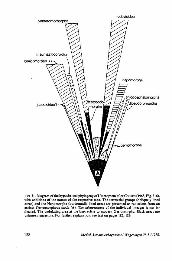
reduvioidea pentatomomorpha
thaumastocoroidea
cimicomorpha s.s.^
joppeicidae?
nepomorpha
enicocephalomorpha
dipsocoromorpha
*-H^~t~fz=* gerromorpha
FIG. 71. Diagram of the hypothetical phylogeny of Heteroptera after COBBEN ( 1968, Fig. 316), with additions of the names of the respective taxa. The terrestrial groups (obliquely lined areas) and the Nepomorpha (horizontally lined area) are presented as radiations from an extinct Gerromorphous stock (A). The arborescence of the individual lineages is not indicated. The undulating area at the base refers to modern Gerromorpha. Black areas are unknown ancestors. For further explanation, see text on pages 187, 189.
188 Meded. Landbouwhogeschool Wageningen 78-5 (1978)
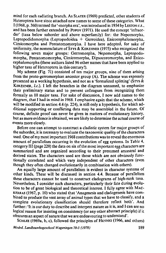
mind for each radiating branch. As SLATER (1969) predicted, other students of Heteroptera have since attached new names to some of these categories. What I (1968, p. 360) termed the '-morpha era', was introduced in 1954 by LESTON C.S., and has been further extended by POPOV (1971). He used the concept 'infraor-der' (taxa below suborder and above superfamily) for: the Nepomorpha, Leptopodidomorpha (Leptopodoidea + Gerroidea), Enicocephalomorpha, Cimicomorpha and Pentatomomorpha. I have here adopted, for sake of uniformity, the nomenclature ofSTYS & KERZHNER (1975) who recognized the following seven major groups: Gerromorpha, Nepomorpha, Leptopodo-morpha, Pentatomomorpha, Cimicomorpha, Dipsocoromorpha, and Enicocephalomorpha (these authors listed 96 other names that have been applied to higher taxa of Heteroptera in this century !).
My scheme (Fig. 71) consisted of ten major groups, nine of them arising from the proto-gerromorphan ancestor group (A). The scheme was expressly presented as a working hypothesis, and not as a 'formalized splitting' (STYS & KERZHNER, I.e.). I left the branches in the diagram unnamed, to emphasize their preliminary status and to prevent colleagues from recognizing them formally as 10 major taxa. For sake of discussion, I now add names to the diagram, that I had in mind in 1968.1 emphasize again that the scheme, which will be modified in section 4.4 (p. 224), is still only a hypothesis, for which additional supporting or conflicting data may be assembled in the future. Of course, definite proof can never be given in matters of evolutionary history, but as more evidence is obtained, we are likely to determine the actual course of events more closely.
Before one can attempt to construct a cladistic system for major groups of the suborder, it is necessary to evaluate the taxonomie quality of the characters used. One of my most important 1968 contributions was to reveal the enormous amount of parallelism occurring in the evolution of egg systems. In Table 4, category III (page 220) the data on six of the most important egg characters are summarized and are organized according to their presumed ancestral and derived states. The characters used are those which are not obviously functionally correlated and which vary independent of other characters (even though they often changed evolutionarily in combination with others).
An equally large amount of parallelism is evident in character systems of other kinds. These will be discussed in section 4.4. Because of parallelism these characters cannot be used to construct cladograms of high-rank taxa. Nevertheless, I consider such characters, particularly their fate during evolution to be of great biological and theoretical interest. I fully agree with MAC-KERRAS (1967, p. 10) who stated that 'Anagenesis and cladogenesis have combined to produce the vast array of animal types that we have to classify, and a complete evolutionary classification should therefore reflect both'. And further: 'It is our duty to describe and interpret nature as it is, and I can see no logical reason for insisting on consistency (or any other abstract principle) if it obscures an aspect of nature that we are endeavouring to understand'.
SCHLEE (1969a, b, c), followed the principles of HENNIG (1966, and others)
Meded. Landbouwhogeschool Wageningen 78-5 (1978) 189
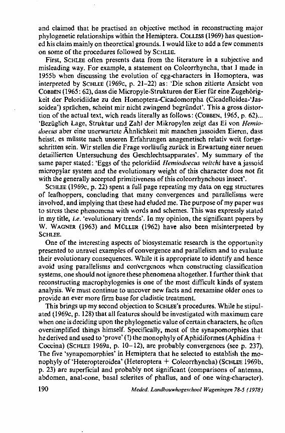
and claimed that he practised an objective method in reconstructing major phylogenetic relationships within the Hemiptera. COLLESS (1969) has questioned his claim mainly on theoretical grounds. I would like to add a few comments on some of the procedures followed by SCHLEE.
First, SCHLEE often presents data from the literature in a subjective and misleading way. For example, a statement on Coleorrhyncha, that I made in 1955b when discussing the evolution of egg-characters in Homoptera, was interpreted by SCHLEE (1969c, p. 21-22) as: 'Die schon zitierte Ansicht von COBBEN (1965: 62), dass die Micropyle-Strukturen der Eier für eine Zugehörigkeit der Peloridiidae zu den Homoptera-Cicadomorpha (Cicadelloidea-'Jas-soidea') sprächen, scheint mir nicht zwingend begründet'. This a gross distortion of the actual text, wich reads literally as follows: (COBBEN, 1965, p. 62)... 'Bezüglich Lage, Struktur und Zahl der Mikropylen zeigt das Ei von Hetnio-doecits aber eine unerwartete Ähnlichkeit mit manchen jassoiden Eieren, dass heisst, es müsste nach unseren Erfahrungen anagenetisch relativ weit fortgeschritten sein. Wir stellen die Frage vorläufig zurück in Erwartung einer neuen detaillierten Untersuchung des Geschlechtsapparates'. My summary of the same paper stated: 'Eggs of the peloridiid Hemiodoecus veitchi have a jassoid micropylar system and the evolutionary weight of this character does not fit with the generally accepted primitiveness of this coleorrhynchous insect'.
SCHLEE (1969c, p. 22) spent a full page repeating my data on egg structures of leafhoppers, concluding that many convergences and parallelisms were involved, and implying that these had eluded me. The purpose of my paper was to stress these phenomena with words and schemes. This was expressly stated in my title, i.e. 'evolutionary trends'. In my opinion, the significant papers by W. WAGNER (1963) and MÜLLER (1962) have also been misinterpreted by SCHLEE.
One of the interesting aspects of biosystematic research is the opportunity presented to unravel examples of convergence and parallelism and to evaluate their evolutionary consequences. While it is appropriate to identify and hence avoid using parallelisms and convergences when constructing classification systems, one should not ignore these phenomena altogether. I further think that reconstructing macrophylogenies is one of the most difficult kinds of system analysis. We must continue to uncover new facts and reexamine older ones to provide an ever more firm base for cladistic treatment.
This brings up my second objection to SCHLEE'S procedures. While he stipulated (1969c, p. 128) that all features should be investigated with maximum care when one is deciding upon the phylogenetic value of certain characters, he often oversimplified things himself. Specifically, most of the synapomorphies that he derived and used to 'prove' ( !) the monophyly of Aphidiformes (Aphidina + Coccina) (SCHLEE 1969a, p. 10-12), are probably convergences (see p. 237). The five 'synapomorphies' in Hemiptera that he selected to establish the monophyly of'Heteropteroidea' (Heteroptera + Coleorrhyncha) (SCHLEE 1969b, p. 23) are superficial and probably not significant (comparisons of antenna, abdomen, anal-cone, basal sclerites of phallus, and of one wing-character).
190 Meded. Landbouwhogeschool Wageningen 78-5 (1978)
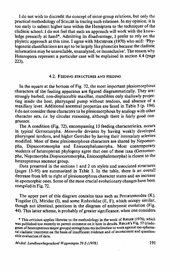
I do not wish to discredit the concept of sister-group relations, but only the practical methodology of SCHLEE in tracing such relations. In my opinion, it is too early to submit higher taxa within the Hemiptera to the techniques of the cladistic school. I do not feel that such an approach will work with the knowledge presently at hand*. Admitting its disadvantage, I prefer to rely on the phyletic approach at this time. I agree with MICHENER (1970) who said: 'Phy-logenetic classifications are apt to be largely like phenetics because the cladistic information may be unavailable, unanalyzed, or inconclusive'. The reason why Heteroptera represent a particular case will be explained in section 4.4 (page 223).
4.2. FEEDING STRUCTURES AND FEEDING
In the square at the bottom of Fig. 72, the most important plesiomorphous characters of the feeding apparatus are figured diagrammatically. They are: strongly barbed, non-displaceable maxillae, mandibles only shallowly projecting inside the host, pharyngeal pump without tendons, and absence of a maxillary lever. Additional ancestral properties are listed in Table 3 (p. 194). I do not consider these characters to be plesiomorphous by analogy with other character sets, i.e. by circular reasoning, although there is fairly good congruence.
The A condition (Fig. 72), encompassing 13 feeding characteristics, occurs in typical Gerromorpha. Mesovelia deviates by having weakly developed pharyngeal tendons, and higher Gerridae by having their intercalary sclerites modified. Most of these plesiomorphous characters are shared by Nepomor-pha, Dipsocoromorpha and Enicocephalomorpha. Most contemporary students of heteropteran phylogeny agree that one of these taxa (Gerromorpha, Nepomorpha Dispsocoromorpha, Enicocephalomorpha) is closest to the heteropterous ancestor group.
Data presented in the sections 1 and 2 on stylets and associated structures (pages 13-95) are summarized in Table 3. In the table, there is an overall decrease from left to right of plesiomorphous character states and an increase in apomorphic ones. Some of the most crucial evolutionary changes have been compiled in Fig. 72.
The upper part of this diagram contains taxa such as Pentatomoidea (K), Tingidae (J), Miridae (I), and some Reduviidae (E, F), which occupy similar, though not identical, positions in the diagram of embryonic evolution (Fig. 44). This latter scheme, is probably of greater significance, when one considers
* This criticism applies likewise to the methodology in the work of RIEGER (1976), which was published too recently to permit comment on it here in details. RIEGER'S Fig. 57 (clado-gram of heteropterous major groups) strengthens my inclination to warn against too ephemeral cladistic treatment on the basis of insufficient evidence and of inconsistent and questionable evaluation of data.
Meded. Landbouwhogeschool Wageningen 78-5 (1978) 191
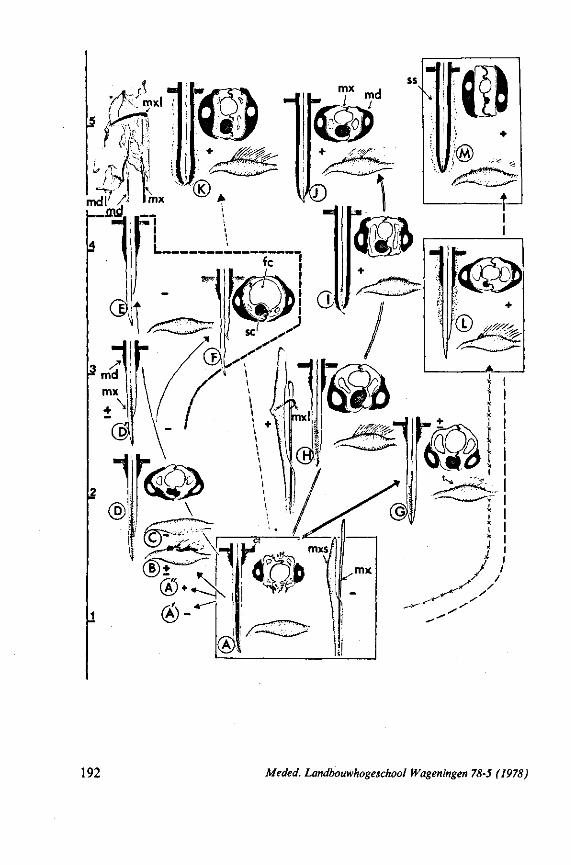
192 Meded. Landbouwhogeschool Wageningen 78-5 (1978)
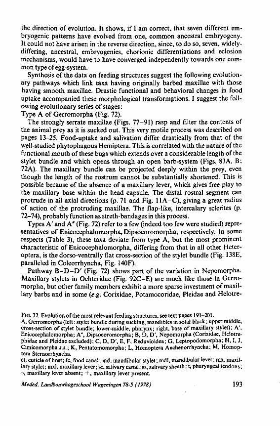
the direction of evolution. It shows, if I am correct, that seven different embryogénie patterns have evolved from one, common ancestral embryogeny. It could not have arisen in the reverse direction, since, to do so, seven, widely-differing, ancestral, embryogénies, chorionic differentiations and eclosion mechanisms, would have to have converged independently towards one common type of egg-system.
Synthesis of the data on feeding structures suggest the following evolutionary pathways which link taxa having originally barbed maxillae with those having smooth maxillae. Drastic functional and behavioral changes in food uptake accompanied these morphological transformations. I suggest the following evolutionary series of stages : Type A of Gerromorpha (Fig. 72).
The strongly serrate maxillae (Figs. 77-91) rasp and filter the contents of the animal prey as it is sucked out. This very motile process was described on pages 13—25. Food-uptake and salivation differ drastically from that of the well-studied phytophagous Hemiptera. This is correlated with the nature of the functional mouth of these bugs which extends over a considerable length of the stylet bundle and which opens through an open barb-system (Figs. 83A, B: 72A). The maxillary bundle can be projected deeply within the prey, even though the length of the rostrum cannot be substantially shortened. This is possible because of the absence of a maxillary lever, which gives free play to the maxillary base within the head capsule. The distal rostral segment can protrude in all axial directions (p. 71 and Fig. 11A-C), giving a great radius of action of the protruding maxillae. The flap-like, intercalary sclerites (p. 72-74), probably function as streth-bandages in this process.
Types A' and A* (Fig. 72) refer to a few (indeed too few were studied) representatives of Enicocephalomorpha, Dipsocoromorpha, respectively. In some respects (Table 3), these taxa deviate from type A, but the most prominent characteristic of Enicocephalomorpha, differing from that in all other Heter-optera, is the dorso-ventrally flat cross-section of the stylet bundle (Fig. 138E, paralleled in Coleorrhyncha, Fig. 140F).
Pathway B -D-D ' (Fig. 72) shows part of the variation in Nepomorpha. Maxillary stylets in Ochteridae (Fig. 92C-E) are much like those in Gerromorpha, but other family members exhibit a more sparse investment of maxillary barbs and in some (e.g. Corixidae, Potamocoridae, Pleidae and Helotre-
FIG. 72. Evolution of the most relevant feeding structures, see text pages 191-201. A, Gerromorpha (left: stylet bundle during sucking, mandibles in solid black; upper middle, cross-section of stylet bundle; lower-middle, pharynx; right, base of maxillary stylet); A', Enicocephalomorpha; A", Dipsocoromorpha; B, D, D', Nepomorpha (Corixidae, Helotre-phidae and Pleidae excluded); C, D, D', E, F, Reduvioidea; G, Leptopodomorpha; H, I, J, Cimicomorpha s.S.; K, Pentatomomorpha; L, Homoptera Auchenorrhyncha; M, Homop-tera Sternorrhyncha. ct, cuticle of host; fc, food canal; md, mandibular stylet; mdl, mandibular lever; mx, maxillary stylet; mxl, maxillary lever; sc, salivary canal; ss, salivary sheath; t, pharyngeal tendons; - , maxillary lever absent; + , maxillary lever present.
Meded. Landbouwhogeschool Wageningen 78-5 (1978) 193
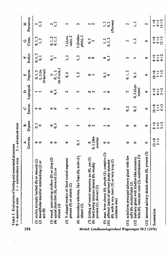
E o
X 5 c «
CU
o e '0
>
e w 8..
2 •o o
Q 2
U.l c u
co 15
o
O
, o o
N . g a
o o)
Is co —
o ca
- =5 o ü c ^
tS E e
CN X
©-• ' g = <->
o . o
o . ( N U , w
IS O 60 E.S •» . *
•o i . I - o o ^
£ ^
*« o CÄ ^ B0O C w
S * O. o , w
• d a c g-a 8 g o. .o G S M
•3 3
<N
O*
.O (N •a ~ <N
S;! o "
E 60
U u> t/5 O 1« •o £ Ci o g
l - s I ° •o £ S s 'S **
a o
a es
en
'H
"M o u
ç
<N
C
u CS
c1 > *
>..* - es
S - * o .-C c S E g B J C
"> c o •s ° c
O ä ^
o E C-60 3 *•*
c °- c -a ? » 2 0 u O u i ;
<N <N S
O . O
o o
•s 2?î Q. ca ^
E S » O o . c
ö u » — O.C w c o o § >
^ t > -w ca o , . Cfl «-• C U <A «J O v
« ü > 3 IH 3 - - o
o -o * z,
M £ * i
E ^ s S
<N
o"
fr~ w </]
•o c «
"ëo
e? > <N
.£•=5 c l
E H
•E = ca o
ca
ca e
'JE Ji E s o i u
u ,_ç "O w ca &)
11 •s.§
60 >
> 0>
« • o t>0 . 2
o. o
ca
o — <N X X X
O — IN X X X *0 ON CN
O — <N X X X oo oo •*
O — <N X X X o r~ c—
O — <N X X X \ 0 t v>
O ~ <N x x x o — t
O — <N
x x x
o — X x en (N
194 Meded. Landbouwhogeschool Wageningen 78-5 (1978)
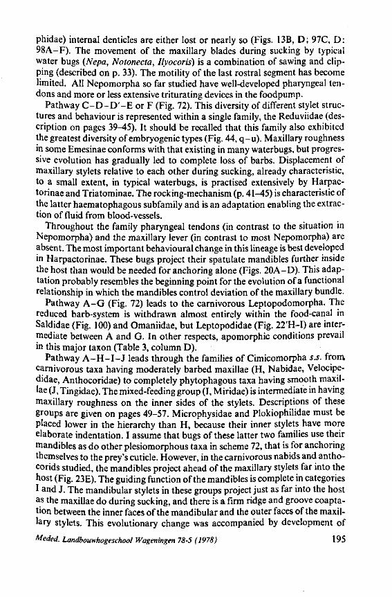
phidae) internal denticles are either lost or nearly so (Figs. 13B, D; 97C, D: 98A-F). The movement of the maxillary blades during sucking by typical water bugs (Nepa, Notonecta, Ilyocoris) is a combination of sawing and clipping (described on p. 33). The motility of the last rostral segment has become limited. All Nepomorpha so far studied have well-developed pharyngeal tendons and more or less extensive triturating devices in the foodpump.
Pathway C -D-D ' -E or F (Fig. 72). This diversity of different stylet structures and behaviour is represented within a single family, the Reduviidae (description on pages 39-45). It should be recalled that this family also exhibited the greatest diversity of embryogénie types (Fig. 44, q-u). Maxillary roughness in some Emesinae conforms with that existing in many waterbugs, but progressive evolution has gradually led to complete loss of barbs. Displacement of maxillary stylets relative to each other during sucking, already characteristic, to a small extent, in typical waterbugs, is practised extensively by Harpac-torinae and Triatominae. The rocking-mechanism (p. 41-45) is characteristic of the latter haematophagous subfamily and is an adaptation enabling the extraction of fluid from blood-vessels.
Throughout the family pharyngeal tendons (in contrast to the situation in Nepomorpha) and the maxillary lever (in contrast to most Nepomorpha) are absent. The most important behavioural change in this lineage is best developed in Harpactorinae. These bugs project their spatulate mandibles further inside the host than would be needed for anchoring alone (Figs. 20A-D). This adaptation probably resembles the beginning point for the evolution of a functional relationship in which the mandibles control deviation of the maxillary bundle.
Pathway A-G (Fig. 72) leads to the carnivorous Leptopodomorpha. The reduced barb-system is withdrawn almost entirely within the food-canal in Saldidae (Fig. 100) and Omaniidae, but Leptopodidae (Fig. 22'H-I) are intermediate between A and G. In other respects, apomorphic conditions prevail in this major taxon (Table 3, column D).
Pathway A - H - I - J leads through the families of Cimicomorpha s.s. from carnivorous taxa having moderately barbed maxillae (H, Nabidae, Velocipe-didae, Anthocoridae) to completely phytophagous taxa having smooth maxillae (J, Tingidae). The mixed-feeding group (I, Miridae) is intermediate in having maxillary roughness on the inner sides of the stylets. Descriptions of these groups are given on pages 49-57. Microphysidae and Plokiophilidae must be placed lower in the hierarchy than H, because their inner stylets have more elaborate indentation. I assume that bugs of these latter two families use their mandibles as do other plesiomorphous taxa in scheme 72, that is for anchoring themselves to the prey's cuticle. However, in the carnivorous nabids and antho-corids studied, the mandibles project ahead of the maxillary stylets far into the host (Fig. 23E). The guiding function of the mandibles is complete in categories I and J. The mandibular stylets in these groups project just as far into the host as the maxillae do during sucking, and there is a firm ridge and groove coaptation between the inner faces of the mandibular and the outer faces of the maxillary stylets. This evolutionary change was accompanied by development of
Meded. Landbouwhogeschool Wageningen 78-5 (1978) 195
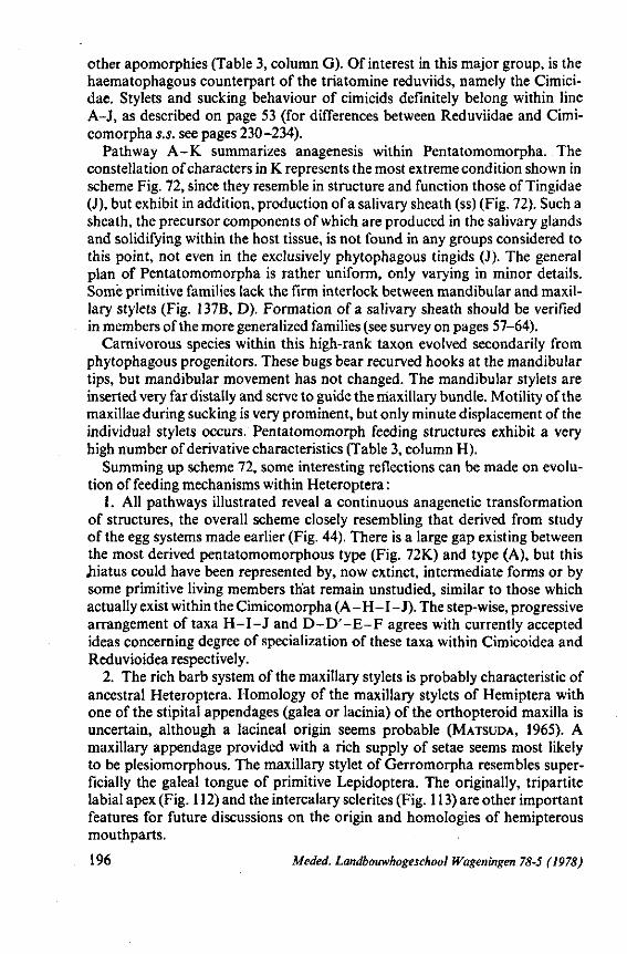
other apomorphies (Table 3, column G). Of interest in this major group, is the haematophagous counterpart of the triatomine reduviids, namely the Cimici-dae. Stylets and sucking behaviour of cimicids definitely belong within line A-J, as described on page 53 (for differences between Reduviidae and Cimi-comorpha s.s. see pages 230-234).
Pathway A-K summarizes anagenesis within Pentatomomorpha. The constellation of characters in K represents the most extreme condition shown in scheme Fig. 72, since they resemble in structure and function those of Tingidae (J), but exhibit in addition, production of a salivary sheath (ss) (Fig. 72). Such a sheath, the precursor components of which are produced in the salivary glands and solidifying within the host tissue, is not found in any groups considered to this point, not even in the exclusively phytophagous tingids (J). The general plan of Pentatomomorpha is rather uniform, only varying in minor details. Some primitive families lack the firm interlock between mandibular and maxillary stylets (Fig. 137B, D). Formation of a salivary sheath should be verified in members of the more generalized families (see survey on pages 57-64).
Carnivorous species within this high-rank taxon evolved secondarily from phytophagous progenitors. These bugs bear recurved hooks at the mandibular tips, but mandibular movement has not changed. The mandibular stylets are inserted very far distally and serve to guide the maxillary bundle. Motility of the maxillae during sucking is very prominent, but only minute displacement of the individual stylets occurs. Pentatomomorph feeding structures exhibit a very high number of derivative characteristics (Table 3, column H).
Summing up scheme 72, some interesting reflections can be made on evolution of feeding mechanisms within Heteroptera :
1. All pathways illustrated reveal a continuous anagenetic transformation of structures, the overall scheme closely resembling that derived from study of the egg systems made earlier (Fig. 44). There is a large gap existing between the most derived pentatomomorphous type (Fig. 72K) and type (A), but this hiatus could have been represented by, now extinct, intermediate forms or by some primitive living members that remain unstudied, similar to those which actually exist within the Cimicomorpha (A-H-I-J ) . The step-wise, progressive arrangement of taxa H - I - J and D - D ' - E - F agrees with currently accepted ideas concerning degree of specialization of these taxa within Cimicoidea and Reduvioidea respectively.
2. The rich barb system of the maxillary stylets is probably characteristic of ancestral Heteroptera. Homology of the maxillary stylets of Hemiptera with one of the stipital appendages (galea or lacinia) of the orthopteroid maxilla is uncertain, although a lacineal origin seems probable (MATSUDA, 1965). A maxillary appendage provided with a rich supply of setae seems most likely to be plesiomorphous. The maxillary stylet of Gerromorpha resembles superficially the galeal tongue of primitive Lepidoptera. The originally, tripartite labial apex (Fig. 112) and the intercalary sclerites (Fig. 113) are other important features for future discussions on the origin and homologies of hemipterous mouthparts.
196 Meded. Landbouwhogeschool Wageningen 78-5 (1978)
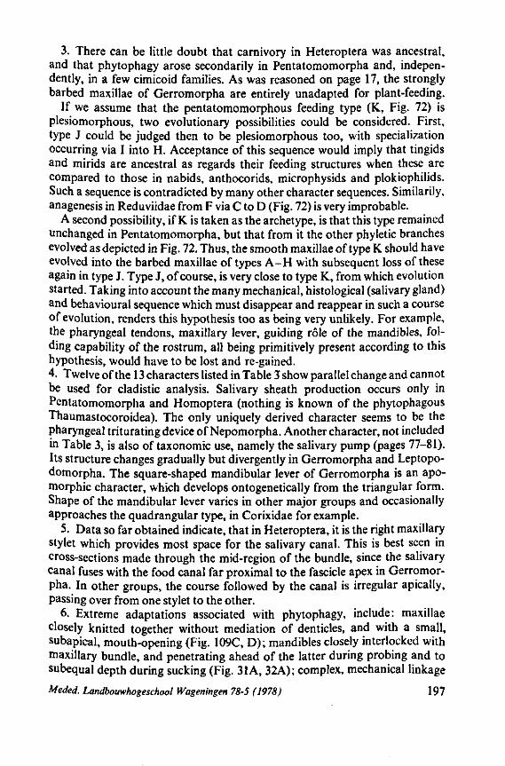
3. There can be little doubt that carnivory in Heteroptera was ancestral, and that phytophagy arose secondarily in Pentatomomorpha and, independently, in a few cimicoid families. As was reasoned on page 17, the strongly barbed maxillae of Gerromorpha are entirely unadapted for plant-feeding.
If we assume that the pentatomomorphous feeding type (K, Fig. 72) is plesiomorphous, two evolutionary possibilities could be considered. First, type J could be judged then to be plesiomorphous too, with specialization occurring via I into H. Acceptance of this sequence would imply that tingids and mirids are ancestral as regards their feeding structures when these are compared to those in nabids, anthocorids, microphysids and plokiophilids. Such a sequence is contradicted by many other character sequences. Similarily, anagenesis in Reduviidae from F via C to D (Fig. 72) is very improbable.
A second possibility, if K is taken as the archetype, is that this type remained unchanged in Pentatomomorpha, but that from it the other phyletic branches evolved as depicted in Fig. 72. Thus, the smooth maxillae of type K should have evolved into the barbed maxillae of types A-H with subsequent loss of these again in type J. Type J, of course, is very close to type K, from which evolution started. Taking into account the many mechanical, histological (salivary gland) and behavioural sequence which must disappear and reappear in such a course of evolution, renders this hypothesis too as being very unlikely. For example, the pharyngeal tendons, maxillary lever, guiding rôle of the mandibles, folding capability of the rostrum, all being primitively present according to this hypothesis, would have to be lost and re-gained. 4. Twelve of the 13 characters listed in Table 3 show parallel change and cannot be used for cladistic analysis. Salivary sheath production occurs only in Pentatomomorpha and Homoptera (nothing is known of the phytophagous Thaumastocoroidea). The only uniquely derived character seems to be the pharyngeal triturating device of Nepomorpha. Another character, not included in Table 3, is also of taxonomie use, namely the salivary pump (pages 77-81). Its structure changes gradually but divergently in Gerromorpha and Leptopo-domorpha. The square-shaped mandibular lever of Gerromorpha is an apo-morphic character, which develops ontogenetically from the triangular form. Shape of the mandibular lever varies in other major groups and occasionally approaches the quadrangular type, in Corixidae for example.
5. Data so far obtained indicate, that in Heteroptera, it is the right maxillary stylet which provides most space for the salivary canal. This is best seen in cross-sections made through the mid-region of the bundle, since the salivary canal fuses with the food canal far proximal to the fascicle apex in Gerromorpha. In other groups, the course followed by the canal is irregular apically, passing over from one stylet to the other.
6. Extreme adaptations associated with phytophagy, include: maxillae closely knitted together without mediation of denticles, and with a small, subapical, mouth-opening (Fig. 109C, D); mandibles closely interlocked with maxillary bundle, and penetrating ahead of the latter during probing and to subequal depth during sucking (Fig. 31 A, 32A); complex, mechanical linkage
Meded. Landbouwhogeschool Wageningen 78-5 (1978) 197
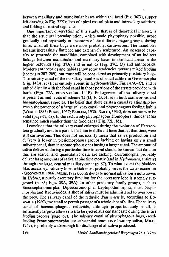
between maxillary and mandibular bases within the head (Fig. 36D), (upper left drawing in Fig. 72K) ; loss of apical rostral plate and intercalary sclerites ; and folding of rostral segments.
One important observation of this study, that is of theoretical interest, is that the structural preadaptation, which made phytophagy possible, arose gradually and separately in ancestors of the different major groups, during times when all these bugs were most probably, carnivorous. The mandibles became increasingly flattened and extensively sculptured. An increased capacity to protrude the mandibles, combined with development of an indirect linkage between mandibular and maxillary bases in the head arose in the higher reduviids (Fig. 37A) and in nabids (Fig. 37C, D) and anthocorids. Modern anthocorids and nabids show some tendencies towards mixed feeding (see pages 207-209), but must still be considered as primarily predatory bugs. The salivary canal of the maxillary bundle is of small calibre in Gerromorpha (Fig. 143A, sc) (it is entirely absent in Hydrometridae, Fig. 147A-C), and is united distally with the food canal in those portions of the stylets provided with barbs (Figs. 72A, cross-section; 144F). Enlargement of the salivary canal is present at mid levels of scheme 72 (D, F, G, H, sc in both carnivorous and haematophagous species. The belief that there exists a causal relationship between the presence of a large salivary canal and phytophagous feeding habits (WEDDE, 1885;ELSON, 1937;EKBLOM, 1930;BARTH, 1954), does not seem to be valid (page 67,68). In the exclusively phytophagous Homoptera, this canal has remained much smaller than the food canal (Fig. 72L, M).
I conclude that the salivary canal enlarged during the evolution of Heterop-tera gradually and in a parallel fashion in different lines that, at that time, were still carnivorous. This does not necessairly mean that saliva production and delivery is lower in plesiomorphous groups lacking or having only a small salivary canal, than in apomorphous ones having a larger canal. The amount of saliva delivered during a particular time interval should be known, but data on this are scarce, and quantitative data are lacking. Gerromorpha probably deliver large amounts of saliva at one time mostly (and in Hydrometra, entirely) through the large, central maxillary canal (p. 67). To what extent the bladderlike, accessory, salivary lobe, which most probably serves for water excretion (GOODCHILD, 1966; MILES, 1972), contributes to normal salivation is not known. In Hebrus, a purely excretory function for the accessory lobe is strongly suggested (p. 83; Figs. 38A, 39A). In other predatory family groups, such as Enicocephalomorpha, Dipsocoromorpha, Leptopodomorpha, most Nepo-morpha and Reduvioidea, a shot of saliva must be administered to overpower the prey. The salivary canal of the reduviid Platymeris is, according to EDWARDS (1960), too small to permit passage of a whole shot of saliva. The salivary canal of haematophagous reduviids, although proportionately small, is sufficiently large to allow saliva to be ejected at a constant rate during the entire feeding process (page 67). The salivary canal of phytophagous bugs, (seed-feeding Pentatomomorpha use substantial amounts of watery saliva, MILES, 1959), is probably wide enough for discharge of all saliva produced.
198 Meded. Landbouwhogeschool Wageningen 78-5 (1978)
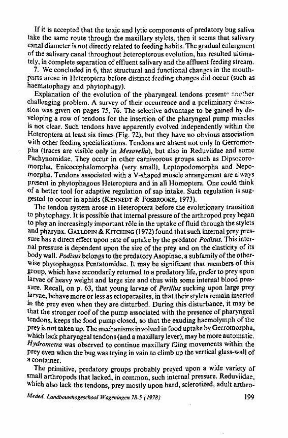
If it is accepted that the toxic and lytic components of predatory bug saliva take the same route through the maxillary stylets, then it seems that salivary canal diameter is not directly related to feeding habits. The gradual enlargment of the salivary canal throughout heteropterous evolution, has resulted ultimately, in complete separation of effluent salivary and the affluent feeding stream.
7. We concluded in 6, that structural and functional changes in the mouth-parts arose in Heteroptera before distinct feeding changes did occur (such as haematophagy and phytophagy).
Explanation of the evolution of the pharyngeal tendons present- another challenging problem. A survey of their occurrence and a preliminary discussion was given on pages 75, 76. The selective advantage to be gained by developing a row of tendons for the insertion of the pharyngeal pump muscles is not clear. Such tendons have apparently evolved independently within the Heteroptera at least six times (Fig. 72), but they have no obvious association with other feeding specializations. Tendons are absent not only in Gerromor-pha (traces are visible only in Mesovelid), but also in Reduviidae and some Pachynomidae. They occur in other carnivorous groups such as Dipsocoro-morpha, Enicocephalomorpha (very small), Leptopodomorpha and Nepo-morpha. Tendons associated with a V-shaped muscle arrangement are always present in phytophagous Heteroptera and in all Homoptera. One could think of a better tool for adaptive regulation of sap intake. Such regulation is suggested to occur in aphids (KENNEDT & FOSBROOKE, 1973).
The tendon system arose in Heteroptera before the evolutionary transition to phytophagy. It is possible that internal pressure of the arthropod prey began to play an increasingly important rôle in the uptake of fluid through the stylets and pharynx. GALLOPIN & KITCHING (1972) found that such internal prey pressure has a direct effect upon rate of uptake by the predator Podisus. This internal pressure is dependent upon the size of the prey and on the elasticity of its body wall. Podisus belongs to the predatory Asopinae, a subfamily of the otherwise phytophagous Pentatomidae. It may be significant that members of this group, which have secondarily returned to a predatory life, prefer to prey upon larvae of heavy weight and large size and thus with some internal blood pressure. Recall, on p. 63, that young larvae of Perillus sucking upon large prey larvae, behave more or less as ectoparasites, in that their stylets remain inserted in the prey even when they are disturbed. During this disturbance, it may be that the stronger roof of the pump associated with the presence of pharyngeal tendons, keeps the food pump closed, so that the exuding haemolymph of the prey is not taken up. The mechanisms involved in food uptake by Gerromorpha, which lack pharyngeal tendons (and a maxillary lever), may be more automatic. Hydrometra was observed to continue maxillary filing movements within the prey even when the bug was trying in vain to climb up the vertical glass-wall of a container.
The primitive, predatory groups probably preyed upon a wide variety of small arthropods that lacked, in common, such internal pressure. Reduviidae, which also lack the tendons, prey mostly upon hard, sclerotized, adult arthro-
Meded. Landbouwhogeschool Wageningen 78-5 (1978) 199
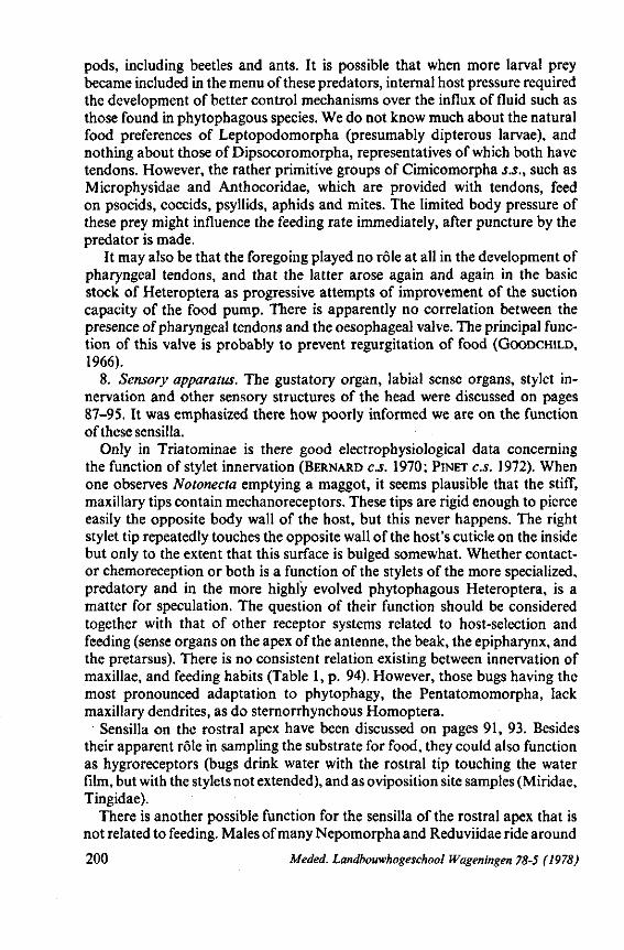
pods, including beetles and ants. It is possible that when more larval prey became included in the menu of these predators, internal host pressure required the development of better control mechanisms over the influx of fluid such as those found in phytophagous species. We do not know much about the natural food preferences of Leptopodomorpha (presumably dipterous larvae), and nothing about those of Dipsocoromorpha, representatives of which both have tendons. However, the rather primitive groups of Cimicomorpha s.S., such as Microphysidae and Anthocoridae, which are provided with tendons, feed on psocids, coccids, psyllids, aphids and mites. The limited body pressure of these prey might influence the feeding rate immediately, after puncture by the predator is made.
It may also be that the foregoing played no rôle at all in the development of pharyngeal tendons, and that the latter arose again and again in the basic stock of Heteroptera as progressive attempts of improvement of the suction capacity of the food pump. There is apparently no correlation between the presence of pharyngeal tendons and the oesophageal valve. The principal function of this valve is probably to prevent regurgitation of food (GOODCHILD, 1966).
8. Sensory apparatus. The gustatory organ, labial sense organs, stylet innervation and other sensory structures of the head were discussed on pages 87-95. It was emphasized there how poorly informed we are on the function of these sensilla.
Only in Triatominae is there good electrophysiological data concerning the function of stylet innervation (BERNARD C.S. 1970; PINET C.S. 1972). When one observes Notonecta emptying a maggot, it seems plausible that the stiff, maxillary tips contain mechanoreceptors. These tips are rigid enough to pierce easily the opposite body wall of the host, but this never happens. The right stylet tip repeatedly touches the opposite wall of the host's cuticle on the inside but only to the extent that this surface is bulged somewhat. Whether contactor chemoreception or both is a function of the stylets of the more specialized, predatory and in the more highly evolved phytophagous Heteroptera, is a matter for speculation. The question of their function should be considered together with that of other receptor systems related to host-selection and feeding (sense organs on the apex of the antenne, the beak, the epipharynx, and the pretarsus). There is no consistent relation existing between innervation of maxillae, and feeding habits (Table 1, p. 94). However, those bugs having the most pronounced adaptation to phytophagy, the Pentatomomorpha, lack maxillary dendrites, as do sternorrhynchous Homoptera.
Sensilla on the rostral apex have been discussed on pages 91, 93. Besides their apparent rôle in sampling the substrate for food, they could also function as hygroreceptors (bugs drink water with the rostral tip touching the water film, but with the stylets not extended), and as oviposition site samples (Miridae, Tingidae).
There is another possible function for the sensilla of the rostral apex that is not related to feeding. Males of many Nepomorpha and Reduviidae ride around
200 Meded. Landbouwhogeschool Wageningen 78-5 (1978)
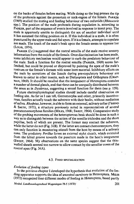
on the backs of females before mating. While doing so the bug presses the tip of the proboscis against the pronotum or neck-region of the female. PARKER (1969) studied the mating and feeding behaviour of two reduviids (Rhinocoris spp.). The position of the male proboscis during copulation is, according to PARKER, part of the sequence of events involved in response to insect prey. The male is apparently unable to distinguish the sex of another individual until it has assumed the riding position on it. If this individual is a male, it is often punctured by the upper male and fed upon. If it is a female, attempts to copulate are made. The touch of the male's beak upon the female seems to appease her (Louis, 1974).
PARKER (I.e.) suggested that the rostral sensilla of the male receive sensory information from the cuticle of the female. Once discrimination has been made, some inhibitory mechanism would appear to curb the predatory behaviour of the male. Such a function for the rostral sensilla (PARKER, 1969) seems farfetched, but could be proved or disproved by painting the apex of the male's rostrum or the female's dorsum with some inert material. Inhibitory effects on the male by secretions of the female during pre-copulatory behaviour are known to occur in other insects, such as Dictyoptera and Orthoptera (CHAP-MAN, 1969). It should be recalled that the integument of Heteroptera contains a number of dermal glands, and that these can be unequally distributed between the sexes as in Dysdercus, suggesting a sexual function for them (see p. 159).
Future electrophysiological studies should include careful observation on feeding site. As far as I can tell, Gerromorpha and other, primarily insectivorous, families actually touch the substrate with their beaks, without mediation of saliva. Rhodnius, however, is able to form an external, salivary collar (FRIEND & SMITH, 1971), a structure previously noted in representatives of several pentatomomorphous families (MILES, 1968; SWEET, 1964). Comparative study of the probing movements of the heteropterous beak should be done in such a way as to distinguish between the action of the sensilla trichodea and the short papillae, both of which are present. The former may contact the substrate, while the latter do not (Fig. 3 IB). If the latter are contact chemoreceptors, they can only function in monitoring stimuli from the host by means of a salivary cone. The predatory Perillus forms an external stylet sheath, which extended from the labial groove towards the puncture made in the host's integument (SWEET, 1964). My observations on the same species suggest that the thin-walled sheath seemed too narrow to allow contact by the sensillar cones of the rostral apex (Fig. 34A).
4.3. FOOD SPECIALIZATION
Evolution of feeding types In the previous chapter I developed the hypothesis that evolution of the fee
ding apparatus supports the idea of ancestral carnivory in Heteroptera. MILES (1972) recognized four different modes of feeding in Heteroptera, based partly
Meded. Landbouwhogeschool Wageningen 78-5 (1978) 201
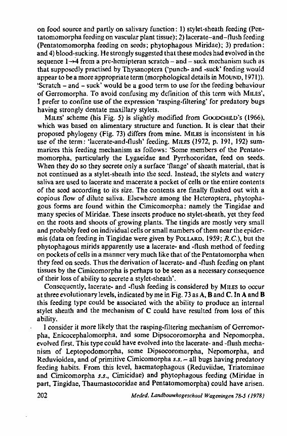
on food source and partly on salivary function: 1) stylet-sheath feeding (Pen-tatomomorpha feeding on vascular plant tissue); 2) lacerate-and-flush feeding (Pentatomomorpha feeding on seeds; phytophagous Miridae); 3) prédation: and 4) blood-sucking. He strongly suggested that these modes had evolved in the sequence l-*4 from a pre-hemipteran scratch - and - suck mechanism such as that supposedly practised by Thysanoptera ('punch- and -suck' feeding would appear to be a more appropriate term (morphological details in MOUND, 1971)). 'Scratch - and - suck' would be a good term to use for the feeding behaviour of Gerromorpha. To avoid confusing my definition of this term with MILES', I prefer to confine use of the expression 'rasping-filtering' for predatory bugs having strongly dentate maxillary stylets.
MILES' scheme (his Fig. 5) is slightly modified from GOODCHILD'S (1966), which was based on alimentary structure and function. It is clear that their proposed phylogeny (Fig. 73) differs from mine. MILES is inconsistent in his use of the term: 'lacerate-and-flush' feeding. MILES (1972, p. 191, 192) summarizes this feeding mechanism as follows : 'Some members of the Pentatomomorpha, particularly the Lygaeidae and Pyrrhocoridae, feed on seeds. When they do so they secrete only a surface 'flange' of sheath material, that is not continued as a stylet-sheath into the seed. Instead, the stylets and watery saliva are used to lacerate and macerate a pocket of cells or the entire contents of the seed according to its size. The contents are finally flushed out with a copious flow of dilute saliva. Elsewhere among the Heteroptera, phytophagous forms are found within the Cimicomorpha : namely the Tingidae and many species of Miridae. These insects produce no stylet-sheath, yet they feed on the roots and shoots of growing plants. The tingids are mostly very small and probably feed on individual cells or small numbers of them near the epidermis (data on feeding in Tingidae were given by POLLARD, 1959; R.C.), but the phytophagous mirids apparently use a lacerate- and -flush method of feeding on pockets of cells in a manner very much like that of the Pentatomorpha when they feed on seeds. Thus the derivation of lacerate- and -flush feeding on plant tissues by the Cimicomorpha is perhaps to be seen as a necessary consequence of their loss of ability to secrete a stylet-sheath'.
Consequently, lacerate- and -flush feeding is considered by MILES to occur at three evolutionary levels, indicated by me in Fig. 73 as A, B and C. In A and B this feeding type could be associated with the ability to produce an internal stylet sheath and the mechanism of C could have resulted from loss of this ability.
I consider it more likely that the rasping-filtering mechanism of Gerromorpha, Enicocephalomorpha, and some Dipsocoromorpha and Nepomorpha, evolved first. This type could have evolved into the lacerate- and -flush mechanism of Leptopodomorpha, some Dipsocoromorpha, Nepomorpha, and Reduvioidea, and of primitive Cimicomorpha s.s. - all bugs having predatory feeding habits. From this level, haematophagous (Reduviidae, Triatominae and Cimicomorpha s.S., Cimicidae) and phytophagous feeding (Miridae in part, Tingidae, Thaumastocoridae and Pentatomomorpha) could have arisen.
202 Meded. Landbouwhogeschool Wageningen 78-5 (1978)
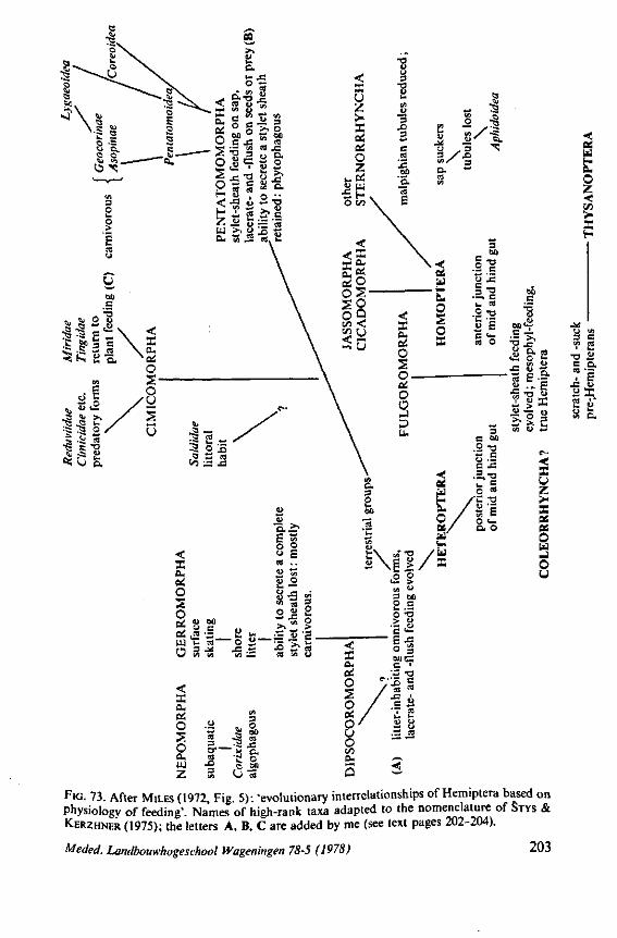
< x u z >-X ei
O z *B6
0> m • S H . o w \
3 •o 1> fc-t/l
u 3 3 e S IS DO 'E.
75
e
W5
o. 1/5
S - 3
ules
los
\ A
phid
o
X> 3
e« fad
O z •«:
s
u s.s-? S a ï 2 Ó'
FIG. 73. After MILES (1972, Fig. 5): 'evolutionary interrelationships of Hemiptera based on Physiology of feeding'. Names of high-rank taxa adapted to the nomenclature of STYS & KERZHNER (1975); the letters A, B, C are added by me (see text pages 202-204).
Meded. Landbouwhogeschool Wageningen 78-5 (1978) 203

Only the Pentatomomorpha (the feeding mechanism of Thaumastocoridae is not known) achieved the capacity to feed on plant material by means of an internal salivary sheath. Many Pentatomomorpha have retained the ability to use their ancestral lacerate- and -flush mechanism, while, at the same time, being able to feed on phloem. Therefore, it is not surprising that those bugs that returned to animal feeding have shifted back 100% to a lacerate-flush feeding mechanism.
Thus, the groups which should be placed at level A in MILES' scheme (Fig. 73) are those having strongly barbed maxillary stylets. These stylets bear an extended functional mouth opening that reaches from the apex of the stylet bundle back to about one third the length of the protruded bundle. Saliva and food material which enters must pass through the long grating system formed by the interwoven barbs of both stylets (pages 13-25, Figs. 77-96). Formation óf a solidifying salivary sheath around this grating would decrease the efficiency of food uptake by this mechanism. On page 17, it was shown that barbed stylets are unlikely to aid in extracting sap from plant tissue.
MILES ( 1972), following GOODCHILD ( 1966), considered ancestral Heteroptera to be litter-inhabiting, omnivores (the word 'omnivorous' can be interpreted in many ways, whereas I argue for a completely predatory ancestral life, a conclusion already emphasized in my 1968 book). CHINA (1933) stated that the primitive Heteroptera were undoubtedly phytophagous. KUNKEL (1967) considered the original feeding of Heteroptera to be varied with plant-feeding predominating. ELSON (1937) and SOUTHWOOD (1973) supposed them to be carnivorous or perhaps fungivorous, whereas EASTOP (1973) favoured the idea of entomophagy in the fore-runners of Hemiptera.
Ancestral fungus-feeding is unlikely. The fungivorous Aradidae have pen-tatomomorphous, and hence specialized feeding structures. Loss of maxillary barbs was apparently necessary for the evolution of mycetosuccivorous habits. Special, crumena-like, adaptations for coiling the stylet-bundle (Fig. 137E-G), have evolved at least four times independently in some pentatomomorphous families (see p. 59), as result of their successful exploitation of this source of food (fungus and roots; Aradus frigidus is actually a root-feeder, TAMANINI, 1955; LINDBERG, 1963).
Finally, evidence could be marshalled to support the idea that polliniphagy was the original way of feeding. It occurs in Anthocoridae. The suggestion has been made on page 207,208 that feeding-evolution in Cimicomorpha s.s. could have passed through predation->-polliniphagy-»-phytophagy. The rasping-filing mouthparts of primitive predators, however, are not the proper tools for emptying pollen-grains.
The feeding of proto-heteroptera can not be considered without reference to Homoptera; this will be discussed in Section 4.5 (pages 234-243).
About half the 70 families of Heteroptera are exclusively carnivorous and three distantly - related taxa are specialized for feeding on vertebrate blood (Reduviidae Triatominae, Cimicidae, and Polyctenidae). Most other bugs are exclusively phytophagous but six families, although predominantly phytopha-
204 Meded. Landbouwhogeschool Wageningen 78-5 (1978)
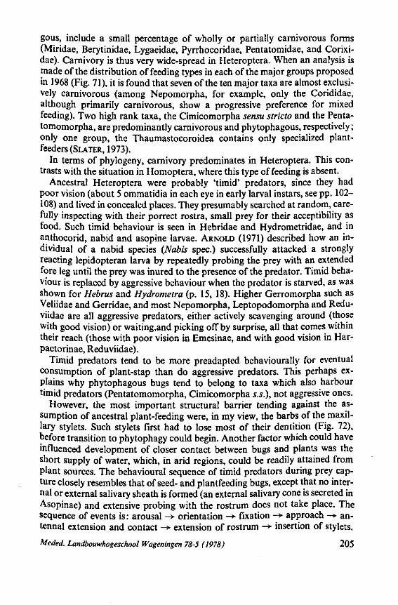
gous, include a small percentage of wholly or partially carnivorous forms (Miridae, Berytinidae, Lygaeidae, Pyrrhocoridae, Pentatomidae, and Corixi-dae). Carnivory is thus very wide-spread in Heteroptera. When an analysis is made of the distribution of feeding types in each of the major groups proposed in 1968 (Fig. 71), it is found that seven of the ten major taxa are almost exclusively carnivorous (among Nepomorpha, for example, only the Corididae, although primarily carnivorous, show a progressive preference for mixed feeding). Two high rank taxa, the Cimicomorpha sensu stricto and the Penta-tomomorpha, are predominantly carnivorous and phytophagous, respectively; only one group, the Thaumastocoroidea contains only specialized plant-feeders (SLATER, 1973).
In terms of phylogeny, carnivory predominates in Heteroptera. This contrasts with the situation in Homoptera, where this type of feeding is absent.
Ancestral Heteroptera were probably 'timid' predators, since they had poor vision (about 5 ommatidia in each eye in early larval instars, see pp. 102— 108) and lived in concealed places. They presumably searched at random, carefully inspecting with their porrect rostra, small prey for their acceptibility as food. Such timid behaviour is seen in Hebridae and Hydrometridae, and in anthocorid, nabid and asopine larvae. ARNOLD (1971) described how an individual of a nabid species (Nabis spec.) successfully attacked a strongly reacting lepidopteran larva by repeatedly probing the prey with an extended fore leg until the prey was inured to the presence of the predator. Timid behaviour is replaced by aggressive behaviour when the predator is starved, as was shown for Hebrus and Hydrometra (p. 15, 18). Higher Gerromorpha such as Veliidae and Gerridae, and most Nepomorpha, Leptopodomorpha and Redu-viidae are all aggressive predators, either actively scavenging around (those with good vision) or waiting.and picking off by surprise, all that comes within their reach (those with poor vision in Emesinae, and with good vision in Har-pactorinae, Reduviidae).
Timid predators tend to be more preadapted behaviourally for eventual consumption of plant-stap than do aggressive predators. This perhaps explains why phytophagous bugs tend to belong to taxa which also harbour timid predators (Pentatomomorpha, Cimicomorpha s.S.), not aggressive ones.
However, the most important structural barrier tending against the assumption of ancestral plant-feeding were, in my view, the barbs of the maxillary stylets. Such stylets first had to lose most of their dentition (Fig. 72), before transition to phytophagy could begin. Another factor which could have influenced development of closer contact between bugs and plants was the short supply of water, which, in arid regions, could be readily attained from plant sources. The behavioural sequence of timid predators during prey capture closely resembles that of seed- and plantfeeding bugs, except that no internal or external salivary sheath is formed (an external salivary cone is secreted in Asopinae) and extensive probing with the rostrum does not take place. The sequence of events is: arousal -> orientation -*• fixation -*• approach -> an-tennal extension and contact -»• extension of rostrum -> insertion of stylets.
Meded. Landbouwhogeschool Wageningen 78-5 (1978) 205
Highly aggressive predators, grab prey with their fore legs immediately and omit prior antennal contact and rostral extension. The generalizations made above are based on data extracted from the literature (e.g. EDWARDS, 1962; EGGERMAN & BONGERS, 1971 ; WOLFRAM, 1972) and on my own observations.
The predominance of prédation in Heteroptera is not, in itself, evidence of this being the original way of feeding. As shown in section 4.2., mouthpart structures, considered on their own merits, establish the plesiomorphy of predatory behaviour. Much symplesiomorphy is shown by other, independently varying, character states in the carnivorous categories Gerromorpha, Nepomorpha, Dipsocoromorpha and Enicocephalomorpha (Table 4, p. 220).
It was concluded on p. 198 that a number of changes in the feeding apparatus, particularly the gradual loss of maxillary barbs, anticipated the advance from predatory to haematophagous and phytophagous feeding. A dorsally and ventrally closed maxillary bundle, functioning as a syringe, and in increased ability to protrude the mandibles were apparently preadaptive features enabling plant-feeding to develop. The great diversity of phytophagous forms seems to be related to these structural modifications. Although relatively few major groups of Heteroptera became phytophagous, those that did underwent extensive adaptive radiation once they penetrated the defences of the vegetative habitat. An increase in individual population density, in number of species (Miridae, Tingidae) and in number of families (Pentatomomorpha) occurred. The carnivorous families, Nabidae, Anthocoridae and Reduviidae, are on an intermediate anagenetic level both in terms of their feeding structures and in their level of ecological adaptation (they are no longer restricted to the litter-zone).
Unbalanced feeding types The fact that a fire-bug like Pyrrhocoris, usually a plant-feeder, can readily
become carnivorous under suboptimal conditions, may seem strange of those concerned with insect-plant relations or with insect (as predator or parasite) -insect (as host) relations. Host-selection is now a feavoured subject of research in entomology. Factors responsible for eliciting each response in the sequence of reactions, that result in ultimate acceptance or rejection of food, have been studied for both phytophagous and carnivorous species (reviews in KENNEDY, 1965 ; SCHOONHOVEN, 1968 ; THORSTEINSON, 1960 ; VINSON, 1976). It is of interest to consider that evolutionary boundary where mixed animal and plantfeeding occurs, or where circumstances may induce a switch from animal to plant-feeding or the reverse.
A consequence of the conclusion of Fig. 72, discussed on pages 191-201, is that such flexible feeding occurred and still occurs in Heteroptera. A short survey follows below.
In the primarily carnivorous groups, Gerromorpha, Nepomorpha, and Leptopodomorpha, only occasional feeding on berries by Gerris (p. 20) and mixed feeding by Corixinae (p. 35) has been recorded.
206 Meded. Landbouwhogeschool Wageningen 78-5 (1978)
Reduviidae Although the Reduviidae, as implied by their common name 'assassin bugs'
are pre-eminently carnivorous, many Harpactorinae live in shrubs and trees, and are sometimes associated with particular plants (COBBEN & WYGODZINSKY, 1975). Larvae and adults of many species, again particularly in the Harpactorinae, use sticky material produced by their own body (p. 146) or originating from the plant, for camouflage or prey-catching. Selection of a particular plant involves behavioural responses by the bugs to stimuli emanating from that plant. These cues may be visual (form, colour) or chemical, or may involve humidity differences.
The association of reduviids with particular plants could be indirect, since they prey on insects present only on these plants. STONER C.S. (1975) tested the plant-feeding capability of first instar larvae of four harpactorine species. The larvae survived significantly longer when fed on dandelion pollen and safflower seed than on water alone, but no larvae developed past the first instar. Seeds of other plants, cotton leaves and bean slices were not better than water in supporting life in the bugs. In spite of this observation that certain reduviids may supplement their usual animal diet to a limited degree with plant substances (nectar is also sometimes taken, WYGODZINSKY, 1947), it is unlikely that Harpactorinae bear the right set of preadaptations for eventual full assumption of phytophagy. A similar but better-developed tendency is found in Anthocoridae.
Movement of prey is essential for predatory response in Harpactorinae (EDWARDS, 1966; PARKER, 1965, 1969). The dilator muscles of the pharyngeal pump of harpactorines lack the tendons, which apparently occur in all phytophagous Hemiptera. The greater ability of these bugs to protrude their mandibular stylets during sucking (p. 41 and Fig. 20) is found also in phytophagous bugs. In Harpactorinae and other reduviids it is probably an adaptation enabling the stylet bundle to be forced deeply through the intersegmental folds of their hard-bodied prey. Their shovel-shaped mandibles (Fig. 22A, 102A-D) would probably be unsuited for regular penetration of plant tissue.
The situation may be otherwise in Rhaphidosominae, which have a long slender rostrum, in contrast to those of most other Reduviidae. It has been suggested by MILLER (1956) that Rhaphidosominae live on stem-borers or on other prey hidden deep within the plant. If so, the stylets of these bugs come in contact with plant-tissue as they attempt to pierce the prey.
Anthocoridae This taxon is primarily predatory, but feeding on other food sources has been
repeatedly mentioned (survey in CARA YON & STEFAN, 1959, and PÉRICART, 1972). Adults of Anthocoris nemorum were regularly observed to search for and to feed on larvae of the microlepidopteron Stigmella malella (miss Prinsen, internal student report, Wageningen). The prey larvae are leaf-miners in apple and the bug must perforate the plant cuticle to reach them. This suggests that the bug responds to characteristics of the plant in which its prey lives. Such a
Meded. Landbouwhogeschool Wageningen 78-5 (1978) 207
relationship might have existed between plants and the predatory progenitors of phytophagous taxa.
Infestation of vegetative and generative plant parts by Orius spp. have been recorded by BODENHEIMER (1921), DOEKSEN (1944), HYSLOP (1916), and SCHOEVERS (1930). BACHELER & BARANOWSKI (1975) confirmed in feeding tests that Paratriphleps laeviusculus (Oriini) lives primarily from the sap of sapodilla flowers. Pollen-feeding is often practised by other Oriini. For Orius pallidi-cornis, this food source seems to be sufficient for complete development. Such polliniphagous behaviour has been extensively studied by FAUVEL (1974) in Orius vicinus. He found differences to exist in the nutritive value of pollen from different plants. Although the bugs can reach maturity on pollen alone, this food is suboptimal for permanent growth and reproduction. BRÖNNIMANN
( 1964) was able to rear Anthocoris nemorum to maturity on a medium consisting of one part water and two parts sweetened condensed milk. Very surprisingly, growth of all instars on this diet, was faster than that of those reared on natural, animal food and the adults that resulted produced viable offspring.
The great flexibility in anthocorid feeding is further illustrated by occasional reports of haematophagy. Anthocoris pilosus (Anthocorinae) and Lyctocoris campes tris (Lyctocorinae) are known to suck blood from vertebrates (STYS & DANIEL, 1957; STYS, 1973, and others). It should be recalled here that the specialized, blood-sucking, Cimicidae are closely related to Anthocoridae (USINGER, 1966).
Nabidae and other Cimicomorpha s.S. Although generally known to be active predators, nabids sometimes suck
from plants and can survive for a while on a diet of pollen; however, complete development on vegetarian food seems impossible (KOSCHEL, 1971; STONER,
1972). RIDGEWAY & JONES (1968), using radiolabelled cotton plants, showed that Nabis americoferus may occasionally consume plant sap in substantial amounts. As is true of some Reduviidae Harpactorinae (p. 207) and Anthocoridae (COBBEN & ARNOUD, 1969), some nabid species always occur on a particular plant taxon. Thus, Aspilaspis viridis is restricted to Tamarix and is green, an unusual colour for a nabid. The nutritional aspect of such insect-plant associations is not known. These are certainly important for the Miridae, the next family on the anagenetic scale.
I do not agree with DRAKE & DA VIES (1960) who believe that Miridae and related families are primarily phytophagous. That Bryocorinae (Miridae) and Tingidae are exclusively phytophagous may have contributed to this idea. Bryocorinae is currently considered to be primitive, but there are arguments against this (COBBEN, 1968a, and this paper, p. 122; SCHUH, 1974). The least specialized subfamily of Miridae is probably the Cylapinae, which, like the primitive mirid group, the Isometopinae, contains only predators as far as is known.
A concise survey, by subfamily, of the gross feeding habits of Miridae has been presented by LESTON (1961). It may be of significance that subfamilies that
208 Meded. Landbouwhogeschool Wageningen 78-5 (1978)
are primarily carnivorous contain relatively few species, as compared with those in which phytophagy prevails. Deraeocorinae is a moderately sized, but carnivorous group. However, Deraeocoris ruber appears to be capable of completing its first larval instar on an exclusively plant diet (VIGGIANI, 1971)*.
Pentatomomorpha This very large taxon, consisting of some 26 families, contains the most
specialized plant sap-feeders in the Heteroptera : the Pentatomoidea, Coreoi-dea, and Piesmatoidea. If the evolutionary scheme presented in Fig. 72 is true, Pentatomomorpha (level K) should have arisen from an ancestral line that was predatory. However, stylet structure in none of the representatives studied exhibit the maxillary barbs from which primary carnivory could be deduced (discussion on pp. 196, 197).
Considering the life habits of extant groups, the most logical conclusion is that the plantsap-sucking taxa originated from a seed-feeding stock. Seed-sucking is still practised by representatives of many families such as Lygaeidae, Pyrrhocoridae, Largidae, Stenocephalidae, Hyocephalidae, Alydidae, Cyd-nidae and Scutelleridae (KUMAR, 1966; MCDONALD, 1960; SCHAEFER, 1972b: SLATER, 1975; SWEET, 1960, 1964; among many others). Of these, Lygaeidae and Pyrrhocoridae are particularly specialized to feed on mature seed. Unfortunately, we are mostly ignorant of the food habits of the relict families Idiostolidae and Thaumastellidae (although one species of the latter family could be fed for some time as adults with various seeds, SWEET in SCHAEFER & WILCOX, 1971). Stylet structure of representatives of these two families is not fundamentally different from that of other primitive pentatomomorphous groups, and this suggests that they could be seed-feeders as well. Lygaeidae live mostly in the litter-zone and SWEET (1960) showed that many Rhyparo-chrominae, can be reared readily on sunflower seed : 'The alacrity with which the various species fed upon a seed which is not indigenous to the study area, and the ease and success of feeding, suggest that most of the species are at least potentially polyphagous or oligophagous upon seeds. This also means that in habitat selection host plant preferences are likely to be of lesser importance than edaphic factors'. The european lygaeine Lygaeus equestris favours ascle-piad seeds, but utilizes also seeds of other plants in the field which support good larval growth and some reproduction (KUGELBERG, 1973, 1974: SOL-BRECK& KUGELBERG, 1972).
This flexibility in food habit reminds us of those carnivorous groups occupying the litter zone and the water surface. Most Gerromorpha and Lep-topodomorpha are polyphagous, when maintained in captivity, but in nature are probably less so because of the impoverished menu available to them. Polyphagy in lygaeids might also be shown to be less prevalent than now believ-
* It was not stated whether Deraeocoris actually fed. I assume that Miridae do require food in first instar forms, as is true of Gerromorpha, Nepomorpha and Reduviidae. Lygaeid larvae also feed in the first instar, but those of pentatomoids and many coreoids apparently do not (OETTING & YONKE, 1971a, b ; SCHAEFER, 1975).
Meded. Landbouwhogeschool Wageningen 78-5 (1978) 209
ed when food preferences of additional species are checked. Many true predators, such as saldids, have very exacting ecological demands. The same is true of many ground-inhabiting lygaeids. It is feasible that the lygaeid line originated from predacious ancestors, which already were associated closely with particular plant communities. Mature seeds lying on the soil, could be increasingly used as food, supplementing the original animal food of the bugs, and ultimately replacing it altogether. This would imply that seed-feeding had originated at different times in different lines of pre-lygaeoids living in different, but fixed ecological situations. In other words, seed-feeding might have arisen on several occasions in several different, vegetational zones, to which the predacious ancestral bugs were already adapted. This could account for the supposed polyphyletic origin of modern lygaeoids s.l. (STYS, 1967b), the phy-logeny of which has so puzzled outstanding specialists such as SCHAEFER
(1963), SCUDDER (1963) and STYS (1961) (see also p. 228). The phloem-feeding habit of higher Pentatomoidea and Coreoidea has
probably developed from an ancestral seed-feeding habit. A diet of dry seeds must be regularly supplemented by uptake of water, if copious saliva is to be produced. Seed-feeding Brachypelta (Cydnidae) and Oncopehus (Lygaeidae) may extract water from succulent parts of the host-plant, and from a wide variety of non-hosts. Water-uptake from these plants is not induced by specific attractants as is food-uptake (SCHORR, 1957; EGGERMANN & BONGERS, 1971). The ability of seed-sucking bugs to perceive various secondary plant substances dissolved in water, might have provided a regulatory cue to them when they were sucking on non-host plants for water (SCHOONHOVEN & DERKSEN-KOP-
PERS, 1973). Seed-feeding thus provides many opportunities to the bugs to pierce leaf and stem tissue, a habit which might ultimately result in their evolutionary progeny becoming sap-suckers. There are different degrees of relations to seed - and sap-feeding within the lygaeid genus Oncopehus. O. fas-ciatus and O. unifasciatellus cannot complete larval development on vegetative milkweed tissue alone, but O. cingulifer does survive under such conditions (RALPH, 1976; ROOT & CHAPLIN,-1976).
Few families of Heteroptera are exclusively sap-sucking (possibly Piesmati-dae, Malcidae, Colobathristidae, and some exotic pentatomoid families). Usually both sap-sucking and seed-sucking taxa occur in a particular family. Within Coreidae, for example, all species in some genera are sap-suckers, whereas those of other genera can feed on sap from stems, fruits, or (rarely) on dry seeds (KUMAR, 1966). This diversity in food habit within a single large taxon presumably indicates that evolution of feeding mechanisms within that taxon is in a phase of unbalanced equilibrium, from which shifts to one or another feeding type can readily occur.
Partial or complete reversal to an animal diet has occurred several times in Lygaeidae, Pyrrhocoridae, Berytinidae and Pentatomidae. The common potential for such reversal, shown in rearing experiments, reveals that lygaeids and pyrrhocorids are not far removed from a predatory existence.
SWEET (1960) attempted in vain to induce various lygaeids to feed on small
210 Meded. Landbouwhogeschool Wageningen 78-5 (1978)
insects, while EYLES (1964) reared larvae of the seed-feeding Drymus sylvaticus to the fifth instar on a diet of beetle larvae. The lygaeid seed-bug Oncopeltus behaves like a predator and aggressively attacks weakened individuals of its own kind. They react only to moving prey, not to killed individuals offered to them (BONGERS, 1969b). HENRICI (1938) reared Pyrrhocoris apterus entirely on pieces of Tenebrio larvae. It should be emphasized that the Pyrrhocoridae harbours two exclusively predacious genera (MILLER, 1956).
The feeding habits of Lygaeidae are summarized by SLATER (1975) as follows:... 'the majority of species feed on mature plant seeds, others utilize sap (Blissinae, many Lygaeinae, etc.). Others are predatory (Geocorinae) and a few suck vertebrate blood (Cleradini)'. Blood-sucking by Cleradini has been reported by LENT (1939), DA COSTA LIMA (1940) and CORREÂ (1956). These bugs comprise a tribe of the otherwise seed-feeding Rhyparochrominae, the only exception being the predatory Mizaldus nidulus (SLATER & CARAYON,
1963). Geocoris spp. (Geocorinae) have captured the interest of applied entomologists because they appear to be polyphagous predators (CHAMPLAIN
& SHOLDT, 1966; V. WADDILL & SHEPARD, 1974; YORK, 1944). However, they do seem to require some plant food, and complete postembryonic development is possible on a solely vegetarian diet (SWEET, 1960; STONER, 1970). TAMAKI & WEEKS (1972) concluded from extensive feeding experiments, that it is advisable to add sunflower seeds in cultivated fields, when Geocoris spp. are considered for biological control ; this source of supplemented food should augment the Geocoris population. Sunflower seeds would also extend the length of survival of adult Geocoris. Berytinidae are probably predominantly sap-feeders, although records of sap-feeding by these bugs are scarce (HERTEL, 1955). Jalysus spinosus, however, may be considered to be a predator, since it requires considerable animal food for optimal growth and reproduction (ELSEY & STIN-
NER, 1971).
The Pentatomoidea contains very few, non-phytophagous taxa. Cydnidae and Corymaelidae are mostly seed- and sap-suckers, but specimens of some small ground species of both families have been found in carrion, where they possibly prey on insect larvae (WEBER, 1930; MICHALK, 1935).
Some remarks on the pentatomid subfamily Asopinae have been given on pages 61, 196. Carnivory in asopine bugs appears to have originated independently ofthat of the groups discussed above, since Pentatomidae are generally highly-specialized plant-feeders. Most produce an internal, salivary sheath and feed at one feeding site for prolonged periods. Asopinae prefer large, soft-bodied prey, in which blood turgor may influence feeding rate (p. 199). Feeding in one spot can continue for hours. BAKER (1927) suggested that predacious pentatomids inject a poison during an attack. The toxic properties of the saliva of these bugs, however may be less than that of other carnivorous groups. Immobilization of prey by some Asopinae is said by GALLOPIN & KITCHING
(1972) to occur after 1-2 minutes and by OETTING & YONKE (1971b) after 5-10 minutes.* Phytophagous pentatomoids and coreoids apparently require no
* See my remark on page 63.
Meded. Landbouwhogeschool Wageningen 78-5 (1978) 211
food in the first instar (MAYNÉ & BRENY, 1948a; SCHAEFER, 1975). However, MOENS (1965) mentioned for the asopine Perillus that plant-sap imbibed by the first instar considerably prolonged duration of this stage. This could have survival value for this bug when prey density is low and greater searching activity is needed.
We may conclude by saying that where phytophagy has evolved in Heterop-tera, feeding behaviour has remained flexible in most families. One can expect that when the chemosensory basis of pre-feeding and feeding behaviour has been better studied, that it also will reveal much more plasticity than is present in the completely phytophagous Homoptera. The experiments of JERMY et al. (1968) and SCHOONHOVEN (1967, 1969, 1976) on exclusively phytophagous insects generated the hypothesis that the sensory system of insects is flexible and can be modified by conditioning and intraspecific variation, which evolutiona-arily may have influenced the behaviour of food-choice (SCHOONHOVEN, pers. comm.). In Heteroptera, having diverse feeding mechanisms, plant feeding may either supplement a regular animal diet or sustain a predacious species for short periods when prey is in short supply. The same should hold for phytophagous species which occasionally take animal food. The significance of predacious Heteroptera in economic entomology has been reviewed by CARA YON
(1961). Asopinae sometimes regulate pest densities effectively when these are high (review in MAYNÉ & BRENY, 1948 b, c).
It is clear from the above survey that the ability of some bugs to switch feeding habits under certain circumstances can influence their impact as biological control agents. FAUVEL (1974) emphasized that a similar ambiguity exists in some carnivorous Acari, which now and then suck from plant tissue. Acari also are considered to be primitively predatory.
Host range A few comments are in order on host range in respect to the feeding strategies
discussed above. Although I do not presume to be complete in my accumulation of feeding data by family group, I here suggest that most, more primitive taxa of Heteroptera such as the carnivorous, pelagic, shore and litter-dwelling bugs are predominantly polyphagous. The more advanced, phytophagous groups living in the overstory tend progressively towards oligophagy-monophagy.
I first illustrate the extremes : species of Gerromorpha, Nepomorpha, Lep-topodomorpha and Enicocephalomorpha studied in the lab, behave, with few exceptions, as general predators (access to prey in the field would be less). Exceptional surface bugs are the open-ocean Halobates species, which may specialize on sea-anemones. HERRING (1961) found that H. sericeus, an offshore species, fed readily on sea-anemones in captivity but refused arthropods, whereas the coastal H. hawaiiensis accepted all kinds of small insects. In Nepomorpha, the belostomatid, Limnogeton fieberi, specializes on fresh-water snails. VOELKER (1966,1968) studied this species extensively and concluded that specialization has apparently reached the stage at which cannibalism no longer occurs in the absence of their snail-prey. There may be more exceptions found
212 Meded. Landbouwhogeschool Wageningen 78-5 (1978)
in small, aberrant families of waterbugs such as Aphelocheiridae and Helotre-phidae when representative species are studied.
In the large, but frequently neglected Dipsocoromorpha, nearly nothing is known about food-choice. A few european species of Pachycoleus, and Cerato-combus, and a Japanese Hypselosoma sp. can be reared for some time on small insects (my own observations, ESAKI & MIYAMOTO, 1959), but EMSLEY (1969) could not induce Trinidad schizopterids to feed on small insects, mites, nematodes etc. Most dipsocoromorph bugs live in litter-zone, or under bark, in the humid tropics, where one would expect rather constant habitat conditions to exist throughout the year and across large surface areas (MOUND & O 'NEILL,
1974). Nevertheless, when one considers the rich schizopterid diversity of Trinidad (EMSLEY, I.e.) one would expect the fauna of the southern continents to be very large. There are species having long and others having short rostra, and much ecological and nutritional diversification should be presumed.
Although generally more advanced than the taxa mentioned above, the Reduviidae all live on an animal diet. Most of them are polyphagous, and attack insects and other arthropods falling within appropriate size classes. Members of one of the more primitive subfamilies, the Emesinae, probably have no decided food preferences, their choice of prey seemingly being determined by circumstance (WYGODZINSKY, 1966). Since this subfamily is very rich in species, greater feeding specialization may be expected to emerge. Stenokmus spp. are highly specialized predators of spiders and possess typical antennal adaptations which are suggested to aid in locating the spiders in their web (MALDONADO-CAPRILES & v. DOESBURG, 1966; SNODDY C.S., 1976). Louis (1974) showed that specimens of Eugubinus annulatus accepted only mosquitoes and drosophilid flies. Harpactorinae are usually not very discriminating either, but Phonoctonus spp. have been seen to attack only pyrrhocorids (STRI
DE, 1956). Specialization on one sort of prey is recorded for some Reduviinae too: Acanthaspis vitticollis on ants (Louis, 1974) and Platymeris on rhinoceros beetles (VAN DER PLANK, 1958). The holoptiline Ptilocerus ochraceus, lures ant prey to it by releasing a chemical substance (JACOBSON, 1911).
Millipedes are the only prey of Ectrichodiinae (MILLER, 1953) since they reject insects and other arthropods even when starved (Louis, 1974). Larvae of Ectrichodia gigas prey gregariously, but adults feed in isolation (CACHAN,
1952). Up to 50 larvae may be found feeding upon one millipede. A single larva is unable to paralyse and kill a millipede. Because attacked individuals continued to move for a long time, CACHAN suggested that these bugs do not produce toxic saliva. The first punctures are always made by the bugs near the head of the prey. The prey is detected only by vision and attack is elicited by prey movement. Aggregation of additional larvae about the prey after initial attack may be caused by release of scent, either from bugs or from prey (p. 146).
In the Cimicomorpha s.S., Joppeicus paradoxus (Joppeicidae) is catholic in its choice of prey and lives in many diverse habitats, even though its distribution is very restricted (DAVIS & USINGER, 1970; STYS, 1971). Species of Plokio-philidae inhabit the webs of spiders (Plokiophilinae) or Embioptera (Embiophi-
Meded. Landbouwhogeschool Wageningen 78-5 (1978) 213
linae). Some species have been found occupying the humus layer and CARA YON
(1974) has concluded from rearing experiments that the optimal microhabitat for these bugs is more or less comparable with that of the litter-zone. Particular ecological conditions and the presence of a fibrous substance, in which small insects can be caught and used as food, seem to outweigh preference for special hosts.
Members of purely phytophagous families and subfamilies, exhibit considerable hostplant specificity. Tingidae and Piesmatidae are all phytophagous and each family harbours important virus vectors : Stephanitis typica ('wilt of coconut palm' in India, SHANTA et al. 1960; MATHEN, 1960) and Piesma qua-drata ('beet crinckle disease' in easteurope, VOLK & KRCZAL, 1957). Piesma cinereum is vector of beet virus in the USA and Canada (COONS et al. 1958). The Copiwm-lace bugs are gall formers (MONOD & CARAYON, 1958). Nearly 2000 species of Tingidae are now described (DRAKE & RUHOFF, 1965) and their food plant records indicate, that the majority of species are probably mono-phagous. Some 40 species of Piesmatidae have been described, most of which seem to prefer chenopod weeds (DRAKE & DAVIS, 1958). The 15 species of the isolated family Thaumastocoridae are likewise very probably all monophagous plant-feeders (SLATER, 1973).
The Miridae comprises a hodge-podge of phytophagous, carnivorous, and mixed-feeding bugs. Of some 100 species studied by KULLENBERG (1946), 70% were phytophagous, 25% mixed-feeding and 5% predominantly carnivorous. Within the first category, 39% were polyphagous, 27% oligophagous and only 4% monophagous. Of the mixed-feeders, about 3/4 ,s frequent many plants, with most others being restricted to one botanical group. Only one species (Phylus coryli) lives on a single plant species. The five definitely predacious species are considered by KULLENBERG to be polyphagous because of their distribution on plants. Although these data cannot stand for the whole family, it may be true that polyphagy prevails in Miridae contrasting to the monophagy of the related Tingidae.
The group of families having unbalanced dietary patterns were discussed in the previous section, p. 207, 208. Most are moderately derived families of Cimicomorpha s.S. and Pentatomomorpha. The direct predecessors of Cimicomorpha s.S. were undoubtedly exclusively predacious. In the cimico-morphan Anthocoridae, Oriini are mostly polyphagous predators (CARAYON,
1961), whereas Anthocorini contains many oligophagous and perhaps some monophagous species (data in COBBEN & ARNOUD, 1969; PÉRICART, 1972). Species of some genera live exclusively on thrips (CARAYON, 1961). Anthocoris spp. find their hosts (psyllids or aphids) by non-random searching on the leaves (DIXON & RÜSSEL, 1972; BRUNNER & BURTS, 1975). ANDERSON (1962) found that the black bean aphid is an inappropriate diet for six Anthocoris spp. Psyllid food resulted in the highest growth indices, in spite of the fact that some of these Anthocoris species prefer aphids in the field. The order of acceptance of each prey species for each Anthocoris species, and the quantity, quality and taste of the food were considered to be responsible for this seeming contradiction.
214 Meded. Landbouwhogeschool Wageningen 78-5 (1978)
In the plant-feeding Pentatomomorpha, no comparative data can be summarized, but mono- or oligophagous species are numerous. In Coreoidea where carnivory does not occur, there may be proportionately more of such species, than in the Pentatomoidea.
On pages 209-211, the Lygaeoidea were suggested to have evolved directly from seed-sucking bugs, having carnivorous ancestors. Secondary carnivory in this group and in the Pentatomoidea probably arose at a time when considerable hostplant-specificity had already developed. Despite the fact that many lygaeids in captivity behave like polyphagous seed-feeders (SWEET, 1960), their close association with only a limited number of seed plants, and thus more olyophagous feeding habits, is apparent for many species in the field (SWEET, 1964). Restricted oligophagy in Lygaeidae is not rare, especially in arboreal species, and is presumably more determined by specific plant substances than by other characteristics of the habitat (for example, in Western Europe: Acotn-pus ruficeps on Valleriana officinalis, Chilacis typhae on Typha; Oxycarenus modestus on Alnus). Heterogastrinae are closely associated with Labiatae and Urticaceae-Moraceae; for example all species of four genera live exclusively on figs in South Africa (SLATER, 1971). Cyminae and Pachygronthini are restricted to Cyperales and Juncales and Blissinae and Teracrini to grasses (SLATER, 1975). On the other hand, species of the genus Dieuches occur on plants in 23 families (EYLES, 1973).
EYLES (1964) tested the food value of seeds of seven plants for five rhyparo-chromines and revealed great differences in suitability. Glucosides produced by the host play an important rôle in host selection by Oncopeltus, particularly in orientation and probing (EGGERMANN & BONGERS, 1972). Oncopeltus clearly prefers milkweed seed above others (BONGERS, 1969a). Nevertheless, this bug can be reared successfully on Helianthus seeds with only slight reduction in growth rate and in reproductive capacity. In regard to what was said on P- 212, it is important to recall that Oncopeltus will feed continuously on sunflower seeds, even when reared on a mixture of milkweed and sunflower seeds in previous generations (STADDON, pers. comm.).
In conclusion, polyphagous carnivory seems to have prevailed originally. Plants began to play a gradually more important rôle in the diets of those predatory bugs which commenced to extend their activities outside the litter zone. It is important to realize that the preference of some predatory Cimico-morpha for certain plants as 'hunting-grounds' may have influenced specia-tion, subsequent species tending more towards phytophagy. Phytophagy in a particular line would enhance the chances of oligophagy and monophagy arising later. Pentatomomorphous stock probably fed upon fallen seeds. As SOUTH WOOD (1972) pointed out, such insects are plant predators. Seed-feeding Heteroptera were faced with the problem of supplementing their diet with water. In arid regions, this supply had to be extracted from succulent plant parts, not necessarily belonging to the producers of such seeds. Such occasional encounters with vegetative plant-substances presumably gave rise from time to
Meded. Landbouwhogeschool Wageningen 78-5 (1978) 215
time to continuously sap-sucking forms. Such changes in feeding behaviour may have arisen very gradually in association with the slow transformation of species. On other occasions, it may have originated more rapidly as a result of isolating factors and subsequent multiplication of species. The number of exposures a mixed-feeding bug would have to a particular plant would have depended on the abundance ofthat plant; with more encounters increasing the chances that such a plant became a host. Numerical analyses of insects living on trees in different geographic regions strongly indicate that the number of species present is proportional to that tree's recent abundance (SOUTHWOOD, 1972).
The whole evolutionary history of Heteroptera suggests that polyphagous carnivory was the principal feeding habit at a time when Gerromorpha, Nepo-morpha, Dipsocoromorpha, Enicocephalomorpha and basic stocks of Redu-vioidea, Cimicomorpha s.s. and Pentatomomorpha were already present. The relation between such bugs and their prey was probably based on prey accessibility in a particular habitat. Habitat selection was, in all probability, more important that the nature of the prey itself. Escape from the litter-zone was achieved by Reduviidae, which persisted to be carnivorous, and by Cimicomorpha s.S. and Pentatomomorpha, which, each in their own way, had, by now, become phytophagous (the disputed position of Thaumastocoridae will not be considered here). The ultimate phytophagous state was gained after the predators had evolved modified stylet structures, increased control of mandibular movement and widened salivary canals, p. 196-199 and Fig. 72. Such predators then came under the influence of plants which affected subsequent host selection in several directions.
Certainly it is not my intention to give the impression to the reader that my reasoning is towards a straight forward change of Heteroptera from polyphagy towards monophagy. Coevolution of insects and plants is a very complex evolutionary phenomenon and I refer here with full agreement to CROWSON (1970, p. 130), when he concluded his chapter on 'Hosts, parasites and classification' as following: 'The general evolutionary pattern has not been one of progressive restriction in the choice of food-plants, but one of evolutionary divergence with the food-plants, punctuated by switches - occasional in some lines, relatively frequent in others, to new types of food-plant. When such switches occur, the new food-plant will presumably be selected for some particular physiological or chemical similarity to the original one; such similarities may or may not be indicators of phylogenetic relationship between the plants showing them. Changes of range, either in the insect or in the food-plants, may often play a part in such food-plant switches'.
One of the evolutionary hurdles facing to those insects slowly adapting to life on the aerial parts of plants, was that of attachment (SOUTHWOOD, 1972). Heteroptera, particularly the phytophagous groups, are notable for the diversity of their pretarsi. On the other hand, it was shown on page 126 that the adaptive value of these structures are difficult to interpret in direct functional
216 Meded. Landbouwhogeschool Wageningen 78-5 (1978)
connection with the substrate, in the absence of detailed experimental analyses. The same is true in trying to understand the data on eye evolution in relation to host finding (pp. 104, 109).
Some peculiar host-plant relations should be considered in relation to speciation. 1. The association of mirid species of the genus Dicyphus (Dicyphinae) with
particular plants has attracted the attention of several heteropterists (REUTER, 1913;CHINA, 1953; SOUTH WOOD, 1972). I refer to this relationship again in a somewhat different context, and to stimulate experimental work on these bugs. Dicyphus spp. are largely confined to plants having either glandular hairs or poisonous properties (Digitalis, Atropis, Hyoscyamus), or to those with a mixture of these characteristics (Nicotiana)*. In Australia, two dicyphine species of Cyrtopeltis and one of Setocoris live on the sticky leaves of sundews and relatives (Drosera, Byblis) without becoming entangled in their mucilage. They walk in such way that they usually do not come in contact with the glandular hairs, and, when they do, it is only with one or two legs, which they rapidly pull free and clean of the viscous substance. The bugs are said to feed upon insects trapped by these plants (LLOYD, 1942; CHINA, 1953; RUSSELL, 1953). SLATER (pers. communication), however, thinks that the sundew-dicyphi-nes are phytophagous, because he found their population-densities to be very high. CHINA (I.e.) reasoned that originally dicyphine bugs were phytophagous but gradually switched to preying on those insects trapped in the mucilage. I here suggest that we can argue also in the reverse way, since it was suggested on page 208 that Miridae were originally carnivorous. A number of European Dicyphus species live on a variety of plants and are partially polyphagous mixed-feeders (KULLENBERG, 1946; SOUTHWOOD & LESTON, 1959). SOUTHWOOD & LESTON
(1959) compared those species, probably mixed-feeding, which seldom occur in large number, with those which do, but only on single plant-species on which they are presumably primarily phytophagous**. Advanced phytophagy would then be more successful in terms of population density.
Dicyphus species are interesting also in discussions on speciation. There is no congruency between the taxonomy of the bug species and that of the plants on which they catch their prey. The reason for this is probably that some of the physical-mechanical properties of the plants, such as gland-secretion, evolved in a haphazard way as secondary substances, for resisting insect attack. As did
* Dicyphus constrictus prefers to suck from the apices of the glandular hairs of labiate plants (KULLENBERG, 1946). The pentatomomorphous stiltbug Gampsocoris punctipes (Berytinidae) also selects the secretory hairs of Ononis for feeding but avoids foot contact with such hairs (SCHWOERBEL, 1956; PÉRICART, 1975).
** Dicyphus pallicornis, restricted everywhere to Digitalis in its wide geographic range, appeared to be entirely phytophagous. Bruised drosophilid flies, suitable food for many semi-carnivorous bugs in rearing experiments, were consistently refused by D. pallicornis (Wageningen, internal studentreport of H. Mutsaerts). The localized dense colonies of young larvae of this mirid bug and the type of infestation is much like in Tingidae.
Meded. Landbouwhogeschool Wageningen 78-5 (1978) 217
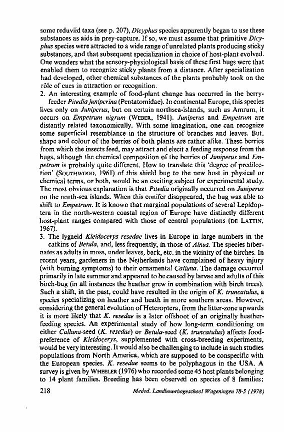
some reduviid taxa (see p. 207), Dicyphus species apparently began to use these substances as aids in prey-capture. If so, we must assume that primitive Dicyphus species were attracted to a wide range of unrelated plants producing sticky substances, and that subsequent specialization in choice of host-plant evolved. One wonders what the sensory-physiological basis of these first bugs were that enabled them to recognize sticky plants from a distance. After specialization had developed, other chemical substances of the plants probably took on the rôle of cues in attraction or recognition. 2. An interesting example of food-plant change has occurred in the berry-
feeder Pitediajuniperina (Pentatomidae). In continental Europe, this species lives only on Juniperus, but on certain northsea-islands, such as Amrum, it occurs on Empetrum nigrum (WEBER, 1941). Juniperus and Empetrum are distantly related taxonomically. With some imagination, one can recognize some superficial resemblance in the structure of branches and leaves. But, shape and colour of the berries of both plants are rather alike. These berries from which the insects feed, may attract and elecit a feeding response from the bugs, although the chemical composition of the berries of Juniperus and Empetrum is probably quite different. How to translate this 'degree of predilection' (SOUTHWOOD, 1961) of this shield bug to the new host in physical or chemical terms, or both, would be an exciting subject for experimental study. The most obvious explanation is that Pitedia originally occurred on Juniperus on the north-sea islands. When this conifer disappeared, the bug was able to shift to Empetrum. It is known that marginal populations of several Lepidop-tera in the north-western coastal region of Europe have distinctly different host-plant ranges compared with those of central populations (DE LATTIN, 1967). 3. The lygaeid Kleidocerys resedae lives in Europe in large numbers in the
catkins of Betula, and, less frequently, in those of Alnus. The species hibernates as adults in moss, under leaves, bark, etc. in the vicinity of the birches. In recent years, gardeners in the Netherlands have complained of heavy injury (with burning symptoms) to their ornamental Calluna. The damage occurred primarily in late summer and appeared to be caused by larvae and adults of this birch-bug (in all instances the heather grew in combination with birch trees). Such a shift, in the past, could have resulted in the origin of K. truncatulus, a species specializing on heather and heath in more southern areas. However, considering the general evolution of Heteroptera, from the litter-zone upwards it is more likely that K. resedae is a later offshoot of an originally heather-feeding species. An experimental study of how long-term conditioning on either Calluna-seed (K. resedae) or Betula-seed (K. truncatulus) affects food-preference of Kleidocerys, supplemented with cross-breeding experiments, would be very interesting. It would also be challenging to include in such studies populations from North America, which are supposed to be conspecific with the European species. K. resedae seems to be polyphagous in the USA. A survey is given by WHEELER (1976) who recorded some 45 host plants belonging to 14 plant families. Breeding has been observed on species of 8 families;
218 Meded. Landbouwhogeschool Wageningen 78-5 (1978)
favoured hosts other than Betula are: Typha, Spiraea, Kalmia, Pieris and Rhododendron species. Life history studies revealed further that development of populations on weed species was generally 1-2 weeks ahead ofthat on birch (WHEELER, I.e.).
Plasticity in feeding behavior of Heteroptera included : occasional switches from 1) prédation towards phytophagy, 2) phytophagy to prédation, and 3) switches in food choice within one of these two feeding categories. Switch 1) occurred at a mid point in heteropterous anagenesis, when the maxillae had lost most of their barb and when the mandibles had assumed a guiding rôle for the stylets within the host (level 3 in scheme Fig. 72, text on pages 195-198).
Switch 2) happened at higher anagenetic levels where chemo-sensory mechanisms of food-selection had presumably become more specialized, and less plastic, such that returns to carnivory became less frequent. Switches in 3) must have been more frequent in phytophagous than in carnivorous lines, considering the wide variety of hosts and the present diversity of phytophagous species (Reduviidae form an exception among carnivorous bugs). Conditioning, modification in the function of taste-receptors and varying sensitivity of receptors may influence changes in food-preferences (p. 212).
4.4. A TENTATIVE, EVOLUTIONARY SCHEME FOR HETEROPTERA
1. Evaluation of characters and construction of the diagram The ideas on heteropterous phylogeny, which I developed in 1968, were
summarized on pages 187-189 and in Fig. 71. I shall now test the validity of this model in light of the new data presented here. In Table 3 (p. 194), a rough, numerical, analysis was made of the ancestral and derived states of various characters of the feeding structures of the principal taxa. This is continued here as Table 4 incorporating other characters but following the same procedure (Pachynomidae and Thaumastocoridae are omitted). Only those characters which vary independently of other characters are used (some often changed evolutionary in combination with others). The scores 0, 1 and 2 indicate the plesiomorphic, intermediate and apomorphic states of the character in question. The relative states of these 38 characters are summarized in Fig. 74 which shows clearly the overall plesiomorphy of Gerromorpha (A). The apomorphic characters in this group are mainly confined to a few, somewhat specialized taxa such as Mesovelia and Hydrometra. Dipsocoromorpha (B) and Enicocephalomorpha (C) also have a relatively high percentage of primitive features; this percentage may be increased or decreased when the states of characters indicated by question-marks are settled. Leptopodomor-pha (D) and Nepomorpha (E) also have a proportionately high degree of plesiomorphy, but these primitive attributes are outweighed by derived ones, when columns 2 and 3 are combined.
The character content of the bars in Fig. 74, row X is not the same. A is based on 54 values, but E on 68. The reason for this is that for each character in E
Meded. Landbouwhogeschool Wageningen 78-5 (1978) 219
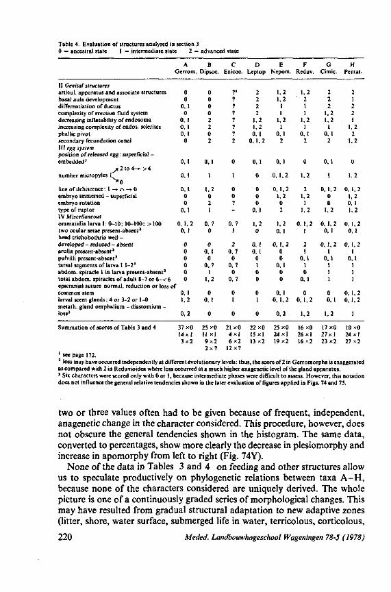
Table 4. Evaluation of structures analysed in section 3 0 = ancestral state 1 = intermediate state 2 = advanced state
11 Genital structures anicul. apparatus and associate structures basal aula development differentiation of ductus complexity of erection fluid system decreasing inflatability of endosoma increasing complexity of endos, sclerites phallic pivot secondary fecundation canal III egg system position of released egg: superficial -embedded3
^ 2 t o 4 - » >4 number micropyles 1
^ 0 line of dehiscence : I ~* n -* 0 embryo immersed - superficial embryo rotation type of ruptor IV Miscellaneous ommatidia larva 1: 0-10; 10-100; > 100 two ocular setae present-absent* head trichobothria well -developed - reduced - absent arolia present-absent3
pulvilli present-absent3
tarsal segments of larva I 1-23
abdom. spiracle 1 in larva present-absent3
total abdom. spiracles of adult 8-7 or 6-< 6
A Gerrom.
0 0
0, 1 0
0. 1 0. 1 0, 1 0
0.1
0,1
0,1 0 0
0,1
0. 1.2 0,1
0 0 0 0 0 0
epicranial suture normal, reduction or loss of common stem larval scent glands: 4 or 3-2 or 1-0 metath. gland omphatium - diastomium -loss3
0,1 1,2
0,2
B Dipsoc.
0 0 0 0 2 2 0 2
0,1
1
1,2 0 2 1
0, ? 0
0 0, I 0
0,? 1
1,2
0 0. 1
0
C Enicoc.
7' 7 ? ? ? ? ? 2
0
1
0 0 7 -
0,? 1
2 0,? 0
0,? 0
0,?
0 1
0
D Leptop.
2 2 2 2
1,2 1,2 0,1
0,1,2
0,1
0
0 0 0
0, 1
1,2 0
0, 1 0,1 0 1 0 0
0 1
0
E Nepom.
1,2 1,2 1 1
1,2 1
0.1 2
0, 1
0,1,2
0,1,2 1,2 0 2
1,2 0. 1
0 ,1,2 0 0
0,1 0 0
0, 1 0, 1,2
0,2
F Reduv.
1,2 2 1 1
1.2 1
0.1 2
0
1.2
2 1.2 1
1,2
0, 1,2 1
2 1
0.1 1 0
0,1
0 0,1,2
1,2
G Cimic.
2 2 2
1,2 1.2 1
0,1 2
0,1
1
0,1,2 0 0
1.2
0,1.2 0,1
0 ,1,2 1
0.1 1 1 1
0 0,1
1.2
H Pentat.
2 1 2 2 1
1.2 2
1.2
0
1,2
0, 1,2 1,2 0. 1 1.2
0. 1,2 0. 1
0 ,1,2 1
0,1 1 1 1
0, 1,2 0.1,2
1
Summation of scores of Table 3 and 4 37x0 14x1 3x2
21x0 4 x | 6x2
12x7
22x0 15x1 13x2
25x0 24x1 19x2
16x0 26x1 16x2
17x0 27x1 23x2
10x0 24x1 27x2
25x0 II x l 9 x2 2 x ?
1 see page 172. 3 loss may have occurred independently at different evolutionary levels: thus, the score of 2 in Gerromorpha is exaggerated as compared with 2 in Reduvioidea where loss occurred at a much higher anagenetic level of the gland apparatus. 3 Six characters were scored only with 0 or 1, because intermediate phases were difficult to assess. However, this notation does not influence the general relative tendencies shown in the later evaluation of figures applied in Figs. 74 and 75.
two or three values often had to be given because of frequent, independent, anagenetic change in the character considered. This procedure, however, does not obscure the general tendencies shown in the histogram. The same data, converted to percentages, show more clearly the decrease in plesiomorphy and increase in apomorphy from left to right (Fig. 74Y).
None of the data in Tables 3 and 4 on feeding and other structures allow us to speculate productively on phylogenetic relations between taxa A-H, because none of the characters considered are uniquely derived. The whole picture is one of a continuously graded series of morphological changes. This may have resulted from gradual structural adaptation to new adaptive zones (litter, shore, water surface, submerged life in water, terricolous, corticolous,
220 Meded. Landbouwhogeschool Wageningen 78-5 (1978)
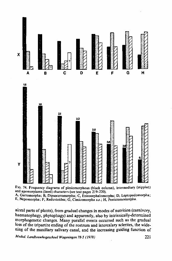
T5
18
2.1
?
FIG. 74. Frequency diagrams of ptesiomorphous (black column), intermediary (stippled) and apomorphous (lined) characters (see text pages 219-220). A, Gerromorpha; B, Dipsocoromorpha; C, Enicocephalomorpha; D, Leptopodomorpha ; E, Nepomorpha; F, Reduvioidea; G, Cimicomorpha s.S.; H, Pentatomomorpha.
aireal parts of plants), from gradual changes in modes of nutrition (carnivory, haematophagy, phytophagy) and apparently, also by intrinsically-determined morphogenetic changes. Many parallel events occurred such as the gradual loss of the tripartite ending of the rostrum and intercalary sclerites, the widening of the maxillary salivary canal, and the increasing guiding function of
Meded. Landbouwhogeschool Wageningen 78-5 (1978) 221
the mandibular stylets (all these modifications took place before any Heterop-tera had become phytophagous) ; other parallel changes appeared in reproductive organs, embryogénie processes, salivary glands, etc. Such parallelisms are difficult to explain as having arisen by action of external selection pressure, although Ross (1969) attributed the many parallelisms shown in my study on the egg-systems of Heteroptera, to be due to factors of natural selection.
The only approach possible, at this time to study the Hemipteran phylogeny seems to me to be the phyletic one, admitting at the outset its weakness of subjective weighting of characters. It is significant that the key-characters used to delimit major groups (e.g. LESTON C.S., 1954; CHINA & MILLER, 1959) are not clear-cut and not applicable to all members. For example, the reduction and ventral shift in position of antennae in Nepomorpha is gradual. Ventral, abdominal trichobothria do not characterize all families of Pentatomomorpha. In the check-list of CHINA & MILLER (1959), the 'Amphibicorisa' (conceived as Gerromorpha plus Leptopodomorpha) should be recognized as having : 'Three pairs of trichobothria on head, (difficult to observe in some genera such as Aepophilus, Hermatobates etc.), rarely only 2 pairs (Limnobatodinae), or confused with bristles (Leptopodidae and Leotichiidae)'. This contribution to construction of a family key for all Heteroptera was superb, but plesiomorphous members of other major groups also have a similar, trichobothrial, head pattern (p. 160). Recognition of the Pentatomomorpha and Cimicomorpha by LESTON C.S. (1954) was a big step forward in the classification of Heteroptera, but resulted in a number of families being left in a doubtful position. It was hardly possible at that time to speculate on the genealogical relations existing both between these two major groups and with others.
HENNIG (1969), when applying his own cladistic method to the phylogeny of the insect Orders, relied for Heteroptera upon the publications of CHINA (1955a), BECKER-MIGDISOVA in ROHDENDORF C.S. (1962), GUPTA (1963a, b), LESTON e.a. (1954), PARSONS (1964), POISSON (1951), and Ross (1965). His conclusions could only be superficial: Nepomorpha (= Cryptocerata) and the terrestrial groups (= Gymnocerata, i.e. all other Heteroptera) were considered to have a sister group relationship, which presumably originated in the Triassic or earlier. The land bugs had already split into several subunits by the Lias, but into how many and how related was not clear from the fossil record (fossils from this epoque have so far yielded only lygaeoid, coreoid, mirid and nabid types). Fossil Nepomorpha were extensively treated by POPOV (1971); they were represented abundantly both in species and in specimens in the lower Jurassic.
Although Nepomorpha do have many primitive features (see Fig. 74, column E), in my view Gerromorpha and the fore-runners of the Dipsocoro-morpha must have originated earlier. Their absence from the fossil record at this time must have some particular historical cause. Reasons could be their scattered distribution in the litter-zone, and a habitat probably less than ideal for fossilization, and their low population densities. They were also small. The chances of finding fossil Gerromorpha are thus much less than for the larger
222 Meded. Landbouwhogeschool Wageningen 78-5 (1978)
sized Nepomorpha ; bugs which frequented local water bodies and concentrated in large numbers in dry years in places ideal for fossilization. It must be remembered that late, mesozoic remains of terrestrial bugs are also relatively scarce.
The Gerromorpha and Dipsocoromorpha probably date back to the lower Mesozoic or earlier. A fossil saldid of lower to middle Jurassic age supposed to represent a saldid, has been described recently by POPOV (1973). The fossil record at present is thus too limited in diversity and in character information (mostly wings) to provide information of use in clarifying the phylogeny of Heteroptera.
The principal reason that the cladistic method cannot be used to unravel the phylogeny of Heteroptera has to do with the very gradual nature of evolution within this suborder. This progression was accompanied by an enormous amount of parallel evolution (in fact all 38 chracters in Tables 3 and 4 are subject to parallel change). Parallelism is considered here (SIMPSON, 1961) to be 'the development of similar characters separatately in two or more lineages of common ancestry and on the basis of, or channelled by, characteristics of that ancestry'. SNEATH & SOKAL (1973, p. 32-37) pointed out the implications that convergent and parallel evolution have for phenetic taxonomy. They are inclined to believe that parallel evolution is generally due to the appearance of parallel trends in a few characters only, and they opt for quantitative studies of this problem.
So far, the cladistic approach has yielded reasonably good results only in lower level analyses (e.g. in some coreid groups, SCHAEFER, 1968). An admirable attempt at a large scale analysis was undertaken by BROTHERS (1975), and resulted in the development of a new cladogram for 38 families of aculeate Hymenoptera. Another fine example was the numerical treatment of orthop-teroid insects by BLACKITH & BLACKITH (1968), although LEQUESNE (1972) showed that at least 54 of the 80 characters tested were not uniquely derived. SCHLIEPHAKE (1965) used Hennig's - techniques to solve the phylogenetic relations of the five families and some of the genera of Thysanoptera, but his use of apomorphic characters seens debatable to me.
The phylogeny of clusters of insect orders, is likewise doubtfully amendable to cladistic analysis. SCUDDER (1973), when considering the higher systematics of insects, suggested that polyphyly seems to be evident everywhere. He supported MACKERRAS (1967) in believing that the evolution of insects and the resulting natural classification was more governed by grades than by clades. This, I believe, was true also for Heteroptera as expressed in Figs. 71 and 72.
The data needed for constructing a reliable cladogram of heteropterous evolution are far from sufficient, and I refrain, for the present, from undertaking such a speculative enterprise (see pages 189-191). In part III of this series, I will attempt to reconstruct such a phylogeny using genitalic structures. As MICHENER (1970, p. 16) stated: 'The best acceptable morphological characters, that is, those which have a high content of historical information, will be those which are intricate or involve intricate structural relationship, since
Meded. Landbouwhogeschool Wageningen 78-5 (1978) 223
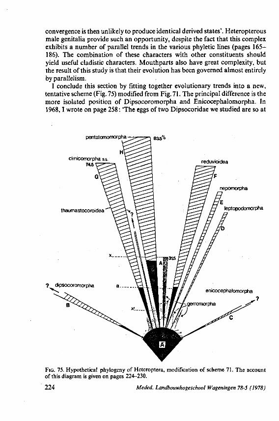
convergence is then unlikely to produce identical derived states'. Heteropterous male genitalia provide such an opportunity, despite the fact that this complex exhibits a number of parallel trends in the various phyletic lines (pages 165-186). The combination of these characters with other constituents should yield useful cladistic characters. Mouthparts also have great complexity, but the result of this study is that their evolution has been governed almost entirely by parallelism.
I conclude this section by fitting together evolutionary trends into a new, tentative scheme (Fig. 75) modified from Fig. 71. The principal difference is the more isolated position of Dipsocoromorpha and Enicocephalomorpha. In 1968,1 wrote on page 258: 'The eggs of two Dipsocoridae we studied are so at
pentatomomorpha
H
835%
ciinicomorpha s.s. 745 reduvioidea
leptopodomorpha
FIG. 75. Hypothetical phylogeny of Heteroptera, modification of scheme 71. The account of this diagram is given on pages 224-230.
224 Meded. Landbouwhogeschool Wageningen 78-5 (1978)
variance with what we now know about shape and aeropylar system of heterop-terous eggs that there is doubt whether these species belong to the same suborder' ; and on p. 378 : There is, however, a discrepancy between the Dipso-coroidea and whole the rest of Heteroptera which is greater than was concluded from the eggs. This fact will make it necessary later to modify fig. 316, which was based on egg evolution'. Indeed, Dipsocoromorpha remain a mysterious group, nothwithstanding the brilliant study devoted to one of the families by EMSLEY (1969) and, fragmentary contributions of other authors. Also, their male genitalia (p. 172), reproductive organs, the often sexual, dimorphism of their pretarsal structures (p. 118) and their spiracular system (p. 139) all deviate strongly from what is seen in other major taxa. Many dipsocoro-morph characters are highly derived; others, such as the basal mechanism of the phallus, wing venation and, to some extent, also stylet structures, are plesiomorphous. The line B of Dipsocoromorpha (Fig. 75) is shown as being open apically, because we do not, as yet, know the range of anagenesis to be expected in this group (e.g. several values in diagram Fig. 74, column B, were indicated by a question mark).
The same is true of Enicocephalomorpha (Fig. 75, line C). Ovariole type (CARAYON, 1950c; personal observation), micropylar apparatus (p. 96), eclosion procedure of prolarva from egg (p. 99), and structure of the front tarsus (p. 118) all exhibit character states unique in Heteroptera. Upper-cretaceous fossils of this group were considered by STYS (1969c), who is presently engaged in a modern treatment of Enicocephalidae (STYS, 1969b, 1970a, b). Wing-venation too is not comparable to that of recent Gerromorpha, bugs considered to be the most plesiomorphous 'typical' Heteroptera. This disjunct position of Dipsocoromorpha (B) and Enicocephalomorpha (C) in relation to other Heteroptera, is expressed in Fig. 75 by their early origin from sector A' and by their greater angle of deviation from the vertical than that shown for taxaA,D-H.
The width of the hatched areas in lines D-H (Fig. 75) show more realistically the relative number of species contained than does Fig. 71. The extremes of anagenetic development in living members of taxa D-H (Fig. 75) are estimated more accurately than in Fig. 71. The length of lines D-H (Fig. 75) correspond to the percentages of evolved characters shown in the histograms presented in Fig. 74 (the scores of columns 2 and 3 are summed). The interface between the hatched and black areas of each line in Fig. 75 represents the lower, anagenetic level of extant species. The position of this border was calculated from the reciprocal of the percentages of ancestral characters (Fig. 74, numbers given above the black columns). The lowest score of 1.5 thus gives the closest approach of living species (level X' in Fig. 75) to the archetypical conditions of sector A'.
The resulting picture (Fig. 75) differs little from Fig. 71. The lower anagenetic extreme level (the interface between hatched and black) shown for Reduvioidea is the only one exhibiting a marked difference from that of Fig. 71. Both Nepomorpha and Leptopodomorpha have reached a rather advanced upper
Meded. Landbouwhogeschool Wageningen 78-5 (1978) 225
anagenetic extreme. The highest level is attained by Pentatomomorpha (line H). This is not due to the fact that greater weight was attributed to the feeding structures of these bugs even though they are the most evolved in Heteroptera. All 38 characters selected received equal weight. Some members of Reduvioidea have the most derived embryogenesis of all Heteroptera, the reason I scored this group anagenetically highest in 1968 (Fig. 71). Using the more neutral classification of characters of this study has resulted in apomorphic extreme of this group (line F in Fig. 75) being reduced to a small extent.
The Joppeicidae (indicated by a question-mark in Fig. 316 of 1968) have now been omitted since a detailed study of Joppeicus by DAVIS & USINGER (1970) has provided evidence to. suggest that they are members of Cimico-morpha s.S.
The evolutionary phylosophy behind Fig. 75 is that typical Heteroptera (A, D-H) constituted 6 or 7 (Thaumastocoroidea are treated as an autonomous offshoot as their place is still obscure*) equivalent high rank taxa, with pre-Gerromorpha (A') as the ancestral group, and modern Gerromorpha (A) as the stem-group. Arrangement of the terrestrial groups D, F-H, side by side, is arbitrary, but the origins of E and F, nearly touching, indicate a closer mutual relationship between them than with any other group. I uphold here the two new views introduced in 1968a; the subordinate position of Gerromorpha and the division between Cimicomorpha s.S. and Reduvioidea. These will be commented upon separately below.
2. The gerromorphous stem-group A stem-group is defined by STYS (1967), following NAEF (1919), as 'a modern
group retaining many primitive features of its extinct ancestral group and differing by these features from the more derivative group (groups) of the same ancestry'. The Gerromorpha are indicated in Fig. 75 with stippling in two ways. The central, vertical ray with dense stippling (from X' to X) represents the anagenetic range existing between the extremes of the 31.5% of advanced characters of Gerromorpha (of the 38 selected for this purpose and evaluated in tables 3 and 4 and in the histogram in Fig. 74). This line is the shortest of those indicated. Its short, black, base (X') indicates that the degree of plesiomorphy in Gerromorpha is greatest of all other major groups. Both X' and X seem exceptionally high when one considers that the frequency value applies to all Gerromorpha. However, Gerromorpha are a very diversified group as was shown in section 3, even though they share similar mouth-part-structure and function. Recall that such stylet structures were found also in representatives of taxa B, C, E, F and G.
* Note remarks made on the pretarsus on p. 119. Whether Thaumastocoroidea belong within one of the major groups or originated independently from Gerromorpha is still open to question. In 1968 I studied their shell structure. SCHAEFER (1969) concluded from these data that Thaumastocoroidea are possibly annectant between Cimicomorpha and Pentatomomorpha. This is not my view. STYS & KERZHNER (1975) classified them provisionally within the Cimicomorpha s.l.
226 Meded. Landbouwhogeschool Wageningen 78-5 (1978)
The pseudopodial representation of Gerromorpha (less densely stippled, irregular area surrounding the surface of sector A ) is more realistic. It shows the innate potentials of this group that are realized, in a rather haphazard manner, in other major groups. The individual taxa of surface bugs show a very low, and varying level of anagenesis, and more importantly, unrelated tendencies that connect some of the terrestrial groups. Some examples are given below : a. The secondary fecundation canal present in nearly all adult female Gerro
morpha, also occurs in primitive members of Leptopodomorpha and rudiments of it are found in some Pentatomomorpha. b. Cephalic trichobothria, best-developed in Gerromorpha, occur also in
primitive members of groups B, D, E (probably), H and G, again with gradual loss in more specialized members. c. Hydrometridae is the only gerromorphous family completely lacking
eye-trichobothria even in first instar larvae and having strong endosomal reduction (with spermatophore-production. I predicted this in 1968, and it has now been confirmed by K. DEN BIEMAN, internal student report, 1976, Wageningen). The combination of these two characters is found only in Reduvioidea: the eye-character in all Reduviidae checked on this point, and the phallic character in more specialized members of Reduviidae. The stilt-legged condition of hydrometrids, associated with slow-motion and rocking behaviour and with reduction or loss of larval defensive glands is paralleled in primitive Reduviidae, Nepomorpha and Pentatomomorpha (p. 145). d. The end apparatus of the phallus of Mesovelia resembles equivalent struc
tures in Nabidae. An aberrant Mesovelia sp. from New Zealand also has a similar basal sperm-conduit and a similarily-developed bulbus ejaculatorius (p. 172). e. The ductus ejaculatorius, primitively thin, circular in cross-section and
coiled when not functioning, has become greatly shortened, flattened and highly distensible in Mesoveloidea, Heterocleptes and the Macrovelia-group (p. 170, 172). This condition is further elaborated in Leptopodomorpha and in other high rank groups. f. An embryonic, cuticular, eclosion bladder is present in both Mesoveliidae
and Nabidae (Fig. 45E, O). g. Although the basal articulatory apparatus of the phallus of representative
Gerromorpha and groups D-H (Fig. 75), seem to differ greatly, some characteristics found in genitalia of some aberrant Gerromorpha appear to bridge the gaps. The simple, trough-shaped excavation of the pygophore diaphragm, so typical for Gerromorpha, has become a dorsally-closed cylinder in Ocellovelia, an aberrant genus of this group (p. 172). This change represents a first step towards formation of the basal aula, present in Nepomorpha and the terrestrial groups. Anchorage of the ductus seminis to a strut-like structure within the phallus base, a condition seen in most terrestrial major groups, is already realized in the gerromorphous Macrovelia, Oravelia and Chepuvelia (p. 172). h. Transformation of the typical gerromorphous pretarsus (arolia and parem-
podia) occur only in a few aberrant surface bugs (p. 115). Further reduction of
Meded. Landbouwhogeschool Wageningen 78-5 (1978) 227
these structures occurred in Leptopodomorpha (p. 115). The bladder-like parempodia of the aberrant hydrometrid Heterocleptes are unique for Ger-romorpha(p. 115) and are reminiscent of structures found in more specialized members of Cimicomorpha s.s.
In listing these gradient features, I do not mean to suggest that straight-line evolutionary transition from A towards D-H (Fig. 75) always occurred. The facts suggest, however, that Gerromorpha had the potential for evolutionary diversification. This potential was realized only fragmentarily in modern surface bugs, but was expressed on a larger scale in the other phyletic lines, originating from this stem-group. The many anagenetic, parallelisms shown to exist in this study, could thus have common genetic components.
The idea that taxa D-H arose from gerromorphous-like ancestors via grade evolution resembles the situation in pentatomomorph Coreoidea as put forward by STYS (1965, 1967b). A survey of different opinion on the phylogeny of the lygaeid-coreid complex of families was given earlier (COBBEN, 1968a, p. 353). The existence of many controversies in the literature are understandable if STYS' hypothesis is correct. He included 10 families in the Coreoidea, the Lygaeidae having about the same evolutionary rôle in this taxon as Gerromorpha had in Heteroptera. Nine coreoid families could have arisen from different pre-lygaeid subfamilies (Fig. 76, after STYS). The arguments supporting this arrangement are given on pages 421-423 in STYS (1967b), and are entirely relevant to our own case of evolution in Heteroptera.
Major groups A, D-H (Fig. 75) form a close unit, whereas groups B and C are isolated branches. This configuration is best solved categorically by giving both Dipsocoromorpha and Enicocephalomorpha separate ranks equal to that of the major groups combined. I will not formalize this view nomen-claturally until more detailed information on Dipsocoromorpha and Enicocephalomorpha to the depth known for typical Heteroptera has accumulated.
STYS & KERZHNER (1975) considered all '-morpha' groups to be of equal rank as infra-orders and discussed the nomenclatural alternatives when Heteroptera is treated as an Order or Suborder. It is clear, when Heteroptera are divided into Dipsocoromorpha, Enicocephalomorpha, and let us say, Heteroptera s.s., that the '-morpha' notation becomes inconsistent (the position of Coleorrhyncha remains uncertain, although SCHLEE (1969b) included them with Heteroptera to form the Heteropteroidea (see p. 190)). If Heteroptera are regarded as a distinct Order, the three subdivisions would merit subordinal status*. If Heteroptera are regarded as a Suborder, further subdivision into three infraorders would be the consequence, and the '-morpha' groups of the Heteroptera s.s. would have to be lowered in rank. The suggestion of POPOV (1971) to divide living Heteroptera into three suborders: 1. Nepomorpha + Leptopodomorpha, 2. Enicocephalomorpha + Cimicomorpha, and 3. Penta-tomomorpha, conflicts with the view presented here.
* STYS (1970C) prefers to consider the Dipsocoromorpha as a suborder, but it is not clear from his paper whether he used Heteroptera in the sense of Order or Suborder.
228 Meded. Landbouwhogeschool Wageningen 78-5 (1978)
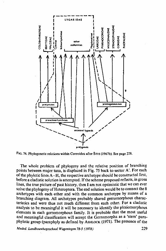
prelygaeids
FIG. 76. Phylogenese relations within Coreoidea after STYS (1967b). See page 228.
The whole problem of phylogeny and the relative position of branching points between major taxa, is displaced in Fig. 75 back to sector A'. For each of the phyletic lines A-H, the respective archetype should be constructed first, before a cladistic solution is attempted. If the scheme proposed reflects, in gross lines, the true picture of past history, then I am not optimistic that we can ever solve the phylogeny of Heteroptera. The end solution would be to connect the 8 archetypes with each other and with the common archetype by means of a branching diagram. All archetypes probably shared gerromorphous characteristics and were thus not much different from each other. For a cladistic analysis to be meaningful it will be necessary to identify the plesiomorphous elements in each gerromorphous family. It is probable that the most useful and meaningful classification will accept the Gerromorpha as a 'stem' para-phyletic group (paraphyly as defined by ASHLOCK (1971). The presence of the
Meded. Landbouwhogeschool Wageningen 78-5 (1978) 229

diverging, basic, phyletic lines B and C, considered now as atypical Heteroptera, make it impossible to decide for sure whether the common archetype of Heteroptera was a gerromorphous, a dipsocoromorphous or an enicocephalo-morphous-like bug.
I concluded in 1968 that the heteropterous archetype could have been a gerromorphous-like insect, and I still believe this when I consider the characters analysed in this paper. Wing-venation was not considered, and I ended my 1968 contribution (p. 378) with the sentence: 'The data from wing venation seem at first sight contradictory to our main conclusion that Amphibicorisae are the most ancestral bugs'. I finish now by saying that the final discussion may focus on the following dilemma: Did we have a gerromorphous ancestor with a dipsocoromorphous or an enicocephalomorphous wing type, or a dispoco-romorphous or an enicocephalomorphous ancestor with gerromorphous reproductive organs and ectodermal genitalia? I doubt whether use of the Hennig-method will here give us a decisive answer.
3. The discrepancies between Cimicomorpha s.S. and Reduvioidea A preliminary proposal, put forward in 1968, to keep Cimicomorpha s.S.
and Reduvioidea separate as two major groups of a rank about equivalent to that of the other groups depicted in Fig. 71, is still supported (Fig. 75). Some colleagues disagreed with this separation (CARAYON, 1971; KERZHNER, 1974; POPOV, 1971). STYS& KERZHNER (1975) continued to treat Cimicomorpha in the original broad sense of LESTON e.a. (1954) in which Reduvioidea is included. STYS in STYS & KERZHNER (I.e.) stated, however, that this thesis is purposely conservative for the time being, because he felt that the Cimicomorpha s.l. represents 'probably the most heterogeneous and controversial infraorder of all those proposed'. The differences between the two groups are listed in Table 5.
Most of these characters have no cladistic value because they show parellel development, but their great number irregularly varying in both taxa, suggests that the origin of G and F and their mutual relations are not fundamentally different from those existing between the clusters G-D in relation to stem group A in Fig. 75. Characters 2-5 (of Table 5) of the head capsule and pronotum, character 15, and those of the egg (21-23) are more plesiomorphous in Cimicomorpha s.S. than in Reduvioidea. On the other hand, characters 14 and 17 and those related to feeding (6-13) and insemination (18-20) are more primitive in Reduvioidea. The discrepancy between G and F is clearly evident in those taxa within each phyletic line which have adopted haematophagy, the Cimicidae and Triatominae respectively. In contrast to Triatoma spp., Cimex spp. have rudimentary maxillary barbs, perform only very limited maxillary displacement protrude the mandibular stylets to the same depth as the maxillary stylets during feeding, and, finally, fold the labial segments during feeding. The feeding procedure of Cimex spp. is more like that of phytophagous bugs, whereas haematophagous reduviids behave more like originally predatory bugs.
It is surprising that phyletic line G (Cimicomorpha s.s.) (Fig. 75), constituting some 10 families, displays about the same range of anagenetic progression
230 Meded. Landbouwhogeschool Wageningen 78-5 (1978)
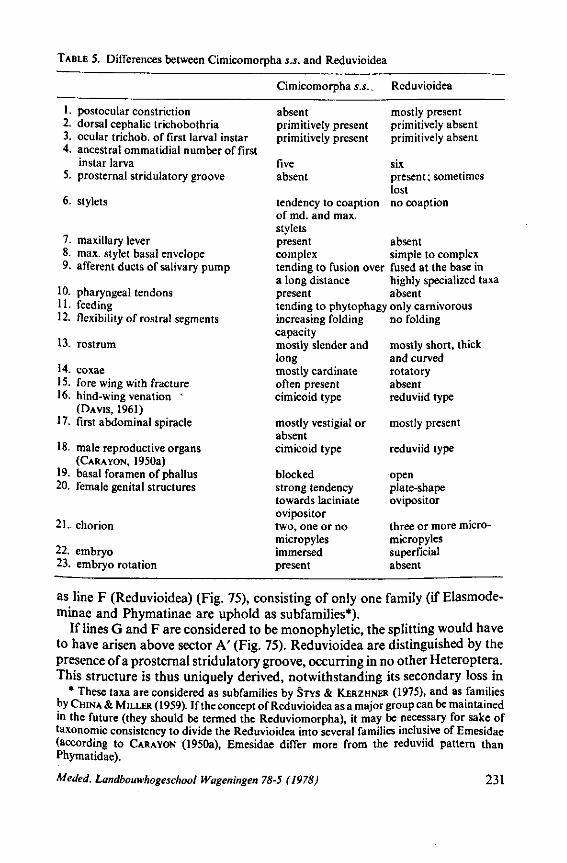
TABLE 5. Differences between Cimicomorpha s.s. and Reduvioidea
1. postocular constriction 2. dorsal cephalic trichobothria 3. ocular trichob. of first larval instar 4. ancestral ommatidial number of first
instar larva 5. prosternai stridulatory groove
6. stylets
7. maxillary lever 8. max. stylet basal envelope 9. afferent ducts of salivary pump
10. pharyngeal tendons 11. feeding 12. flexibility of rostral segments
13. rostrum
14. coxae 15. fore wing with fracture 16. hind-wing venation '
(DAVIS, 1961) 17. first abdominal spiracle
18. male reproductive organs (CARA YON, 1950a)
19. basal foramen of phallus 20. female genital structures
21. chorion
22. embryo 23. embryo rotation
Cimicomorpha s.s.,
absent primitively present primitively present
five absent
tendency to coaption of md. and max. stylets present complex
Reduvioidea
mostly present primitively absent primitively absent
six present; sometimes lost no coaption
absent simple to complex
tending to fusion over fused at the base in a long distance present
highly specialized taxa absent
tending to phytophagy only carnivorous increasing folding capacity mostly slender and long mostly cardinate often present cimicoid type
mostly vestigial or absent cimicoid type
blocked strong tendency towards laciniate ovipositor two, one or no micropyles immersed present
no folding
mostly short, thick and curved rotatory absent reduviid type
mostly present
reduviid type
open plate-shape ovipositor
three or more micro-micropyles superficial absent
as line F (Reduvioidea) (Fig. 75), consisting of only one family (if Elasmode-minae and Phymatinae are uphold as subfamilies*).
If lines G and F are considered to be monophyletic, the splitting would have to have arisen above sector A' (Fig. 75). Reduvioidea are distinguished by the presence of a prosternai stridulatory groove, occurring in no other Heteroptera. This structure is thus uniquely derived, notwithstanding its secondary loss in
* These taxa are considered as subfamilies by STYS & KERZHNER (1975), and as families by CHINA & MILLER (1959). If the concept of Reduvioidea as a major group can be maintained in the future (they should be termed the Reduviomorpha), it may be necessary for sake of taxonomie consistency to divide the Reduvioidea into several families inclusive of Emesidae (according to CARA YON (1950a), Emesidae differ more from the reduviid pattern than Phymatidae).
Meded. Landbouwhogeschool Wageningen 78-5 (1978) 231
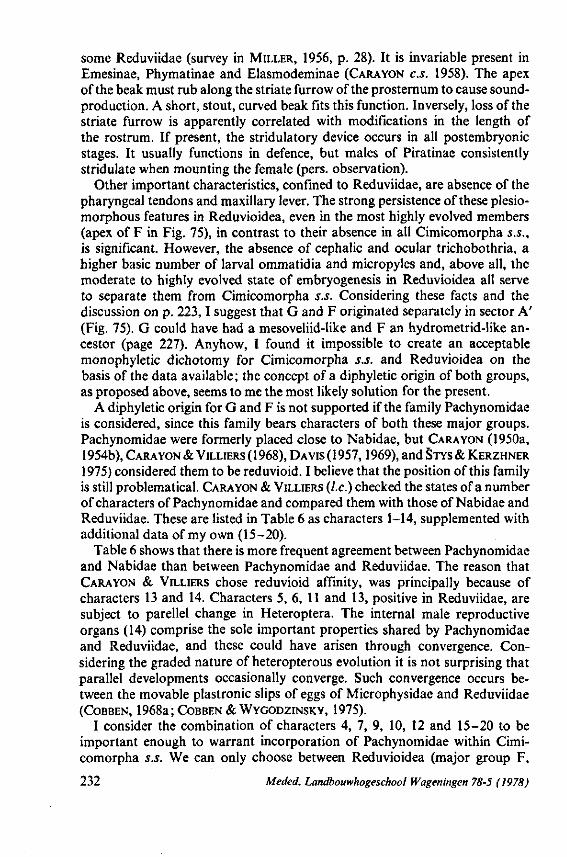
some Reduviidae (survey in MILLER, 1956, p. 28). It is invariable present in Emesinae, Phymatinae and Elasmodeminae (CARAYON C.S. 1958). The apex of the beak must rub along the striate furrow of the prosternum to cause sound-production. A short, stout, curved beak fits this function. Inversely, loss of the striate furrow is apparently correlated with modifications in the length of the rostrum. If present, the stridulatory device occurs in all postembryonic stages. It usually functions in defence, but males of Piratinae consistently stridulate when mounting the female (pers. observation).
Other important characteristics, confined to Reduviidae, are absence of the pharyngeal tendons and maxillary lever. The strong persistence of these plesio-morphous features in Reduvioidea, even in the most highly evolved members (apex of F in Fig. 75), in contrast to their absence in all Cimicomorpha s.S., is significant. However, the absence of cephalic and ocular trichobothria, a higher basic number of larval ommatidia and micropyles and, above all, the moderate to highly evolved state of embryogenesis in Reduvioidea all serve to separate them from Cimicomorpha s.S. Considering these facts and the discussion on p. 223,1 suggest that G and F originated separately in sector A' (Fig. 75). G could have had a mesoveliid-like and F an hydrometrid-like ancestor (page 227). Anyhow, I found it impossible to create an acceptable monophyletic dichotomy for Cimicomorpha s.s. and Reduvioidea on the basis of the data available; the concept of a diphyletic origin of both groups, as proposed above, seems to me the most likely solution for the present.
A diphyletic origin for G and F is not supported if the family Pachynomidae is considered, since this family bears characters of both these major groups. Pachynomidae were formerly placed close to Nabidae, but CARA YON (1950a, 1954b), CARA YON & VILLIERS (1968), DAVIS (1957,1969), and STYS& KERZHNER
1975) considered them to be reduvioid. I believe that the position of this family is still problematical. CARA YON & VILLIERS (I.e.) checked the states of a number of characters of Pachynomidae and compared them with those of Nabidae and Reduviidae. These are listed in Table 6 as characters 1-14, supplemented with additional data of my own (15-20).
Table 6 shows that there is more frequent agreement between Pachynomidae and Nabidae than between Pachynomidae and Reduviidae. The reason that CARAYON & VILLIERS chose reduvioid affinity, was principally because of characters 13 and 14. Characters 5, 6, 11 and 13, positive in Reduviidae, are subject to parellel change in Heteroptera. The internal male reproductive organs (14) comprise the sole important properties shared by Pachynomidae and Reduviidae, and these could have arisen through convergence. Considering the graded nature of heteropterous evolution it is not surprising that parallel developments occasionally converge. Such convergence occurs between the movable plastronic slips of eggs of Microphysidae and Reduviidae (COBBEN, 1968a; COBBEN & WYGODZINSKY, 1975).
I consider the combination of characters 4, 7, 9, 10, 12 and 15-20 to be important enough to warrant incorporation of Pachynomidae within Cimicomorpha s.S. We can only choose between Reduvioidea (major group F,
232 Meded. Landbouwhogeschool Wageningen 78-5 (1978)
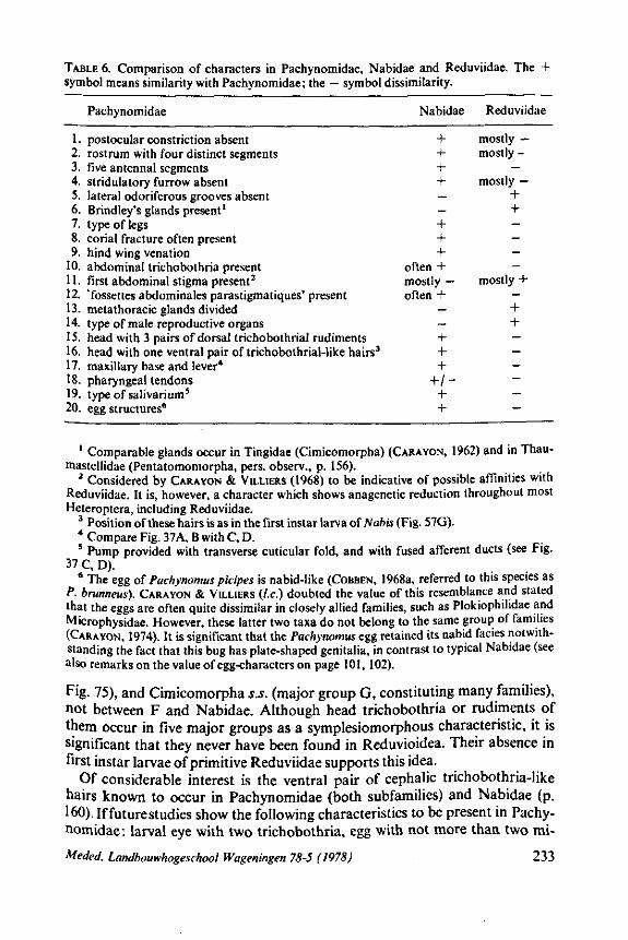
+ + + + _ _ + + +
often + mostly — often +
_ _ + + 4-
+ / -+ +
mostly — mostly -
— mostly —
+ + — — — -
mostly + — + + -— — — — —
TABLE 6. Comparison of characters in Pachynomidae, Nabidae and Reduviidae. The + symbol means similarity with Pachynomidae; the — symbol dissimilarity.
Pachynomidae Nabidae Reduviidae
1. postocular constriction absent 2. rostrum with four distinct segments 3. five antennal segments 4. stridulatory furrow absent 5. lateral odoriferous grooves absent 6. Brindley's glands present1
7. type of legs 8. corial fracture often present 9. hind wing venation
10. abdominal trichobothria present 11. first abdominal stigma present2
12. 'fossettes abdominales parastigmatiques' present 13. metathoracic glands divided 14. type of male reproductive organs 15. head with 3 pairs of dorsal trichobothrial rudiments 16. head with one ventral pair of trichobothrial-like hairs3
17. maxillary base and lever* 18. pharyngeal tendons 19. type of sal i va ri um 5
20. egg structures6
1 Comparable glands occur in Tingidae (Cimicomorpha) (CARAYON, 1962) and in Thau-mastellidae (Pentatomomorpha, pers. observ., p. 156).
2 Considered by CARAYON & VILLIERS (1968) to be indicative of possible affinities with Reduviidae. It is, however, a character which shows anagenetic reduction throughout most Heteroptera, including Reduviidae.
3 Position of these hairs is as in the first instar larva ofNabis (Fig. 57G). * Compare Fig. 37A, B with C, D. 5 Pump provided with transverse cuticular fold, and with fused afferent ducts (see Fig.
37 C, D). 6 The egg of Pachynomus picipes is nabid-like (COBBEN, 1968a, referred to this species as
P. brunneus). CARAYON & VILLIERS (I.e.) doubted the value of this resemblance and stated that the eggs are often quite dissimilar in closely allied families, such as Plokiophilidae and Microphysidae. However, these latter two taxa do not belong to the same group of families (CARAYON, 1974). It is significant that the Pachynomus egg retained its nabid facies notwithstanding the fact that this bug has plate-shaped genitalia, in contrast to typical Nabidae (see also remarks on the value of egg-characters on page 101, 102).
Fig. 75), and Cimicomorpha s.s. (major group G, constituting many families), not between F and Nabidae. Although head trichobothria or rudiments of them occur in five major groups as a symplesiomorphous characteristic, it is significant that they never have been found in Reduvioidea. Their absence in first instar larvae of primitive Reduviidae supports this idea.
Of considerable interest is the ventral pair of cephalic trichobothria-like hairs known to occur in Pachynomidae (both subfamilies) and Nabidae (p. 160). If future studies show the following characteristics to be present in Pachynomidae: larval eye with two trichobothria, egg with not more than two mi-
Meded. Landbouwhogeschool Wageningen 78-5 (1978) 233
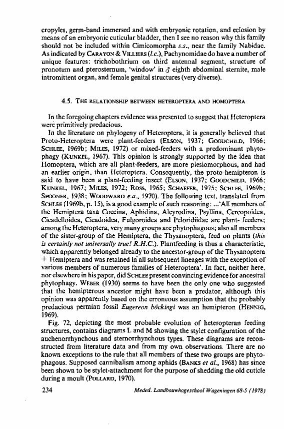
cropyles, germ-band immersed and with embryonic rotation, and eclosion by means of an embryonic cuticular bladder, then I see no reason why this family should not be included within Cimicomorpha s.S., near the family Nabidae. As indicated by CARA YON & VILLIERS (I.e.), Pachynomidae do have a number of unique features: trichobothrium on third antennal segment, structure of pronotum and pterosternum, 'window' in <$ eighth abdominal sternite, male intromittent organ, and female genital structures (very diverse).
4.5. THE RELATIONSHIP BETWEEN HETEROPTERA AND HOMOPTERA
In the foregoing chapters evidence was presented to suggest that Heteroptera were primitively predacious.
In the literature on phylogeny of Heteroptera, it is generally believed that Proto-Heteroptera were plant-feeders (ELSON, 1937; GOODCHILD, 1966; SCHLEE, 1969b; MILES, 1972) or mixed-feeders with a predominant phyto-phagy (KUNKEL, 1967). This opinion is strongly supported by the idea that Homoptera, which are all plant-feeders, are more plesiomorphous, and had an earlier origin, than Heteroptera. Consequently, the proto-hemipteron is said to have been a plant-feeding insect (ELSON, 1937; GOODCHILD, 1966; KUNKEL, 1967; MILES, 1972; Ross, 1965; SCHAEFER, 1975; SCHLEE, 1969b; SPOONER, 1938; WOODWARD e.a., 1970). The following text, translated from SCHLEE (1969b, p. 15), is a good example of such reasoning: ...'All members of the Hemiptera taxa Coccina, Aphidina, Aleyrodina, Psyllina, Cercopoidea, Cicadelloidea, Cicadoidea, Fulgoroidea and Peloridiidae are plant- feeders; among the Heteroptera, very many groups are phytophagous; also all members of the sister-group of the Hemiptera, the Thysanoptera, feed on plants (this is certainly not universally true! R.H.C.). Plantfeeding is thus a characteristic, which apparently belonged already to the ancestor-group of the Thysanoptera + Hemiptera and was retained in all subsequent lineages with the exception of various members of numerous families of Heteroptera'. In fact, neither here, nor elsewhere in his paper, did SCHLEE present convincing evidence for ancestral phytophagy. WEBER (1930) seems to have been the only one who suggested that the hemipterous ancestor might have been a predator, although this opinion was apparently based on the erroneous assumption that the probably predacious permian fossil Eugereon böckingi was an hemipteron (HENNIG,
1969).
Fig. 72, depicting the most probable evolution of heteropteran feeding structures, contains diagrams L and M showing the stylet configuration of the auchenorrhynchous and sternorrhynchous types. These diagrams are reconstructed from literature data and from my own observations. There are no known exceptions to the rule that all members of these two groups are phytophagous. Supposed cannibalism among aphids (BANKS et ai, 1968) has since been shown to be stylet-attachment for the purpose of shedding the old cuticle during a moult (POLLARD, 1970).
234 Meded. Landbouwhogeschool Wageningen 68-5 (1978)
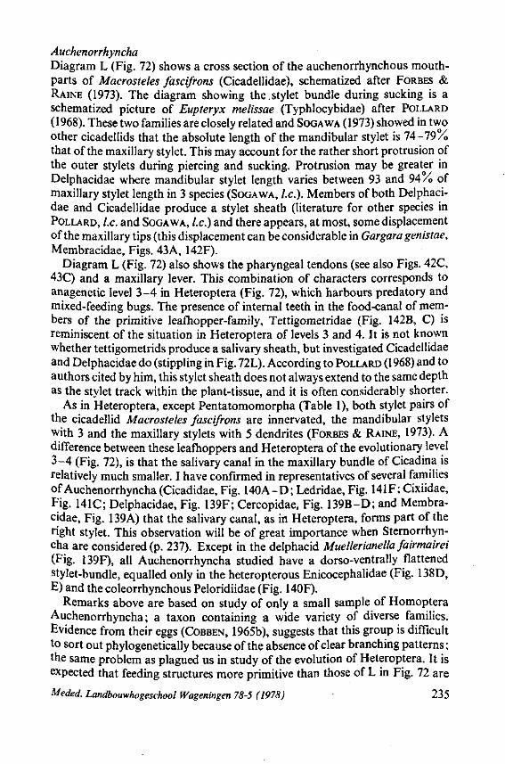
Auchenorrhyncha Diagram L (Fig. 72) shows a cross section of the auchenorrhynchous mouth-parts of Macrosteles fascifrons (Cicadellidae), schematized after FORBES & RAINE (1973). The diagram showing the.stylet bundle during sucking is a schematized picture of Eupteryx melissae (Typhlocybidae) after POLLARD (1968). These two families are closely related and SOGAWA (1973) showed in two other cicadellids that the absolute length of the mandibular stylet is 74-79% that of the maxillary stylet. This may account for the rather short protrusion of the outer stylets during piercing and sucking. Protrusion may be greater in Delphacidae where mandibular stylet length varies between 93 and 94 A of maxillary stylet length in 3 species (SOGAWA, I.e.). Members of both Delphacidae and Cicadellidae produce a stylet sheath (literature for other species in POLLARD, I.e. and SOGAWA, I.e.) and there appears, at most, some displacement of the maxillary tips (this displacement can be considerable in Gargara genistae, Membracidae, Figs. 43A, 142F).
Diagram L (Fig. 72) also shows the pharyngeal tendons (see also Figs. 42C, 43C) and a maxillary lever. This combination of characters corresponds to anagenetic level 3-4 in Heteroptera (Fig. 72), which harbours predatory and mixed-feeding bugs. The presence of internal teeth in the food-canal of members of the primitive leafhopper-family, Tettigometridae (Fig. 142B, C) is reminiscent of the situation in Heteroptera of levels 3 and 4. It is not known whether tettigometrids produce a salivary sheath, but investigated Cicadellidae and Delphacidae do (stippling in Fig. 72L). According to POLLARD (1968) and to authors cited by him, this stylet sheath does not always extend to the same depth as the stylet track within the plant-tissue, and it is often considerably shorter.
As in Heteroptera, except Pentatomomorpha (Table 1), both stylet pairs of the cicadellid Macrosteles fascifrons are innervated, the mandibular stylets with 3 and the maxillary stylets with 5 dendrites (FORBES & RAINE, 1973). A difference between these leafhoppers and Heteroptera of the evolutionary level 3-4 (Fig. 72), is that the salivary canal in the maxillary bundle of Cicadina is relatively much smaller. I have confirmed in representatives of several families of Auchenorrhyncha (Cicadidae, Fig. 140A-D; Ledridae, Fig. 141F; Cixiidae, Fig. 141C; Delphacidae, Fig. 139F; Cercopidae, Fig. 139B-D; and Membracidae, Fig. 139A) that the salivary canal, as in Heteroptera, forms part of the right stylet. This observation will be of great importance when Sternorrhyn-cha are considered (p. 237). Except in the delphacid Muellerianella fairmairei (Fig. 139F), all Auchenorrhyncha studied have a dorso-ventrally flattened stylet-bundle, equalled only in the heteropterous Enicocephalidae (Fig. 138D, E) and the coleorrhynchous Peloridiidae (Fig. 140F).
Remarks above are based on study of only a small sample of Homoptera Auchenorrhyncha; a taxon containing a wide variety of diverse families. Evidence from their eggs (COBBEN, 1965b), suggests that this group is difficult to sort out phylogenetically because of the absence of clear branching patterns; the same problem as plagued us in study of the evolution of Heteroptera. It is expected that feeding structures more primitive than those of L in Fig. 72 are
Meded. Landbouwhogeschool Wageningen 78-5 (1978) 235
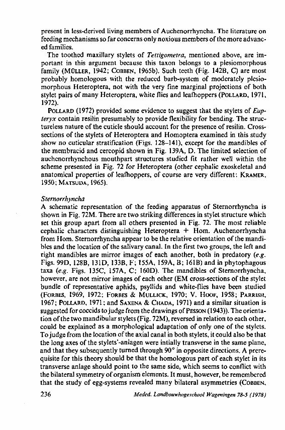
present in less-derived living members of Auchenorrhyncha. The literature on feeding mechanisms so far concerns only noxious members of the more advanced families.
The toothed maxillary stylets of Tettigometra, mentioned above, are important in this argument because this taxon belongs to a plesiomorphous family (MÜLLER, 1942; COBBEN, 1965b). Such teeth (Fig. 142B, C) are most probably homologous with the reduced barb-system of moderately plesiomorphous Heteroptera, not with the very fine marginal projections of both stylet pairs of many Heteroptera, white flies and leafhoppers (POLLARD, 1971, 1972).
POLLARD (1972) provided some evidence to suggest that the stylets of Eup-ieryx contain resilin presumably to provide flexibility for bending. The structureless nature of the cuticle should account for the presence of resilin. Cross-sections of the stylets of Heteroptera and Homoptera examined in this study show no cuticular stratification (Figs. 128-141), except for the mandibles of the membracid and cercopid shown in Fig. 139A, D. The limited selection of auchenorrhynchous mouthpart structures studied fit rather well within the scheme presented in Fig. 72 for Heteroptera (other cephalic exoskeletal and anatomical properties of leafhoppers, of course are very different : KRAMER, 1950; MATSUDA, 1965).
Sternorrhyncha A schematic representation of the feeding apparatus of Sternorrhyncha is shown in Fig. 72M. There are two striking differences in stylet structure which set this group apart from all others presented in Fig. 72. The most reliable cephalic characters distinguishing Heteroptera + Hom. Auchenorrhyncha from Hom. Sternorrhyncha appear to be the relative orientation of the mandibles and the location of the salivary canal. In the first two groups, the left and right mandibles are mirror images of each another, both in predatory {e.g. Figs. 99D, 128B, 131D, 133B, F; 155A, 159A, B; 161B) and in phytophagous taxa {e.g. Figs. 135C, 157A, C; 160D). The mandibles of Sternorrhyncha, however, are not mirror images of each other (EM cross-sections of the stylet bundle of representative aphids, psyllids and white-flies have been studied (FORBES, 1969, 1972; FORBES & MULLICK, 1970; V. HOOF, 1958; PARRISH, 1967; POLLARD, 1971 ; and SAXENA & CHADA, 1971) and a similar situation is suggested for coccids to judge from the drawings of PESSON (1943)). The orientation of the two mandibular stylets (Fig. 72M), reversed in relation to each other, could be explained as a morphological adaptation of only one of the stylets. To judge from the location of the axial canal in both stylets, it could also be that the long axes of the stylets'-anlagen were intially transverse in the same plane, and that they subsequently turned through 90° in opposite directions. A prerequisite for this theory should be that the homologous part of each stylet in its transverse anläge should point to the same side, which seems to conflict with the bilateral symmetry of organism elements. It must, however, be remembered that the study of egg-systems revealed many bilateral asymmetries (COBBEN,
236 Meded. Landbouwhogeschool Wageningen 78-5 (1978)
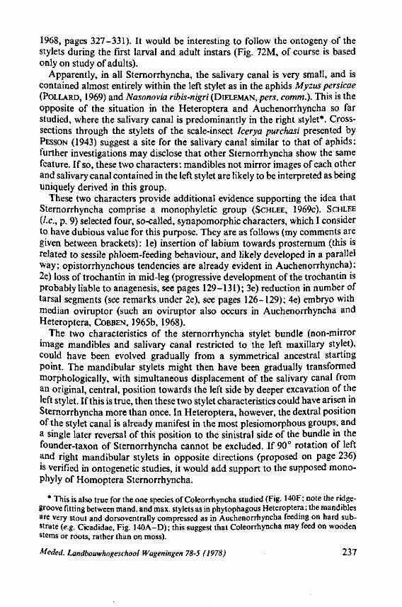
1968, pages 327-331). It would be interesting to follow the ontogeny of the stylets during the first larval and adult instars (Fig. 72M, of course is based only on study of adults).
Apparently, in all Sternorrhyncha, the salivary canal is very small, and is contained almost entirely within the left stylet as in the aphids Myzus persicae (POLLARD, 1969) and Nasonovia ribis-nigri (DIELEMAN, pers. comm.). This is the opposite of the situation in the Heteroptera and Auchenorrhyncha so far studied, where the salivary canal is predominantly in the right stylet*. Cross-sections through the stylets of the scale-insect Icerya purchasi presented by PESSON (1943) suggest a site for the salivary canal similar to that of aphids: further investigations may disclose that other Sternorrhyncha show the same feature. If so, these two characters: mandibles not mirror images of each other and salivary canal contained in the left stylet are likely to be interpreted as being uniquely derived in this group.
These two characters provide additional evidence supporting the idea that Sternorrhyncha comprise a monophyletic group (SCHLEE, 1969C). SCHLEE (/.c, p. 9) selected four, so-called, synapomorphic characters, which I consider to have dubious value for this purpose. They are as follows (my comments are given between brackets): le) insertion of labium towards prosternum (this is related to sessile phloem-feeding behaviour, and likely developed in a parallel way; opistorrhynchous tendencies are already evident in Auchenorrhyncha): 2e) loss of trochantin in mid-leg (progressive development of the trochantin is probably liable to anagenesis, see pages 129-131); 3e) reduction in number of tarsal segments (see remarks under 2e), see pages 126-129); 4e) embryo with median oviruptor (such an oviruptor also occurs in Auchenorrhyncha and Heteroptera, COBBEN, 1965b, 1968).
The two characteristics of the sternorrhyncha stylet bundle (non-mirror image mandibles and salivary canal restricted to the left maxillary stylet), could have been evolved gradually from a symmetrical ancestral starting point. The mandibular stylets might then have been gradually transformed morphologically, with simultaneous displacement of the salivary canal from an original, central, position towards the left side by deeper excavation of the left stylet. If this is true, then these two stylet characteristics could have arisen in Sternorrhyncha more than once. In Heteroptera, however, the dextral position of the stylet canal is already manifest in the most plesiomorphous groups, and a single later reversal of this position to the sinistral side of the bundle in the founder-taxon of Sternorrhyncha cannot be excluded. If 90° rotation of left and right mandibular stylets in opposite directions (proposed on page 236) is verified in ontogenetic studies, it would add support to the supposed mono-phyly of Homoptera Sternorrhyncha.
* This is also true for the one species of Coleorrhyncha studied (Fig. 140F; note the ridge-groove fitting between mand. and max. stylets as in phytophagous Heteroptera ; the mandibles are very stout and dorsoventrally compressed as in Auchenorrhyncha feeding on hard substrate (e.g. Cicadidae, Fig. 140A-D); this suggest that Coleorrhyncha may feed on wooden stems or roots, rather than on moss).
Meded. Landbouwhogeschool Wageningen 78-5 (1978) 237
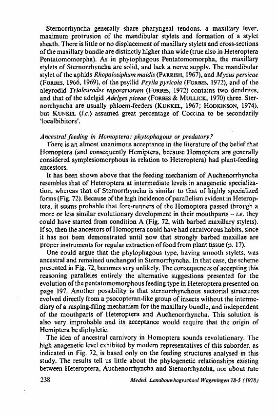
Sternorrhyncha generally share pharyngeal tendons, a maxillary lever, maximum protrusion of the mandibular stylets and formation of a stylet sheath. There is little or no displacement of maxillary stylets and cross-sections of the maxillary bundle are distinctly higher than wide (true also in Heteroptera Pentatomomorpha). As in phytophagous Pentatomomorpha, the maxillary stylets of Sternorrhyncha are solid, and lack a nerve supply. The mandibular stylet of the aphids Rhopalosiphum maidis (PARRISH, 1967), and Myzus persicae (FORBES, 1966, 1969), of the psyllid Psylla pyricola (FORBES, 1972), and of the aleyrodid Trialeurodes vaporariorum (FORBES, 1972) contains two dendrites, and that of the adelgid Adelges piceae (FORBES & MULLICK, 1970) three. Sternorrhyncha are usually phloem-feeders (KUNKEL, 1967; HODKINSON, 1974), but KUNKEL (I.e.) assumed great percentage of Coccina to be secondarily 'localbibitors'.
Ancestral feeding in Homoptera: phytophagous or predatory? There is an almost unanimous acceptance in the literature of the belief that
Homoptera (and consequently Hemiptera, because Homoptera are generally considered symplesiomorphous in relation to Heteroptera) had plant-feeding ancestors.
It has been shown above that the feeding mechanism of Auchenorrhyncha resembles that of Heteroptera at intermediate levels in anagenetic specialization, whereas that of Sternorrhyncha is similar to that of highly specialized forms (Fig. 72). Because of the high incidence of parallelism evident in Heteroptera, it seems probable that fore-runners of the Homoptera passed through a more or less similar evolutionary development in their mouthparts - i.e. they could have started from condition A (Fig. 72, with barbed maxillary stylets). If so, then the ancestors of Homoptera could have had carnivorous habits, since it has not been demonstrated until now that strongly barbed maxillae are proper instruments for regular extraction of food from plant tissue (p. 17).
One could argue that the phytophagous type, having smooth stylets, was ancestral and remained unchanged in Sternorrhyncha. In that case, the scheme presented in Fig. 72, becomes very unlikely. The consequences of accepting this reasoning paralleles entirely the alternative suggestions presented for the evolution of the pentatomomorphous feeding type in Heteroptera presented on page 197. Another possibility is that sternorrhynchous suctorial structures evolved directly from a psocopteran-like group of insects without the intermediary of a rasping-filing mechanism for the maxillary bundle, and independent of the mouthparts of Heteroptera and Auchenorrhyncha. This solution is also very improbable and its acceptance would require that the origin of Hemiptera be diphyletic.
The idea of ancestral carnivory in Homoptera sounds revolutionary. The high anagenetic level exhibited by modern representatives of this suborder, as indicated in Fig. 72, is based only on the feeding structures analysed in this study. The results tell us little about the phylogenetic relationships existing between Heteroptera, Auchenorrhyncha and Sternorrhyncha, nor about rate
238 Meded. Landbouwhogeschool Wageningen 78-5 (1978)
of change in the structures considered. It is quite probable that the transition towards phytophagy occurred relative
ly rapidly and quite early in the history of both Homoptera (Fig. 72 A->-L ; A-*M), and Pentatomomorpha (A->K), since intermediate phases are not known. The phyletic line leading to phytophagous Tingidae passed through stages exhibited by representatives of various, still extant family groups having carnivorous and mixed-feeding types (A->H->I-»J).
The overall, parallel, evolution of feeding structures in Hemiptera, presumably started from ancestral type A (Fig. 72), and reached its climax in the three types of phytophagous mouth parts shown in Fig. 72, K, J and M. Superficially, these seem similar to each other, except for the odd position of the salivary canal and the cross-sectional shape of the mandibles shown in M (p. 192). Thus, evolution of feeding structures occurred within rather narrow limits which seemed not to have permitted much divergence.
However, POLLARD (1969) has shown that intrinsic features of the stylets and their muscular control reveal more diversity to be present in phytophagous Hemiptera than is apparent from study of their gross structure alone. Factors affecting the piercing act and deviation of the stylet-bundle within plant tissue are manifold : different stylet length, different stylet diameter and flexibility ; absence or presence of stylet top-curvature; absence or presence of a longitudinal interlocking ridge and groove system; variable rotation capacity of the stylet bundle either by spiral interlocking of the stylets or by action of a labial clamp.
Phytophagous Heteroptera (Miridae, Tingidae, most Pentatomomorpha) can be considered to be predatory with regard to their selective and mobile way of feeding (SOUTHWOOD, 1972) and their rather rough and rapid laceration of plant tissue. Although completely phytophagous, Tingidae produce no salivary sheath and they manoeuvre their stylets in a coarse manner, as evidenced by the rapid up and down movement of their heads during feeding (personal observation on Serenthia laeta - feeding on stems of Luzula ; for symptoms of damage by other tingids see JOHNSON, 1937 and POLLARD, 1959). Representatives of the higher Pentatomomorpha do produce a stylet sheath, and practise a more balanced and specialized phloem-feeding as evidenced by the prolonged and 'quiet' feeding at one spot, in the gregarious coreid, Acanthocoris sp. (Fig. 3 IB).
Sternorrhyncha are 'refined' phloem-feeders. As expressed by BORNMAN & BOTHA (1973): 'no micro-manipulator can yet hope to emulate the finesse it {the aphid) shows in penetrating and puncturing tissues and cells'. Usually, it takes an aphid considerably longer than 15 minutes to reach the appropriate phloem-tissue with its stylets (data compiled by POLLARD, 1973). The feeding mechanism of Homoptera seems much more delicate and controlled than that of Heteroptera. The opistorhynchous head and presence of tentorial rudiments may contribute to this.
The principal reasons that Heteroptera are usually considered to be derived in comparison with Homoptera are: tentorium lost, gula developed, forewing
Meded. Landbouwhogeschool Wageningen 78-5 (1978) 239
structured as hemielytron (Ross, 1965). The forewings of Dipsocoromorpha and Enicocephalomorpha have homopterous features and lack most hemielytron structure (a phylogenetic study of hemipterous wings is in preparation by STYS). The primitive presence of a gula and absence of a tentorium fit well with the evolutionary concept presented here, but are diametrically opposed to current interpretations in comparative morphology. It is urgently required that future studies analyse the extent of tentorium-development within distinct phyletic lines of Homoptera in which, on other grounds, clear progressive evolution has been suggested (for example, the tettigometrid-cixiid-delphacid line in Auchenorrhyncha). Heteroptera generally lack a tentorium, but apode-mal ingrowths are present in Leptopodomorpha (PARSONS, 1962; pers. observ.), Rhopalidae (AKBAR, 1957), Dinidoridae (BANERJI, 1960) and Tingidae (LIVINGSTONE, 1969). Both MATSUDA (1965) and LIVINGSTONE (I.e.) considered these to be tentorial arms. It is of great significance to me that none of these heteropte-rous taxa are considered as primitive and that, except for Leptopodomorpha, which are carnivorous, such apodemes occur in phytophagous groups and seem best developed in the most 'enthusiastic' plant-feeders, the Tingidae.
It seems quite plausible to me that increasing specialization for plant feeding necessitated a shortening and loss of the gular region and a resulting caudal shift in rostral implantation. The stylet bases in the head of Homoptera point more or less anteriad whereas they are directed posteriad in Heteroptera*. Homoptera have an entirely different, and more sophisticated feeding mechanism as illustrated by the intricate, directional, muscular control of the stylets of an aphid. It is assumed that the different relative position of the mouthparts and the more refined movements of individual stylet pairs, brought about a change in the origin and insertion of the corresponding muscles. Considering the evolutionary plasticity of the cuticle and ontogenetic flexibility of the epidermis, it is possible that a secondarily-derived tentorium could have evolved. The tentorium is highly variable in Auchenorrhyncha and is seldom present in the complete, original constitution it has in orthopteroid insects. It may be significant in this connection, that of the five families studied by KRAMER (1950), the dorsal and anterior tentorial arms are connected only in Cica-didae and Cercopidae (1 species studied of each). Based on their egg structure, these two families seem more specialized than Fulgoromorpha (COBBEN 1965b). Scolops spec, and other Fulgoridae have a more primitive sucking pump and ovipositor, and a poorly developed tentorium (KRAMER, I.e.).
Structure and function of the labium also deserves more attention. We concluded on pages 69-75 that the labium of the heteropterous archetype had a trilobed apical segment (remnants of the glossae and paraglossae?), lacking an
* The carnivorous family of toad-bugs (Gelastocoridae) consists of the Gelastocorinae and Nerthrinae. The first subfamily is undoubtedly the most primitive based on leg structure, abdominal segmentation and male genitalia (TODD, 1955). In Gelastocorinae, the stout rostrum arises from the apex of the head, and is recurved posteriorly. In its derived condition in Nerthrinae, the origin of the rostrum is displaced caudad, and the rostrum projects ven-trally or anteriorly.
240 Meded. Landbouwhogeschool Wageningen 78-5 (1978)
intrinsic musculature, and bearing intercalary sclerites (remnants of labial palps?*). These characteristics are strongly reduced or are absent in the most highly evolved Heteroptera, and seem to be absent in all Homoptera so far studied (it is questionable whether the separate apical sclerites in some cicadel-lids (EVANS, 1938 ; KRAMER, 1950) are homologous structures). MATSUDA (1965, p. 154) argued that the most proximal of the four rostral segments in Heteroptera is secondarily acquired, and that the 3-segmented condition in most Homoptera is therefore more primitive. This reasoning seems to be contradicted by his own reference to the embryological facts presented by HEYMONS (1899) and does not justify his following phrase : 'The primitiveness of the three-segmented rostrum in Homoptera thus appears to be well established. This is what is naturally expected, because the auchenorrhynchous Homoptera are more primitive than the Heteroptera in almost all other structures'. Both PESSON (1951, p. 1392-93) and MATSUDA (1965, p. 154-156) reviewed older literature on homologies of the labial parts and MATSUDA considered plausible the possibility that the greater part of the beak may be of palpal origin as it seems to be in Siphonaptera and Diptera. The number of rostral segments in Ster-norrhyncha may vary from one to five. Both Heteroptera (PARSONS' papers: data presented here), and Auchenorrhyncha lack intrinsic muscles in the distal segment (KRAMER, 1950).
Regulation of stylet protrusion in Sternorrhyncha, especially in those species having looped stylets, may be governed by the so-called labial clamp (see discussion of different theories in POLLARD, 1970). In testing the hypothesis of the primitiveness of the gerromorphan labium, detailed comparison should be made of the anatomy and musculature of all the diverse labial types found in Homoptera.
The labial groove in Heteroptera, primitively lacks extensive areas of strengthenings but it may have a lining of microtrichiae as was mentioned in Fig. 10. The intercalary sclerites may have evolved to strengthen the labial groove between the distal and penultimate rostral segments in higher Gerro-morpha and Nepomorpha (p. 73). Such differentiations could have been pre-adapted to develop into the labial clamp of Sternorrhyncha. The photomicrographs of labial cross sections and their captions (Figs. 143-161) in this paper can be compared with similar sections of representative Sternorrhyncha in FORBES (1969), FORBES & MULLICK (1970) and others. They require additional study.
MATSUDA (1963, p. 156) ended his chapter on the hemipterous labium with the following comment: Tn both suborders (Heteroptera and Homoptera)
* Reference is made of the derivation of hemipterous musculature (adductors of palpi labialis and flexors of glossae and paraglossae) from the orthopteroid labium (PUCHKOVA, 1968). PARSONS (1966b, p. 1074) discussed briefly the possible origin of the intercalary sclerites and reviewed the various points of view presented by other authors. She denied the idea that the intercalary sclerites might be vestigial labial palps, because she considered these sclerites as secondary developments. The question becomes ictual again, now I believe that the intercalary sclerites belonged to the attributes of the ancestral Heteroptera.
Meded. Landbouwhogeschool Wageningen 78-5 (1978) 241
the last segment is often devoid of a muscle'. I am not aware of any hemipteron having muscles in the last rostral segment, but if so, this would be of great help in homologizing the rostral segments of the two suborders. SCHLEE (1970) labelled dark structures in the distal segment of the cretaceous aleyrodid Heidea cretacica (his Fig. 4) as muscles but I think that these represent the labial groove.
There is no fossil evidence to support the theory that Homoptera were originally predacious. But, we must remember that fine details of the stylets (maxillary barbs indicate predatism) are not visible in minute fossil species. Homoptera seem not an ancient, relict group compared with Heteroptera. Phloem- and xylemfeeding are both associated with special adaptations of the alimentary canal (GOODCHILD, 1966) and of endosymbiontic relationships (MÜLLER, 1962).
Other derived characteristics are: (1) sound production (this occurs universally in Auchenorrhyncha ; a voluminous literature has developed, stimulated by the pioneering work of OSSIANNILSSON (1949) (survey in LESTON & PRINGLE,
1964). It probably was originally characteristic of both sexes. All instars of Aetalion reticulatum (Aetalionidae) except stage one, posses tymbals (EVANS,
1957). It also occurs in Psyllidae (CAMPBELL, 1964; HESLOP-HARRISON, 1960; OSSIANNILSSON, 1950), and in some Aphididae (WILLIAMS, 1922; EASTOP,
1952; BROUGHTON & HARRIS, 1971). (2) alarm-pheromones in Sternorrhyncha (e.g. BOWERS C.S. 1972; KISLOW & EDWARDS, 1972; NAULT et al., 1973) and
in Auchenorrhyncha (NAULT et al., 1974). (3) sex-pheromones in Sternorrhyncha (e.g. DOANE, 1966; MARSH, 1975; MORENO, 1972). (4) complex life histories. (5) polymorphism and seasonal alternation of hosts in Sternorrhyncha, and, finally, (6) tendencies towards holometabolous development in psyllids, aleyrodids and coccoids (HINTON, 1963b; HEMING, 1971, 1972). Such specializations apparently evolved in association with the later invasion by these insects of the over-story (compare with Heteroptera, p. 220,221), with their transition to vascular tissue feeding and during their subsequent explosive speciations. Also associated with this speciation, was the wide spread assumption by these insects of a sessile, gregarious life style and complex adaptations to seasonal fluctuations in food supply. Holometabolism is associated with sessile behaviour of feeding immatures (HEMING, pers. comm.). Whereas Heteroptera are characterized by their volatile, chemical defense mechanisms, Auchenorrhyncha rely on motility to escape and on accoustic communication, and Sternorrhyncha on the production of protective secretory substances (wax in particular). In numbers of individuals, the Sternorrhyncha seem to be the most successful, not in the least because of their feeding strategies.
I do not wish to imply that the ancestors of Hemiptera were heteropterous-rather than homopterous-like insects. No decision on this can be made as yet. I do question the idea that Homoptera are generally more plesiomorphous than Heteroptera, and suggest that study of feeding structures points to an opposite view. Reconstruction of an hemipterous hypothetical archetype (Heteroptera and Homoptera are here considered to be suborders of Hemiptera) is infinitely more difficult than that of the heteropterous archetype. This
242 Meded. Landbouwhogeschool Wageningen 78-5 (1978)
latter problem was discussed in section 4.4 where a rather pessimistic view on the scientific possibilities of solving this question was presented.
The tentative, evolutionary scheme presented for Heteroptera (Fig. 75) could be enlarged to encompass Homoptera in such a way that the central segment A' is transformed into a sphere, from which Auchenorrhyncha and Sternorrhyn-cha would originate-each as separate branches in directions opposite to those of Heteroptera. Provisionally, I would add a separate line for Coleorrhyncha, since the place of this moss-inhabiting taxon in relation to either Heteroptera or Homoptera is still not clear (see p. 190). The scheme resulting from these additions would still be far from adequate. On page 220-1 it was said with regard to the relations within Heteroptera that the whole picture is one of a continuously graded series of morphological changes. This may have resulted from gradual structural adaptation to new adaptive zones (...), from gradual changes in modes of nutrition (...) and apparently, also by intrinsically -determined morphogenetic changes. Many parallel events occurred such as...
This remark is pertinent when Heteroptera s.l. are tried to be linked phy-logenetically to Homoptera s.l., for 'the further one has to go back in time to find the origins of a major group of animals, the clearer do grades become and the more obscure becomes the phylogeny' (MACKERRAS, 1967, p. 10).
The highest taxonomie units of hemipterous insects could probably be better manipulated taxonomically were they all given equal rank. The no-menclatorial consequences of high-rank classification of Heteroptera were considered on page 228, 229. One of the alternatives is to consider Heteroptera an Order and to divide it into three Suborders: the Heteroptera s.S., Dipsocoromorpha and Enicoce-phalomorpha. Auchenorrhyncha and Sternorrhyncha should then also each be treated as separate Orders.
Meded. Landbouwhogeschool Wageningen 78-5 (1978) 243
5. SUMMARY AND CONCLUSIONS
1. This work, the second volume of a series dealing with evolutionary events within the insect taxon Heteroptera, is concerned with the mouthpart
structures and feeding behaviour of about 150 species representing 56 families of all major divisions. The data are presented systematically in the first two chapters with a critical review of the literature. Miscellaneous characteristics of many more species are investigated in the third chapter preceding the general discussion in chapter 4. 2. Chapter one evaluates facts about stylet characteristics, which are studied
in toto light-optically and with the stereoscan EM, and in sections with the transmission EM. The morphological aspects are clarified functionally by presenting observations on feeding in a number of species. 3. All postembryonic stages of all Gerromorpha ('water-striders' or 'surface
bugs', including also some semi-terrestrial taxa) are characterized by very serrated maxillary stylets, which protrude far beyond the apex of the rostrum during feeding (pages 13-25). The maxillary stylets operate as a single unit because they cannot glide independently one ahead of the other. The right stylet is slightly longer than the left stylet and it curves at the top. The barb-system of the right maxillary stylet is more conspicuous than that of the left stylet. The maxillary bundle is extremely flexible and can be curved in all radial directions. All Gerromorpha are predators (pages 17-20) and the maxillary barbs are used to lacerate, triturate and filter host-tissue. As soon as the mandibles have harpooned the prey's cuticle, the maxillary stylets rapidly rasp the prey tissue by a drilling-filing process. The functional mouth of the maxillary stylets is not merely apical ; it extends a considerable length along the barbed area. The fluid contents of the prey easily enter through the barb-grating, and salivation also occurs along the whole length of the functional mouth. Saliva is most probably mainly ejected along the stylet food canal, for the salivary canal of Gerromorpha is small (it is entirely absent in Hydrometra). The method used by Gerromorpha to take in food is termed 'rasping-filtering feeding'. 4. Some taxa of Nepomorpha ('waterbugs', pages 27-37) have many, partly
exposed maxillary barbs, although of varying length and size (Ochteridae, Gelastocoridae, Belostomatidae, Nepidae, Naucoridae, Notonectidae) ; others have only a few barbs, limited to the food canal of the stylets (Potamocoridae, Pleidae, Helotrephidae and Corixidae). Most, if not all members are predators and even the most evolved taxa of corixids apparently need some animal food. The elaborate barb-system of Ochteridae resembles that found in Gerromorpha. The strongly barbed maxillary stylets of Gelastocoridae are more robust and less flexible than those of Gerromorpha and Ochteridae. Within the small families of Ochteridae and Gelastocoridae, closely related taxa may have strikingly different maxillary files. The structures and functioning of Belostomatidae and Nepidae stylets more or less bridge Ochteridae and Gelastocori-
244 Meded. Landbouwhogeschool Wageningen 78-5 (1978)
dae on the one hand, and Naucoridae and Notonectidae on the other. The way the stylets are manoeuvred in the latter two families can be described as 'sawing-clipping feeding'. The stiff maxillary stylets operate only in a straight line by regularly alternating forward and backward movements, the right stylet performing a much longer stroke than the left one. Directional change of the pathway of the stylet-bundle is accomplished wholly by lateral deviation of the last rostral segment. 5. Stylet structures of Reduvioidea ('assassin bugs', pages 39-45) reveal a
surprisingly wide range of piercing-sucking instruments within one family (Reduviidae). Without exception, the species have adapted to feed on animal juices. Some Emesinae have a highly elaborate array of maxillary bristles, whereas haematophagous species show the other extreme of maxillary evolution, i.e. disappearance of maxillary dentition. All kinds of intermediate types of maxillary differentiations occur, and the diversity within each of the 10 subfamilies studied may prove to be greater, when more representatives are investigated. The morphological deviations of the mandibular stylets is another striking characteristic of many reduviids (spatulate apex; asymmetry between left and right stylet in Holoptilinae, the first example in Hemiptera). Harpactorinae insert their mandibular stylets deep into the host's tissue; this helps to determine the direction of food-sampling in the maxillary bundle. There is considerable displacement of the maxillary stylets in Harpactorinae (pushing forward of right maxillary stylet, combined with withdrawal of left stylet). This type of maxillary stylet behaviour is best developed in Triatominae, where the displaced maxillary apices use a rocking mechanism to locate and utilize the contents of bloodvessels. Data from literature on this subject are discussed. 6. Leptopodomorpha (pages 45-47) are certainly all predators (aberrant taxa
could not be studied alive, but morphological and ecological data suggest predatory behaviour). They are called 'shore-bugs', although representatives of some small and strange families (Leotichiidae, Omaniidae) hardly deserve this term. The maxillary stylets of Leptopodidae and Leotichiidae are fairly barbed; those of Omaniidae and Saldidae much less so. The central stylets of the latter family form a closed cylinder with a preapical slit-like mouth-opening. There is no displacement of the maxillary apices and the bundle does not curve during sucking. Mandibular penetration is only superficial to anchor the host's cuticle. 7. The stylets of Cimicomorpha s.s. (pages 49-57, the Reduvioidea are not
included, but the Joppeicidae are) show a gradual reduction in their maxillary barbs according to the following sequence of families: Microphysidae, Plokiophilidae, Nabidae, Anthocoridae, Miridae, Tingidae and Cimicidae. Concomitantly, there is a progressively deeper penetration of the mandibular stylets. In Miridae, Tingidae and Cimicidae, which contain carnivorous, phytophagous and haematophagous forms, the mandibular stylets function entirely as guiding rods for the maxillary bundle; mandibular and maxillary stylets have an interlocking groove-ridge. There is only slight displacement of the maxillary apices during feeding.
Meded. Landbouwhogeschool Wageningen 78-5 (1978) 245
8. The literature on the stylets and feeding of Pentatomomorpha is surveyed and new data are presented on some rather plesiomorphous families:
Thaumastellidae, Urostylidae, Phloeidae (pages 57-63). These families lack the distinct groove-ridge device between outer and inner stylets, which is present in the other 16 families investigated. Obvious maxillary barbs are completely absent. Most, if not all, Pentatomomorpha (data on the sucking act of the plesiomorphous families are not available) intrude their mandibular stylets at maximum depth into the substrate when directing the maxillary bundle into animal or plant tissue. Pentatomomorpha is the only known group of Heterop-tera that produces a salivary sheath inside succulent plant tissue. The stylets of the phytophagous Thaumastocoroidea, which is provisionally treated as a separate major group, are slightly more pentatomomorphous than cimicomor-phous. However, the width of the cross-section of the total stylet-bundle of Thaumastocoridae is distinctly greater than the height, as in lower Cimico-morpha (unlike the better known families of Pentatomomorpha). It is not known whether Thaumastocoridae can produce stylet-sheaths. 9. Barbed maxillary stylets occur in the predatory major taxa Dipsocoromor-
pha and Enicocephalomorpha (page 64); the files of barbs in the latter group are as elaborate as in Gerromorpha. 10. For all Heteroptera, the left-right asymmetry of the maxillary stylets is
noticeable. The right maxillary stylet is slightly longer than the left one and has the more pronounced differentiations. 11. Cross-sectional characteristics of the stylet bundle and rostrum are
reviewed on pages 65-69. The left and rightmanibular stylets are mirror images of each other throughout the Heteroptera. The mandibles of the generalized Gerromorpha have a proportionately small diameter, the centre of which is positioned mid-laterally of the maxillary stylets ; there is no obvious interlocking between external and internal stylets. From the more derived Gerromorpha through all other Heteroptera, mandibular stylet diameter increases and the stylets are increasingly "encompassing the maxillary bundle: thus rotation of mandibular stylets independently from the maxillary stylets is hampered. The closest combination of the two pairs of stylets with groove-ridge interlockings (Miridae, Tingidae, Thaumastocoridae, most Pentatomomorpha) results in a coaptation of the lateral stylets for guiding the route of the inner stylets.
The intricate pattern of interweaving barbs belonging to the rasping-filtering method of feeding is three-dimensionally apparent in Figs. 78F, 87D, 130E, F. The dorso-ventrally flattened maxillary stylet-bundle of Enicocephalomorpha (only one species studied) is atypical of Heteroptera. The cross-section of the maxillary bundle is square in most Heteroptera, with a preponderance of cuticular material in the dorsal half. This is also true for the exclusively phytophagous Tingidae and some phytophagous Miridae. In other phytophagous Miridae, lateral and median axes are subequal, and it appears that in all Pentatomomorpha, whether phytophagous or carnivorous, the maxillary bundle is more high than broad.
246 Meded. Landbouwhogeschool Wageningen 78-5 (197^)
12. The ventral salivary canal (only completely absent in Hydrometra) is predominantly confined to the right maxillary stylet inalltaxa studied so far
(its position is lateral to the food canal in the microphysid and enicocephalid studied). The size of the canal in relation to the size of the food canal is discussed on pages 67-69. The lumen of the salivary canal seems, in an evolutionary sense, not to bear a causal relation to the amount of saliva produced and to the type of feeding (phytophagous, carnivorous or haematophagous). 13. Chapter 2 deals with structures related to feeding other than the piercing-
sucking stylets. Attention is paid to some important,varying properties of the labium, the apex of the terminal segment, and the articulatory devices between this and the penultimate segment (pages 69-75). All Gerromorpha have a tripartite rostral apex with a distinct apical plate, and a great intrinsic motility of the terminal segment, governed by four groups of muscles. Furthermore, they have well-developed intercalary sclerities. Such rostral differentiations vary by taxa in other major groups and may even be absent. Their function in relation to feeding, and extent of development through Heteroptera is discussed, as well as the elbow-folding capacities of the rostral segments. (This latter peculiarity occurs only in part of Cimicomorpha s.s. and in the Pen-tatomomorpha). 14. Additional internal structures associated with the process of foodextrac-
tion are dealt with on pages 75-77. The quadrangular mandibular lever of Gerromorpha, which enforces the predatory piercing-attack (no selection for intersegmental membranes of prey), originates ontogenetically from the triangular lever type, present in young larval instars. The enveloping sheath of the maxillary base of Heteroptera may be simple or complex. The occurrence or absence of a maxillary lever and of food-pump tendons is summarized : phytophagous taxa have a maxillary lever and food pump tendons, whereas carnivorous and haematophagous forms may or may not have them. 15. The salivary pump and afferent and efferent ducts show various modifica
tions, and often a gradation of different types occurs within major groups (pages 77-81). Additional data are presented on the salivary glands (pages 81-87) and the histology of the glands of Hebridae is described. The posterior lobe of the principal gland is composed of two groups of 3 and 6 cells, respectively, which produce a histochemically different secretion. A spherical annulus functions as a sphincter to allow secretions to pass. The anterior lobe contains about 7 secretory cells. The so-called accessory gland is actually a reservoir which probably has an excretory function. The terminology of this accessory lobe in Heteroptera is discussed. 16. A cursory survey is made of the cuticular composition of the anterior
and posterior epipharyngeal sense organ and of the sensilla on the rostral apex (pages 87-93). No significant taxonomie implications can be inferred from this survey; the implications of stylet innervation are also unclear (representatives of 13 families from 5 major groups were sectioned for this purpose. Table 1, p. 94). The number of neurons in the mandibular stylets ranges from 3 to 6, and in the maxillary stylets from 0 {Corixa, and phytophagous
Meded. Landbouwhogeschool Wageningen 78-5 (1978) 247
Pentatomomorpha) to 3-5 (in predatory and haematophagous groups, also in phytophagous Miridae). Flexibility of the nerve fibres is shown by the apparently secondary re-attainment of maxillary dendrites in the predatory pentatomomorphous Asopinae. The sensory apparatus is further discussed on pages 93-95; 200-201; 212. 17. Before analysing the phylogenetic consequences of the investigated facts
on feeding structures, the data on some neglected parts of the egg phase and postembryological features are presented and their anagenetic and cladistic significance is evaluated. 18. Embryogénie, chorionic diversifications and eclosion mechanics of the
eggs are summarized from my publication of 1968 and new data are added. Some Russian opinions on phylogenetic interpretations of these data are commented upon (pages 96-102). 19. The survey of the eyes of the first larval instar (pages 102-111, Figs. 46,
47) indicates that an ocular disc with 5 ommatidia and two trichobothria might represent the plesiomorphous condition. Anagenetic increase of the number of ommatidia and a concomitant reduction and ultimate loss of ocular trichobothria often bears no obvious relation to change in lifestyle. Discontinuities in the total picture of ocular anagenesis are found in Reduviidae, Hydrometridae and Enicocephalidae. The rates of increase of ommatidia and gradual reduction of trichobothria throughout ontogenesis more or less equals the anagenetic course of evolution. 20. Pretarsal structures (pages 111 -126, Figs. 48-51 ) show an unusual array of
variation within one insect suborder, but, as is the case with eve evolution the adaptive value of pretarsal modifications remains obscure in the absence of sufficient experimental data on their functions. Most Gerromorpha have a unique pretarsus with two arolia in all postembryonic stages. It is argued that spinous parempodia and two arolia are plesiomorphous in Heteroptera, and some functional aspects are considered. Reduced or modified arolia and parempodia are found in aberrant, more terrestrial Gerromorpha, and, both onto-genetically and anagenetically, in other major groups that live in or near water or in the damp litter-zone. Other new pretarsal structures arose in those groups that invaded the overstory. The taxonomie value of such structures varies by major group. The problem of pretarsal heterogeneity in Miridae is discussed. 21. Strong evidence is presented (pages 126-129) that the ancestral tarsal
formula is 1:1:1 in larvae and 2:2:2 in adults. This implies that during the evolution of the Heteroptera segments were mainly added. Secondary reduction often occurred in legs that had other functions besides locomotion.
From the discussion on coxal types (pages 129-131), it is concluded that the division into pagiopodous and trochalopodous types is only of historical interest. The absence of a trochantin is considered to be plesiomorphous, and is usually associated with rotatory coxae. A trochantin developed secondarily and independent of whether the coxae were rotatory or cardinate. 22. Larval chaetotaxy of the first instar is too inconsistent within larger
248 Meded. Landbouwhogeschool Wageningen 78-5 (1978)
taxonomie units to allow useful speculations to be made on its phylogene-tic importance. The original ten-spiracle condition is retained in the first larval instar of many families, particularly in the aquatic and semiaquatic ones (excepting Dipsocoromorpha) ; the most primitive condition of the spiracles is found in Hebridae. The total of 10 tracheo-spiracular metamera exists in adults of Gerromorpha and Leptopodomorpha. The first or eighth abdominal spiracle tends to be reduced or absent in other major groups.
The fate of the dorso-cephalic ecdysial lines during Heteroptera evolution is marked by strong reduction of the common stem in higher Pentatomomorpha due to loss of the vertex. Hebridae and Veliidae exhibit a slight tendency towards this. 23. Data on the larval abdominal scent glands are compiled in Table 2, pages
140-146 and Figs. 52-56. Extant species have a maximum of four unpaired glands, situated mid-dorsally between the third and the seventh tergites. The gland formula cannot be used in higher ranking phylogeny because of the many parallels in the reduction of the glands. Most Gerromorpha have no abdominal glands, and the plesiomorphous taxa of this group possess only a small anterior gland, as in Hebrus, the simple cellular structure of which is described. This lack of glands in surface bugs seems to contradict the usual generalized status of this group. Chemical and functional analyses of the glands must be done before this dilemma can be solved. Possible functions of the glands, other than defence, are suggested. 24. A special section is devoted to the occurrence, structure and functions
of the metasternal scent glands of the adult (pages 146-159, Figs. 58-61). It is now commonly agreed that the paired gland apparatus represents the advanced state. A description is given of the omphalium type of gland in Hebrus, which typifies the plesiomorphous condition. The rôle of cuticular pits that have a thick layer of microtrichia, is discussed in relation to the gland. The plesiomorphous metasternal scent gland apparatus of Gerromorpha shows varieties that result from ten more or less independently acting transformation trends, including loss of the whole system in some taxa. The distribution of unpaired and paired metasternal glands with associated structures in other major groups is discussed on the basis of the scheme presented by CARA-YON (1971). Unpaired glands occur in representatives of 5 major groups, but this symplesiomorphy does not allow taxonomie conclusions to be drawn. 25. Three pairs of cephalic trichobothria were most probably present in the
heteropterous archetype. They occur in members of most major groups except, so far, in Reduvioidea and apparently in Enicocephalidae. A review of other sets of trichobothria is given (pages 159-161). Of particular interest are the trichobothria of Pachynomidae. The common occurrence of a ventral cephalic pair of presumably sensory hairs in this group and in nabid larvae is stressed. Other cuticular differentiations of unknown function such as 'pit-organs', 'dips' and 'sieve-pores' (probably epidermal glands) of Gerromorpha are also considered (pages 161-165). 26. Since it has recently been stated in the literature that Leptopodomorpha
Meded. Landbouwhogeschool Wageningen 78-5 (1978) 249
are more primitive than Gerromorpha, and that the value of genital structures for phylogeny has been overestimated, I have considered the importance of such structures in section 3.7 (pages 165-186, Figs. 62-70). I believe that the ectodermal characteristics of the intromittent organ, in particular of the basal parts, are of great value for unravelling macro-evolutionary processes ; they must, however, be studied properly in great detail in taxa of all levels within each major group.
Six different phallic types can be recognized (Enicocephalomorpha and Thaumastocoroidea omitted) corresponding with the major taxonomie groups, despite a great amount of parallel evolution in the subunits of the phallus. The Gerromorpha type is unequivocally the most plesiomorphous in its very elementary functional morphology. The gaps between this and all other types still cannot be bridged by reasonable speculation, but a hypothesis has been presented to link this type with the others using the conditions in aberrant Gerromorpha. The leptopodoid phallus is treated in some detail in order to show its extreme complexity, which is proportional to the progressive degree of coiling of the unique reel-system. 27. All data presented that do not relate to feeding and feeding structures
lead to the assumption that the most plesiomorphous taxa of Gerromorpha share the greatest combination of original characters of Heteroptera. The heteropterous archetype might be specified by the following set of characters (feeding structures not yet included, see 29) : a) hygrophilous, living in litter-zone. b) small bodied. c) eggs proportionally large with a spacious external hexagonal pattern. d) eggs deposited superficially. e) solid chorion without pores for storing atmospheric air. 0 single, simple micropyle in centre of cephalic pole. g) irregular longitudinal line of dehiscence of the shell. h) invagination of the germ-band caudo-ventrally of the egg, without loss
of contact between head lobes and serosa, i) presence of a posterior serosal hydropyle. j) 180 ° rotation of the embryo before revolution, k) transverse, paired ruptor ovi of embryonic cuticle between boundary of
anteclypeus and postclypeus. 1) eye of first larval instar with 5 ommatidia and two trichobothria. m) pretarsus with spinous parempodia and two arolia. n) larvae with one tarsal segment, adults with two tarsal segments, o) abdomen with eight pairs of stigmata, p) head with Y-shaped ecdysial suture, q) head with three pairs of trichobothria. r) larvae with four abdominal scent glands, s) unpaired metathoracal scent gland of the omphalium type, t) male intromittent organ with simple, completely open basal apparatus,
erection by blood pressure, maximal inflatability of endosoma, sperm-
250 Meded. Landbouwhogeschool Wageningen 78-5 (1978)
duct without bulbus ejaculatorius or other differentiations, u) female with secondary fecundation canal and without laciniate ovipositor. The most plesiomorphous taxa of modern Gerromorpha posses all these characteristics except for 1) and r). 28. The taxonomie procedures I followed in this book and earlier (1965,
1968) in order to trace the relationships between the high-ranking categories, are discussed on pages 187-191. Comments are made on SCHLEE'S practice of cladistic treatment of some hemipterous groups. 29. The supposed evolution of the feeding apparatus is schematically presented
in Fig. 72 (page 192) and explained on pages 191-201. Thirteen plesiomorphous feeding structures are listed in Table 3, page 194. Among these belong: strongly barbed, non-displaceable maxillary stylets ; mandibular stylets penetrating the prey only shallowly ; pharyngeal pump without tendons; absence of maxillary lever ; rostrum with apical plate and intercalary sclerites ; head with cephalic glands. Gerromorpha share the maximum combination of such ancestral characteristics, whereas Pentatomomorpha reveal the most derived conditions. Plesiomorphous characteristics prevail in the Nepomorpha, Dip-socoromorpha and Enicocephalomorpha. Some degree of anagenetic change of the feeding structures considered is shown within each of the major groups. The most pronounced anagenesis is shown within Reduviidae, which also exhibits the greatest progression of embryogénie types. 30. The general trend of progressive evolution of mouth-part structures is as
follows: gradual loss of the barb-system of the maxillary stylets; division of the central lumen into a food canal and a salivary canal and gradual increase of the diameter of the salivary canal ; increasing mandibular guidance of the route of the central stylet-bundle ; gradual development of a maxillary lever that limits the protrusion of the central stylets ; gradual coaption of outer and inner stylets and connection between the mandibular and maxillary bases within the head ; gradual development of food pump tendons ; gradual reduction of the apical plate, intercalary sclerites and motility of last rostral segment. 31. The illogical consequences that result when the direction of evolution of
feeding structures, as indicated in Fig. 72, is considered in reverse order, are discussed. If the picture presented in Fig. 72 indeed reflects the course of events, then it is clear that the phytophagous way of life originated in Heterop-tera secondarily. Some structural and behavioural transformations were prerequisites for the shift to phytophagy, namely loss of maxillary barbs and the intimate co-operation of mandibular and maxillary stylets. Such preadaptations are already partly realized in families that are predatory by origin and whose species are exclusively carnivorous, namely Nabidae and Reduviidae. The strengthening of the pharynx with tendons likewise arose before the evolutionary transition to phytophagy. Stylet-sheath feeding (only in Pentatomomorpha) is considered to be an apomorphous characteristic. 32. The hypothesis of ancestral carnivorousness in Heteroptera was derived
first from the data on the egg-systems and is now independently confirmed from studying the feeding structures. Other character-sets presented in this
Meded. Landbouwhogeschool Wageningen 78-5 (1978) 251
publication also support this hypothesis. But, the idea of ancestral carnivo-rousness conflicts with other theories published recently : this is discussed on pages 201-206. It is also argued that fungus-feeding or polliniphagy were unlikely to be the original methods of feeding in Heteroptera. More likely, primitive bugs were small, timid, haphazardly searching, general predators, provided with poor vision, living in concealed humid places, and equipped with the rasping-filing type of stylets. After critical structural modifications of the feeding instruments had occurred, the change of feeding towards phytophagy in the new adaptive zones above the litter-stratum resulted in abundant specia-tion and further taxonomie proliferation in some groups, and in increase of population densities in others. 33. The interesting evolutionary boundaries in extant Heteroptera, where
' species with mixed carnivorous and phytophagous tendencies occur within the same lower taxonomie units are considered on pages 206-212. Specialization in seed-feeding with sequential phloem-feeding in Pentatomomorpha might have been preceded by ecological specializations of these bugs, when they preyed on other insects. It is very important for biological control to know the exact feeding requirements of species at present in the boundary zone between carnivorousness and phytophagy. On pages 212-219, the data from a survey that measured food selection in carnivorous and phytophagous species arj presented. Some challenging aspects of intraspecific flexibility in feeding behaviour, well worth experimental analysis, are mentioned at the end of chapter 4.3. 34. A numerical analysis of the ancestral and derived states of all appropriate
characteristics studied is summarized in Table 4 (page 220) and in the histograms in Fig. 74 (page 221). The overall plesiomorphy of Gerromorpha is obvious. The decrease in plesiomorphy and consequent increase in apomor-phy is shown in the other groups according to the following sequence : Lepto-podomorpha, Nepomorpha, Reduvioidea, Cimicomorpha s.S. and Pentatomomorpha. Dipsocoromorpha and Enicocephalomorpha also reveal a fairly high degree of plesiomorphy, but data on these groups need to be supplemented. 35. None of the characteristics considered in Table 2 and 3 are uniquely
derived, so that the facts gathered do not allow useful speculation on phylogenetic relations between the high-rank taxa. The whole picture is one of a continuously graded series of morphological and behavioural changes. The major groups represent stages of organization resulting from repeated successful invasions, into new habitats : from the damp litter and shore-habitats to life upon the water-surface, to submerged life in water; or from a terrestrial habitat into the overstory. The macro-evolution of Heteroptera is consequently marked more by grades than by clades. The evolution of feeding and associated structures also is characterized predominently by parallelisms.
The only reliable uniquely derived characteristic seems to be the square-shaped mandibular lever, which developed ontogenetically and represents an autapomorphic characteristic of Gerromorpha. The pharyngeal triturating device characterizes Nepomorpha, although it is liable to further impressive
252 Meded. Landbouwhogeschool Wageningen 78-5 (1978)
anagenesis within this group. The salivary pump exhibits divergent change within the major groups, such as the Gerromorpha and the Leptopodomorpha. In the Heteroptera only the Pentatomomorpha produce salivary sheaths (plesiomorphous taxa in this group still need to have this ability checked): this is parelleled in Homoptera. 36. Having stressed that a cladistic analysis of the subdivisions of Heteroptera
is not yet possible at present, I have fitted evolutionary trends together in a new, tentative scheme, Fig. 75, which is elucidated on pages 219-226. In this scheme the whole problem of phylogeny and the relative position of branching points between major taxa is displaced back to sector A'. For each of the phyletic lines A-H, the respective archetype should be constructed first, before a cladistic solution is attempted. The final untangling would involve connecting the 8 archetypes to each other and to the common archetype by means of a branching diagram. All archetypes probably shared many gerromorphous features and thus did not differ much from each other.
The graphical representation in Fig. 75 of the phyletic lines, within which anagenetic levelling is indicated, is more accurately estimated than in Fig. 71 (my scheme of 1968). Dipsocoromorpha and Enicocephalomorpha are now shown to be more isolated from all other Heteroptera, and the reasons for this are given on pages 224-225. However, the essential matter of Fig. 71 is upheld in this book, namely the subordinate position of the Gerromorpha and the division between Cimicomorpha s.s. and Reduvioidea. 37. The diversified Gerromorpha are considered as the stem-paraphyletic
group for most of the heteropterous family groups. A number of characteristics of this stem-group are listed (pages 226-228) which, to some extent, link the taxa of terrestrial groups. The hypothesis of a polyphyletic origin of most Heteroptera from gerromorphous-like ancestors via grade-evolution resembles the situation in coreoid families, for which Lygaeidae should have served as the stem-group (STYS, 1967). 38. The taxonomie and nomenclatural consequences of Fig. 75 are discussed
on pages 228-230 and, in relation to Homoptera, on page 243 ; the current inconsistent use of the '-morpha' notation is pointed out. I have, however, refrained from formalizing desirable changes at this stage of knowledge. 39. Cimicomorpha and Reduvioidea are treated as independently evolved
equivalent major groups. Since some authors have objected to such a splitting, the discrepancies between both groups are emphasized in Table 5 and discussed on pages 230-232. The Pachynomidae, which most authors consider to be reduvioid, might be placed more properly within Cimicomorpha s.s., (see Table 6 and pages 232-234). 40. The book concludes with a brief discussion of the relationship between
Heteroptera and Homoptera on the basis of feeding structures (pages 234-243).AH members of Homoptera are phytophagous and there is an almost unanimous acceptance in the literature of the belief that the proto-homopteron was phytophagous. If the course of evolution of selected feeding structures did indeed occur as depicted in Fig. 72, it appears that the corresponding structures
Meded. Landbouwhogeschool Wageningen 78-5 (1978) 253
of Auchenorrhyncha (diagram L in Fig. 72) resemble those of Heteroptera at intermediate levels of anagenetic specialization; the sternorrhynchous type (diagram M in Fig. 72) is more or less comparable with highly specialized forms in Heteroptera. Because of the high incidence of parallel trends within Heteroptera, it seems most probably that forerunners of Homoptera passed through a more or less similar evolutionary development of some mouthpart components; they presumably also started from condition A (Fig. 72) with barbed maxillary stylets. If so, then the ancestors of Homoptera would have had carnivorous habits, since to date it has not been demonstrated that strongly barbed maxillae in Heteroptera are proper instruments for regular extraction of food from plant tissue. 41. It is hypothesized (pages 240-241) that tentorial structures might have
arisen secondarily in Homoptera as the consequence of more sophisticated feeding manoeuvring and intricate, directional, muscular control of the individual stylets, involving opistorrhynchous transformation of the head capsule. The idea that Homoptera are generally more plesiomorphous than Heteroptera is further questioned. 42. Two clade characteristics of the feeding apparatus of Sternorrhyncha
(mandibles with reversed orientation ; salivary canal confined to left maxillary stylet) seem to confirm its monophyly and to distinguish this taxon from the remaining Hemiptera, both Heteroptera and Auchenorrhyncha (pages 236-238).
254 Meded. Landbouwhogeschool Wageningen 78-5 (1978)
ACKNOWLEDGEMENTS
My thanks are due to the many who co-operated in the production of this book. Jack Lattin (Univ. Oregon, Corvallis) and Norman Davis (Univ. Connecticut, Storrs) corrected parts of a preliminary short draft of the paper during their sabbatical leave in Wageningen. Joy Boenisch Burrough (Renkum, Netherlands) corrected the summary. Bryan Staddon (Univ. South Wales. Cardiff) commented on the chapter on glands, and I had useful discussions with James Slater (Univ. Connecticut, Storrs) on theoretical aspects of phytogeny, and with Nils Miller Anderson (Zool. Museum, Copenhagen) on taxonomie problems in surface bugs. In particular, I am most grateful to Bruce Heming (Univ. Alberta, Edmonton), who invested much time and energy in correcting the English of the entire final text. I profited greatly from his broad knowledge in the field of comparative morphology and histology. Karin Heming-v. Battum placed serial sections of the salivary and metathoracal glands of Hebrus at my disposal. Margaret Parsons (Univ. Toronto, Ontario) rendered Heteropterology a great serivce by making Miss H. Vaitaitis' English translation of Popov's book (1971) widely available. At my request, Z.T. Dabrowski (Dept. Appl. Ent. Warsaw) was kind enough to translate into English Kerzhner's review (1974) of my book (1968).
Part of the material presented in this study was collected for other purposes during expeditions in the Caribbean area (1956/57) and in Africa (Ivory Coast, 1965; Ethiopia, 1969). I acknowledge the financial support of the Netherlands Foundation for the Advancement of Tropical Research (WOTRO), and the Board of the Agricultural University, Wageningen, which made such collecting trips possible.
The following colleagues supplied or identified material: J. Carayon (Museum National d'Histoire Naturelle, Paris), N. T. Davis (University of Connecticut, Storrs), P. H. van Doesburg (Rijksmuseum van Natuurlijke Historic Leiden), H. Eckerlein (Coburg), I. Kerzhner (Zoological Museum, Leningrad), S. Miyamoto (Biological Laboratory, Fukuoka), J. T. Polhemus (Englewood, Colorado), P. STYS (Department of Systematic Zoology, Prague), T. E. Woodward (University of Queensland, Brisbane), P. Wygodzinsky (American Museum of Natural History, New York).
S. Henstra and, especially, Felix Tiel of the Electron Microscopy Section of the Service Institute for Technical Physics in Agriculture, Wageningen, made the many superb photomicrographs with the Jeol scanning electron-microscope. Ine Derksen-Koppers skilfully prepared the microsections and photographs with the TEM, for which C. van der Scheer and J. Groenewegen (Virology Lab., Wageningen) provided technical assistance. Incidental sections and photographs (Fig. 161A-D) were made by H. Schoenmaker. J. W. Brangert and Th. Hogen Esch were responsible for producing the photographs taken through optical microscope. F. J. J. v. Planta and W. C. Th. Middelplaats
Meded. Landbouwhogeschool Wageningen 78-5 (1978) 255
expertly retouched the scanning electron-micrographs. Jan Rozeboora and Dinie Noordeloos assisted in mounting the photographs. A special expression of thanks is devoted to Ria Cuperus-Bodt, who tediously typed various versions of the manuscript with great speed and precision.
256 Meded. Landbouwhogeschool Wageningen 78-5 (1978)
REFERENCES*
ABRAHAM, G. R., 1944. Über die Beteiligung der Vorderbeine an der Chemorezeption bei einigen Wasserwanzen. - Z. vergl. Physiol., 30: 321-342.
AKBAR, S. S., 1957. The morphology and life-history of Leptocorisa varicornis Fabr. (Corei-dae, Hemiptera) - a pest of paddy crop in India. Part I. Head and thorax. - Aligarh Musi. Univ. Pubis. Zoological Series Ind. Ins. Typ., 5: 1-53.
ALDRICH, J. R., BLUM, M. S., DUFFEY, S. S., FALES, H. M., 1976. Male specific natural products in the bug, Leptoglossus phyllopus: chemistry and possible function. - J . Insect Physiol., 22: 1201-1206.
ALDRICH, J. R., YONKE, T. R., OETTING, R. D., 1972. Histology and morphology of the abdominal scent apparatus in three alydids. - J. Kans. ent. Soc, 45: 162-171.
ALDRICH, J. R., YONKE, T. R., 1975. Natural products of abdominal and metathoracic glands of coreoid bugs. - Ann. ent. Soc. Am., 68:955-960.
ANDERSON, N. H., 1962. Growth and fecundity of Anthocoris spp. reared on various prey (Heteroptera: Anthocoridae). - Entomologia exp. appl., 5: 40-52.
ANDO, H., 1962. The comparative embryology of Odonata with special reference to a relic dragonfly Epiophlebia superstes Selys. - Japan Soc. prom. Sei. Tokyo : 205 pp.
ANONYMUS, 1971. In: Res. Brch. Rep. 1971, Res. Stn. Vancouver, B.C.: 355-356. ARNOLD, J. W., 1971. Feeding behaviour of a predaceous bug (Hemiptera: Nabidae). -
Can. J. Zool., 49:131-132. ARYEETEY, E. A., KUMAR, R., 1973. Structure and function of the dorsal abdominal gland
and defence mechanism in cocoa-capsids (Miridae: Heteroptera). - J. Ent. (A), 47 (2): 181-189.
ASHLOCK, P. D., 1971. Monophyly and associated terms. -Syst. Zool., 20: 63-69. AwATi, P. R., 1914. The mechanism of suction in the potato capsid bug, Lygus pabulinus
Linn. - Proc. zool. Soc. Lond., 2: 685-733. BACHELER, J. S., BARANOWSKI, R. M., 1975. Paratriphleps laeviusculus, a phytophagous
anthocorid new to the United States (Hemiptera : Anthocoridae). - Fla Ent., 58:157-163. BAGGINI, A., BERNARDI, R., CASNATI, G., PA VAN, M., RICCA, A., 1966. Ricerche sulle secre-
zioni difensive di insetti emitteri eterotteri (Hem. Heteroptera). - Eos, Revta esp. En-tom., 42: 7-26.
BAKER, A. D., 1927. Some remarks on the feeding process of the Pentatomidae (Hemiptera -Heteroptera). - 19th Ann. Rep. Queb. Soc. Prot. PL: 24-34.
BAKER, J. T., JONES, P. A., 1969. Volatile constituents of the scent-gland reservoir of the nymph of the coreoid Pternistria bispina Stâl. (Hem. Heteroptera). - Aust. J. Chem., 22: 1793-1796.
BALASUBRAMANIAN, A., DAVIS, R. G., 1968. The histology of the labial glands of some Delphacidae (Hemiptera : Homoptera). - Trans. R. ent. Soc. Lond., 120:239-251.
BALDWIN, W. F., KNIGHT, A. G., LYNN, K. R„ 1971. A sex pheromone in the insect Rhodnius prolixus (Hemiptera: Reduviidae). - Can. Ent., 103: 18-22.
BALDWIN, W. F., SALTHOUSE, T. N., 1959. Dermal glands and muscin in the moulting cycle of Rhodnius prolixus S tâl . -J . Insect Physiol., 3 : 345-348.
BALLARD, E., EVANS, M. G., 1928. Dysdercus sidae Montr, in Queensland. - Bull. ent. Res., 18: 405-432.
BANERJI, L. G., 1960. Morphology of the head capsule of Aspongopus janus Fabr. (Pentatomidae: Heteroptera). - Agra Univ. J. Res., 9:47-64.
BANKS, C. J., MACAULAY, E. D. M., HOLMAN, J., 1968. Cannibalism and prédation by aphids. -Nature, Lond., 218:491.
* Abbreviations according to World List of Scientific Periodicals.
Meded. Landbouwhogeschool Wageningen 78-5 (1978) 257
BAPTIST, B. A., 1941. The morphology and physiology of the salivary glands of Hemiptera -Heteroptera. - Q. Jl microsc. Sei. 82: 91-139.
BARBAGALLO, S., 1970. Contributo alia conoscenza del Calocoris (Closterotonus) trivialis (Costa) (Rhynchota - Heteroptera, Miridae). - Ent. Bari, 6: 1-104.
BARTH, R., 1952. Estudos anatômicos e histológicos sobre a subfamilia Triatominae (Heteroptera, Reduviidae). - Mem. Inst. Oswaldo Cruz., 50: 69-196.
BARTH, R., 1953. Anatomische und histologische Studien ueber die Unterfamilie Triatominae (Heteroptera, Reduviidae). III Teil: Untersuchungen ueber den Stechvorgang bei Triatominae. - Mem. Inst. Oswaldo Cruz., 51: 69-94.
BARTH, R., 1954. Estudos anatômicos e histológicos sobre a subfamilia Triatominae (Heteroptera, Reduviidae). - Mem. Inst. Oswaldo Cruz., 52: 517-585.
BARTH, R., 1961. Sobre o ogâo abdominal glandular de Arilus carinatus (Forster, 1771) (Heteroptera, Reduviidae). - Mem. Inst. Oswaldo Cruz., 59: 37-43.
BAUDOIN, R., 1951. Le réflexe d'hydranapheuxis chez les insectes du bord des eaux. - Bull. biol. Fr. Belg., 85: 88-104.
BAUDOIN, R., 1955. La physico-chimie des surfaces dans la vie des arthropodes aériens des miroirs d'eau, des rivages marins et lacustres et de la zone intercotidale. - Bull. biol. Fr. Belg., 89: 16-164.
BAUDOIN, R., 1961. La physico-chimie des surfaces liquides et oecologie des rivages et des miroirs d'eau. -Bull. biol. Fr. Belg., 68:153-171.
BAUDOIN, R., 1963. Les invertébrés organisés pour la vie aérienne et terrestre vivant à la limite des eaux et dans la zone intertidale. - Cah. Etud. Biol., 8-9, Fac. cath. Lyon, : 45-70.
BEDAU, K., 1911. Das Facettenauge der Wasserwanzen. ~Z. wiss. Zool. 97:417-456. BENWITZ, G., 1956. Der Kopf von Corixa punctata Jl 1. (geoffroyi Leach) (Hemiptera -
Heteroptera). -Zool. Jb. Abt. f. Anat., 75: 312-378. BERNARD, J., 1974. Mécanisme d'ouverture de la bouche chez l'Hémiptère hématophage,
Triatoma infestons. - J. Insect Physiol., 20: 1-8. BERNARD, J., PINET, J. M., BOISTEL, J., 1970. Électrophysiologie des récepteurs des stylets
maxillaires de Triatoma infestons - action de la température et de la teneur en eau de l'air. -J. Insect Physiol., 16:2157-2180.
BETTEN, H., 1943. Die Stinkdrüsen der Corixiden. -Zool. Jb., Anat., 68:137-176. BILIOTTI, E., RIOM, J., 1967. Faune corticole du pin maritime: Elatophilus nigricornis (Hern.
Anthocoridae). - Annls Soc. ent. Fr. (N.S.), 3 : 1103-1108. BLACKITH, R. E., BLACKITH, R. M., 1968. A numerical taxonomy of orthopteroid insects.-
Aust. J. Zool., 16:111-131. BLÖTE, H. C , 1945. On the systematic position of Scotomedes (Heteroptera - Nabidae). -
Zool. Meded., 24: 321-324. BODENHEIMER, F., 1921. Zur Kenntnis der Chrysanthemum Wanzen, sowie der durch die
hervorgerufenen Gallbildung. - Z. PflKrankh., 31: 97-100. BONGERS, J., 1968. Subsozialphänomene bei Oncopeltus fasciatus Dali. (Heteroptera, Lygaei-
dae). - Insectes soc, 15: 309-317. BONGERS, J., 1969a. Zur Frage der Wirtsspezifität bej Oncopeltus fasciatus (Heteroptera:
Lygaeidae). - Entomologia exp. appl., 12:147-156. BONGERS, J., 1969b. Saugverhalten und Nahrungsaufnahme von Oncopeltus fasciatus Dallas
(Heteroptera, Lygaeidae). - Oecologia, 3 : 374-389. BONNEMAISON, L., 1952. La punaise ornée du chou. II. morphologie, anatomie et physiologie.
- Annls Épiphyt., 3: 143-253. BORNER, C , 1934. Über System und Stammesgeschichte der Schnabelkerfe. - Ent. Beih., 1:
138-144. BORNMAN, C. H., BOTHA, E. J., 1973. The role of aphids in phloem research. - Endeavour,
32:129-133. BÖTTGER, K. 1974. Zur Biologie von Sphaerodema grassei ghesquierei. Studien an zentral
afrikanischen Belostomatiden (Heteroptera, Insecta) I. - Arch. Hydrobiol., 74: 100-122. BOWERS, W. S., NAULT, L. R., WEBB, R. E., DUTKY, S. R., 1972. Aphid alarm pheromone:
isolation, identification, synthesis. - Science, 177: 1121-1122.
258 Meded. Landbouwhogeschool Wageningen 78-5 (1978)
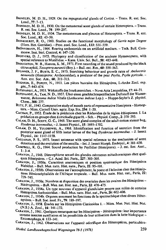
BRINDLEY, M. D. H., 1929. On the repugnatorial glands of Corixa. - Trans. R. ent. Soc. Lond.,77:7-13.
BRINDLEY, M. D. H., 1930. On the metasternal scent glands of certain Heteroptera. - Trans. R. ent. Soc. Lond., 78: 199-207.
BRINDLEY, M. D. H., 1934. The metasternum and pleuron of Heteroptera. - Trans. R. ent. Soc. Lond., 82: 43-50.
BRINKHURST, R. O., 1960. Studies on the functional morphology of Gerris najas Degeer (Hem. Het. Gerridae). - Proc. zool. Soc. Lond., 133: 531-559.
BRÖNNIMANN, H., 1964. Rearing anthocorids on an artificial medium. - Tech. Bull. Com-monw. Inst. biol. Control, 4: 147-150.
BROTHERS, D. J., 1975. Phylogeny and classification of the aculeate Hymenoptera, with special reference to Mutillidae. - Kans. Univ. Sei. Bull., 50: 483-648.
BROUGHTON, W. B., HARRIS, K. M., 1971. First recording of the sound produced by the black citrus aphid, Toxoptera auranlii (Boy.). - Bull. ent. Res., 60: 559-563.
BRUNNER, J. F., BURTS, E. C , 1975. Searching behaviour and growth rates of Anthocoris nemoralis (Hemiptera: Anthocoridae), a predator of the pear Psylla, Psylla pyricola. -Ann. ent. Soc. Am., 68; 311-315.
BUGNION, E., POPOFF, N., 1911. Les pièces buccales des Hémiptères, I.-Archs Zool. exp. gén., 7:643-674.
BUTENANDT, A., 1955. Wirkstoffe des Insektenreiches. - Nova Acta Leopoldina, 17:44-71. BUTENANDT, A., TAM, N. D., 1957. Über einen geschlechtsspezifischen Duftstoff der Wasser
wanze, Belostoma indica Vitalis (Leihocerus indicus Lep.). - Hoppe-Seyler's Z. physiol. Chem.,308:277.
BUTT, F. H., 1943. Comparative study of mouth parts of representative Hemiptera - Homop-tera. - Mem. Cornell Univ. agric. Exp. Stn, 254:1-20.
CACHAN, P., 1952. Étude de la prédation chez les Réduvides de la région éthiopienne. I. La prédation en groupe chez Ectrichodia gigas H. - Sch. - Physiol. Comp., 2:378-395.
CALAM, D. H., SCOTT, G. C , 1969. The scent gland complex of the adult cotton stainer bug, Dysdercus intermedins,-]. Insect Physiol., 15: 1695-1702.
CALAM, D. H., YOUDEOWEI, A., 1968. Identification and function of secretion from the posterior scent gland of fifth instar larvae of the bug Dysdercus intermedius. - J. Insect Physiol., 14: 1147-1158.
CALLAHAN, P. S., 1975. Insect antennae with special reference to the mechanism of scent detection and the evolution of the sensilla. - Int. J. Insect Morph. Embryol., 4: 381-430.
CAMPBELL, K. G., 1964. Sound production by Psyllidae (Hemiptera). - J . ent. Soc. Aust., 1:3-4.
CARAYON, J., 1948. Dimorphisme sexuel des glandes odorantes métathoraciques chez quelques Hémiptères. - C r . Acad. Sei. Paris., 227: 303-305.
CARAYON, J., 1950a. Caractères anatomiques et position systématique des Hémiptères Nabidae. - Bull. Mus. natn. Hist, natn., Paris, 22: 95-101.
CARAYON, J., 1950b. Observations sur l'accouplement, la ponte et l'éclosion chez des Hémiptères Hénicocéphalidés de l'Afrique tropicale. - Bull. Mus. natn. Hist, nat., Paris, 22: 739-745.
CARAYON, J., 1950c. Nombres et disposition des ovarioles dans les ovaires des Hémiptères -Hétéroptères. - Bull. Mus. nat. Hist, nat., Paris, 22: 470-475.
CARAYON, J., 1954a. Un type nouveau d'appareil glandulaire propre aux mâles de certains Hémiptères Anthocoridae. - Bull. Mus. natn. Hist, nat., Paris, 26: 602-606.
CARAYON, J., 1954b. Organes assumant les fonctions de la spermathèque chez divers Hétéroptères. - Bull. Soc. zool. Fr., 79: 189-197.
CARAYON, J., 1958. Études sur les Hémiptères Cimicoidea 1. - Mém. Mus. Nat. Hist. Nat. (N.S.) A. Zool., 16: 141-172.
CARAYON, J., 1961. Quelques remarques sur les Hémiptères - Hétéroptères: leur importance comme insectes auxilliaires et les possibilités de leur utilisation dans la lutte biologique -Entomophaga,6:133-141.
CARAYON, J., 1962. Observations sur l'appareil odorifique des Hétéroptères, particulière-
Meded. Landbouwhogeschool Wageningen 78-5 (1978) 259
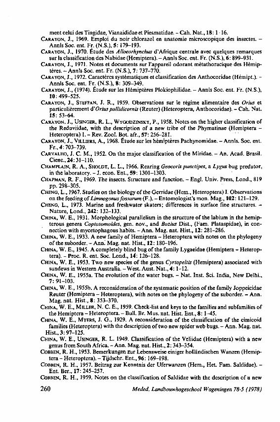
ment celui des Tingidae, Vianaididae et Piesmatidae. - Cah. Nat., 18:1-16. CARAYON, J., 1969. Emploi du noir chlorazol en anatomie microscopique des insectes. -
Annls Soc. ent. Fr. (N.S.), 5: 179-193. CARAYON, J., 1970. Étude des Alloeorhynchus d'Afrique centrale avec quelques remarques
sur la classification des Nabidae (Hemiptera). - Annls Soc. ent. Fr. (N.S.), 6: 899-931. CARAYON, J., 1971. Notes et documents sur l'appareil odorant métathoracique des Hémip
tères. - Annls Soc. ent. Fr. (N.S.), 7: 737-770. CARAYON, J., 1972. Caractères systématiques et classification des Anthocoridae (Hémipt.). -
Annls Soc. ent. Fr. (N.S.), 8: 309-349. CARAYON, J., (1974). Étude sur les Hémiptères Plokiophilidae. - Annls Soc. ent. Fr. (N.S.),
10:499-525. CARAYON, J., STEFFAN, J. R., 1959. Observations sur le régime alimentaire des Orius et
particulièrement ôi'Orius pallidicornis (Reuter) (Heteroptera, Anthocoridae). - Cah. Nat. 15: 53-64.
CARAYON, J., USINGER, R. L., WYGODZINSKY, P., 1958. Notes on the higher classification of the Reduviidae, with the description of a new tribe of the Phymatinae (Hemiptera -Heteroptera) 1. - Rev. Zool. Bot. afr., 57: 256-281.
CARAYON, J., VILLIERS, A., 1968. Étude sur les hémiptères Pachynomidae. - Annls. Soc. ent. Fr., 4:703-739.
CARVALHO, J. C. M., 1952. On the major classification of the Miridae. - An. Acad. Brasil. Ciena, 24:31-110.
CHAMPLAIN, R. A., SHOLDT, L. L., 1966. Rearing Geocoris punctipes, a Lygus bug predator, in the laboratory. - J. econ. Ent., 59: 1301-1303.
CHAPMAN, R. F., 1969. The insects. Structure and function. - Engl. Univ. Press, Lond., 819 pp. 298-305.
CHENG, L., 1967. Studies on the biology of the Gerridae (Hem., Heteroptera) I. Observations on the feeding of Limnogonus fossarum (F.). - Entomologist's mon. Mag., 102: 121-129.
CHENG, L., 1973. Marine and freshwater skaters; differences in surface fine structures. -Nature, Lond., 242: 132-133.
CHINA, W. E., 1931. Morphological parallelism in the structure of the labium in the hemip-terous genera Coptosomoides, gen. nov., and Bozius Dist., (Fam. Plataspidae), in connection with mycetophagous habits. -Ann. Mag. nat. Hist., 12: 281-286.
CHINA, W. E., 1933. A new family of Hemiptera - Heteroptera with notes on the phylogeny of the suborder. - Ann.' Mag. nat. Hist., 12: 180-196.
CHINA, W. E., 1945. A completely blind bug of the family Lygaeidae (Hemiptera - Heteroptera). - Proc. R. ent. Soc. Lond., 14": 126-128.
CHINA, W. E., 1953. Two new species of the genus Cyrtopeltis (Hemiptera) associated with sundews in Western Australia. -West. Aust. Nat., 4:1-12.
CHINA, W. E., 1955a. The evolution of the water bugs. - Nat. Inst. Sei. India, New Delhi., 7:91-103.
CHINA, W. E., 1955b. A reconsideration of the systematic position of the family Joppeicidae Reuter (Hemiptera - Heteroptera), with notes on the phylogeny of the suborder. - Ann. Mag. nat. Hist., 8: 353-370.
CHINA, W. E., MILLER, N. C. E., 1959. Check-list and keys to the families and subfamilies of the Hemiptera - Heteroptera. - Bull. Br. Mus. nat. Hist. Ent., 8: 1-45.
CHINA, W. E., MYERS, J. G., 1929. A reconsideration of the classification of the cimicoid families (Heteroptera) with the description of two new spider web bugs. - Ann. Mag. nat.
Hist., 3: 97-125. CHINA, W. E., USINGER, R. L. 1949. Classification of the Veliidae (Hemiptera) with a new
genus from South Africa. - Ann. Mag. nat. Hist., 2: 343-354. COBBEN, R. H., 1953. Bemerkungen zur Lebensweise einiger holländischen Wanzen (Hemip
tera - Heteroptera). - Tijdschr. Ent., 96: 169-198. COBBEN, R. H., 1957. Beitrag zur Kenntnis der Uferwanzen (Hem., Het. Fam. Saldidae). -
Ent. Ber., 17: 245-257. COBBEN, R. H., 1959. Notes on the classification of Saldidae with the description of a new
260 Meded. Landbouwhogeschool Wageningen 78-5 (1978)
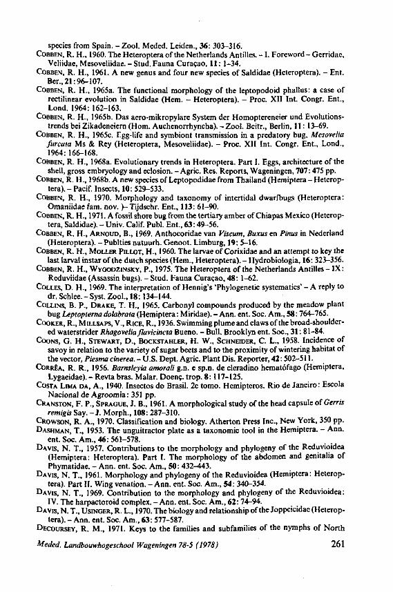
species from Spain. - Zool. Meded. Leiden., 36: 303-316. COBBEN, R. H., 1960. The Heteroptera of the Netherlands Antilles. - 1 . Foreword - Gerridae,
Veliidae, Mesoveliidae. - Stud. Fauna Curaçao, 11: 1-34. COBBEN, R. H., 1961. A new genus and four new species of Saldidae (Heteroptera). - Ent.
Ber., 21:96-107. COBBEN, R. H., 1965a. The functional morphology of the leptopodoid phallus: a case of
rectilinear evolution in Saldidae (Hem. - Heteroptera). - Proc. XII Int. Congr. Ent., Lond. 1964: 162-163.
COBBEN, R. H., 1965b. Das aero-mikropylare System der Homoptereneier und Evolutions-trends bei Zikadeneiern (Hom. Auchenorrhyncha). - Zool. Beitr., Berlin, 11: 13-69.
COBBEN, R. H., 1965C. Egg-life and symbiont transmission in a predatory bug, Mesovelia furcata Ms & Rey (Heteroptera, Mesoveliidae). - Proc. XII Int. Congr. Ent., Lond., 1964:166-168.
COBBEN, R. H., 1968a. Evolutionary trends in Heteroptera. Part I. Eggs, architecture of the shell, gross embryology and eclosion. - Agric. Res. Reports, Wageningen, 707:475 pp.
COBBEN, R. H., 1968b. A new species of Leptopodidae from Thailand (Hemiptera - Heteroptera). - Pacif. Insects, 10: 529-533.
COBBEN, R. H., 1970. Morphology and taxonomy of intertidal dwarfbugs (Heteroptera: Omaniidae fam. nov. )-Tijdschr. Ent., 113: 61-90.
COBBEN, R. H., 1971. A fossil shore bug from the tertiary amber of Chiapas Mexico (Heteroptera, Saldidae). -Univ. Calif. Publ. Ent., 63:49-56.
COBBEN, R. H., ARNOUD, B., 1969. Anthocoridae van Viscum, Buxus en Pinus in Nederland (Heteroptera). -Publties natuurh. Genoot. Limburg, 19: 5-16.
COBBEN, R. H., MOLLER PILLOT, H., 1960. The larvae of Corixidae and an attempt to key the last larval instar of the dutch species (Hem., Heteroptera). - Hydrobiologia, 16: 323-356.
COBBEN, R. H., WYGODZINSKY, P., 1975. The Heteroptera of the Netherlands Antilles - IX: Reduviidae (Assassin bugs). - Stud. Fauna Curaçao, 48: 1-62.
COLLES, D. H., 1969. The interpretation of Hennig's 'Phylogenetic systematics' - A reply to dr. Schlee. - Syst. Zool., 18: 134-144.
COLLINS, B. P., DRAKE, T. H., 1965. Carbonyl compounds produced by the meadow plant bug Leptopterna dolabrata (Hemiptera: Miridae). - Ann. ent. Soc. Am., 58: 764-765.
COOKER, R., MILLSAPS, V., RICE, R., 1936. Swimming plume and claws of the broad-shouldered waterstrider Rhagovelia flavicincta Bueno. - Bull. Brooklyn ent. Soc., 31: 81-84.
COONS, G. H., STEWART, D., BOCKSTAHLER, H. W., SCHNEIDER, C L., 1958. Incidence of savoy in relation to the variety of sugar beets and to the proximity of wintering habitat of the vector, Piesma cinerea. - U.S. Dept. Agric. Plant Dis. Reporter, 42: 502-511.
CORRÊA, R. R., 1956. Barnsleyia amorali g.n. e sp.n. de cleradino hematófago (Hemiptera, Lygaeidae). - Revta bras. Malar. Doenç. trop. 8:117-125.
COSTA LIMA DA, A., 1940. Insectos do Brasil. 2e tomo. Hemipteros. Rio de Janeiro: Escola Nacional de Agroomia: 351 pp.
CRANSTON, F. P., SPRAGUE, J. B., 1961. A morphological study of the head capsule of Gerris remigisSay. - J . Morph., 108: 287-310.
CROWSON, R. A., 1970. Classification and biology. Atherton Press Inc., New York, 350 pp. DASHMAN, T., 1953. The unguitractor plate as a taxonomie tool in the Hemiptera. - Ann.
ent. Soc. Am., 46: 561-578. DAVIS, N. T., 1957. Contributions to the morphology and phylogeny of the Reduvioidea
(Hemiptera: Heteroptera). Part I. The morphology of the abdomen and genitalia of Phymatidae. - Ann. ent. Soc. Am., 50: 432-443.
DAVIS, N. T., 1961. Morphology and phylogeny of the Reduvioidea (Hemiptera: Heteroptera). Part II. Wing venation. - Ann. ent. Soc. Am., 54: 340-354.
DAVIS, N. T., 1969. Contribution to the morphology and phylogeny of the Reduvioidea: IV. The harpactoroid complex. - Ann. ent. Soc. Am., 62: 74-94.
DAVIS, N. T., USINGER, R. L., 1970. The biology and relationship of the Joppeicidae (Heteroptera). - Ann. ent. Soc. Am., 63: 577-587.
DECOURSEY, R. M., 1971. Keys to the families and subfamilies of the nymphs of North
Meded. Landbouwhogeschool Wageningen 78-5 (1978) 261
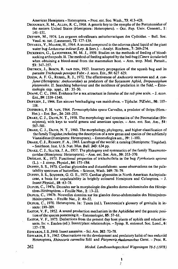
American Hemiptera- Heteroptera. -Proc. ent. Soc. Wash., 73:413-428. DECOURSEY, R. M., ALLEN, R. C , 1968. A generic key to the nymphs of the Pentatomidae of
the eastern United States (Hemiptera: Heteroptera). - Occ. Pap. Univ. Connect., 1: 141-151.
DETHIER, M., 1974. Les organes odoriférants métathoraciques des Cydnidae. - Bull. Soc. Vaud. se. nat. (Lausanne), 72:127-139.
DEVAKUL, V., MAARSE, H., 1964. A second compound in the odorous gland liquid of the giant water bug Lethocerus indicus (Lep. & Serv.). - Analyt. Biochem., 7: 269-274.
DICKERSON, G., LAVOIPIERRE, M. M. J., 1959. Studies on the methods of feeding of bloodsucking arthropods. II. The method of feeding adopted by the bed-bug (Cimex lectularius) when obtaining a blood-meal from the mammalian host. - Ann. trop. Med. Parasit., 53: 347-357.
DIETRICK, J., BOSCH, R. VAN DEN, 1957. Insectary propagation of the squash bug and its parasite Trichopoda pennipes Fabr.- J. econ. Ent., 50:627-629.
DIXON, A. F. G., RÜSSEL, R. J., 1972. The effectiveness of Anthocoris nemorum and A. con-fusus (Hemiptera: Anthocoridae) as predators of the Sycamore Aphid, Drepanosiphum platanoides. II. Searching behaviour and the incidence of prédation in the field. - Ento-mologia exp. appl., 15: 35-50.
DOANE, C. C , 1966. Evidence for a sex attractant in females of the red pine scale. - J. econ. Ent., 59:1539-1540.
DOEKSEN, J., 1944. Een nieuwe beschadiging van maiskolven. -Tijdschr. PIZiekt., 50: 107— 108.
DOESBURG, P. H. VAN, 1964. Termatophylidea opaca Carvalho, a predator of thrips (Hem.-Het.). -Ent. Ber., 24:248-253.
DRAKE, C. J., DAVIS, N. T., 1958. The morphology and systematics of the Piesmatidae (Hemiptera), with keys to world genera and american species. - Ann. ent. Soc. Am., 51 : 567-581.
DRAKE, C. J., DAVIS, N. T., 1960. The morphology, phylogeny, and higher classification of the family Tingidae.including the description of a new genus and species of the subfamily Vianaidinae(Hemiptera: Heteroptera).-Entomologicaam., 39: 1-100.
DRAKE, C. J., RUHOFF, F. A., 1965. Lacebugs of the world: a catalog (Hemiptera: Tingidae). -Smithson. Inst. U.S. Nat. Mus. Bull. 243: 634 pp.
DRAKE, C. J., SLATER, J. A., 1957. The phylogeny and systematics of the family Thaumasto-coridae (Hemiptera : Heteroptera). - Ann. ent. Soc. Am., 50: 353-370.
DRÂSLAR, K., 1973. Functional properties of trichobothria in the bug Pyrrhocoris apterus (L.). - J. comp. Physiol., 84: 175-184.
DUFFEY, S. S., 1970. Cardiac glycosides and distastefulness: some observations on the pala-tability spectrum of butterflies. - Science, Wash. 169: 78-79.
DUFFEY, S. S., SCUDDER, G. G. E., 1972. Cardiac glycosides in North American Asclepiada-ceae, a basis for unpalatability in brightly coloured Hemiptera and Coleoptera. - J. Insect Physiol., 18:63-78.
DUPUIS, C , 1947a. Données sur la morphologie des glandes dorso-abdominales des Hémip-tères-Hétéroptères. - Feuille Nat., 2: 13-22.
DUPUIS, C , 1947b. Nouvelles données sur les glandes dorso-abdominales des Hémiptères-Hétéroptères. - Feuille Nat., 2: 49-52.
DUPUIS, C , 1970. Heteroptera. In: Tuxen (ed.), Taxonomist's glossary of genitalia in insects: 190-209.
EASTOP, V. F., 1952. A sound production mechanism in the Aphididae and the generic position of the species possessing it. - Entomologist, 85: 57-61.
EASTOP, V. F., 1973. Deductions from the present day host plants of aphids and related insects. In: v. Emden (ed.). Insect/plant relationships. - Symp. R. entomol. Soc. Lond., 6: 157-178.
EDWARDS, J. S.,1960. Insect assassins. - Sei. Am. 202:72-78. EDWARDS, J. S., 1962. Observations on the development and predatory habit of two reduviid
Heteroptera, Rhinocoris carmelita Stâl and Platymeris rhadamanthus Gerst. - Proc. R.
262 Meded. Landbouwhogeschool Wageningen 78-5 (1978)
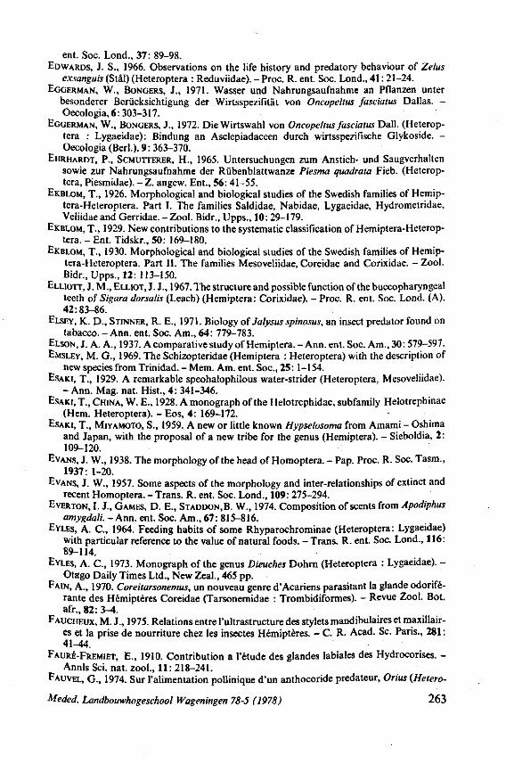
ent. Soc. Lond., 37: 89-98. EDWARDS, J. S., 1966. Observations on the life history and predatory behaviour of Zelus
exsanguis (Stâl) (Heteroptera : Reduviidae). - Proc. R. ent. Soc. Lond., 41: 21-24. EGGERMAN, W., BONGERS, J., 1971. Wasser und Nahrungsaufnahme an Pflanzen unter
besonderer Berücksichtigung der Wirtsspezifität von Oncopeltus fasciatus Dallas. -Oecologia, 6: 303-317.
EGGERMAN, W., BONGERS, J., 1972. Die Wirtswahl von Oncopeltus fasciatus Dali. (Heteroptera : Lygaeidae): Bindung an Asclepiadaceen durch wirtsspezifische Glykoside. -Oecologia (Bed.), 9: 363-370.
EHRHARDT, P., SCMUTTERER, H., 1965. Untersuchungen zum Anstich- und Saugverhalten sowie zur Nahrungsaufnahme der Rübenblattwanze Piesma quadrata Fieb. (Heteroptera, Piesmidae). - Z. angew. Ent., 56: 41-55.
EKBLOM, T., 1926. Morphological and biological studies of the Swedish families of Hemip-tera-Heteroptera. Part I. The families Saldidae, Nabidae, Lygaeidae, Hydrometridae, Veliidaeand Gerridae. - Zool. Bidr., Upps., 10: 29-179.
EKBLOM, T., 1929. New contributions to the systematic classification of Hemiptera-Heterop-tera. - Ent. Tidskr., 50: 169-180.
EKBLOM, T., 1930. Morphological and biological studies of the Swedish families of Hemip-tera-Heteroptera. Part II. The families Mesoveliidae, Coreidae and Corixidae. - Zool. Bidr., Upps., 12: 113-150.
ELLIOTT, J. M., ELLIOT, J. J., 1967. The structure and possible function of the buccopharyngeal teeth of Sigara dorsalis (Leach) (Hemiptera: Corixidae). - Proc. R. ent. Soc. Lond. (A), 42:83-86.
ELSEY, K. D., STINNER, R. E., 1971. Biology of July sus spinosus, an insect predator found on tabacco. - Ann. ent. Soc. Am., 64: 779-783.
ELSON, J. A. A., 1937. A comparative study of Hemiptera. - Ann. ent. Soc. Am., 30: 579-597. EMSLEY, M. G., 1969. The Schizopteridae (Hemiptera : Heteroptera) with the description of
new species from Trinidad. - Mem. Am. ent. Soc, 25:1-154. EsAKi, T., 1929. A remarkable speohalophilous water-strider (Heteroptera, Mesoveliidae).
- Ann. Mag. nat. Hist., 4: 341-346. ESAKI, T., CHINA, W. E., 1928. A monograph of the Helotrephidae, subfamily Helotrephinae
(Hem. Heteroptera). - Eos, 4: 169-172. ESAKI, T., MIYAMOTO, S., 1959. A new or little known Hypselosoma from Amami - Oshima
and Japan, with the proposal of a new tribe for the genus (Hemiptera). - Sieboldia, 2: 109-120.
EVANS, J. W., 1938. The morphology of the head of Homoptera. - Pap. Proc. R. Soc. Tasm., 1937: 1-20.
EVANS, J. W., 1957. Some aspects of the morphology and inter-relationships of extinct and recent Homoptera. - Trans. R. ent. Soc. Lond., 109: 275-294.
EVERTON, I. J., GAMES, D. E., STADDON,B. W., 1974. Composition of scents from Apodiphus amygdali. - Ann. ent. Soc. Am., 67: 815-816.
EYLES, A. C , 1964. Feeding habits of some Rhyparochrominae (Heteroptera: Lygaeidae) with particular reference to the value of natural foods. - Trans. R. ent. Soc. Lond., 116: 89-114.
EYLES, A. C , 1973. Monograph of the genus Dieuches Dohrn (Heteroptera : Lygaeidae). -Otago Daily Times Ltd., New Zeal., 465 pp.
FAIN, A., 1970. Coreitarsonemus, un nouveau genre d'Acariens parasitant la glande odoriférante des Hémiptères Coreidae (Tarsonemidae : Trombidiformes). - Revue Zool. Bot. afr.,82:3-4.
FAUCHEUX, M. J., 1975. Relations entre Infrastructure des stylets mandibulaires et maxillaires et la prise de nourriture chez les insectes Hémiptères. - C. R. Acad. Se. Paris., 281 : 41-44.
FAURÉ-FREMIET, E., 1910. Contribution a l'étude des glandes labiales des Hydrocorises. -Annls Sei. nat. zool., 11: 218-241.
FAUVEL, G., 1974. Sur l'alimentation pollinique d'un anthocoride prédateur, Orius {Hetero-
Meded. Landbouwhogeschool Wageningen 78-5 (1978) 263
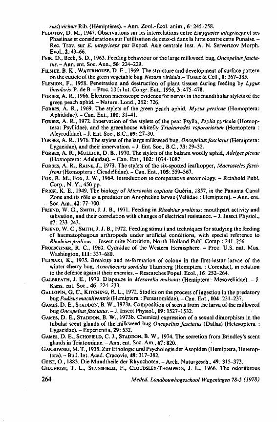
rius) vicinus Rib. (Hémiptères). - Ann. Zool.-Écol. anim., 6: 245-258. FEDOTOV, D. M., 1947. Observations sur les interrelations entre Eurygaster integriceps et ses
Phasiinae et considérations sur l'utilisation de ceux-ci dans la lutte contre cette Punaise. -Ree. Trav. sur E. integriceps par Exped. Asie centrale Inst. A. N. Servertzov Morph. Evol.,2:49-66.
FEIR, D., Beek, S. D., 1963. Feeding behaviour of the large milkweed bug, Oncopeltus fascia-tus. - Ann. ent. Soc. Ann., 56: 224-229.
FILSHIE, B. K., WATERHOUSE, D. F., 1969. The structure and development of surface pattern on the cuticle of the green vegetable bug Nezara viridula.- Tissue & Cell., 1: 367-385.
FLEMION, F., 1958. Penetration and destruction of plant tissues during feeding by Lygus lineolaris P. de B. -Proc. 10th Int. Congr. Ent., 1956,3:475-478.
FORBES, A. R., 1966. Electron microscope evidence for nerves in the mandibular stylets of the green peach aphid. - Nature, Lond., 212: 726.
FORBES, A. R., 1969. The stylets of the green peach aphid, Myzus persicae (Homoptera: Aphididae). - Can. Ent., 101: 31-41.
FORBES, A. R., 1972. Innervation of the stylets of the pear Psylla, Psylla pyricola (Homoptera: Psyllidae), and the greenhouse whitefly Trialeurodes vaporariorum (Homoptera : Aleyrodidae). - J. Ent. Soc, B.C., 69: 27-30.
FORBES, A. R., 1976. The stylets of the large milkweed bug, Oncopeltus fasciatus (Hemiptera: Lygaeidae), and their innervation. - J. Ent. Soc, B.C., 73: 29-32.
FORBES, A. R., MULLICK, D. B., 1970. The stylets of the balsam woolly aphid, Adelges piceae (Homoptera: Adelgidae). - Can. Ent., 102: 1074-1082.
FORBES, A. R., RAINE, J., 1973. The stylets of the six-spotted leafhopper, Macrosteles fasci-frons (Homoptera : Cicadellidae). - Can. Ent., 105: 559-567.
Fox, R. M., Fox, J. W., 1964. Introduction to comparative entomology. - Reinhold Publ. Corp., N. Y., 450 pp.
FRICK, K. E., 1949. The biology of Microvelia capitata Guérin, 1857, in the Panama Canal Zone and its rôle as a predator on Anopheline larvae (Veliidae : Hemiptera). - Ann. ent. Soc. Am., 42:77-100.
FRIEND, W. G., SMITH, J. J. B., 1971. Feeding in Rhodnius prolixus: mouthpart activity and salivation, and their correlation with changes of electrical resistance. - J. Insect Physiol., 17: 233-243.
FRIEND, W. C , SMITH, J. J. B., 1972. Feeding stimuli and techniques for studying the feeding of haematophagous arthropods under artificial conditions, with special reference to Rhodnius prolixus. - Insect-mite Nutrition, North-Holland Publ. Comp.: 241-256.
FROESCHNER, R. C , 1960. Cydnidae of the Western Hemisphere. - Proc. U.S. nat. Mus. Washington, 111: 337-680.
FUJISAKI, K., 1975. Breakup and re-formation of colony in the first-instar larvae of the winter cherry bug, Acanthocoris sordidus Thunberg (Hemiptera : Coreidae), in relation to the defence against their enemies. - Researches Popul. Ecol., 16: 252-264.
GALBREATH, J. E., 1973. Diapauze in Mesovelia mulsanti (Hemiptera: Mesoveliidae). - J. Kans. ent. Soc, 46: 224-233.
GALLOPÎN, G. C , KITCHING, R. L., 1972. Studies on the process of ingestion in the predatory bug Podisus maculiventris (Hemiptera : Pentatomidae). - Can. Ent., 104: 231-237.
GAMES, D. E., STADDON, B. W., 1973a. Composition of scents from the larva of the milkweed bug Oncopeltus f asciatus.-J. Insect Physiol., 19: 1527-1532.
GAMES, D. E., STADDON, B. W., 1973b. Chemical expression of a sexual dimorphism in the tubular scent glands of the milkweed bug Oncopeltus fasciatus (Dallas) (Heteroptera : Lygaeidae). - Experientia, 29: 532.
GAMES, D. E., SCHOFIELD, C. J., STADDON, B. W., 1974. The secretion from Brindley's scent glands in Triatominae. - Ann. ent. Soc. Am., 67: 820.
GARBOWSKI, M. T., 1935. Zur Ethologie und Psychologie der Asopiden (Hemiptera, Heteroptera). - Bull. Int. Acad. Cracovie, 48: 317-382.
GEISE, O., 1883. Die Mundtheile der Rhynchoten. - Arch. Naturgesch., 49: 315-373. GILCHRIST, T. L., STANSFIELD, F., CLOUDSLEY-THOMPSON, J. L., 1966. The odoriferous
264 Meded. Landbouwhogeschool Wageningen 78-5 (1978)
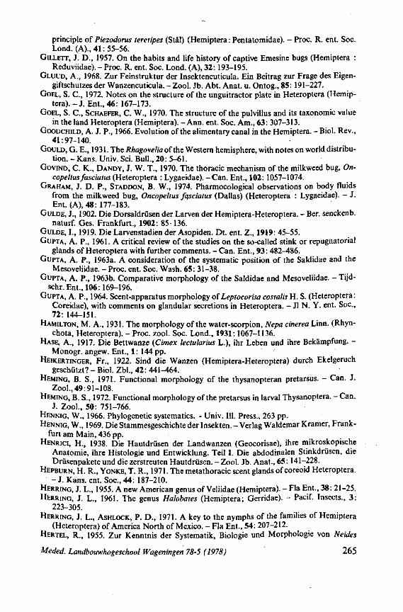
principle of Piezodorus teretipes (Stàl) (Hemiptera : Pentatomidae). - Proc. R. ent. Soc. Lond. (A)., 41: 55-56.
GILLETT, J. D., 1957. On the habits and life history of captive Emesine bugs (Hemiptera : Reduviidae). - Proc. R. ent. Soc. Lond. (A), 32: 193-195.
GLUUD, A., 1968. Zur Feinstruktur der Insektencuticula. Ein Beitrag zur Frage des Eigengiftschutzes der Wanzencuticula. - Zool. Jb. Abt. Anat. u. Ontog., 85:191-227.
GOEL, S. C , 1972. Notes on the structure of the unguitractor plate in Heteroptera (Hemiptera).-J. Ent., 46: 167-173.
GOEL, S. C , SCHAEFER, C. W., 1970. The structure of the pulvillus and its taxonomie value in the land Heteroptera (Hemiptera). - Ann. ent. Soc. Am., 63: 307-313.
GOODCHILD, A. J. P., 1966. Evolution of the alimentary canal in the Hemiptera. - Biol. Rev., 41:97-140.
GOULD, G. E., 1931. The Rhagovelia of the Western hemisphere, with notes on world distribution. - Kans. Univ. Sei. Bull., 20: 5-61.
GOVIND, C. K., DANDY, J. W. T., 1970. The thoracic mechanism of the milkweed bug, On-copeltus fasciatus (Heteroptera : Lygaeidae). - Can. Ent., 102:1057-1074.
GRAHAM, J. D. P., STADDON, B. W., 1974. Pharmocological observations on body fluids from the milkweed bug, Oncopeltus fqsciatus (Dallas) (Heteroptera : Lygaeidae). - J. Ent. (A), 48: 177-183.
GULDE, J., 1902. Die Dorsaldrüsen der Larven der Hemiptera-Heteroptera. - Ber. senckenb. naturf. Ges. Frankfurt., 1902: 85-136.
GULDE, I., 1919. Die Larvenstadien der Asopiden. Dt. ent. Z., 1919: 45-55. GUPTA, A. P., 1961. A critical review of the studies on the so-called stink or repugnatorial
glands of Heteroptera with further comments. - Can. Ent., 93: 482-486. GUPTA, A. P., 1963a. A consideration of the systematic position of the Saldidae and the
Mesoveliidae. - Proc. ent. Soc. Wash. 65: 31-38. GUPTA, A. P., 1963b. Comparative morphology of the Saldidae and Mesoveliidae. - Tijd-
schr. Ent., 106:169-196. GUPTA, A. P., 1964. Scent-apparatus morphology of Leptocorisa costalis H. S. (Heteroptera :
Coreidae), with comments on glandular secretions in Heteroptera. - Jl N. Y. ent. Soc, 72: 144-151.
HAMILTON, M. A., 1931. The morphology of the water-scorpion, Nepa cinerea Linn. (Rhyn-chota, Heteroptera). - Proc. zool. Soc. Lond., 1931:1067-1136.
HASE, A., 1917. Die Bettwanze (Cimex lectularius L.), ihr Leben und ihre Bekämpfung. -Monogr. angew. Ent., 1: 144 pp.
HEIKERTINGER, Fr., 1922. Sind die Wanzen (Hemiptera-Heteroptera) durch Ekelgeruch geschützt? - Biol. Zbl., 42: 441-^64.
HEMING, B. S., 1971. Functional morphology of the thysanopteran pretarsus. - Can. J. Zool., 49:91-108.
HEMING, B. S., 1972. Functional morphology of the pretarsus in larval Thysanoptera. - Can. J. Zool., 50: 751-766.
HENNIG, W., 1966. Phylogenetic systematics. - Univ. 111. Press., 263 pp. HENNIG, W., 1969. Die Stammesgeschichte der Insekten. - Verlag Waldemar Kramer, Frank
furt am Main, 436 pp. HENRICI, H., 1938. Die Hautdrüsen der Landwanzen (Geocorisae), ihre mikroskopische
Anatomie, ihre Histologie und Entwicklung. Teil I. Die abdodinalen Stinkdrüsen, die Drüsenpakete und die zerstreuten Hautdrüsen. -Zool. Jb. Anat., 65:141-228.
HEPBURN, H. R., YONKE, T. R., 1971. The metathoracic scent glands of coreoid Heteroptera. - J. Kans. ent. Soc, 44: 187-210.
HERRING, J. L., 1955. A new American genus of Veliidae (Hemiptera). - Fla Ent., 38: 21-25. HERRING, J. L., 1961. The genus Halobates (Hemiptera; Gerridae). - Pacif. Insects., 3:
223-305. HERRING, J. L., ASHLOCK, P. D., 1971. A key to the nymphs of the families of Hemiptera
(Heteroptera) of America North of Mexico. - Fla Ent., 54: 207-212. HERTEL, R., 1955. Zur Kenntnis der Systematik, Biologie und Morphologie von Neides
Meded. Landbouwhogeschool Wageningen 78-5 (1978) 265
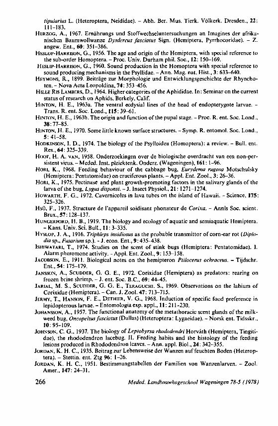
tipularius L. (Heteroptera, Neididae). - Abh. Ber. Mus. Tierk. Völkerk. Dresden., 22: 111-183.
HERZOG, A., 1967. Ernährungs und Stoffwechseluntersuchungen an Imagines der afrikanischen Baumwollwanze Dysdercus fasciatus Sign. (Hemiptera, Pyrrhocoridae). - Z. angew. Ent., 60: 351-386.
HESLOP-HARRISON, G., 1956. The age and origin of the Hemiptera, with special reference to the sub-order Homoptera. - Proc. Univ. Durham phil. Soc, 12: 150-169.
HESLIP-HARRISON, G., 1960. Sound production in the Homoptera with special reference to sound producing mechanisms in the Psyllidae. - Ann. Mag. nat. Hist., 3: 633-640.
HEYMONS, R., 1899. Beiträge zur Morphologie und Entwicklungsgeschichte der Rhyncho-ten. - Nova Acta Leopoldina, 74: 353-456.
HILLE Ris LAMBERS, D., 1964. Higher categories of the Aphididae. In: Seminaron the current status of research on Aphids, Berkely, Calif.
HINTON, H. E., 1963a. The ventral ecdysial lines of the head of endopterygote larvae. -Trans. R. ent. Soc. Lond., 115: 39-61.
HINTON, H. E., 1963b. The origin and function of the pupal stage. - Proc. R. ent. Soc. Lond., 38:77-85.
HINTON, H. E., 1970. Some little known surface structures. - Symp. R. entomol. Soc. Lond., 5: 41-58.
HODKINSON, I. D., 1974. The biology of the Psylloidea (Homoptera): a review. - Bull. ent. Res., 64.325-339.
HOOF, H. A. VAN, 1958. Onderzoekingen over de biologische overdracht van een non-persistent virus. -Meded. Inst, plziektenk. Onderz. (Wageningen), 161: 1-96.
HORI, K., 1968. Feeding behaviour of the cabbage bug, Eurydema rugosa Motschulsky (Hemiptera: Pentatomidae) on cruciferous plants. - Appl. Ent. Zool., 3: 26-36.
HORI, K., 1975. Pectinase and plant growth-promoting factors in the salivary glands of the larva of the bug, Lygus disponsi. - J . Insect Physiol., 21: 1271-1274.
HOWARTH, F. G., 1972. Cavernicoles in lava tubes on the island of Hawaii. - Science, 175: 325-326.
Hsü, F., 1937. Structure de l'appareil soidisant phonateur de Corixa. - Annls Soc. scient. Brux., 57:128-137.
HUNGERFORD, H. B., 1919. The biology and ecology of aquatic and semiaquatic Hemiptera. - Kans. Univ. Sei. Bull., 11: 3-335.
HYSLOP, J. A., 1916. Triphleps insidiosus as the probable transmittor of corn-ear rot (Diplo-dia sp., Fusarium sp.). - J. econ. Ent., 9: 435-438.
IsHiWATARi, T., 1974. Studies on the scent of stink bugs (Hemiptera: Pentatomidae). I. Alarm pheromone activity. - Appl. Ent. Zool., 9: 153-158.
JACOBSON, E., 1911. Biological notes on the hemipteron Ptilocerus ochraceus. - Tijdschr. Ent., 54: 175-179.
JANSSON, A., SCUDDER, G. G. E., 1972. Corixidae (Hemiptera) as predators: rearing on frozen brine shrimp. - J. ent. Soc. B.C., 69: 44-45.
JARIAL, M. S., SCUDDER, G. G. E., TERAGUCHI, S., 1969. Observations on the labium of Corixidae (Hemiptera). - Can. J. Zool. 47: 713-715.
JERMY, T., HANSON, F. E., DETHIER, V. G., 1968. Induction of specific food preference in lepidopterous larvae. - Entomologia exp. appl., 11:211-230.
JOHANSSON, A., 1957. The functional anatomy of the metathoracic scent glands of the milkweed bug, Oncopeltus fasciatus (Dallas) (Heteroptera: Lygaeidae). - Norsk ent. Tidsskr., 10:95-109.
JOHNSON, C. G., 1937. The biology of Lepiobyrsa rhododendri Horvâth (Hemiptera, Tingiti-dae), the rhododendron lacebug. II. Feeding habits and the histology of the feeding lesions produced in Rhododendron leaves. -Ann. appl. Biol., 24: 342-355.
JORDAN, K. H. C , 1935. Beitrag zur Lebensweise der Wanzen auf feuchten Boden (Heteroptera). - Stettin, ent. Ztg 96: 1-26.
JORDAN, K. H. C , 1951. Bestimmungstabellen der Familien von Wanzenlarven. - Zool. Amer., 147:24-31.
266 Meded. Landbouwhogeschool Wageningen 78-5 (1978)
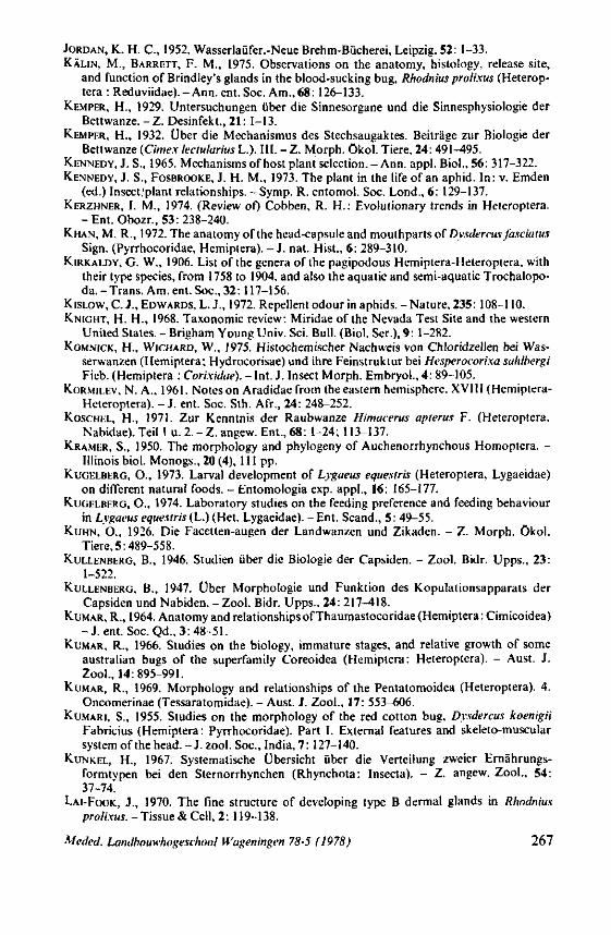
JORDAN, K. H. C , 1952. Wasserlaüfer.-Neue Brehm-Bücherei, Leipzig, 52: 1-33. KÄLIN, M., BARRETT, F. M., 1975. Observations on the anatomy, histology, release site,
and function of Brindley's glands in the blood-sucking bug, Rhodnius prolixus (Heterop-tera : Reduviidae). - Ann. ent. Soc. Am., 68: 126-133.
KEMPER, H., 1929. Untersuchungen über die Sinnesorgane und die Sinnesphysiologie der Bettwanze. - Z. Desinfekt., 21: 1-13.
KEMPER, H., 1932. Über die Mechanismus des Stechsaugaktes. Beiträge zur Biologie der Bettwanze (Cime.x lectularius L.). III. - Z. Morph, ökol. Tiere, 24: 491-495.
KENNEDY, J. S., 1965. Mechanisms of host plant selection. -Ann. appl. Biol., 56: 317-322. KENNEDY, J. S., FOSBROOKE, J. H. M., 1973. The plant in the life of an aphid. In: v. Emden
(ed.) Insect/plant relationships. - Symp. R. entomol. Soc. Lond., 6: 129-137. KERZHNER, I. M., 1974. (Review of) Cobben, R. H.: Evolutionary trends in Heteroptera.
-Ent . Obozr., 53:238-240. KHAN, M. R., 1972. The anatomy of the head-capsule and mouthparts of Dysdercus fasciaius
Sign. (Pyrrhocoridae, Hemiptera). - J . nat. Hist., 6: 289-310. KIRKALDY, G. W., 1906. List of the genera of the pagipodous Hemiptera-Heteroptera. with
their type species, from 1758 to 1904, and also the aquatic and semi-aquatic Trochalopo-da. - Trans. Am. ent. Soc, 32:117-156.
KISLOW, C. J., EDWARDS, L. J., 1972. Repellent odour in aphids. - Nature, 235: 108-110. KNIGHT, H. H., 1968. Taxonomie review: Miridae of the Nevada Test Site and the western
United States. - Brigham Young Univ. Sei. Bull. (Biol. Ser.), 9: 1-282. KOMNICK, H., WICHARD, W., 1975. Histochemischer Nachweis von Chloridzellen bei Was
serwanzen (Hemiptera; Hydrocorisae) und ihre Feinstruktur bei Hesperocorixa sahlbergi Ficb. (Hemiptera : Corixidae). - Int. J. Insect Morph. Embryol., 4: 89-105.
KORMILEV, N. A., 1961. Notes on Aradidae from the eastern hemisphere. XVIII (Hemiptera-Heteroptera). - J. ent. Soc. Sth. Afr., 24: 248-252.
KOSCHEL, H., 1971. Zur Kenntnis der Raubwanze Himucerus apterus F. (Heteroptera, Nabidae). Teil 1 u. 2. - Z. angew. Ent., 68: 1-24; 113-137.
KRAMER, S., 1950. The morphology and phylogeny of Auchenorrhynchous Homoptera. -Illinois biol. Monogs., 20 (4), 111 pp.
KUGELBERG, O., 1973. Larval development of Lygaeus equesiris (Heteroptera, Lygaeidae) on different natural foods. - Entomologia exp. appl., 16: 165-177.
KUGELBERG, O., 1974. Laboratory studies on the feeding preference and feeding behaviour in Lygaeus equestris (L.) (Het. Lygaeidae). - Ent. Scand., 5: 49-55.
KUHN, O., 1926. Die Facetten-augen der Landwanzen und Zikaden. - Z. Morph, ökol. Tiere, 5:489-558.
KULLENBERG, B., 1946. Studien über die Biologie der Capsiden. - Zool. Bidr. Upps., 23: 1-522.
KULLENBERG, B., 1947. Über Morphologie und Funktion des Kopulationsapparats der Capsiden und Nabiden. - Zool. Bidr. Upps.. 24: 217-418.
KUMAR, R., 1964. Anatomy and relationships of Thaumastocoridae (Hemiptera : Cimicoidea) - J .ent . Soc. Qd., 3: 48-51.
KUMAR, R., 1966. Studies on the biology, immature stages, and relative growth of some australian bugs of the superfamily Coreoidea (Hemiptera: Heteroptera). - Aust. J. Zool., 14:895-991.
KUMAR, R., 1969. Morphology and relationships of the Pentatomoidea (Heteroptera). 4. Oncomerinae (Tessaratomidae). - Aust. J. Zool., 17: 553-606.
KUMARI, S., 1955. Studies on the morphology of the red cotton bug. Dysdercus koenigii Fabricius (Hemiptera: Pyrrhocoridae). Part I. External features and skeleto-muscular system of the head. -J.zool. Soc, India, 7: 127-140.
KUNKEL, H., 1967. Systematische Übersicht über die Verteilung zweier Ernährungsformtypen bei den Sternorrhynchen (Rhynchota: Insecta). - Z. angew. Zool., 54: 37-74.
LAI-FOOK, J., 1970. The fine structure of developing type B dermal glands in Rhodnius prolixus. - Tissue & Cell, 2: 119-138.
Meded. Landbouwhogeschool Wageningen 78-5 (1978) 267
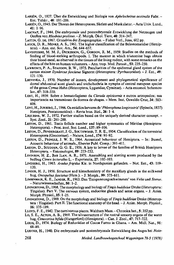
LARSÉN, O., 1927. Über die Entwicklung und Biologie von Aphelocheirus aestivalis Fabr. -Ent. Tidskr., 48: 181-206.
LARSÉN, O., 1945. Der Thorax der Heteropteren. Skelett und Muskulatur. - Acta Univ. Lund., 41: 1-96.
LASCHAT, F., 1944. Die embryonale und postembryonale Entwicklung der Netzaugen und Ocellen von Rhodniusprolixus.-Z. Morph, ökol. Tiere, 40: 314-347.
LATTIN, G. DE, 1967. Grundriss der Zoogeographie. - Fisher Verl., Jena, 602 pp. LAUCK, D. R., MENKE, A. S., 1961. The higher classification of the Belostomatidae (Hemip-
tera). - Ann. ent. Soc. Am., 54: 644-657. LAVOIPIERRE, M. M. J., DICKERSON, G., GORDON, R. M., 1959. Studies on the methods of
feeding of blood-sucking arthropods. 1. The manner in which triatomine bugs obtain their blood-meal, as observed in the tissues of the living rodent, with some remarks on the effects of the bite on human volunteers. - Ann. trop. Med. Parasit., 53:235-250.
LAWRENCE, P. A., STADDON, B. W., 1975. Peculiarities of the epidermal gland system of the cotton stainer Dysdercus fasciatus Signoret (Heteroptera : Pyrrhocoridae). - J . Ent., 49: 121-130.
LEDVINKA, J., 1970. Number of instars, development and phylogenetical significance of dorsal abdominal scent glands and trichobothria in nymphs of central european species of the genus Cymus Hahn (Heteroptera, Lygaeidae, Cyminae). - Acta entomol. bohemos-lav., 67:318-330.
LENT, H., 1939. Sobre o hematofagismo da Clerada apicicornis e outros artropodos; sua importancia na transmissao da doenca de chagas. - Mem. Inst. Oswaldo Cruz, 34: 583-606.
LENT, H., JURBERG, J., 1966. Os estâdios larvares de 'Phloeophana longirostris' (Spinola, 1837) Hemiptera, Pentatomoidea). - Revta bras. Biol., 26: 1-4.
LEQUESNE, W. J., 1972. Further studies based on the uniquely derived character concept. -Syst. Zool., 21:281-288.
LESTON, D., 1961. Testis follicle number and higher systematics of Miridae (Hemiptera-Heteroptera). -Proc. zool. Soc. Lond., 137: 89-106.
LESTON, D., PENDERGRAST, J. G., SOUTHWOOD, T. R. E., 1954. Classification of the terrestrial Heteroptera (Geocorisae). - Nature, Lond., 174: 91-92.
LESTON, D., PRINGLE, J. W. S., 1964. Acoustical behaviour of Hemiptera. - In: Busnel, Acoustic behaviour of animals., Elsevier Publ. Comp.: 391-411.
LESTON, D., SCUDDER, G. G. E., 1956. A key to larvae of the families of British Hemiptera Heteroptera. - Entomologist, 89: 223-232.
LEVINSON, H. Z., BAR ILAN, A. R., 1971. Assembling and alerting scents produced by the bedbug Cimex lecturalius L. - Experientia, 27:102-103.
LINDBERG, H., 1963. Aradus frigidus Kir. in Nordspanien gefunden. - Not. Ent., 43: 119-120.
LINDER, H. J., 1956. Structure and histochemistry of the maxillary glands in the milkweed bug, Oncopeltus fasciatus (Hem.). - J. Morph., 99: 575-611.
LINSENMAIR, K. E., JANDER, R., 1963. Das 'Entspannungsschwimmen' von Velia und Stenus. - Naturwissenschaften, 50: 1-2.
LIVINGSTONE, D., 1968. The morphology and biology of Tingis buddleiae Drake (Heteroptera : Tingidae). Part V. The nervous system, endocrine glands and sense organs. - J. Anim. Morph. Physiol., 15:1-25.
LIVINGSTONE, D., 1969. On the morphology and biology of Tingis buddleiae Drake (Heteroptera : Tingidae). Part II. The functional anatomy of the head. - J. Anim. Morph. Physiol., 16: 135-189.
LLOYD, F. E., 1942. The carnivorous plants. Waltham Mass. - Chronica bot., 5:352 pp. Lo, S. E., ACTON, A. B., 1969. The ultrastructure of the rostral sensory organs of the water
bug, Cenocorixa bifida (Hungerford) (Hemiptera). - Can. J. Zool., 47: 717-722. Louis, D., 1974. Biology of Reduviidae of Cocoa Farms in Ghana. - Am. Midi. Nat., 91 :
68-89. LUDTKE, H., 1940. Die embryonale and postembryonale Entwicklung des Auges bei Noto-
268 Meded. Landbouwhogeschool Wageningen 78-5 (1978)
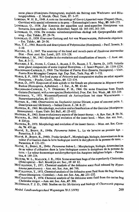
necta glauca (Hemiptera-Heteroptera), zugleich ein Beitrag zum Wachtums- und Häutungsproblem. - Z. Morph, ökol. Tiere, 37: 1-37.
LUMSDEN, W. H. R., 1949. A note on the ecology oiGerris (Aquarius) najas (Degeer) (Hem., Gerridae) with special references to its prey. - Entomologist's mon. Mag., 85:169-173.
LUNDBLAD, O., 1934. Zur Kenntnis der aquatilen und semi-aquatilen Hemipteren von Sumatra, Java und Bali. - Arch. Hydrobiol. Suppl. 12: 185-195, 263-319.
LUNDBLAD, O., 1936. De svenska vattenhemipterernas ekologi och djurgeografiska ställ-ning. - Ent. Tidskr., 57: 29-74.
LUNDBLAD, O., 1939. Eine neue Gattung und Art von Wasserwanzen, Hebrovelia singularis. - Ent. Tidskr., 60: 29-36.
MAA, T. C , 1961. Records and descriptions of Polyctenidae (Hemiptera). - Pacif. Insects, 3: 1-10.
MACGILL, E. I., 1947. The anatomy of the head and mouth-parts of Dysdercus intermedius Dist. -Proc. zool. Soc. Lond., 117: 115-128.
MACKERRAS, I. M., 1967. Grades in the evolution and classification of insects. - J. Aust. ent. Soc, 6:3-11.
MACLEOD, J. K., HOWE, I., CABLE, J., BLAKE, J. D., BAKER, J. T., SMITH, D., 1975. Volatile scent gland components of some tropical Hemiptera. - J. Insect Physiol., 21: 1219-1224.
MALDONADO-CAPRILES, J., 1969. The Miridae of Puerto Rico (Insecta, Hemiptera). - Univ. Puerto Rico Mayagiiez Campus. Agr. Exp. Stat., Tech. Pap. 45: 1-133.
MARKS, E. P., 1959. The food pump of Pelocoris and comparative studies on other aquatic Hemiptera. -Psyche, Camb., Mass., 64: 123-134.
MARSH, D„ 1975. Responses of male aphids to the female sex pheromone in Megoura viciae Bucton (Homoptera, Aphididae). - J. Ent., A, 50: 43-64.
MALDONADO-CAPRILES, J., V. DOESBURG, P. H., 1966. On some Emesinae from Dutch Guiana (Surinam), with a new species (Reduviidae). Proc. Ent. Soc. Wash., 68: 325-329.
MASCHWITZ, V., 1971. Wasserstoffperoxid als Antiseptikum bei einer Wasserwanze. -Naturwissenschaften, 11:572.
MATHEN, K., 1960. Observations on Stephanitis typicus Distant, a pest of coconut palm. 1. Description and life history. - Indian Cocon. J., 14: 8-27.
MATSUDA, R., 1960. Morphology, evolution and a classification of the Gerridae (Hemiptera-Heteroptera). - Kans. Univ. Sei. Bull., 41:25-632.
MATSUDA, R., 1963. Some evolutionary aspects of the insect thorax. - A. Rev. Ent., 8:59-76. MATSUDA, R., 1965. Morphology and evolution of the insect head. - Mem. Am. ent. Inst.,
4:334 pp. MATSUDA, R., 1970. Morphology and evolution of the insect thorax. - Mem. ent. Soc. Cana
da, 76: 431 pp. MAYNÉ, R., BRENY, R., 1948a. Picromerus bidens. L., La vie larvaire au premier âge. -
Parasitica, 3:1-20. MAYNÉ, R., BRENY, R., 1948b. Troilus luridus F., Morphologie, biologie, determination de sa
valeur d'utilisation dans la lutte biologique contre le doryphore de la pomme de terre. -Parasitica, 4: 131-151.
MAYNÉ, R., BRENY, R., 1948c. Picromerus bidens L.: Morphologie, biologie, determination de sa valeur d'utilisation dans la lutte biologique contre le doryphore de la pomme de terre. - La valeur économique antidoryphorique des Asopines indigènes belges. - Parasitica, 4: 189-224.
MCATEE, W. L., MALLOCK, J. R., 1924. Some annectant bugs of the superfamily Cimicoidea (Heteroptera). - Bull. Brooklyn ent. Soc, 19: 69-82.
MCCULLOUGH, T., 1971. Chemical analysis of the defensive scent fluid released by Hypse-lonotuspunctiventris. - Ann. ent. Soc. Am., 64: 749.
MCCULLOUGH, T., 1973. Chemical analysis of the defensive scent fluid from the bug Morena obtusa (Hemiptera : Coreidae). - Ann. ent. Soc Am., 66:231-232.
MCCULLOUGH, T., 1974. Chemical analysis of the defensive scent fluid from the cactus bug Chelinidea vittiger.- Ann. ent. Soc. Am., 67: 300.
MCDONALD, F. J. D., 1960. Studies on the life-history and biology of Choerocoris paganus
Meded. Landbouwhogeschool Wageningen 78-5 (1978) 269
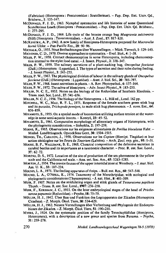
(Fabricius) (Heteroptera : Pentatomidae : Scutellerinae). - Pap. Dep. Ent. Univ. Qd., Brisbane., 1: 135-147.
MCDONALD, F. J. D., 1963. Nymphal systematics and life histories of some Queensland Scutellerinae Leach (Hemiptera : Pentatomidae). - Pap. Dep. Ent. Univ. Qd. Brisbane., 1: 277-295.
MCDONALD, F. J. D., 1969. Life cycle of the bronze orange bug Musgraveia sulciventris (Stâl) (Hemiptera : Tessarotomidae). - Aust. J. Zool., 17: 817-820.
MCKINSTRY, A. P., 1942. A new family of Hemiptera-Heteroptera proposed for Macrovelia hornii Uhler. - Pan-Pacific Ent., 28: 90-96.
MICHALK, O., 1935. Neue Beobachtungen über Wanzenfliegen. - Mark.Tierwelt, 1:129-140. MICHENER, C. D., 1970. Diverse approaches to systematics. - Evol. Bio)., 4: 1-38. MILES, P. W., 1958. Contact chemoreception in some Heteroptera, including chemorecep-
tion internal to the stylet food canal. - J . Insect. Physiol., 2: 338-347. MILES, P. W., 1959. The salivary secretions of a plant-sucking bug, Oncopeltus fasciatus
(Dall.) (Heteroptera : Lygaeidae). I. The types of secretion and their roles during feeding. - J. Insect Physiol., 3: 243-255.
MILES, P. W., 1967. The physiological division of labour in the salivary glands of Oncopeltus fasciatus (Dall.) (Heteroptera : Lygaeidae). - Aust. J. biol. Sei., 20: 785-797.
MILES, P. W., 1968. Insect secretions in plants. - A. Rev. Phytopathol., 6:137-164. MILES, P. W., 1972. The saliva of Hemiptera. -Adv. Insect Physiol., 9: 183-255. MILLER, N. C. E., 1953. Notes on the biology of the Reduviidae of Southern Rhodesia. -
Trans, zool. Soc. Lond., 27: 541-656. MILLER, N. C. E., 1956. The biology of the Heteroptera. - L. Hill ed., Lond. 162 pp. MITCHELL, W. C , MAU, R. F. L., 1971. Response of the female southern green stink bug
and its parasite, Trichopoda pennipes, to male stink bug pheromones. - J. econ. Ent., 64: 856-859.
MIYAMOTO, S., 1955. On a special mode of locomotion utilizing surface tension at the water-edge in some semi-aquatic insects. - Kontyû, 23: 45-52.
MIYAMOTO, S., 1961. Comparative morphology of alimentary organs of Heteroptera, with the phylogenetic consideration. -Sieboldia, 2: 197-259.
MOENS, R., 1965. Observations sur les exigences alimentaires de Perillus bioculatus Fabr. -Meded. LandbHogesch. OpzoekStns Gent, 30: 1504-1515.
MONOD, TH., CARA YON, J., 1958. Observations sur les Copium (Hemipt. Tingidae) et leur action cécidogène sur les fleurs de Teucrium (Labiées). - Arch. Zool. exp. gén., 95: 1-31.
MOORE, B. P., WALLBANK, B. E., 1968. Chemical composition of the defensive secretion in carabid beetles and its importance as a taxonomie character. - Proc. R. ent. Soc. Lond., 37: 62-72.
MORENO, D. S., 1972. Location of the site of production of the sex pheromone in the yellow scale and the California red scale. - Ann. ent. Soc. Am., 65: 1283-1285.
MORTON, J., 1954. The crevice faunas of the upper intertidal zone at Wembury.- J. mar. biol. Ass. U. K., 33: 187-224.
MOUND, L. A., 1971. The feeding apparatus of thrips.- Bull. ent. Res., 60: 547-548. MOUND, L. A., O'NEILL, K., 1974. Taxonomy of the Merothripidae, with ecological and
phylogenetic considerations (Thysanoptera). - J. nat. Hist., 8: 481-509. MUIR, F. 1907. Notes on the stridulating organ and stink glands of Tessaratoma papulosa
Thunb. -Trans. R. ent. Soc. Lond., 1907: 256-258. MUIR, F., KERSHAW, J. C , 1911. On the later embryological stages of the head of Pristhe-
sancuspapuensis (Reduviidae). - Psyche, 18:75-79. MÜLLER, H. J., 1942. Über Bau und Funktion des Legeapparates der Zikaden (Homoptera
Cicadina). - Z. Morph, ökol. Tiere, 38: 534-629. MÜLLER, H. J., 1962. Neuere Vorstellungen über Verbreitung und Phylogenie der Endosym-
biosen der Zikaden.-Z. Morph, ökol. Tiere, 51: 190-210. MYERS, J., 1924. On the systematic position of the family Termitaphididae (Hemiptera,
Heteroptera), with a description of a new genus and species from Panama. - Psyche, 31:259-278.
270 Meded. Landbouwhogeschool Wageningen 78-5 (1978)
NAEF, A., 1919. Idealistische Morphologie und Phylogenetik. (Zur Methodik der systematischen Morphologie). - G. Fischer, Jena, 77 pp.
NAKASHIMA, T., 1975. Several types of the mycetangia found in platypodid ambrosia beetles (Coleoptera : Platypodidae). - Insecta matsum., 7: 1-69.
NAULT, L. R., EDWARDS, L. J., STYER, W. E., 1973. Aphid alarm pheromones: secretion and reception. - Envir. Ent., 2: 101-105.
NAULT, L. R., WOOD, T. K., GOFF, A. M., 1974. Treehopper (Membracidae) alarm pheromones.-Nature, 249:387-389.
NEESE, V., 1965. Zur Funktion der Augenborsten bei der Honigbiene. - Z. vergl. Physiol., 49: 543-585.
NEISWANDER, C. R., 1926. On the anatomy of the head and thorax in Ranatra (Heteroptera). - Trans. Am. ent. Soc, 51: 311-320.
NUORTEVA, P., 1956. Studies on the comparative anatomy of the salivary glands in four families of Heteroptera.- Ann. Ent. Fenn., 22:45-54.
ODHIAMBO, T. R., 1959. An account of parental care in Rhinocoris albopilosus Sign. (Hem. Het. Reduviidae), with notes on its life history. - Proc. R. ent. Soc. Lond. (A), 34: 175-185.
OETTING, R. D., YONKE, T. R., 1971a. Biology of some Missouri stink bugs. - J. Kans. ent. Soc, 44:446-459.
OETTING, R. D., YONKE, T. R., 1971b. Immature stages and biology of Podisus placidus and Stiretrus fimbriatus (Hemiptera : Pentatomidae). - Can. Ent., 103: 1505-1516.
OsMANi, Z., NAIDU, M. B., 1966. Evidence of sex attractant in female Dysdercus cingulatus Fabr. - Indian, J. exp. Biol. 5:51.
OSSIANNILSSON, F., 1949. Insect drummers. - Opusc. ent. (Suppl.), 10: 1-146. OSSIANNILSSON, F., 1950. Sound production in Psyllids (Hem. Horn.). - Opusc. ent., 15: 202. PAJUNEN, V. I. 1970. Adaptation of Arctocorisa carinata (Sahib.) and Callicorixa producta
(Reut.) populations to a rock pool environment. In: Dynamics of Populations. Proc. Adv. Study Inst. Dynamics Numbers Popul. (Oosterbeek): 148-158.
PANTLE, C. FEIR, D., 1976. Olfactory response to milkweed seed extracts in the milkweed bug. - J . Insect. Physiol. 22: 285-289.
PARK, R. J., SUTHERLAND, M. D., 1962. Volatile constituents of the bronze orange bug, Rhoecocoris sulciventris. - Aust. J. Chem., 15: 172.
PARKER, A. H., 1965. The predatory behaviour and life history of Pisilus tipuliformis Fabri-cius (Hemiptera : Reduviidae). - Entomologia exp. appl. 8: 1-12.
PARKER, A. H., 1969. Predatory and reproductive behavior of Rhinocoris bicolor and R. tropicus (Hemiptera: Reduviidae). - Entomologia exp. appl. 12: 107-117.
PARRISH, W. B., 1967. The origin, morphology, and innervation of aphid stylets (Homoptera). - Ann. ent. Soc. Am., 60: 273-276.
PARSONS, M. C , 1958. Cephalic glands in Gelastocoris (Hemiptera - Heteroptera). - Psyche, Camb. Mass., 65:99-107.
PARSONS, M. C , 1959. Skeleton and musculature of the head of Gelastocoris oculatus (Fabri-cius) (Hemiptera - Heteroptera). - Bull. Mus. comp. Zool. Harv., 122:299-357.
PARSONS, M. C , 1962. Skeleton and musculature of the head of Saldula paliipes (F.) (Heteroptera : Saldidae). - Trans. R. ent. Soc. Lond., 114: 97-130.
PARSONS, M. C , 1963. The endoskeletal salivary pumping apparatus in representative Belostomatidae (Heteroptera). - Can. J. Zool., 41: 1017-1024.
PARSONS, M. G, 1964. The origin and development of the hemipteran cranium. - Can. J. Zool., 42: 409-432.
PARSONS, M. C , 1965. Clypeal modification in some littoral and aquatic Heteroptera. -Can. J. Zool., 43: 161-166.
PARSONS, M. C , 1966a. Studies on the cephalic anatomy of Naucoridae (Heteroptera). -Trans. R. ent. Soc. Lond., 118: 119-151.
PARSONS, M. C , 1966b. Labial skeleton and musculature of the Hydrocorisae (Heteroptera). - Can. J. Zool., 44: 1051-1084.
PARSONS, M. C , 1968. The cephalic and prothoracic skeletomusculature and nervous system
Meded. Landbouwhogeschool Wageningen 78-5 (1978) 271
in Lethocerus (Heteroptera, Belostomatidae). - J. Lin. Soc. (Zool.), 47: 349-406. PARSONS, M. C , 1969a. The labium of Aphelocheirus aestivalis F. as compared with that of
typical Naucoridae (Heteroptera). - Can. J. Zool., 47: 295-306. PARSONS, M. C , 1969b. The food pump of Aphelocheirus aestivalis F. as compared with that
of typical Naucoridae (Heteroptera). - J. Morph., 129: 17-30. PARSONS, M. C , 1972. Fine structure of the triturating devices in the food pump of Notonecta
(Heteroptera : Notonectidae). - J. Morph., 138: 141-168. PATTENDEN, G., STADDON, B. W., 1968. Secretion of the metathoracic glands of the water-
bug Notonectaglauca L. (Heteroptera; Notonectidae). - Experientia, 24: 1092. PATTENDEN, G., STADDON, B. W., 1970. Observations on the metasternal scent glands of
Lethocerus spp. (Heteroptera : Belostomatidae). - Ann. ent. Soc. Am., 63: 900-901. PATTENDEN, G., Staddon, B. W., 1972. Identification of iso-butyric acid in secretion from
Brindley's scent glands in Rhodnius prolixus (Heteroptera : Reduviidae). - Ann. ent. Soc. Am., 65: 1240-1241.
PATTON, W. S., CRAGG, W. A., 1913. A textbook of medical entomology. London Christian Literature Soc, India, 768 pp.
PENDERGRAST, J. G., 1952. Studies on the biology of pentatomid bugs of the genus Rhopali-morpha Dallas (Heteroptera). -Trans. R. Soc, N.Z., 80:143-153.
PEREGRINE, D. J., 1972. Some evidence of gustatory discrimination in the cotton bug Dys-dercus fasciatus Sign. (Hemiptera, Pyrrhocoridae). - Bull. ent. Res., 62: 211-214.
PÉRICART, J., 1972. Hémiptères, Anthocoridae, Cimicidae et Microphysidae de l'ouest-paléarctique. - Faune de l'Europe et du Bassin méditerranéen., 7:402 pp.
PÉRICART, J., 1975. Note sur le mode de vie des Gampsocoris et la présence en France de G. culicinus (Hem., Berytinidae). - L'entomologiste, 31: 215-219.
PESSON, P., 1944. Contribution à l'étude morphologique et fonctionelle de la tête, de l'appareil buccal et du tube digestif des femelles de coccides. - Monogrs Publ. Stn. Lab. Rech. agron. Paris, 1: 1-266.
PESSON, P., 1951. Homoptères. In : Grassé's Traité de Zoologie, 10:1390-1656. PETERS, W., ULBRICH, R., 1973. The life history of the water-boatman Trichocorixella mexi-
cana (Heteroptera : Corixidae). - Can. Ent., 105: 277-282. PINDER, A. R., STADDON, B. W., 1965. The odoriferous secretion of the waterbug, Sigara
falleni(Fieb.).-J.chem. Soc, 530: 2955-2958. PINET, J. M., 1963. L'innervation sensorielle des stylets mandibulaires et maxillaires de
Rhodnius prolixus Stâl. (Insecte Hémiptère Hétéroptère). - C. r. hebd. Séanc. Acad. Sei. Paris., 257: 3666-3668.
PINET, J. M., BERNARD, J., BOISTEL, T., 1969. Étude électrophysiologique des récepteurs des stylets chez une punaise hématophage Triatoma infestons. - C. R. Séanc. Soc. Biol., Paris, 163:1939-1948.
PINET, J. M., BERNARD, J., 1972. Essai d'interprétation du mode d'action de la vapeur d'eau et de la température sur un récepteur d'insecte. - Ann. Zool. - Écol. anim., 4: 483-495.
PLANK, F. L. VAN DER, 1958. The assassin bug Platymeris rhadamanthus Gaerst., a useful predator of the rhinoceros beetles Oryctes boas F. and O. monoceros (Oliv.). - J. ent. Soc. sth. Afr., 19: 14-28.
POISSON, R. A., 1924. Contribution à l'étude des Hémiptères aquatiques. - Bull. biol. Fr. Belg., 58:49-305.
POISSON, R. A., 1951. Hétéroptères. In : Grassé's Traité de Zoologie, 10: 1657-1803. POISSON, R. A., 1952. Contribution à l'étude des Hydrocorises de Madagascar (missions J.
Millot et R. Paulian, 1949). - Mém. Inst. scient. Madagascar, 1: 23-70. POISSON, R. A., 1956. Contribution à l'étude des Hydrocorises de Madagascar (Heteroptera).
- Mém. Inst. scient. Madagascar, 7: 243-265. POISSON, R. A., 1957. Hemiptera Heteroptera : Hydrocorisae & Geocorisae - Gerroidea.
- S. Afr. Animal Life, Uppsala, 4: 327-373. POLIVANOVA, E. N., 1960. Osobjennosti ontogeneza pentatomid (Hemiptera, Pentatomoidea)
i jevo znacenije dlja jich sistematiki i filogenii. - The Ontogeny of Insects (Acta Symp. evol. ins. Praha), 1: 75-81.
272 Meded. Landbouwhogeschool Wageningen 78-5 (1978)
POLLARD, D. G., 1955. Feeding habits of the cotton whitefly, Bemisia tabaci Genn. (Homop-tera : Aleurodidae). - Ann. appl. Biol., 43: 664-671.
POLLARD, D. G., 1959. Feeding habits of the lace-bug Urentius aegypliacus Bergevin (Hemip-tera : Tingidae). - Ann. appl. Biol., 47: 778-782.
POLLARD, D. G., 1968. Stylet penetration and feeding damage of Eupleryx melissae Curtis (Hemiptera, Cicadellidae) on sage. - Bull. ent. Res., 58: 55-71.
POLLARD, D. G., 1969. Directional control of the stylets in phytophagous Hemiptera. -Proc. R. ent. Soc. Lond. (A)., 44: 173-185.
POLLARD, D. G., 1970. Simulated cannibalism in an aphid. - Proc. R. ent. Soc. Lond. (A)., 45:98-103.
POLLARD, D. G., 1971. Some observations on the mouth-parts of white-flies (Hem., Aleyrodi-dae). - Entomologist's mon. Mag., 107: 81-88.
POLLARD, D. G., 1972. The stylet structure of a leafhopper (Eupleryx melissae Curtis : Ho-moptera, Cicadellidae). - J . nat. Hist., 6: 261-271.
POLLARD, D. G., 1973. Plant penetration by feeding aphids (Hemiptera, Aphidoidea) : a review. - Bull. ent. Res. 62: 631-714.
POPHAM, E. J., 1962. On the salivary pump of Dysdercus iniermedius Dist. (Hemiptera Heter-optera) and other bugs. - Proc. zool. Soc. Lond., 139: 489-493.
POPOV, Y. A., 1971. Historical development of Hemiptera infraorder Nepomorpha (Heter-optera). (in Russian, informal translation in english by Miss H. Vaitaitis). - Trudy Pal. Inst. Acad. Sc. USSR, 129: 1-141.
POPOV, Y. A., 1973. First discovery of an Hemiptera of the family Saldidae (Heteroptera) in the Mesozoic of Siberia (In Russian). - Doklady (Reports) Acad. Sei. USSR, 209: 703-705.
PRIESNER, H., 1960. A monograph of the Thysanoptera of the Egyptian deserts. - Pubis Inst. Désert Egypte, 13: 1-549.
PUCHKOV, V. G., 1958. Larvae of Hemiptera-Heteroptera. I. Lygaeidae. - Ént. Obozr. (Moscow), 37:392-413.
PUCHKOV, V. G., 1959a. Larvae of Hemiptera-Heteroptera belonging to the superfamily Pentatomoidea. I. Identification keys of the families Pentatomoidea and of the species of Acanthosomidae, Cydnidaeand Scutelleridae(In Russian). -Zool. Zh., 38:1190-1206.
PUCHKOV, V. G„ 1959b. Larvae of the families Coreidae and Arenocoreidae (Coreoidea, Heteroptera) of the fauna of the European part of the USSR (In Russian, English summary). - Dopov. Akad. Nauk. URSR, 10: 1156-1161.
PUCHKOVA, L. V., 1965. Maxillary glands of Heteroptera, their function and origin (in Russian with English summary). - Zool. Zh. Moscow, 34: 1801-1807.
PUCHKOVA, L. V., 1966. The morphology and biology of the egg of the terrestrial bugs (Hemiptera) (In Russian). - Horae Soc. Ent. Un. Sovet., 51: 75-132.
PUCHKOVA, L. V., 1968. Development of the beak musculature and sclerites in Heteroptera (In Russian, English summary). - Ent. Obozr., 47: 51-60.
PUCHKOVA, L. V., 1969. (Review of) Cobben, R. H.: Evolutionary trends in Heteroptera. -Vêstnik Zool. Kiev., 4: 93-94.
PUCHKOVA, L. V., PUCHKOV, V. G., 1958. Certain morphological peculiarities of the abdominal odoriferous glands in Pentatomidae larvae (Pentatomoidea, Hemiptera-Heteroptera). No. 1 (In Ukrainian). - Dopov. Akad. Nauk. URSR, 1:100-104.
QUADRI, M. A. H., 1951. On the anatomy of the mouth-parts and the mode of feeding in the aquatic bugs (Cryptocerata). - Proc. zool. Soc. Béng., 4:117-135.
QUADRI, M. A. H., 1959. Mechanism of feeding in Hemiptera. - Smithson. misc. Colins, 137: 237-245.
RALPH, C. P., 1976. Natural food requirements, host-finding, and population density of the large milkweed bug, Oncopeltus fasciatus (Hemiptera : Lygaeidae), in relation to host plant density and morphology. - Thesis, Cornell Univ., Ithaca, N. Y.
RAMAMURTY, p. S., KRISHNANANDAM, Y., 1967. Morphological and histochemical study of the scent gland in the pond-skater, Gerris spinolae Leth. et Serv. (Gerridae-Insecta). -Zool. Anz., 178:198-210.
Meded. Landbouwhogeschool Wageningen 78-5 (1978) 273
RA WAT, B. L., 1939. On the habits, metamorphosis and reproductive organs of Naucoris cimicoides L. (Hemiptera-Heteroptera). - Trans. R. ent. Soc. Lond., 88: 119-138.
REMOLD, H., 1962. Über die biologische Bedeutung der Duftdrüsen bei den Landwanzen (Geocorisae). - Z. vergl. Physiol., 45: 636-694.
RENSING, L., 1962. Beiträge zur vergleichenden Morphologie, Physiologie und Ethologie der Wasserlaüfer (Gerroidea). - Zool. Beitr., 7:447-485.
REUTER, O. M., 1910. Neue Beiträge zur Phylogenie und Systematik der Miriden nebst einleitenden Bemerkungen über die Phylogenie der Heteropterenfamilien. - Acta Soc. Sei. fenn.,37: 1-172.
REUTER, O. M., 1913. Insekternas levnadsvanor och instinkter. Stockholm. RICHARDS, W. R., 1965. The Callaphidini of Canada (Homoptera : Aphididae). - Mem.
ent. Soc. Canada, 44: 1-149. RICHARDS, W. R., 1968. Generic classification, evolution, and biogeography of the Sminthu-
ridae of the world (Collembola). - Mem. ent. Soc. Canada, 53: 1-54. RIDGEWAY, R. L., JONES, S. L., 1968. Plant feeding by Geocoris pallens and Nabis americo-
ferus. - Ann. ent. Soc. Am., 61: 232-233. RIEGER, C , 1976. Skelett und Muskulatur des Kopfes und Prothorax von Ochterus marginatus
Latreille. - Zoomorph., 83: 109-191. RILEY, G. F. C , 1918. Food of aquatic Hemiptera. - Science, 48: 545-546. ROHDENDORF, B. B., (Edit.), 1962. Tschlenistonogie-Tracheiny i Chelitzerowye. - Osnowy
Palaeontologii, 9, Moscow, U.S.S.R. Acad, of Sciences. ROOT, R. B., CHAPLIN, S. J., 1976. The life-styles of tropical milkweed bugs, Oncopeltus
(Hemiptera : Lygaeidae) utilizing the same hosts. - Ecology, 57:132-140. Ross, H. H., 1965. A textbook of entomology. - J . Wiley & Sons, Inc. 539 pp. Ross, H. H., 1969. (Review of) Cobben, R. H.: Evolutionary trends in Heteroptera. Part I.
1968. - Evolution, 23: 357-358. ROTH, L. M., 1961. A study of the odoriferous glands of Scaptocoris divergens (Hemiptera :
Cydnidae). - Ann. ent. Soc. Am., 54: 900-911. RUSSELL, M. C , 1953. Notes on insects associated with sundews (Drosera) at Lesmurdie. -
W. Aust. Nat., 4: 9-12. SAXENA, K. N., 1963. Mode of ingestion in a heteropterous insect, Dysdercus koenigii (F.)
(Pyrrhocoridae). - J . Insect Physiol., 9: 47-71. SAXENA, P. N., CHADA, H. L., 1971. The greenbug, Schizaphis graminum. I. Mouth parts and
feeding habits. -Ann. ent. Soc. Am., 64: 897-904. SCALES, A. L., 1968.Fema!e tarnished plant bugs attract males.-J. econ. Ent.,61:1466-1467. SCHAEFER, C. W., 1963. Remarks on Scudder's classification of the Lygaeoid-Coreoid
complex of the Heteroptera. - Can. J. Zool., 41:1174-1175. SCHAEFER, C. W., 1965. The morphology and higher classification of the Coreoidea (Hemip
tera-Heteroptera). Part III. The families Rhopalidae, Alydidae, and Coreidae. - Misc. Pubis ent. Soc. Am., 5: 1-76.
SCHAEFER, C. W., 1966. The morphology and higher systematics of the Idiostolinae (Hemiptera : Lygaeidae). Ann. ent. Soc. Am., 59: 602-613.
SCHAEFER, C. W., 1968. The morphology and higher classification of the Coreoidea (Hemiptera-Heteroptera). Part IV. The Acanthocephala-group and the position of Slenoscelidea Westwood (Coreidae). - Occ. Pap. Univ. Conn., 1: 153-199.
SCHAEFER, C. W., 1969. Morphological and phylogenetic notes on the Thaumastocoridae (Hemiptera, Heteroptera). - J. Kans. ent. Soc, 42: 251-256.
SCHAEFER, C. W., 1972a. Degree of metathoracic scent-gland development in the Trichopho-rous Heteroptera (Hemiptera). - Ann. ent. Soc. Am., 65: 810-821.
SCHAEFER, C. W., 1972b. Cladesand grades in the Alydidae.-J. Kans. ent. Soc, 45:135-141. SCHAEFER, C. W., 1975. Heteropteran trichobothria (Hemiptera : Heteroptera) - Int. J.
Insect Morph. Embryol., 4: 193-264. SCHAEFER, C. W., WILCOX, D. B., 1969. Notes on the morphology, taxonomy and distribu
tion of the Idiostolidae (Hemiptera - Heteroptera). - Ann. ent. Soc. Am., 62: 485-502. SCHAEFER, C. W., WILCOX, D. B., 1971. A new species of Thaumastellidae (Hemiptera:
274 Meded. Landbouwhogeschool Wageningen 78-5 (1978)
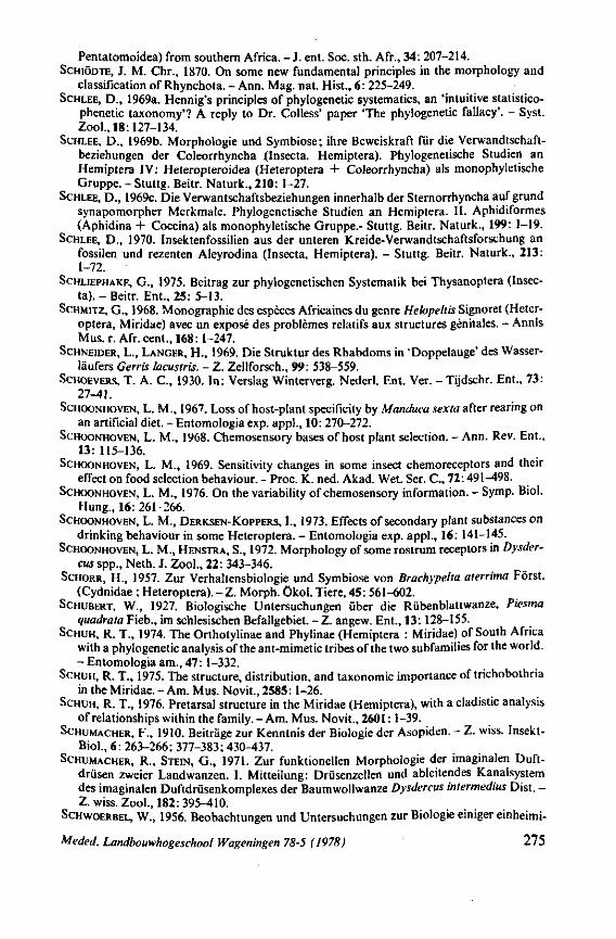
Pentatomoidea) from southern Africa. - J . ent. Soc. sth. Afr., 34: 207-214. SCHIÖDTE, J. M. Chr., 1870. On some new fundamental principles in the morphology and
classification of Rhynchota. - Ann. Mag. nat. Hist., 6: 225-249. SCHLEE, D., 1969a. Hennig's principles of phylogenetic systematics, an 'intuitive statistico-
phenetic taxonomy'? A reply to Dr. Colless' paper 'The phylogenetic fallacy'. - Syst. Zool., 18:127-134.
SCHLEE, D., 1969b. Morphologie und Symbiose; ihre Beweiskraft für die Verwandtschaftbeziehungen der Coleorrhyncha j(Insecta. Hemiptera). Phylogenetische Studien an Hemiptera IV: Heteropteroidea (Heteroptera + Coleorrhyncha) als monophyletische Gruppe. - Stuttg. Beitr. Naturk., 210: 1-27.
SCHLEE, D., 1969C. Die Verwantschaftsbeziehungen innerhalb der Sternorrhyncha aufgrund synapomorpher Merkmale. Phylogenetische Studien an Hemiptera. II. Aphidiformes (Aphidina + Coccina) als monophyletische Gruppe.- Stuttg. Beitr. Naturk., 199: 1-19.
SCHLEE, D., 1970. Insektenfossilien aus der unteren Kreide-Verwandtschaftsforschung an fossilen und rezenten Aleyrodina (Insecta, Hemiptera). - Stuttg. Beitr. Naturk., 213: 1-72.
SCHLIEPHAKE, G., 1975. Beitrag zur phylogenetischen Systematik bei Thysanoptera (Insecta). - Beitr. Ent., 25: 5-13.
SCHMITZ, G., 1968. Monographie des espèces Africaines du genre Helopeltis Signoret (Heteroptera, Miridae) avec un exposé des problèmes relatifs aux structures génitales. - Annls Mus. r. Afr. cent., 168: 1-247.
SCHNEIDER, L., LANGER, H., 1969. Die Struktur des Rhabdoms in 'Doppelauge' des Wasserläufers Gerris lacustris. - Z. Zeilforsch., 99: 538-559.
SCHOEVERS, T. A. C , 1930. In: Verslag Winterverg. Nederl. Ent. Ver. - Tijdschr. Ent., 73: 27-41.
SCHOONHOVEN, L. M., 1967. Loss of host-plant specificity by Manduca sexta after rearing on an artificial diet. - Entomologia exp. appl., 10: 270-272.
SCHOONHOVEN, L. M., 1968. Chemosensory bases of host plant selection. - Ann. Rev. Ent., 13:115-136.
SCHOONHOVEN, L. M., 1969. Sensitivity changes in some insect chemoreceptors and their effect on food selection behaviour. - Proc. K. ned. Akad. Wet. Ser. C , 72:491-498.
SCHOONHOVEN, L. M., 1976. On the variability of chemosensory information. - Symp. Biol. Hung., 16: 261-266.
SCHOONHOVEN, L. M., DERKSEN-KOPPERS, I., 1973. Effects of secondary plant substances on drinking behaviour in some Heteroptera. - Entomologia exp. appl., 16: 141-145.
SCHOONHOVEN, L. M., HENSTRA, S., 1972. Morphology of some rostrum receptors in Dysder-cus spp., Neth. J. Zool., 22: 343-346.
SCHORR, H., 1957. Zur Verhaltensbiologie und Symbiose von Brachypelta aterrima Forst. (Cydnidae ; Heteroptera). - Z . Morph, ökol. Tiere, 45: 561-602.
SCHUBERT, W., 1927. Biologische Untersuchungen über die Rübenblattwanze, Piesma quadrata Fieb., im schlesischen Befallgebiet. - Z. angew. Ent., 13: 128-155.
SCHUH, R. T., 1974. The Orthotylinae and Phylinae (Hemiptera : Miridae) of South Africa with a phylogenetic analysis of the ant-mimetic tribes of the two subfamilies for the world. - Entomologia am., 47:1-332.
SCHUH, R. T., 1975. The structure, distribution, and taxonomie importance of trichobothria in the Miridae. -Am. Mus. Novit., 2585:1-26.
SCHUH, R. T., 1976. Pretarsal structure in the Miridae (Hemiptera), with a cladistic analysis of relationships within the family. -Am. Mus. Novit., 2601:1-39.
SCHUMACHER, F., 1910. Beiträge zur Kenntnis der Biologie der Asopiden. - Z. wiss. Insekt-Biol., 6: 263-266; 377-383; 430-437.
SCHUMACHER, R., STEIN, G., 1971. Zur funktionellen Morphologie der imaginalen Duftdrüsen zweier Landwanzen. I. Mitteilung: Drüsenzellen und ableitendes Kanalsystem des imaginalen Duftdrüsenkomplexes der Baumwollwanze Dysdercus intermedius Dist. -Z. wiss. Zool., 182:395-410.
SCHWOERBEL, W., 1956. Beobachtungen und Untersuchungen zur Biologie einiger einheimi-
Meded. Landbouwhogeschool Wageningen 78-5 (1978) 275
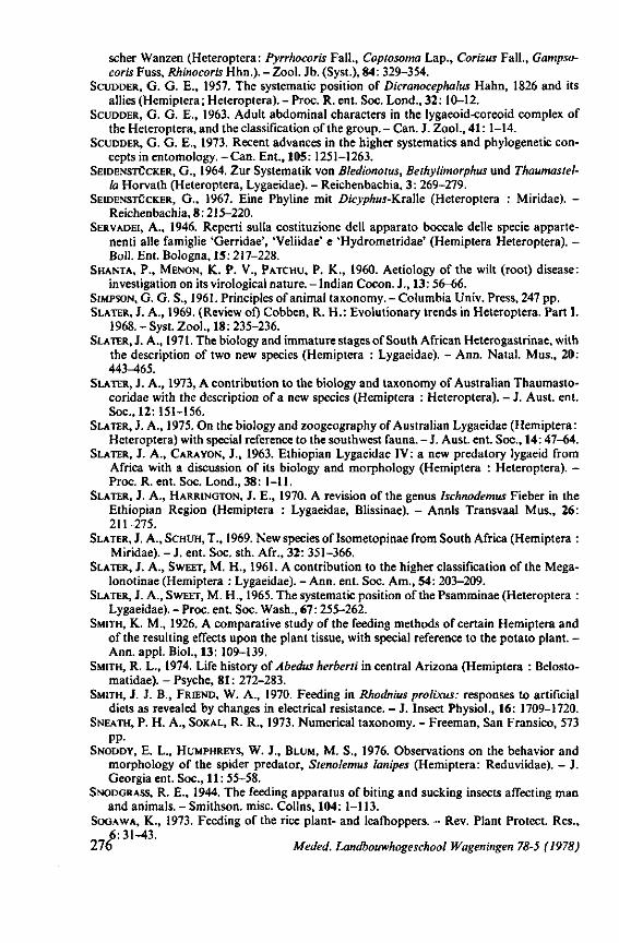
scher Wanzen (Heteroptera: Pyrrhocoris Fall., Coptosoma Lap., Corizus Fall., Gampso-coris Fuss, Rhinocoris Hhn.).-Zool. Jb. (Syst.), 84: 329-354.
SCUDDER, G. G. E., 1957. The systematic position of Dicranocephalus Hahn, 1826 and its allies (Hemiptera; Heteroptera). - Proc. R. ent. Soc. Lond., 32: 10-12.
SCUDDER, G. G. E., 1963. Adult abdominal characters in the lygaeoid-coreoid complex of the Heteroptera, and the classification of the group. - Can. J. Zool., 41: 1-14.
SCUDDER, G. G. E., 1973. Recent advances in the higher systematics and phylogenetic concepts in entomology. - Can. Ent., 105: 1251-1263.
SEIDENSTÜCKER, G., 1964. Zur Systematik von Bledionotus, Bethylimorphus und Thaumastel-la Horvath (Heteroptera, Lygaeidae). - Reichenbachia, 3: 269-279.
SEIDENSTÜCKER, G., 1967. Eine Phyline mit Dicyphus-KraWe (Heteroptera : Miridae). -Reichenbachia, 8:215-220.
SERVADEI, A., 1946. Reperti sulla costituzione dell apparato boccale delle specie apparte-nenti alle famiglie 'Gerridae', 'Veliidae' e 'Hydrometridae' (Hemiptera Heteroptera). -Boll. Ent. Bologna, 15: 217-228.
SHANTA, P., MENON, K. P. V., PATCHU, P. K., 1960. Aetiology of the wilt (root) disease: investigation on its virological nature. - Indian Cocon. J., 13: 56-66.
SIMPSON, G. G. S., 1961. Principles of animal taxonomy. - Columbia Univ. Press, 247 pp. SLATER, J. A., 1969. (Review of) Cobben, R. H.: Evolutionary trends in Heteroptera. Part I.
1968. - Syst. Zool., 18: 235-236. SLATER, J. A., 1971. The biology and immature stages of South African Heterogastrinae, with
the description of two new species (Hemiptera : Lygaeidae). - Ann. Natal. Mus., 20: 443-465.
SLATER, J. A., 1973, A contribution to the biology and taxonomy of Australian Thaumasto-coridae with the description of a new species (Hemiptera : Heteroptera). - J. Aust. ent. Soc, 12:151-156.
SLATER, J. A., 1975. On the biology and zoogeography of Australian Lygaeidae (Hemiptera: Heteroptera) with special reference to the southwest fauna. - J . Aust. ent. Soc., 14: 47-64.
SLATER, J. A., CARA YON, J., 1963. Ethiopian Lygaeidae IV: a new predatory lygaeid from Africa with a discussion of its biology and morphology (Hemiptera : Heteroptera). -Proc. R. ent. Soc. Lond., 38: 1-11.
SLATER, J. A., HARRINGTON, J. E., 1970. A revision of the genus hchnodemus Fieber in the Ethiopian Region (Hemiptera : Lygaeidae, Blissinae). - Annls Transvaal Mus., 26: 211-275.
SLATER, J. A., SCHUH, T., 1969. New species of Isometopinae from South Africa (Hemiptera : Miridae). - J . ent. Soc. sth. Afr., 32: 351-366.
SLATER, J. A., SWEET, M. H., 1961. A contribution to the higher classification of the Mega-lonotinae (Hemiptera : Lygaeidae). - Ann. ent. Soc. Am., 54: 203-209.
SLATER, J. A., SWEET, M. H., 1965. The systematic position of the Psamminae (Heteroptera : Lygaeidae). - Proc. ent. Soc. Wash., 67: 255-262.
SMITH, K. M., 1926. A comparative study of the feeding methods of certain Hemiptera and of the resulting effects upon the plant tissue, with special reference to the potato plant. -Ann. appl. Biol., 13: 109-139.
SMITH, R. L., 1974. Life history of Abedus herberti in central Arizona (Hemiptera : Belosto-matidae). - Psyche, 81: 272-283.
SMITH, J. J. B., FRIEND, W. A., 1970. Feeding in Rhodnius prolixus: responses to artificial diets as revealed by changes in electrical resistance. - J. Insect Physiol., 16: 1709-1720.
SNEATH, P. H. A., SOKAL, R. R., 1973. Numerical taxonomy. - Freeman, San Fransico, 573 pp.
SNODDY, E. L., HUMPHREYS, W. J., BLUM, M. S., 1976. Observations on the behavior and morphology of the spider predator, Stenolemus lanipes (Hemiptera: Reduviidae). - J. Georgia ent. Soc., 11: 55-58.
SNODGRASS, R. E., 1944. The feeding apparatus of biting and sucking insects affecting man and animals. - Smithson. misc. Colins, 104: 1-113.
SOGAWA, K., 1973. Feeding of the rice plant- and leafhoppers. - Rev. Plant Protect. Res., 6:31-43.
276 Meded. Landbouwhogeschool Wageningen 78-5 (1978)
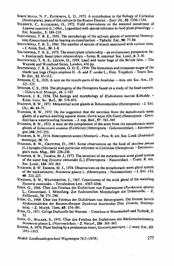
SOKOL'SKAYA, N. P., ZHITENEVA, L. D. , 1973. A contribution to the biology of Corixidae (Heteroptera), pests offish culture in the Rostov Dis tr ic t . -Zool . Zh., 52:1330-1334.
SOLBRECK, C , KUGELBERG, O., 1972. Field observations on the seasonal occurrence of Lygaeus equestris (L.) (Het., Lygaeidae) with special reference to food plant phenology. -Ent. Scandin., 3 : 189-210.
SOUTHWOOD, T. R. E., 1955. The morphology of the salivary glands of terrestrial Heteroptera (Geocorisae) and its bearing on classification. - Tijdschr. Ent., 98: 77-84.
SOUTHWOOD, T. R. E., 1961. The number of species of insects associated with various trees. - J . Anim. Ecol. ,30: 1-8.
SOUTHWOOD, T. R. E., 1973. The insect/plant relationship - an evolutionary perspective. In: v. Emden (ed). Insect/plant relationships. - Symp. R. entomol. Soc. Lond., 6: 3-30.
SOUTHWOOD, T. R. E., LESTON, D., 1959. Land and water bugs of the British Isles. - The Wayside and Woodland Series, London, 436 pp.
SOUTHWOOD, T. R. E., SCUDDER, G. G. E„ 1956. The bionomics and immature stages of the thistle lace bugs (Tingis ampliata H. - S . and T. cardui L., Hem. Tingidae). - Trans. Soc. Br. Ent., 12:93-112.
SPOONER, C. S., 1920. A note on the mouth-parts of the Aradidae. - Ann. ent. Soc. Am., 13: 121-122.
SPOONER, C. S., 1938. The phylogeny of the Hemiptera based on a study of the head capsule. - Illinois biol. Monogr., 16: 1-102.
SPRAGUE, J. B., 1956. The biology and morphology of Hydrometra martini Kirkaldy. -Kans. Univ. Sei. Bull , 38: 579-693.
STADDON, B. W., 1971. Metasternal scent glands in Belostomatidae (Heteroptera). - J . Ent. (A)., 46: 69-71.
STADDON, B. W., 1972. On the suggestion that the secretion from the metathoracic scent glands of a surface-dwelling aquatic insect, Gerris najas (De Geer) (Heteroptera : Gerri-dae) has a waterproofing function. - J. exp. Biol., 57: 765-769.
STADDON, B. W., 1973. A note on the composition of the scent from the metathoracic scent glands of Gelastocoris oculatus (Fabricius) (Heteroptera : Gelastocoridae). - Entomologist, 106: 253-255.
STADDON, B. W., 1974. Heteropteran scents (Abstract). - Proc. R. ent. Soc. Lond. (Journal of meetings), 38: 35.
STADDON, B. W., GRIFFITHS, D., 1967. Some observations on the food of Aeschna juncea (L.) nymphs (Odonata) with particular reference to Corixidae (Hemiptera). - Entomologist's mon. Mag., 103: 226-230.
STADDON, B. W., THORNE, M. J., 1973. The structure of the metathoracic scent gland system of the water bug Ilyocoris cimicoides (L.) (Heteroptera : Naucoridae). - Trans. R. ent. Soc. Lond., 124: 343-363.
STADDON, B. W. THORNE, M. J., 1974. Observations on the metathoracic scent gland system of the backswimmer, Notonecta glauca L. (Heteroptera : Notonectidae). - J. Ent. (A), 48: 223-227.
STADDON, B. W., WEATHERSTON, J., 1967. Constituents of the stink gland of the waterbug Ilyocoris cimicoides. - Tetrahedron Lett. : 4567-4568.
STEIN, G., 1966. Über den Feinbau der Duftdrüsen von Feuerwanzen (Pyrrhocoris apterus L., Geocorisae). I. Mitteilung. Zur funktionellen Morphologie der Drüsenzelle. - Z. Zellforsch., 74: 271-290.
STEIN, G., 1969. Über den Feinbau der Duftdrüsen von Heteropteren. Die hintere larvale Abdominaldrüse der Baumwollwanze Dysdercus intermedius Dist. (Insecta, Heteroptera). - Z. Morph. Tiere, 65 : 374-391.
STEIN, G., 1971. Giftige Duftstoffe bei Wanzen. - Umschau in Wissenschaft und Technik, 2 : 52.
STEIN, G., WALKER, S., 1970. Über den Feinbau der Duftdrüsen des Rückenschwimmers, Notonecta glauca L. (Notonectidae). - Z. Naturf., 256: 561-562.
STONER, A. 1970. Plant feeding by a predaeeous insect, Geocorispunetipes. - J. econ. Ent., 63 : 1911-1915.
Meded. Landbouwhogeschool Wageningen 78-5 (1978) 2 77
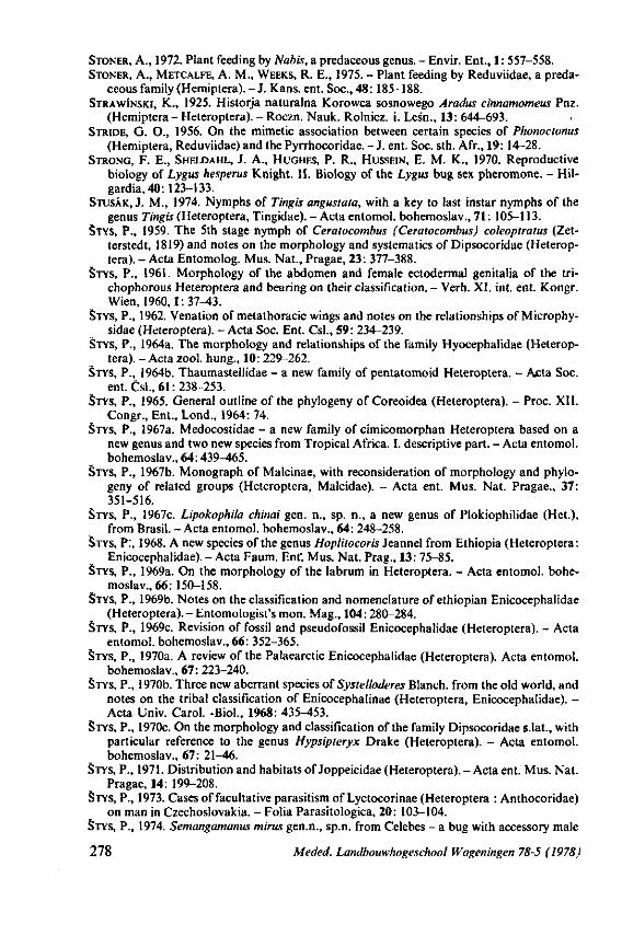
STONER, A., 1972. Plant feeding by Nabis, a predaceous genus. - Envir. Ent., 1: 557-558. STONER, A., METCALFE, A. M., WEEKS, R. E., 1975. - Plant feeding by Reduviidae, a preda
ceous family (Hemiptera). - J. Kans. ent. Soc , 48:185-188. STRAWINSKI, K., 1925. Historja naturalna Korowca sosnowego Aradus cinnamomeus Pnz.
(Hemiptera - Heteroptera). - Roczn. Nauk. Rolnicz. i. Les'n., 13: 644-693. STRIDE, G. O., 1956. On the mimetic association between certain species of Phonoctonus
(Hemiptera, Reduviidae) and the Pyrrhocoridae. - J. ent. Soc. sth. Afr., 19: 14-28. STRONG, F. E., SHELDAHL, J. A., HUGHES, P. R., HUSSEIN, E. M. K., 1970. Reproductive
biology of Lygus hesperus Knight. II. Biology of the Lygus bug sex pheromone. - Hil-gardia, 40:123-133.
STUSÂK, J. M., 1974. Nymphs of Tingis angustata, with a key to last instar nymphs of the genus Tingis (Heteroptera, Tingidae). - Acta entomol. bohemoslav., 7 1 : 105-113.
STYS, P., 1959. The 5th stage nymph of Ceralocombus (Ceratocombus) coleoptratus (Zet-terstedt, 1819) and notes on the morphology and systematics of Dipsocoridae (Heteroptera). - Acta Entomolog. Mus. Nat., Pragae, 23: 377-388.
STYS, P., 1961. Morphology of the abdomen and female ectodermal genitalia of the tri-chophorous Heteroptera and bearing on their classification. - Verh. XI. int. ent. Kongr. Wien, 1960,1:37-43.
STYS, P., 1962. Venation of metathoracic wings and notes on the relationships of Microphy-sidae (Heteroptera). - Acta Soc. Ent. Csl., 59: 234-239.
STYS, P., 1964a. The morphology and relationships of the family Hyocephalidae (Heteroptera). - Acta zool. hung., 10: 229-262.
STYS, P., 1964b. Thaumastellidae - a new family of pentatomoid Heteroptera. - Acta Soc. ent. Csl., 6 1 : 238-253.
STYS, P., 1965. General outline of the phylogeny of Coreoidea (Heteroptera). - Proc. XII. Congr., Ent., Lond., 1964: 74.
STYS, P., 1967a. Medocostidae - a new family of cimicomorphan Heteroptera based on a new genus and two new species from Tropical Africa. I. descriptive part. - Acta entomol. bohemoslav., 64:439-465.
STYS, P., 1967b. Monograph of Malcinae, with reconsideration of morphology and phylogeny of related groups (Heteroptera, Malcidae). - Acta ent. Mus. Nat. Pragae., 37: 351-516.
STYS, P., 1967c. Lipokophila chinai gen. n., sp. n., a new genus of Plokiophilidae (Het.), from Brasil. - Acta entomol. bohemoslav., 64: 248-258.
STYS, P., 1968. A new species of the genus Hopliiocoris Jeannel from Ethiopia (Heteroptera: Enicocephalidae). - Acta Faum. Ent'. Mus. Nat. Prag., 13: 75-85.
STYS, P., 1969a. On the morphology of the labrum in Heteroptera. - Acta entomol. bohemoslav., 66: 150-158.
STYS, P., 1969b. Notes on the classification and nomenclature of ethiopian Enicocephalidae (Heteroptera). - Entomologist's mon. Mag., 104: 280-284.
STYS, P., 1969c. Revision of fossil and pseudofossil Enicocephalidae (Heteroptera). - Acta entomol. bohemoslav., 66: 352-365.
STYS, P., 1970a. A review of the Palaearctic Enicocephalidae (Heteroptera). Acta entomol. bohemoslav., 67: 223-240.
STYS, P., 1970b. Three new aberrant species of Systelloderes Blanch, from the old world, and notes on the tribal classification of Enicocephalinae (Heteroptera, Enicocephalidae). -Acta Univ. Carol. -Biol., 1968: 435-453.
STYS, P., 1970c. On the morphology and classification of the family Dipsocoridae s.lat., with particular reference to the genus Hypsipteryx Drake (Heteroptera). - Acta entomol. bohemoslav., 67: 21-46.
STYS, P., 1971. Distribution and habitats of Joppeicidae (Heteroptera). - Acta ent. Mus. Nat. Pragae, 14: 199-208.
STYS, P., 1973. Cases of facultative parasitism of Lyctocorinae (Heteroptera : Anthocoridae) on man in Czechoslovakia. - Folia Parasitologica, 20: 103-104.
STYS, P., 1974. Semangamanus mints gen.n., sp.n. from Celebes - a bug with accessory male
278 Meded. Landbouwhogeschool Wageningen 78-5 (1978)
genitalia (Heteroptera, Schizopteridae). Acta entomol. bohemoslav., 71: 382-397. STYS, P., 1975. Redescription of Ptenidiophyes mirabilis Reuter (Heteroptera: Schizopteri
dae). - Vest. csl. zool. Spol., 39: 154-159. STYS, P., DANIEL, M., 1956. Lycotocoris'campestris (Fabricius, 1794) (Heteroptera, Anthoco-
ridae) jako fakultativni ektoparasit cloveka. - Vestnik Csl. Spol. zool., 1:100. STYS, P., KERZHNER, J., 1975. The rank and nomenclature of higher taxa in recent Heterop
tera. - Acta entomol. bohemoslav., 72: 65-79. SUTTON, M., 1947. Feeding mechanism of water-bugs. - Nature, 160: 122. SWEET, M. H., 1960. The seed bugs: a contribution to the feeding habits of the Lygaeidae
(Hemiptera : Heteroptera). - Ann. ent. Soc. Am., 53: 317-321. SWEET, M. H., 1964. The biology and ecology of the Rhyparochrominae of New England
(Heteroptera : Lygaeidae). Part I. - Entomologica am., 43:1-124. SWEET, M. H., SLATER, J. A., 1961. A generic key to the nymphs of North America Lygaeidae
(Hemiptera - Heteroptera). - Ann. ent. Soc. Am., 54: 333-340. TAMAKI, G., WEEKS, R. E., 1972. Biology and ecology of two predators, Geocoris pollens
Stàl and G. bullatus(Say.). -Tech. Bull. no. 1446, US Dep. of Agric. 46 pp. TAMANINI, L., 1955. Ricerche zoologiche sui Monti Sibillini (Appennino umbro - marchi-
giano). I. Descrizione di un nuovo Miride, Plagiothylus zorzii n.sp. II. Caratteri morfolo-gici e cenni biologici suil' Aradus frigidus Kiritschenko (Hemiptera, Heteroptera, Ara-didae). - Mem. Mus. Stor. Nat. Verona, 5: 31-38; 45-59.
THORSTEINSON, A. J., 1960. Host selection in phytophagous insects. -A . Rev. Ent., 5:193-218 TIMONIN, M., 1957. Relationship of Scaptocoris talpa Champ, to F. oxysporum f. cubense,
soil microflora, and host plant. - Unit. Fruit Co. Rept. TIMONIN, M., 1961. The interaction of plant pathogen, and Scaptocoris talpa Champ., Can.
J. Bot., 39:695-703. TODD, E. L., 1955. A taxonomie revision of the family Gelastocoridae (Hemiptera). - Kans.
Univ. Sei. Bull., 37: 277-475. TOWER, D. G. 1914. The mechanism of the mouth-parts of the squash bug Anasa tristis
De Geer. - Psyche, 21: 99-108. TREMBLEY, E„ ROTUNDO, G., 1975. The pheromones of the Coccoidea and their possible use
in integrated control. - 8th Int. Plant Prot. Congr. Sect. 5, Biological and Genetical Control.: 195-200.
TSUYUKI, T., OGATA, Y., YAMAMOTO, J., SHIMI, K., 1965. Stink bug aldehydes. - Agr. Biol. Chem., 29:419-427.
TULLGREN, A., 1918. Zur Morphologie und Systematik der Hemipteren. - J. Ent. Tidskr., 39:113-132.
USINGER, R. L., 1943. A revised classification of the Reduvioidea with a new subfamily from South America (Hemiptera). - Ann. ent. Soc. Am., 36: 602-618.
USINGER, R. L., 1966. Monograph of Cimicidae (Hemiptera-Heteroptera). - Thomas Say Found., 7: 585 pp.
USINGER, R. L., MATSUDA, R., 1959. Classification of the Aradidae (Hemiptera-Heteroptera). - Brit. Mus. (Nat. Hist.). 410 pp.
VANCE, T. C , 1974. Larvae of the Sericothripini (Thysanoptera : Thripidae), with reference to other larvae of the Terebrantia of Illinois. - 111. nat. Hist. Surv. Bull., 31:143-208.
VECHT, J. VAN DER, 1933. De groote peperwants of semoenjoeng (Dasynus piperis China). -Proefschr. Rijksuniv. Leiden, 101 pp.
VIGGIANI, G., 1971. Osservazioni biologicke sul Miride predatore Deraeocoris ruber (L.) (Rhynchota, Heteroptera). - Boll. Lab. Ent. agr., 29: 270-286.
VILLIERS, A., 1948. Hémiptères Reduviidés de l'Afrique Noire. - Faune Emp. fr., 9, 488 pp. VINSON, B. S., 1976. Host selection by insect parasitoids. - Ann. Rev. Ent., 21: 109-134. VODJDANI, S., 1954. Contribution à l'étude des punaises des céréales et en particulier d'Eury-
gaster integriceps Put. (Hemiptera, Pentatomidae, Scutellerinae). - Annls Epiphyt., 5: 105-160.
VOELKER, J., 1966. Wasserwanzen als obligatorische Schneckenfresser im Nildelta (Limno-getonßeberi Mayr: Belostomatidae, Hemiptera). - Z. Tropenmed. Parasit., 17: 155-165.
Meded. Landbouwhogeschool Wageningen 78-5 (1978) 279
VOELKER, J., 1968. Untersuchungen zu Ernährung, Fortpflanzungsbiologie und Entwicklung von Limnogeton fiebri Mayr (Belostomatidae, Hemiptera) als Beitrag zur Kenntnis von natürlichen Feinden tropischer Süsswasserschnecken. - Ent. Mitt. zool. Stlnst., zool. Mus. Hamb., 3: 201-224.
VOLK, J., KRCZAI, H., 1957. Ubertragungsversuche mit Piesma quadratum Fieb., dem Vektor der Kräuselkrankheit der Zucker- und Futterrübe. - NachbrBl. dt. PflSchutzdienst, 9: 17-22.
WADDILL, V., SHEPARD, M., 1974. Potential of Geocoris punctipes (Hemiptera : Lygaeidae) and Nabis spp. (Hemiptera : Nabidae) as predators of Epilachna varivestis (Coleoptera : Coccinellidae). - Entomophaga, 19: 421-426.
WAGNER, E., 1955. Bemerkungen zum System der Miridae (Hem. Het.). - Dt. ent. Z. (N.F.)., 2: 230-242.
WAGNER, W., 1963. Dynamische Taxionomie, angewandt auf die Delphaciden Mitteleuropas. - Mitt. Hamb. zool. Mus. Inst., 60:111-180.
WAGNER, E., WEBER, H. H., 1964. Hétéroptères Miridae. - Faune Fr., 67, 592 pp. WALKER, S., 1972. Die Feinstruktur der imaginalen Duftdrüsen von Corixa punctata (Heter-
optera, Hydrocorisae). - Z. wiss. Zool., 183: 190-205. WALTON, G. A., 1943. The water bugs (Rhynchota - Hemiptera) of North Somerset. - Trans.
Soc. Br. Ent., 8: 231-290. WATASE, A., 1961. Studies on the visual organs of the Aphids (Part 2). On the outer structure
of the compound eyes of hatching larvae (In Japanese with English summary). - J. appl. Ent. Zool., Tokyo, 5: 230-234.
WATERHOUSE, D. F., FORSS, D. A., HACKMAN, R. H., 1961. Characteristic odour components of the scent of stink bugs., J. Insect Physiol., 6: 113-121.
WEAVER, E. C , CLARKE, E. T., WEAVER, N., 1975. Attractiveness of an assassin bug to sting-less bees. - J. Kans. ent. Soc., 48:17-18.
WEBER, H., 1930. Biologie der Hemipteren. - Biologische Studienbücher Berlin, 11, 543 pp. WEBER, H. H., 1941. Beitrag zur Kenntnis der Hemipterenfauna der Nordseeinsel Amrum.
- Kieler Meeresforsch., 4: 109-158. WEDDE, H., 1885. Beiträge zur Kenntnis des Rhynchotenrüssels. - Arch. Naturgesch., 51:
113-143. WEFELSCHEID, H., 1912. Über die Biologie und Anatomie von Plea minutissima Leach. -
Zool. Jb., 32: 389-474. WELLS, M. J., The thoracic glands of Hemiptera Heteroptera. - Q. Jl. microsc. Sei., 95:
231-244. WENSLER, R. J., FILSHIE, B. K., 1969. Gustatory sense organs in the food canal of aphids. -
J. Morph., 129: 473-492. WHEELER, A. G., 1976. Life history of Kleidocerys resedae on European White Birch and
Ericaceous shrubs. - Ann. ent. Soc. Am., 69: 459-463. WIESE, K., 1972. Das mechanorezeptorische Beuteortungssystem von Notonecta. I. Die
Funktion des tarsalen Scolopidialorgans. - J . comp. Physiol., 78: 83-102. WIGGLESWORTH, V. B., 1947. The epicuticle in an insect, Rhodnius prolixus (Hemiptera). -
Proc. R. Soc. Lond., 134: 163-181. WILLIAMS, C. B., 1922. Co-ordinated rhythm in insects; with a record of sound production in
an aphid. - Entomologist, 55:173-176. WOLFRAM, E. M., 1972. Versuch einer Determination und Typisierung der Heteropteren
Mitteleuropas nach ihrer Ernährungsbiologie. - Inaug. Diss., Rheinische Friedrich-Wilhelm-Univ., Bonn, 321 pp.
WOODWARD, T. E., 1956. The Heteroptera of New Zealand. Part II. - The Enicocephalidae with a supplement to Part I (Cydnidae and Pentatomidae). - Trans. R. Soc. N.Z., 84: 391-430.
WOODWARD, T. E., 1958. Studies on Queensland Hemiptera. Part III. A remarkable new intertidal saldid. - Pap. Dep. Ent. Univ. Qd., 1:101-110.
WOODWARD, T. E., 1968. The australian Idiostolidae (Hemiptera: Heteroptera). - Trans. R. ent. Soc. Lond., 120: 253-261.
280 Meded. Landbouwhogeschool Wageningen 78-5 (1978)
WOODWARD, T. E., EVANS, J. W., EASTOP, V. F., 1970. In: The insects of Australia, chapter 26, Hemiptera: 387-457.
WYGODZINSKY, P., 1947. Contribuiçao ao conhecimento do gênero Heniartes Spinola, 1837 (Apiomerinae, Reduviidae, Hemiptera). - Archos Mus. nac, 41: 3-65.
WYGODZINSKY, P., 1950. Schizopterinae from Angola (Cryptostemmatidae, Hemiptera). -Publ. cult. Mus. Dundo, Comp. Diam., Ang., 7: 9-47.
WYGODZINSKY, P., 1952. Sobre algunos Cryptostemmatidae, principalmente de la Argentina (Hemiptera). - Acta zool. lilloana, 10: 51-74.
WYGODZINSKY, P., 1953. Cryptostemmatinae from Angola (Cryptostemmatidae, Hemiptera). - Publ. cult. Mus. Dundo, Comp. Diam. Ang., 16: 29-47.
WYGODZINSKY, P., 1966. A monograph of the Emesinae (Reduviidae, Hemiptera). - Bull. Am. Mus. nat. Hist., 133, 614 pp.
WYGODZINSKY, P., USINGER, R. L., 1963. Classification of the Holoptilinae and description of the first representative from the New World (Hemiptera : Reduviidae). - Proc. R. ent. Soc. Lond., 32:47-52.
YONKE, T. R., MEDLER, J. T., 1969. Biology of the Coreidae in Wisconsin. - Wis. Acad. Sei., 57:163-188.
YORK, G. T., 1944. Food studies of Geocoris spp', predators of the beet leafhopper. - J. econ. Ent., 37: 25-29.
YOUDEOWEI, A., CALAM, D. H., 1969. The morphology of the scent glands of Dysdercus intermedius Distant (Hemiptera : Pyrrhocoridae) and a preliminary analysis of the scent gland secretions of the fifth instar larvae. - Proc. R. ent. Soc. Lond., 44: 38-44.
Young, E. C , 1969. Eye growth in Corixidae (Hemiptera: Heteroptera). - Proc. R. ent. Soc. Lond.,44: 71-78.
ZDÄREK, J., 1970. Mating behaviour in the bug, Pyrrhocoris apterus L. (Heteroptera): ontogeny and its environmental control. - Behaviour 37: 253-268.
ZWART, K.. W. R., 1965. On the influence of some food substances on survival of Corixidae (Heteroptera). - Proc. 12th int. Congr. Ent. Lond., 1964: 411—412.
Meded. Landbouwhogeschool Wageningen 78-5 (1978) 281
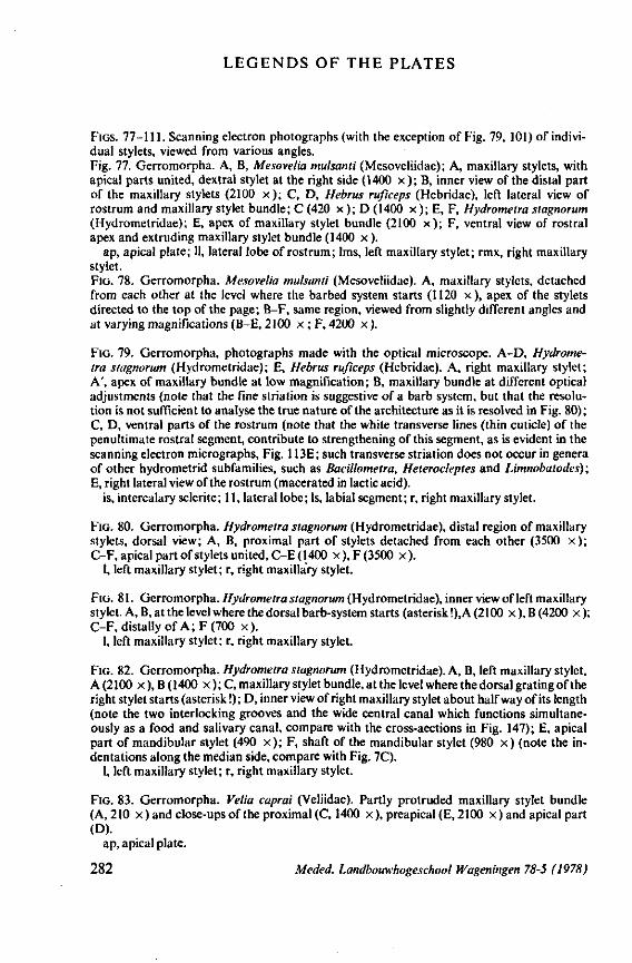
LEGENDS OF THE PLATES
FIGS. 77-111. Scanning electron photographs (with the exception of Fig. 79, 101) of individual stylets, viewed from various angles. Fig. 77. Gerromorpha. A, B, Mesovelia mulsanti (Mesoveliidae); A, maxillary stylets, with apical parts united, dextral stylet at the right side (1400 x ) ; B, inner view of the distal part of the maxillary stylets (2100 x ) ; C, D, Hebrus ruficeps (Hebridae), left lateral view of rostrum and maxillary stylet bundle; C (420 x ); D (1400 x ); E, F, Hydrometra stagnorum (Hydrometridae); E, apex of maxillary stylet bundle (2100 x ) ; F, ventral view of rostral apex and extruding maxillary stylet bundle (1400 x ).
ap, apical plate; 11, lateral lobe of rostrum; 1ms, left maxillary stylet; rmx, right maxillary stylet. FIG. 78. Gerromorpha. Mesovelia mulsanti (Mesoveliidae). A, maxillary stylets, detached from each other at the level where the barbed system starts (1120 x) , apex of the stylets directed to the top of the page; B-F, same region, viewed from slightly different angles and at varying magnifications (BE, 2100 x ; F, 4200 x ).
FIG. 79. Gerromorpha, photographs made with the optical microscope. A-D, Hydrometra stagnorum (Hydrometridae); E, Hebrus ruficeps (Hebridae). A, right maxillary stylet; A', apex of maxillary bundle at low magnification; B, maxillary bundle at different optical adjustments (note that the fine striation is suggestive of a barb system, but that the resolution is not sufficient to analyse the true nature of the architecture as it is resolved in Fig. 80) ; C, D, ventral parts of the rostrum (note that the white transverse lines (thin cuticle) of the penultimate rostral segment, contribute to strengthening of this segment, as is evident in the scanning electron micrographs, Fig. 113E; such transverse striation does not occur in genera of other hydrometrid subfamilies, such as Bacillometra, Heterocleptes and Limnobatodes); E, right lateral view of the rostrum (macerated in lactic acid),
is, intercalary sclerite; 11, lateral lobe; Is, labial segment; r, right maxillary stylet.
FIG. 80. Gerromorpha. Hydrometra stagnorum (Hydrometridae), distal region of maxillary stylets, dorsal view; A, B, proximal part of stylets detached from each other (3500 x ) ; C-F, apical part of stylets united, C-E (1400 x),F(3500 x) .
1, left maxillary stylet ; r, right maxillary stylet.
FIG. 81. Gerromorpha. Hydrometra stagnorum (Hydrometridae), inner view of left maxillary stylet. A, B, at the level where the dorsal barb-system starts (asterisk !), A (2100 x ), B (4200 x ); C-F, distally of A; F (700 x) .
1, left maxillary stylet; r, right maxillary stylet.
FIG. 82. Gerromorpha. Hydrometra stagnorum (Hydrometridae). A, B, left maxillary stylet, A (2100 x ), B (1400 x ); C, maxillary stylet bundle, at the level where the dorsal grating of the right stylet starts (asterisk !); D, inner view of right maxillary stylet about halfway of its length (note the two interlocking grooves and the wide central canal which functions simultaneously as a food and salivary canal, compare with the cross-aections in Fig. 147); E, apical part of mandibular stylet (490 x ) ; F, shaft of the mandibular stylet (980 x) (note the indentations along the median side, compare with Fig. 7C).
1, left maxillary stylet; r, right maxillary stylet.
FIG. 83. Gerromorpha. Velia caprai (Veliidae). Partly protruded maxillary stylet bundle (A,210 x ) and close-ups of the proximal (C, 1400 x), preapical (E, 2100 x ) and apical part (D).
ap, apical plate.
282 Meded. Landbouwhogeschool Wageningen 78-5 (1978)
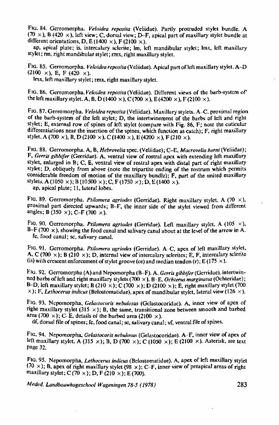
FIG. 84. Gerromorpha. Veloidea reposita (Veliidae). Partly protruded stylet bundle. A (70 x), B (420 x), left view; C, dorsal view; D-F, apical part of maxillary stylet bundle at different orientations, D, E (1400 x), F (2100 x) .
ap, apical plate; is, intercalary sclerite; lm, left mandibular stylet; lmx, left maxillary stylet; rm, right mandibular stylet; rmx, right maxillary stylet.
FIG. 85. Gerromorpha. Veloidea reposita (Veliidae). Apical part of left maxillary stylet. A-D (2100 x) . E, F (420 x) .
lmx, left maxillary stylet; rmx, right maxillary stylet.
FIG. 86. Gerromorpha. Veloidea reposita (Veliidae). Different views of the barb-system of the left maxillary stylet. A, B, D ( 1400 x ), C (700 x ), E (4200 x ), F (2100 x ).
FIG. 87. Gerromorpha. Veloidea reposita (Veliidae). Maxillary stylets. A-C, proximal region of the barb-system of the left stylet; D, the intertwinement of the barbs of left and right stylet; E, external row of spines of left stylet (compare with Fig. 86, F; note the cuticular differentiations near the insertion of the spines, which function as catch); F, right maxillary stylet.A(700 x),B,D(2100 x),C(1400 x),E(4200 x),F(210 x) .
FIG. 88. Gerromorpha. A, B, Hebrovelia spec. (Veliidae); C-E, Macrovelia horni(Veliidae); F, Gerris gibbifer (Gerridae). A, ventral view of rostral apex with extending left maxillary stylet, enlarged in B; C, E, ventral view of rostral apex with distal part of right maxillary stylet; D, obliquely from above (note the tripartite ending of the rostrum which permits considerable freedom of motion of the maxillary bundle); F, part of the united maxillary stylets.A(1050 x);B(10500 x);C,F(1750 x);D,E(1400 x) .
ap, apical plate; 11, lateral lobes.
FIG. 89. Gerromorpha. Ptilomera agriodes (Gerridae). Right maxillary stylet. A (70 x ), proximal part directed upwards; B-F, the inner side of the stylet viewed from different angles; B (350 x ) ; C-F (700 x) .
FIG. 90. Gerromorpha. Ptilomera agriodes (Gerridae). Left maxillary stylet. A (105 x), B-F (700 x ), showing the food canal and salivary canal about at the level of the arrow in A.
fc, food canal; sc, salivary canal.
FIG. 91. Gerromorpha. Ptilomera agriodes (Gerridae). A-C, apex of left maxillary stylet, A, C (700 x ) ; B (210 x ) ; D, internal view of intercalary sclerites; E, F, intercalary sclerite (is) with crescent enforcement of stylet groove (ce) and median tendon (r) ; E ( 175 x ).
FIG. 92. Gerromorpha (A) and Nepomorpha (B-F). A, Gerris gibbifer (Gerridae), intertwin-ned barbs of left and right maxillary stylets (700 x ). B-E, Ochterus marginatus (Ochteridae) ; B-D, left maxillary stylet; B (210 x);C(700 x);D(2100 x ) ; E, right maxillary stylet (700 x ) ; F, Lethocerus indicus (QeXostomatidde), apex of mandibular stylet, lateral view (126 x) .
FIG. 93. Nepomorpha, Gelastocoris nebulosus (Gelastocoridae). A, inner view of apex of right maxillary stylet (315 x ) ; B, the same, transitional zone between smooth and barbed area (700 x ) ; C-E, details of the barbed area (2100 x) .
df, dorsal file of spines; fc, food canal; sc, salivary canal; vf, ventral file of spines.
FIG. 94. Nepomorpha, Gelastocoris nebulosus (Gelastocoridae). A-F, inner view of apex of left maxillary stylet. A (315 x ) ; B, D (700 x ) ; C (1050 x ) ; E (2100 x). Asterisk, see text Page 32.
FIG. 95. Nepomorpha, Lethocerus indicus (Belostomatidae). A, apex of left maxillary stylet (70 x ) ; B, apex of right maxillary stylet (98 x ) ; C-F, inner view of preapical areas of right maxillary stylet; C (70 x ) ;D , F (210 x);E(700).
Meded. Landbouwhogeschool Wageningen 78-5 (1978) 283
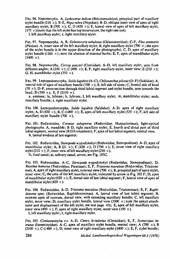
FIG. 96. Nepomorpha. A, Lethocerus indicus (Belostomatidae), preapical part of maxillary stylet bundle (210 x) . B-E, Nepa rubra (Nepidae); B-D, oblique inner view of apex of right maxillary stylet; B (700 x ) ; C, D (420 x ) ; E, lateral view of apex of left maxillary stylet, (175 x ) (note that the left stylet has top incurvature, the right one not).
1, left maxillary stylet; r, right maxillary stylet.
FIG. 97. Nepomorpha. A, B, Gelastocoris nebulosus (Gelastocoridae); C-F, Plea atomaria (Pleidae). A, inner view of the left maxillary stylet; B, right maxillary stylet (700 x ; the apex of the stylet bundle is in the upper direction of the photographs). C, D, apex of maxillary stylet bundle (1260 x ) ; note the absence of external barbs; E, F, apex of mandibular stylet (1400 x) .
FIG. 98. Nepomorpha. Corixa panzeri (Corixidae). A-D, left maxillary stylet, seen from different angles; A (350 x ) ; C (490 x ) ; E, F, right maxillary stylet, inner view; E (210 x ) ; G, H, mandibular stylet (350 x ).
FIG. 99. Leptopodomorpha. Salda lugubris (A-C), Chiloxanthuspilosus (D-F) (Saldidae). A, ventral side of apex of maxillary bundle (700 x ) ; B, left side of same; C, frontal side of head (70 x ); D-F, cross-section through third labial segment and stylet bundle, seen towards the head ; D (700 x ) ; E, F (2100 x).
a, antenna; la, labium; Ir, labrum; 1, left maxillary stylet; m, mandibular stylet; mxb, maxillary bundle; r, right maxillary stylet.
FIG. 100. Leptopodomorpha, Salda lugubris (Saldidae). A-D, apex of right maxillary stylet; A, D (420 x ) ;B , C(1400 x ) ; E, apex of left maxillary stylet (525 x ) ; F, left side of maxillary stylet bundle (700 x) .
FIG. 101. Reduvioidea, Coranus subapterus (Reduviidae, Harpactorinae), light-optical photographs. A, mandible; B-D, right maxillary stylet; E, fourth and distal part of third labial segment, ventral view (KOH-treatment); F, apex of last labial segment, ventral view.
It, lateral tendons of last segment.
FIG. 102. Reduvioidea, Stenopoda wygodzinskyi (Reduviidae, Stenopodinae). A-D, apex of mandibular stylet; A, B (21 x ) ; C (420 x ) ; D (700 x ) ; E, inner view of right maxillary stylet (210 x ) ; F, inner view of left maxillary stylet (210 x) .
fc, food canal; sc, salivary canal; arrow, see Fig. 103C.
FIG. 103. Reduvioidea. A-C, Stenopoda wygodzinskyi (Reduviidae, Stenopodinae); D, Rasahus hamulus (Reduviidae, Piratinae); E, F, Triatoma maculata (Reduviidae, Triatomi-nae). A, apex of right maxillary stylet, external view (700 x ) ; B, preapical part of same stylet, inner view; C, the area of the left maxillary stylet, indicated by arrow in Fig. 102 F; D, apex of mandibular stylet (420 x ) ; E, dorsal side of last labial segment; F, lateral view of apex of mandibular stylet (420 x ).
FIG. 104. Reduvioidea. A-D, Triatoma maculata (Reduviidae, Triatominae); E, F, Raphi-dosoma spec. (Reduviidae, Raphidosominae). A, lateral view of last labial segment; B, extreme apex of rostrum, dorsal view, with extending maxillary bundle; C, left maxillary stylet, inner view; D, maxillary stylet bundle, lateral view (3500 x ; note the apical attachment and displacement of the left stylet, see text page 43); E, apex of left maxillary stylet, inner view (490 x ) ; F, apex of right maxillary stylet, inner view (350 x) .
1, left maxillary stylet; r, right maxillary stylet.
FIG. 105. Cimicomorpha s.s. A-D, Cimex lectularius (Cimicidae); E, F, Isometopus in-trusus (Isometopidae). A-C, apex of maxillary stylet bundle, ventral view; A (700 x), B (2100 x), C (1400 x ) ; D, inner view of right maxillary stylet (1400 x ) ; E, F, stylet bundle;
284 Meded. Landbouwhogeschool Wageningen 68-5 (1978)
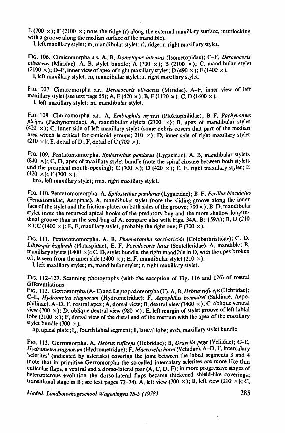
E (700 x ) ; F (2100 X ; note the ridge (r) along the external maxillary surface, interlocking with a groove along the median surface of the mandible).
1, left maxillary stylet; m, mandibular stylet; ri, ridge; r, right maxillary stylet.
FIG. 106. Cimicomorpha S.J. A, B, Isometopus intrusus (Isometopidae); C-F, Deraeocoris olivaceus (Miridae). A, B, stylet bundle; A (700 x ) ; B (2100 x ) ; C, mandibular stylet (2100 x ) ;D-F , inner view of apex of right maxillary stylet; D (490 x);F(1400 x).
1, left maxillary stylet; m, mandibular stylet; r, right maxillary stylet.
FIG. 107. Cimicomorpha s.S.. Deraeocoris olivaceus (Miridae). A-F, inner view of left maxillary stylet (see text page 55) ; A, E (420 x ) ; B, F ( 1120 x ) ; C, D ( 1400 x ).
1, left maxillary stylet; m, mandibular stylet.
FIG. 108. Cimicomorpha s.S.. A, Embiophila myersi (Plokiophilidae); B-F, Pachynomus picipes (Pachynomidae). A, mandibular stylets (2100 x ) ; B, apex of mandibular stylet (420 x ) ; C, inner side of left maxillary stylet (some debris covers that part of the median area which is critical for cimicoid groups; 210 x ) ; D, inner side of right maxillary stylet (210 x ); E, detail of D ;F , detail of C (700 x) .
FIG. 109. Pentatomomorpha, Spilostethus pandurus (Lygaeidae). A, B, mandibular stylets (840 x ) ; C, D, apex of maxillary stylet bundle (note the spiral closure between both stylets and the preapical mouth-opening); C (700 x ) ; D (420 x ) ; E, F, right maxillary stylet; E (420 x) ;F(700 x) .
lmx, left maxillary stylet; rmx, right maxillary stylet.
FIG. 110. Pentatomomorpha, A, Spilostethus pandurus (Lygaeidae); B-F, Perillus bioculatus (Pentatomidae, Asopinae). A, mandibular stylet (note the sliding-groove along the inner face of the stylet and the friction-plates on both sides of the groove; 700 x ); B-D, mandibular stylet (note the recurved apical hooks of the predatory bug and the more shallow longitudinal groove than in the seed-bug of A, compare also with Figs. 34A, B; 159A); B, D (210 x ) ; C ( 1400 x ) ; E, F, maxillary stylet, probably the right one ; F (700 x ).
FIG. 111. Pentatomomorpha. A, B, Phaenacantha saccharicida (Colobathristidae); C, D, Libyaspis haglundi (Plataspidae); E, F, Poecilocoris latus (Scutelleridae). A, mandible; B, maxillary stylets (1400 x ) ; C, D, stylet bundle, the right mandible in D, with the apex broken off, is seen from the inner side (1400 x ) ;E ,F , mandibular stylet (210 x) .
1, left maxillary stylet; m, mandibular stylet; r, right maxillary stylet.
FIG. 112-127. Scanning photographs (with the exception of Fig. 116 and 126) of rostral differentiations. FIG. 112. Gerromorpha (A-E)and Leptopodomorpha (F). A, B, Hebrusruficeps(Hebridae); C-E, Hydrometra stagnorum (Hydrometridae); F, Aepophilus bonnairei (Saldinae, Aepo-philinae). A-D, F, rostral apex; A, dorsal view; B, dextral view (1400 x ); C, oblique ventral view (700 x ) ; D, oblique dextral view (980 x ) ; E, left margin of stylet groove of left labial lobe (2100 x ) ; F, dorsal view of the distal end of the rostrum with the apex of the maxillary stylet bundle (700 x) .
ap, apical plate; 14, fourth labial segment; II, lateral lobe; mxb, maxillary stylet bundle.
FIG. 113. Gerromorpha. A, Hebrus ruficeps (Hebridae); B, Oravelia pege (Veliidae); C-E, Hydrometra stagnorum (Hydrometridae) ; F, Macrovelia horni (Veliidae). A-D, F, intercalary 'sclerites' (indicated by asterisks) covering the joint between the labial segments 3 and 4 (note that in primitive Gerromorpha the so-called intercalary sclerites are more like thin cuticular flaps, a ventral and a dorso-lateral pair (A, C, D, F); in more progressive stages of heteropterous evolution the dorso-lateral flaps became thickened shield-like coverings; transitional stage in B; see text pages 72-74). A, left view (700 x ) ; B, left view (210 x ) ; C,
Meded. Landbouwhogeschool Wageningen 78-5 (1978) 285
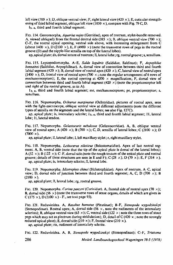
left view (700 x ); D, oblique ventral view; F, right lateral view (420 x ); E, cuticular strengthening of third labial segment, oblique left view (1050 x ), compare with Fig. 79 C, D.
b , 4, third and fourth labial segment.
FIG. 114. Gerromorpha, Aquarius najas (Gerridae), apex of rostrum, stylet-bundle removed. A, viewed obliquely from the frontal dextral side (385 x ) ; B, oblique ventral view (700 x ) ; C-F, the rostral apical opening, ventral side above, with increasing enlargement from C (about 1400 x) , D (2100 x) , E, F (4900 x ) (note the transverse rows of pegs in the rostral groove (D) and the nipple-like sensilla on top of the lateral lobes).
ap, apical plate; ds, dorsal suture of rostrum; 11, lateral lobe; rg, rostral groove; s, sensillum.
FIG. 115. Leptopodomorpha. A-E, Salda lugubris (Saldidae, Saldinae); F, Aepophilus bonnairei (Saldidae, Aepophilinae). A, dorsal view of connection between third and fourth labial segment (420 x ) ; B, dorsal view of rostral apex (420 x ) ; C, lateral view of rostral apex (1400 x ); D, frontal view of rostral apex (700 x ; note the regular arrangement of 6 rows of mechanoreceptors); E, the rostral opening at 4200 x magnification; F, dorsal view of connection betweem third and fourth labial segment (420 x ) (note the proprioreceptor left and right of the rostral groove, as in A).
I3, 4, third and fourth labial segment; mr, mechanoreceptors; pr, proprioreceptor; s, sensillum.
FIG. 116. Nepomorpha, Ochterus marginatus (Ochteridae), pictures of rostral apex, seen with the light-microscope, oblique ventral view at different adjustments (note the different types of sensilla on the segments and the lateral lobes; see also Fig. 127C).
ap, apical plate; is, intercalary sclerite; b , 4, third and fourth labial segment; 11, lateral lobe; It, lateral tendon.
FIG. 117. Nepomorpha. Gelastocoris nebulosus (Gelastocoridae). A, B, oblique ventral view of rostral apex; A (420 x ) ; B (700 x ) ; C, D, sensilla of lateral lobes; C (2100 x ) ; D (7000 x).
ap, apical plate ; 11, lateral lobe ; 1, left maxillary stylet ; r, right maxillary stylet.
FIG. 118. Nepomorpha. Lethocerus niloticus (Belostomatidae). Apex of last rostral segment. A, B, ventral side (note that the tip of the apical plate is dorsal of the lateral lobes); A (52 x ) ; B (122 x ) ;C-F , dorsal side.(note the rough texture of the apical plate and rostral groove; details of these structures are seen in E and F); C (28 x ) ; D (70 x ) ; E, F (314 x) .
ap, apical plate; is, intercalary sclerite; 11, lateral lobe.
FIG. 119. Nepomorpha. Idiotrephes chinai (Helotrephidae). Apex of rostrum. A~C, apical view; D, dorsal side of junction between third and fourth segment; A, C, D (700 x ) ; B (2100 x) .
ap, apical plate; 11, lateral lobe; rg, rostral groove.
FIG. 120. Nepomorpha. Corixa panzeri (Corixidae). A, frontal side of rostral apex (70 x ) ; B, dorsal side (56 x ) (note the transverse rows of sense organs, details of which are given in C(175 x), D(2100 x ) - F., see text page 93).
FIG. 121. Reduvioidea. A, Rasahus hamatus (Piratinae); B-F, Stenopoda wygodzinskyi (Stenopodinae). Rostral apex; A, dorsal side (56 x , note the rudiments of the intercalary sclerites); B, oblique ventral view (63 x ); C, ventral side (122 x ; note the three rows of stout pegs which may act as plectrum during stridulation); D, detail of C (420 x ; note the strongly reduced apical plate); E, dorsal side (210 x ); F, frontal view (210 x ).
ap, apical plate; ris, rudiment of intercalary sclerite.
FIG. 122. Reduvioidea. A, B, Stenopoda wygodzinskyi (Stenopodinae); C-F, Triatoma
286 Meded. Landbouwhogeschool Wageningen 78-5 (1978)
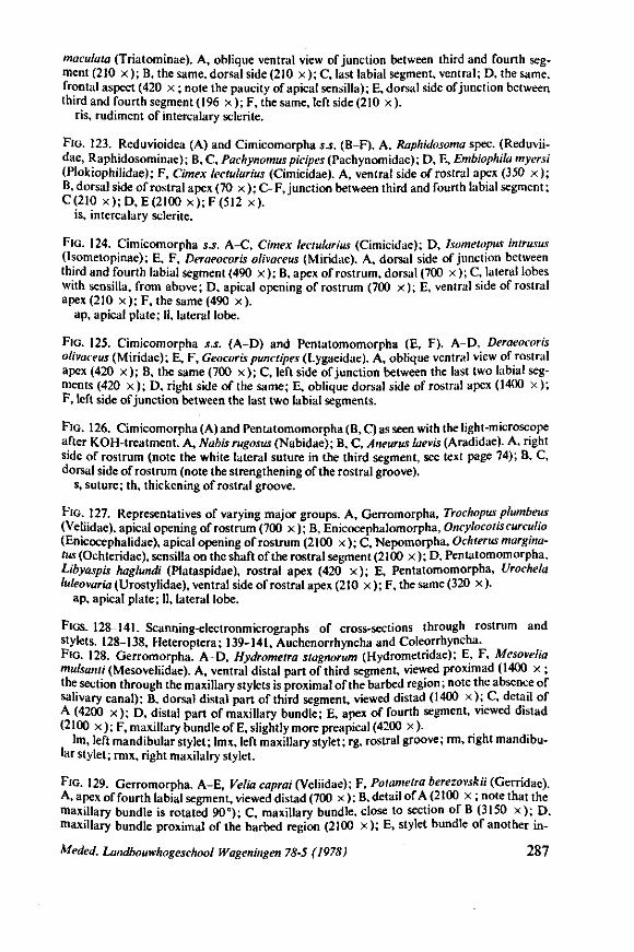
maculata (Triatominae). A, oblique ventral view of junction between third and fourth segment (210 x ) ; B, the same, dorsal side (210 x ) ; C, last labial segment, ventral; D, the same, frontal aspect (420 x ; note the paucity of apical sensilla); E, dorsal side of junction between third and fourth segment (196 x ); F, the same, left side (210 x ).
ris, rudiment of intercalary sclerite.
Flo. 123. Reduvioidea (A) and Cimicomorpha 5.s. (B-F). A, Raphidosoma spec. (Reduvii-dae, Raphidosominae); B, C, Pachynomus picipes (Pachynomidae); D, E, Embiophila myersi (Plokiophilidae); F, Cimex lectularius (Cimicidae). A, ventral side of rostral apex (350 x ) ; B, dorsal side of rostral apex (70 x ); C-F, junction between third and fourth labial segment; C(210 x);D,E(2100 x);F(512 x) .
is, intercalary sclerite.
FIG. 124. Cimicomorpha s.S. A-C, Cimex lectularius (Cimicidae); D, hometopus intrusus (Isometopinae); E, F, Deraeocoris olivaceus (Miridae). A, dorsal side of junction between third and fourth labial segment (490 x ) ; B, apex of rostrum, dorsal (700 x ) ; C, lateral lobes with sensilla, from above; D, apical opening of rostrum (700 x ); E, ventral side of rostral apex (210 x ); F, the same (490 x ).
ap, apical plate; 11, lateral lobe.
FIG. 125. Cimicomorpha s.s. (A-D) and Pentatomomorpha (E, F). A-D, Deraeocoris olivaceus (Miridae); E, F, Geocoris punctipes (Lygaeidae). A, oblique ventral view of rostral apex (420 x ) ; B, the same (700 x ) ; C, left side of junction between the last two labial segments (420 x ); D, right side of the same; E, oblique dorsal side of rostral apex (1400 x ); F, left side of junction between the last two labial segments.
FIG. 126. Cimicomorpha (A) and Pentatomomorpha (B, C) as seen with the light-microscope after KOH-treatment. A, Nabis rugosus (Nabidae); B, C, Aneurus laevis (Aradidae). A, right side of rostrum (note the white lateral suture in the third segment, see text page 74); B, C, dorsal side of rostrum (note the strengthening of the rostral groove),
s, suture; th, thickening of rostral groove.
FIG. 127. Representatives of varying major groups. A, Gerromorpha, Trochopus plumbeus (Veliidae), apical opening of rostrum (700 x ); B, Enicocephalomorpha, Oncylocotis curculio (Enicocephalidae), apical opening of rostrum (2100 x ) ; C, Nepomorpha, Ochterus margina-tus (Ochteridae), sensilla on the shaft of the rostral segment (2100 x ); D, Pentatomomorpha, Libyaspis haglundi (Plataspidae), rostral apex (420 x ) ; E, Pentatomomorpha, Urochela luleovaria (Urostylidae), ventral side of rostral apex (210 x ); F, the same (320 x ).
ap, apical plate; 11, lateral lobe.
FIGS. 128-141. Scanning-electronmicrographs of cross-sections through rostrum and stylets. 128-138, Heteroptera; 139-141, Auchenorrhyncha and Coleorrhyncha. FIG. 128. Gerromorpha. A-D, Hydrometra stagnorum (Hydrometridae); E, F, Mesovelia mulsanti (Mesoveliidae). A, ventral distal part of third segment, viewed proximad (1400 x ; the section through the maxillary stylets is proximal of the barbed region ; note the absence of salivary canal); B, dorsal distal part of third segment, viewed distad (1400 x ) ; C, detail of A (4200 x ) ; D, distal part of maxillary bundle; E, apex of fourth segment, viewed distad (2100 x ) ;F , maxillary bundle ofE, slightly more preapical (4200 x).
lm, left mandibular stylet; lmx, left maxillary stylet; rg, rostral groove; rm, right mandibular stylet; rmx, right maxilalry stylet.
FIG. 129. Gerromorpha. A-E, Velio, caprai (Veliidae); F, Polametra berezovskii (Genidae). A, apex of fourth labial segment, viewed distad (700 x ); B, detail of A (2100 x ; note that the maxillary bundle is rotated 90°); C, maxillary bundle, close to section of B (3150 x ) ; D, maxillary bundle proximal of the barbed region (2100 x ) ; E, stylet bundle of another in-
Meded. Landbouwhogeschool Wageningen 78-5 (1978) 287
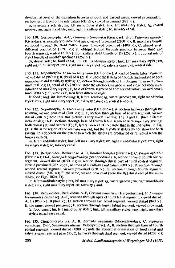
dividual, at level of the transition between smooth and barbed areas, viewed proximad ; F, section just in front of the intercalary sclerites, viewed proximad (980 x ).
is, intercalary sclerite; lm, left mandibular stylet; lmx, left maxillary stylet; rg, rostral groove; rm, right mandible; rmx, right maxillary stylet; sc, salivary canal.
FIG. 130. Gerromorpha. A-C, Potametra berezovskii (Gerridae); D-F, Ptilomera agriodes (Gerridae). A, maxillary bundle near apex, viewed proximad (2100 x ) ; B, maxillary bundle sectioned through the third rostral segment, viewed proximad (1400 x ) ; C, almost as A, different orientation (1750 x ) ; D, oblique section through junction between third and fourth segment, ventral (210 x ); E, maxillary stylet bundle of D (1258 x ) ; F, dorsal side of stylet bundle of another specimen (1258 x) .
ds, dorsal side; fc, food canal; lm, left mandibular stylet; lmx, left maxillary stylet; rm, right mandibular stylet; rmx, right maxillary stylet; sc, salivary canal; vs, ventral side.
FIG: 131. Nepomorpha. Ochterus marginatus (Ochteridae). A, end of fourth labial segment, viewed distad (980 x ) ; B, detail of A (2100 x ;note the fluting on the external surface of both mandibular and maxillary stylets); C, section through middle of third segment, viewed proximad (980 x ) ; D, detail of C (2100 x ; note the interlocking groove and ridge between mandibular and maxillary stylet); E, base of fourth segment of another individual, viewed proximad (7000 x ); F, same as E, seen from different angle.
fc, food canal; int. interlocking; It, lateral tendon; rg, rostral groove; rm, right mandibular stylet; rmx, right maxillary stylet; sc, salivary canal; vt, ventral tendons.
FIG. 132. Nepomorpha. Ochterus marginatus (Ochteridae). A, section half-way through the rostrum, viewed proximad (280 x ) ; B, C, section through base of fourth segment, viewed distad (280 x ; note that this picture is very much like Fig. 131 B and E, three different individuals); D-F, sections through base of fourth labial segment with maxillary gratings both dorsal (D) and ventral (F); E, lateral view (2100 x ; note that in the individual in Fig. 131 F the same region of the rostrum was cut, but the maxillary stylets do not show the barb system ; this depends on the extent to which the stylets are protracted or retracted when the bug was killed).
lm, left mandibular stylet ; lmx, left maxillary stylet ; rm, right mandibular stylet ; rmx, right maxillary stylet; sc, salivary canal.
FIG. 133. Reduvioidea, Reduviidae. A, B, Rasahus hamatus (Piratinae); C, Pirates hybridus (Piratinae); D-F, Stenopoda wygodzinslcyi (Stenopodmné). A, section through fourth rostral segment, viewed distad (1050 x ) ; B, section through distal part of third rostral segment, viewed proximad (702 x ) ; C, neurons of maxillary axial canal (9600 x ) ; D, section through second rostral segment, viewed proximad (250 x ) ; E, section through fourth segment, viewed distad (840 x ) ; F, the same, viewed proximad (note the flat distal end of the mandibles, see Figs. 102A-D).
lm, left mandibular stylet ; lmx, left maxillary stylet ; rg, rostral groove ; rm, right mandibular stylet; rmx, right maxillary stylet; sc, salivary gland.
FIG. 134. Reduvioidea, Reduviidae. A-E, Coranus subapterus (Harpactorinae); F, Emesaya brevipennis (Emesinae). A-C, sections through apex of third labial segment, viewed distad; A, C (1050 x ) ; B (840 x ) ; D, section through last labial segment, viewed distad (840 x ) ; E, the same, viewed proximad; F, section through fourth labial segment, viewed proximad.
fc, food canal; lm, left mandibular stylet; lmx, left maxillary stylet; rmx, right maxillary stylet; sc, salivary canal.
FIG. 135. Cimicomorpha s.s. A, B, Loricula elegantula (Microphysidae); C, Joppeicus paradoxus; D-F, Scotomedes alienus (Velocipedidae). A, B, section through base of fourth rostral segment, viewed distad (4200 x ; note the abnormal orientation of food canal and salivary canal, see text page 69); C, half-way through third segment, viewed distad (4200 x ) ;
288 Meded. Landbouwhogeschool Wageningen 78-5 (1978)
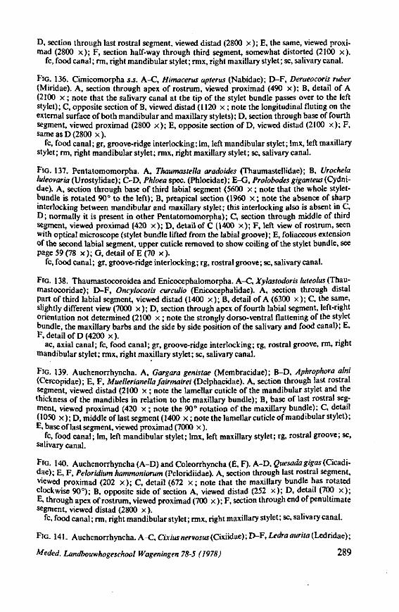
D, section through last rostral segment, viewed distad (2800 x ) ; E, the same, viewed proxi-mad (2800 x ) ; F, section half-way through third segment, somewhat distorted (2100 x) .
fc, food canal ; rm, right mandibular stylet ; rmx, right maxillary stylet ; sc, salivary canal.
FIG. 136. Cimicomorpha s.S. A-C, Himacerus apterus (Nabidae); D-F, Deraeocoris ruber (Miridae). A, section through apex of rostrum, viewed proximad (490 x ) ; B, detail of A (2100 x ; note that the salivary canal at the tip of the stylet bundle passes over to the left stylet); C, opposite section of B, viewed distad (1120 x ; note the longitudinal fluting on the external surface of both mandibular and maxillary stylets) ; D, section through base of fourth segment, viewed proximad (2800 x ) ; E, opposite section of D, viewed distad (2100 x ) ; F, same as D (2800 x ).
fc, food canal; gr, groove-ridge interlocking; lm, left mandibular stylet; lmx, left maxillary stylet; rm, right mandibular stylet; rmx, right maxillary stylet; sc, salivary canal.
FIG. 137. Pentatomomorpha. A, Thaumastella aradoides (Thaumastellidae); B, Urochela luleovaria (Urostylidae); C-D, Phloea spec. (Phloeidae); E-G, Prolobodes giganteus (Cydni-dae). A, section through base of third labial segment (5600 x ; note that the whole stylet-bundle is rotated 90° to the left); B, preapical section (1960 x ; note the absence of sharp interlocking between mandibular and maxillary stylet; this interlocking also is absent in C, D ; normally it is present in other Pentatomomorpha) ; C, section through middle of third segment, viewed proximad (420 x ) ; D, detail of C (1400 x ) ; F, left view of rostrum, seen with optical microscope (stylet bundle lifted from the labial groove); E, foliaceous extension of the second labial segment, upper cuticle removed to show coiling of the stylet bundle, see page 59 (78 x ); G, detail of E (70 x ).
fc, food canal ; gr, groove-ridge interlocking; rg, rostral groove; sc, salivary canal.
FIG. 138. Thaumastocoroidea and Enicocephalomorpha. A-C, Xylastodoris luteolus (Thau-mastocoridae); D-F, Oncylocotis curculio (Enicocephalidae). A, section through distal part of third labial segment, viewed distad (1400 x ) ; B, detail of A (6300 x ) ; C, the same, slightly different view (7000 x ); D, section through apex of fourth labial segment, left-right orientation not determined (2100 x ; note the strongly dorso-ventral flattening of the stylet bundle, the maxillary barbs and the side by side position of the salivary and food canal); E, F, detail of D (4200 x) .
ac, axial canal; fc, food canal; gr, groove-ridge interlocking; rg, rostral groove, rm, right mandibular stylet; rmx, right maxillary stylet; sc, salivary canal.
FIG. 139. Auchenorrhyncha. A, Gargara genistae (Membracidae) ; B-D, Aphrophora alni (Cercopidae); E, F, Muellerianella fairmairei (Delphacidae). A, section through last rostral segment, viewed distad (2100 x ; note the lamellar cuticle of the mandibular stylet and the thickness of the mandibles in relation to the maxillary bundle); B, base of last rostral segment, viewed proximad (420 x ; note the 90° rotation of the maxillary bundle); C, detail (1050 x ); D, middle of last segment (1400 x ; note the lamellar cuticle of mandibular stylet); E, base of last segment, viewed proximad (7000 x ).
fc, food canal; lm, left mandibular stylet; lmx, left maxillary stylet; rg, rostral groove; sc, salivary canal.
FIG. 140. Auchenorrhyncha (A-D) and Coleorrhyncha (E, F). A-D, Quesadagigas (Cicadi-dae); E, F, Peloridium hammoniorum (Peloridiidae). A, section through last rostral segment, viewed proximad (202 x ) ; C, detail (672 x ; note that the maxillary bundle has rotated clockwise 90°); B, opposite side of section A, viewed distad (252 x ) ; D, detail (700 x ) ; E, through apex of rostrum, viewed proximad (700 x ) ; F, section through end of penultimate segment, viewed distad (2800 x ).
fc, food canal ; rm, right mandibular stylet ; rmx, right maxillary stylet ; sc, salivary canal.
FIG. 141. Auchenorrhyncha. A-C, Cuius nervosus (Cixiidae) ; D-F, Ledra aurita (Ledridae) ;
Meded. Landbouwhogeschool Wageningen 78-5 (1978) 289
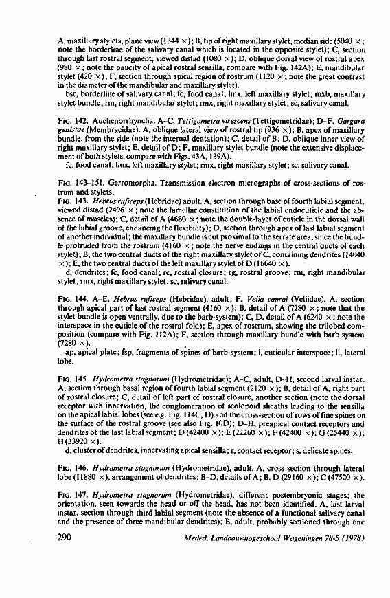
A, maxillary stylets, plane view (1344 x ); B, tip of right maxillary stylet, median side (5040 x ; note the borderline of the salivary canal which is located in the opposite stylet) ; C, section through last rostral segment, viewed distad (1080 x ) ; D, oblique dorsal view of rostral apex (980 x ; note the paucity of apical rostral sensilla, compare with Fig. 142A); E, mandibular stylet (420 x ) ; F, section through apical region of rostrum (1120 x ; note the great contrast in the diameter of the mandibular and maxillary stylet).
bsc, borderline of salivary canal; fc, food canal; lmx, left maxillary stylet; mxb, maxillary stylet bundle; rm, right mandibular stylet; rmx, right maxillary stylet; sc, salivary canal.
FIG. 142. Auchenorrhyncha. A-C, Tettigometra virescens (Tettigometridae); D-F, Gargara genistae (Membracidae). A, oblique lateral view of rostral tip (936 x ) ; B, apex of maxillary bundle, from the side (note the internal dentation); C, detail of B; D, oblique inner view of right maxillary stylet; E, detail of D; F, maxillary stylet bundle (note the extensive displacement of both stylets, compare with Figs. 43A, 139 A),
fc, food canal; lmx, left maxillary stylet; rmx, right maxillary stylet; sc, salivary canal.
FIG. 143-151. Gerromorpha. Transmission electron micrographs of cross-sections of rostrum and stylets. FIG. 143. Hebrus ruficeps (Hebridae) adult. A, section through base of fourth labial segment, viewed distad (2496 x ; note the lamellar constitution of the labial endocuticle and the absence of muscles); C, detail of A (4680 x ; note the double-layer of cuticle in the dorsal wall of the labial groove, enhancing the flexibility); D, section through apex of last labial segment of another individual ; the maxillary bundle is cut proximal to the serrate area, since the bundle protruded from the rostrum (4160 x ; note the nerve endings in the central ducts of each stylet) ; B, the two central ducts of the right maxillary stylet of C, containing dendrites (14040 x ) ; E, the two central ducts of the left maxillary stylet of D ( 16640 x ).
d, dendrites; fc, food canal; re, rostral closure; rg, rostral groove; rm, right mandibular stylet; rmx, right maxillary stylet; sc, salivary canal.
FIG. 144. A-E, Hebrus ruficeps (Hebridae), adult; F, Velia caprai (Veliidae). A, section through apical part of last rostral segment (4160 x ); B, detail of A (7280 x ; note that the stylet bundle is open ventrally, due to the barb-system); C, D, detail of A (6240 x ; note the interspace in the cuticle of the rostral fold); E, apex of rostrum, showing the trilobed composition (compare with Fig. 112A); F, section through maxillary bundle with barb system (7280 x) .
ap, apical plate; fsp, fragments of spines of barb-system ; i, cuticular interspace; 11, lateral lobe.
FIG. 145. Hydrometra stagnorum (Hydrometridae); A-C, adult, D-H, second larval instar. A, section through basal region of fourth labial segment (2120 x ) ; B, detail of A, right part of rostral closure; C, detail of left part of rostral closure, another section (note the dorsal receptor with innervation, the conglomeration of scolopoid sheaths leading to the sensilla on the apical labial lobes (see e.g. Fig. 114C, D) and the cross-section of rows of fine spines on the surface of the rostral groove (see also Fig. 10D); D-H, preapical contact receptors and dendrites of the last labial segment; D (42400 x ) ; E (22260 x ) ; F (42400 x ) ; G (25440 x ) ; H (33920 x) .
d, cluster of dendrites, innervating apical sensilla ; r, contact receptor; s, delicate spines.
FIG. 146. Hydrometra stagnorum (Hydrometridae), adult. A, cross section through lateral lobe(11880 x), arrangement of dendrites; B-D, details o fA;B,D (29160 x);C(47520 x) .
FIG. 147. Hydrometra stagnorum (Hydrometridae), different postembryonic stages; the orientation, seen towards the head or off the head, has not been identified. A, last larval instar, section through third labial segment (note the absence of a functional salivary canal and the presence of three mandibular dendrites); B, adult, probably sectioned through one
290 Meded. Landbouwhogeschool Wageningen 78-5 (1978)
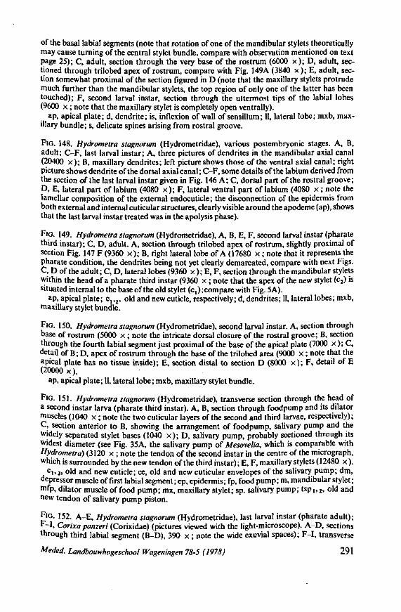
of the basal labial segments (note that rotation of one of the mandibular stylets theoretically may cause turning of the central stylet bundle, compare with observation mentioned on text page 25); C, adult, section through the very base of the rostrum (6000 x ) ; D, adult, sectioned through trilobed apex of rostrum, compare with Fig. 149A (3840 x ) ; E, adult, section somewhat proximal of the section figured in D (note that the maxillary stylets protrude much further than the mandibular stylets, the top region of only one of the latter has been touched); F, second larval instar, section through the uttermost tips of the labial lobes (9600 x ; note that the maxillary stylet is completely open ventrally).
ap, apical plate; d, dendrite; is, inflexion of wall of sensillum; 11, lateral lobe; mxb, maxillary bundle; s, delicate spines arising from rostral groove.
FIG. 148. Hydrometra stagnorum (Hydrometridae), various postembryonic stages. A, B, adult; C-F, last larval instar; A, three pictures of dendrites in the mandibular axial canal (20400 x ) ; B, maxillary dendrites; left picture shows those of the ventral axial canal; right picture shows dendrite of the dorsal axial canal; C-F, some details of the labium derived from the section of the last larval instar given in Fig. 146 A; C, dorsal part of the rostral groove; D, E, lateral part of labium (4080 x ); F, lateral ventral part of labium (4080 x ; note the lamellar composition of the external endocuticle; the disconnection of the epidermis from both external and internal cuticular structures, clearly visible around the apodeme (ap), shows that the last larval instar treated was in the apolysis phase).
FIG. 149. Hydrometra stagnorum (Hydrometridae), A, B, E, F, second larval instar (pharate third instar); C, D, adult. A, section through trilobed apex of rostrum, slightly proximal of section Fig. 147 F (9360 x ) ; B, right lateral lobe of A (17680 x ; note that it represents the pharate condition, the dendrites being not yet clearly demarcated, compare with next Figs. C, D of the adult; C, D, lateral lobes (9360 x ) ; E, F, section through the mandibular stylets within the head of a pharate third instar (9360 x ; note that the apex of the new stylet (c2) is situated internal to the base of the old stylet (c,) ; compare with Fig. 5A).
ap, apical plate; c,,2, old and new cuticle, respectively; d, dendrites; 11, lateral lobes; mxb, maxillary stylet bundle.
FIG. 150. Hydrometra stagnorum (Hydrometridae), second larval instar. A, section through base of rostrum (5000 x ; note the intricate dorsal closure of the rostral groove; B, section through the fourth labial segment just proximal of the base of the apical plate (7000 x ); C, detail of B ; D, apex of rostrum through the base of the trilobed area (9000 x ; note that the apical plate has no tissue inside); E, section distal to section D (8000 x ) ; F, detail of E (20000 x) .
ap, apical plate ; 11, lateral lobe ; mxb, maxillary stylet bundle.
FIG. 151. Hydrometra stagnorum (Hydrometridae), transverse section through the head of a second instar larva (pharate third instar). A, B, section through foodpump and its dilator muscles (1040 x ; note the two cuticular layers of the second and third larvae, respectively); C, section anterior to B, showing the arrangement of foodpump, salivary pump and the widely separated stylet bases (1040 x ) ; D, salivary pump, probably sectioned through its widest diameter (see Fig. 35A, the salivary pump of Mesovelia, which is comparable with Hydrometra) (3120 x ; note the tendon of the second instar in the centre of the micrograph, which is surrounded by the new tendon of the third instar); E, F, maxillary stylets (12480 x ).
c i , 2. old and new cuticle; ce, old and new cuticular envelopes of the salivary pump; dm, depressor muscle of first labial segment; ep, epidermis; fp, food pump; m, mandibular stylet; mfp, dilator muscle of food pump; mx, maxillary stylet; sp. salivary pump; tsp1>2, old and new tendon of salivary pump piston.
FIG. 152. A-E, Hydrometra stagnorum (Hydrometridae), last larval instar (pharate adult); F-I, Corixa panzeri (Corixidae) (pictures viewed with the light-microscope). A-D, sections through third labial segment (B-D), 390 x ; note the wide exuvial spaces); F-I, transverse
Meded. Landbouwhogeschool Wageningen 78-5 (1978) 291
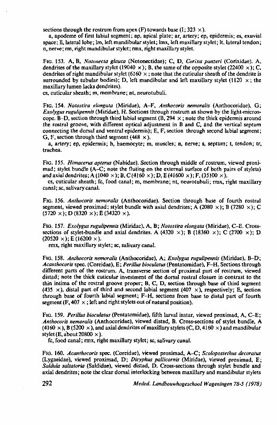
sections through the rostrum from apex (F) towards base (I; 323 x ). a, apodeme of first labial segment; ap, apical plate; ar, artery; ep, epidermis; es, exuvial
space; 11, lateral lobe; lm, left mandibular stylet; lmx, left maxillary stylet; It, lateral tendon; n, nerve ; rm, right mandibular stylet ; rmx, right maxillary stylet.
FIG. 153. A, B, Notonecta glauca (Notonectidae); C, D, Corixa panzeri (Corixidae). A, dendrites of the maxillary stylet (19040 x ) ; B, the same of the opposite stylet (22400 x ) ; C, dendrites of right mandibular stylet (6160 x ; note that the cuticular sheath of the dendrite is surrounded by tubular bodies); D, left mandibular and left maxillary stylet (1120 x ; the maxillary lumen lacks dendrites), cs, cuticular sheath; m, membrane; nt, neurotubuli.
FIG. 154. Notostira elongata (Miridae), A-F, Anthocoris nemoralis (Anthocoridae), G; Exolygus rugulipennis (Miridae), H. Sections through rostrum as shown by the light-microscope. B-D, section through third labial segment (B, 294 x ; note the thick epidermis around the rostral groove, with different optical adjustment in B and C, and the vertical septum connecting the dorsal and ventral epidermis); E, F, section through second labial segment; G, F, section through third segment (468 x ).
a, artery; ep, epidermis; h, haemocyte; m, muscles; n, nerve; s, septum; t, tendon; tr, trachea.
FIG. 155. Himacerus apterus (Nabidae). Section through middle of rostrum, viewed proxi-mad; stylet bundle (A-C; note the fluting on the external surface of both pairs of stylets) and axial dendrites; A(1040 x ) ; B, C (4160 x ) ;D , E (41600 x ) ; F, (35100 x) .
cs, cuticular sheath; fc, food canal; m, membrane; nt, neurotubuli; rmx, right maxillary canal; sc, salivary canal.
FIG. 156. Anthocoris nemoralis (Anthocoridae). Section through base of fourth rostral segment, viewed proximad; stylet bundle with axial dendrites; A (2080 x ) ; B (7280 x ) ; C (5720 x);D(8320 x ) ; E (34320 x) .
FIG. 157. Exolygus rugulipennis (Miridae), A, B; Notostira elongata (Miridae), C-E. Cross-sections of stylet-bundle and axial dendrites. A (4320 x ) ; B (18360 x ) ; C (2700 x ) ; D (20520 x ) ; E (16200 x) .
rmx, right maxillary stylet; sc, salivary canal.
FIG. 158. Anthocoris nemoralis (Anthocoridae), A; Exolygus rugulipennis (Miridae), B-D; Acanthocoris spec. (Coreidae), E; Perillus bioculatus (Pentatomidae), F-H. Sections through different parts of the rostrum. A, transverse section of proximal part of rostrum, viewed distad ; note the thick cuticular investment of the dorsal rostral closure in contrast to the thin intima of the rostral groove proper; B, C, D, section through base of third segment (435 x ), distal part of third and second labial segment (407 x ), respectively; E, section through base of fourth labial segment; F-H, sections from base to distal part of fourth segment (F, 407 x ; left and right stylets out of natural position).
FIG. 159. Perillus bioculatus (Pentatomidae), fifth larval instar, viewed proximad, A, C-E; Anthocoris nemoralis (Anthocoridae), viewed distad, B. Cross-sections of stylet bundle, A (4160 x),B(5200 x), and axial dendrites of maxillary stylets (C,D, 4160 x ) and mandibular stylet (E, about 20800 x) .
fc, food canal; rmx, right maxillary stylet; sc, salivary canal.
FIG. 160. Acanthocoris spec. (Coreidae), viewed proximad, A-C; Scolopostethus decoratus (Lygaeidae), viewed proximad, D; Dicyphus pall kornis (Miridae), viewed proximad, E; Saldula saltatoria (Saldidae), viewed distad, D. Cross-sections through stylet bundle and axial dendrites; note the clear dorsal interlocking between maxillary and mandibular stylets
292 Meded. Landbouwhogeschool Wageningen 78-5 (1978)
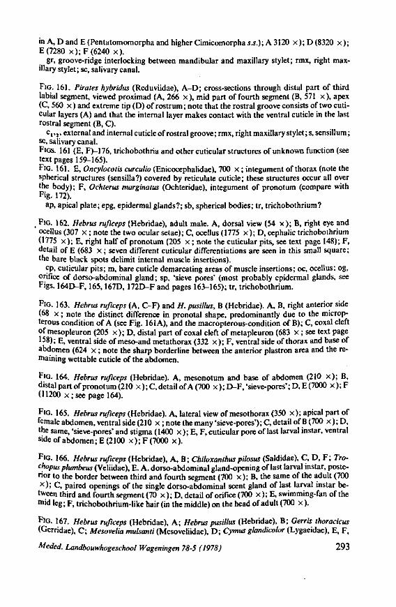
in A, D and E (Pentatomomorpha and higher Cimicomorpha s.S.); A 3120 x ) ; D (8320 x ) ; E (7280 x ) ; F (6240 x) .
gr, groove-ridge interlocking between mandibular and maxillary stylet; rmx, right maxillary stylet; sc, salivary canal.
FIG. 161. Pirates hybridus (Reduviidae), A-D; cross-sections through distal part of third labial segment, viewed proximad (A, 266 x ), mid part of fourth segment (B, 571 x ), apex (C, 560 x ) and extreme tip (D) of rostrum ; note that the rostral groove consists of two cuti-cular layers (A) and that the internal layer makes contact with the ventral cuticle in the last rostral segment (B, C).
c,,2, external and internal cuticle of rostral groove ; rmx, right maxillary stylet; s, sensillum ; sc, salivary canal. FIGS. 161 (E, F)-176, trichobothria and other cuticular structures of unknown function (see text pages 159-165). FIG. 161. E, Oncylocotis curculio (Enicocephalidae), 700 x ; integument of thorax (note the spherical structures (sensilla?) covered by reticulate cuticle; these structures occur all over the body); F, Ochterus marginatus (Ochteridae), integument of pronotum (compare with Fig. 172).
ap, apical plate; epg, epidermal glands?; sb, spherical bodies; tr, trichobothrium?
t FIG. 162. Hebrus rufweps (Hebridae), adult male. A, dorsal view (54 x ) ; B, right eye and ocellus (307 x ; note the two ocular setae); C, ocellus (1775 x ) ; D, cephalic trichobothrium ( 1775 x ) ; E, right half of pronotum (205 x ; note the cuticular pits, see text page 148) ; F, detail of E (683 x ; seven different cuticular differentiations are seen in this small square; the bare black spots delimit internal muscle insertions).
cp, cuticular pits; m, bare cuticle demarcating areas of muscle insertions; oc, ocellus; og, orifice of dorso-abdominal gland; sp, 'sieve pores' (most probably epidermal glands, see Figs. 164D-F, 165,167D, 172D-F and pages 163-165); tr, trichobothrium.
FIG. 163. Hebrus rufweps (A, C-F) and H. pusillus, B (Hebridae). A, B, right anterior side (68 x ; note the distinct difference in pronotal shape, predominantly due to the microp-terous condition of A (see Fig. 161A), and the macropterous-condition of B); C, coxal cleft of mesopleuron (205 x ) ; D, distal part of coxal cleft of metapleuron (683 x ; see text page 158); E, ventral side of meso-and metathorax (332 x ) ; F, ventral side of thorax and base of abdomen (624 x ; note the sharp borderline between the anterior plastron area and the remaining wettable cuticle of the abdomen.
FIG. 164. Hebrus rufweps (Hebridae). A, mesonotum and base of abdomen (210 x ) ; B, distal part of pronotum (210 x ); C, detail of A (700 x ); D-F, 'sieve-pores'; D, E (7000 x ); F (11200 x ; see page 164).
FIG. 165. Hebrus rufweps (Hebridae). A, lateral view of mesothorax (350 x ) ; apical part of female abdomen, ventral side (210 x ; note the many 'sieve-pores'); C, detail of B (700 x ); D, the same, 'sieve-pores' and stigma (1400 x ); E, F, cuticular pore of last larval instar, ventral sideofabdomen;E(2100 x ) ; F (7000 x) .
FIG. 166. Hebrus rufweps (Hebridae), A, B; Chiloxanthuspilosus (Saldidae), C, D, F ; Tro-chopus plumbeus (Veliidae), E. A. dorso-abdominal gland-opening of last larval instar, posterior to the border between third and fourth segment (700 x ) ; B, the same of the adult (700 x ) ; C, paired openings of the single dorso-abdominal scent gland of last larval instar between third and fourth segment (70 x ) ; D, detail of orifice (700 x ) ; E, swimming-fan of the mid leg; F, trichobothrium-like hair (in the middle) on the head of adult (700 x ).
FIG. 167. Hebrus rufweps (Hebridae), A; Hebrus pusillus (Hebridae), B; Gerris thoracicus (Gerridae), C; Mesovelia mulsanti (Mesoveliidae), D; Cymus glandicolor (Lygaeidae), E, F,
Meded. Landbouwhogeschool Wageningen 78-5 (1978) 293
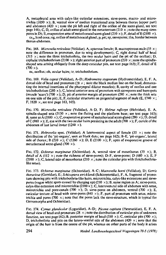
A, metapleural area with calyx-like cuticular extensions, sieve-pores, macro- and micro-trichia (1050 x ) ; B, ventral view of median transitional area between thorax (upper part) and abdomen (420 x ; note the pit left and right of the orifice of the scent-gland, see text page 148); C, D, orifice of adult scent-gland in the metasternum (210 x ; note the many sieve-pores in D); E, evaporative area of meta thoracal scent-gland (210 x ); F, detail of E (2100 x ).
cx3, hind coxa; og, orifice of metathoracal gland ; p, pit; sp, sievepores; tha, border between thorax-abdomen.
FIG. 168. Microvelia reticulata (VeYiidae). A, apterous female ; B, macropterous male (35 x ; note the difference in pronotum, due to wing development); C, right dorsal half of head (315 x ; note the three trichobothria, the two ocular hairs and the pit of the ocellus); D, cephalic trichobothrium (2100 x ); right anterior part of pronotum (210 x ; note the spindle-shaped seta arising obliquely from the deep cuticular pits, see text page 163); F, detail of E (700 x) .
oc, ocellus; oh, ocular hairs; tr, trichobothrium.
FIG. 169. Velia caprai (Veliidae), A-D; Hydrometra stagnorum (Hydrometridae), E, F. A, dorsal side of head and pronotum (28 x ; note the black median line on the head, demarcating the internal insertions of the pharyngeal dilator muscles); B, cavity of ocellus and one trichobothrium (280 x ) ; C, lateral anterior area of pronotum with sievepores and bare spots (muscle 'scars') (700 x ) ; D, pit of anterior margin of pronotum (280 x ; note the thick seta on one side of the pit); E, F, cuticular structures on pregenital segment of male (E, 1740 x ; F, 1820 x , see text page 163, 165).
FIG. 170. Microvelia reticulata (Veliidae), A-D, F; Hebrus ruficeps (Hebridae), E. A, spindle-shaped seta (3150 x ; note the fluting and the platform base; see text page 163); B, same as A (3500 x ) ; C, evaporative groove of metathoracal scent-gland (280 x ) ; D, detail of C (980 x ); E, eye with the two ocular hairs persisting in the adult (700 x ); F, cuticle of the abdomen of last larval instar (1260 x) .
FIG. 171. Hebrovelia spec. (Veliidae). A lateroventral aspect of female (35 x ; note the distribution of the 'pit-organs', seen as black dots; see page 162); B-E, 'pit-organs', lateral side of thorax; B (210 x ) ; C (1260 x ) ; D, E (2100 x ) ; F, apex of evaporative groove of metathoracal scent-gland (700 x ).
FIG. 172. Ochterus marginatus (Ochteridae). A, ventral view of mesothorax (35 x ) ; B, detail of A (112 x ; note the richness of sieve-pores); D-F, sieve-pores; D (680 x ) ; E, F (2100 x ); C, lateral side of mesothorax (210 x ; note the cuticular pits with thrichobothria-like setae).
FIG. 173. Ochterus marginatus (Ochteridae), A-C; Macrovelia horni (Veliidae), D; Gerris thoracicus (Gerridae), E; Schizoptera stricklandi (Schizopteridae), F. A, fragment of pronotum showing pits with trichobothria-like hairs, microtrichia, calyx-like extensions and sievepores (vague white spots caused by charging up) (210 x ) ; B, same region as A; sieve-pores, calyx-like extension and microtrichia (2100 x ); C, lateroveniral side of abdomen with setae, microtrichia and pore-canals (700 x ) ; D, sieve-pores on abdomen, ventral (700 x ) ; E, cuticular texture of head with sieve-pores (840 x ) ; F, part of pronotum with setae, microtrichia and pores (700 x ; note that the pores lack the sieve-structure, which is typical for Gerromorpha and Ochteridae).
FIG. 174. Cymus glandicolor (Lygaeidae), A-D; Piesma capitata (Piesmatidae), E, F. A, dorsal view of head and pronotum (28 x ; note the distribution of cuticular pits of unknown function, see text page 162) ; B, posterior margin of head (350 x ) ; C, cuticular pits (700 x ) ; D, trichobothria and pits on the latero-ventral side of the abdomen (420 x ; note that the origin of the hair is from the centre of the pit, whereas on other parts of the body it arises
294 Meded. Landbouwhogeschool Wageningen 78-5 (1978)
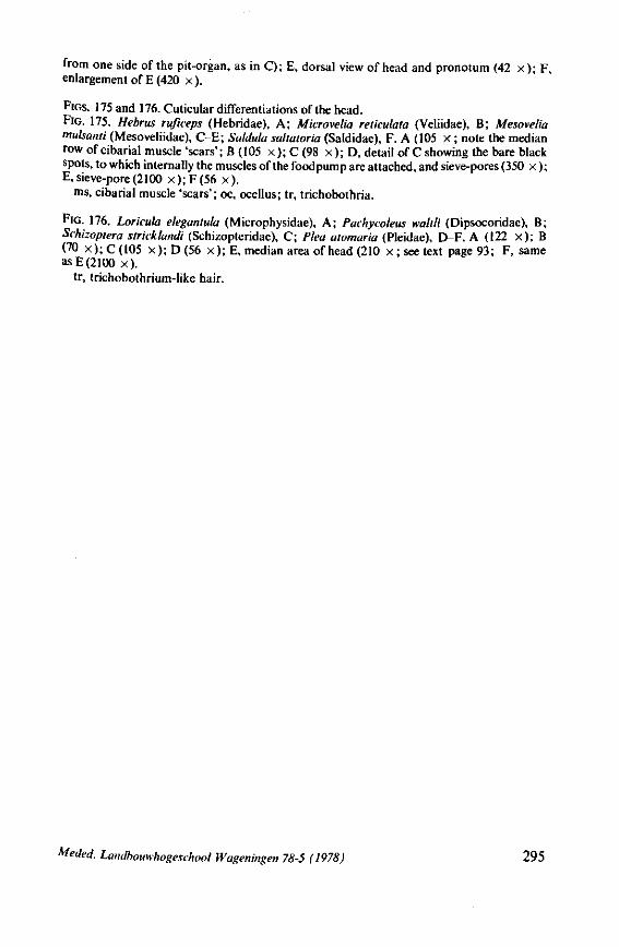
from one side of the pit-organ, as in C); E, dorsal view of head and pronotum (42 x ) ; F, enlargement of E (420 x ).
FIGS. 175 and 176. Cuticular differentiations of the head. FIG. 175. Hebrus ruficeps (Hebridae), A; Microvelia reticulata (Veliidae), B; Mesovelia mulsanti (Mesoveliidae), C-E; Saldula saltatoria (Saldidae), F. A (105 x ; note the median row of cibarial muscle 'scars'; B (105 x ) ; C (98 x ) ; D, detail of C showing the bare black spots, to which internally the muscles of the foodpump are attached, and sieve-pores (350 x ); E, sieve-pore (2100x) ;F(56x) .
ms, cibarial muscle 'scars'; oc, ocellus; tr, trichobothria.
FIG. 176. Loricula elegantula (Microphysidae), A; Pachycoleus waltli (Dipsocoridae), B; Schizoptera strkklandi (Schizopteridae), C; Plea atomaria (Pleidae), E>-F. A (122 x ) ; B (70 x);C(105 x ) ;D(56 x ) ; E, median area of head (210 x ; see text page 93; F, same as E (2100 x) .
tr, trichobothrium-like hair.
Meded. Landbouwhogeschool Wageningen 78-5 (1978) 295
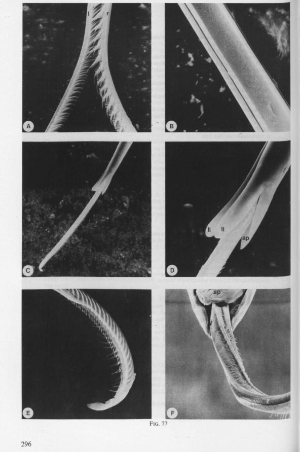
FIG. 77
296
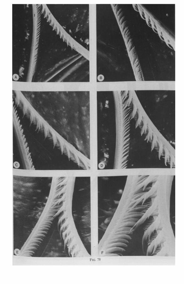
FIG. 78
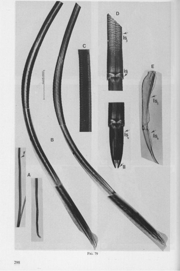
Is,
Is.
I
FIG. 79
298
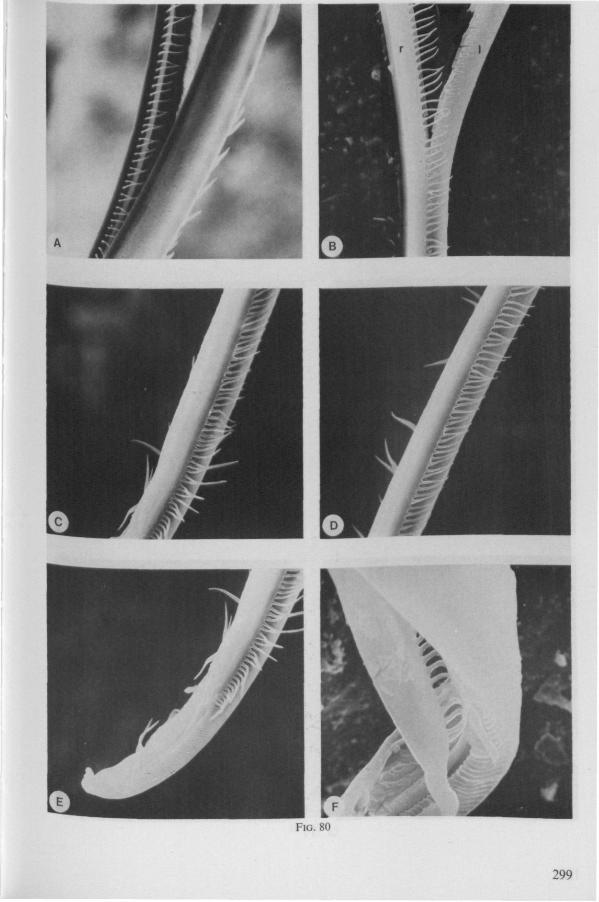
FIG. 80
299
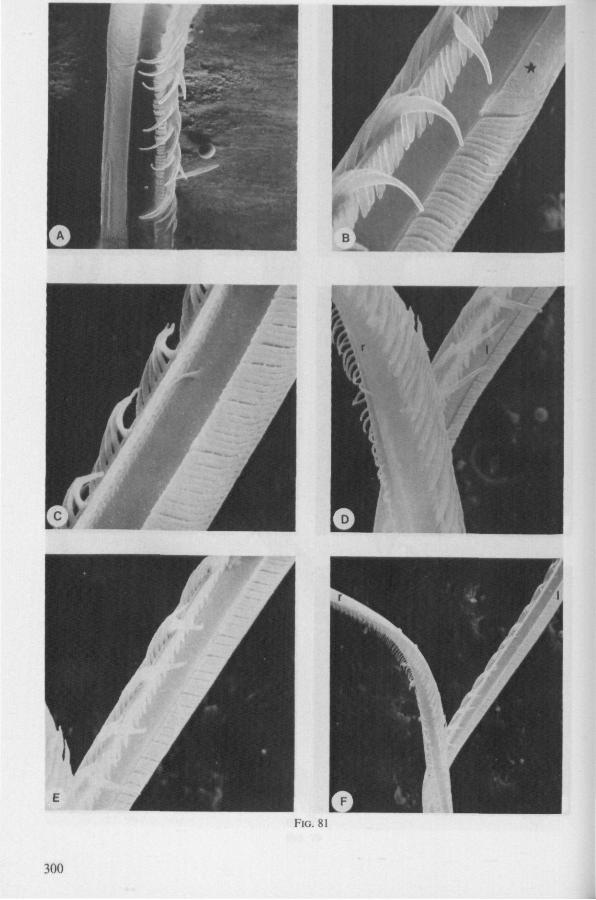
FIG. 81
300
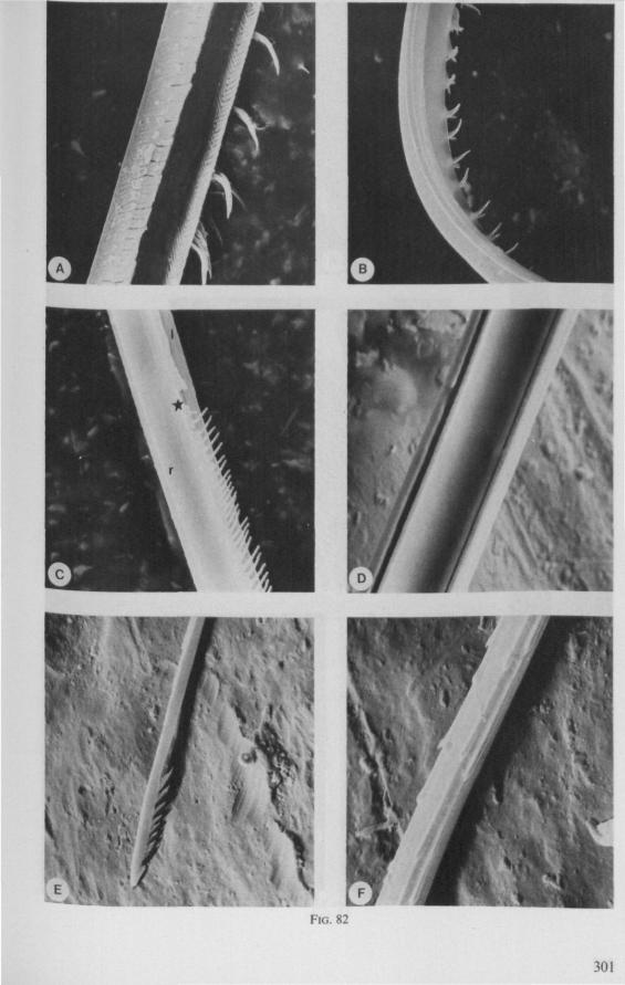
À;
FIG. 82
301
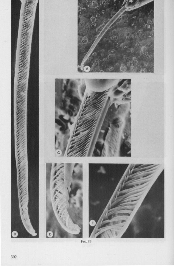
w?
FIG. 83
302
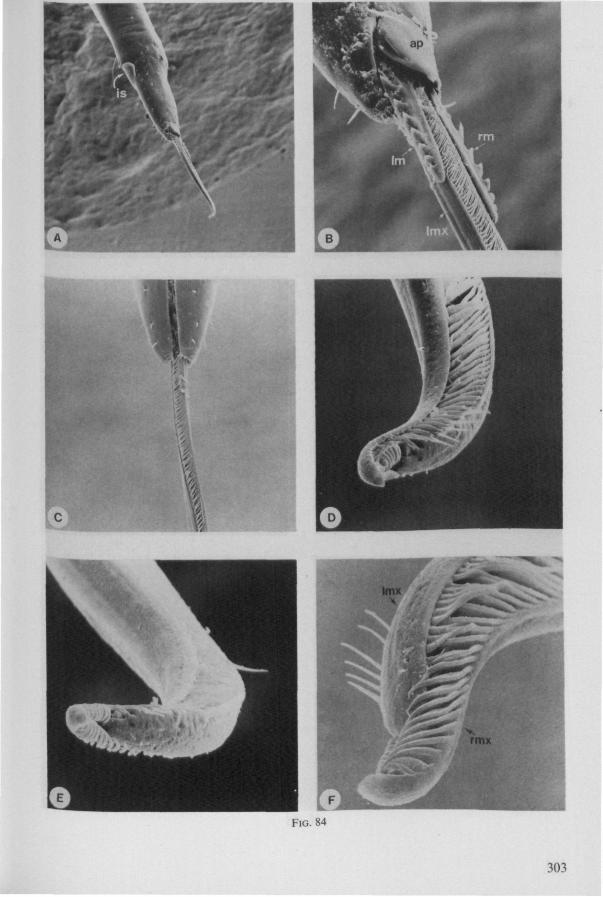
c
f
1
FIG. 84
303
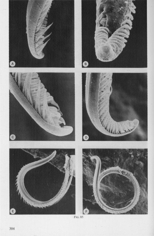
FIG. 85
304
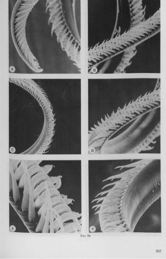
FIG. 86
305
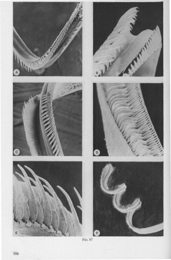
FIG. 87
306
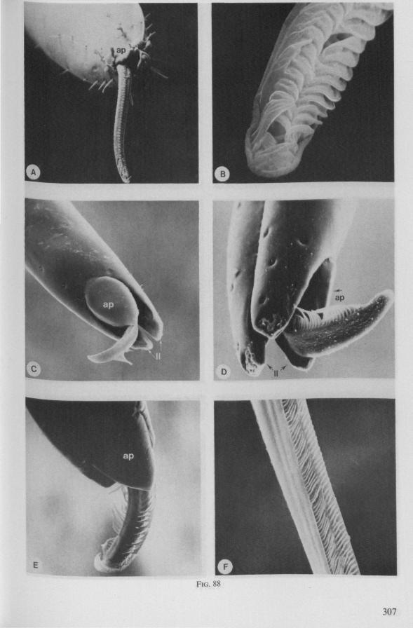
FIG. 88
307
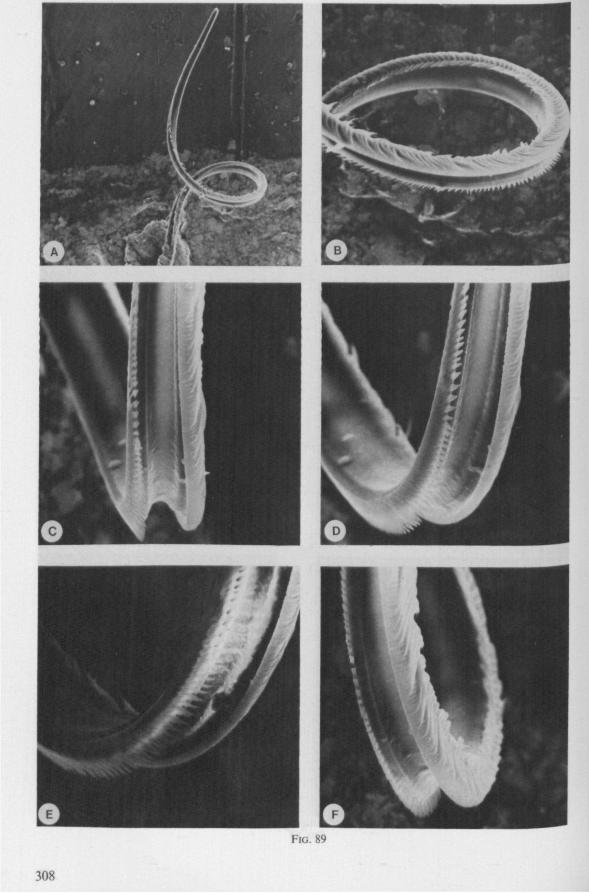
FIG. 89
308
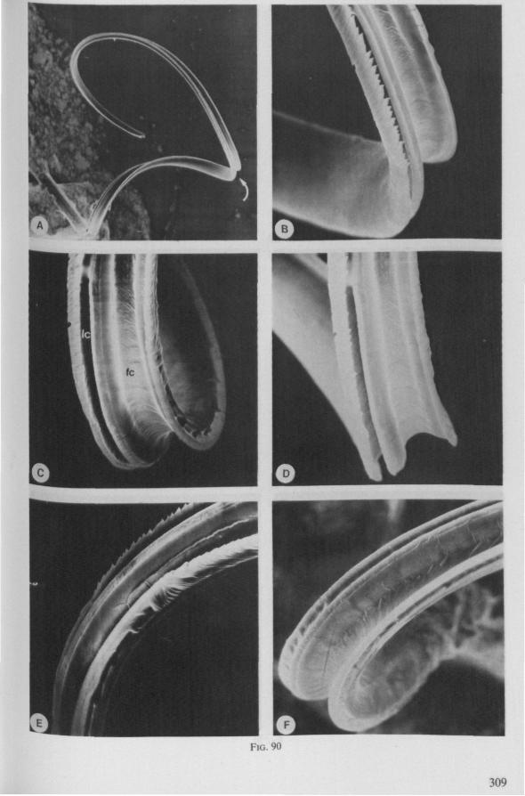
FIG. 90
309
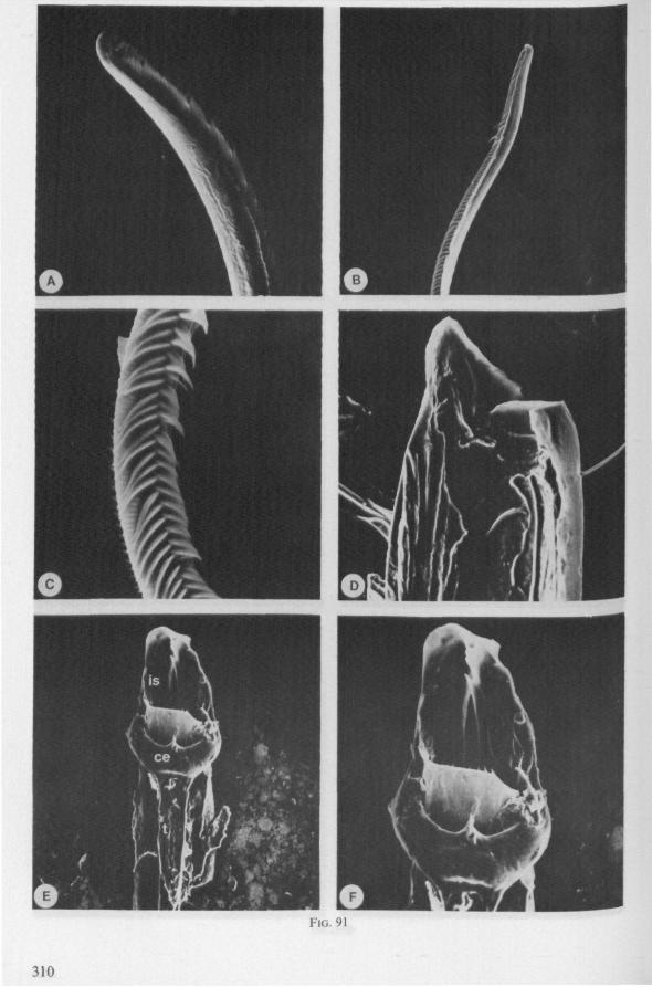
fl
FIG. 91
310
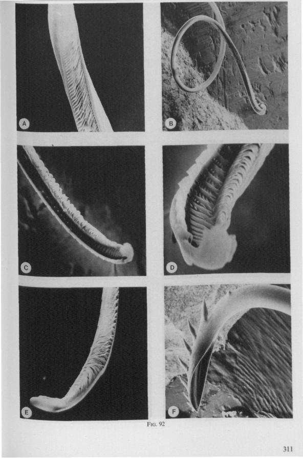
FIG. 92
311
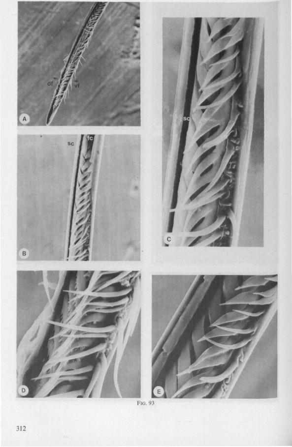
FIG. 93
312
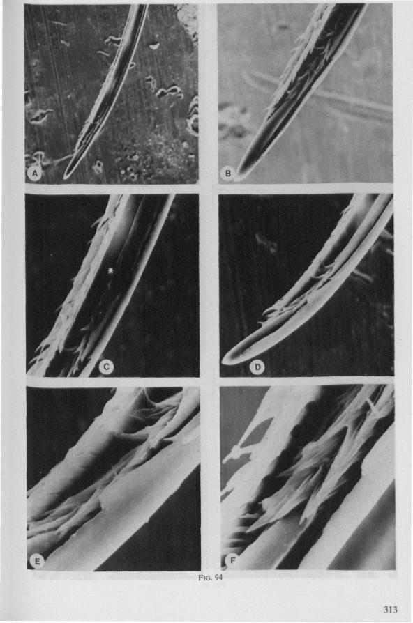
FIG. 94
313
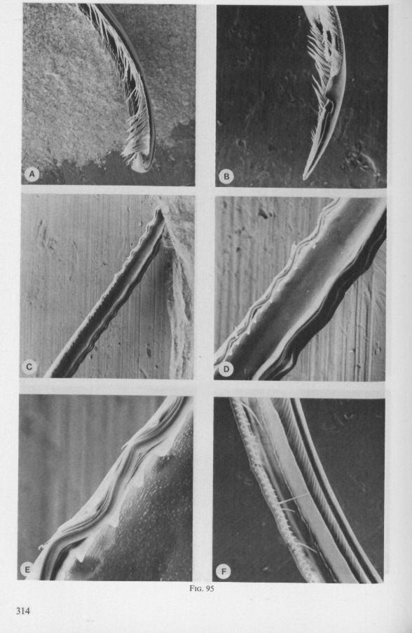
FIG. 95
314
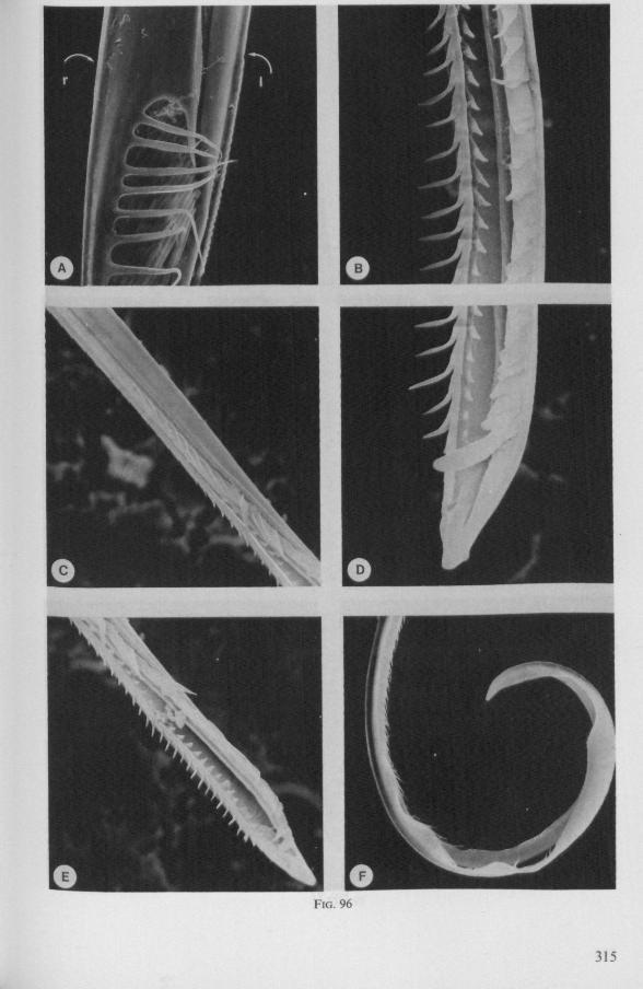
FIG. 96
315
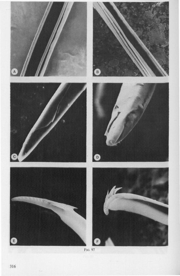
FIG. 97
316
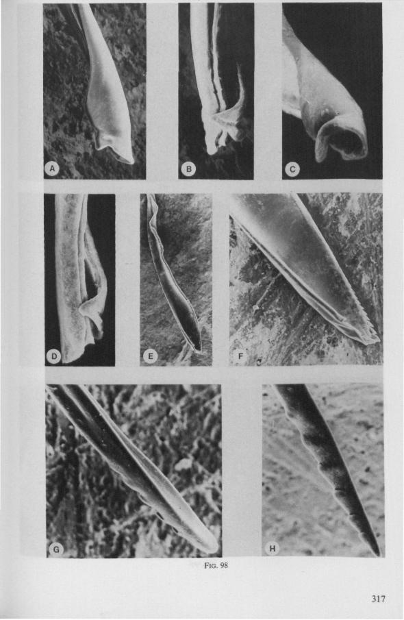
FIG. 98
317
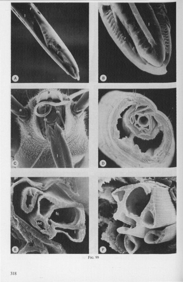
FIG. 99
318
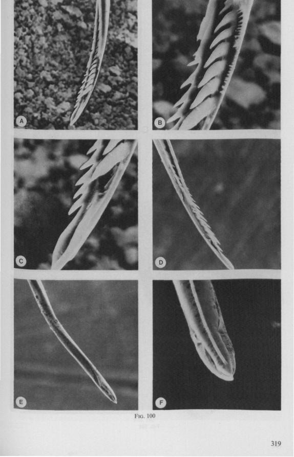
FIG. 100
319
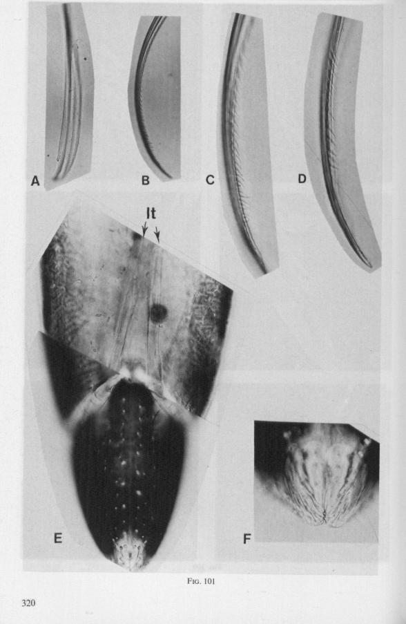
FIG. 101
320
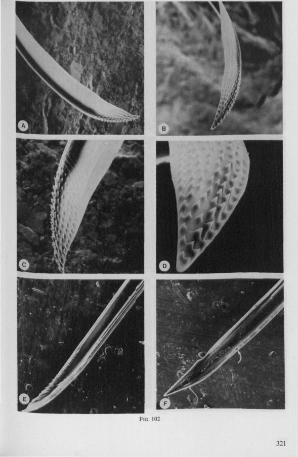
FIG. 102
321
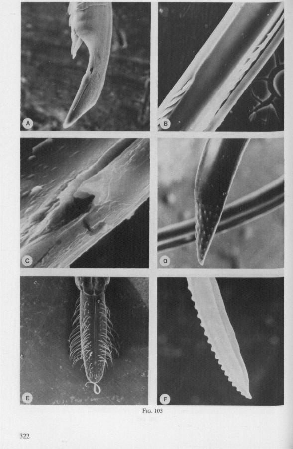
FIG. 103
322
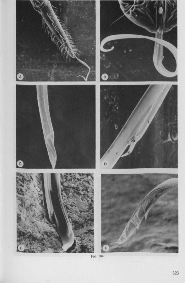
FIG. 104
323
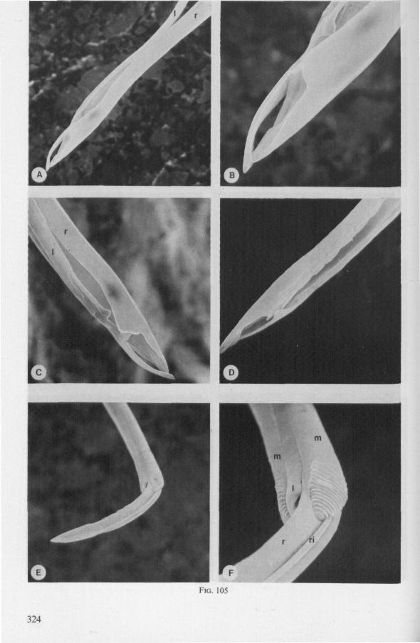
FIG. 105
324
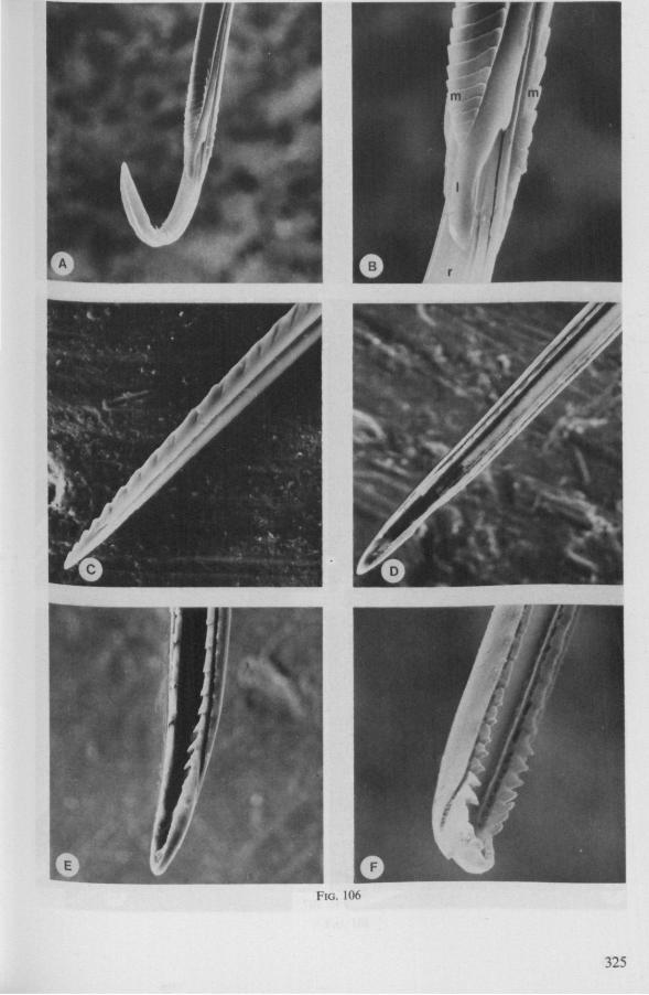
FIG. 106
325
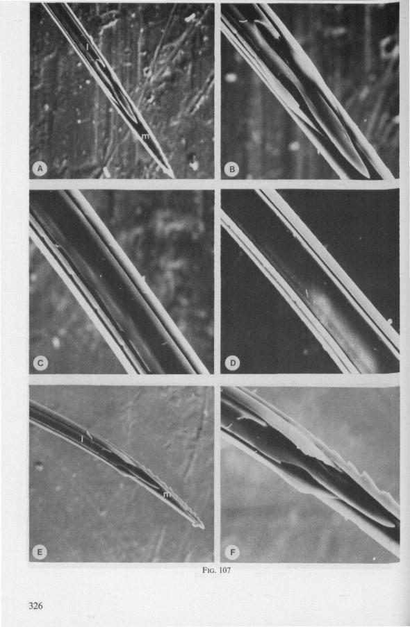
FIG. 107
326
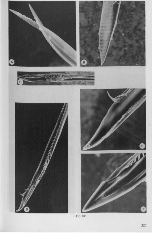
FIG. 108
327
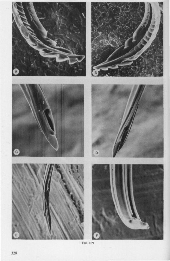
\
\
FIG. 109
328
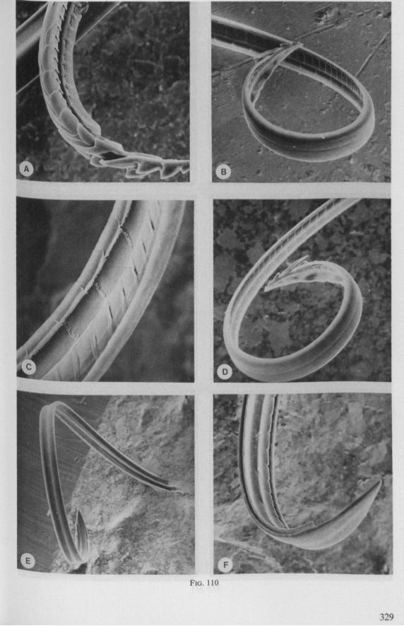
329
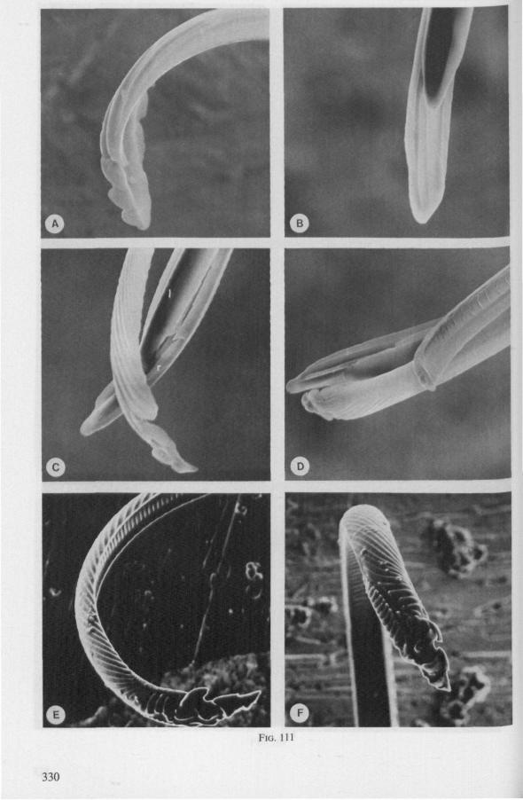
FIG. 111
330
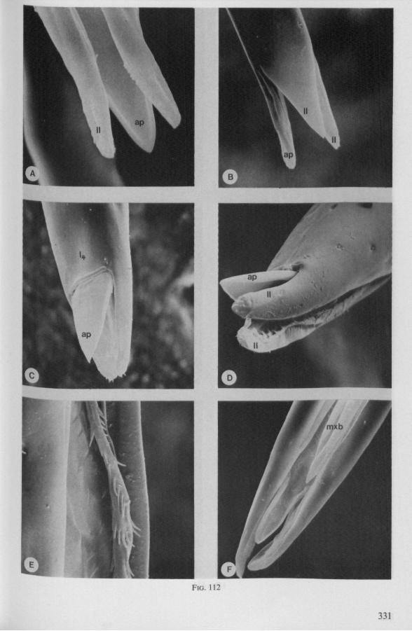
FIG. 112
331
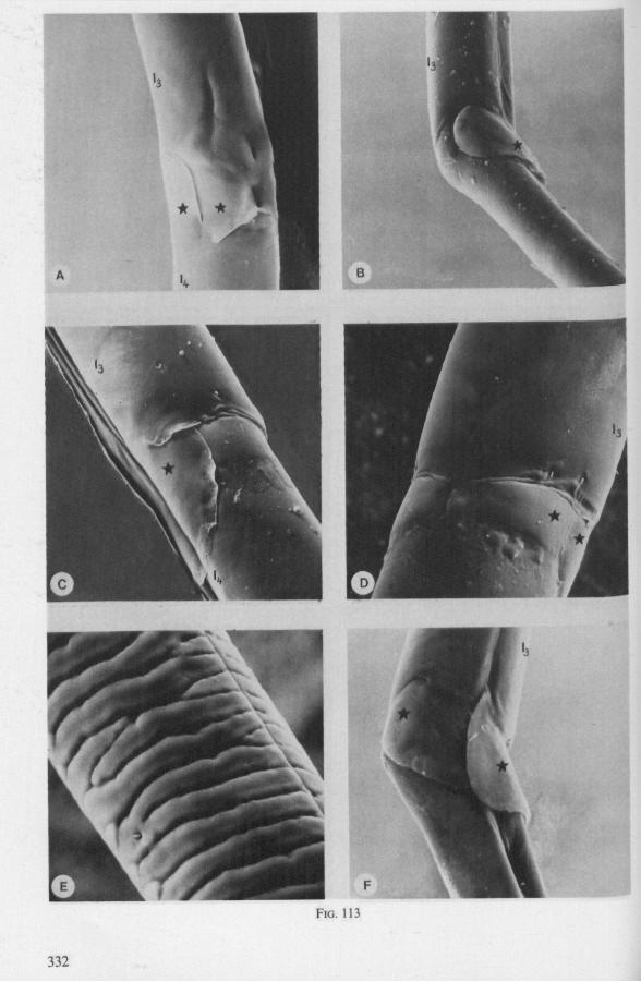
FIG. 113
332
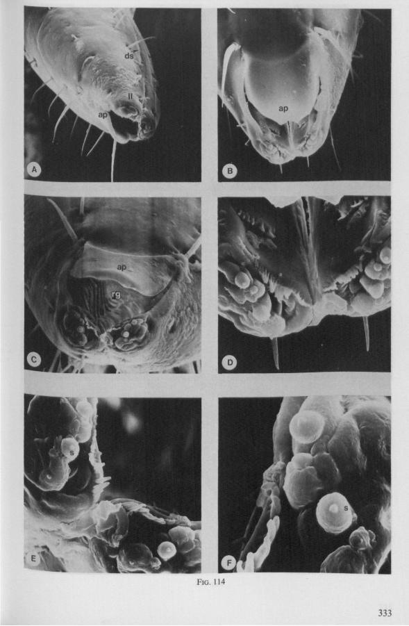
FIG. 114
333
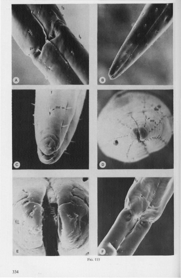
334
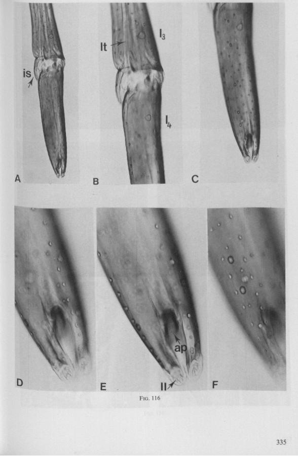
"A
It
I
B \
E 11/ F FIG. 116
335
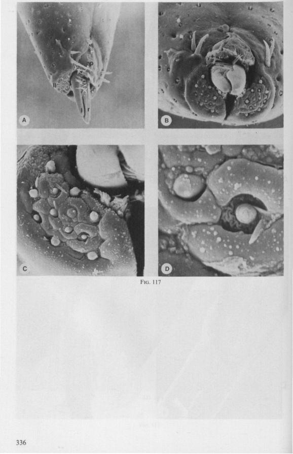
FIG. 117
336
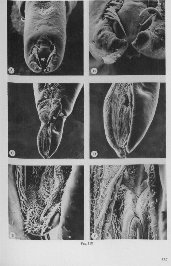
FIG. 118
337
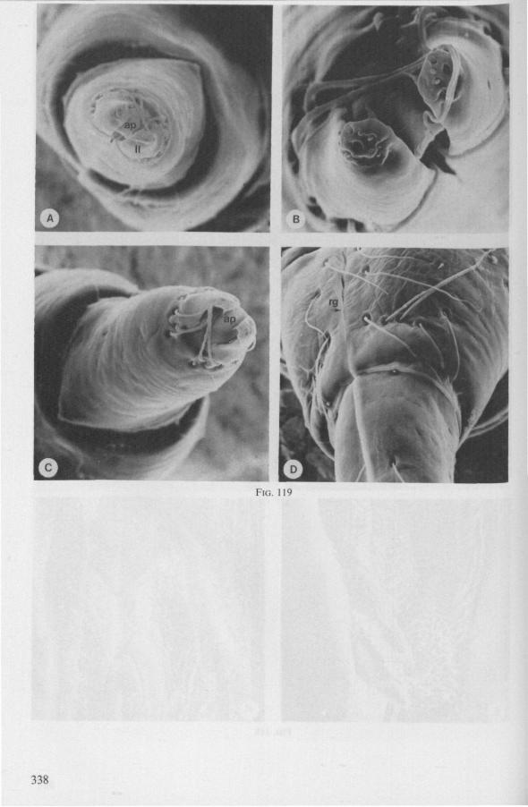
IG. 119
338
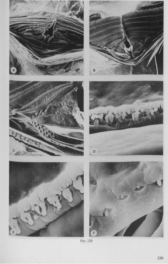
FIG. 120
339
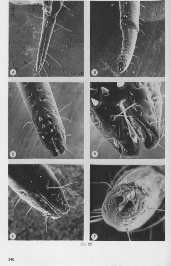
FIG. 121
340
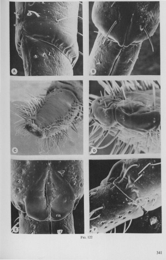
FIG. 122
341
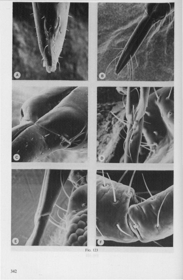
FIG. 123
342
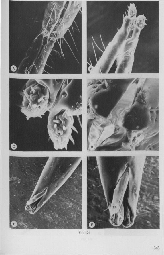
FIG. 124
343
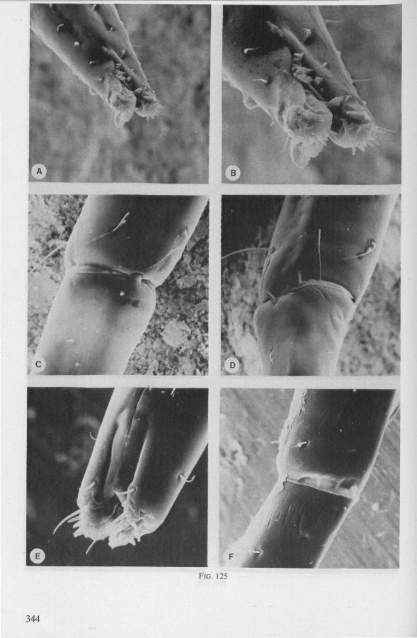
FIG. 125
344
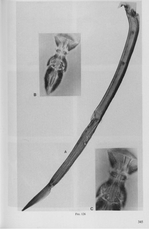
B
FIG. 126
345
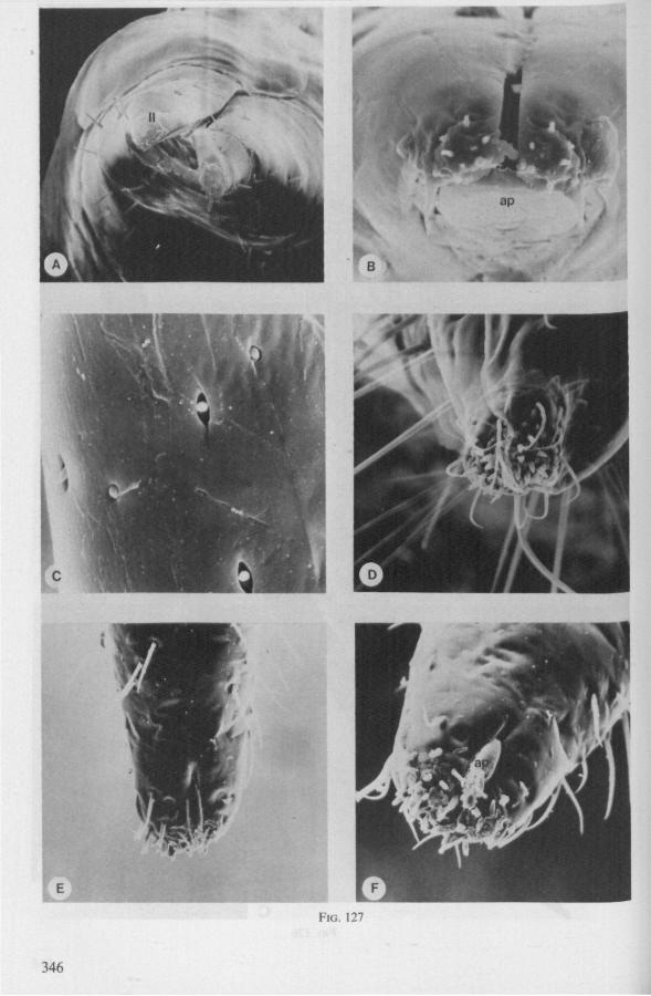
t
ap
FIG. 127
346
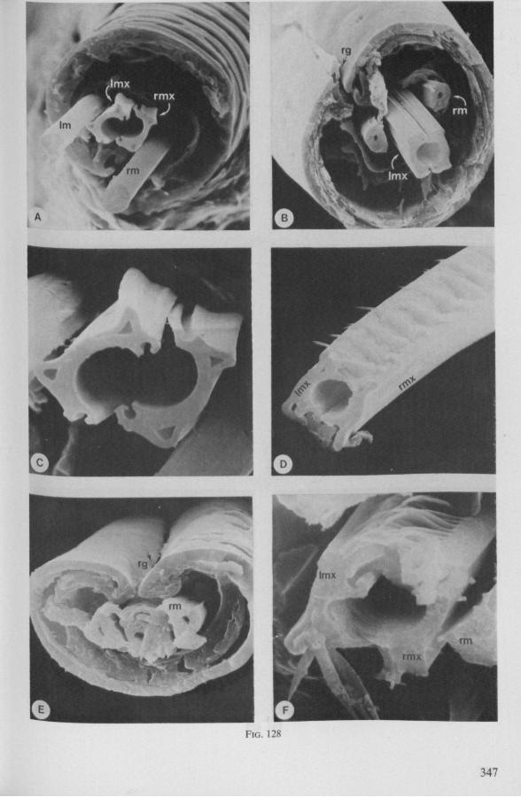
FIG. 128
347
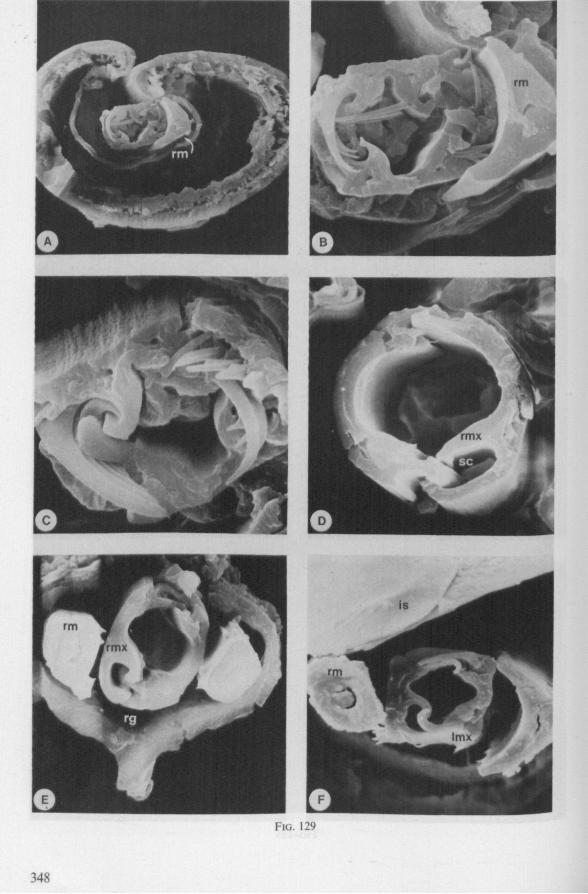
FIG. 129
348
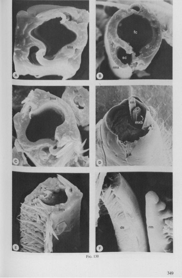
FIG. 130
349
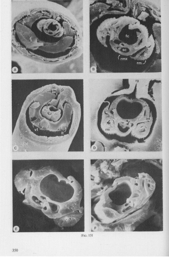
FIG. 131
350
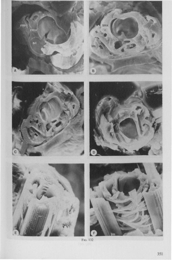
FIG. 132
351
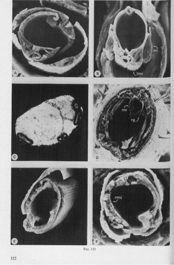
FIG. 133
352
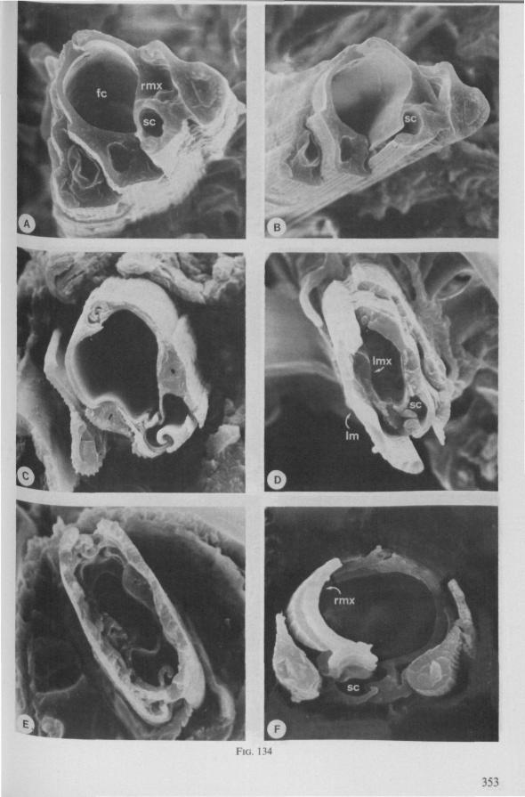
FIG. 134
353
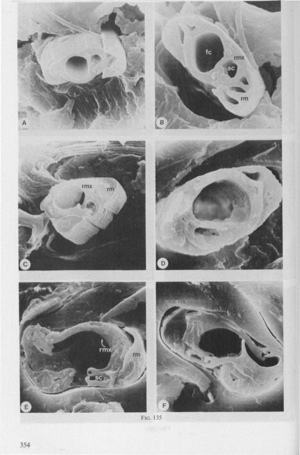
FIG. 135
354
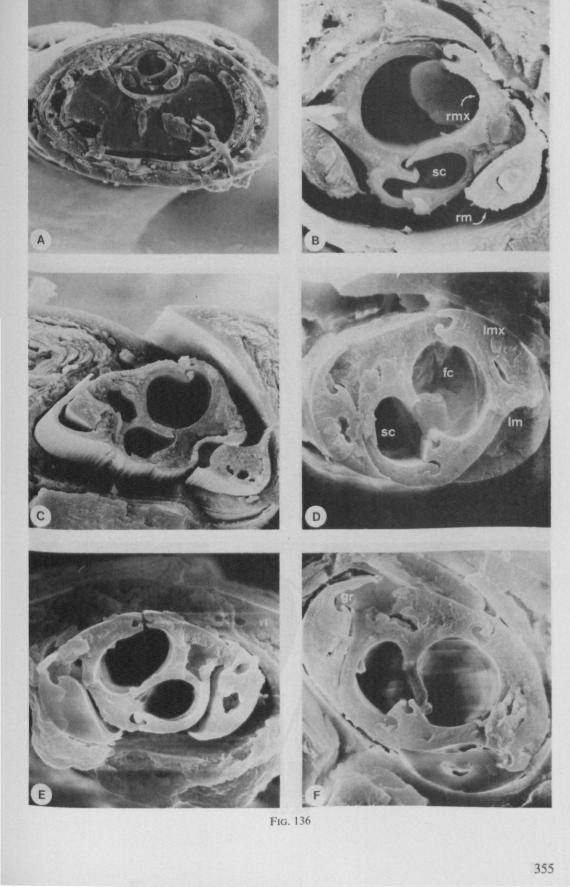
355
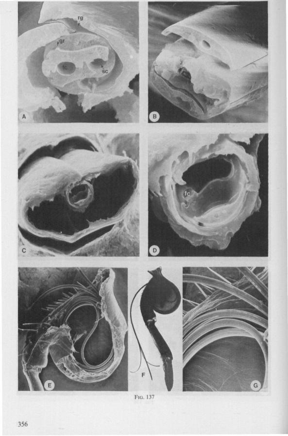
FIG. 137
356
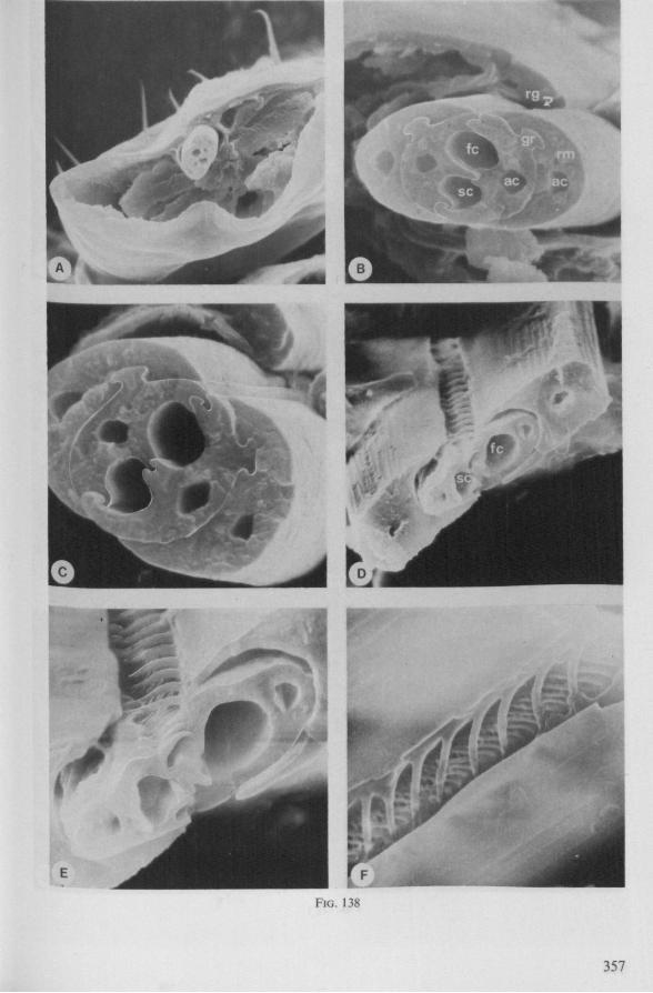
FIG. 138
357
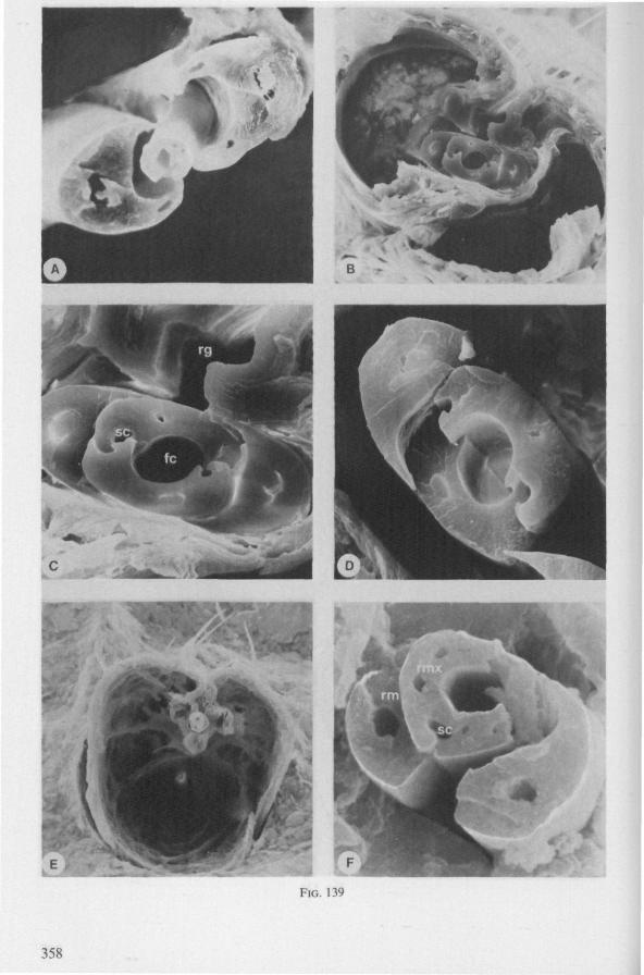
358
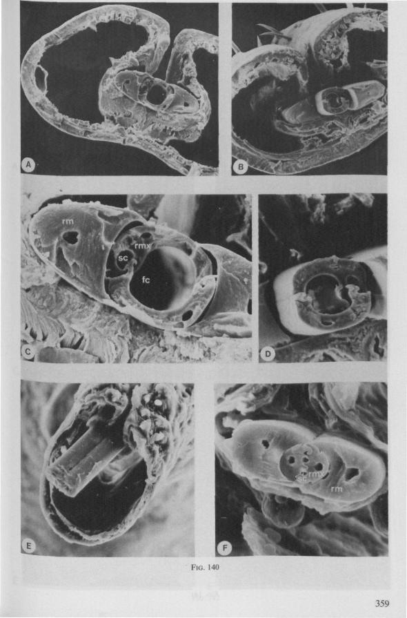
FIG. 140
359
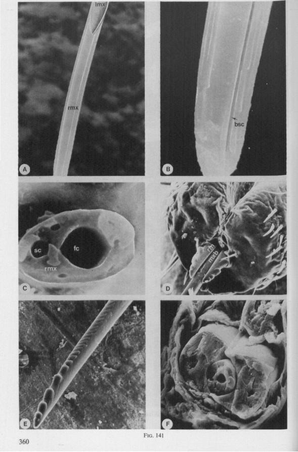
360 FIG. 141
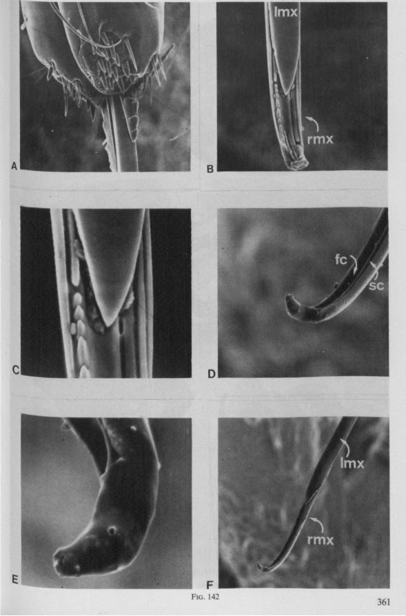
FIG. 142 361
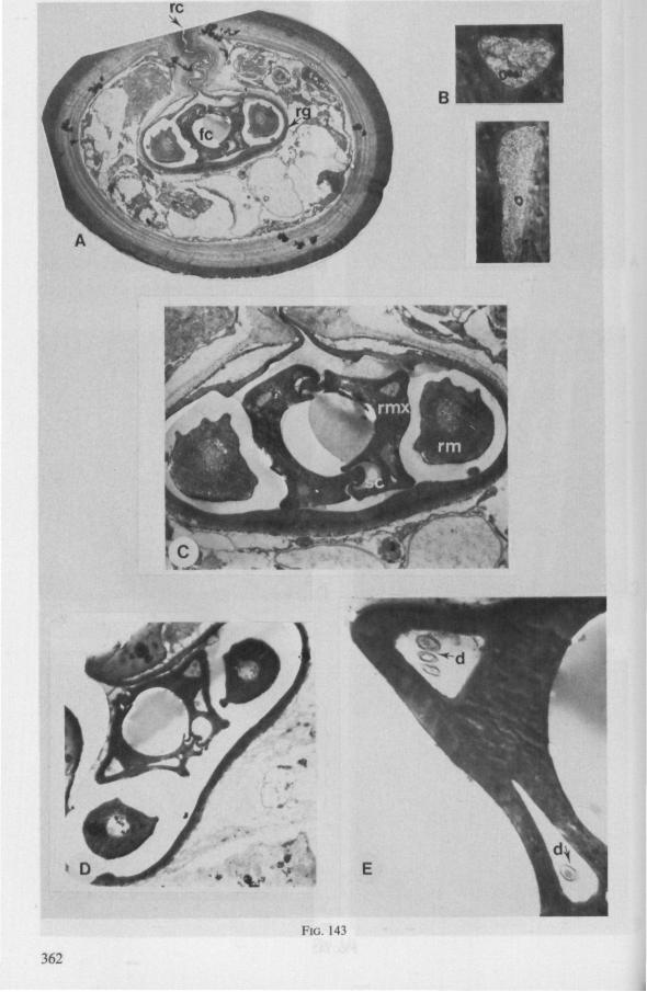
FIG. 143
362

FIG. 144
363
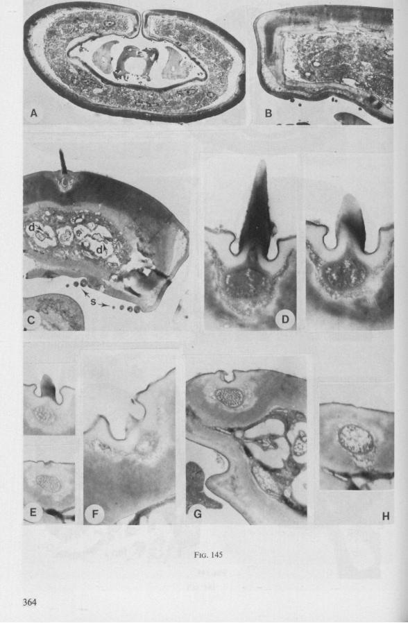
FIG. 145
364
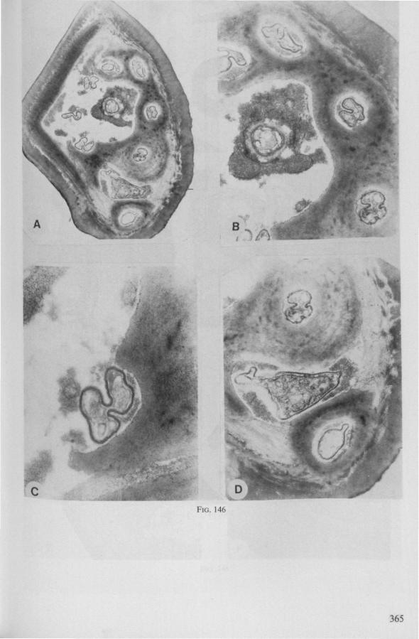
î
,^A
FIG. 146
365
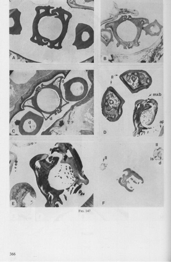
x h
FIG. 147
366
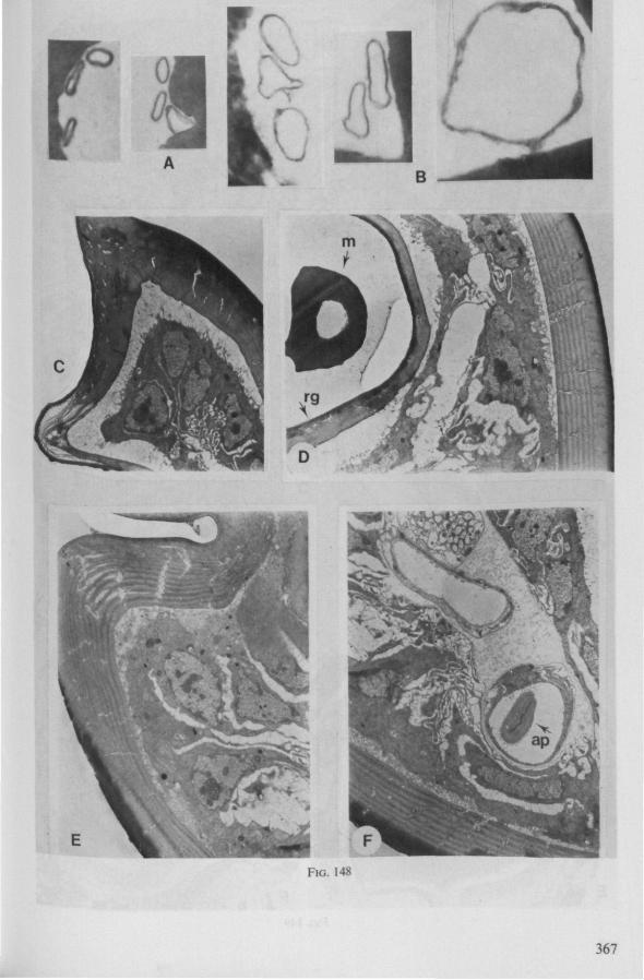
FIG. 148
367
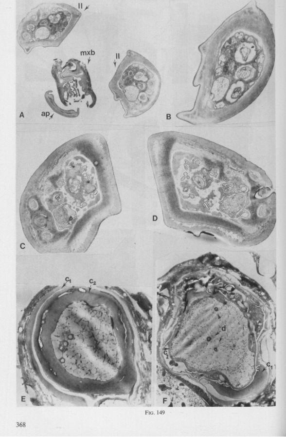
FIG. 149
368
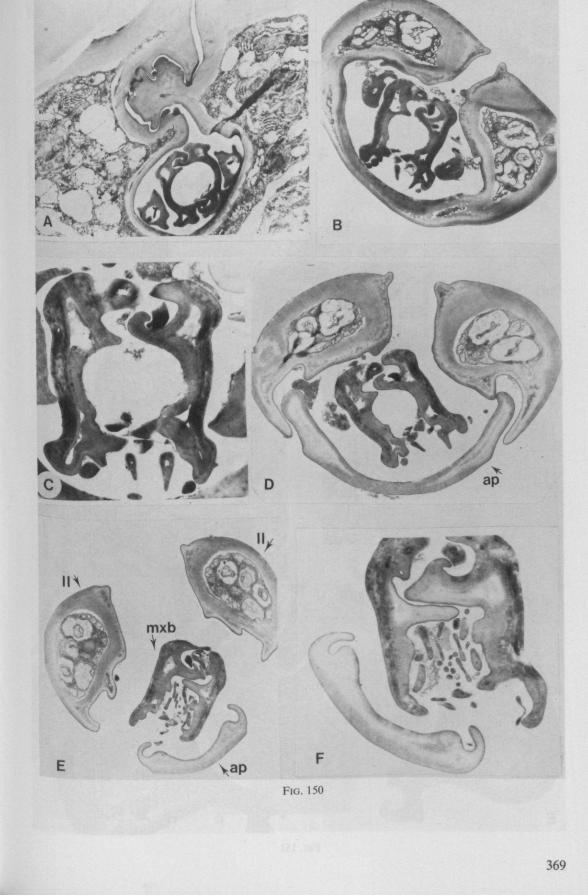
>• yi&
369
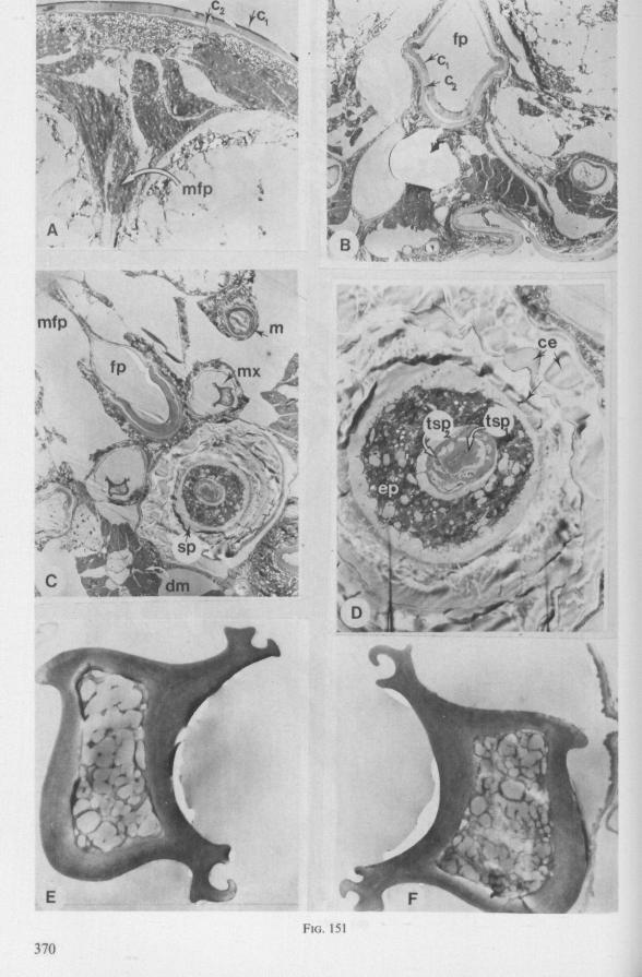
« » •
^SSÄ
B -:
FIG. 151
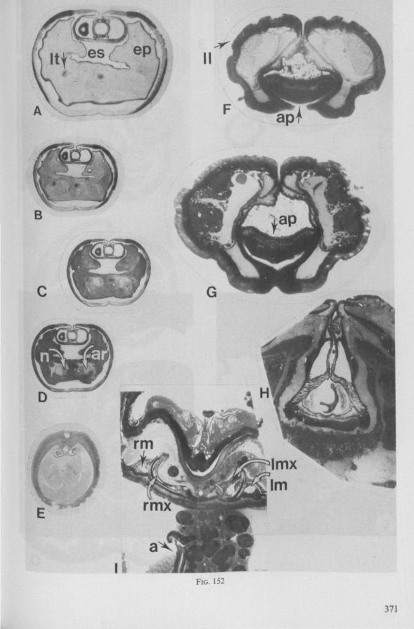
F ^ p *
FIG. 152
371
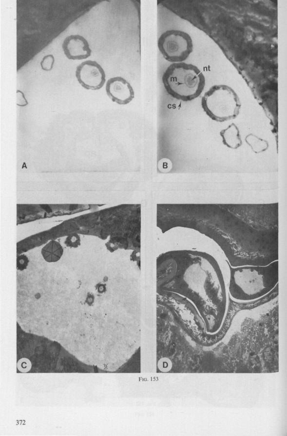
1
i
0 o o
41 4»
tl
(
^v* . . . , . *^^
FIG. 153
372
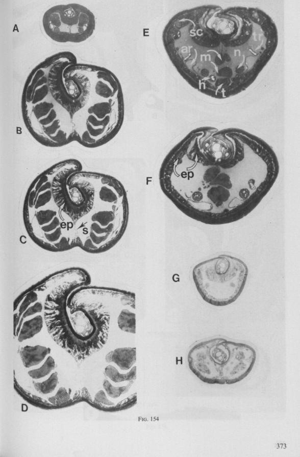
H
FIG. 154
373
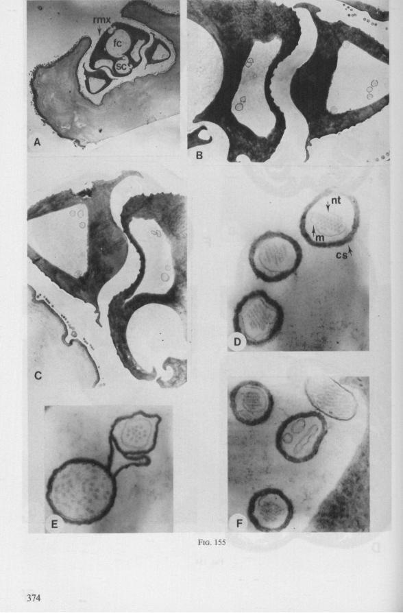
FIG. 155
374
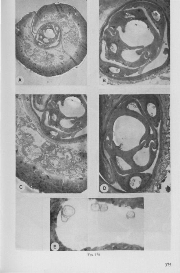
FIG. 156
375
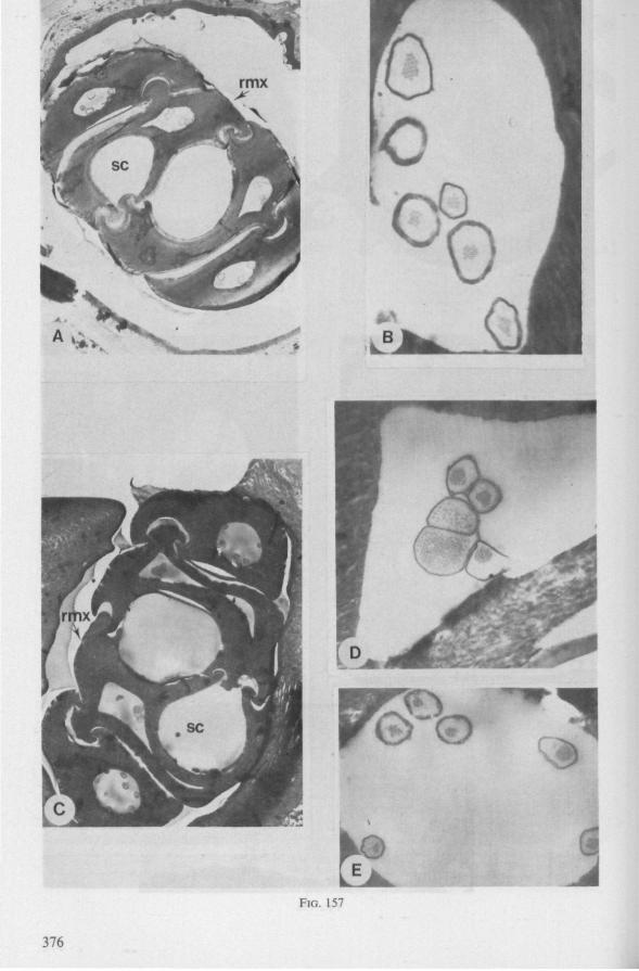
P^b ^ 1
FIG. 157
376
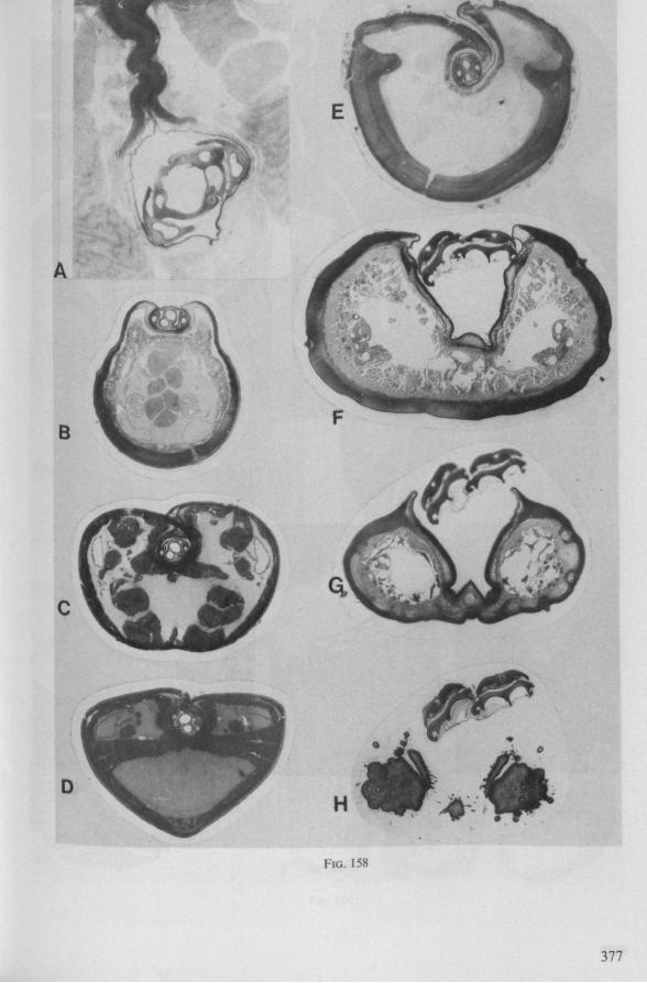
FIG. 158
377
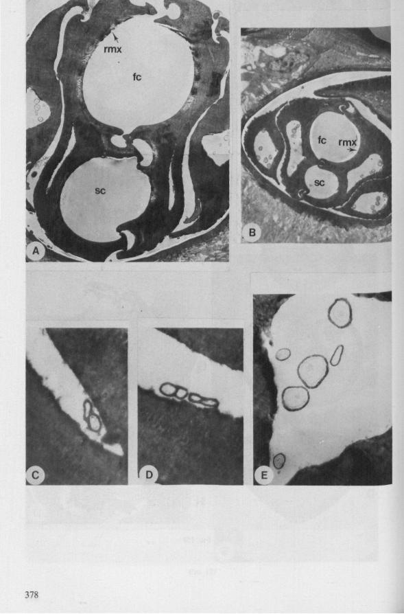
378
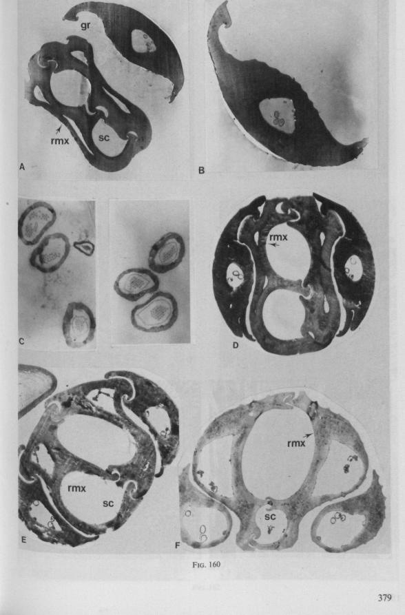
B
FIG. 160
379
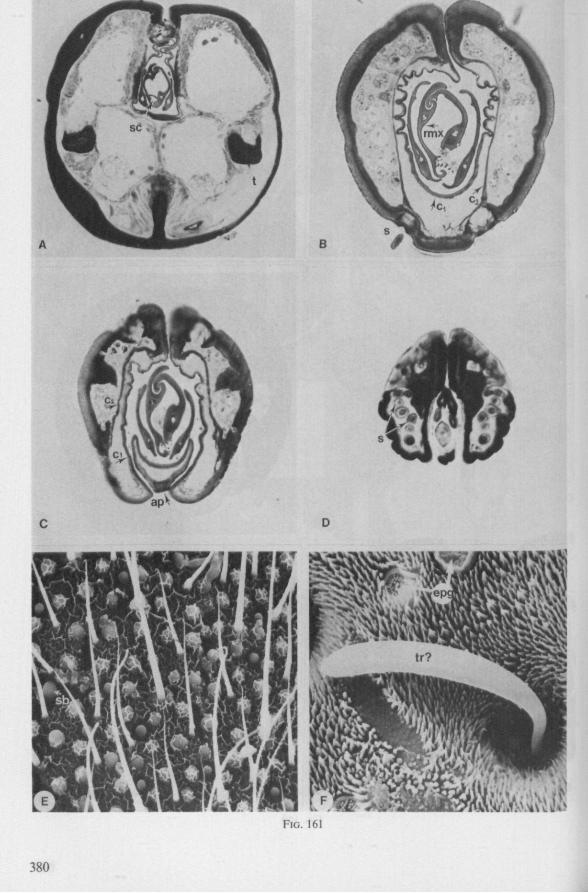
380
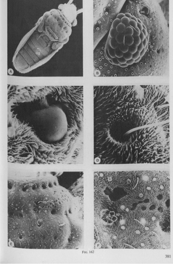
* * % %
i ,, mm FIG. 162
381
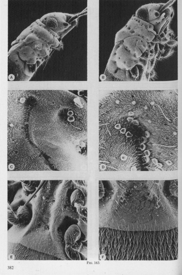
FIG. 163 382
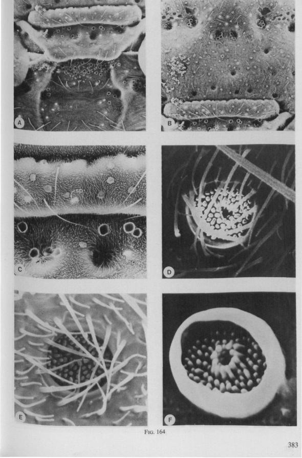
M i • •«—»»'•' '
FIG. 164
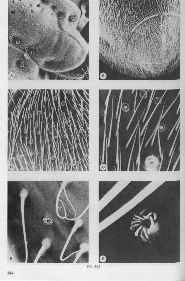
FIG. 165
384
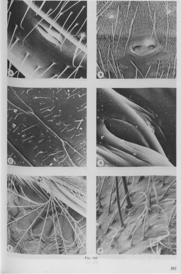
w ße
L F
FIG. 166
385
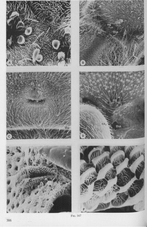
FIG. 167
386
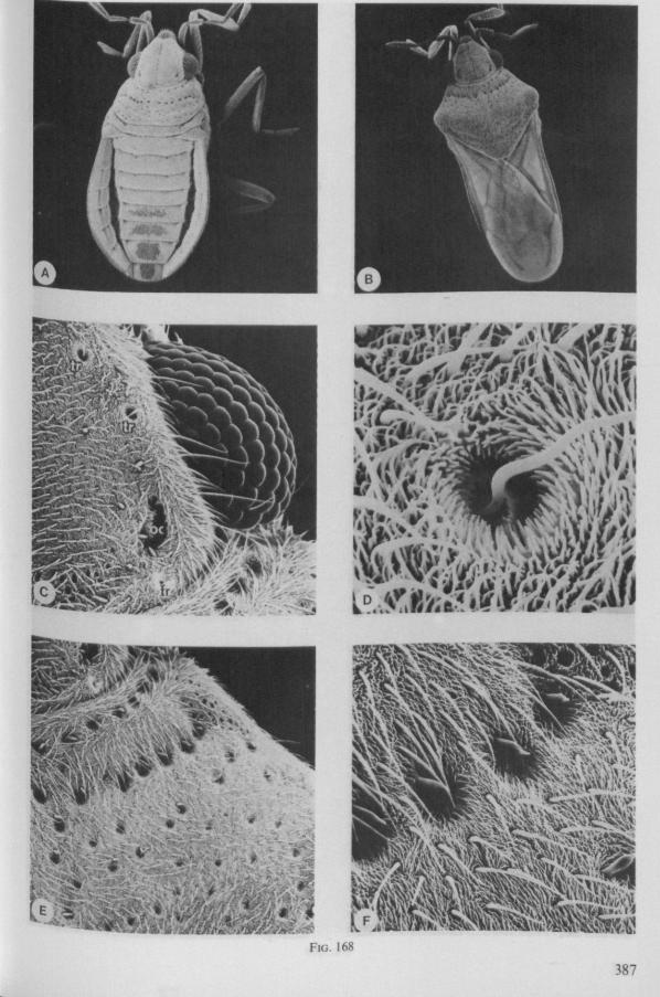
..y.
m
* * .
FIG. 168
387
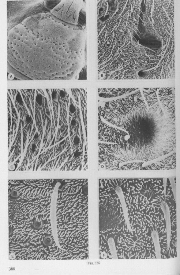
, J
à &«k^
Ai
ï'*Sfi
FIG. 169
388
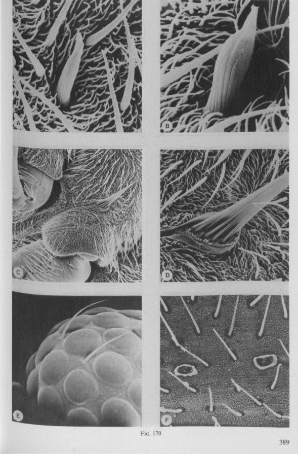
FIG. 170
389
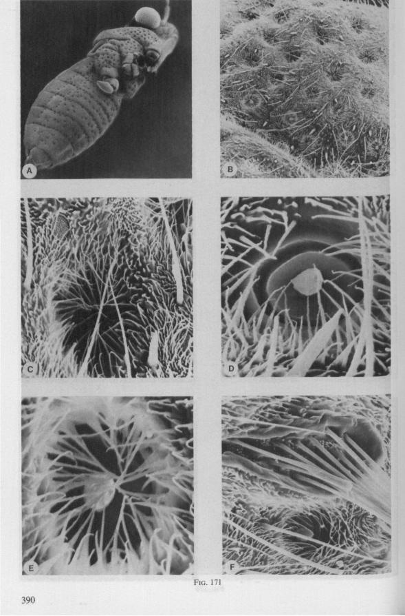
rw'li%iX
« 5
sPfp
FIG. 171
390
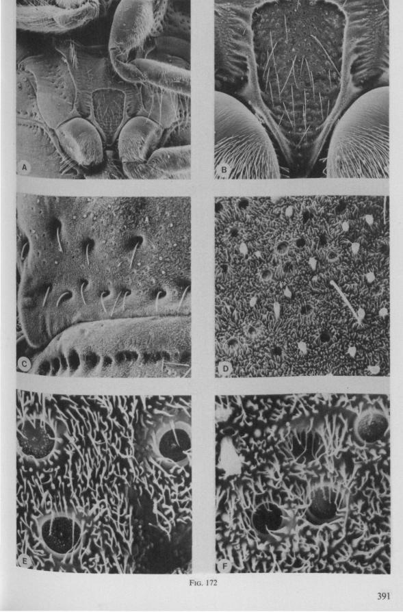
E l l M@-
m
: * "wlflTiTOClBi—TSfeBR
m
É ^
. t
: i }"'2 4fe
I^* ,n \?W' ,
FIG. 172
391
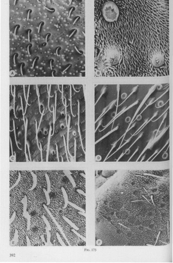
392
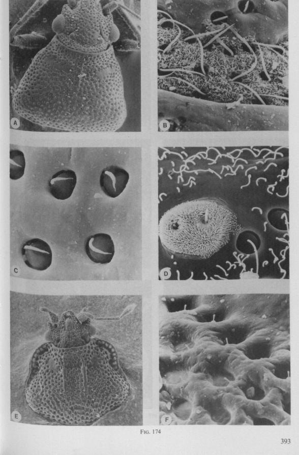
9 9 O
S o
FIG. 174
393
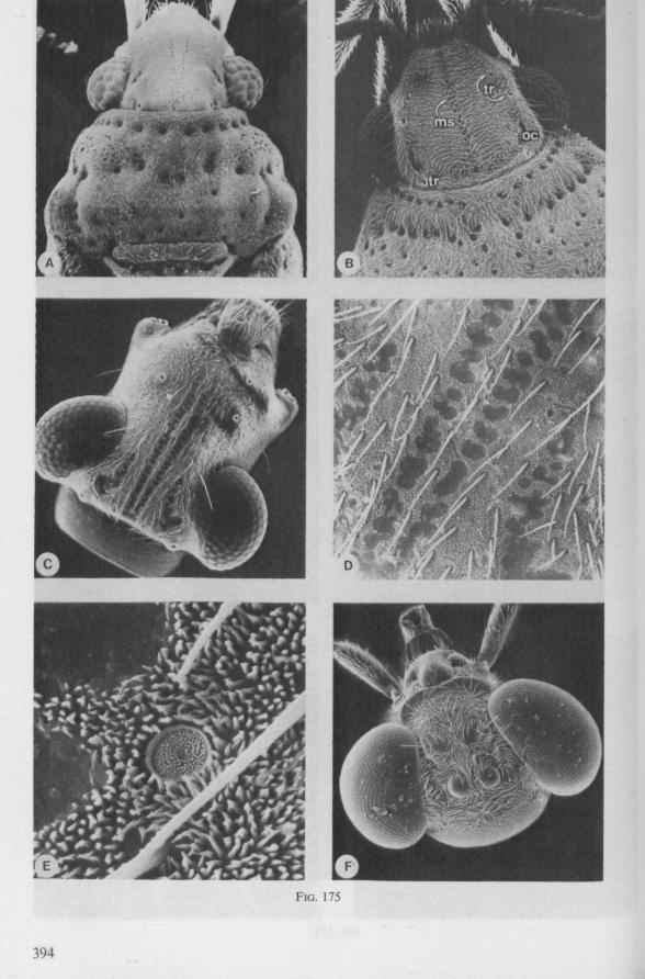
FIG. 175
394
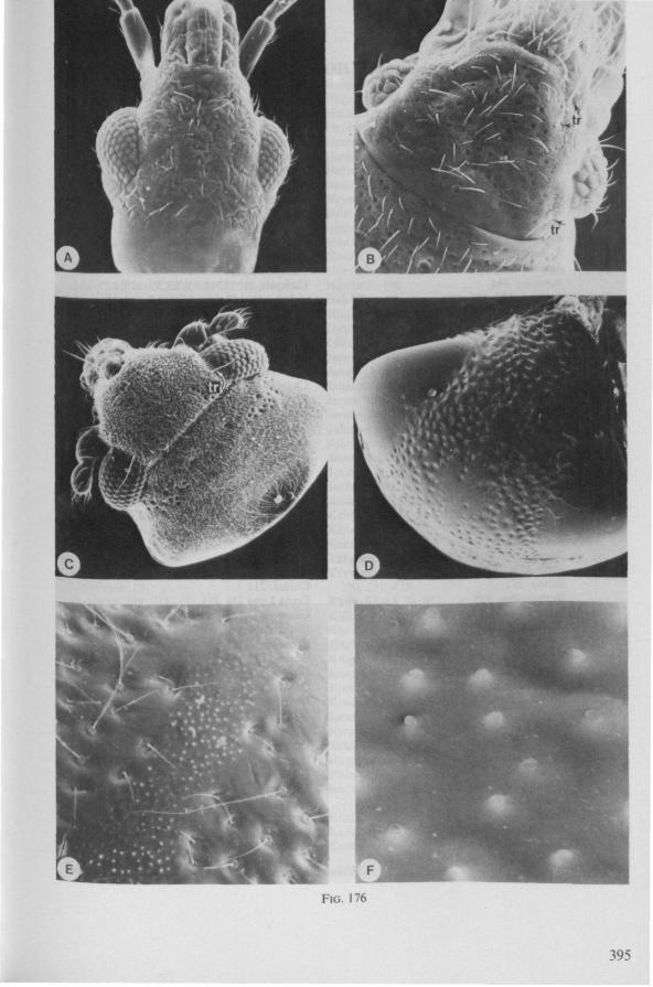
mj* isla
[cj
FIG. 176
395
AUTHOR INDEX
Abraham 125 Acton 93 Akbar 139,240 Aldrich 143, 154, 159 Allen 126 Anderson 99, 113, 156, 214 Ando 102 Anonymus 91 Arnold 205 Arnoud 208, 214 Aryeetey 144 Ashlock 126, 127,229 Awati 65, 75, 89 Bacheler 208 Baggini 154 Baker 211 Baldwin 159, 164 Ballard 75 Banerji 74, 240 Banks 234 Baptist 81, 85, 87 Baranowski 208 Barbagallo 75 Bar-Ilan 146, 158 Barret 154, 156 Barth 65,67,72,75,79,89,198 Baudoin 123, 124, 125, 144, 146, 158 Becker-Migdisova 222 Bedau 109 Benwitz 15, 35, 65, 67, 77, 79, 87, 93, 94, 117 Bernard 41,43,91,93,94,200 Betten 142 Bieman, den 227 Blackith, R. E. 223 Blackith, R. M. 223 Blöte 51 Bodenheimer 208 Bongers 62, 74, 206, 210, 211, 215 Bonnemaison 57, 74 Borner 111 Bornman 239 Bosch v. d. 155 Botha 239 Bowers 242 Breny 211,212 Brindley 142, 153 Brinkhurst 75,131,148,158 Brönnimann 208 Brothers 223 Broughton 242
Brunner 214 Bueno 123 Bugnion 57, 65, 74, 75 Burts 214 Butenandt 159 Butt 65,77, 139 Cachan 146,213 Calam 142, 144, 145, 146 Callahan 162, 165 Campbell 242 Carayon 11,15, 49, 51, 53,99,120, 127,132,
140, 142, 146, 150, 152, 153, 154, 155, 156, 159, 160, 161, 180,207,211,212,214,225, 230,231,232,233,234,249
Carvalho 120 Chada 236 Champlain 211 Chaplin 210 Chapman 201 Cheng 13,65,87,158 China 59, 108, 117, 123, 127, 128, 146, 148,
150,160,204,217,222,231 Cobben 6, 13, 18, 39, 45, 47, 49, 53, 57, 75,
77,96,97,99,101,102, 106,111,113, 115, 122, 125, 132, 135, 139, 140, 142, 143, 146, 156, 160, 165, 166, 168, 172, 173, 175, 180, 188, 190, 207, 208, 214, 228, 232, 233, 235, 236, 237, 240
Colless 190 Cooker 123 Coons 214 Correâ 211 Costa Lima Da 211 Cragg 65 Cranston 15, 65, 67, 70, 71, 74, 87 Crowson 216 Dandy 130 Daniel 208 Dashman 113 Davis 39, 51,81.108, 127, 129, 131, 132,
142,146, 156, 208, 213, 214, 226, 232 DeCoursey 126, 132 Derksen- Koppers 210 Dethier 154, 155 Dickerson 53, 74 Dieleman 237 Dietrick 155 Dixon 214 Doane 242 Doeksen 208
396 Meckd. Landbouwhogeschool Wugeningen 78-5 (1978)
Doesburg v. 104,213 Drake 64, 108, 119, 127, 128, 129, 131, 132,
146,208,214 Drâslar 160 Duffey 156 Dupuis 140, 142, 166 Eastop 204, 242 Edwards 41, 65, 67, 119, 156, 198, 206, 207,
242 Eggerman 206,210,215 Ehrhardt 62 Ekblom 13, 15, 18, 25, 35, 47, 51, 57, 65,
67, 76, 198 Elsey 211 Elson 13, 27,67, 75, 76,198, 204, 234 Emsley 10, 64, 72, 75, 81, 87, 99, 118, 127,
131,139,213,225 Esaki 108,117, 139,213 Evans 75,241,242 Everton 142, 154 Eyles 211,215 Fain 155 Faucheux 13 Fauré-Fremiet 81 Fauvel 208, 212 Fedotov 158 Filshie 143, 154 Flemion 55 Forbes 94, 235, 236, 238, 241 Fosbrooke 199 Fox, J. W. 130 Fox, R. M. 130 Frick 13, 18 Friend 41,43,67,89,201 Froeschner 59 Fujisaki 145, 146 Galbreath 18 Gallopin 199,211 Games 146, 156, 159 Geise 65 Gillett 107 Gluud 143 Goel 111,113,117,118,119,122 Goodchild 5,198, 200, 202, 204,234, 242 Gould 123 Govind 130 Graham 156 Griffiths 157 Guide 63, 140 Gupta 130,222 Hamilton 27, 35. 77 Harris 242 Hase 74 Heikertinger 155 Heming 83, 102, 242
Heming-v. Battum 18, 83, 168 Hennig 189,222,234 Henrici 140, 142, 164,211 Henstra 91 Hepburn 143, 154 Herring 126, 127,212 Hertel 211 Herzog 91 Heslop-Harrison 242 Heymons 140, 241 Hille Ris Lambers 102 Hinton 111, 139,242 Hodkinson 238 Hoofv. 236 Hori 62, 74 Howarth 108 Hsü 89 Hungerford 18,35,37, 115 Hyslop 208 Ishiwatari 145 Jacobson 41, 213 Jander 124, 156 Jansson 35 Jarial 93 Jermy 212 Johansson 159 Johnson 55, 239 Jones 208 Jordan 18, 126 Jurberg 140, 142 Kälin 154, 156 Kemper 74. 87 Kennedt 199 Kennedy 206 Kershaw 75 Kerzhner 6, 101, 153, 189, 203, 226, 228,
230,231,232 Khan 62, 65, 74, 91 Kirkaldy 129 Kislow 242 Kitching 199,211 Knight 120, 159 Komnick 93, 164, 165 Kormilev 127 Koschel 208 Kramer 240, 241 Krczal 214 Krishnanandam 148 Kugelberg 209 Kuhn 109 Kullenberg 53, 55, 65, 67, 68, 74, 126, 155,
214,217 Kumar 64,139,140,144, 209, 210 Kumari 139 Kunkel 204, 234, 238
Meded. Landbouwhogeschool Wageningen 78-5 (1978) 397
Langer 109 Larichi 155 Larsén 117,131 Laschat 110 Lattin 123,218 Lauck 115,127 Lavoipierre 41, 43, 45, 53, 65, 74 Lawrence 159, 164 Ledvinka 142, 161 Lent 140, 142,211 Lequesne 223 Leston 18, 49, 122, 126, 145, 189, 208, 217,
222, 230, 242 Levinson 146, 158 Lindberg 204 Linder 15 Linsenmair 124, 156 Livingstone 55, 65, 74, 75, 89, 91, 240 Lloyd 217 Lo 93 Louis 145,155,201,213 Ludtke 110 Lumsden 18 Lundblad 18, 127, 161 Lynn 159 Macgill 65, 75, 87 Mackerras 189,223,243 Macleod 159 Maldonado-Capriles 120, 213 Malloch 41, 127 Marks 76 Marsh 242 Maschwitz 143, 147 Mathen 214 Matsuda 73,77,106,119,127,131,144,150,
196,236,240,241 Mau 155, 159 Mayné 211,212 McAtee 41, 127 McCullough 154 McDonald 140,209 McKinstry 76 Menke 115, 127 Michalk 211 Michener 191,223 Miles 5, 61, 62, 89, 198, 201, 202, 203, 204,
234 Miller 41, 145, 155, 207, 211, 213, 222, 231,
232 Mitchell 155, 159 Miyamoto 81, 85, 87, 89, 124, 125, 139, 213 Moens 212 Moller-Pillot 142, 143 Monod 214 Moore 144
Moreno 242 Morton 144 Mound 202,213 Muir 75, 142 Müller 190,236,242 Mullick 236, 238, 241 Murphey 123 Myers 59, 108, 127, 128 Naef 226 Naidu 159 Nakashima 162 Nault 242 Neese 111 Neiswander 15, 35, 77 Nuorteva 81 Odhiambo 145 Oetting 63, 143,209,211 O'Neill 213 Osmani 159 Ossiannilsson 242 Pajunen 35 Park 159 Parker 201, 207 Parrish 236, 238 Parsons 11,15, 20, 27, 47. 65, 68, 69, 71, 72,
73,74,75,76,77, 79, 87,127,131,132,139, 142,157,222,240,241
Pattenden 153, 156, 157,159 Patton 65 Pendergrast 74 Peregrine 89, 94 Péricart 49, 51,104, 110,129, 207, 214, 217 Pesson 236, 237, 241 Peters 35 Pinder 157 Pinet 93, 94, 200 Plank v. d. 213 Poisson 15, 67, 117, 127, 142, 222 Polivanova 140 Pollard 55, 57,61,65, 68, 202,234, 235, 236,
237, 239, 241 Popham 79 Popoff 57, 65, 74 Popov 15, 101, 131, 165, 166, 173, 189, 222,
223, 230 Priesner 132 Pringle 242 Puchkov 126. 140. 142 Puchkova 15, 79, 102, 140, 241 Quadri 27, 35, 69. 77 Raine 235 Ralph 210 Ramamurty 148 Rawat 131 Remold 143, 144, 146, 154, 155
398 Meded. Landbouwhogeschool Wageningen 78-5 (1978)
Rensing 109, 124 Reuter 128, 129,217 Richards 102, 132, 164 Ridgeway 208 Rieger 191 Riley 20 Rohdendorf 222 Root 210 Ross 222, 234, 239 Roth 59, 142, 143 Ruhoff 214 Rüssel 214 Rüssel 217 Sailer 161 Salthouse 164 Saxena 62,236 Scales 159 Schaefer 61, 111, 118, 119, 122, 132, 140,
142,156,160,161,209, 210, 211,223,226, 234
Schiödte 129, 131 Schlee 5, 189, 190, 191, 228, 234, 237, 242,
251 Schliephake 223 Schmitz 122 Schneider 109 Schoevers 208 Schoonhoven 91,206,210,212 Schorr 210 Schubert 74, 75 Schuh 53, 122, 160, 208 Schumacher 63 Schwoerbel 217 Scott 142 Scudder 35,102,110,126,156,210,223 Seidenstücker 120, 127 Servadei 13, 65, 67, 76 Shanta 214 Shepard 211 Sholdt 211 Simpson 223 Slater 15, 53, 63, 119, 126, 127, 128, 132,
139,142,189, 205, 209, 211, 214, 215, 217 Smith J . J .B . 41,43,67,89,201 Smith K. M. 55 Smith R. L. 115 Smutterer 62 Sneath 223 Snoddy 119,213 Snodgrass 65, 67, 76 Sogawa 235 Sokal 223 Sokol'skaya 37 Solbreck 209 Southwood 18, 49, 81, 102, 110, 145, 204,
215,216,217,218,239 Spooner 13,20,59,76,234 Spraque 13,15,65, 67, 70, 71, 74, 87,131 Staddon 146, 153, 154, 156, 157, 158, 159,
164,215 Stefan 207 Stein 143 Stinner 211 Stoner 207,208,211 Strawinski 59 Stride 213 Strong 159 Stusâk 126 Stys 6,10, 51. 64. 75.118.127,129.140,142.
153, 161. 189. 203. 208. 210. 213, 225. 226. 228,229.230.231,232.240
Sutherland 159 Sutton 35 Sweet 5, 15, 61, 62, 126, 127, 128, 139, 142,
154,161,201,209,210,211,215 Tamaki 211 Tamanini 204 Timonin 59, 143 Thome 153, 154, 157 Thorsteinson 206 Todd 240 Tower 65, 75 Tsuyuki 154, 155 Tullgren 111, 161 Ulbrich 37 Usinger 49, 51, 53, 77,81,119,123,127,128,
132,142,156,208,213,226 Vance 132 Vecht v. d. 62 Viggiani 209 Villiers 132,145,160,161, 232,234 Vinson 206 Vodjdani 158 Voegelé 155 Voelker 212 Volk 214 Waddill 211 Wagner E. 120,122 Wagner W. 190 Wallbank 144 Walton 13,35,37 Watase 102 Waterhouse 143, 154, 155 Weatherston 157 Weaver 155 Weber H. 20, 59, 66, 74, 211, 234 Weber H.H. 120,218 Wedde 65, 198 Weeks 211 Wefelscheid 37,93
Meded. Landbouwhogeschool Wageningen 78-5 (1978) 399
Wheeler 218,219 Wygodzinsky 41, 109, 118, 119, 127, 139, Wichard 93, 164, 165 140, 142, 156, 207, 213, 232 Wiese 125 Yonke 63, 143, 154, 209, 211 Wigglesworth 164 York 211 Wilcox 61,140,142,209 Youdeowei 144, 145, 146 Williams 242 Young 110 Wolfram 18, 74, 206 Zdarek 159 Woodward 61, 109, 110, 127, 161, 234 Zhiteneva 37
Zwart 35
400 Meckel. Landbouwhogeschool Wagenmgen 78-5 (1978)
SUBJECT INDEX
Page numbers in bold print denote drawings, in italic type denote electron-micrographs; numerals in ordinary print refer to text pages.
Subfamily and family names and higher categories are not listed in entirety but are only selectively indexed by subjects.
Acalypta carinata 9. 52 Acanthaspis vilticollis 145, 213 Acanthocoris 9, 62, 70, 94, 107, 145, 214, 58,
377, 379 accessory parempodia 113, 115, 118, 119,
112 Acompocoris pygmaeus 136 Acompus ruficeps 107, 215, 105 Adelgus piceae 238 Aelia germari 155 Aepophilus 69, 70, 72, 89,108, 115,125,132,
144, 146, 150, 153, 158, 160, 173, 175, 177, 181, 222, 176
Aepophilus bonnairei 8, 106, 180, 48, 78, 80, 114,134,149, 33J, 334
Aetalion reticulatum 242 Alloeorhynchus chinai 8, 51, 52, 82 allomone 154-158 Alydus calcaratus 9, 107,151 Ambrysus 73 anagenesis, this item emerges time and time
again in chapters 3 and 4 (96-234) Anasa 65, 75, 155 Ancyrosoma 107 Aneurus laevis 9, 108, 119, 105,137 Annomatocoris 108 Anoplocnemis 107,98 Amhocoris 51, 110, 111,165 Anthocoris gallarum-ulmi 136 Anthocoris nemoralis 8, 94, 373, 375, 377,
378 Anthocoris nemorum 109, 110, 207, 208,103 Anthocoris pilosus 208 Anthocoris visci 110, 151 Aphelocheirus 69, 73, 117, 142 Aphelocheirus aestivalis 7, 33 Aphelonotus 75, 79, 89 Aphrophoraalni 10, 358 apical plate, see rostrum Apiomerus 155 Apodiphus 142, 154 Aquarius 65, 73, 75, 131, 148, 158 Aquarius najas 6, 18, 156, 333 Arachnocoris dispar 120,116
Arachnocoristrinitatis 8, 120 Aradus cinnamomeus 9, 57, 107, 108,105 Aradus frigidus 204 arolia 111-126, 112, 114, 116 Arvelius atropunctatus 107, 105 Aspilaspis viridis 208 asymmetry 41, 43, 115, 119 Auchenorryncha 235, 236 Bacillometra 6, 16, 127 Baclozygum depressum 119,116 Berytinus 107 blood-feeding (from vertebrates) 41, 43, 45,
53,202,208,211 Bozius 59 Brachyceratocoris 120 Br achy pelt a 210 Brontostoma discus 46 Bryocoris 104, 122 Bryocoris pteridis 8, 121 Calocoris 55, 75 Camptopus 107 Carpocoris 107, 164 cephalic glands 15 Ceratocombus 96,99,104,109,110,126,139,
213,98 Ceratocombus coleoptratus 10, 64, 32, 78 Cercopis vulnerata 10, 92 Chartoscirta 114, 135 Chepuveliausingeri 115, 150, 172,227,112 Chiloxanthus 89, 106, 165, 177, 178, 181,
114 Chiloxanthus pilosus 8, 47, 180, 134, 149,
174, 176, 318, 385 Chlamydatus evanescens 136 Chlamydatus saltitans 136,138 chorion structures 96 Cimex 53, 65, 76, 89, 91, 104,142, 146 Cimex lectularius 8, 54, 324, 342, 343 Cimicomorpha s. str. - stylets and feeding 49-57, 207-209, 213,
214 - evaluation ancestral-advanced state 194,
221 - versus Reduvioidea 230-234
Meded. Landbouwhogeschool Wageningen 78-5 (1978) 401
- evolution 195,192, 224 Cixius nervosus 10, 92, 360 cladistic analysis, see phylogenetic procedure Coleopterocoris 37, 73, 76, 77, 79, 106, 108,
157 Coleopterocoris kleerekoperi 1 Coleorrhyncha 190, 237 Collar tida 109 Copium 214 Coptosoma coleoptrata 10, 107 Coptosoma scutellatum 116 Coptosomoides 59 Corallocoris marksae 8,149 Coranus 97, 107 Coranus aegyplius 82, 105, 138 Coranus subapterus 8, 41, 42, 116, 320, 353 Coreus marginatus 9, 107 Coridius brunneus 10 Corixa 65,67,87,94,106,110 Corixa panzeri 7, 94, 317, 339, 371, 372 Corixa punctata 35, 135 coxaltypes 129-131 Cosmoclopius curacavensis 116 Cryptostemma 139 Cryptostemmatidae 10 cuticular structures of unknown function
161-165, 387-393 Cylindrostethus 112 Cylindrostethus hungerfordi 6 Cymatia 37 Cymatia coleoptrata 7,135 Cymatia bondsdorffi 7, 36 Cymus 142, 165 Cymus glandicolor 386, 393 Cyrtopeltis 217 Dasynus piperis 62 Deraeocoris 55, 209 Deraeocoris olivaceus 9, 525, 526, J4J, 544 Deraeocoris ruber 9,209,555 Diaprepocoris 117 Diaprepocoris zealandiae 7, 37 Dictyla symphyti 9 Dicyphus 120,217 Dicyphus epilobii 121 Dicyphus constrictus 217 Dicyphus pallicornis 9, 94, 217, 579 Dieuches 215 Dindymus 89 Diplonychus rusticum 117 'dip'-structure 162,163,557-5*9 Dipsocoromorpha - stylets and feeding 64,213 - evaluation ancestral-advanced state 194,
221 - evolution 193,192, 224
Dolycoris baccarum 10, 70, 80 Drymus sylvaticus 211 Dysdercus 62, 65, 75, 79, 87, 89, 91, 94, 107,
142, 143,144,145,146, 159,164,116 Dysodius lunatus 9, 57 ecdysial lines 139,140,133-138 Ectrichodea gigas 146,213 egg-systems 96-102,98 Elasmostethus interstinctus 10, 107 Embiophila 74, 81, 120 Embiophila myersi 8, 327, 342 Embolorrhinus tuberculatus 10, 32 embryogénie patterns 96, 97, 98 Emesaya brevipennis 7,555 Empicoris 39, 77, 79, 106 Empicoris culiciformis 7,105,182 Empicoris vagabundus 7, 48, 82 Enalosalda mexicana 89 En icocephalomorpha
- stylets 64 - evaluation ancestral-advanced state 194,
221 - evolution 193,192, 224 Eoctenes sinae 120 epipharyngeal organ 87, 89, 91, 22, 27, 38,
80,90 Eremocoris 107,137 Erianotus lanosus 8, 45, 47 Eugereon bockingi 234 Eugubinus annulatus 213 Eupteryx melissae 235, 236 Eurydema 57, 107, 145 Eurydema rugosa 62 Eurygaster integriceps 158 Eurygaster testudinarius 107,105 Evolution - of feeding structures 191-201,191 - of feeding strategies 201-206 - general 187,189,219-230,242,243,188 Exolygus lineolaris 55 Exolygus rugulipennis 9, 94, 373, 376, 377 eye - first larval instar 102-108,103, 105 - adult 108-110 - setae 110, 111, 103,105, 389 feeding - general, see evolution - unbalanced feeding types 206-212 - Heteroptera-Homoptera 234-243 food-pump 75-76, 199, 38, 90, 92, 370 Fulvius oxycarenoides 8 fungus-feeding 59, 204 gall-formation 214 Gampsocoris punctipes 217 Gardenapipara 1, 39, 40
402 Meded. Landbouwhogeschool Wageningen 78-5 (1978)
Gargara genistae 10,235,92,358,361 Gelastocoris 29, 31, 65, 69, 87, 131, 157 Gelastocoris nebulosus 7, 312, 313, 316, 336 Gelastocoris oculatus 27, 32 Geocoris 70, 107,211 Geocorispunctipes 9,105, 344 Gerris 6, 15, 20, 25, 67, 87, 104, 109, 130,
132,144,148, 164, 168,170,206,98 Gerris gibbifer 307,311 Gerris remigis 70,71 Gerris thoracicus 84,103,133,138,147,167,
386, 392 Gerromorpha - stylets and feeding 13-29,212 - evaluation ancestral-advanced state 194,
221 - evolution 193,192, 224 - characters as stem-group 226-230 glands, see cephalic, epidermal, salivary or
scents glands Glochocoris 119 grade-evolution, see evolution, general Graphosoma 65, 164 Graphosoma lineatum 10, 94, 60 gustatory organ, see epipharyngeal organ haemotophagy, see blood-feeding Hallodapus 104 Halobates 71,73,212 Halobates princeps 7, 147 Halosalda 181,114,135,171,174 Hebrovelia 6, 20, 71, 77, 104, 113, 123, 127,
158,161,168,146,167,307,390 Hebrus 6, 15, 17, 18, 25, 66, 70, 77, 81, 85,
89, 101, 104, 109, 124, 127, 130, 131, 132, 139, 143,144, 148, 150,157,158, 162, 164, 165,168,170,198,205
Hebrus pusillus 6, 83, 86,382,386 Hebrus ruficeps 6, 83, 94, 111, 32, 84, 103,
112,133,138,147, 296, 298, 331, 332, 362, 363, 381-386, 389, 394
Heidea cretacica 242 Helopeltis 119, 122,121 Hemiodoecus veitchi 10, 190 Henestaris laticeps 9 Hermatobates 144, 222 Hesperocorixa 93, 164 Heterocleptes 15, 20,115,123,144,148,150,
170, 227, 228 Heterocleptes hoberlandti 6,147 Himacerus 74 Himacerus apterus 8, 182, 355, 374 Himacerus myrmecoides 8, 82 Holoptilus melanospilus 7, 41, 42 Homoptera 234-243 Hoplitocoris 87,89, 118
Hoplonannus 81, 99 host range 212-219 Hotea curculionoides 10,107 Hotea subfasciata 162 Husseyella 127 Hyaliodes opaca 121 hydranapheuxis 124, 125 Hydrometra 13,15, 16, 17,18, 20, 25, 65, 66,
70, 77, 79, 85, 94, 104, 109, 124, 127, 130, 131, 144, 150, 162, 163, 165,168,170, 198, 199,205,219,67,89
Hydrometra martini 71 Hydrometra stagnorum 6, 14, 19, 21, 24, 26,
28, 30, 84, 103, 133, 138, 169, 296, 298, 299, 300, 301, 331, 332, 347, 364-371, 388
Hypselosoma hiroshimai 10, 64, 70, 213 Hypsipteryx machadoi 10, 64, 72,118,116 Icera purchasi 237 Idiotrephes 69,72,73,117 Idiotrephes chinai 7, 338 llyocoris 73, 106, 117, 127, 131, 153, 157 Ilyocoris cimicoides 1, 33, 35, 36, 98 intercalary sclerites, see rostrum intromittent organ of male, see phallus Ischnodemus 107, 137 Isometopus 66, 69, 74, 111 Isometopus intrusus 8, 53, 104, 88, 103, 121,
136, 138, 324, 325, 343 Jalysus spinosus 211 Joppeicus 127 Joppeicus paradoxus 8, 51, 213, 354 kairomone 154-158 Kleidocerys resedae 218 Kleidocerys truncatulus 218,219 lacerate-and-flush feeding 202-204 Laccotrephes 160 larvae, first instar 126-140,133-138 Ledra aurita 10, 360 Leotichius 108, 150 Leotichius speluncarum 8, 47, 149 Leptocorisa 107 Leptopodomorpha - stylets and feeding 45-48 - evaluation ancestral-advanced state 221 - evolution 195,192, 224 Leptopus 115, 181,100 Leptopus marmoratus 8, 45, 103, 114, 134,
149, 171 Lethocerus 33, 35, 69, 79, 131, 153 Lethocerus cordofanus ) 59 Lethocerus indicus 117, 118, 159, 311, 314,
315 Lethocerus niloticus 7,34,337 Libyaspis haglundi 10, 330, 346 Limnobatodes 6, 15, 115, 127
Meded. Landbouwhogeschool Wageningen 78-5 (1978) 403
Limnobatodes paradoxus 112 Limnogeton fleberi 212 Limnogonus fossarum 13, 65, 87 Liocoris 104 Loricula 69, 72, 127 Loricula ekgantula 8, 103, 184, 354, 395 Loricula pselaphiformis 8, 49, 184 Lyctocoris campestris 208 Lygaeus equestris 209 Lygi« 55, 65, 75, 89, 159 Lyguspabulinus 9, 54 Macrolophus nubilus 104,103 Macroscytus javanus 9 Macrostelesfascifrons 235 Macrotylus 120 Macrovelia 85, 99, 104, 115, 123, 127, 144,
158,164,168,170,172,227,169 Macrovelia horni 6, 20, 507, Ü2, 5P2 Madeovelia 127 Malcus 165 mandibular lever 13, 15, 76, 80, 82 Maorislolusionnoiri 161 maxillary lever 76-77, 78, 80, 82 Mesovelia 15, 18, 65, 70, 75, 77, 85, 89, 104,
127, 130, 131, 132, 139, 143, 144, 164, 168, 172,191,199,219,227,167
Mesovelia furcata 6, 84,102,112,133,138 Mesovelia mulsanti 6, 78, 296, 297, 347, 386,
394 Mesoveloidea 115, 127, 170,227, 169 Metapterus banksii 106, 105 Metatropis rufescens 9 Metrobates 71, 73 Micracanthia marginalis 135 Micronecta 37 Micronecta meridionalis 7 Microvelia 13, 77, 104, 111, 124, 132, 156,
162,163,164,168,98,167 Microvelia capitata 18 Microvelia longipes 147 Microvelia reticulata 6, 84, 103, 133, 387,
389, 394 Mictis 107 Millotella major 150, 147 M iris striatus 121 Mizaldus nidulus 211 Monalocoris 122 Monathia humuli 103 Muellerianella fairmairei 10, 235, 55S Musgraevia 159 Myrmedobia coleoptrata 8 Myrmus 57, 65, 107 Myrmus miriformis 9, 58 Myzus persicae 10, 237, 238 JVató 51, 65, 74,104,160, 205, 233
Nabis americoferus 208 Nabisflavomarginatus 182 Nabis limbatus 103 Wate rugoiMj 8, 94, 50,136,138, J*5 Nannocoris 72 Nasonovia ribis-nigri 111 Neides 70 Neides tipularius 9, 56 M>/w 35, 65, 69 Afep« ri/Ara 7, i / 5 Nepomorpha - stylets and feeding 27-38 - evaluation ancestral-advanced state 194,
221 - evolution 193, 195,192 Nerthra 117,127,157,116 Nerthra colaticollis 7, 31 Nerthra laticollis 7, 31 Nezaraviridula 155,159 Notonecta 33, 35, 65, 69, 73, 85, 106, 110,
125,153,154,157,200,98 Notonecta glauca 7, 94, 372 Notonecta maculala 98 Notonecta obliqua 1 Notostira elongata 8, 94, 373, 376 Nysius 107 Ocellovelia 113, 127, 150, 172, 227, 169 Ochterus 35, 69, 77, 79, 87, 106, 117, 127,
162, 165 Ochterus marginatus 7, 31,38,114,147,57/,
335, 346, 350, 351, 380, 391, 392 Ochterus perbosci 7, 31,36 Odontotarsus purpureolineatus 107, 105 Omania coleoptrata 8,149 Oncerotrachelus acuminatus 7,42 Oncopeltus 61, 62, 65, 89, 94, 107, 130, 146,
159,210,211,215,98,100 Oncopeltus fasciatus 9, 210,105,137,138 Oncylocotis 69, 77, 87, 96, 97, 99, 108, 109,
111,118,150,153,160,165,98 Oncylocotis curculio 10, 78, 88, 103, 116,
147,546,557,550 Oravelia 77, 127, 144, 172, 227 Oraveliapege 6, 150,147, 332 Orius 74, 208 Onus minutus 8 Orius pallidicornis 208 Orthocephalus saltator 121 Ort hops campestris 104, 103 Orthotylus diaphanus 121 Oxycarenus modestus 215 Pachycoleus 89, 99, 104, 126, 132, 150, 172,
213,98 Pachycoleus waltli 10, 69, 118, 88, 103, 116,
134,147,182,395
404 Meded. Landbouwhogeschool Wageningen 78-5 (1978)
Pachynomidae 233 Pachynomus 233 Pachynomus brunneus 233 Pachynomus picipes 8, 233,116, 327, 342 Palomena 140 Pantilius tunicalus 9 parellelism 197, 198, 239, among other
pages Paraphrynovelia 127 paraphyly 229 Paratriphleps laeviusculus 208 parempodia 111-126, 112, 114, 116, 121 Patapius thaiensis 160 Peloridium hammoniorum 10,359 Pentacora signoreti 8, 177, 178, 114, 134,
138, 174 Penlatoma 70 Pentatomomorpha - stylets and feeding 57-63,209-212,215 - evaluation ancestral-advanced state 194,
221 -evolution 196,221,192,229 Per ill us 61, 62, 65, 70,107,199,201,212 Perillus bioculatus 10, 63, 94, 60, 137, 138,
329, 377, 378 Peritopus 127 Peritrechus geniculatus 107, 105 Phaenacantha saccharicida 9,330 phallus 165-186, 167, 169, 171, 174, 176,
179,182,184 pheromone 158, 159 Philaenus spumarius 10,92 Phloea 10,66,356 Phloeophana 142 Phonoctonus 213 Phlyia 137 phyletic analysis, see phylogenetic procedure phylogenetic procedure 187-191, 219-226,
188, 224 Phylus coryli 214 Phymata 107 Phytocoris 104 Piesma 70, 79, 165 Piesma capitata 393 Piesma cinereum 9, 214 Piesma quadrata 62, 214 Pilophorus clavatus 121 Pirates hybridus 7, 94, 95, 107, 352, 380 Pitedia juniperina 218 'pit-organs' 161, 162, 390 Platymeris rhadamanthus 41, 65, 67, 156,
198,213 Plea 37, 69, 73, 93, 106, 143,157,151 Plea atomaria 7, 37, 316, 395 Plinthisus brevipennis 107, 118, 105
Podisus 62, 199 Podops inuncta 107, 105 Poecilocoris latus 10, 70, 330 pollen-feeding 204 Potametra berezovskii 7. 68. 348. 349 Potamocoridae 7, 37 Potamocoris 117,36,114 preadaptation 198 pretarsus 111-126,112,114,116,121 Pristhesancus 75 Procilia 107 Prolobodes 59 Prolobodesgiganteus 9, 356 Prostemmaguttula 184 Psallus 104 Psylia pyrkola 238 Ptilocerus ochraceus 213 Ptilomera 70, 71., 73 Ptilomera agriodes 7, 308, 309, 310, 349 pulvillus 118-122,116,121 Pyrrhocoris 57, 65, 75, 107,159,164, 206 Pyrrhocoris apterus 9, 94, 211,105 Quesada gigas 10, 359 Ranatra 35 Raphidosoma 8, 107, 323, 342 Rasahus hamatus 7,39,48,322,340,352 rasping-filtering feeding 15-17, 202, 244 Reduvius personatus 107,105, 116 Reduvioidea - stylets and feeding 39^»5, 207, 213 - evaluation ancestral-advanced state 194,
221 - versus Cimicomorpha s.s. 230-234 - versus Pachynomidae 233 - evolution 195, 192, 224 Rhagovelia 127, 156 Rhinocoris 201,105 Rhinocoris carmelita 41 Rhinocoris iracundus 137 Rhodnius 43, 65,67, 89,93,94,110,159,164,
201 Rhopalosiphum maidis 238 Rhopalus parumpunctatus 151 Rhyparochromus 57, 65 Rhyparochromus pini 151 rostrum - apical plate 69-70,331,335, 337, 343, 346 - cross-sections 347-360, 362-380 - folding of segments 74-75 - intercalary sclerites 72-74, 332 - motility of last segment 71-72.29 - sense organs 91, 93, 58, 60, 333, 335, 336,
338, 339, 341-343 ruptor ovi 97,99,100 Solda 89, 178,114,179
Meded. Landbouwhogeschool Wageningen 78-5 (1978) 405
Solda buenoi 8, 80 Solda littoralis 8, 47, 177, 183, 88,174 Saldo lugubris 8,47,183,174,318,319,334 Saldula 8,47,65,68, 72,89,131,165,114 Saldula orthochila 103, 114, 135, 138 Saldula pallipes 87, 114 Saldula palustris 135 Saldula saltatoria 94,379, 394 salivary canal 67-69,197,198 salivary glands 81-87,84,86,88 salivary pump 77-81, 78, 80, 82, 90, 92, 370 salivary sheath 61-63,197,201,204,235,58,
60 salivation 15,16,17,63,67,201,211,213 sawing-clipping feeding 33,245 Scaptocoris 59, 142, 143 scent glands - dorso-abdominal 140-146,135-138,385 - metasternal 146-159, 147, 149, 151, 152,
386 - functions 145, 146, 154-159 Schidium callipygum 7, 106,105 Schizoptera 81 Schizoptera stricklandi 10, 64, 392, 395 Scolopostethus 107 Scolopostethus decoratus 94,379 Scolopostethus thomsoni 105, 116,138 Scolops 240 Scotomedes alienus 8, 51, 354 seed-feeding 202, 209-211, 215 Sehirusbiguttatus 9, 107,105,184 sensory apparatus 87-95, 125, 200, 212 Serenthia laeta 239 Setocoris 217 'sieve-pores' 163-165, 381, 383, 384, 386,
388 Sigara 65,89,110,157 Sigara fossarum 7 Sinea diadema 8, 41, 44 Spaihocera batatus 9, 58 Speovelia 108 Speovelia aaa 112 Sphaeridops amoenus 8, 33,40 Spilostethus pandurus 9, 62, 63, 328, 329 spiracles 132-139 stem-group 226, 229 Stenolemus lanipes 119 Stenopirates 87, 89, 161 Stenopoda 70 Stenopoda wygodzinskyi 7,48,321,322,340,
341,352 Stephanitis typica 214 Sternorrhyncha 236-238 stylet sheath, see salivary sheath stylets
- apical differentiations 13-64, 296-330 - cross-sections 65-67, 347-360,362-380 - innervation 93-95,362,366,367,372,374,
376, 378, 379 Submicrovelia 127 tables - innervation of stylets 94 - distribution abdominal glands 141 - evaluation feeding structures 194 - evaluation miscellaneous characters 220 - differences Cimicomorpha-Reduvioidea
231 - differences Pachynomidae-Nabidae-
Reduviidae 233 tarsal segments 126-129 taxonomie implications 228 Temnostethus 110 Tenagobia 131 Termatophylidea 104 Tessarotoma javanka 10 Tessarotoma papulosa 142 Tettigometra virescens 10, 236,90,361 Thaumastella 74, 79, 89, 150 Thaumastella aradoides 9, 59, 142, 56, 80
151, 356 Thaumastella namaquensis 61, 142 Thaumastocoroidea - stylets 64 - position 226 Themonocoris 156 Tingis 65,75,89,91,110,111 Tingis ampliata 103 Tingis cardui 109, 103, 136 Trapezonotus arenarius 107, 105 Trepobates 11 Trialeurodes vaporariorum 238 Triatoma 43,65, 70,72, 75,79, 89,91,93,94,
107 Triatoma infestans 41 Triatoma maculata 8, 41, 44, 105, 137, 151,
322,323,341 trichobothria 110, 111, 160, 161, 103, 133-
138,380,381,387,394 Trichotonannus dundo 10,139 Trisecus pictus 9 Trochopus 77,113,118,127 Trochopus plumbeus 6,158,112,346,385 Tropidotylus 59 Urentius 65 Urochela luleovaria 10, 59, 356 Urostvlis woodwardi 10 Valleriola 69,89, 115 Valleriola assouanensis 8, 45, 47, 48 Velia 15, 25, 65, 68, 77, 109, 115, 127, 130,
131, 144, 156,162-164. 170
406 Meded. Landbouwhogeschool Wageningen 78-5 (1978)
Velia caprai 6,18,78,167,302,348,363,388 Xylastodoris 108, 111,119 Veloidea reposita 6, 127, 303-306 Xylastodoris luteolus 10, 66, 68, 103, 151, virus transmission 214 357 Xenophytes cascus 10,90 Xylocoris 110
Meded. Landbouwhogeschool Wageningen 68-5 (1978) 407


