Embed Size (px)
Citation preview
HAL Id: hal-03133179https://hal.inrae.fr/hal-03133179
Submitted on 25 Aug 2021
HAL is a multi-disciplinary open accessarchive for the deposit and dissemination of sci-entific research documents, whether they are pub-lished or not. The documents may come fromteaching and research institutions in France orabroad, or from public or private research centers.
L’archive ouverte pluridisciplinaire HAL, estdestinée au dépôt et à la diffusion de documentsscientifiques de niveau recherche, publiés ou non,émanant des établissements d’enseignement et derecherche français ou étrangers, des laboratoirespublics ou privés.
Distributed under a Creative Commons Attribution| 4.0 International License
Exposure of wild ungulates to the Usutu and Tick-BorneEncephalitis Viruses in France in 2009–2014: evidence of
undetected Flavivirus circulation a decade agoLaure Bournez, Gérald Umhang, Eva Faure, Jean-Marc Boucher, Franck
Boué, Elsa Jourdain, Mathieu Sarasa, Francisco Llorente, MiguelJiménez-Clavero, Sara Moutailler, et al.
To cite this version:Laure Bournez, Gérald Umhang, Eva Faure, Jean-Marc Boucher, Franck Boué, et al.. Exposure of wildungulates to the Usutu and Tick-Borne Encephalitis Viruses in France in 2009–2014: evidence of unde-tected Flavivirus circulation a decade ago. Viruses, MDPI, 2020, 12 (1), pp.10. �10.3390/v12010010�.�hal-03133179�
viruses
Article
Exposure of Wild Ungulates to the Usutu andTick-Borne Encephalitis Viruses in France in2009–2014: Evidence of Undetected FlavivirusCirculation a Decade AgoLaure Bournez 1,*, Gérald Umhang 1, Eva Faure 2, Jean-Marc Boucher 1, Franck Boué 1 ,Elsa Jourdain 3, Mathieu Sarasa 2,4, Francisco Llorente 5, Miguel A. Jiménez-Clavero 5,6 ,Sara Moutailler 7 , Sandrine A. Lacour 8, Sylvie Lecollinet 8 and Cécile Beck 8
1 Nancy Laboratory for Rabies and Wildlife, The French Agency for Food, Environmental and OccupationalHealth and Safety (ANSES), CS 40009 54220 Malzéville, France; [email protected] (G.U.);[email protected] (J.-M.B.); [email protected] (F.B.)
2 National Hunters Federation, 92130 Issy-les-Moulineaux, France; [email protected] (E.F.);[email protected] (M.S.)
3 Université Clermont Auvergne, INRAE, VetAgro Sup, Unité mixte de recherche Epidémiologie des maladiesanimales et zoonotiques (UMR EPIA), 63122 Saint-Genès-Champanelle, France; [email protected]
4 Biologie et Ecologie des Organismes et Populations Sauvages (BEOPS), 1 Esplanade Compans Caffarelli,31000 Toulouse, France
5 Centro de Investigación en Sanidad Animal, Instituto Nacional de Investigación y Tecnología Agraria yAlimentaria (INIA-CISA), 28130 Valdeolmos, Spain; [email protected] (F.L.); [email protected] (M.A.J.-C.)
6 Centro de Investigación Biomédica en Red de Epidemiología y Salud Pública (CIBERESP),28029 Madrid, Spain
7 Unité mixte de recherche Biologie moléculaire et Immunologie Parasitaire (UMR BIPAR), ANSES, INRAE,Ecole Nationale Vétérinaire d’Alfort, Université Paris-Est, Maisons-Alfort 94700, France;[email protected]
8 Unité mixte de recherche (UMR) Virologie, INRAE, Ecole Nationale Vétérinaire d’Alfort, ANSES, UniversitéParis-Est, 94700 Maisons-Alfort, France; [email protected] (S.A.L.); [email protected] (S.L.);[email protected] (C.B.)
* Correspondence: [email protected]
Received: 20 November 2019; Accepted: 16 December 2019; Published: 19 December 2019 �����������������
Abstract: Flaviviruses have become increasingly important pathogens in Europe over the past fewdecades. A better understanding of the spatiotemporal distribution of flaviviruses in France isneeded to better define risk areas and to gain knowledge of the dynamics of virus transmission cycles.Serum samples from 1014 wild boar and 758 roe deer from 16 departments (administrative units) inFrance collected from 2009 to 2014 were screened for flavivirus antibodies using a competitive ELISA(cELISA) technique. Serum samples found to be positive or doubtful by cELISA were then testedfor antibodies directed against West Nile virus (WNV), Usutu virus (USUV), Bagaza virus (BAGV),and tick-borne encephalitis/Louping ill viruses (TBEV/LIV) by microsphere immunoassays (exceptBAGV) and micro-neutralization tests. USUV antibodies were detected only in southeastern andsouthwestern areas. TBEV/LIV antibodies were detected in serum samples from eastern, southwesternand northern departments. The results indicate continuous circulation of USUV in southern Francefrom 2009 to 2014, which was unnoticed by the French monitoring system for bird mortality. Thefindings also confirm wider distribution of TBEV in the eastern part of the country than of humanclinical cases. However, further studies are needed to determine the tick-borne flavivirus responsiblefor the seroconversion in southwestern and northern France.
Viruses 2020, 12, 10; doi:10.3390/v12010010 www.mdpi.com/journal/viruses
Viruses 2020, 12, 10 2 of 16
Keywords: flavivirus; usutu virus; tick-borne encephalitis virus; west nile virus; seroprevalence; wildruminants; roe deer; wild boar
1. Introduction
The importance of the Flavivirus genus (family Flaviviridae) in terms of public and animal healthhas increased in Europe over the last few decades [1–3]. Many of these viruses, such as West Nilevirus (WNV) or tick-borne encephalitis virus (TBEV), are major human pathogens. In nature, theseviruses are maintained in an enzootic cycle involving ornithophilic mosquitoes (WNV, Usutu virus, andBagaza virus) or ticks (TBEV and Louping ill virus) as competent vectors, and birds or small mammalsas main reservoir hosts. Most medium and large mammalian species are susceptible to infections, butare considered dead-end or incidental hosts as they are not able to transmit the viruses. Louping illvirus (LIV) is an exception, as sheep, hares and grouse are considered to be the main reservoirs [4].
In recent decades, vector-borne flavivirus infections have emerged in new areas worldwide, andthere have been continuous and growing reports of outbreaks in humans or animals [3,5,6]. In WesternEurope, an increase in the number of WNV cases in horses and humans and of TBEV cases in humanshas been reported broadly, while both viruses have been spreading to previously unaffected areas [3,5,6].In addition, the Usutu virus (USUV), a virus belonging to the Japanese encephalitis complex, likeWNV, was detected for the first time in Italy in 1996 and in Austria in 2001, causing deaths amongEurasian blackbirds (Turdus merula) [7]. In the following years, the USUV genome or antibodieswere found in several Central and Western European countries including Austria, Spain, Hungary,Belgium, the Czech Republic, Germany, Poland, Greece, France, Croatia, Serbia, Slovakia, Switzerlandand the Netherlands, in areas that frequently overlapped with WNV circulation zones [2,8]. Twomajor USUV outbreaks affecting wild birds were identified with extensive circulation in NorthernEurope in 2016 (Belgium, Germany, the Netherlands and France) [9], and in Western and CentralEurope in 2018 (France, Austria) [2,10]. Given that USUV and WNV are genetically, antigenically andepidemiologically closely related, in Europe the question emerged of the influence of one virus on thespatiotemporal dynamics of the other. This highlights the need to obtain more epidemiological data onthe spatiotemporal dynamics of USUV in endemic and nonendemic areas of WNV.
A better understanding of the spatiotemporal distribution of flaviviruses is needed to define riskareas of virus circulation, and to gain knowledge on the dynamics of virus transmission cycles. Althoughglobal changes (climatic, environmental or anthropogenic factors) may impact the geographicaldistribution of tick-borne flaviviruses, these viruses generally exhibit more epidemiological stabilityover time than mosquito-borne flaviviruses, which evolve by epizootic waves [6]. WNV, USUV andTBEV are the main flaviviruses that have been circulating in France. Two other seabird-related tick-borneflaviviruses have occasionally been found on the Atlantic coast: Meaban virus and Tyuleniy virus [6].The presence of Louping ill virus (LIV) or Louping ill-like virus (LI-like virus) and Bagaza virus (BAGV)in neighbouring countries, including Spain [6], raises questions about the potentially undetectedcirculation of these flaviviruses in France. The endemic area for WNV in France is located along theMediterranean coast, more specifically in the Camargue region, a natural wetland. However, the virushas been detected irregularly during the past two decades. WNV epizootics with neuroinvasive casesin horses were reported in France in 2000, 2003, 2004, 2006, 2015 and 2018-2019 [11–13]. Betweenthese epidemic years, it is unknown whether the virus circulated every year in some areas, or whetherepizootics are caused by new strains introduced by migrating birds, even though the identificationof silent circulation episodes by active surveillance and virus genome analysis tend to support thefirst hypothesis [14,15]. The presence of USUV was suspected in 2009–2010 in Camargue, with sixEurasian magpies (Pica pica) displaying USUV-specific neutralizing antibodies [15]. Since then, severallineages of the virus have been detected from 2015 to 2018 in several French regions from dead birds,
Viruses 2020, 12, 10 3 of 16
one human patient or mosquito pools [13,16–19], but there is no information on the spatiotemporalcirculation of USUV in France between 2009 and 2015.
France is located in the western area of the TBEV geographical distribution. Compared toneighbouring countries (e.g., Switzerland and Germany), the number of human TBEV cases is relativelystable, from two to 29 per year [3]. Clinical cases in humans have only been reported in some areasof eastern France: Mainly in Alsace-Lorraine, in the Alpine area (Haute-Savoie), and recently in theLoire and Haute-Loire [20,21]. Although human infection seems to be restricted to certain areas, thevirus is thought to circulate to a larger extent in eastern France. In fact, TBEV-positive serum samplesfrom forest workers from other regions of eastern France (Bourgogne, Franche-Comté and Ardennes)have been detected [22]. Nevertheless, as it is often difficult to know the exact location where peoplewere infected through the bites of TBEV-infected ticks, these results require further investigation tobetter define the areas where TBEV circulates. Other viruses antigenically closely related to TBEVbelonging to the TBEV serocomplex, LIV and LI-like viruses, such as Turkish sheep encephalomyelitis(TSE) and Spanish sheep encephalomyelitis (SSE), are well known in the British Isles and Ireland, buthave also been detected sporadically in northern Spain (Basque Country, Asturias), Norway, Denmark,Turkey, Bulgaria and Greece [23–27]. In these countries, except for Great Britain (LIV), data on thenatural transmission cycle are not available. Recently, TBEV serocomplex neutralizing antibodies weredetected in dogs in the south of Spain [28] and in horses on the island of Mallorca (off the eastern coastof Spain) [29]. Based on our current knowledge of the TBEV epidemiological cycle, it is more likelythat the virus involved is LIV or a LI-like virus. The key factor in TBEV persistence in a given area isthought to be co-feeding of tick larvae and nymphs. These stages have synchronous activities onlyunder specific climatic conditions that are not found in Spain [30].
Hunter-harvested mammals are more accessible than wild birds or small mammals and maybe used as sentinels for monitoring flavivirus activity [31–33]. Antibodies against WNV, USUV andTBEV have been detected in naturally exposed roe deer (Capreolus capreolus), red deer (Cervus elaphus)and wild boar (Sus scrofa) [31,32,34–36]. These species have a long life span and antibodies againstflaviviruses may persist for more than one year [31,32,34,35,37]. Thus, analysing serum from all ageclasses is useful to explore the spatiotemporal flavivirus trends in a given area. In this study, we usedserum banking of samples from hunter-harvested wildlife to estimate the exposure level of wild boarand roe deer to flaviviruses in several regions of France from 2009 to 2014. The main objectives were:1) to improve knowledge on TBEV distribution; 2) to determine whether LIV or BAGV are presentin France; and 3) to determine whether USUV and WNV circulated from 2009 to 2014 and if so, inwhich areas.
2. Materials and Methods
2.1. Data and Study Area
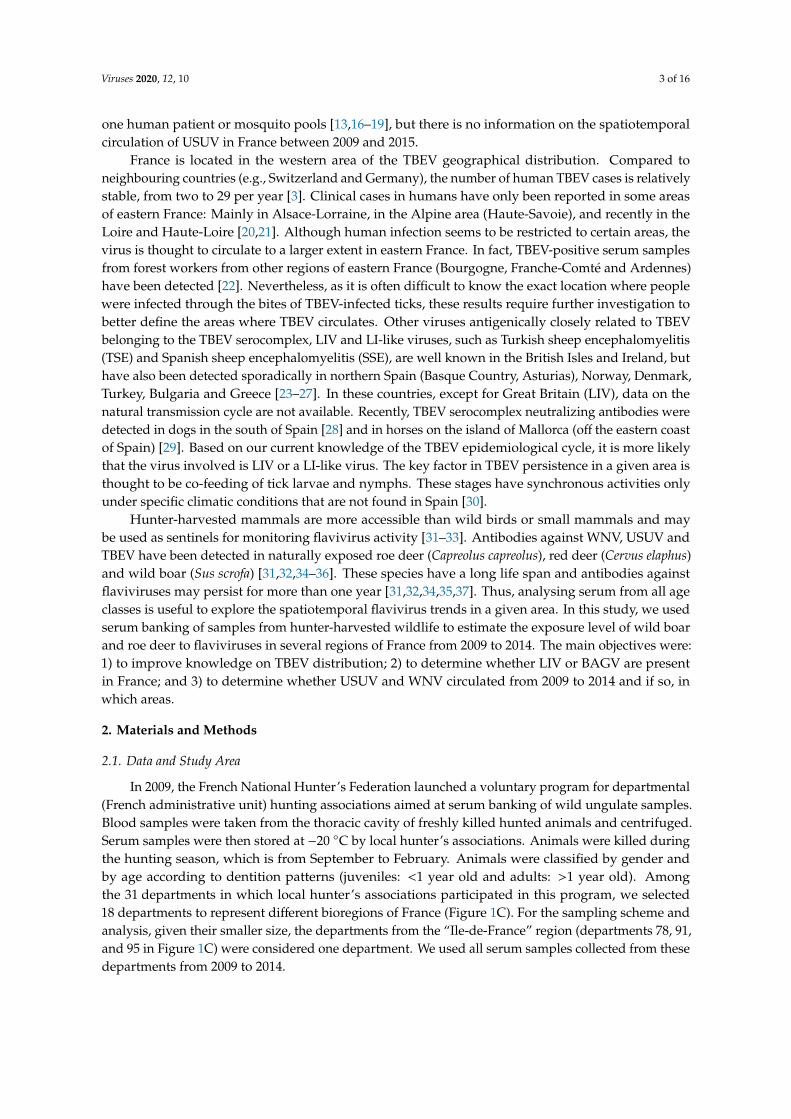
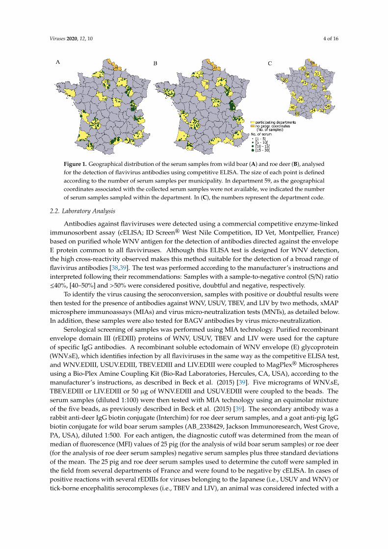
In 2009, the French National Hunter’s Federation launched a voluntary program for departmental(French administrative unit) hunting associations aimed at serum banking of wild ungulate samples.Blood samples were taken from the thoracic cavity of freshly killed hunted animals and centrifuged.Serum samples were then stored at −20 ◦C by local hunter’s associations. Animals were killed duringthe hunting season, which is from September to February. Animals were classified by gender andby age according to dentition patterns (juveniles: <1 year old and adults: >1 year old). Amongthe 31 departments in which local hunter’s associations participated in this program, we selected18 departments to represent different bioregions of France (Figure 1C). For the sampling scheme andanalysis, given their smaller size, the departments from the “Ile-de-France” region (departments 78, 91,and 95 in Figure 1C) were considered one department. We used all serum samples collected from thesedepartments from 2009 to 2014.
Viruses 2020, 12, 10 4 of 16
Viruses 2019, 11, x FOR PEER REVIEW 4 of 17
Figure 1. Geographical distribution of the serum samples from wild boar (A) and roe deer (B), analysed for the detection of flavivirus antibodies using competitive ELISA. The size of each point is defined according to the number of serum samples per municipality. In department 59, as the geographical coordinates associated with the collected serum samples were not available, we indicated the number of serum samples sampled within the department. In (C), the numbers represent the department code.
2.2. Laboratory Analysis
Antibodies against flaviviruses were detected using a commercial competitive enzyme-linked immunosorbent assay (cELISA; ID Screen® West Nile Competition, ID Vet, Montpellier, France) based on purified whole WNV antigen for the detection of antibodies directed against the envelope E protein common to all flaviviruses. Although this ELISA test is designed for WNV detection, the high cross-reactivity observed makes this method suitable for the detection of a broad range of flavivirus antibodies [38,39]. The test was performed according to the manufacturer’s instructions and interpreted following their recommendations: samples with a sample-to-negative control (S/N) ratio ≤ 40%, [40–50%] and > 50% were considered positive, doubtful and negative, respectively.
To identify the virus causing the seroconversion, samples with positive or doubtful results were then tested for the presence of antibodies against WNV, USUV, TBEV, and LIV by two methods, xMAP microsphere immunoassays (MIAs) and virus micro-neutralization tests (MNTs), as detailed below. In addition, these samples were also tested for BAGV antibodies by virus micro-neutralization.
Serological screening of samples was performed using MIA technology. Purified recombinant envelope domain III (rEDIII) proteins of WNV, USUV, TBEV and LIV were used for the capture of specific IgG antibodies. A recombinant soluble ectodomain of WNV envelope (E) glycoprotein (WNV.sE), which identifies infection by all flaviviruses in the same way as the competitive ELISA test, and WNV.EDIII, USUV.EDIII, TBEV.EDIII and LIV.EDIII were coupled to MagPlex® Microspheres using a Bio-Plex Amine Coupling Kit (Bio-Rad Laboratories, Hercules, CA, USA), according to the manufacturer’s instructions, as described in Beck et al. (2015) [39]. Five micrograms of WNV.sE, TBEV.EDIII or LIV.EDIII or 50 µg of WNV.EDIII and USUV.EDIII were coupled to the beads. The serum samples (diluted 1:100) were then tested with MIA technology using an equimolar mixture of the five beads, as previously described in Beck et al. (2015) [39]. The secondary antibody was a rabbit anti-deer IgG biotin conjugate (Interchim) for roe deer serum samples, and a goat anti-pig IgG biotin conjugate for wild boar serum samples (AB_2338429, Jackson Immunoresearch, West Grove, PA, USA), diluted 1:500. For each antigen, the diagnostic cutoff was determined from the mean of median of fluorescence (MFI) values of 25 pig (for the analysis of wild boar serum samples) or roe deer (for the analysis of roe deer serum samples) negative serum samples plus three standard deviations of the mean. The 25 pig and roe deer serum samples used to determine the cutoff were sampled in the field from several departments of France and were found to be negative by cELISA. In cases of positive reactions with several rEDIIIs for viruses belonging to the Japanese (i.e., USUV and WNV) or tick-borne encephalitis serocomplexes (i.e., TBEV and LIV), an animal was considered
Figure 1. Geographical distribution of the serum samples from wild boar (A) and roe deer (B), analysedfor the detection of flavivirus antibodies using competitive ELISA. The size of each point is definedaccording to the number of serum samples per municipality. In department 59, as the geographicalcoordinates associated with the collected serum samples were not available, we indicated the numberof serum samples sampled within the department. In (C), the numbers represent the department code.
2.2. Laboratory Analysis
Antibodies against flaviviruses were detected using a commercial competitive enzyme-linkedimmunosorbent assay (cELISA; ID Screen® West Nile Competition, ID Vet, Montpellier, France)based on purified whole WNV antigen for the detection of antibodies directed against the envelopeE protein common to all flaviviruses. Although this ELISA test is designed for WNV detection,the high cross-reactivity observed makes this method suitable for the detection of a broad range offlavivirus antibodies [38,39]. The test was performed according to the manufacturer’s instructions andinterpreted following their recommendations: Samples with a sample-to-negative control (S/N) ratio≤40%, [40–50%] and >50% were considered positive, doubtful and negative, respectively.
To identify the virus causing the seroconversion, samples with positive or doubtful results werethen tested for the presence of antibodies against WNV, USUV, TBEV, and LIV by two methods, xMAPmicrosphere immunoassays (MIAs) and virus micro-neutralization tests (MNTs), as detailed below.In addition, these samples were also tested for BAGV antibodies by virus micro-neutralization.
Serological screening of samples was performed using MIA technology. Purified recombinantenvelope domain III (rEDIII) proteins of WNV, USUV, TBEV and LIV were used for the captureof specific IgG antibodies. A recombinant soluble ectodomain of WNV envelope (E) glycoprotein(WNV.sE), which identifies infection by all flaviviruses in the same way as the competitive ELISA test,and WNV.EDIII, USUV.EDIII, TBEV.EDIII and LIV.EDIII were coupled to MagPlex® Microspheresusing a Bio-Plex Amine Coupling Kit (Bio-Rad Laboratories, Hercules, CA, USA), according to themanufacturer’s instructions, as described in Beck et al. (2015) [39]. Five micrograms of WNV.sE,TBEV.EDIII or LIV.EDIII or 50 µg of WNV.EDIII and USUV.EDIII were coupled to the beads. Theserum samples (diluted 1:100) were then tested with MIA technology using an equimolar mixtureof the five beads, as previously described in Beck et al. (2015) [39]. The secondary antibody was arabbit anti-deer IgG biotin conjugate (Interchim) for roe deer serum samples, and a goat anti-pig IgGbiotin conjugate for wild boar serum samples (AB_2338429, Jackson Immunoresearch, West Grove,PA, USA), diluted 1:500. For each antigen, the diagnostic cutoff was determined from the mean ofmedian of fluorescence (MFI) values of 25 pig (for the analysis of wild boar serum samples) or roe deer(for the analysis of roe deer serum samples) negative serum samples plus three standard deviationsof the mean. The 25 pig and roe deer serum samples used to determine the cutoff were sampled inthe field from several departments of France and were found to be negative by cELISA. In cases ofpositive reactions with several rEDIIIs for viruses belonging to the Japanese (i.e., USUV and WNV) ortick-borne encephalitis serocomplexes (i.e., TBEV and LIV), an animal was considered infected with a
Viruses 2020, 12, 10 5 of 16
specific flavivirus if the corresponding bead coupled to rEDIII generated an MFI at least double thatgenerated with the other beads.
MNTs were performed for the detection of specific neutralizing antibodies against WNV (strainIs98, Genbank ID AF481864.1, provided by Philippe Desprès, IPP, Paris), USUV (strain Italy 2012,206795-3/2012, Genbank ID KX816653.1, provided by Davide Lelli, IZSLER, Brescia), TBEV (strain Hypr,Genbank ID U39292.1), LIV (strain Li 3/1, Genbank ID KP144331.1, provided by Nicholas Johnson,APHA, Weybridge), and BAGV (strain Spain 2010, Genbank KR108244) as described in Beck et al.(2015) [39]. A serum sample was considered positive if cells were protected at the 1:10 serum dilutionfor WNV, USUV and BAGV, and 1:20 for TBEV and LIV. When results indicated cross-neutralizationbetween flaviviruses, we identified the infecting flavivirus by considering the virus with the highestneutralization capacity, and with neutralization titres that differ by at least 4-fold.
2.3. Laboratory Result Interpretation
Detection of flaviviruses. We considered a serum sample to be i) “flavivirus-negative” when theresult of the pan-flavivirus cELISA was negative or was doubtful and negative in MNTs or MIAs;or ii) “flavivirus-positive” when flavivirus antibodies were detected by the pan-flavivirus cELISA(positive serum in cELISA), MNTs or MIAs (doubtful serum in cELISA).
Identification of flaviviruses. As MNTs and MIAs gave complementary and specific results andidentified the same viruses (see Results 3.1), we considered that a serum sample was positive for aspecific virus (i.e., USUV-, TBEV-, LIV-, WNV- or BAGV-positive) when antibodies towards this viruswere detected by at least one of the methods, MNTs or MIAs, given the conditions described above.TBEV and LIV could not be differentiated by MNTs and MIAs, and hereafter results are noted as“TBEV/LIV positive.”
We considered a positive or doubtful cELISA serum sample as “confirmed” when the flavivirusresponsible for the seroconversion was identified by one of the confirmatory tests, MNTs or MIAs.Positive cELISA serum samples not identified by MNTs or MIAs were considered “non-confirmed”.We calculated the proportion of confirmatory results as the proportion of confirmed serum samplesamong positive or doubtful cELISA serum samples. We compared the S/N ratio of the cELISA betweenserum samples confirmed by two or only one test to assess whether the difference in the results wascorrelated to the S/N ratio.
2.4. Statistical Analyses
The apparent seroprevalence of antibodies against flaviviruses was estimated by species from theratio of cELISA positives to the total number of samples, with the exact binomial confidence intervalsof 95% (CI95%). Differences between species were tested by a Pearson’s Chi-square test. The meancELISA S/N ratio values were compared between species by a Student’s t-test.
As no antibodies were detected against WNV and BAGV, we only analysed the factors associatedwith USUV and TBEV/LIV antibody seroprevalence. This analysis was only conducted in USUV- andTBEV/LIV-infected departments. A department was considered USUV-infected (or TBEV/LIV-infected)when at least one serum sample was USUV- (or TBEV/LIV-) positive in MNT or MIA. For TBEV/LIV,we only included the departments of eastern France, the known geographical distribution area ofthe TBE virus. We considered that the TBEV/LIV-positive serum samples from eastern France werelikely “TBEV-positive.” In infected departments, the exposure status of animals for each virus wasmodelled using binomial generalized linear mixed model (GLMM), as a function of sampling period,ungulate species and age. The department was included as a random factor to take into account thepotential spatial aggregation in virus circulation. We defined the sampling period from 1 September to28 February of the following year, in order to take into account the hunting season and the probable lowflavivirus circulation during the winter. Odds ratios and 95% confidence intervals were estimated bybootstrapping. All statistical analyses were performed with R 3.5.0 software (Vienna, Austria) [40]. All
Viruses 2020, 12, 10 6 of 16
maps were created with QGIS 2.18.11 (QGIS Geographic Information System. Open Source GeospatialFoundation Project).
3. Results
3.1. Serological Results
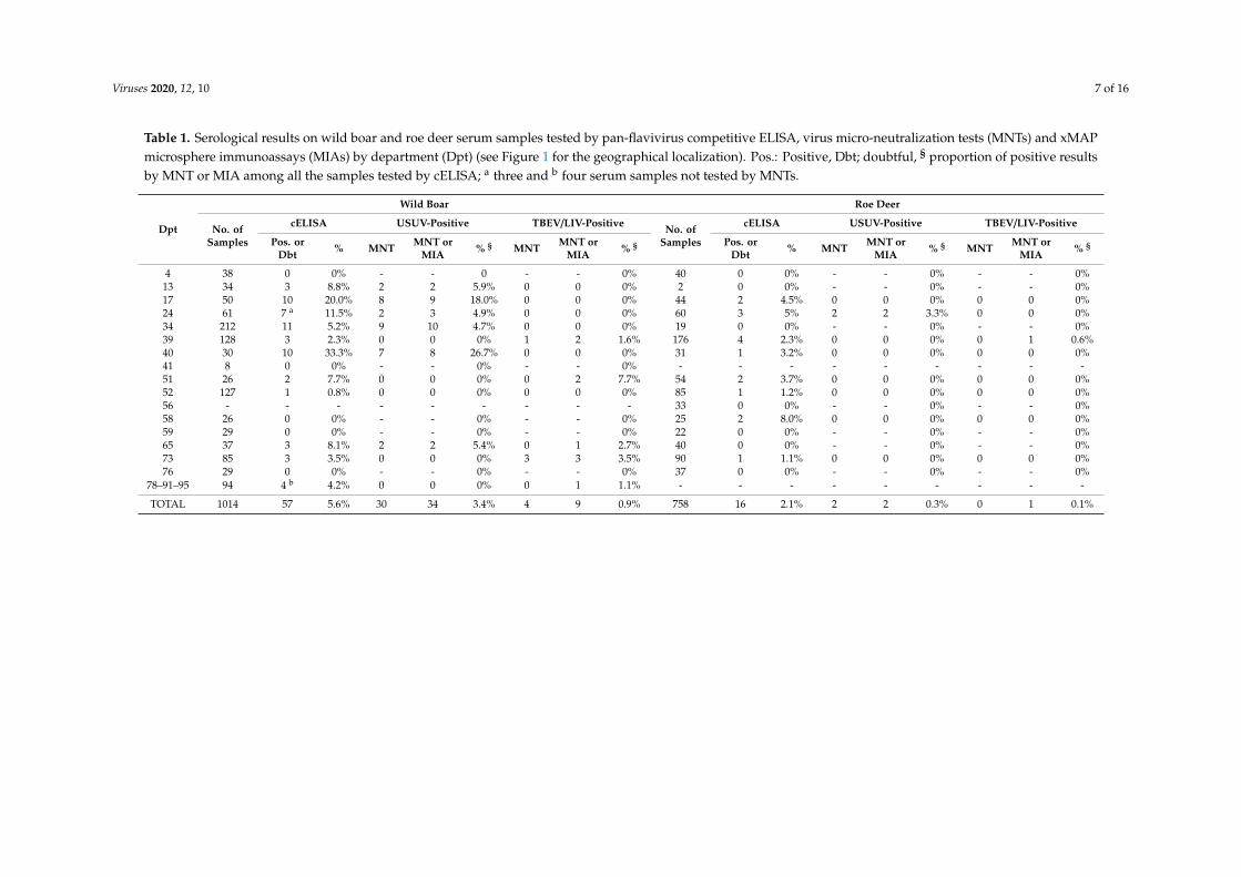
In total, 758 roe deer serum samples and 1014 wild boar serum samples were collected fromSeptember 2009 to November 2014 (Figure 1). From eight to 304 serum samples were collected perdepartment (Figure 1 and Table 1). Pan-flavivirus antibodies (i.e., positive and doubtful cELISA results)were detected in 16/758 (2.1%, CI95%: 1.2-3.4%) roe deer serum samples and 57/1014 (5.6%, CI95%:4.3-7.2%) wild boar serum samples. These data represent a significantly higher seroprevalence in wildboar than in roe deer (Chi2 p < 0.001). Among positive pan-flavivirus cELISA samples, the mean % S/Nvalue of wild boar serum samples was lower than that of roe deer (Student’s t-test, t = −3.7, df = 29,p < 0.001), suggestive of a stronger antibody response in wild boar.
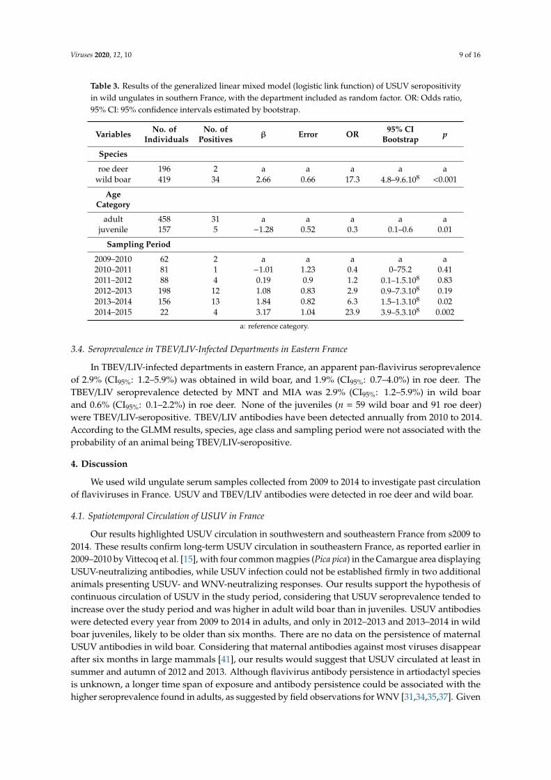
Among the ELISA positive and doubtful samples (n = 73), seven could not be tested by MNTs dueto serum cytotoxicity or low serum quantity (Table 1). None of the serum samples tested containedspecific antibodies against WNV or BAGV. Specific antibodies against USUV or TBEV/LIV weredetected in 32 and four serum samples by MNTs, and in 30 and seven serum samples by MIAs,respectively. The results were identical between both methods for 49 serum samples (Table 2): 22were negative, 26 positive for USUV, and one positive for TBEV/LIV, with a higher proportion ofidentical results among the USUV-positive serum samples (26/35, 74%) than the TBEV/LIV-positiveserum samples (1/9, 11%). The mean % S/N value was lower for samples confirmed by MNT and MIAthan that confirmed by only one test, or that not confirmed by any test (Mann–Whitney U test, W = 330,p = 0.007), suggesting that high antibody levels facilitated the confirmation of flavivirus infection.
Overall, specific antibodies against USUV were detected by either method in two roe deer and34 wild boar serum samples, and those against TBEV/LIV were detected in one roe deer and ninewild boar serum samples. Neutralizing antibody titres were around 1/10–1/20 for 11 samples andranged from 1/40 to 1/320 for 23 samples. The mean % S/N value was positively correlated with theneutralizing antibody titres (Spearman correlation, rs = 0.88, p-value = 0.03), suggesting again thathigh antibody levels facilitated the confirmation of flavivirus infection by MNT. The proportion ofconfirmatory results (i.e., positive for at least one test) was higher in wild boar (43/57, 75%) than in roedeer (2/16, 13%).
3.2. Geographical Distribution
USUV antibodies were detected in animals from six departments, located in southwestern andsoutheastern France by MNT and/or MIA (Figure 2). TBEV/LIV MNT- and MIA-positive antibodies weredetected in three departments of eastern France (Figure 2). TBEV/LIV antibodies were detected only byMIA in three departments located in eastern (Marne, department 51), southwestern (Hautes-Pyrénées,department 65) and northern France (Yvelines, department 78) (Figure 2, Table 1). The 28 serumsamples found to be positive or doubtful with the pan-flavivirus cELISA that could not be tested (lowserum quantity or too hemolysed) or confirmed by MIA and MNT were from 11 departments (Figure 2).They included nine departments in which USUV or TBEV/LIV antibodies had been confirmed, andtwo from eastern France close to the TBEV endemic area. No flavivirus antibodies were detected infour departments with >30 serum samples tested: Three were located in western and northern Franceand one was located in far southeastern France.
Viruses 2020, 12, 10 7 of 16
Table 1. Serological results on wild boar and roe deer serum samples tested by pan-flavivirus competitive ELISA, virus micro-neutralization tests (MNTs) and xMAPmicrosphere immunoassays (MIAs) by department (Dpt) (see Figure 1 for the geographical localization). Pos.: Positive, Dbt; doubtful, § proportion of positive resultsby MNT or MIA among all the samples tested by cELISA; a three and b four serum samples not tested by MNTs.
Dpt
Wild Boar Roe Deer
No. ofSamples
cELISA USUV-Positive TBEV/LIV-Positive No. ofSamples
cELISA USUV-Positive TBEV/LIV-Positive
Pos. orDbt % MNT MNT or
MIA % § MNT MNT orMIA % § Pos. or
Dbt % MNT MNT orMIA % § MNT MNT or
MIA % §
4 38 0 0% - - 0 - - 0% 40 0 0% - - 0% - - 0%13 34 3 8.8% 2 2 5.9% 0 0 0% 2 0 0% - - 0% - - 0%17 50 10 20.0% 8 9 18.0% 0 0 0% 44 2 4.5% 0 0 0% 0 0 0%24 61 7 a 11.5% 2 3 4.9% 0 0 0% 60 3 5% 2 2 3.3% 0 0 0%34 212 11 5.2% 9 10 4.7% 0 0 0% 19 0 0% - - 0% - - 0%39 128 3 2.3% 0 0 0% 1 2 1.6% 176 4 2.3% 0 0 0% 0 1 0.6%40 30 10 33.3% 7 8 26.7% 0 0 0% 31 1 3.2% 0 0 0% 0 0 0%41 8 0 0% - - 0% - - 0% - - - - - - - - -51 26 2 7.7% 0 0 0% 0 2 7.7% 54 2 3.7% 0 0 0% 0 0 0%52 127 1 0.8% 0 0 0% 0 0 0% 85 1 1.2% 0 0 0% 0 0 0%56 - - - - - - - - - 33 0 0% - - 0% - - 0%58 26 0 0% - - 0% - - 0% 25 2 8.0% 0 0 0% 0 0 0%59 29 0 0% - - 0% - - 0% 22 0 0% - - 0% - - 0%65 37 3 8.1% 2 2 5.4% 0 1 2.7% 40 0 0% - - 0% - - 0%73 85 3 3.5% 0 0 0% 3 3 3.5% 90 1 1.1% 0 0 0% 0 0 0%76 29 0 0% - - 0% - - 0% 37 0 0% - - 0% - - 0%
78–91–95 94 4 b 4.2% 0 0 0% 0 1 1.1% - - - - - - - - -
TOTAL 1014 57 5.6% 30 34 3.4% 4 9 0.9% 758 16 2.1% 2 2 0.3% 0 1 0.1%
Viruses 2020, 12, 10 8 of 16
Table 2. Contingency table of and xMAP microsphere immunoassays (MIAs) and virusmicro-neutralization tests (MNTs) results for 66 samples revealed as positive or doubtful by competitiveELISA and tested by both MIAs and MNTs.
ConfirmationMethod
MIATotalPositive for
USUVPositive forTBEV/LIV Negative
MNT
Positive for USUV 26 0 6 32
Positive for TBEV/LIV 0 1 3 4
Negative 3 5 22 30
Total 29 6 31 66
Viruses 2019, 11, x FOR PEER REVIEW 1 of 17
Table 2. Contingency table of and xMAP microsphere immunoassays (MIAs) and virus micro-neutralization tests (MNTs) results for 66 samples revealed as positive or doubtful by competitive ELISA and tested by both MIAs and MNTs.
Confirmation Method
MIA Total Positive for
USUV Positive for TBEV/LIV Negative
MNT
Positive for USUV 26 0 6 32 Positive for TBEV/LIV
0 1 3 4
Negative 3 5 22 30 Total 29 6 31 66
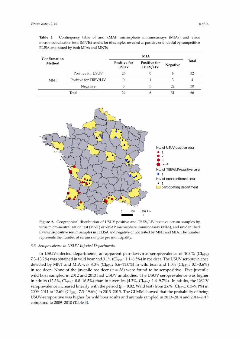
Figure 2. Geographical distribution of USUV-positive and TBEV/LIV-positive serum samples by virus micro-neutralization test (MNT) or xMAP microsphere immunoassay (MIA), and unidentified flavivirus-positive serum samples in cELISA and negative or not tested by MNT and MIA. The number represents the number of serum samples per municipality.
Figure 2. Geographical distribution of USUV-positive and TBEV/LIV-positive serum samples byvirus micro-neutralization test (MNT) or xMAP microsphere immunoassay (MIA), and unidentifiedflavivirus-positive serum samples in cELISA and negative or not tested by MNT and MIA. The numberrepresents the number of serum samples per municipality.
3.3. Seroprevalence in USUV-Infected Departments
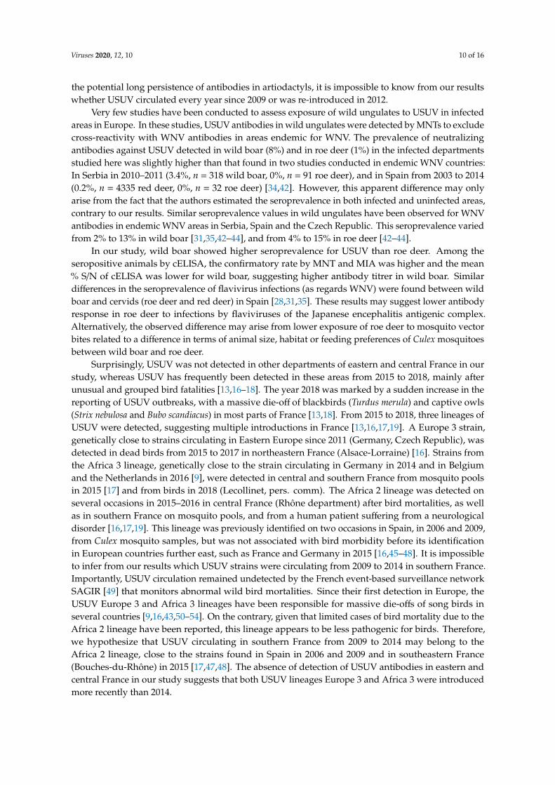
In USUV-infected departments, an apparent pan-flavivirus seroprevalence of 10.0% (CI95%:7.3–13.2%) was obtained in wild boar and 3.1% (CI95%: 1.1–6.5%) in roe deer. The USUV seroprevalencedetected by MNT and MIA was 8.0% (CI95%: 5.6–11.0%) in wild boar and 1.0% (CI95%: 0.1–3.6%)in roe deer. None of the juvenile roe deer (n = 38) were found to be seropositive. Five juvenilewild boar sampled in 2012 and 2013 had USUV antibodies. The USUV seroprevalence was higherin adults (12.3%, CI95%: 8.8–16.5%) than in juveniles (4.3%, CI95%: 1.4–9.7%). In adults, the USUVseroprevalence increased linearly with the period (p = 0.02, Wald test) from 2.6% (CI95%: 0.3–9.1%) in2009–2011 to 12.6% (CI95%: 7.3–19.6%) in 2013–2015. The GLMM showed that the probability of beingUSUV-seropositive was higher for wild boar adults and animals sampled in 2013–2014 and 2014–2015compared to 2009–2010 (Table 3).
Viruses 2020, 12, 10 9 of 16
Table 3. Results of the generalized linear mixed model (logistic link function) of USUV seropositivityin wild ungulates in southern France, with the department included as random factor. OR: Odds ratio,95% CI: 95% confidence intervals estimated by bootstrap.
Variables No. ofIndividuals
No. ofPositives β Error OR 95% CI
Bootstrap p
Species
roe deer 196 2 a a a a awild boar 419 34 2.66 0.66 17.3 4.8–9.6.108 <0.001
AgeCategory
adult 458 31 a a a a ajuvenile 157 5 −1.28 0.52 0.3 0.1–0.6 0.01
Sampling Period
2009–2010 62 2 a a a a a2010–2011 81 1 −1.01 1.23 0.4 0–75.2 0.412011–2012 88 4 0.19 0.9 1.2 0.1–1.5.108 0.832012–2013 198 12 1.08 0.83 2.9 0.9–7.3.108 0.192013–2014 156 13 1.84 0.82 6.3 1.5–1.3.108 0.022014–2015 22 4 3.17 1.04 23.9 3.9–5.3.108 0.002
a: reference category.
3.4. Seroprevalence in TBEV/LIV-Infected Departments in Eastern France
In TBEV/LIV-infected departments in eastern France, an apparent pan-flavivirus seroprevalenceof 2.9% (CI95%: 1.2–5.9%) was obtained in wild boar, and 1.9% (CI95%: 0.7–4.0%) in roe deer. TheTBEV/LIV seroprevalence detected by MNT and MIA was 2.9% (CI95%: 1.2–5.9%) in wild boarand 0.6% (CI95%: 0.1–2.2%) in roe deer. None of the juveniles (n = 59 wild boar and 91 roe deer)were TBEV/LIV-seropositive. TBEV/LIV antibodies have been detected annually from 2010 to 2014.According to the GLMM results, species, age class and sampling period were not associated with theprobability of an animal being TBEV/LIV-seropositive.
4. Discussion
We used wild ungulate serum samples collected from 2009 to 2014 to investigate past circulationof flaviviruses in France. USUV and TBEV/LIV antibodies were detected in roe deer and wild boar.
4.1. Spatiotemporal Circulation of USUV in France
Our results highlighted USUV circulation in southwestern and southeastern France from s2009 to2014. These results confirm long-term USUV circulation in southeastern France, as reported earlier in2009–2010 by Vittecoq et al. [15], with four common magpies (Pica pica) in the Camargue area displayingUSUV-neutralizing antibodies, while USUV infection could not be established firmly in two additionalanimals presenting USUV- and WNV-neutralizing responses. Our results support the hypothesis ofcontinuous circulation of USUV in the study period, considering that USUV seroprevalence tended toincrease over the study period and was higher in adult wild boar than in juveniles. USUV antibodieswere detected every year from 2009 to 2014 in adults, and only in 2012–2013 and 2013–2014 in wildboar juveniles, likely to be older than six months. There are no data on the persistence of maternalUSUV antibodies in wild boar. Considering that maternal antibodies against most viruses disappearafter six months in large mammals [41], our results would suggest that USUV circulated at least insummer and autumn of 2012 and 2013. Although flavivirus antibody persistence in artiodactyl speciesis unknown, a longer time span of exposure and antibody persistence could be associated with thehigher seroprevalence found in adults, as suggested by field observations for WNV [31,34,35,37]. Given
Viruses 2020, 12, 10 10 of 16
the potential long persistence of antibodies in artiodactyls, it is impossible to know from our resultswhether USUV circulated every year since 2009 or was re-introduced in 2012.
Very few studies have been conducted to assess exposure of wild ungulates to USUV in infectedareas in Europe. In these studies, USUV antibodies in wild ungulates were detected by MNTs to excludecross-reactivity with WNV antibodies in areas endemic for WNV. The prevalence of neutralizingantibodies against USUV detected in wild boar (8%) and in roe deer (1%) in the infected departmentsstudied here was slightly higher than that found in two studies conducted in endemic WNV countries:In Serbia in 2010–2011 (3.4%, n = 318 wild boar, 0%, n = 91 roe deer), and in Spain from 2003 to 2014(0.2%, n = 4335 red deer, 0%, n = 32 roe deer) [34,42]. However, this apparent difference may onlyarise from the fact that the authors estimated the seroprevalence in both infected and uninfected areas,contrary to our results. Similar seroprevalence values in wild ungulates have been observed for WNVantibodies in endemic WNV areas in Serbia, Spain and the Czech Republic. This seroprevalence variedfrom 2% to 13% in wild boar [31,35,42–44], and from 4% to 15% in roe deer [42–44].
In our study, wild boar showed higher seroprevalence for USUV than roe deer. Among theseropositive animals by cELISA, the confirmatory rate by MNT and MIA was higher and the mean% S/N of cELISA was lower for wild boar, suggesting higher antibody titrer in wild boar. Similardifferences in the seroprevalence of flavivirus infections (as regards WNV) were found between wildboar and cervids (roe deer and red deer) in Spain [28,31,35]. These results may suggest lower antibodyresponse in roe deer to infections by flaviviruses of the Japanese encephalitis antigenic complex.Alternatively, the observed difference may arise from lower exposure of roe deer to mosquito vectorbites related to a difference in terms of animal size, habitat or feeding preferences of Culex mosquitoesbetween wild boar and roe deer.
Surprisingly, USUV was not detected in other departments of eastern and central France in ourstudy, whereas USUV has frequently been detected in these areas from 2015 to 2018, mainly afterunusual and grouped bird fatalities [13,16–18]. The year 2018 was marked by a sudden increase in thereporting of USUV outbreaks, with a massive die-off of blackbirds (Turdus merula) and captive owls(Strix nebulosa and Bubo scandiacus) in most parts of France [13,18]. From 2015 to 2018, three lineages ofUSUV were detected, suggesting multiple introductions in France [13,16,17,19]. A Europe 3 strain,genetically close to strains circulating in Eastern Europe since 2011 (Germany, Czech Republic), wasdetected in dead birds from 2015 to 2017 in northeastern France (Alsace-Lorraine) [16]. Strains fromthe Africa 3 lineage, genetically close to the strain circulating in Germany in 2014 and in Belgiumand the Netherlands in 2016 [9], were detected in central and southern France from mosquito poolsin 2015 [17] and from birds in 2018 (Lecollinet, pers. comm). The Africa 2 lineage was detected onseveral occasions in 2015–2016 in central France (Rhône department) after bird mortalities, as wellas in southern France on mosquito pools, and from a human patient suffering from a neurologicaldisorder [16,17,19]. This lineage was previously identified on two occasions in Spain, in 2006 and 2009,from Culex mosquito samples, but was not associated with bird morbidity before its identificationin European countries further east, such as France and Germany in 2015 [16,45–48]. It is impossibleto infer from our results which USUV strains were circulating from 2009 to 2014 in southern France.Importantly, USUV circulation remained undetected by the French event-based surveillance networkSAGIR [49] that monitors abnormal wild bird mortalities. Since their first detection in Europe, theUSUV Europe 3 and Africa 3 lineages have been responsible for massive die-offs of song birds inseveral countries [9,16,43,50–54]. On the contrary, given that limited cases of bird mortality due to theAfrica 2 lineage have been reported, this lineage appears to be less pathogenic for birds. Therefore,we hypothesize that USUV circulating in southern France from 2009 to 2014 may belong to theAfrica 2 lineage, close to the strains found in Spain in 2006 and 2009 and in southeastern France(Bouches-du-Rhône) in 2015 [17,47,48]. The absence of detection of USUV antibodies in eastern andcentral France in our study suggests that both USUV lineages Europe 3 and Africa 3 were introducedmore recently than 2014.
Viruses 2020, 12, 10 11 of 16
4.2. No Detection of WNV and BAGV Antibodies
No WNV antibodies were detected in wild ungulates sampled from the known endemic areas forWNV in southeastern France, the two departments Bouches-du-Rhône (department 13, Figure 1C) andHérault (department 34, Figure 1C), whereas Vittecoq et al. [15] found WNV antibodies in a second-yearcommon magpie in 2010 in the Bouches-du-Rhône, suggesting recent WNV infection (in 2009 or 2010)in the area. In southeastern France, WNV has been reported irregularly and is generally detectedwhen the virus circulation is high and causes clinical outbreaks in humans and horses. Such epizooticsin the Camargue area have been observed in 2000, 2004, 2015 and 2018 [11,13,55,56]. Between twoepizootics, the virus could be silently maintained, as suggested by the detection of WNV antibodies inwild birds [15,57]. In our study, few animals were sampled in areas located close to the wetlands ofthe Hérault and Bouches-du-Rhône departments, where WNV cases were mostly detected during theoutbreaks. This may have limited the detection of WNV circulation in our study. This result confirmsthat the virus, if present, probably circulates very locally close to wetland areas that provide sustainablehabitats for the nesting of many wild resident or migrating birds, and for the multiplication of Culexvectors [58].
Bagaza virus antibodies were not detected. In Europe, this virus was only detected in southernSpain in 2010 and 2011, associated with high mortality of red-legged partridges (Alectoris rufa) andring-necked pheasants (Phasianus colchicus) [59,60]. The main hosts of BAGV are birds and there islittle evidence of BAGV antibodies in mammals. Neutralizing antibodies to BAGV have been found inserum samples from encephalitis cases in humans in India, and in experimentally infected mice [61,62].
4.3. TBEV Serocomplex Distribution
We detected TBEV/LIV antibodies in three departments of eastern France (Savoie department73, Marne department 51, and Jura department 39 in Figure 1C), in one department in the south(Hautes-Pyrénées, department 65) and one department in northern France (Yvelines, department 78).Given the genetic and antigenic relatedness between TBEV and LIV, serological responses induced afterTBEV and LIV infection could not be differentiated by MNT and MIA. Although the distribution ofTBEV in France is not known precisely and clinical cases have mainly been reported in the far easternparts of France (Alsace-Lorraine and Alpine regions), the virus is thought to be broadly distributed inthe east of France. We detected TBEV/LIV antibodies in wild ungulates in one department (Savoie),where TBE clinical cases in humans have been reported [20], and in two departments (Marne and Jura),where forest workers were previously found to be seropositive to TBEV [22]. It is therefore likely thatseropositive wild ungulates observed in our study in eastern France have been exposed to TBEV.
The wild boar found to be TBEV/LIV-seropositive in the Hautes-Pyrénées department by MIAwas not confirmed by MNT, whereas the serum was strongly positive in cELISA (low mean % S/N).For the wild boar in the Yvelines department, the serum was too hemolysed and the result could notbe confirmed by MNT. The result obtained by MIA could be interpreted, with caution, as an indicatorof a tick-borne flavivirus infection. TBEV/LIV seropositivity in Hautes-Pyrénées could originate froman infection with LIV or LI-like virus, since these viruses have been reported in sheep, goats andCantabrian chamois (Rupicapra pyrenaica parva) in close geographical areas in Spain: In the Basqueand Asturias regions [23,25,63]. The presence of a virus from the TBEV-serocomplex was found at abroader scale in Spain by serology, with antibodies neutralizing TBEV reported in southern Spain andon the island of Mallorca [28,29]. The positive serum in the Yvelines might also reveal the westernlimit of distribution of TBEV in France. Interestingly, TBEV was recently detected in ticks in the UnitedKingdom, which represents the western-most distribution limit for TBEV [64]. Finally, in both cases, wecannot rule of the hypothesis that this might be due to another tick-borne flavivirus. The diversity anddistribution of tick-borne flaviviruses are not well known, and new species are regularly discovered inticks [65]. Further investigations are needed to confirm the circulation of another tick-borne flavivirusin France and to isolate this virus.
Viruses 2020, 12, 10 12 of 16
The overall TBEV seroprevalence found in wild boar and roe deer in the eastern departments(2.9% in wild boar and 0.3% in roe deer) was much lower than that found in TBEV endemic areasin Germany, Poland, Slovakia, the Czech Republic and the south of Sweden, varying from 10% to40% [32,36,44,66–69]. However, it was similar to the seroprevalences found in areas with the sameepidemiological situation as eastern France, with no or very few human clinical cases: From 2% to 6%seropositive wild boar and roe deer have been reported in Belgium, in some areas of Germany, and inthe south of Denmark [66,70–73]. We did not find any significant difference in TBEV seroprevalencebetween wild boar and roe deer, which is consistent with previous observations [44,66,69,71,72]. Theseresults are unexpected as roe deer are considered to be the main hosts for the nymphs and adults ofIxodes ricinus, the main tick vector for TBEV in Western Europe [74]. However, there is a lack of data onthe role of wild boar as a feeding host for this tick or for other potential vector tick species of TBEV,such as Dermacentor spp. [75]
4.4. Unidentified Flaviviruses
Twenty-seven samples were positive or doubtful according to cELISA and negative according toMNT and MIA for WNV, USUV, TBEV, LIV and BAGV, or negative according to MIA and not tested byMNT. All but four samples were located in departments where USUV or TBEV/LIV antibodies hadbeen detected in our study. The four other samples were located in two eastern departments, whereTBEV antibodies had previously been detected in forest workers [22]. Three samples were located inthe Yvelines (department 78), where antibodies against TBEV/LIV were detected in one serum sample.We cannot exclude the possibility that another flavivirus may be circulating. However, given theirlocalization, it is more likely that the animals were exposed to USUV or TBEV. Most probably, ELISApositive or doubtful results could not be confirmed by MNT or MIA due to serum quality or the loweranalytical sensitivity of these tests, which is consistent with the low antibody titres (high % S/N) foundby cELISA for most of these serum samples.
5. Conclusions
The results obtained in this study documented the distribution and spatiotemporal dynamics offlaviviruses in France from 2009 to 2014. They indicate silent and probable continuous circulation ofUSUV in southwestern and southeastern France from 2009 to 2014. Antibodies against TBEV/LIV weredetected in two eastern departments, where no TBE clinical cases have been reported so far, a finding thatconfirms the wider distribution of the TBE virus in the east of France. The serum samples with TBEV/LIVantibodies found in southwestern and northern France warrant further exploration to determine thevirus responsible for the seroconversion. This study shows the usefulness of serum banking by huntersconcerning wildlife for retrospective epidemiological surveys of pathogen circulation.
Author Contributions: G.U., E.J., E.F., M.S., S.M., C.B. and S.L. conceived the study and designed the methodology.E.F. and M.S. collected the serum available. J.-M.B., F.B., S.A.L., C.B., F.L. and M.A.J.-C. performed the laboratorytests. L.B. performed the analysis and led the writing of the manuscript. All authors have read and agreed to thepublished version of the manuscript.
Funding: This research was funded by the Fédération Nationale des Chasseurs, grant number FNC-PSN-PS1-2013.
Acknowledgments: The authors are very grateful to all hunters and local hunter’s associations who helped tobuild the serum banking and who agreed to make samples available for our study. We are grateful to PhilippeDesprès from IPP (Paris, France), Davide Lelli from IZSLER (Brescia, Italy), and Nicholas Johnson (Weybridge,United Kingdom) from APHA for providing reference flavivirus strains.
Conflicts of Interest: The authors declare that they have no conflict of interest.
References
1. Weissenböck, H.; Hubálek, Z.; Bakonyi, T.; Nowotny, N. Zoonotic mosquito-borne flaviviruses: Worldwidepresence of agents with proven pathogenicity and potential candidates of future emerging diseases. Vet.Microbiol. 2010, 140, 271–280. [CrossRef] [PubMed]
Viruses 2020, 12, 10 13 of 16
2. Clé, M.; Beck, C.; Salinas, S.; Lecollinet, S.; Gutierrez, S.; Van de Perre, P.; Baldet, T.; Foulongne, V.; Simonin, Y.Usutu virus: A new threat? Epidemiol. Infect. 2019, 147, e232. [CrossRef] [PubMed]
3. Erber, W.; Schmitt, H.-J.; Jankovic, T.V. Chapter 12a: Epidemiology by country–an overview. Tick-BorneEnceph.-Book 2019. [CrossRef]
4. Gilbert, L. Louping ill virus in the UK: A review of the hosts, transmission and ecological consequences ofcontrol. Exp. Appl. Acarol. 2016, 68, 363–374. [CrossRef] [PubMed]
5. European Centre for Disease Prevention and Control Surveillance and Disease Data for West Nile Fever.Available online: https://ecdc.europa.eu/en/west-nile-fever/surveillance-and-disease-data (accessed on16 July 2019).
6. Beck, C.; Jimenez-Clavero, M.; Leblond, A.; Durand, B.; Nowotny, N.; Leparc-Goffart, I.; Zientara, S.;Jourdain, E.; Lecollinet, S. Flaviviruses in Europe: Complex circulation patterns and their consequencesfor the diagnosis and control of West Nile disease. Int. J. Environ. Res. Public. Health 2013, 10, 6049–6083.[CrossRef]
7. Weissenböck, H.; Bakonyi, T.; Rossi, G.; Mani, P.; Nowotny, N. Usutu Virus, Italy, 1996. Emerg. Infect. Dis.2013, 19, 274–277. [CrossRef]
8. Nikolay, B. A review of West Nile and Usutu virus co-circulation in Europe: How much do transmissioncycles overlap? Trans. R. Soc. Trop. Med. Hyg. 2015, 109, 609–618. [CrossRef]
9. Cadar, D.; Lühken, R.; van der Jeugd, H.; Garigliany, M.; Ziegler, U.; Keller, M.; Lahoreau, J.; Lachmann, L.;Becker, N.; Kik, M.; et al. Widespread activity of multiple lineages of Usutu virus, western Europe, 2016.Euro Surveill 2017, 22, 30452. [CrossRef]
10. Weidinger, P.; Kolodziejek, J.; Bakonyi, T.; Brunthaler, R.; Erdélyi, K.; Weissenböck, H.; Nowotny, N. Differentdynamics of Usutu virus infections in Austria and Hungary, 2017–2018. Transbound. Emerg. Dis. 2019.[CrossRef]
11. Bahuon, C.; Marcillaud-Pitel, C.; Bournez, L.; Leblond, A.; Beck, C.; Hars, J.; Leparc-Goffart, I.; L’Ambert, G.;Paty, M.-C.; Cavalerie, L.; et al. West Nile virus epizootics in the Camargue (France) in 2015 and reinforcementof surveillance and control networks: -EN- -FR- Épizootie due au virus de West Nile survenue en Camargue(France) en 2015 et renforcement des réseaux de surveillance et de contrôle -ES- Epizootia causada por elvirus West Nile en la Camarga (Francia) en 2015 y refuerzo de las redes de vigilancia y control. Rev. Sci. Tech.OIE 2016, 35, 811–824.
12. Maquart, M.; Dahmani, M.; Marié, J.-L.; Gravier, P.; Leparc-Goffart, I.; Davoust, B. First serological evidenceof West Nile Virus in horses and dogs from Corsica island, France. Vector-Borne Zoonotic Dis. 2017, 17,275–277. [CrossRef]
13. Johnson, N.; Fernández de Marco, M.; Giovannini, A.; Ippoliti, C.; Danzetta, M.; Svartz, G.; Erster, O.;Groschup, M.; Ziegler, U.; Mirazimi, A.; et al. Emerging Mosquito-Borne Threats and the Response fromEuropean and Eastern Mediterranean Countries. Int. J. Environ. Res. Public. Health 2018, 15, 2775. [CrossRef]
14. Jourdain, E.; Toussaint, Y.; Leblond, A.; Bicout, D.J.; Sabatier, P.; Gauthier-Clerc, M. Bird species potentiallyinvolved in introduction, amplification, and spread of West Nile Virus in a mediterranean wetland, theCamargue (Southern France). Vector-Borne Zoonotic Dis. 2007, 7, 15–33. [CrossRef]
15. Vittecoq, M.; Lecollinet, S.; Jourdain, E.; Thomas, F.; Blanchon, T.; Arnal, A.; Lowenski, S.; Gauthier-Clerc, M.Recent circulation of West Nile Virus and potentially other closely related flaviviruses in Southern France.Vector-Borne Zoonotic Dis. 2013, 13, 610–613. [CrossRef]
16. Lecollinet, S.; Blanchard, Y.; Manson, C.; Lowenski, S.; Laloy, E.; Quenault, H.; Touzain, F.; Lucas, P.; Eraud, C.;Bahuon, C.; et al. Dual emergence of Usutu virus in common blackbirds, Eastern France, 2015. Emerg. Infect.Dis. 2016, 22, 2225. [CrossRef]
17. Eiden, M.; Gil, P.; Ziegler, U.; Rakotoarivony, I.; Marie, A.; Frances, B.; L’Ambert, G.; Simonin, Y.; Foulongne, V.;Groschup, M.H.; et al. Emergence of two Usutu virus lineages in Culex pipiens mosquitoes in the Camargue,France, 2015. Infect. Genet. Evol. 2018, 61, 151–154. [CrossRef]
18. Beck, C.; Gonzales, G.; Decors, A.; Lemberger, K.; Lowenski, S.; Dumarest, D.; Lecollinet, S. Surveillanceépidémiologique du virus Usutu dans l’avifaune. Virologie 2018, 22, 261–263.
19. Simonin, Y.; Sillam, O.; Carles, M.J.; Gutierrez, S.; Gil, P.; Constant, O.; Martin, M.F.; Grard, G.; Van dePerre, P.; Salinas, S.; et al. Human Usutu Virus Infection with Atypical Neurologic Presentation, Montpellier,France, 2016. Emerg. Infect. Dis. 2018, 24, 875–878. [CrossRef]
Viruses 2020, 12, 10 14 of 16
20. Velay, A.; Argemi, X.; Wendling, M.-J.; Martinot, M.; Hansmann, Y.; Fafi-Kremer, S. L’encéphalite à tique enFrance: Qu’en savons-nous aujourd’hui? Rev. Francoph. Lab. 2019, 2019, 34–43. [CrossRef]
21. Botelho-Nevers, E.; Gagneux-Brunon, A.; Velay, A.; Guerbois-Galla, M.; Grard, G.; Bretagne, C.; Mailles, A.;Verhoeven, P.O.; Pozzetto, B.; Gonzalo, S.; et al. Tick-Borne Encephalitis in Auvergne-Rhône-Alpes Region,France, 2017–2018. Emerg. Infect. Dis. 2019, 25, 1944–1948. [CrossRef]
22. Thorin, C.; Rigaud, E.; Capek, I.; André-Fontaine, G.; Oster, B.; Gastinger, G.; Abadia, G. Séroprévalence dela borréliose de Lyme et de l’encéphalite à tiques chez des professionnels exposés dans le Grand Est de laFrance. Médecine Mal. Infect. 2008, 38, 533–542. [CrossRef]
23. Balseiro, A.; Royo, L.J.; Martínez, C.P.; Fernández de Mera, I.G.; Höfle, Ú.; Polledo, L.; Marreros, N.; Casais, R.;Marín, J.F.G. Louping ill in goats, Spain, 2011. Emerg. Infect. Dis. 2012, 18, 976–978. [CrossRef]
24. Dobler, G. Zoonotic tick-borne flaviviruses. Vet. Microbiol. 2010, 140, 221–228. [CrossRef]25. Marin, M.S.; McKenzie, J.; Gao, G.F.; Reid, H.W.; Antoniadis, A.; Gould, E.A. The virus causing
encephalomyelitis in sheep in Spain: A new member of the tick-borne encephalitis group. Res. Vet.Sci. 1995, 58, 11–13. [CrossRef]
26. Gao, G.F.; Zanotto, P.M.; Holmes, E.C.; Reid, H.W.; Gould, E.A. Molecular variation, evolution andgeographical distribution of louping ill virus. Acta Virol. 1997, 41, 259–268.
27. Jensen, P.M.; Skarphedinsson, S.; Semenov, A. Densities of the tick (Ixodes ricinus) and coexistence of theLouping ill virus and tick borne encephalitis on the island of Bornholm. Ugeskr. Laeger 2004, 166, 2563–2565.
28. García-Bocanegra, I.; Jurado-Tarifa, E.; Cano-Terriza, D.; Martínez, R.; Pérez-Marín, J.E.; Lecollinet, S.Exposure to West Nile virus and tick-borne encephalitis virus in dogs in Spain. Transbound. Emerg. Dis. 2018,65, 765–772. [CrossRef]
29. Vanhomwegen, J.; Beck, C.; Desprès, P.; Figuerola, A.; García, R.; Lecollinet, S.; López-Roig, M.;Manuguerra, J.-C.; Serra-Cobo, J. Circulation of zoonotic arboviruses in equine populations of Mallorcaisland (Spain). Vector-Borne Zoonotic Dis. 2017, 17, 340–346. [CrossRef]
30. Randolph, S.E. The shifting landscape of tick-borne zoonoses: Tick-borne encephalitis and Lyme borreliosisin Europe. Philos. Trans. R. Soc. Lond. B. Biol. Sci. 2001, 356, 1045–1056. [CrossRef]
31. Boadella, M.; Díez-Delgado, I.; Gutiérrez-Guzmán, A.V.; Höfle, U.; Gortázar, C. Do wild ungulates allowimproved monitoring of flavivirus circulation in Spain? Vector-Borne Zoonotic Dis. 2012, 12, 490–495.[CrossRef]
32. Gerth, H.-J.; Grimshandl, D.; Stage, B.; Döller, G.; Kunz, C. Roe deer as sentinels for endemicity of tick-borneencephalitis virus. Epidemiol. Infect. 1995, 115, 355–365. [CrossRef]
33. Roelandt, S.; Suin, V.; Van Gucht, S.; Van der Stede, Y.; Roels, S. Comparative tick-borne encephalitis (virus)surveillance in Belgium 2009–2015: Experiences with diagnostic tests, sentinel species and surveillancedesigns. J Zoonotic Dis Public Health 2017, 1, 1–4.
34. García-Bocanegra, I.; Paniagua, J.; Gutiérrez-Guzmán, A.V.; Lecollinet, S.; Boadella, M.; Arenas-Montes, A.;Cano-Terriza, D.; Lowenski, S.; Gortázar, C.; Höfle, U. Spatio-temporal trends and risk factors affecting WestNile virus and related flavivirus exposure in Spanish wild ruminants. BMC Vet. Res. 2016, 12, 249. [CrossRef]
35. Gutiérrez-Guzmán, A.-V.; Vicente, J.; Sobrino, R.; Perez-Ramírez, E.; Llorente, F.; Höfle, U. Antibodies toWest Nile virus and related flaviviruses in wild boar, red foxes and other mesomammals from Spain. Vet.Microbiol. 2012, 159, 291–297. [CrossRef]
36. Labuda, M.; Elecková, E.; Licková, M.; Sabó, A. Tick-borne encephalitis virus foci in Slovakia. Int. J. Med.Microbiol. 2002, 291, 43–47. [CrossRef]
37. Geevarghese, G.; Shaikh, B.H.; Jacob, P.G.; Bhat, H.R. Persistence of haemagglutination-inhibition antibodiesto JE and WN viruses in naturally infected domestic pigs in Karnataka State, India. Acta Virol. 1994, 38,235–237.
38. Llorente, F.; García-Irazábal, A.; Pérez-Ramírez, E.; Cano-Gómez, C.; Sarasa, M.; Vázquez, A.;Jiménez-Clavero, M.Á. Influence of flavivirus co-circulation in serological diagnostics and surveillance: Amodel of study using West Nile, Usutu and Bagaza viruses. Transbound. Emerg. Dis. 2019, 66, 2100–2106.[CrossRef]
39. Beck, C.; Desprès, P.; Paulous, S.; Vanhomwegen, J.; Lowenski, S.; Nowotny, N.; Durand, B.; Garnier, A.;Blaise-Boisseau, S.; Guitton, E.; et al. A high-performance multiplex immunoassay for serodiagnosis offlavivirus-associated neurological diseases in horses. BioMed Res. Int. 2015, 2015, 1–13. [CrossRef]
Viruses 2020, 12, 10 15 of 16
40. R Development Core team. R: A Language and Environment for Statistical Computing; R Foundation forStatistical Computing: Vienna, Austria, 2018.
41. Niewiesk, S. Maternal Antibodies: Clinical Significance, Mechanism of Interference with Immune Responses,and Possible Vaccination Strategies. Front. Immunol. 2014, 5, 446. [CrossRef]
42. Escribano-Romero, E.; Lupulovic, D.; Merino-Ramos, T.; Blázquez, A.-B.; Lazic, G.; Lazic, S.; Saiz, J.-C.;Petrovic, T. West Nile virus serosurveillance in pigs, wild boars, and roe deer in Serbia. Vet. Microbiol. 2015,176, 365–369. [CrossRef]
43. Hubálek, Z.; Juricová, Z.; Straková, P.; Blazejová, H.; Betásová, L.; Rudolf, I. Serological survey for West Nilevirus in wild artiodactyls, Southern Moravia (Czech Republic). Vector-Borne Zoonotic Dis. 2017, 17, 654–657.[CrossRef]
44. Juricová, Z.; Hubálek, Z. Serological surveys for arboviruses in the game animals of southern Moravia (CzechRepublic). Folia Zool. 1999, 48, 185–189.
45. Ziegler, U.; Fast, C.; Eiden, M.; Bock, S.; Schulze, C.; Hoeper, D.; Ochs, A.; Schlieben, P.; Keller, M.; Zielke, D.E.;et al. Evidence for an independent third Usutu virus introduction into Germany. Vet. Microbiol. 2016, 192,60–66. [CrossRef]
46. Sieg, M.; Schmidt, V.; Ziegler, U.; Keller, M.; Höper, D.; Heenemann, K.; Rückner, A.; Nieper, H.; Muluneh, A.;Groschup, M.H.; et al. Outbreak and Cocirculation of Three Different Usutu Virus Strains in Eastern Germany.Vector-Borne Zoonotic Dis. 2017, 17, 662–664. [CrossRef]
47. Moreno, J.; Tenorio, A.; Figuerola, J.; Sánchez-Seco, M.P.; Ruiz, S.; Vázquez, A.; Magallanes, A.; Molero, F.;Herrero, L. West Nile and Usutu Viruses in Mosquitoes in Spain, 2008–2009. Am. J. Trop. Med. Hyg. 2011, 85,178–181.
48. Busquets, N.; Alba, A.; Allepuz, A.; Aranda, C.; Nuñez, J.I. Usutu Virus Sequences in Culex pipiens (Diptera:Culicidae), Spain. Emerg. Infect. Dis. 2008, 14, 861–863. [CrossRef]
49. Decors, A.; Hars, J.; Faure, E.; Quintaine, T.; Chollet, J.-Y.; Rossi, S. Le réseau SAGIR: Un outil de vigilancevis-à-vis des agents pathogènes exotiques. Bull. Épidémiologique Santé Anim.-Aliment. 2014, 66, 35–39.
50. Chvala, S.; Bakonyi, T.; Bukovsky, C.; Meister, T.; Brugger, K.; Rubel, F.; Nowotny, N.; Weissenböck, H.Monitoring of Usutu virus activity and spread by using dead bird surveillance in Austria, 2003–2005. Vet.Microbiol. 2007, 122, 237–245. [CrossRef]
51. Steinmetz, H.W.; Bakonyi, T.; Weissenböck, H.; Hatt, J.-M.; Eulenberger, U.; Robert, N.; Hoop, R.; Nowotny, N.Emergence and establishment of Usutu virus infection in wild and captive avian species in and around Zurich,Switzerland—Genomic and pathologic comparison to other central European outbreaks. Vet. Microbiol. 2011,148, 207–212. [CrossRef]
52. Bakonyi, T.; Busquets, N.; Nowotny, N. Comparison of complete genome sequences of Usutu Virus strainsdetected in Spain, Central Europe, and Africa. Vector-Borne Zoonotic Dis. 2014, 14, 324–329. [CrossRef]
53. Ziegler, U.; Jöst, H.; Müller, K.; Fischer, D.; Rinder, M.; Tietze, D.T.; Danner, K.-J.; Becker, N.; Skuballa, J.;Hamann, H.-P.; et al. Epidemic spread of Usutu virus in Southwest Germany in 2011 to 2013 and monitoringof wild birds for Usutu and West Nile viruses. Vector-Borne Zoonotic Dis. 2015, 15, 481–488. [CrossRef]
54. Rijks, J.; Kik, M.; Slaterus, R.; Foppen, R.; Stroo, A.; IJzer, J.; Stahl, J.; Gröne, A.; Koopmans, M.; van derJeugd, H.; et al. Widespread Usutu virus outbreak in birds in the Netherlands, 2016. Euro Surveill 2016, 21,30391. [CrossRef]
55. Murgue, B.; Murri, S.; Zientara, S.; Durand, B.; Durand, J.P.; Zeller, H. West Nile outbreak in horses insouthern France, 2000: The return after 35 years. Emerg. Infect. Dis. 2001, 7, 692–696. [CrossRef]
56. Del Giudice, P.; Schuffenecker, I.; Vandenbos, F.; Counillon, E.; Zellet, H. Human West Nile virus, France.Emerg. Infect. Dis. 2004, 10, 1885–1886. [CrossRef]
57. Jourdain, E.; Gauthier-Clerc, M.; Sabatier, P.; Grège, O.; Greenland, T.; Leblond, A.; Lafaye, M.; Zeller, H.G.Magpies as hosts for West Nile Virus, Southern France. Emerg. Infect. Dis. 2008, 14, 158–160. [CrossRef]
58. Sánchez-Gómez, A.; Amela, C.; Fernández-Carrión, E.; Martínez-Avilés, M.; Sánchez-Vizcaíno, J.M.;Sierra-Moros, M.J. Risk mapping of West Nile virus circulation in Spain, 2015. Acta Trop. 2017, 169,163–169. [CrossRef]
59. Aguero, M. Bagaza Virus in Partridges and Pheasants, Spain, 2010. Emerg. Infect. Dis. 2011, 17, 1498–1501.[CrossRef]
60. Llorente, F.; Pérez-Ramírez, E.; Fernández-Pinero, J.; Soriguer, R.; Figuerola, J.; Jiménez-Clavero, M.Á.Flaviviruses in game birds, Southern Spain, 2011–2012. Emerg. Infect. Dis. 2013, 19, 1023–1025. [CrossRef]
Viruses 2020, 12, 10 16 of 16
61. Llorente, F.; Pérez-Ramírez, E.; Fernández-Pinero, J.; Elizalde, M.; Figuerola, J.; Soriguer, R.C.;Jiménez-Clavero, M.Á. Bagaza virus is pathogenic and transmitted by direct contact in experimentallyinfected partridges, but is not infectious in house sparrows and adult mice. Vet. Res. 2015, 46, 93. [CrossRef]
62. Bondre, V.P.; Sapkal, G.N.; Yergolkar, P.N.; Fulmali, P.V.; Sankararaman, V.; Ayachit, V.M.; Mishra, A.C.;Gore, M.M. Genetic characterization of Bagaza virus (BAGV) isolated in India and evidence of anti-BAGVantibodies in sera collected from encephalitis patients. J. Gen. Virol. 2009, 90, 2644–2649. [CrossRef]
63. Ruiz-Fons, F.; Balseiro, A.; Willoughby, K.; Oleaga, Á.; Dagleish, M.P.; Pérez-Ramírez, E.; Havlíková, S.;Klempa, B.; Llorente, F.; Martín-Hernando, M.P. Clinical infection of Cantabrian chamois (Rupicaprapyrenaica parva) by louping ill virus: New concern for mountain ungulate conservation? Eur. J. Wildl. Res.2014, 60, 691–694. [CrossRef]
64. Holding, M.; Dowall, S.D.; Medlock, J.M.; Carter, D.P.; Pullan, S.T.; Lewis, J.; Vipond, R.; Rocchi, M.S.;Baylis, M.; Hewson, R. Tick-Borne Encephalitis Virus, United Kingdom. Emerg. Infect. Dis. 2020, 26.[CrossRef]
65. Kuivanen, S.; Levanov, L.; Kareinen, L.; Sironen, T.; Jääskeläinen, A.J.; Plyusnin, I.; Zakham, F.; Emmerich, P.;Schmidt-Chanasit, J.; Hepojoki, J.; et al. Detection of novel tick-borne pathogen, Alongshan virus, in Ixodesricinus ticks, south-eastern Finland, 2019. Euro Surveill 2019, 24. [CrossRef]
66. Balling, A.; Plessow, U.; Beer, M.; Pfeffer, M. Prevalence of antibodies against tick-borne encephalitis virus inwild game from Saxony, Germany. Ticks Tick-Borne Dis. 2014, 5, 805–809. [CrossRef]
67. Kiffner, C.; Vor, T.; Hagedorn, P.; Niedrig, M.; RüHe, F. Determinants of tick-borne encephalitis virus antibodypresence in roe deer (Capreolus capreolus) sera. Med. Vet. Entomol. 2012, 26, 18–25. [CrossRef]
68. Cisak, E.; Wójcik-Fatla, A.; Sroka, J.; Zajac, V.; Bilska-Zajac, E.; Chmurzynska, E.; Dutkiewicz, J. Prevalence oftick-borne encephalitis virus antibodies in domestic and game animals from Eastern Poland. Bull. Vet. Inst.Pulawy 2012, 56, 275–278. [CrossRef]
69. Gomez Martinez, C. Role of Cervids and Wild Boar on the Presence of Tick-Borne Encephalitis Virus inSweden. Master’s Thesis, Department of Wildlife, Fish and Environmental Studies, Swedish University ofAgricultural Sciences, Umea, Sweden, 2014; p. 17.
70. Linden, A.; Wirtgen, M.; Nahayo, A.; Heyman, P.; Niedrig, M.; Schulze, Y. Tickborne encephalitis virusantibodies in wild cervids in Belgium. Vet. Rec. 2012, 170, 108. [CrossRef]
71. Roelandt, S.; Suin, V.; der Stede, Y.V.; Lamoral, S.; Marche, S.; Tignon, M.; Saiz, J.C.; Escribano-Romero, E.;Casaer, J.; Brochier, B.; et al. First TBEV serological screening in Flemish wild boar. Infect. Ecol. Epidemiol.2016, 6, 31099. [CrossRef]
72. Tavernier, P.; Sys, S.U.; De Clercq, K.; De Leeuw, I.; Caij, A.B.; De Baere, M.; De Regge, N.; Fretin, D.;Roupie, V.; Govaerts, M.; et al. Serologic screening for 13 infectious agents in roe deer (Capreolus capreolus)in Flanders. Infect. Ecol. Epidemiol. 2015, 5, 29862. [CrossRef]
73. Skarphédinsson, S.; Jensen, P.M.; Kristiansen, K. Survey of tickborne infections in Denmark. Emerg. Infect.Dis. 2005, 11, 1055–1061. [CrossRef]
74. Van Wieren, S.E.; Hofmeester, T.R. The role of large herbivores in Ixodes ricinus and Borrelia burgdorferis.l. dynamics. In Ecology and Control of Vector-Borne Diseases; Braks, M.A.H., van Wieren, S.E., Takken, W.,Sprong, H., Eds.; Wageningen Academic Publishers: Wageningen, The Netherlands, 2016; Volume 4,pp. 75–89, ISBN 978-90-8686-293-1.
75. Földvári, G.; Široký, P.; Szekeres, S.; Majoros, G.; Sprong, H. Dermacentor reticulatus: A vector on the rise.Parasit. Vectors 2016, 9, 314. [CrossRef]
© 2019 by the authors. Licensee MDPI, Basel, Switzerland. This article is an open accessarticle distributed under the terms and conditions of the Creative Commons Attribution(CC BY) license (http://creativecommons.org/licenses/by/4.0/).


