Embed Size (px)
Citation preview
Expression and Function of Variants of HumanCatecholamine Transporters Lacking the FifthTransmembrane Region Encoded by Exon 6Chiharu Sogawa1, Chieko Mitsuhata2, Kei Kumagai-Morioka3, Norio Sogawa1, Kazumi Ohyama4, Katsuya
Morita3, Katsuyuki Kozai2, Toshihiro Dohi3, Shigeo Kitayama1*
1 Department of Dental Pharmacology, Okayama University Graduate School of Medicine, Dentistry and Pharmaceutical Sciences, Okayama, Japan, 2 Department of
Pediatric Dentistry, Hiroshima University Graduate School of Biomedical Sciences, Hiroshima, Japan, 3 Department of Dental Pharmacology, Hiroshima University Graduate
School of Biomedical Sciences, Hiroshima, Japan, 4 RI Research Center, Okayama University Dental School, Okayama, Japan
Abstract
Background: The transporters for dopamine (DAT) and norepinephrine (NET) are members of the Na+- and Cl2-dependentneurotransmitter transporter family SLC6. There is a line of evidence that alternative splicing results in several isoforms ofneurotransmitter transporters including NET. However, its relevance to the physiology and pathology of theneurotransmitter reuptake system has not been fully elucidated.
Methodology/Principal Findings: We found novel isoforms of human DAT and NET produced by alternative splicing inhuman blood cells (DAT) and placenta (NET), both of which lacked the region encoded by exon 6. RT-PCR analyses showed adifference in expression between the full length (FL) and truncated isoforms in the brain and peripheral tissues, suggestingtissue-specific alternative splicing. Heterologous expression of the FL but not truncated isoforms of DAT and NET in COS-7cells revealed transport activity. However, immunocytochemistry with confocal microscopy and a cell surface biotinylationassay demonstrated that the truncated as well as FL isoform was expressed at least in part in the plasma membrane at thecell surface, although the truncated DAT was distributed to the cell surface slower than FL DAT. A specific antibody to the C-terminus of DAT labeled the variant but not FL DAT, when cells were not treated with Triton for permeabilization,suggesting the C-terminus of the variant to be located extracellulary. Co-expression of the FL isoform with the truncatedisoform in COS-7 cells resulted in a reduced uptake of substrates, indicating a dominant negative effect of the variant.Furthermore, an immunoprecipitation assay revealed physical interaction between the FL and truncated isoforms.
Conclusions/Significance: The unique expression and function and the proposed membrane topology of the variantssuggest the importance of isoforms of catecholamine transporters in monoaminergic signaling in the brain and peripheraltissues.
Citation: Sogawa C, Mitsuhata C, Kumagai-Morioka K, Sogawa N, Ohyama K, et al. (2010) Expression and Function of Variants of Human CatecholamineTransporters Lacking the Fifth Transmembrane Region Encoded by Exon 6. PLoS ONE 5(8): e11945. doi:10.1371/journal.pone.0011945
Editor: Hendrik W. van Veen, University of Cambridge, United Kingdom
Received December 9, 2009; Accepted July 11, 2010; Published August 5, 2010
Copyright: � 2010 Sogawa et al. This is an open-access article distributed under the terms of the Creative Commons Attribution License, which permitsunrestricted use, distribution, and reproduction in any medium, provided the original author and source are credited.
Funding: This work was supported by a Grant-in-Aid for Scientific Programs from the Japanese Ministry of Education, Culture, Sports, Science and Technology.The funder had no role in study design, data collection and analysis, decision to publish, or preparation of the manuscript.
Competing Interests: The authors have declared that no competing interests exist.
* E-mail: [email protected]
Introduction
Neurotransmitter transporters accumulate extracellular neuro-
transmitters released from nerve terminals to maintain synaptic
clearance, thereby controlling the fine-tuning of neurotransmission
[1]. Psychostimulants including cocaine and amphetamines exert
their pharmacological effects by acting on monoamine neuro-
transmitter transporters for dopamine (DAT), norepinephrine
(NET) and serotonin (SERT) [2,3]. DAT, NET, and SERT, along
with transporters for GABA and glycine, are Na+- and Cl2-
dependent neurotransmitter transporters, having 12 hydrophobic
transmembrane domains (TMDs) and intracellular N- and C-
termini [4,5].
There is increasing evidence that neurotransmitter transporters
are not constitutively expressed at nerve endings, but rather,
dynamically regulated by various cellular mechanisms. One such
mechanism could be alternative splicing. We and others have
reported various NET splice variants in different species including
rats, cows and humans [see ref. 6 for review]. However, there is no
evidence for the occurrence of DAT isoforms produced by
alternative splicing.
Recently, Miller et al. [7] reported a variant of monkey NET,
generated by alternative splicing in the region encoded by exon 6.
They reported that when expressed in COS-7 cells, the variant failed
to reveal any transport activity. However, few details were given about
the structure, function and expression of this mutant. Skipping exon 6
causes the nucleotide sequence to shift in frame, resulting in evasion of
nonsense mediated decay (NMD) operating to eliminate aberrant
RNA [8]. This isoform of monkey NET was predicted to lack the 5th
TMD, leading to a potentially unique membrane topology.
PLoS ONE | www.plosone.org 1 August 2010 | Volume 5 | Issue 8 | e11945
Recently, Yamashita et al. [9] elucidated the structure of a
leucine transporter (LeuT), a bacterial homologue of the Na+- and
Cl2-dependent neurotransmitter transporters, using X-ray crys-
tallography. These results confirmed the previously predicted 12
TMD structure, and provided new structural details. Furthermore,
they suggested the dimerization of LeuT, with the 9th and 12th
TMDs interacting [9]. A similar conclusion was reached for
mammalian Na+- and Cl2-dependent neurotransmitter transport-
ers including DAT based on biochemical and molecular biological
evidence [10]. We also reported that variants of rat NET produced
by alternative splicing in the C-terminal region had a dominant
negative effect on the functional expression of not only wild-type
NET but also wild-type DAT [11]. Furthermore, Hahn et al. [12]
recently demonstrated that a mutation in the human NET gene
associated with orthostatic intolerance disrupted the surface
expression of mutant and wild-type transporters, resulting from
oligomerization as a potential mechanism of the dominant
negative effect. Given these results, it is difficult to speculate on
the structure of the isoform lacking the 5th TMD, and how splice
variants lacking exon 6 might exhibit a dominant negative effect
via interaction with wild-type DAT.
We initially identified isoforms of human NET (hNET) and
DAT (hDAT) as counterparts of the monkey NET variant missing
exon 6, in various tissues, since both DAT and NET are known to
exist not only in the central nervous system but also in peripheral
tissues, e.g. lymphocytes and gut for DAT [13,14] and placenta for
NET [15]. Thus, we further explored the mechanism of the
functional expression of the novel hDAT and hNET isoforms. The
current study demonstrated the unique expression and function
and proposed a membrane topology of the hDAT and/or hNET
variants that might contribute to our understanding of the
importance of alternative splicing for monoamine neurotransmit-
ter transporters.
Results
Identification and characterization of variants of humancatecholamine transporters
The screening of hNET cDNAs by RT-PCR in SK-N-SH cells
[16] resulted in the identification of additional hNET variants, one
of which lacked the region encoded by exon 6. We designated this
clone hNETDEX6. Since a monkey NET variant missing exon 6
has been reported, we explored the possible occurrence of
counterparts among hDAT and hNET.
An initial search of the EST database identified human NET
but not DAT transcripts, which skipped the exon 6 region (e.g.
BE260309, BE314831, etc). We could not find any candidates for
variants of the DAT and NET transcripts in rat and mouse EST
database. Since the organization of the hDAT and hNET genes
has been well characterized [17,18] (Fig. S1A), we compared the
nucleotide sequences of the hDAT and hNET genes at exon 6 and
nearby introns predicting different splicing between hDAT and
hNET. A search with the web-based program ESEfinder, which
predicts putative binding sites (exonic splicing enhancer elements,
ESE) six to eight nucleotides long for the SR proteins SF2/ASF,
SC35, SRp40, and SRp55 in any gene of interest [19], uncovered
ESE motifs for all these proteins in the nucleotide sequences of
hNET and hDAT cDNAs, although the hNET sequence lacked
the SF2/ASF motif. These results together with a weak consensus
sequence of the pyrimidine-rich intronic 59-region at the junction
in the hNET but not hDAT gene (Fig. S1B) suggested an increased
occurrance of alternative splicing at exon 6 for hNET rather than
hDAT.
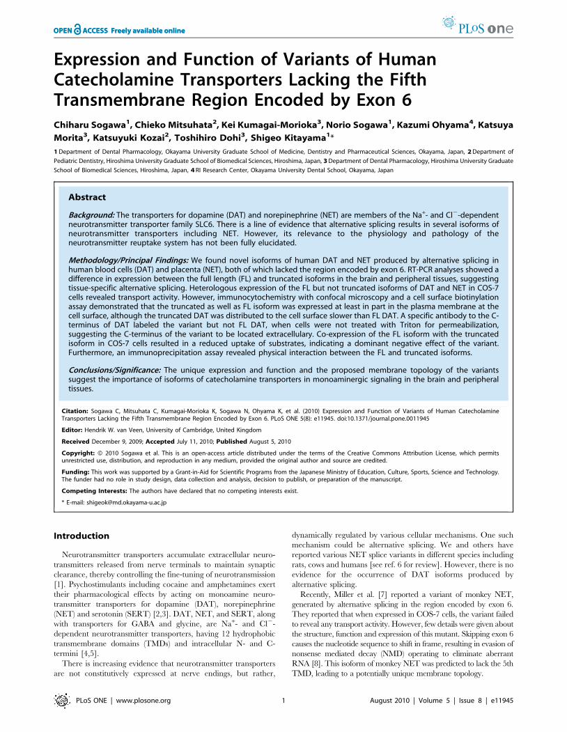
For human DAT, we found a counterpart of the splice variant
in peripheral white blood cells (Fig. 1A), where wild-type DAT
mRNA has been found [13]. Changes in nucleotide sequence of
the variant, designated hDATDEX6, were in frame due to loss of
the region encoded by exon 6. However, we could not detect
the variant in total RNA from human brain tissue, or in a cDNA
library from human substantia nigra, both obtained from
commercial sources, under any PCR conditions tested (data
not shown). Also, in preliminary experiments, attempts to
amplify the splice variant from the brain tissue of patients with
Parkinson’s disease failed (Fig. S2). The presence of DAT mRNA
and protein in the murine bowel, possibly in enteric dopami-
nergic neurons, has been reported [14], but we did not examine
the variant there.
For human NET, variant mRNA lacking the region encoded
by exon 6 was detecteded in placenta and fetal brain by RT-
PCR (Fig. 1B). Changes in the nucleotide sequence of the
variant, designated hNETDEX6, were in frame due to loss of the
region encoded by exon 6. In addition, we found another variant
lacking the region encoded by exons 5 and 6, which causes a
frame shift, resulting in a stop codon in the region encoded by
exon 7. hNETDEX6 mRNA was also detected in the adrenals
and fetal brain but not in the adult brain, suggesting a
tissue-specific and development-dependent splicing of hNET
transcripts.
Figure 1. RT-PCR analysis of the human dopamine andnorepinephrine transporter variants produced by alternativesplicing. A. Total RNA from white blood cells of three healthyvolunteers (lane 1–3) was used to synthesize first strand cDNA foranalyzing the expression of a variant of the dopamine transporter byRT-PCR and agarose gel electrophoresis. B. Expression of variants of thenorepinephrine transporter was examined in adult whole brain (lane 1),fetal whole brain (lane 2), placenta (lane 3) and SK-N-SH cells (lane 4)with controls (FL hNET, lane 5 and hNETDEX6, lane 6). The datarepresent a typical example from one experiment followed by at least 2additional experiments with similar results. M; DNA marker of 100 bpladders.doi:10.1371/journal.pone.0011945.g001
DAT and NET Splice Variants
PLoS ONE | www.plosone.org 2 August 2010 | Volume 5 | Issue 8 | e11945
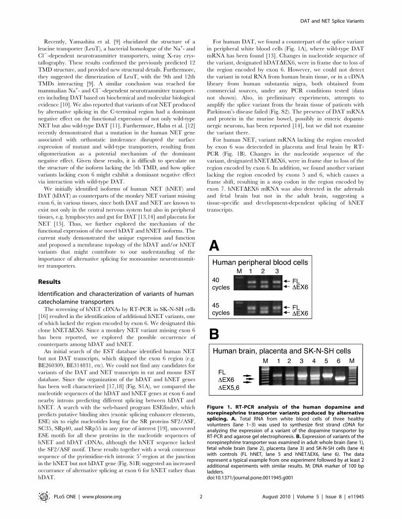
Functional characterization of human DAT and NETvariants
Figure 2A shows the uptake of [3H]DA in COS-7 cells
transiently expressing the full-length (FL) hDAT and the splice
variant, hDATDEX6. While the cells expressing FL hDAT
revealed a robust uptake of [3H]DA, hDATDEX6 failed to induce
any uptake of [3H]DA. Furthermore, no specific binding of the
cocaine analogue WIN35,428 was found in COS-7 cells
expressing hDATDEX6 (Fig. 2B).
Similarly, hNETDEX6 did not transport [3H]NE, when
transiently expressed in COS-7 cells (Fig. 2C).
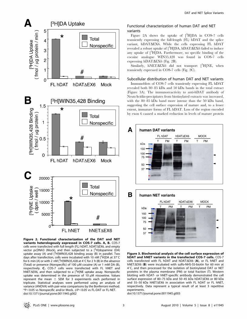
Subcellular distribution of human DAT and NET variantsImmunoblots of COS-7 cells transiently expressing FL hDAT
revealed both 80–85 kDa and 50 kDa bands in the total extract
(Figure 3A). The immunoreactivity to anti-hDAT antibody of
NeutrAvidin-precipitates from biotinylated extracts was increased,
with the 80–85 kDa band more intense than the 50 kDa band,
suggesting the cell surface expression of mature and, to a lesser
extent, immature forms of FL hDAT. Loss of the region encoded
by exon 6 caused a marked reduction in levels of mature protein
Figure 2. Functional characterization of the DAT and NETvariants heterologously expressed in COS-7 cells. A, B. COS-7cells were transfected with full length (FL) hDAT, hDATDEX6, and emptyvector pcDNA3 (Mock), and then subjected to a [3H]dopamine (DA)uptake assay (A) and [3H]WIN35,428 binding assay (B) in parallel. Twodays after transfection, cells were incubated with 10 nM [3H]DA at 37uCfor 6 min (A) or with 2 nM [3H]WIN35,428 at 4uC for 2 h (B) in the absence(Total) or presence (Nonspecific) of 100 mM cocaine (A) or 1 mM DA (B),respectively. C. COS-7 cells were transfected with FL hNET andhNETDEX6, and then subjected to a [3H]NE uptake assay. Nonspecificuptake was determined in the presence of 10 mM nisoxetine. Valuesrepresent the mean 6 SEM for 3 experiments each performed intriplicate. Statistical analyses were performed using an analysis ofvariance (ANOVA) with pair-wise comparisons by the Bonferroni method.*P,0.05 vs Nonspecific and/or Mock, #P,0.05 vs FL-DAT or FL-NET.doi:10.1371/journal.pone.0011945.g002
Figure 3. Biochemical analysis of the cell surface expression ofhDAT and hNET variants in the transfected COS-7 cells. COS-7cells transfected with FL hDAT and hDATDEX6 (A), or FL hNET andhNETDEX6 (B) were incubated with sulfo-NHS-SS-biotin for 60 min at4uC, and then processed for the isolation of biotinylated DAT or NETproteins in the plasma membrane (PM) or total fraction (T). Westernblotting with hDAT- or hNET-specific antibody demonstrated the cellsurface expression of 80–75 kDa and 50–45 kDa hDATDEX6 or 80 kDaand 55–50 kDa hNETDEX6 in association with FL hDAT or FL hNET,respectively. Data represent a typical result of at least 3 repetitiveexperiments.doi:10.1371/journal.pone.0011945.g003
DAT and NET Splice Variants
PLoS ONE | www.plosone.org 3 August 2010 | Volume 5 | Issue 8 | e11945
(80–85 kDa band) with a greater increase in levels of immature
protein (50 kDa band) in total fractions, in association with a
striking loss of mature but not immature protein in cell surface
fractions (Figure 3A). hDATDEX6 looked slightly larger than FL
hDAT in the biotinylated fraction. One explanation for this
apparent discrepancy is that the conformational change due to
truncation at TM5 reduces the mobility of hDATDEX6 in the gel.
Alternatively, hDATDEX6 may be glycosylated differently due to
the conformational change, resulting in a larger molecular weight.
Further study is needed to clarify this issue.
Cell surface biotinylation and Western blot analyses in COS
cells transfected with FL and truncated hNET cDNAs using a
specific antibody to hNET revealed results similar to those
observed for hDAT (Fig. 3B).
Immunocytochemical detection with confocal microscopy
demonstrated the transient expression of FL hDAT at the surface
of COS-7 cells and an intracellular distribution (Fig. 4A). In
contrast, hDATDEX6 was mainly located in the cytosolic
compartment with less expression at the cell surface (Fig. 4A).
We further analyzed the subcellular distribution of hDAT variants
in MDCK cells stably expressing FL hDAT and hDATDEX6.
The cell surface expression of FL hDAT and to a lesser extent,
hDATDEX6, was observed (Fig. 4B). A Z-axis analysis revealed
that hDAT localized apically (Fig. 4B) consistent with a previous
report for DAT [20], which was in contrast to the basolatelal
distribution of hNET we observed previously [21].
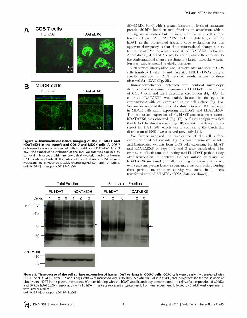
We further analyzed the time-course of the cell surface
expression of hDAT variants. Fig. 5 shows immunoblots of total
and biotinylated extracts from COS cells expressing FL hDAT
and hDATDEX6 at days 1, 2 and 3 after transfection. The
expression of both total and biotinylated FL hDAT peaked 1 day
after transfection. In contrast, the cell surface expression of
hDATDEX6 increased gradually, reaching a maximum at 3 days,
while the total protein level was constant after transfection. During
these periods, no transport activity was found in the cells
transfected with hDATDEX6 cDNA (data not shown).
Figure 4. Immunofluorescence imaging of the FL hDAT andhDATDEX6 in the transfected COS-7 and MDCK cells. A. COS-7cells were transiently transfected with FL hDAT and hDATDEX6. After 2days, the subcellular distribution of the DAT variants was assessed byconfocal microscopy with immunological detection using a humanDAT-specific antibody. B. The subcellular localization of hDAT variantswas examined in MDCK cells stably expressing FL hDAT and hDATDEX6.doi:10.1371/journal.pone.0011945.g004
Figure 5. Time-course of the cell surface expression of human DAT variants in COS-7 cells. COS-7 cells were transiently transfected withFL DAT or hDATDEX6. After 1, 2, and 3 days, cells were incubated with sulfo-NHS-SS-biotin for 120 min at 4uC, and then processed for the isolation ofbiotinylated hDAT in the plasma membrane. Western blotting with the hDAT-specific antibody demonstrated the cell surface expression of 80 kDaand 50 kDa hDATDEX6 in association with FL hDAT. The data represent a typical result from one experiment followed by 2 additional experimentswith similar results.doi:10.1371/journal.pone.0011945.g005
DAT and NET Splice Variants
PLoS ONE | www.plosone.org 4 August 2010 | Volume 5 | Issue 8 | e11945
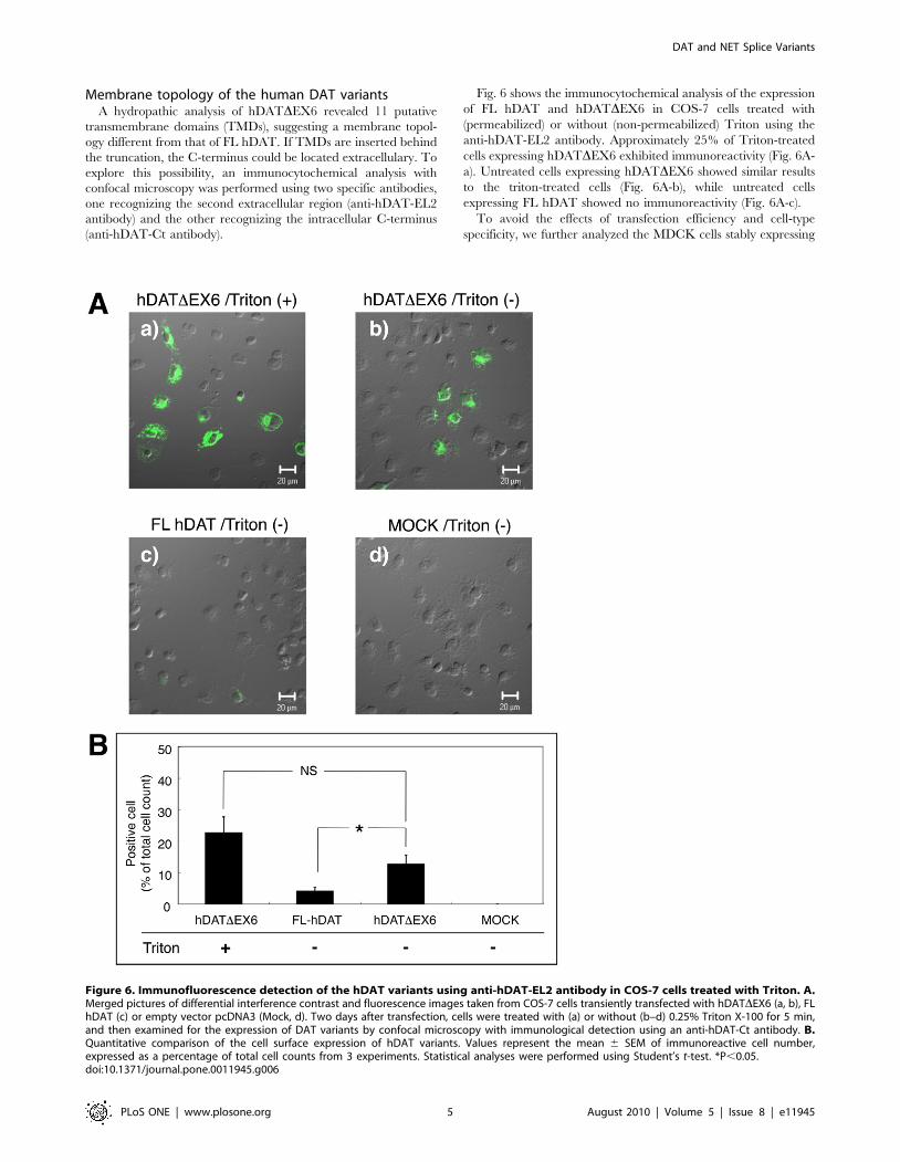
Membrane topology of the human DAT variantsA hydropathic analysis of hDATDEX6 revealed 11 putative
transmembrane domains (TMDs), suggesting a membrane topol-
ogy different from that of FL hDAT. If TMDs are inserted behind
the truncation, the C-terminus could be located extracellulary. To
explore this possibility, an immunocytochemical analysis with
confocal microscopy was performed using two specific antibodies,
one recognizing the second extracellular region (anti-hDAT-EL2
antibody) and the other recognizing the intracellular C-terminus
(anti-hDAT-Ct antibody).
Fig. 6 shows the immunocytochemical analysis of the expression
of FL hDAT and hDATDEX6 in COS-7 cells treated with
(permeabilized) or without (non-permeabilized) Triton using the
anti-hDAT-EL2 antibody. Approximately 25% of Triton-treated
cells expressing hDATDEX6 exhibited immunoreactivity (Fig. 6A-
a). Untreated cells expressing hDATDEX6 showed similar results
to the triton-treated cells (Fig. 6A-b), while untreated cells
expressing FL hDAT showed no immunoreactivity (Fig. 6A-c).
To avoid the effects of transfection efficiency and cell-type
specificity, we further analyzed the MDCK cells stably expressing
Figure 6. Immunofluorescence detection of the hDAT variants using anti-hDAT-EL2 antibody in COS-7 cells treated with Triton. A.Merged pictures of differential interference contrast and fluorescence images taken from COS-7 cells transiently transfected with hDATDEX6 (a, b), FLhDAT (c) or empty vector pcDNA3 (Mock, d). Two days after transfection, cells were treated with (a) or without (b–d) 0.25% Triton X-100 for 5 min,and then examined for the expression of DAT variants by confocal microscopy with immunological detection using an anti-hDAT-Ct antibody. B.Quantitative comparison of the cell surface expression of hDAT variants. Values represent the mean 6 SEM of immunoreactive cell number,expressed as a percentage of total cell counts from 3 experiments. Statistical analyses were performed using Student’s t-test. *P,0.05.doi:10.1371/journal.pone.0011945.g006
DAT and NET Splice Variants
PLoS ONE | www.plosone.org 5 August 2010 | Volume 5 | Issue 8 | e11945
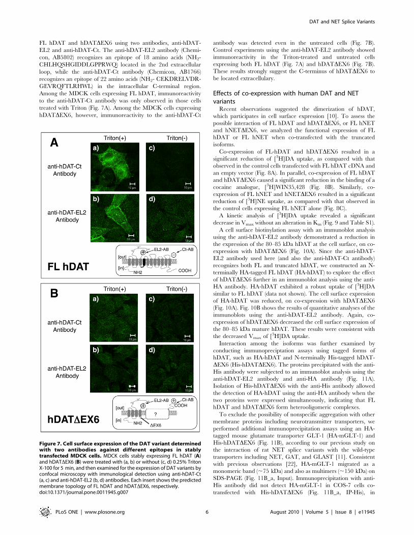
FL hDAT and hDATDEX6 using two antibodies, anti-hDAT-
EL2 and anti-hDAT-Ct. The anti-hDAT-EL2 antibody (Chemi-
con, AB5802) recognizes an epitope of 18 amino acids (NH2-
CHLHQSHGIDDLGPPRWQ) located in the 2nd extracellular
loop, while the anti-hDAT-Ct antibody (Chemicon, AB1766)
recognizes an epitope of 22 amino acids (NH2- CEKDRELVDR-
GEVRQFTLRHWL) in the intracellular C-terminal region.
Among the MDCK cells expressing FL hDAT, immunoreactivity
to the anti-hDAT-Ct antibody was only observed in those cells
treated with Triton (Fig. 7A). Among the MDCK cells expressing
hDATDEX6, however, immunoreactivity to the anti-hDAT-Ct
antibody was detected even in the untreated cells (Fig. 7B).
Control experiments using the anti-hDAT-EL2 antibody showed
immunoreactivity in the Triton-treated and untreated cells
expressing both FL hDAT (Fig. 7A) and hDATDEX6 (Fig. 7B).
These results strongly suggest the C-terminus of hDATDEX6 to
be located extracellulary.
Effects of co-expression with human DAT and NETvariants
Recent observations suggested the dimerization of hDAT,
which participates in cell surface expression [10]. To assess the
possible interaction of FL hDAT and hDATDEX6, or FL hNET
and hNETDEX6, we analyzed the functional expression of FL
hDAT or FL hNET when co-transfected with the truncated
isoforms.
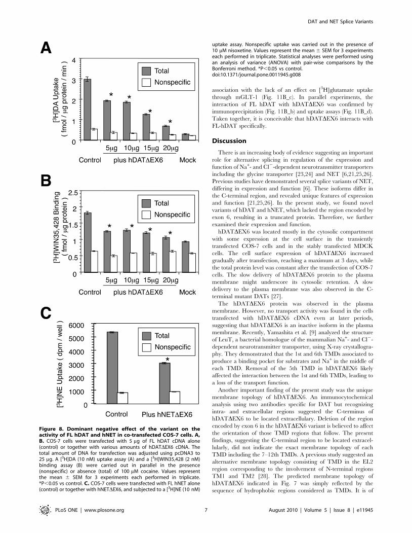
Co-expression of FL-hDAT and hDATDEX6 resulted in a
significant reduction of [3H]DA uptake, as compared with that
observed in the control cells transfected with FL hDAT cDNA and
an empty vector (Fig. 8A). In parallel, co-expression of FL hDAT
and hDATDEX6 caused a significant reduction in the binding of a
cocaine analogue, [3H]WIN35,428 (Fig. 8B). Similarly, co-
expression of FL hNET and hNETDEX6 resulted in a significant
reduction of [3H]NE uptake, as compared with that observed in
the control cells expressing FL hNET alone (Fig. 8C).
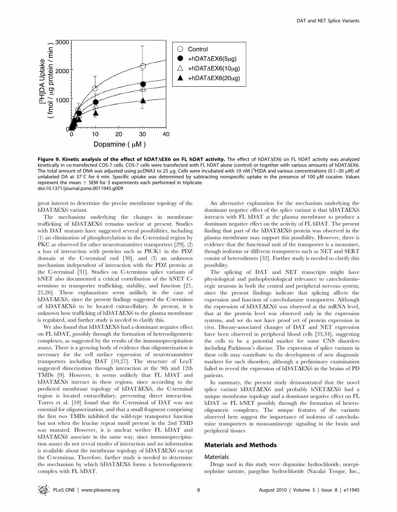
A kinetic analysis of [3H]DA uptake revealed a significant
decrease in Vmax without an alteration in Km (Fig. 9 and Table S1).
A cell surface biotinylation assay with an immunoblot analysis
using the anti-hDAT-EL2 antibody demonstrated a reduction in
the expression of the 80–85 kDa hDAT at the cell surface, on co-
expression with hDATDEX6 (Fig. 10A). Since the anti-hDAT-
EL2 antibody used here (and also the anti-hDAT-Ct antibody)
recognizes both FL and truncated hDAT, we constructed an N-
terminally HA-tagged FL hDAT (HA-hDAT) to explore the effect
of hDATDEX6 further in an immunoblot analysis using the anti-
HA antibody. HA-hDAT exhibited a robust uptake of [3H]DA
similar to FL hDAT (data not shown). The cell surface expression
of HA-hDAT was reduced, on co-expression with hDATDEX6
(Fig. 10A). Fig. 10B shows the results of quantitative analyses of the
immunoblots using the anti-hDAT-EL2 antibody. Again, co-
expression of hDATDEX6 decreased the cell surface expression of
the 80–85 kDa mature hDAT. These results were consistent with
the decreased Vmax of [3H]DA uptake.
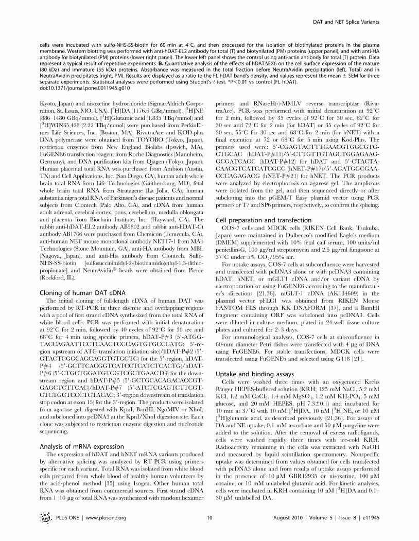
Interaction among the isoforms was further examined by
conducting immunopreciptation assays using tagged forms of
hDAT, such as HA-hDAT and N-terminally His-tagged hDAT-
DEX6 (His-hDATDEX6). The proteins precipitated with the anti-
His antibody were subjected to an immunoblot analysis using the
anti-hDAT-EL2 antibody and anti-HA antibody (Fig. 11A).
Isolation of His-hDATDEX6 with the anti-His antibody allowed
the detection of HA-hDAT using the anti-HA antibody when the
two proteins were expressed simultaneously, indicating that FL
hDAT and hDATDEX6 form heterooligomeric complexes.
To exclude the possibility of nonspecific aggregation with other
membrane proteins including neurotransmitter transporters, we
performed additional immunoprecipitation assays using an HA-
tagged mouse glutamate transporter GLT-1 (HA-mGLT-1) and
His-hDATDEX6 (Fig. 11B), according to our previous study on
the interaction of rat NET splice variants with the wild-type
transporters including NET, GAT, and GLAST [11]. Consistent
with previous observations [22], HA-mGLT-1 migrated as a
monomeric band (,75 kDa) and also as multimers (,150 kDa) on
SDS-PAGE (Fig. 11B_a, Input). Immunoprecipitation with anti-
His antibody did not detect HA-mGLT-1 in COS-7 cells co-
transfected with His-hDATDEX6 (Fig. 11B_a, IP-His), in
Figure 7. Cell surface expression of the DAT variant determinedwith two antibodies against different epitopes in stablytransfected MDCK cells. MDCK cells stably expressing FL hDAT (A)and hDATDEX6 (B) were treated with (a, b) or without (c, d) 0.25% TritonX-100 for 5 min, and then examined for the expression of DAT variants byconfocal microscopy with immunological detection using anti-hDAT-Ct(a, c) and anti-hDAT-EL2 (b, d) antibodies. Each insert shows the predictedmembrane topology of FL hDAT and hDATDEX6, respectively.doi:10.1371/journal.pone.0011945.g007
DAT and NET Splice Variants
PLoS ONE | www.plosone.org 6 August 2010 | Volume 5 | Issue 8 | e11945
association with the lack of an effect on [3H]glutamate uptake
through mGLT-1 (Fig. 11B_c). In parallel experiments, the
interaction of FL hDAT with hDATDEX6 was confirmed by
immunoprecipitation (Fig. 11B_b) and uptake assays (Fig. 11B_d).
Taken together, it is conceivable that hDATDEX6 interacts with
FL-hDAT specifically.
Discussion
There is an increasing body of evidence suggesting an important
role for alternative splicing in regulation of the expression and
function of Na+- and Cl2-dependent neurotransmitter transporters
including the glycine transporter [23,24] and NET [6,21,25,26].
Previous studies have demonstrated several splice variants of NET,
differing in expression and function [6]. These isoforms differ in
the C-terminal region, and revealed unique features of expression
and function [21,25,26]. In the present study, we found novel
variants of hDAT and hNET, which lacked the region encoded by
exon 6, resulting in a truncated protein. Therefore, we further
examined their expression and function.
hDATDEX6 was located mostly in the cytosolic compartment
with some expression at the cell surface in the transiently
transfected COS-7 cells and in the stably transfected MDCK
cells. The cell surface expression of hDATDEX6 increased
gradually after transfection, reaching a maximum at 3 days, while
the total protein level was constant after the transfection of COS-7
cells. The slow delivery of hDATDEX6 protein to the plasma
membrane might underscore its cytosolic retention. A slow
delivery to the plasma membrane was also observed in the C-
terminal mutant DATs [27].
The hDATDEX6 protein was observed in the plasma
membrane. However, no transport activity was found in the cells
transfected with hDATDEX6 cDNA even at later periods,
suggesting that hDATDEX6 is an inactive isoform in the plasma
membrane. Recently, Yamashita et al. [9] analyzed the structure
of LeuT, a bacterial homologue of the mammalian Na+- and Cl2-
dependent neurotransmitter transporter, using X-ray crystallogra-
phy. They demonstrated that the 1st and 6th TMDs associated to
produce a binding pocket for substrates and Na+ in the middle of
each TMD. Removal of the 5th TMD in hDATDEX6 likely
affected the interaction between the 1st and 6th TMDs, leading to
a loss of the transport function.
Another important finding of the present study was the unique
membrane topology of hDATDEX6. An immunocytochemical
analysis using two antibodies specific for DAT but recognizing
intra- and extracellular regions suggested the C-terminus of
hDATDEX6 to be located extracellulary. Deletion of the region
encoded by exon 6 in the hDATDEX6 variant is believed to affect
the orientation of those TMD regions that follow. The present
findings, suggesting the C-terminal region to be located extracel-
lularly, did not indicate the exact membrane topology of each
TMD including the 7–12th TMDs. A previous study suggested an
alternative membrane topology consisting of TMD in the EL2
region corresponding to the involvement of N-terminal regions
TM1 and TM2 [28]. The predicted membrane topology of
hDATDEX6 indicated in Fig. 7 was simply reflected by the
sequence of hydrophobic regions considered as TMDs. It is of
Figure 8. Dominant negative effect of the variant on theactivity of FL hDAT and hNET in co-transfected COS-7 cells. A,B. COS-7 cells were transfected with 5 mg of FL hDAT cDNA alone(control) or together with various amounts of hDATDEX6 cDNA. Thetotal amount of DNA for transfection was adjusted using pcDNA3 to25 mg. A [3H]DA (10 nM) uptake assay (A) and a [3H]WIN35,428 (2 nM)binding assay (B) were carried out in parallel in the presence(nonspecific) or absence (total) of 100 mM cocaine. Values representthe mean 6 SEM for 3 experiments each performed in triplicate.*P,0.05 vs control. C. COS-7 cells were transfected with FL hNET alone(control) or together with hNETDEX6, and subjected to a [3H]NE (10 nM)
uptake assay. Nonspecific uptake was carried out in the presence of10 mM nisoxetine. Values represent the mean 6 SEM for 3 experimentseach performed in triplicate. Statistical analyses were performed usingan analysis of variance (ANOVA) with pair-wise comparisons by theBonferroni method. *P,0.05 vs control.doi:10.1371/journal.pone.0011945.g008
DAT and NET Splice Variants
PLoS ONE | www.plosone.org 7 August 2010 | Volume 5 | Issue 8 | e11945
great interest to determine the precise membrane topology of the
hDATDEX6 variant.
The mechanism underlying the changes in membrane
trafficking of hDATDEX6 remains unclear at present. Studies
with DAT mutants have suggested several possibilities, including
(1) an elimination of phosphorylation in the C-terminal region by
PKC as observed for other neurotransmitter transporters [29], (2)
a loss of interaction with proteins such as PICK1 in the PDZ
domain at the C-terminal end [30], and (3) an unknown
mechanism independent of interaction with the PDZ protein at
the C-terminal [31]. Studies on C-terminus splice variants of
hNET also documented a critical contribution of the hNET C-
terminus to transporter trafficking, stability, and function [21,
25,26]. These explanations seem unlikely in the case of
hDATDEX6, since the present findings suggested the C-terminus
of hDATDEX6 to be located extracellulary. At present, it is
unknown how trafficking of hDATDEX6 to the plasma membrane
is regulated, and further study is needed to clarify this.
We also found that hDATDEX6 had a dominant negative effect
on FL hDAT, possibly through the formation of heterooligomeric
complexes, as suggested by the results of the immunoprecipitation
assays. There is a growing body of evidence that oligomerization is
necessary for the cell surface expression of neurotransmitter
transporters including DAT [10,27]. The structure of LeuT
suggested dimerization through interaction at the 9th and 12th
TMDs [9]. However, it seems unlikely that FL hDAT and
hDATDEX6 interact in these regions, since according to the
predicted membrane topology of hDATDEX6, the C-terminal
region is located extracellulary, preventing direct interaction.
Torres et al. [10] found that the C-terminal of DAT was not
essential for oligomerization, and that a small fragment comprising
the first two TMDs inhibited the wild-type transporter function
but not when the leucine repeat motif present in the 2nd TMD
was mutated. However, it is unclear wether FL hDAT and
hDATDEX6 associate in the same way, since immunoprecipita-
tion assays do not reveal modes of interaction and no information
is available about the membrane topology of hDATDEX6 except
the C-terminus. Therefore, further study is needed to determine
the mechanism by which hDATDEX6 forms a heterooligomeric
complex with FL hDAT.
An alternative explanation for the mechanism underlying the
dominant negative effect of the splice variant is that hDATDEX6
interacts with FL hDAT at the plasma membrane to produce a
dominant negative effect on the activity of FL hDAT. The present
finding that part of the hDATDEX6 protein was observed in the
plasma membrane may support this possibility. However, there is
evidence that the functional unit of the transporter is a monomer,
though isoforms or different transporters such as NET and SERT
consist of heterodimers [32]. Further study is needed to clarify this
possibility.
The splicing of DAT and NET transcripts might have
physiological and pathophysiological relevance to catecholamin-
ergic neurons in both the central and peripheral nervous system,
since the present findings indicate that splicing affects the
expression and function of catecholamine transporters. Although
the expression of hDATDEX6 was observed at the mRNA level,
that at the protein level was observed only in the expression
systems, and we do not have proof yet of protein expression in
vivo. Disease-associated changes of DAT and NET expression
have been observed in peripheral blood cells [33,34], suggesting
the cells to be a potential marker for some CNS disorders
including Parkinson’s disease. The expression of splice variants in
these cells may contribute to the development of new diagnostic
markers for such disorders, although a preliminary examination
failed to reveal the expression of hDATDEX6 in the brains of PD
patients.
In summary, the present study demonstrated that the novel
splice variant hDATDEX6 and probably hNETDEX6 had a
unique membrane topology and a dominant negative effect on FL
hDAT or FL hNET possibly through the formation of hetero-
oligomeric complexes. The unique features of the variants
observed here suggest the importance of isoforms of catechola-
mine transporters in monoaminergic signaling in the brain and
peripheral tissues.
Materials and Methods
MaterialsDrugs used in this study were dopamine hydrochloride, norepi-
nephrine tartrate, pargyline hydrochloride (Nacalai Tesque, Inc.,
Figure 9. Kinetic analysis of the effect of hDATDEX6 on FL hDAT activity. The effect of hDATDEX6 on FL hDAT activity was analyzedkinetically in co-transfected COS-7 cells. COS-7 cells were transfected with FL hDAT alone (control) or together with various amounts of hDATDEX6.The total amount of DNA was adjusted using pcDNA3 to 25 mg. Cells were incubated with 10 nM [3H]DA and various concentrations (0.1–30 mM) ofunlabeled DA at 37uC for 6 min. Specific uptake was determined by subtracting nonspecific uptake in the presence of 100 mM cocaine. Valuesrepresent the mean 6 SEM for 3 experiments each performed in triplicate.doi:10.1371/journal.pone.0011945.g009
DAT and NET Splice Variants
PLoS ONE | www.plosone.org 8 August 2010 | Volume 5 | Issue 8 | e11945
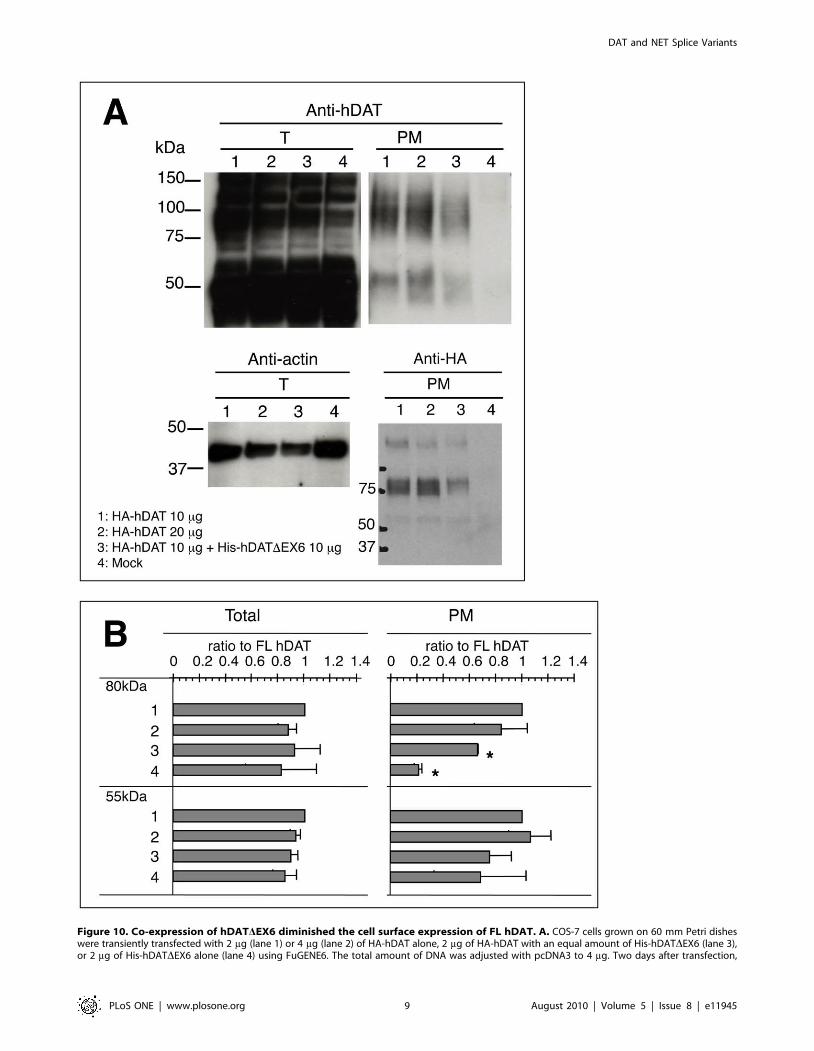
Figure 10. Co-expression of hDATDEX6 diminished the cell surface expression of FL hDAT. A. COS-7 cells grown on 60 mm Petri disheswere transiently transfected with 2 mg (lane 1) or 4 mg (lane 2) of HA-hDAT alone, 2 mg of HA-hDAT with an equal amount of His-hDATDEX6 (lane 3),or 2 mg of His-hDATDEX6 alone (lane 4) using FuGENE6. The total amount of DNA was adjusted with pcDNA3 to 4 mg. Two days after transfection,
DAT and NET Splice Variants
PLoS ONE | www.plosone.org 9 August 2010 | Volume 5 | Issue 8 | e11945
Kyoto, Japan) and nisoxetine hydrochloride (Sigma-Aldrich Corpo-
ration, St. Louis, MO, USA). [3H]DA (1176.6 GBq/mmol), [3H]NE
(886–1480 GBq/mmol), [3H]Glutamic acid (1.835 TBq/mmol) and
[3H]WIN35,428 (2.22 TBq/mmol) were purchased from PerkinEl-
mer Life Sciences, Inc. (Boston, MA). RivatraAce and KOD-plus
DNA polymerase were obtained from TOYOBO (Tokyo, Japan),
restriction enzymes from New England Biolabs (Ipswich, MA),
FuGENE6 transfection reagent from Roche Diagnostics (Mannheim,
Germany), and DNA purification kits from Qiagen (Tokyo, Japan).
Human placental total RNA was purchased from Ambion (Austin,
TX) and Cell Applications, Inc. (San Diego, CA), human adult whole
brain total RNA from Life Technologies (Gaithersburg, MD), fetal
whole brain total RNA from Stratagene (La Jolla, CA), human
substantia nigra total RNA of Parkinson’s disease patients and normal
subjects from Clontech (Palo Alto, CA), and cDNA from human
adult adrenal, cerebral cortex, pons, cerebellum, medulla oblongata
and placenta from Biochain Institute, Inc. (Hayward, CA). The
rabbit anti-hDAT-EL2 antibody AB5802 and rabbit anti-hDAT-Ct
antibody AB1766 were purchased from Chemicon (Temecula, CA),
anti-human NET mouse monoclonal antibody NET17-1 from MAb
Technologies (Stone Mountain, GA), anti-HA antibody from MBL
(Nagoya, Japan), and anti-His antibody from Clontech. Sulfo-
NHS-SS-biotin [sulfosuccinimidyl-2-(biotinamido)ethyl-1,3-dithio-
propionate] and NeutrAvidinH beads were obtained from Pierce
(Rockford, IL).
Cloning of human DAT cDNAThe initial cloning of full-length cDNA of human DAT was
performed by RT-PCR in three discrete and overlapping regions
with a pool of first strand cDNA synthesized from the total RNA of
white blood cells. PCR was performed with initial denaturation
at 92uC for 2 min, followed by 40 cycles of 92uC for 30 sec and
68uC for 4 min using specific primers, hDAT-P#3 (59-ATGG-
TACCAGAATTCCTCAACTCCCAGTGTGCCCATG; 59-re-
gion upstream of ATG translation initiation site)/hDAT-P#2 (59-
GTACTCGGCAGCAGGTGTGGTC) for the 59-region, hDAT-
P#4 (59-GCTTCACGGTCATCCTCATCTCACTG)/hDAT-
P#6 (59-CTGCTGGATGTCGTCGCTGAACTG) for the down-
stream region and hDAT-P#5 (59-GCTGCACAGACACCGT-
GAGCTCTTCAC)/hDAT-P#7 (59-ATCTCGAGTCTTCGT-
CTCTGCTCCCTCTACAC; 39-region downstream of translation
stop codon at exon 15) for the 39-region. The products were isolated
from agarose gel, digested with KpnI, BanHI, NgoMIV or XhoI,
and subcloned into pcDNA3 at the KpnI/XhoI digestion site. Each
clone was subjected to restriction enzyme digestion and nucleotide
sequencing.
Analysis of mRNA expressionThe expression of hDAT and hNET mRNA variants produced
by alternative splicing was analyzed by RT-PCR using primers
specific for each variant. Total RNA was isolated from white blood
cells prepared from whole blood of healthy human volunteers by
the acid-phenol method [35] using Isogen. Other human total
RNA was obtained from commercial sources. First strand cDNA
from 1–10 mg of total RNA was synthesized with random hexamer
primers and RNaseH(-)-MMLV reverse transcriptase (Riva-
traAce). PCR was performed with initial denaturation at 92uCfor 2 min, followed by 35 cycles of 92uC for 30 sec, 62uC for
30 sec and 72uC for 2 min (for hDAT) or 35 cycles of 92uC for
30 sec, 55uC for 30 sec and 68uC for 2 min (for hNET) with a
final extention at 72 or 68uC for 5 min using Kod-Plus. The
primers used were: 59-CGAGTACTTTGAACGTGGCGTG-
CTGCAC (hDAT-P#11)/59-CTTGTTGTAGCTGGAGAAG-
GCGATCAGC (hDAT-P#12) for hDAT and 59-CTACTA-
CAACGTCATCATCGCC (hNET-P#17)/59-AGATGGCGAA-
CCCAGAGACG (hNET-P#21) for hNET. The PCR products
were analyzed by electrophoresis on agarose gel. The amplicons
were isolated from the gel, and then sequenced directly or after
subcloning into the pGEM-T Easy plasmid vector using PCR
primers or T7 and SP6 primers, respectively, to confirm the splicing.
Cell preparation and transfectionCOS-7 cells and MDCK cells (RIKEN Cell Bank, Tsukuba,
Japan) were maintained in Dulbecco’s modified Eagle’s medium
(DMEM) supplemented with 10% fetal calf serum, 100 units/ml
penicillin-G, 100 mg/ml streptomycin and 2.5 mg/ml fungisone at
37uC under 5% CO2/95% air.
For uptake assays, COS-7 cells at subconfluence were harvested
and transfected with pcDNA3 alone or with pcDNA3 containing
hDAT, hNET, or mGLT1 cDNA and/or variant cDNA by
electroporation or using FuGENE6 according to the manufactur-
er’s directions [21,36]. mGLT-1 cDNA (AK134609) in the
plasmid vector pFLC1 was obtained from RIKEN Mouse
FANTOM FLS through KK DNAFORM [37], and a BamHI
fragment containing ORF was subcloned into pcDNA3. Cells
were diluted in culture medium, plated in 24-well tissue culture
plates and cultured for 2–3 days.
For immunological analyses, COS-7 cells at subconfluence in
60-mm diameter Petri dishes were transfected with 4 mg of DNA
using FuGENE6. For stable transfections, MDCK cells were
transfected using FuGENE6 and selected using G418 [21].
Uptake and binding assaysCells were washed three times with an oxygenated Krebs
Ringer HEPES-buffered solution (KRH; 125 mM NaCl, 5.2 mM
KCl, 1.2 mM CaCl2, 1.4 mM MgSO4, 1.2 mM KH2PO4, 5 mM
glucose, and 20 mM HEPES, pH 7.360.1) and incubated for
10 min at 37uC with 10 nM [3H]DA, 10 nM [3H]NE, or 10 nM
[3H]glutamic acid, as described previously [21,36]. For assays of
DA and NE uptake, 0.1 mM ascorbate and 50 mM pargyline were
added to the solution. After the removal of excess radioligands,
cells were washed rapidly three times with ice-cold KRH.
Radioactivity remaining in the cells was extracted with NaOH
and measured by liquid scintillation spectrometry. Nonspecific
uptake was determined from values obtained for cells transfected
with pcDNA3 alone and from results of uptake assays performed
in the presence of 10 mM GBR12935 or nisoxetine, 100 mM
cocaine, or 10 mM unlabeled glutamic acid. For kinetic analyses,
cells were incubated in KRH containing 10 nM [3H]DA and 0.1–
30 mM unlabelled DA.
cells were incubated with sulfo-NHS-SS-biotin for 60 min at 4uC, and then processed for the isolation of biotinylated proteins in the plasmamembrane. Western blotting was performed with anti-hDAT-EL2 antibody for total (T) and biotynilated (PM) proteins (upper panel), and with anti-HAantibody for biotynilated (PM) proteins (lower right panel). The lower left panel shows the control using anti-actin antibody for total (T) protein. Datarepresent a typical result of repetitive experiments. B. Quantitative analysis of the effects of hDATDEX6 on the cell surface expression of the mature(80 kDa) and immature (55 kDa) proteins. Absorbance was measured in the total fraction before NeutraAvidin precipitation (left, Total) and inNeutraAvidin precipitates (right, PM). Results are displayed as a ratio to the FL hDAT band’s density, and values represent the mean 6 SEM for threeseparate experiments. Statistical analyses were performed using Student’s t-test. *P,0.01 vs control (FL hDAT).doi:10.1371/journal.pone.0011945.g010
DAT and NET Splice Variants
PLoS ONE | www.plosone.org 10 August 2010 | Volume 5 | Issue 8 | e11945
Figure 11. hDATDEX6 physically interacts with FL hDAT. A. COS-7 cells grown on 60 mm Petri dishes were transfected with 2 mg of HA-hDATalone (lane 1), 2 mg of His-hDATDEX6 (lane 2), 2 mg of HA-hDAT with an equal amount of His-hDATDEX6 (lane 3), or 4 mg of empty vector pcDNA3.The total amount of DNA was adjusted with pcDNA3 to 4 mg. Two days after transfection, HA-hDAT was co-immunoprecipitated with His-hDATDEX6using anti-His antibody. A western blot analysis of the immunoprecipitates obtained with the anti-His antibody was performed using anti-hDAT-EL2antibody (left) and anti-HA antibody (right). The data represent a typical result from one experiment followed by 2 additional experiments withsimilar results. B. (a, b) COS-7 cells grown on 60 mm Petri dishes were transfected with 0.25 mg of HA-mGLT1 alone (lane 1), 0.25 mg of HA-mGLT1with an equal amount of His-hDATDEX6 (lane 2), 2 mg of empty vector pcDNA3 (lane 3), 0.25 mg of HA-hDAT alone (lane 4), or 0.25 mg of HA-hDATwith an equal amount of His-hDATDEX6 (lane 5). The total amount of DNA was adjusted with pcDNA3 to 2 mg. Two days after transfection, HA-mGLT1 (a) or HA-hDAT (b) was co-immunoprecipitated with His-hDATDEX6 using anti-His antibody. A western blot analysis of theimmunoprecipitates obtained with the anti-His antibody was performed using anti-HA antibody. (c, d) In parallel experiments, COS-7 cellstransfected with HA-mGLT1 alone (control) or together with His-hDATDEX6 (c), or those transfected with HA-hDAT alone (control) or together with
DAT and NET Splice Variants
PLoS ONE | www.plosone.org 11 August 2010 | Volume 5 | Issue 8 | e11945
For binding assays, cells were incubated with 2 nM
[3H]WIN35,428 in KRH buffer at 4uC (on ice) for 2 h in the
absence (total binding) or presence (nonspecific binding) of 1 mM
DA [36].
Kinetic analyses of uptake data and Eadie-Hofstee plots were
conducted using Prism 3 (GraphPad Softwares, San Diego, CA).
Statistical analyses were performed using an analysis of variance
(ANOVA) with pair-wise comparisons by the Bonferroni method.
Immunocytochemistry and confocal microscopyFor immunostaining experiments, MDCK cells were grown on
Costar TranswellH filter supports (Corning, Acton, MA) at 26105
cells/well. The cells were initially rinsed with Ca2+- and Mg2+-
containing PBS, and then fixed in 4% paraformaldehyde. After
three washes with PBS, cells were permeabilized in PBS
containing 0.25% Triton X-100 for 5 min, and incubated in
blocking solution (2% goat serum) for 30 min. Cells were
incubated with a rabbit anti-hDAT polyclonal antibody (anti-
hDAT-Ct antibody (1:500) or anti-hDAT-EL2 antibody (1:250))
overnight at 4uC, followed by a FITC-conjugated anti-rabbit
secondary antibody. Cells were then washed three times with PBS,
and the filter with cells was excised from its support and mounted
on a glass slide with Perma FluorH aqueous mounting medium
(Thermo Shandon, Pittsburgh, PA). Immunostaining of COS-7
cells grown on Falcon BiocoatH culture slides (Becton Dickinson
Labware, Bedford, MA) was performed as above. After a final
wash, the cells were covered with a coverslip and mounting
medium. Immunofluorescent images were generated using a Zeiss
laser scanning confocal microscope (LSM510, Central Research
Laboratory, Okayama University Medical School). The z-series
(for MDCK cells) was collected by focusing initially on the middle
section of the cells (position 0) and scanning 6 mm above to 6 mm
below this plane at 0.2 mm intervals.
Cell surface Biotinylation and Western blottingCells were washed with ice cold Ca2+- and Mg2+-containing
PBS and incubated for 1 h at 4uC with 1 mg/ml sulfo-NHS-SS-
biotin. Cells were then washed with PBS, collected and solubilized
in RIPA buffer, and incubated at 4uC for 30 min. Non lysates
were removed by centrifugation for 20 min at 12000 rpm, and an
aliquot of each sample was used for the isolation of biotinylated
proteins with NeutrAvidinH beads by incubating overnight at 4uCwith gentle shaking. The precipitated proteins were then washed
five times with RIPA buffer, and denatured by heating the beads
in sample buffer at 95uC for 5 min. Samples were analyzed by
SDS-PAGE (5–20% gel, BioRad, Tokyo, Japan), and transferred
onto a PVDF membrane (GE Healthcare Biosciences, Buck-
inghamshire, UK). Western blotting was performed with poly-
clonal rabbit antibodies to hDAT or a monoclonal mouse
antibody to hNET followed by an HRP-conjugated secondary
antibodies, and detected on autoradiographic film. The quantifi-
cation of signals was performed using densitometry and NIH
Image software, as described [21]. Statistical analyses were
performed using Student’s t-test.
ImmunoprecipitationCells co-expressing HA-hDAT and His-hDATDEX6, or those
co-expressing HA-mGLT1 and His-hDATDEX6 were homoge-
nized with RIPA buffer, and the lysates were subjected to
immunoprecipitation overnight at 4uC with 0.5 mg of anti-His
antibody, followed by 1 h of incubation with a 50% slurry of
protein-A Sepharose beads (GE Healthcare Biosciences). Beads
were washed four times with RIPA buffer, and the proteins bound
to the beads were eluted with loading buffer and subjected to SDS-
PAGE, as described above. Immunoblot analyses of the precip-
itates were carried out using the anti-hDAT-EL2 antibody and
anti-HA antibody.
Supporting Information
Figure S1 Schematic representation of the organization of the
human DAT and NET genes (A), and comparison of DAT/NET
genes at the exon 6 - intron5 boundary (B). A. Boxes and bars
represent exons and introns, respectively. A translation initiation
codon exists in exon 2. The arrow in exon 6 represents an
alternative splicing site. Insert shows a scheme of the alternative
splicing. The arrowhead indicates positions of the primers used. B.
The position of exon 6 (capital letters) was assigned based on the
consensus AG-GT (underlined) in the adjacent introns 5 and 6
(small letter). The pyrimidine-rich consensus sequence for splicing
is shaded, a possible exonic splicing enhancer (ESE) motif for the
SR protein SF2/ASF is underlined by a dotted line, and the GA-
rich ESE motif is boxed.
Found at: doi:10.1371/journal.pone.0011945.s001 (0.86 MB TIF)
Figure S2 RT-PCR analysis of the expression of the hDAT
variant in Substantia nigra from normal subjects and Parkinson
disease patients. Total RNA from human Substantia nigra of
patients with Parkinson’s disease (PD) or normal subjects (Cont)
was obtained commercially, and used to synthesize first strand
cDNA (RT+, with RivatraAce; RT-, without RivatraAce). PCR
was performed with initial denaturation at 94uC for 2 min,
followed by 40 cycles of 92uC for 30 sec and 68uC for 2 min with a
final extention at 68uC for 5 min using Kod-Plus. The primers
used were: 59-CGAGTACTTTGAACGTGGCGTGCTGCAC
(hDAT-P#11)/59-GTGGTGACAATCGCGTCCCTGTAGCA-
G (hDAT-P#10). The PCR products were analyzed by electro-
phoresis on agarose gel. C: FL hDAT, V: hDATDEX6, N:
negative control (water as a template), M: DNA marker of 100 bp.
Found at: doi:10.1371/journal.pone.0011945.s002 (0.66 MB
TIF)
Table S1 Kinetic analysis of the effect of hDATDEX6 on hDAT
activity in co-transfected COS-7 cells. COS-7 cells were
transfected with the full-length (FL) hDAT alone (control) or with
various amounts of the splice variant hDATDEX6. The total
amount of DNA for transfection was adjusted with pcDNA3 to
25 mg. Uptake assays were carried out by incubating cells with
10 nM [3H]dopamine in the presence of various concentrations
(0.1–30 mM) of unlabelled DA at 37uC for 6 min. Specific uptake
was determined by subtracting the nonspecific uptake measured in
the presence of 100 mM cocaine. Values represent the mean 6
SEM for 3 experiments each performed in triplicate. Vmax was
expressed as a ratio to the control (FL hDAT alone) value, which
was 2.0360.55 fmol/mg protein/min. *Significantly different from
control at P,0.05.
Found at: doi:10.1371/journal.pone.0011945.s003 (0.03 MB
DOC)
His-hDATDEX6 (d) were subjected to uptake assays for [3H]glutamic acid (10 nM) and [3H]DA (10 nM), respectively. Values represent the mean 6 SEMfor three experiments each performed in duplicate, and are expressed as a percentage of HA-mGLT alone (94976960 dpm/well/10 min) or HA-hDATalone (4276965903 dpm/well/10 min). Statistical analyses were performed using the Mann-Whitney U-test. *P,0.05 vs control.doi:10.1371/journal.pone.0011945.g011
DAT and NET Splice Variants
PLoS ONE | www.plosone.org 12 August 2010 | Volume 5 | Issue 8 | e11945
Author Contributions
Conceived and designed the experiments: CS CM KKM SK. Performed
the experiments: CS CM KKM NS KO KM SK. Analyzed the data: CS
CM KKM NS KO KM KK TD SK. Contributed reagents/materials/
analysis tools: KK TD SK. Wrote the paper: CS TD SK.
References
1. Iversen LL (1971) ‘‘Role of transmitter uptake mechanisms in synaptic
neurotransmission.’’ Br J Pharmacol 41: 571–91.
2. Langer ZS, Schoemaker H (1988) ‘‘Effects of antidepressants on monoaminetransporters.’’ Prog Neuropsychopharmacol Biol Psychiat 12: 193–216.
3. Kitayama S, Dohi T (1996) ‘‘Cellular and molecular aspects of monoamineneurotransmitter transporters.’’ Jpn J Pharmacol 72: 195–208.
4. Amara SG, Kuhar MJ (1993) ‘‘Neurotransmitter transporters: Recent progress.’’Ann Rev Neurosci 16: 73–93.
5. Gether U, Andersen PH, Larsson OM, Schousboe A (2006) ‘‘Neurotransmitter
transporters: molecular function of important drug targets.’’ Trend PharmacolSci 27: 375–83.
6. Kitayama S, Dohi T (2003) ‘‘Norepinephrine transporter splice variants andtheir interaction with substrates and blockers.’’ Eur J Pharmacol 479: 65–70.
7. Miller GM, Yatin SM, De La Garza R II, Goulet M, Madras BK (2001)
‘‘Cloning of dopamine, norepinephrine and serotonin transporters from monkeybrain: relevance to cocaine sensitivity.’’ Mol Brain Res 87: 124–43.
8. Wagner E, Lykke-Andersen J (2002) ‘‘mRNA surveillance: the perfect persist.’’J Cell Sci 115: 3033–8.
9. Yamashita A, Singh SK, Kawate T, Jin Y, Gouaux E (2005) ‘‘Crystal structureof a bacterial homologue of Na+/Cl- -dependent neurotransmitter transporters.’’
Nature 437: 215–23.
10. Torres GE, Carneiro A, Seamans K, Fiorentini C, Sweeney A, et al. (2003)‘‘Oligomerization and trafficking of the human dopamine transporter.’’ J Biol
Chem 278: 2731–9.11. Kitayama S, Ikeda T, Mitsuhata C, Sato T, Morita K, et al. (1999) ‘‘Dominant
negative isoform of rat norepinephrine transporter produced by alternative RNA
splicing.’’ J Biol Chem 274: 10731–6.12. Hahn MK, Robertson D, Blakely RD (2003) ‘‘A mutation in the human
norepinephrine transporter gene (SLC6A2) associated with orthostatic intoler-ance disrupts surface expression of mutant and wild-type transporters.’’
J Neurosci 23: 4470–4478.13. Amenta F, Bronzetti E, Cantalamessa F, El-Assouad D, Felici L, et al. (2001)
‘‘Identification of dopamine plasma membrane and vesicular transporters in
human peripheral blood lymphocytes.’’ J Neuroimmunol 117: 133–142.14. Li ZS, Pham TD, Tamir H, Chen JJ, Gershon MD (2004) ‘‘Enteric
dopaminergic neurons: definition, developmental lineage, and effects of extrinsicdenervation.’’ J Neurosci 24: 1330–1339.
15. Ramamoorthy S, Prasad PD, Kulanthaivel P, Leibach FH, Blakely RD, et al.
(1993) ‘‘Expression of a cocaine-sensitive norepinephrine transporter in thehuman placental syncytiotrophoblast.’’ Biochemistry 32: 1346–1353.
16. Kitayama S, Morita K, Dohi T (2001) ‘‘Functional characterization of thesplicing variants of human norepinephrine transporter.’’ Neurosci Lett 312:
108–112.17. Kawarai T, Kawakami H, Yamamura Y, Nakamura S (1997) ‘‘Structure and
organization of the gene encoding human dopamine transporter.’’ Gene 195:
11–8.18. Porzgen P, Bonisch H, Hammermann R, Bruss M (1998) ‘‘The human
noradrenaline transporter gene contains multiple polyadenylation sites and twoalternatively spliced C-terminal exons.’’ Biochim Biophys Acta 1398: 365–70.
19. Cartegni L, Krainer AR (2003) ‘‘ESEfinder: a web resource to identify exonic
splicing enhancers.’’ Nucleic Acids Res 31: 3568–71.20. Gu HH, Ahn J, Caplan MJ, Blakely RD, Levey AI, et al. (1996) ‘‘Cell-specific
sorting of biogenic amine transporters expressed in epithelial cells.’’ J Biol Chem271: 18100–6.
21. Sogawa C, Kumagai K, Sogawa N, Morita K, Dohi T, et al. (2007) ‘‘C-terminal
region regulates the functional expression of human noradrenaline transporter
splice variants.’’ Biochem J 401: 185–95.
22. Haugeto Ø, Ullensvang K, Levy LM, Chaudhry FA, Honore T, et al. (1996)
Brain glutamate transporter proteins form homomultimers. J Biol Chem 271:
27715–22.
23. Kim KM, Kingsmore SF, Han H, Yang-Feng TL, Godinot N, et al. (1994)
‘‘Cloning of the human glycine transporter type 1: molecular and pharmaco-
logical characterization of novel isoform variants and chromosomal localization
of the gene in the human and mouse genome.’’ Mol Pharmacol 45: 608–17.
24. Borowsky B, Hoffman BJ (1998) ‘‘Analysis of a gene encoding two glycine
transporter variants reveals alternative promoter usage and a novel gene
structure.’’ J Biol Chem 273: 29077–85.
25. Distelmaier F, Wiedemann P, Bruss M, Bonisch H (2004) ‘‘Functional
importance of the C-terminus of the human norepinephrine transporter.’’
J Neurochem 91: 537–46.
26. Bauman PA, Blakely RD (2002) ‘‘Determinants within the C-terminus of the
human norepinephrine transporter dictate transporter trafficking, stability, and
activity.’’ Arch Biochem Biophys 404: 80–91.
27. Miranda M, Sorkina T, Grammatopoulos TN, Zawada WM, Sorkin A (2004)
‘‘Multiple molecular determinants in the carboxy terminus regulate dopamine
transporter export from endoplasmic reticulum.’’ J Biol Chem 279: 30760–70.
28. Masson J, Sagne C, Hamon M, El Mestikawy S (1999) ‘‘Neurotransmitter
transporters in the central nervous system.’’ Pharmacol Rev 51: 439–64.
29. Robinson MB (2002) ‘‘Regulated trafficking of neurotransmitter transporters:
common notes but different melodies.’’ J Neurochem 80: 1–11.
30. Torres GE, Yao WD, Mohn AR, Quan H, Kim KM, et al. (2001) ‘‘Functional
interaction between monoamine plasma membrane transporters and the
synaptic PDZ domain-containing protein PICK1.’’ Neuron 30: 121–34.
31. Bjerggaard C, Fog JU, Hastrup H, Madsen K, Loland CJ, et al. (2004) ‘‘Surface
targeting of the dopamine transporter involves discrete epitopes in the distal C
terminus but does not require canonical PDZ domain interaction.’’ J Neurosci
24: 7024–36.
32. Kocabas AM, Rudnick G, Kilic F (2003) ‘‘Functional consequences of homo-
but not hetero-oligomerization between transporters for the biogenic amine
neurotransmitters.’’ J Neurochem 85: 1513–20.
33. Caronti B, Antonini G, Calderaro C, Ruggieri S, Palladini G, et al. (2001)
‘‘Dopamine transporter immunoreactivity in peripheral blood lymphocytes in
Parkinson’s disease.’’ J Neural Transm 108: 803–7.
34. Mata S, Urbina M, Manzano E, Ortiz T, Lima L (2005) ‘‘Noradrenaline
transporter and its turnover rate are decreased in blood lymphocytes of patients
with major depression.’’ J Neuroimmunol 170: 134–40.
35. Chomczynski P, Sacchi U (1987) ‘‘Single step method of RNA isolation by acid
guanidium-thiocyanate-phenol-chloroform extraction.’’ Anal Biochem 162:
156–9.
36. Kitayama S, Shimada S, Xu H, Markham L, Donovan DM, et al. (1992)
‘‘Dopamine transporter site-directed mutations differently alter substrate
transport and cocaine binding.’’ Proc Natl Acad Sci USA 89: 7782–5.
37. The FANTOM Consortium: Carninci P, Kasukawa T, Katayama S, Gough J,
Frith MC, et al. (2005) ‘‘The transcriptional landscape of the mammalian
genome.’’ Science 309: 1559–63.
DAT and NET Splice Variants
PLoS ONE | www.plosone.org 13 August 2010 | Volume 5 | Issue 8 | e11945


