Embed Size (px)
Citation preview
Expression of Functional CD40 by Vascular Endothelial Cells By Diane Hollenbaugh,* Nicole Mischel-Petty,* Caroline P. Edwards,* Jan C. Simon,r Ralf W. Denfeld, Peter A. Kiener,* and Alejandro Aruffo*
From the *Bristol-Myers Squibb Pharmaceutical Research Institute, Seattle, Washington 98121; and the r of Dermatology, A[bert-Ludwigs-Universitat, Freiburg D-79104, Germany
Summary The interaction between activated vascular endothelium and T cells has been shown to play an important role in the recruitment and activation of T cells at sites of inflammation. Here we report the expression of CD40 by vascular endothelial cells and its regulation by inflammatory agents. Using the soluble recombinant CD40 ligand, sgp39, we show that the interaction of CD40 with its ligand can lead to endothelial cell activation, which in turn leads to leukocyte adhesion. This adhesion is partly mediated by the expression of E-selectin. In addition to E-selectin expression, ~gp39 induces the expression of intercellular adhesion molecule 1 and augments the tumor necrosis factor cz-induced expression of vascular cell adhesion molecule 1. The effects of sgp39 on endothelial cells can be blocked with anti-gp39 monoclonal antibody (mAb), anti- CD40 mAb, or soluble CD40. Staining of tissues from healthy human skin using anti-CD40 mAb showed very weak expression of CD40 by the endothelium, while skin involved in inflam- matory disease showed marked upregulation of CD40 expression. These studies suggest that interactions between cell surface proteins expressed by activated T cells with their receptors on vascular endothelium can stimulate the vasculature at sites of inflammation and may be involved in normal inflammatory responses and in inflammatory disease.
C D40, a 50-kD glycoprotein expressed by a range of cell types of the immune system, functions as a signaling
protein. It was initially described and studied on B cells, where stimulation with anti-CD40 mAb was shown to induce proliferation and isotype switching in the presence of an ap- propriate costimuli. For example, early studies demonstrated that anti-CD40 mAbs are synergistic with PMA, anti-CD20 mAb, or IL-4 for the induction of B cell proliferation (1-4). Further studies have shown that anti-CD40 treatment with IL-4 as the costimulator results in the secretion of IgE, IgM, IgA, and soluble CD23 (4-8). With IL-10 as the costimu- lator, anti-CD40 treatment results in the secretion of IgA, IgM, and IgG (9, 10). In addition, anti-CD40 presented by CD32-transfected L cells in combination with IL-10 and TGF-3 results in the production of IgA from slgD § B cells (9). CD40 is also expressed and functional on several other cell types. Monocytes respond to anti-CD40 or CD40 ligand (gp39) in conjunction with GM-CSF, IL-3, or IFN-3' by producing cytokines (11). Thymic epithelium has also been shown to express CD40, and stimulation with anti-CD40 in conjunction with IFN-3, and IL-1 leads to secretion of GM-CSF (12). CD40 is expressed by normal basal epithe- lium, follicular dendritic cells, and some carcinoma- and melanoma-derived cell lines (13-16). More recently, it has
33 J. Exp. Med. @ The Rockefeller Volume 182 July 1995 33--40
been found that T cells express CD40 and respond to gp39 by the proliferation and expression of cytokines and activa- tion markers (17), and dendritic Langerhans cells respond to ligation of CD40 by altering their morphology and surface molecule and cytokine expression (18).
The molecular steps leading to the recruitment and activa- tion of leukocytes at sites of inflammation are being dissected. The current hypothesis is that leukocyte recruitment and ac- tivation at sites of inflammation is a sequential multistep pro- cess (for review see references 19, 20). First, adhesion pro- teins of the selectin family mediate the rolling of leukocytes on the activated vascular endothelial cells. The rolling leuko- cytes then probe the endothelial cell surface and come into contact with high local concentrations of cytokines and chemokines, which emanate from the site of inflammation and are bound and presented by proteoglycans on the er,- dothelial cell surface. These proinflammatory molecules acti- vate the leukocytes, which then become firmly attached to the vascular cell wall, a process thought to be primarily medi- ated by members of the integrin family of receptors. The firmly bound leukocytes then extravasate into the surrounding tissue and migrate to the site of inflammation. The role of endothelial cell surface proteins in this process has been the subject of intensive study. For example, expression of P-selectin (CD62P),
University Press �9 0022-1007/95/07/0033/08 $2.00
on Novem
ber 7, 2014jem
.rupress.orgD
ownloaded from
Published July 1, 1995
E-selectin (CD62E), intercellular adhesion molecule 1 (ICAM- 1)1; (CD54), and vascular cell adhesion molecule 1 (VCAM-1) (CD106) by activated endothelial cells results in leukocyte binding to the activated endothelial cells. Furthermore, the interaction of molecules such as ICAM-1 and VCAM-1 with their corresponding integrin receptors enhances T cell acti- vation. In addition, in the presence of IFN-3', endothelial ceils can express MHC class II, suggesting that at sites of inflammation these ceils may also present antigen (21).
There is evidence that the cell surface proteins expressed by some of the activated leukocytes transiently bound to the endothelial cells at sites of inflammation provide stimulatory signals to the endothelial cells. For example, studies have shown that activated T cells can alter the expression of MHC or enhance vascular permeability in a cell contact-dependent fashion (22, 23). Thus, signals delivered to the endothelial cells by cell surface proteins that are expressed by activated leukocytes may play an important role in ensuring an effec- tive inflammatory response. These interactions may enhance the ability of the endothelial cell to recruit and activate leu- kocytes, facilitate the molecular events that lead to leukocyte diapedesis, and/or enhance the ability of the endothelial cells to act as APC.
To investigate if the T cell activation antigen gp39 (CD40L, T R A P , T-BAM) is involved in the stimulation of vascular endothelial cells during endothelial cell-T cell interactions at a site of inflammation, we examined if vascular endothelial cells express CD40, how its expression might be regulated, and if ligation of CD40 results in endothelial cell activation.
Materials and Methods Antibodies and Reagents. The mAb against CD40 (G28-5, mouse
IgG1), a gift from J. Ledbetter (Bristol-Myers Squibb, Seattle, WA), has been described previously (1). TNF-ot produced in Escherichia coli was purchased from R&D Systems, Inc. (Minneapolis, MN), recombinant IL-1B produced in E. coli, and IFN-y were purchased from Genzyme Corp. (Cambridge, MA); LPS, isolated from E, coli Ol11:B4 and phenol/water purified, was purchased from Sigma Chemical Co. (St. Louis, MO); sgp39, a fusion protein consisting of the extracellular domain of murine CD8 (Ly2) and the extracell- ular domain of gp39, was produced by transient expression in COS cells as previously described (24) and purified by chromatography on an anti-Ly2 affinity column, mAb 39-1.106 was a gift from A. Siadak (Bristol-Myers Squibb); F(ab')2 fragments were produced by digestion of the intact antibody with pepsin (Boehringer Mann- heim Corp., Indianapolis, IN). The Fc fragment was removed by affinity chromatography using immobilized protein A (IPA-300; Repligen Corp., Cambridge, MA).
Endothelial Cell Culture and Stimulations. Endothelial cells were cultured and expression assays were performed essentially as de- scribed (25). Human umbilical vein endothelial cells (HUVEC) and human aortic endothelial cells (HAEC) from C1onetics Corp. (San Diego, CA) were cultured and stimulated in medium 199 (GIBCO BILL, Gaithersburg, MD) with additions to final concentrations as follows: 4 mM r-glutamine, 48.5 /~g/ml penicillin, 80/~g/ml streptomycin, 1 mM sodium pyruvate (Sigma Chemical Co.), 90
1 Abbreviations used in this paper: HAEC, human aortic endothelial cells; HUVEC, human umbilical vein endothelial cells; ICAM-1, intercellular adhesion molecule 1; VCAM-1, vascular cell adhesion molecule 1.
/xg/ml heparin (Sigma Chemical Co.), 30/~g/ml endothelial growth supplement (Collaborative Biomedical Products, Bedford, MA), and 20% fetal bovine serum. Endothelial cells were grown in tissue culture flasks treated with 1% gelatin and plated at 1.5 x 10⁴ cells per well in flat-bottomed 96-well tissue culture plates (Costar Corp., Cambridge, MA) that had been coated with 1/~g fibronectin per well (Collaborative Biomedical Products). Endothelial cells were stimulated 1-2 d after plating. Cells were used at passage 4 or 5. Optimum doses of cytokines, fusion protein, or LPS were deter- mined by titration.
Flow Cytometry. HUVEC were treated with 0.5 mM EDTA in PBS to remove them from the flask and then stained for CD40 expression by incubating the cells on ice with G28-5 or an isotype- matched control at 2.5 #g per well, followed by a FITC-conjugated goat anti-mouse antibody (1:500; Tago, Inc., Burlingame, CA) or by incubation with supernatant from COS cells expressing ~gp39 or sCD72 (100/~1 per well), followed by PE-conjugated anti-Ly2 (mAb 53-6.7, Boehringer Mannheim Corp.). All antibody incuba- tions were performed in RPMI containing 2% fetal bovine serum and 0.1% NAN3. Cells were analyzed with a FACScan | flow cy- tometer (Becton Dickinson & Co., Mountain View, CA).
E-selectin, ICAM-1, VCAM-1, and CD40 Expression Assays. Stim- ulants were added in medium 199 plus additions at 100/zl/well and incubated at 37~ for 4 h before assaying for E-selectin expres- sion and 24 h before assaying for ICAM-1, VCAM-1, and CD40 expression. Plates were then washed twice with cold PBS, fixed for 10 min with 0.5% glutaraldehyde in PBS at 4~ washed four times with 3% goat serum/PBS/20 mM EDTA (blocking buffer), and blocked for 1 h at 37~ or overnight at 4~ in the same buffer. Cells were treated with 100/zl murine mAbs in blocking buffer for 1 h at 37~ at the following concentrations: anti-E- and anti- P-selectin (R&D Systems, Inc.) at 0.25/~g/ml, anti-ICAM-1 and anti-VCAM-1 (Becton Dickinson & Co.) at 1/zg/ml, or G28-5 at 5 #g/ml. Plates were washed four times with blocking buffer, incubated 1 h at 37~ with horseradish peroxidase--conjugated anti- mouse IgG in blocking buffer (Jackson ImmunoResearch Labora- tories, Inc., West Grove, PA; 100/zl/well, 1:2,000 dilution), and then washed four times. Plates were developed using EIA Chro- magen Reagent in EIA-buffered Substrate (both from Genetic Systems Corp., Redmond, WA; 100/xl per well, 1:100 dilution) and stopped with 100/~l/well of 1 N H2504. The absorbance was determined at dual wave lengths of 450 and 630 nm.
PMN/Endothelial Cell and HL-60/Endothelial Cell Adhesion Assay. Adhesion of PMN cells or the premyelocytic cell line HD60 to endothelial cells was measured essentially as previously described, with modifications (26). Briefly, PMN were separated from whole blood with Polymorphoprep TM (Nycomed), resuspended to a den- sity of 4 x 10⁶ cells per ml in PBS containing 1 mM each of CaC12 and MgClz and 10 mM 2',7'-bis-(2-carboxyethyl)-5 (and- 6)-carboxyfluorescein, ace toxy methyl ester (Molecular Probes, Inc., Eugene, OR), and then incubated in the dark for 15 rain at 37~ An equal volume of RPMI media was added, and the cells were pelleted and washed with 1 mM CaC12/MgC12 PBS and resuspended at 2.5 x 10⁶ cells per ml. Labeled PMN, 100/~1 per well, were added to a monolayer of HUVEC or HAEC that had been plated as described above and activated for 4 h. Plates were swirled gently at room temperature for 30 rain in the dark, the wells were washed two times with PBS, and the adherent neutro- phils were lysed by the addition of 100/~1 per well of 1% SDS in 50 mM "Iris, pH 8. Fluorescence was measured on a microplate fluorescence reader. Assays for the adherence of HL-60 cells were performed using the same procedure.
Patients. Patients with allergic contact dermatitis (n = 5) and
34 Expression of Functional CD40 by Vascular Endothelial Cells
on Novem
ber 7, 2014jem
.rupress.orgD
ownloaded from
Published July 1, 1995
psoriasis (n = 4) were compared with healthy control subjects (n = 5). None of the patients had received previous treatment for their skin condition. After we obtained informed consent, full- thickness 4-mm punch biopsies were taken from the lesional skin of the patients. In the control group, 4-mm punch biopsies were taken from the volar aspects of the forearm. Samples were snap frozen immediately and stored at -80~ until use.
Immunohistochemistry. Frozen skin specimens were embedded in OCT compound (Miles Inc., West Haven, CT), and 5-/xm serial cryostat sections were prepared using a cryostat (Crycut 2000; Reichert Jung, Nussloch, Germany). Air-dried, acetone-fixed frozen sections were stained using a four-step immunohistochemical staining protocol as described previously (27). Briefly, cells were stained by sequential treatment with (a) primary mAb (G28-5, mouse IgG1), (b) biotin-conjugated goat anti-mouse IgG, (c) peroxidase-conjugated streptavidin, and (d) diaminobenzidine as chromogenic substrate. Finally, sections were counterstained with hematoxylin. Control stainings were performed by replacing the primary mAb with isotype-matched control reagents. The stained samples were evaluated by four independent observers in a blinded fashion using an Axioskop equipped with a MC100 camera system (Carl Zeiss, Inc., Thornwood, NY).
Results Endothelial Cells Express CD40. To examine ifCD40/gp39
binding may play a role in the interaction between acti- vated T cells and activated endothelial cells, we first exam-
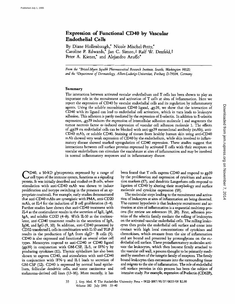
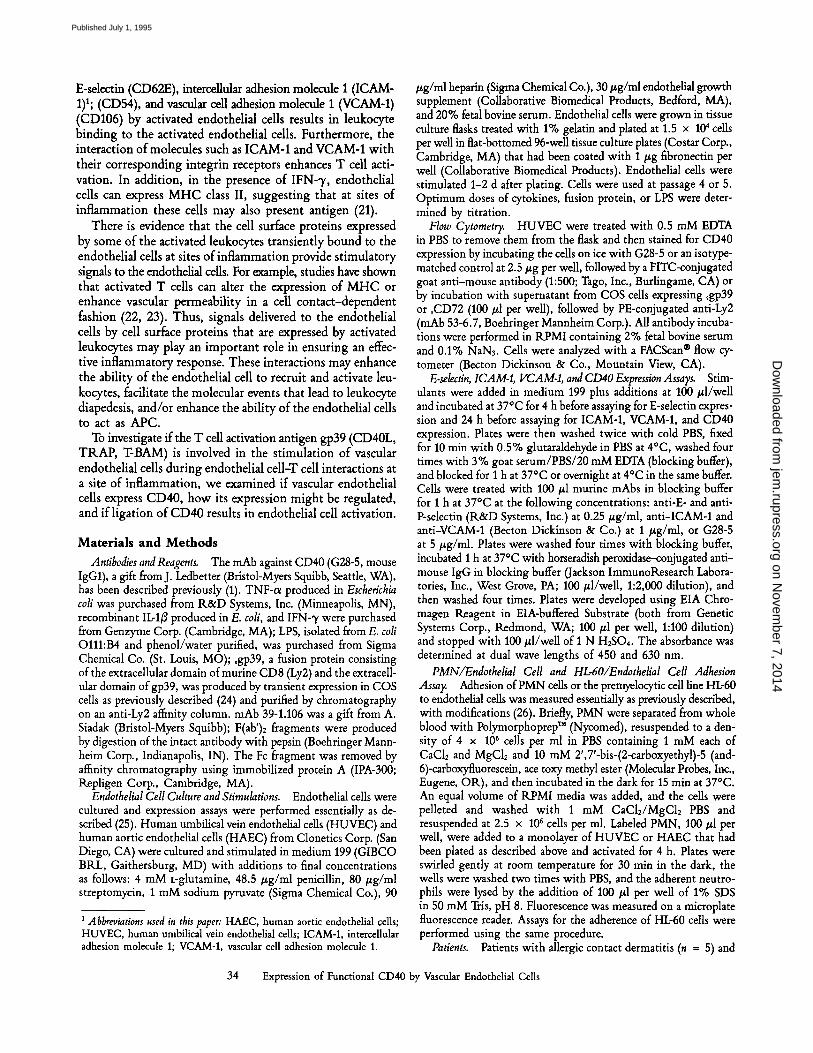
ined whether CD40 is expressed on endothelial cells. HUVEC of passage number 5 were stained with anti-CD40 mAb or sgp39 and examined by flow cytometry. As shown in Fig. 1, CD40 is expressed by resting endothelial cells in culture. HAEC were also found to express CD40, as detected by FACS | analysis (data not shown). To examine if this basal level of CD40 expression can be altered by proinflammatory medi- ators, HUVEC were incubated with TNF, IFN-'y, LPS, or IL-13. After 24 h, cells treated with the inflammatory cytokines or LPS all showed slight but significant increases in expres- sion of CD40 (Fig. 2). Similar effects were seen after stimu- lation for 72 h (data not shown). These results suggest that the expression of CD40 by endothelial cells can be regulated by agents known to play a role in inflammation. Addition- ally, it was found that treatment of the cells with an anti- CD40 antibody for 24 h substantially reduced the level of CD40 detected on the surface of the cells. In these experi- ments, the cells were incubated with the G28-5 antibody, and then subsequently stained with additional G28-5 anti- body. In this way, the total G28-5 bound to the cell surface is detected. The reduced level of CD40 detected is likely to be caused by internalization of the CD40. This possibility is suggested by the observation that a G28-5 immunotoxin was capable of killing CD40-expressing B cell lymphomas, a process dependent on internalization (15).
Leukocyte Binding to Endothelial Cells Can Be Stimulated via CD40. The ability of endothelial cells to respond to liga-
Figure 1. Expression of CD40 by HUVEC. HUVEC were as- sayed by indirect immunofluores- cence using (/1) sgp39, (B) nega- tive control fusion protein, (C) anti-CD40 mAb G28-5, or (D) isotype matched control antibody. Filled histograms represent stain- ing with secondary antibody alone; solid lines represent staining with the indicated protein.
35 Hollenbaugh et al.
on Novem
ber 7, 2014jem
.rupress.orgD
ownloaded from
Published July 1, 1995
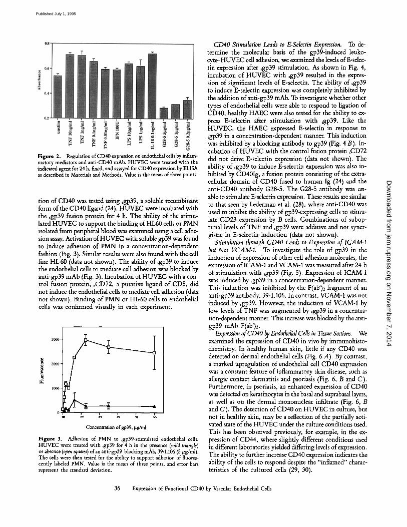
Figure 2. Regulation of CD40 expression on endothelial cells by inflam- matory mediators and anti-CD40 mAb. HUVEC were treated with the indicated agent for 24 h, fixed, and assayed for CD40 expression by ELISA as described in Materials and Methods. Value is the mean of three points.
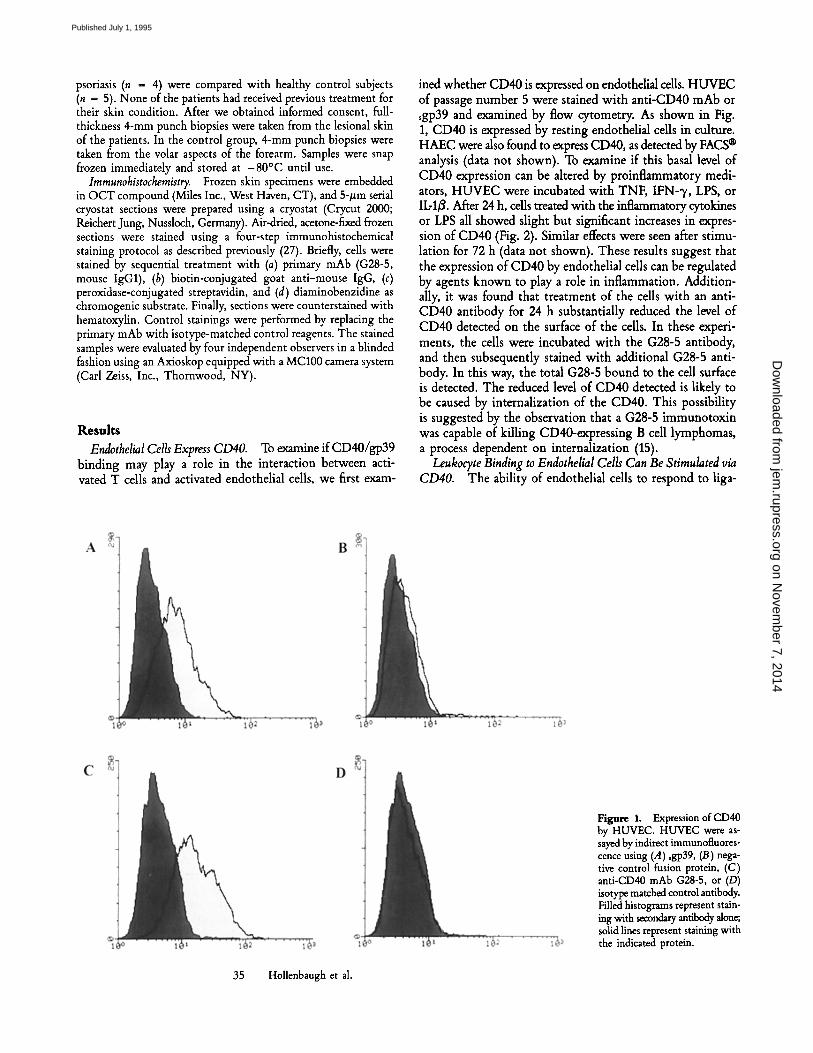
tion of CD40 was tested using ~gp39, a soluble recombinant form of the CD40 ligand (24). HUVEC were incubated with the ~gp39 fusion protein for 4 h. The ability of the stimu- lated HUVEC to support the binding of HL60 cells or PMN isolated from peripheral blood was examined using a cell adhe- sion assay. Activation of HUVEC with soluble gp39 was found to induce adhesion of PMN in a concentration-dependent fashion (Fig. 3). Similar results were also found with the cell line HL-60 (data not shown). The ability of ~gp39 to induce the endothelial cells to mediate cell adhesion was blocked by anti-gp39 mAb (Fig. 3). Incubation of HUVEC with a con- trol fusion protein, ~CD72, a putative ligand of CD5, did not induce the endothelial cells to mediate cell adhesion (data not shown). Binding of PMN or HL-60 cells to endothelial cells was confirmed visually in each experiment.
3000- T
2000-
10011-
I " " J-
0 i i
C o n c e n t r a t i o n o f g p 3 9 , ~tg/ml
Figure 3. Adhesion of PMN to sgp39-stimulated endothelial cells. HUVEC were treated with sgp39 for 4 h in the presence (solid triangle) or absence (open squares) of an anti-gp39-blocking mAb, 39-1.106 (5/zg/ml). The cells were then tested for the ability to support adhesion of fluores- cently labeled PMN. Value is the mean of three points, and error bars represent the standard deviation.
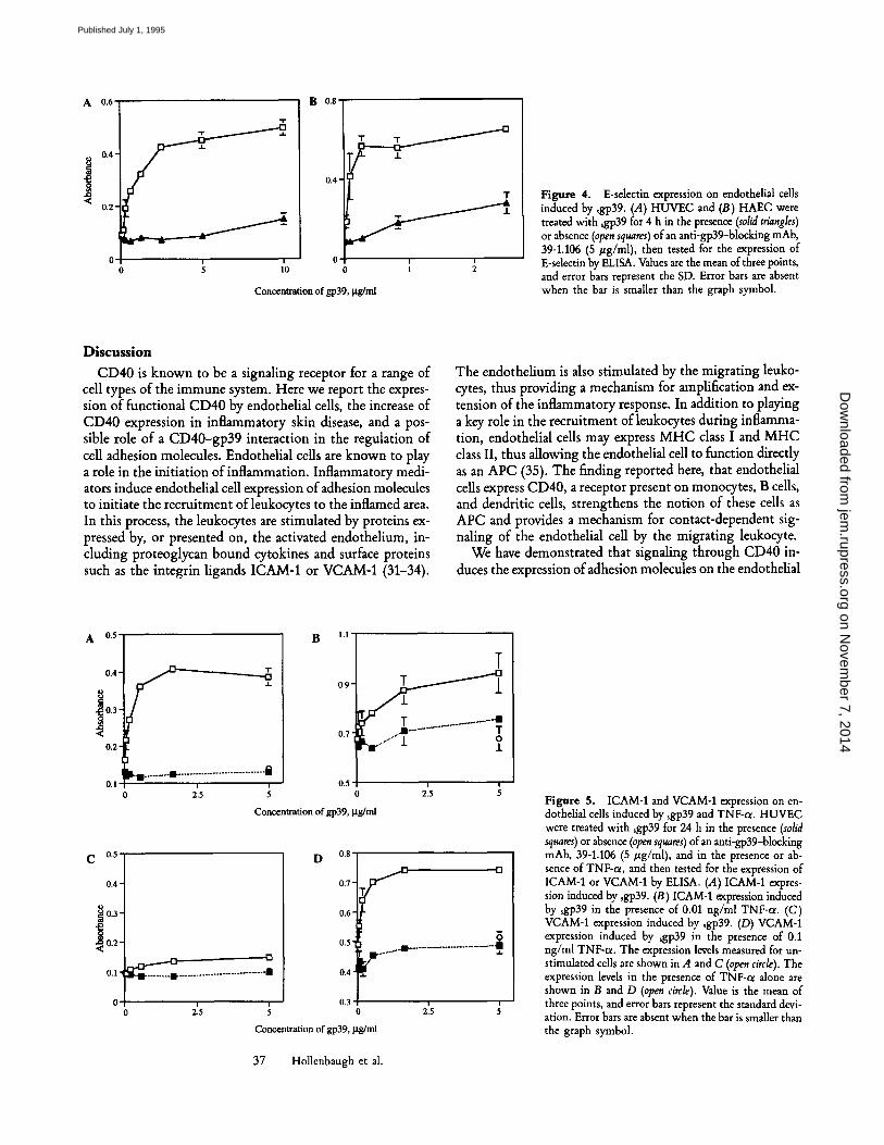
CD40 Stimulation Leads to E-Selectin Expression. To de- termine the molecular basis of the gp39-induced leuko- cyte-HUVEC cell adhesion, we examined the levels of E-sdec- tin expression after sgp39 stimulation. As shown in Fig. 4, incubation of HUVEC with sgp39 resulted in the expres- sion of significant levels of E-selectin. The ability of sgp39 to induce E-selectin expression was completely inhibited by the addition of anti-gp39 mAb. To investigate whether other types of endothelial cells were able to respond to ligation of CD40, healthy HAEC were also tested for the ability to ex- press E-selectin after stimulation with sgp39. Like the HUVEC, the HAEC expressed E-selectin in response to ~gp39 in a concentration-dependent manner. This induction was inhibited by a blocking antibody to gp39 (Fig. 4 B). In- cubation of HUVEC with the control fusion protein sCD72 did not drive E-selectin expression (data not shown). The ability of ~gp39 to induce E-selectin expression was also in- hibited by CD40Ig, a fusion protein consisting of the extra- cellular domain of CD40 fused to human Ig (24) and the anti-CD40 antibody G28-5. The G28-5 antibody was un- able to stimulate E-selectin expression. These results are similar to that seen by Lederman et al. (28), where anti-CD40 was used to inhibit the ability of gp39-expressing cells to stimu- late CD23 expression by B cells. Combinations of subop- timal levels of TNF and ~gp39 were additive and not syner- gistic in E-selectin induction (data not shown).
Stimulation through CD40 Leads to Expression of lCAM-I but Not VCAM-1. To investigate the role of gp39 in the induction of expression of other cell adhesion molecules, the expression of ICAM-1 and VCAM-1 was measured after 24 h of stimulation with ~gp39 (Fig. 5). Expression of ICAM-1 was induced by sgp39 in a concentration-dependent manner. This induction was inhibited by the F(ab')2 fragment of an anti-gp39 antibody, 39-1.106. In contrast, VCAM-1 was not induced by sgp39. However, the induction of VCAM-1 by low levels of TNF was augmented by sgp39 in a concentra- tion-dependent manner. This increase was blocked by the anti- gp39 mAb F(ab')2.
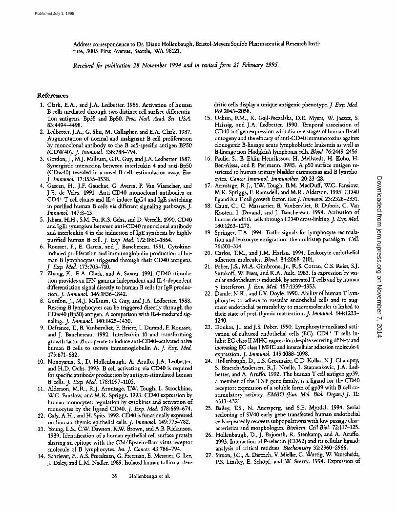
Expression of CD40 by Endothelial Cells in Tissue Sections. We examined the expression of CD40 in vivo by immunohisto- chemistry. In healthy human skin, little if any CD40 was detected on dermal endothelial cells (Fig. 6 A). By contrast, a marked upregulation of endothelial cell CD40 expression was a constant feature of inflammatory skin disease, such as allergic contact dermatitis and psoriasis (Fig. 6, B and C). Furthermore, in psoriasis, an enhanced expression of CD40 was detected on keratinocytes in the basal and suprabasal layers, as well as on the dermal mononuclear infiltrate (Fig. 6,/3 and C). The detection of CD40 on HUVEC in culture, but not in healthy skin, may be a reflection of the partially acti- vated state of the HUVEC under the culture conditions used. This has been observed previously, for example, in the ex- pression of CD44, where slightly different conditions used in different laboratories yielded differing levels of expression. The ability to further increase CD40 expression indicates the ability of the cells to respond despite the "inflamed" charac- teristics of the cultured cells (29, 30).
36 Expression of Functional CD40 by Vascular Endothelial Cells
on Novem
ber 7, 2014jem
.rupress.orgD
ownloaded from
Published July 1, 1995
A 0.6
0.4"
0.2'
0 ! 0 5 llO
B 0.8
0.4
0 1 2
Concen~tion of gp39, gg/ml
Figure 4. E-selectin expression on endothelial cells induced by ~gp39. (A) HUVEC and (B) HAEC were treated with sgp39 for 4 h in the presence (solid triangles) or absence (open squares) of an anti-gp39-blocking mAb, 39-1.106 (5 #g/ml), then tested for the expression of E-selectin by ELISA. Values are the mean of three points, and error bars represent the SD. Error bars are absent when the bar is smaller than the graph symbol.
Discussion CD40 is known to be a signaling receptor for a range of
cell types of the immune system. Here we report the expres- sion of functional CD40 by endothelial cells, the increase of CD40 expression in inflammatory skin disease, and a pos- sible role of a CD40-gp39 interaction in the regulation of cell adhesion molecules. Endothelial cells are known to play a role in the initiation of inflammation. Inflammatory medi- ators induce endothelial cell expression of adhesion molecules to initiate the recruitment of leukocytes to the inflamed area. In this process, the leukocytes are stimulated by proteins ex- pressed by, or presented on, the activated endothelium, in- cluding proteoglycan bound cytokines and surface proteins such as the integrin ligands ICAM-1 or VCAM-1 (31-34).
The endothelium is also stimulated by the migrating leuko- cytes, thus providing a mechanism for amplification and ex- tension of the inflammatory response. In addition to playing a key role in the recruitment of leukocytes during inflamma- tion, endothelial cells may express MHC class I and MHC class II, thus allowing the endothelial cell to function directly as an APC (35). The finding reported here, that endothelial cells express CD40, a receptor present on monocytes, B cells, and dendritic cells, strengthens the notion of these cells as APC and provides a mechanism for contact-dependent sig- naling of the endothelial cell by the migrating leukocyte.
We have demonstrated that signaling through CD40 in- duces the expression of adhesion molecules on the endothelial
A 0.5 B l . l
0.4-
~ 0.3 -
<
0.2-
0.1
C 0.5
0.4-
~ 0.3-
~ 0.2"
0.1"
0
0.9-
0.7'
T
r'- T . . . . . . . . . . . . .,m - . - ~ - - |
0.5 215 5 0
Concentra t ion o f gp39, ~g/ml
215
D 08
0.7
0.6-
0.5
0.4
0.3
Concentration of gp39, ~tg/ml
37 Hollenbaugh et al.
! 215 S
Y
i 2.5
Figure 5. ICAM-1 and VCAM-1 expression on en- dothelial cells induced by ~gp39 and TNF-o~. HUVEC were treated with ~gp39 for 24 h in the presence (solid squares) or absence (open squares) of an anti-gp39-blocking mAb, 39-1.106 (5 #g/ml), and in the presence or ab- sence of TNF-t~, and then tested for the expression of ICAM-1 or VCAM-1 by ELISA. (.4) ICAM-1 expres- sion induced by sgp39. (B) ICAM-1 expression induced by sgp39 in the presence of 0.01 ng/ml TNF-ol. (C) VCAM-1 expression induced by sgp39. (D) VCAM-1 expression induced by ~gp39 in the presence of 0.1 ng/ml TNF-c~. The expression levels measured for un- stimulated cells are shown in A and C (open circle). The expression levels in the presence of TNF-ct alone are shown in B and D (open circle). Value is the mean of three points, and error bars represent the standard devi- ation. Error bars are absent when the bar is smaller than the graph symbol.
on Novem
ber 7, 2014jem
.rupress.orgD
ownloaded from
Published July 1, 1995
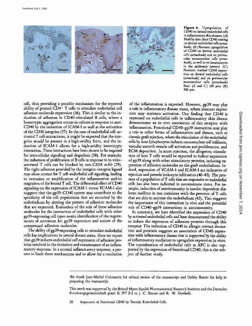
Figure 6. Upregulation of CD40 on dermal endothelial cells in inflammatory skin diseases. (.4) Healthy skin: faint CD40 staining on dermal endothelial cells (arrow- heads). (13) Psoriasis: upregulation of CD40 on dermal endothelial cells (arrowheads) and on perivas- cular mononuclear cells (arrow- heads), as well as on keratinocytes in the epidermis (arrows). (C) Psoriasis: marked CD40 expres- sion on dermal endothelial cells (arrowheads) and on perivascular mononuclear cells (arrowheads) Bars: (.4 and C) 100 /~m; (B) 400/zm.
cell, thus providing a possible mechanism for the reported ability of primed CD4 § T cells to stimulate endothelial cell adhesion molecule expression (36). This is similar to the in- duction of adhesion in CD40-stimulated B cells, where a homotypic aggregation occurs in culture in response to anti- CD40 by the induction of ICAM-1 as well as the activation of the CD18 integrins (37). In the case of endothelial cell-ac- tivated T cell interactions, it might be expected that the inte- grins would be present in a high-avidity form, and the in- duction of ICAM-1 allows for a high-avidity heterotypic interaction. These interactions have been shown to be required for intercellular signaling and diapedesis (38). For example, the induction of proliferation of B cells in response to in vitro- activated T cells can be blocked by anti-CD18 mAb (39). The tight adhesion provided by the integrin-integrin ligand may allow contact for T cell-endothelial cell signaling, leading to extension or amplification of the inflammation and/or migration of the bound T cell. The differential effect of CD40 signaling on the expression of ICAM-1 versus VCAM-1 also suggests that the gp39-CD40 system may contribute to the specificity of the cell populations that are recruited by the endothelium by altering the pattern of adhesion molecules that are expressed. Evaluation of the role of these adhesion molecules for the interaction of endothelial cells with other gp39-expressing cell types awaits identification of the require- ments of activation for gp39 expression and nature of the coexpressed adhesion molecules.
The ability of gp39-expressing cells to stimulate endothelial cells has implications in several disease states. Here we report that gp39 induces endothelial cell expression of adhesion pro- teins involved in the initiation and maintenance of an inflam- matory response. In a normal inflammatory response, a pro- cess to limit these mechanisms and to allow for a resolution
of the inflammation is expected. However, gp39 may play a role in inflammatory disease states, where aberrant expres- sion may maintain activation. Our finding that CD40 is expressed on endothelial cells in inflammatory skin disease demonstrates an in vivo association of this receptor with inflammation. Functional CD40-gp39 interaction may play a role in other forms of inflammation and disease, such as chronic graft rejection, where the stimulation of the endothelial cells by host lymphocytes induces mononuclear cell infiltrate, vascular smooth muscle cell activation and proliferation, and ECM deposition. In acute rejection, the allogeneic stimula- tion of host T cells would be expected to induce expression of gp39 along with other stimulatory proteins, inducing ex- pression of adhesion molecules on the graft endothelium. In- deed, expression of VCAM-1 and ICAM-1 are indicative of rejection and precede leukocyte infiltration (4042). The pres- ence of a population of T cells that are responsive to endothelial cells has also been indicated in autoimmune states. For ex- ample, induction of autoimmunity in insulin-dependent dia- betes mellitus in rats correlates with the presence of T cells that are able to activate the endothelium (43). This suggests the importance of this interaction in vivo and the potential role of CD40-gp39 interactions in autoimmunity.
In summary, we have identified the expression of CD40 by activated endothelial cells and have demonstrated the ability to induce the expression of adhesion proteins through this receptor. The induction of CD40 in allergic contact derma- titis and psoriasis suggests an association of CD40 expres- sion with inflammatory disease that is supported by the ability of inflammatory mediators to upregulate expression in vitro. The consideration of endothelial cells as APC is also sup- ported by the expression of functional CD40; this is the sub- ject of further study.
We thank Jean-Michel Culouscou for critical review of the manuscript and Debby Baxter for help in preparing the manuscript.
This work was supported by the Bristol-Myers Squibb Pharmaceutical Research Institute and the Deutsche Forschungsgemeinschaft grant Si 397 2-2 to J. C. Simon and R. W. Denfield.
38 Expression of Functional CD40 by Vascular Endothelial Cells
on Novem
ber 7, 2014jem
.rupress.orgD
ownloaded from
Published July 1, 1995
Address correspondence to Dr. Diane Hollenbaugh, Bristol-Meyers Squibb Pharmaceutical Research Insti- tute, 3005 First Avenue, Seattle, WA 98121.
Received for publication 28 November 1994 and in revised form 21 February 1995.
References 1. Clark, E.A., and J.A. Ledbetter. 1986. Activation of human
B cells mediated through two distinct cell surface differentia- tion antigens, Bp35 and Bp50. Proc. Natl. Acad. Sci. USA. 83:4494-4498.
2. Ledbetter, J.A., G. Shu, M. Gallagher, and E.A. Clark. 1987. Augmentation of normal and malignant B cell proliferation by monoclonal antibody to the B cell-specific antigen BP50 (CDW40). J. Immunol. 138:788-794.
3. Gordon, J., M.J. MiUsum, G.R. Guy, andJ.A. Ledbetter. 1987. Synergistic interaction between interleukin 4 and anti-Bp50 (CDw40) revealed in a novel B cell restimulation assay. Eur. J. Immunol. 17:1535-1538.
4. Gascan, H., J.F. Gauchat, G. Aversa, P. Van Vlasselaer, and J.E. de "Cries. 1991. Anti-CD40 monoclonal antibodies or CD4 + T cell clones and IL-4 induce IgG4 and IgE switching in purified human B cells via different signaling pathways. J. Immunol. 147:8-13.
5. Jabara, H.H., S.M. Fu, R.S. Geha, and D. Vercelli. 1990. CD40 and IgE: synergism between anti-CD40 monoclonal antibody and interleukin 4 in the induction of IgE synthesis by highly purified human B cell. j. Exp. Med. 172:1861-1864.
6. Rousset, F., E. Garcia, and J. Banchereau. 1991. Cytokine- induced proliferation and immunoglobulin production of hu- man B lymphocytes triggered through their CD40 antigens. J. Extz Med. 173:705-710.
7. Zhang, K., E.A. Clark, and A. Saxon. 1991. CD40 stimula- tion provides an IFN-gamma-independent and Ib4-dependent differentiation signal directly to human B cells for IgE produc- tion. J. Immunol. 146:1836-1842.
8. Gordon, J., M.J. Millsum, G. Guy, and J.A. Ledbetter. 1988. Resting B lymphocytes can be triggered directly through the CDw40 (Bp50) antigen. A comparison with II~4-mediated sig- naling. J. Immunol. 140:1425-1430.
9. Defrance, T., B. Vanbervliet, F. Briere, I. Durand, F. Rousset, and J. Banchereau. 1992. Interleukin 10 and transforming growth factor B cooperate to induce anti-CD40-activated naive human B cells to secrete immunoglobulin A.J. Exp. Med. 175:671-682.
10. Nonoyama, S., D. Hollenbaugh, A. Aruffo, J.A. Ledbetter, and H.D. Ochs. 1993. B cell activation via CD40 is required for specific antibody production by antigen-stimulated human B cells, j. Extz Med. 178:1097-1102.
11. Alderson, M.R., R.J. Armitage, T.W. Tough, L. Strockbine, W.C. Fanslow, and M.K. Spriggs. 1993. CD40 expression by human monocytes: regulation by cytokines and activation of monocytes by the ligand CD40. J. Exp. Med. 178:669-674.
12. Galy, A.H., and H. Spits. 1992. CD40 is functionally expressed on human thymic epithelial cells. J. lmmunol. 149:775-782.
13. Young, L.S., C.W. Dawson, K.W. Brown, and A.B. Rickinson. 1989. Identification of a human epithelial cell surface protein sharing an epitope with the C3d/Epstein-Barr virus receptor molecule of B lymphocytes. Int. J. Cancer. 43:786-794.
14. Schriever, F., A.S. Freedman, G. Freeman, E. Messner, G. Lee, J. Daley, and L.M. Nadler. 1989. Isolated human follicular den-
39 Hollenbaugh et al.
dritic cells display a unique antigenic phenotype.J. Exl~ Med. 169:2043-2058.
15. Uckun, F.M., K. Gajl-Peczalska, D.E. Myers, W. Jaszcz, S. Haissig, and J.A. Ledbetter. 1990. Temporal association of CD40 antigen expression with discrete stages of human B-cell ontogeny and the efficacy of anti-CD40 immunotoxins against clonogenic B-lineage acute lymphoblastic leukemia as well as B-lineage non-Hodgkin's lymphoma cells. Blood. 76:2449-2456.
16. Paulie, S., B. Ehlin-Henriksson, H. Mellstedt, H. Koho, H. Ben-Aissa, and P. Perlmann. 1985. A p50 surface antigen re- stricted to human urinary bladder carcinomas and B lympho- cytes. Cancer Immunol. Immunother. 20:23-28.
17. Armitage, R.J., T.W. Tough, B.M. MacDuff, W.C. Fanslow, M.K. Spriggs, F. Ramsdell, and M.K. Alderson. 1993. CD40 ligand is a T cell growth factor. Eur.J. Immunol. 23:2326-2331.
18. Caux, C., C. Massacrier, B. Vanbervliet, B. Dubois, C. Van Kooten, I. Durand, and J. Banchereau. 1994. Activation of human dendritic cells through CD40 cross-linking.J. Exp Med. 180:1263-1272.
19. Springer, T.A. 1994. Traffic signals for lymphocyte recircula- tion and leukocyte emigration: the multistep paradigm. Cell. 76:301-314.
20. Carlos, T.M., and J.M. Harlan. 1994. Leukocyte-endothelial adhesion molecules. Blood. 84:2068-2101.
21. Pober, J.S., M.A. Gimbrone, Jr., K.S. Cotran, C.S. Reiss, S.J. Surakoff, W. Fiers, and K.A. Auk. 1983. Ia expression by vas- cular endothelium is inducible by activated T cells and by human 3' interferon. J. Exp. Med. 157:1339-1353.
22. Damle, N.K., and L.V. Doyle. 1990. Ability of human T lym- phocytes to adhere to vascular endothelial cells and to aug- ment endothelial permeability to macromolecules is linked to their state of post-thymic maturation. J. Immunol. 144:1233- 1240.
23. Doukas, J., and J.S. Pober. 1990. Lymphocyte-mediated acti- vation of cultured endothelial cells (EC). CD4 § T cells in- hibit EC class II MHC expression despite secreting IFN-3" and increasing EC class I MHC and intercellular adhesion molecule-1 expression. J. Immunol. 145:1088-1098.
24. Hollenbaugh, D., L.S. Grosmaire, C.D. Kullas, N.J. Chalupny, S. Braesch-Andersen, R.J. NoeUe, I. Stamenkovic, J.A. Led- better, and A. Aruffo. 1992. The human T cell antigen gp39, a member of the TNF gene family, is a ligand for the CD40 receptor: expression of a soluble form of gp39 with B cell co- stimulatory activity. EMBO (Eur. Mol. Biol. Organ.)J. 11: 4313-4321.
25. Bailey, T.S., N. Auersperg, and S.E. Myrdal. 1994. Serial recloning of SV40 early gene transfected human endothelial cells repeatedly recovers subpopulations with low passage char- acteristics and morphologies. Biochem. Cell Biol. 72:117-125.
26. Hollenbangh, D., J. Bajorath, K. Stenkamp, and A. Aruffo. 1993. Interaction of P-selectin (CD62) and its cellular ligand: analysis of critical residues. Biochemistry 32:2960-2966.
27. Simon, J.C., A. Dietrich, V. Mielke, C. Wuttig, W. Vanscheidt, P.S. Linsley, E. Schrpf, and W. Sterry. 1994. Expression of
on Novem
ber 7, 2014jem
.rupress.orgD
ownloaded from
Published July 1, 1995
the B7/BB1 activation antigen and its ligand CD28 in T-cell- mediated skin diseases. J. Invest. Dermatol. 103:539-543.
28. Lederman, S., M.J. Yellin, G. Inghirami, J.J. Lee, D.M. Knowles, and L. Chess. 1992. Molecular interactions mediating T-B lym- phocyte collaboration in human lymphoid follicles. Roles of T cell-B cell-activating molecule (5c8 antigen) and CD40 in contact-dependent help. J. Immunol. 149:3817-3826.
29. Mackay, F., H. Loetscher, D. Stueber, G. Gehr, and W. Less- lauer. 1993. Tumor necrosis factor c~ (TNF-ol)-induced cell adhesion to human endothelial cells is under dominant con- trol of one TNF receptor type, TNF-R55. J. Exp. Med. 177: 1277-1286.
30. Bennett, K.L., D.G. Jackson, J.C. Simon, E. Tanczos, R. Peach, B. Modrell, I. Stamenkovic, G. Plowman, and A. Aruffo. 1995. CD44 isoforms containing exon V3 are responsible for the pre- sentation of heparin binding growth factor. J. Cell Biol. 128: 687-698.
31. Tanaka, Y., D.H. Adams, S. Hubscher, H. Hirano, U. Sieben- list, and S. Shaw. 1993. T-cell adhesion induced by proteoglycan- immobilized cytokine MIP-13. Nature (Lond.). 361:79-82.
32. Kuhlman, P., V.T. Moy, B.A. Lollo, and A.A. Brian. 1991. The accessory function of murine intercellular adhesion molecule-1 in T lymphocyte activation. Contributions of adhesion and co-activation. J. Immunol. 146:1773-1782.
33. Van Seventer, G.A., Y. Shimizu, K. Horgan, G.E.G. Luce, D. Webb, and S. Shaw. 1991. Remote T cell co-stimulation via LFA-1/ICAM-1 and CD2/LFA-3: demonstration with immobi- lized ligand/mAb and implication in monocyte-mediated co- stimulation. Eur. j . Immunol. 21:1711-1718.
34. Damle, N.K., and A. Aruffo. 1991. Vascular cell adhesion mol- ecule 1 induces T-cell antigen receptor-dependent activation of CD4 + T lymphocytes. Proc. Natl. Acad. Sci. USA. 88: 6403-6407.
35. Pober, J.S., and R.S. Cotran. 1990. The role of endothelial
cells in inflammation. Transplantation. 50:537-544. 36. Damle, N.K., C. Eberhardt, and M. Van der Vieren. 1991.
Direct interaction with primed CD4 + CD45R0 § memory T lymphocytes induces expression of endothelial leukocyte adhe- sion molecule-1 and vascular cell adhesion molecule-1 on the surface of vascular endothelial cells. Eur. j . Immunot. 21:2915- 2923.
37. Barrett, T.B., G. Shu, and E.A. Clark. 1991. CD40 signalling activates CDlla/CD18 (LFA-1)-mediated adhesion in B cells. J. Immunol. 146:1722-1729.
38. Luscinskas, F.W., M.I. Cybulsky, J.-M. Kiely, C.S. Peckins, V.M. Davis, and M.A. Gimbrone, Jr. 1991. Cytokine-activated human endothelial monolayers support enhanced neutrophil transmigration via a mechanism involving both endothelial- leukocyte adhesion molecule-1 and intercellular adhesion molecule-1. J. Immunol. 146:1617-1625.
39. Tohma, S., S. Hirohata, and P.E. Lipsky. 1991. The role of CDlla/CD18-CD54 interactions in human T cell-dependent B cell activation. J. Immunol. 146:492-499.
40. Briscoe, D.M., F.J. Schoen, G.E. Rice, M.P. Bevilacqua, P. Ganz, and J.S. Pober. 1991. Induced expression of endothelial- leukocyte adhesion molecules in human cardiac allografts. Trans- plantation. 41:537-547.
41. Taylor, P.M., M.L. Rose, M.H. Yacoub, and R. Pigott. 1992. Induction of vascular adhesion molecules during rejection of human cardiac allografts. Transplantation. 54:451-457.
42. Pelletier, R.P., R.G. Ohye, A. Vanbuskirk, D.D. Sedmak, P. Kincade, R.M. Ferguson, and C.G. Orosz. 1992. Importance of endothelial VCAM-1 for inflammatory leukocytic infiltra- tion in vivo. J. Irnmunol. 149:2473-2481.
43. Doukas, J., and J.P. Mordes. 1993. T lymphocytes capable of activating endothelial cells in vitro are present in rats with au- toimmune diabetes. J. Iramunol. 150:1036-1046.
40 Expression of Functional CD40 by Vascular Endothelial Cells
on Novem
ber 7, 2014jem
.rupress.orgD
ownloaded from
Published July 1, 1995


