Embed Size (px)
Citation preview
Page 1/17
Favorable Outcomes with Reduced Steroid Use inJuvenile DermatomyositisAmir B. Orandi
Mayo Clinic https://orcid.org/0000-0002-3720-2402Lampros Fotis
National and Kapodistrian University of AthensJamie Lai
Baylor College of MedicineHallie Morris
The George Washington University School of Medicine and Health SciencesAndrew White
Washington University School of Medicine in Saint Louis: Washington University in St Louis School ofMedicineAnthony R. French ( [email protected] )
Washington University School of MedicineKevin Baszis
Washington University School of Medicine in Saint Louis: Washington University in St Louis School ofMedicine
Research article
Keywords: Juvenile Dermatomyositis, Calcinosis, Biologic Therapy, Glucocorticoids, PediatricRheumatology
Posted Date: January 13th, 2021
DOI: https://doi.org/10.21203/rs.3.rs-142233/v1
License: This work is licensed under a Creative Commons Attribution 4.0 International License. Read Full License
Version of Record: A version of this preprint was published at Pediatric Rheumatology on August 17th,2021. See the published version at https://doi.org/10.1186/s12969-021-00615-0.
Page 2/17
Abstract
BackgroundHigh-intensity glucocorticoid regimens are commonly used to induce and maintain remission in JuvenileDermatomyositis (JDM) but are associated with several adverse side-effects. Eminence-based treatmentguidelines from CARRA and SHARE both advocate induction of intravenous pulse steroids followed byhigh dose oral steroids (2 mg/kg) tapered over 12 months. This study compares the time to diseasecontrol with reduced glucocorticoid dosing.
MethodsWe retrospectively reviewed the records at a single tertiary-care children’s hospital of patients with JDMbetween 2000 and 2014 who had a minimum of 2 years of follow-up. The primary outcome measure wastime to control of muscle and skin disease. Additional outcome measures included glucocorticoid dosing,effect of treatment on height, frequency of calcinosis and complications from treatment.
ResultsOf the 69 patients followed during the study period, 31 ful�lled inclusion criteria. Median length of follow-up was 4.58 years, (3–7.5). Myositis control was achieved in a median of 7.1 months (range 0.9–63.4).Cutaneous disease control was achieved in a median of 16.7 months (range 4.3–89.5). The medianstarting dose of glucocorticoids was 0.85 mg/kg/day, (range 0.5–1.74). The median duration of steroidtreatment was 9.1 months, (range 4.7–17.4), while the median duration of any pharmacotherapy was29.2 months (range 10.4 to 121.3). Sustained disease control off medications was achieved in 21/31(68%) patients by the end of review. Persistent calcinosis was identi�ed in only one patient (3%).
ConclusionCurrent accepted treatment paradigms for JDM include oral glucocorticoids beginning at 2 mg/kg/dreduced over 12 months; however, our results suggest that treatment using reduced doses and durationswith early use of steroid-sparing agents are comparably effective in achieving favorable outcomes inJDM.
BackgroundJuvenile Dermatomyositis (JDM) is a rare in�ammatory myopathy in children, comprising 85% of allidiopathic in�ammatory myopathies of childhood (1). It is a chronic immune-mediated vasculopathyassociated with proximal muscle weakness, characteristic skin involvement, and impairment in physicalfunction (2). Diagnosis has long been based on the clinical and laboratory criteria of Bohan and Peter (3,
Page 3/17
4) , but new classi�cation criteria have been recently developed incorporating weighted scores for clinicalfeatures of characteristic cutaneous changes, symmetric proximal muscle weakness, elevated serummuscle enzymes, myopathic changes on electromyogram, and characteristic muscle biopsyabnormalities, combined with absence of histopathologic signs of other myopathies (5). The outcome ofpatients with JDM prior to the 1960s was poor, as more than one-third of patients died from their illnessand one-third developed permanent limitations (6, 7). Following the introduction of glucocorticoids, whichbecame a mainstay of the treatment in JDM, mortality rates declined. After increased use of otherimmunomodulatory agents such as methotrexate and azathioprine (8, 9), the mortality rate furtherdeclined to estimates of less than 2–3% (10, 11).
During this era of gradual improvement in treatment outcomes, there were no randomized controlled trialsor published corticosteroid treatment regimens to guide therapeutic decisions. Generally, different doses,routes, and duration of glucocorticoids were �rst-line therapy for mild, moderate, and severe JDM incombination with other immunomodulatory agents. However, a number of published reports advocatedthat initial high-dose, intravenous and/or pulsed intervals of glucocorticoids were needed to aggressivelysuppress disease activity, sustain remission, and also to prevent calcinosis, a highly morbid JDMcomplication (12–16). Despite this trend, there remained tremendous variety in practice, with mostauthors advocating an initial oral prednisone dose of 2 mg/kg/day (up to a maximum of 60–80 mg/day),with pulses of high-dose (up to 30 mg/kg/day, maximum 1 g daily) intravenous methylprednisolone(IVMP) for moderate to severe cases as induction therapy, followed by a slow taper of oral steroids overone year (17). However, lower to medium doses of prednisone (1 to 1.5 mg/kg/day) were also reportedeffective, particularly after taking into account the disability associated with higher glucocorticoid dosing(18). Another report showed comparable outcomes using combinations of other immunosuppressiveagents with no use of systemic glucocorticoids, albeit in a mild disease phenotype (19). Regardless, themost current recommendations continue to endorse high-dose glucocorticoids: Consensus-basedtreatment plans published by the Childhood Arthritis and Rheumatology Research Alliance (CARRA) forthe initial treatment of moderate to severe JDM advocated for 2 mg/kg/day oral prednisone incombination with methotrexate in their most conservative protocol, with the two more aggressiveprotocols utilizing intravenous methylprednisolone (30 mg/kg/day, maximum 1 gm) for 3 days andcontinuing with weekly or monthly intravenous methylprednisolone pulses (20, 21) . Similarly, Europeanrecommendations from the Single Hub and Access point for Pediatric Rheumatology in Europe (SHARE)recommended high-dose intravenous dosing of methylprednisolone (15–30 mg/kg/day, maximum1 g/day) for all patients at diagnosis or with a disease �are, followed by an oral prednisone taperbeginning at 1–2 mg/kg/day (22). To our knowledge, there have been no reports describing outcomeswith lower doses of glucocorticoids used consistently in early combination with otherimmunosuppressive agents.
Given that high-dose and/or prolonged use of glucocorticoids have many potential adverse events(including growth retardation, hypertension, impaired glucose tolerance, immunosuppression, osteopenia,recurrent infections, vertebral fractures, and avascular necrosis) (23), the approach at our center has beento use the minimum dose and duration of glucocorticoids necessary, in order to induce remission and
Page 4/17
control disease activity, in combination with early and consistent use of steroid-sparing agents. Here, wereport the outcomes of JDM patients treated in this manner at as single tertiary-care children’s hospitalduring a 14-year period, that coincides with the timeframe of the above referenced publications.
Methods
Study populationWe retrospectively reviewed the charts of patients with JDM diagnosed and treated at St. Louis Children’sHospital between January 2000 and December 2014. We included patients with probable or de�nite JDM,according to the Bohan and Peter criteria (3, 4), with disease onset prior to 18 years of age, and with atleast two years of subsequent follow up. Patients with mixed connective tissue disease, overlapsyndrome, or amyopathic JDM were excluded. Patients were also excluded if they transferred care to ourinstitution after being diagnosed and/or treated at other institutions. This study was approved by theHuman Research Protection O�ce of Washington University School of Medicine in St Louis #201107133.
De�nitionsSymptom onset was de�ned as the time when patient, parent, guardian, or pediatrician �rst observedJDM symptoms (e.g., weakness or rash). Myositis control was de�ned as the time when normal musclestrength (5/5 in all major muscle groups) and normal muscle enzyme levels (creatine kinase, aldolase,lactate dehydrogenase, aspartate aminotransferase, and alanine aminotransferase) were documented.Cutaneous disease control was de�ned as the time when no active JDM rash was documented onphysical exam. Complete disease control was, therefore, de�ned as achievement of both myositis andcutaneous disease control. Medications were gradually withdrawn after complete disease control wasmaintained for at least six months, but most often 12 months. Once all medications were withdrawn, andmuscle and skin disease remained quiescent, the patient was considered to have sustained diseasecontrol.
Disease course was de�ned as 1) monocyclic when the patient had no clinical or laboratory markersconsistent with active disease and was off all medications within 24 months of diagnosis, 2) chroniccontinuous when there was persistent disease or continued treatment with medications for more than 24months after diagnosis, and 3) polycyclic when there was recurrence of disease after at least 6 months ofno clinical or laboratory disease activity. These de�nitions are similar to those used in prior studies (14).Initial disease severity was classi�ed as mild, moderate, or severe based on presenting disease features,including degree of muscle weakness, presence of dysphagia or dysphonia, organ involvement,ulcerations, and degree of skin involvement.
Treatment protocolOur treatment protocol consisted of a stepwise approach depending on disease severity at presentationand evolution of symptoms following treatment initiation. Upon diagnosis, patients with mild disease
Page 5/17
were started on oral prednisone and methotrexate. Patients with moderate to severe disease were treatedwith IVMP, followed by an oral steroid taper, and methotrexate. IVMP was only used either as an initialtherapy within the �rst month of diagnosis and/or in cases of severe relapse. Varying schedules anddosages were used for patients receiving IVMP, ranging from 10 to 30 mg/kg/day for up to 3 days, withmaximum 1 gram/day. The oral steroid taper uniformly began at a dose less than 2 mg/kg/day, with thetaper duration dependent on the clinical and laboratory response of each patient. Methotrexate wasinitiated at or within one month of diagnosis at a dose of 12–15 mg/m2 weekly (primarilysubcutaneously). Patients in whom complete disease control was not achieved within 3–6 months werestarted on monthly intravenous immune globulin (IVIG) at 2 g/kg/dose. Patients with severe diseasereceived IVIG sooner after diagnosis. Hydroxychloroquine was added for those patients with persistentskin disease that did not respond to the above treatments. Alternative agents less commonly usedincluded rituximab, le�unomide, cyclosporine, cyclophosphamide and mycophenolate mofetil. Treatmentwas weaned in a stepwise fashion after the patient had remained in complete disease control for 6–12months.
GrowthGrowth parameters were obtained at diagnosis and at subsequent follow up visits, and the z-scores forheight and BMI were calculated based on the CDC growth charts for the United States published in 2000(24).
Statistical analysisAnalysis was performed using SAS Version 9.3 (SAS Institute) and SPSS Version 22.0 (IBM Analytics).Categorical variables were reported as percentages, and continuous variables as medians and ranges.Time-to-event analyses were conducted with Kaplan-Meier analyses. The starting time was the date ofdiagnosis, and data from participants not experiencing the outcome were censored at the time of lastfollow-up. The effect of steroid medication on participant height was modeled with a random-effectsmixed-model analysis. Participants were considered a random effect, and years from diagnosis and itssquare were considered �xed effects. Mann-Whitney test was used for the comparison between thedisease groups.
Results
Demographic characteristics (Table 1)Of 69 JDM patients diagnosed and/or treated between 2000 and 2014, 19 patients were excluded due totransfer of care from outside institutions or missing initial diagnostic and management data. Elevenpatients had inadequate follow-up for inclusion. Eight patients were excluded due to diagnosis ofamyopathic JDM. Therefore, 31 patients (12 males, 19 females) were included in the study. Medianduration of follow up of this group was 4.6 years (range 3–7.5). The median age of the patients at the
Page 6/17
time of diagnosis was 8.1 years (range 5.6–10.5). The median duration of untreated disease (time fromsymptom onset to diagnosis and treatment) was 2.9 months (range 1.5–8.6).
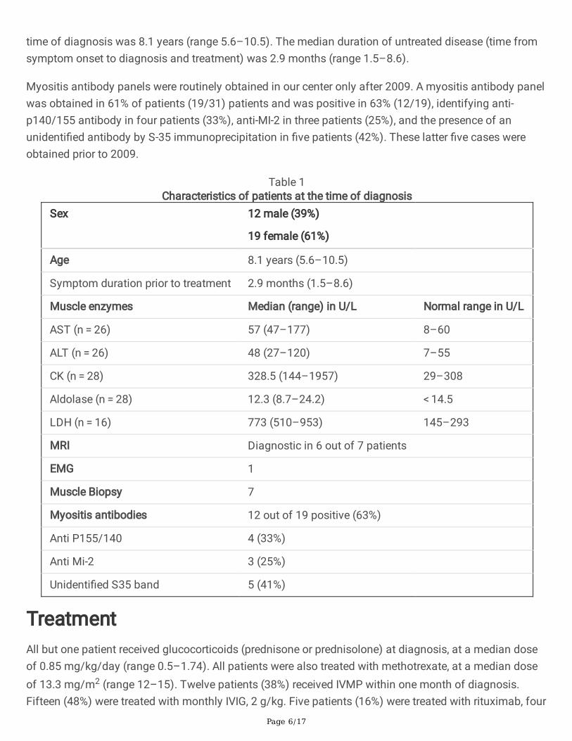
Myositis antibody panels were routinely obtained in our center only after 2009. A myositis antibody panelwas obtained in 61% of patients (19/31) patients and was positive in 63% (12/19), identifying anti-p140/155 antibody in four patients (33%), anti-MI-2 in three patients (25%), and the presence of anunidenti�ed antibody by S-35 immunoprecipitation in �ve patients (42%). These latter �ve cases wereobtained prior to 2009.
Table 1Characteristics of patients at the time of diagnosis
Sex 12 male (39%)
19 female (61%)
Age 8.1 years (5.6–10.5)
Symptom duration prior to treatment 2.9 months (1.5–8.6)
Muscle enzymes Median (range) in U/L Normal range in U/L
AST (n = 26) 57 (47–177) 8–60
ALT (n = 26) 48 (27–120) 7–55
CK (n = 28) 328.5 (144–1957) 29–308
Aldolase (n = 28) 12.3 (8.7–24.2) < 14.5
LDH (n = 16) 773 (510–953) 145–293
MRI Diagnostic in 6 out of 7 patients
EMG 1
Muscle Biopsy 7
Myositis antibodies 12 out of 19 positive (63%)
Anti P155/140 4 (33%)
Anti Mi-2 3 (25%)
Unidenti�ed S35 band 5 (41%)
TreatmentAll but one patient received glucocorticoids (prednisone or prednisolone) at diagnosis, at a median doseof 0.85 mg/kg/day (range 0.5–1.74). All patients were also treated with methotrexate, at a median doseof 13.3 mg/m2 (range 12–15). Twelve patients (38%) received IVMP within one month of diagnosis.Fifteen (48%) were treated with monthly IVIG, 2 g/kg. Five patients (16%) were treated with rituximab, four
Page 7/17
(13%) with le�unomide, 13 (42%) with hydroxychloroquine, two (6%) with mycophenolate mofetil, four(13%) with cyclophosphamide, three (10%) with anti-tumor necrosis factor (TNF)-alpha agents, and onewith cyclosporine. No patients underwent placement of central lines for intravenous medications.
OutcomesMyositis control was achieved within a median of 7.1 months (range 0.9–63.4), and only one patient hadevidence of persistent muscle disease at the end of the study period. (Fig. 1). Cutaneous disease controlwas achieved within a median of 16.7 months (range 4.3–89.5) (Fig. 2). For seven patients (22%), rashnever completely resolved. The median duration of oral prednisone treatment for the cohort was 9.1months (range 4.7–17.4).
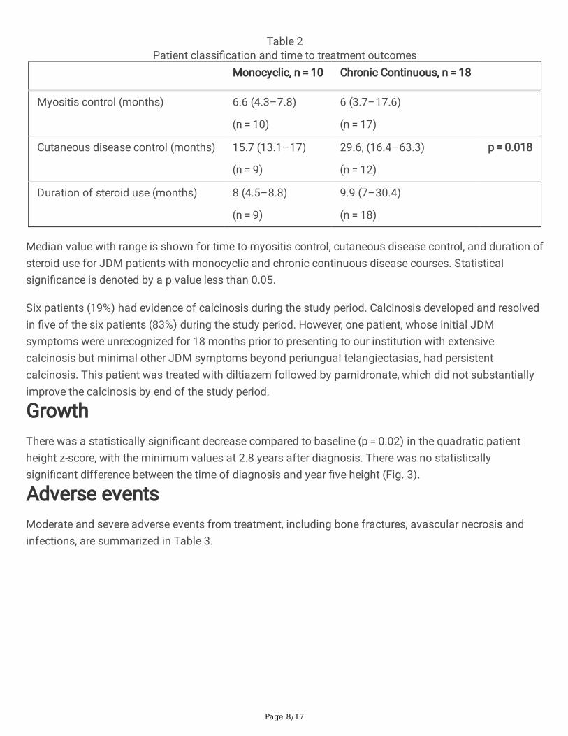
A monocyclic course of disease was observed in 10 patients (32%). In those patients, myositis controlwas reached in 6.6 months (range 4.3–7.8) and cutaneous disease control was achieved in 15.7 months(range 13.1–17). Oral prednisone was administered for 8 months (range 4.5–8.8), and total treatmentduration (all medications) was 19.6 months (range 16.5–22.5). Eighteen patients (58%) followed achronic continuous disease course. In those patients, myositis control was reached in 6 months (range3.7–17.6) and cutaneous disease control in 29.6 months (range 16.4–63.3). Oral prednisone wasadministered for 9.9 months (range 7–30.4). Of those patients with chronic continuous disease, nine(50%) eventually achieved sustained disease control (complete disease control followed by medicationwithdrawal) by the end of the study period (Table 2). Finally, three patients (10%) were classi�ed ashaving a polycyclic course, with symptom relapse occurring a median of 12 months after achievingcomplete disease control and medication withdrawal. In two of these, complete disease control wasagain achieved after relapse, while one continued to have active disease at the end of the study period.Comparison between patients with monocyclic disease versus those with chronic continuous disease didnot reveal a statistically signi�cant difference in the time to myositis control, the dose or duration ofsteroid administration, the patient age at diagnosis, or the duration of untreated disease prior todiagnosis (Table 2). The time to cutaneous disease control was signi�cantly shorter in the patients withmonocyclic disease course, compared to those with chronic continuous disease course (p = 0.018;Table 2). Overall, sustained disease control was achieved in 21 patients (68%) by the end of the studyperiod, with a median treatment duration of 22.3 months (range 18.0–31.4).
Page 8/17
Table 2Patient classi�cation and time to treatment outcomes
Monocyclic, n = 10 Chronic Continuous, n = 18
Myositis control (months) 6.6 (4.3–7.8)
(n = 10)
6 (3.7–17.6)
(n = 17)
Cutaneous disease control (months) 15.7 (13.1–17)
(n = 9)
29.6, (16.4–63.3)
(n = 12)
p = 0.018
Duration of steroid use (months) 8 (4.5–8.8)
(n = 9)
9.9 (7–30.4)
(n = 18)
Median value with range is shown for time to myositis control, cutaneous disease control, and duration ofsteroid use for JDM patients with monocyclic and chronic continuous disease courses. Statisticalsigni�cance is denoted by a p value less than 0.05.
Six patients (19%) had evidence of calcinosis during the study period. Calcinosis developed and resolvedin �ve of the six patients (83%) during the study period. However, one patient, whose initial JDMsymptoms were unrecognized for 18 months prior to presenting to our institution with extensivecalcinosis but minimal other JDM symptoms beyond periungual telangiectasias, had persistentcalcinosis. This patient was treated with diltiazem followed by pamidronate, which did not substantiallyimprove the calcinosis by end of the study period.
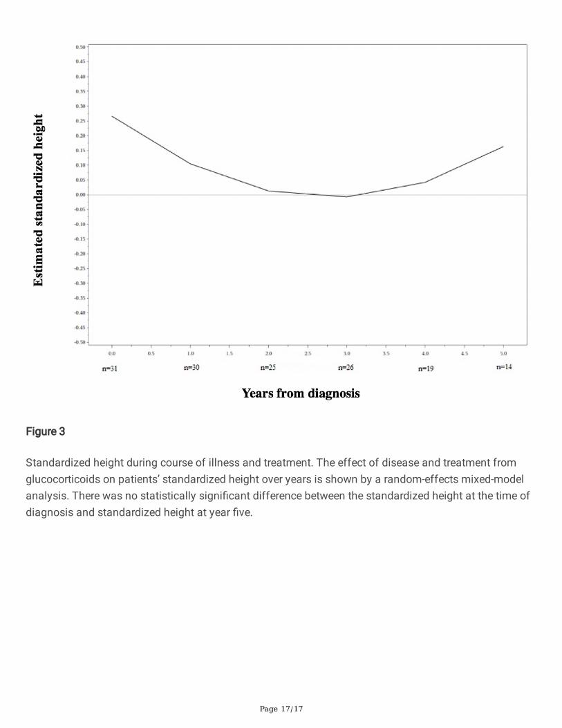
GrowthThere was a statistically signi�cant decrease compared to baseline (p = 0.02) in the quadratic patientheight z-score, with the minimum values at 2.8 years after diagnosis. There was no statisticallysigni�cant difference between the time of diagnosis and year �ve height (Fig. 3).
Adverse eventsModerate and severe adverse events from treatment, including bone fractures, avascular necrosis andinfections, are summarized in Table 3.

Page 9/17
Table 3Adverse effects observed in JDM cohort (n = 31)
Fractures (n = 4) Talus (n = 1)
Multiple thoracic vertebrae, T6-T10, T12 (n = 1)
Avulsion fracture of the 3rd �nger (n = 1)
Distal tibia (following trauma) (n = 1)
Avascular necrosis (n = 2) Knees bilaterally (n = 1)
Ankle bones (n = 1)
Infections (n = 8) * RSV (n = 1)
Campylobacter (n = 1)
Oral thrush (n = 3)
Cellulitis (n = 1)
Pleural effusion (n = 1)
Impetigo (n = 1)
Vaginal candidiasis (n = 1)
Viral meningitis (n = 1)
Pneumonia (n = 1)
Streptococcal bacteremia (n = 1)
CMV reactivation (n = 1)
Herpes zoster (n = 2)
Growth (n = 2) Short stature (n = 1)
Poor weight gain (n = 1)
3 patients had multiple infections
DiscussionIn this single-center 14-year cohort study, we demonstrate that JDM patients treated with low-dose oralglucocorticoids (with IVMP only if dictated by disease severity) and early use of steroid-sparingtreatments have comparable results to published outcomes in other treatment protocols advocatinghigher doses and longer duration of steroids, including a well-designed, similar sized, single-center cohortstudy that used a more conventional glucocorticoid regimen (16). Our treatment protocol includedintravenous steroid use at diagnosis in only 38% of patients, contrasting with 84% in the comparatorsingle-center cohort study (16). Furthermore, our treatment protocol employed lower starting doses of oral
Page 10/17
steroids (median dose of 0.85 mg/kg/day) that were tapered over time as the patient responded, incontrast to a more conventional higher dosing regimen of 2 mg/kg/day given until a complete clinicresponse was achieved (16). We utilized a higher frequency of IVIG (48% versus 20%) and rituximab (16%versus zero); however, this observation must take into account that the study periods do not completelyoverlap and reports of rituximab use in JDM (25, 26) had not yet been published during the study periodof the comparator study (16). Our use of cyclophosphamide in 10% of patients compared to 4% in thecomparator study (16) suggests that both studies saw approximately similar disease severity, ascyclophosphamide is usually reserved for the most severely affected JDM patients. When comparingclinical responses to the different treatment regimens, our patients achieved myositis and cutaneousdisease control with medians of 7.1 and 16.7 months compared to myositis and cutaneous diseasenormalization medians of 13 and 19 months, respectively, in the comparator study, although thede�nitions used were slightly different. Sustained medication-free disease control in both cohorts wascomparable, with 21 patients (68%) achieving medication-free disease control in our study, compared to28 patients (57%) in the comparator study. Overall, it appears that outcome measures were comparablebetween the two studies.
Calcinosis is considered a marker of disease damage rather than an active disease feature itself (27, 28),although that understanding may be changing, as a recent survey of pediatric rheumatologists reported73% of respondents considered the new development of calcinosis in a patient with absent muscle orskin disease as “active JDM disease” (29). In our cohort, calcinosis developed in 19% of patients (6 out of31 patients), but persisted in only one patient (3%). The calcinosis a�icting �ve of the six patientsresolved with standard treatment prescribed for their JDM, without sequelae by the end of the of thestudy period. In the comparator study (16), six patients (12%) developed calcinosis, which was persistentin two patients (4%). The incidence and prevalence of calcinosis in our cohort is comparable to thepublished literature (30), and if calcinosis is understood to be associated with prolonged or inadequatelytreated disease (31), then these �ndings also re�ect the effectiveness of our treatment approach usinglower doses and duration of glucocorticoids.
As described by Stringer et. al., (17), the treatment strategies employed by pediatric rheumatologists intreating JDM are largely anchored around the use of high-dose oral glucocorticoids. This is echoed in thepublished treatment guidelines of CARRA (20) and SHARE (22) which both advocate for the use of IVMPand oral prednisone starting at 2 mg/kg/day tapered over 12 months. At our center, patients were startedat a lower dose of steroids (median 0.85 mg/kg/day), followed by a taper based on the clinical andlaboratory response of the individual patient completing in a median 9.1 months (range 4.7–17.4). High-dose IV methylprednisolone was reserved for moderate to severe JDM cases, comprising only 38% ofpatients. Our alternative approach of lower steroid doses combined with universally early initiation ofsteroid-sparing agents led to lower cumulative glucocorticoid exposure and less associated adverseeffects while retaining comparative favorable outcomes. A previous retrospective study of IVMP therapycompared to oral prednisone (1–2 mg/kg/day) in JDM also found little difference in e�cacy aftercontrolling for disease severity (32).
Page 11/17
We did not identify a signi�cant difference in glucocorticoid usage between JDM patients withmonocyclic and chronic continuous courses. While high-dose intravenous glucocorticoids remain the�rst-line option for patients with moderate to severe initial presentation, following this initial period, ourdata suggests that a taper beginning at a lower steroid dose can be used. Furthermore, our retrospectivecohort suggests that a sustainable result can be achieved without the need for frequent IVMP pulsesfollowing the induction IVMP pulse. In our study, steroid-sparing agents, particularly methotrexate, IVIGand hydroxychloroquine, were used universally early in the disease course and continued afterglucocorticoid discontinuation. Monthly IVIG was used with very good results, despite the disadvantagesof high cost and the need for an infusion facility. No major adverse events attributable to IVIG were seenin our cohort, possibly in�uenced by the intentional avoidance of central line placement.
Cutaneous disease is often more resistant than myositis to initial treatment with glucocorticoids andimmunomodulators, often persists for longer periods of time, and in some cases is refractory to multipletherapies. Hydroxychloroquine has been reported as an effective agent for refractory cutaneous disease(33, 34) and has been included by CARRA in consensus treatment plans of skin-predominant JDM (35). Inour study population, hydroxychloroquine was utilized in 13 patients with persistent skin involvement. Ofthe 15 cases in which IVIG was used, six cases were to treat refractory cutaneous disease, and waseffective in these patients. Four of the �ve patients in this cohort who received rituximab have beenpreviously reported, and rituximab was found to be bene�cial in three of four cases (25). Mycophenolatemofetil has been reported to be a useful steroid-sparing agent in patients with JDM (36), and in this studycohort was used in 2 patients with bene�cial results.
Limitations of the study include the retrospective design and the relatively modest number of subjectsdue to our applied exclusion criteria. Due to the retrospective nature of this study, we were not able toapply PRINTO’s criteria for inactive disease, which includes the childhood myositis assessment scale,manual muscle testing and/or physician global assessments. In reference to the comparator study, somede�nitions in the two studies were not identical making some direct comparisons di�cult. The lack ofuniversal testing of myositis antibodies did not allow su�cient numbers to establish associationsbetween speci�c antibodies and disease features or outcomes.
ConclusionReduced oral corticosteroid dosing in combination with steroid-sparing agents achieved comparableoutcomes to those reported with higher doses and longer duration of steroid therapy, re�ected by rates ofand time to muscle and skin disease control, rate of sustained disease control off medications, andoccurrence of calcinosis. This approach limits the cumulative glucocorticoid exposure and potential long-term adverse events. As increasing numbers of other immunomodulatory agents are investigated in thetreatment of JDM (37–39), ideally cumulative steroid exposure will continue to decrease.
Abbreviations
Page 12/17
JDM (Juvenile Dermatomyositis), IVMP (intravenous pulse methylprednisolone), CARRA (ChildhoodArthritis and Rheumatology Research Alliance), MRI (magnetic resonance imaging), IVIG (intravenousimmune globulin), PRINTO (Paediatric Rheumatology International Trials Organization)
DeclarationsEthics approval and consent to participate: Not applicable
Consent for publication: Not applicable
Availability of data and materials: The datasets used and analyzed during the current study are availablefrom the corresponding author on reasonable request.
Competing interests: The authors declare no con�icts of interest.
Funding: There was no funding source for this work.
Authors contributions: AO and LF contributed equally to manuscript preparation and drafting. AF and KBconceptualized the study and methodology. All authors contributed to patient eligibility and clinical data,and provided critical edits of manuscript including �nal draft approval.
Acknowledgements: The authors would like to acknowledge Maria Karalexi, M.D., Ph.D. for her assistancewith statistical analysis.
References1. Mendez EP, Lipton R, Ramsey-Goldman R, Roettcher P, Bowyer S, Dyer A, et al. US incidence of
juvenile dermatomyositis, 1995-1998: results from the National Institute of Arthritis andMusculoskeletal and Skin Diseases Registry. Arthritis Rheum. 2003;49(3):300-5.
2. Rider LG, Nistala K. The juvenile idiopathic in�ammatory myopathies: pathogenesis, clinical andautoantibody phenotypes, and outcomes. J Intern Med. 2016;280(1):24-38.
3. Bohan A, Peter JB. Polymyositis and dermatomyositis (�rst of two parts). N Engl J Med.1975;292(7):344-7.
4. Bohan A, Peter JB. Polymyositis and dermatomyositis (second of two parts). N Engl J Med.1975;292(8):403-7.
5. Lundberg IE, Tjarnlund A, Bottai M, Werth VP, Pilkington C, de Visser M, et al. 2017 European LeagueAgainst Rheumatism/American College of Rheumatology Classi�cation Criteria for Adult andJuvenile Idiopathic In�ammatory Myopathies and Their Major Subgroups. Arthritis Rheumatol.2017;69(12):2271-82.
�. Bitnum S, Daeschner CW, Jr., Travis LB, Dodge WF, Hopps HC. Dermatomyositis. J Pediatr.1964;64:101-31.
Page 13/17
7. Ravelli A, Trail L, Ferrari C, Ruperto N, Pistorio A, Pilkington C, et al. Long-term outcome andprognostic factors of juvenile dermatomyositis: a multinational, multicenter study of 490 patients.Arthritis Care Res (Hoboken). 2010;62(1):63-72.
�. Jacobs JC, Jr. Treatment of dermatomyositis. Arthritis Rheum. 1977;20(2 Suppl):338-41.
9. Jacobs JC. Methotrexate and azathioprine treatment of childhood dermatomyositis. Pediatrics.1977;59(2):212-8.
10. Huber A, Feldman BM. Long-term outcomes in juvenile dermatomyositis: how did we get here andwhere are we going? Curr Rheumatol Rep. 2005;7(6):441-6.
11. Hidano A, Kaneko K, Arai Y, Kikuchi R. Survey of the prognosis for dermatomyositis, with specialreference to its association with malignancy and pulmonary �brosis. J Dermatol. 1986;13(4):233-41.
12. Bowyer SL, Blane CE, Sullivan DB, Cassidy JT. Childhood dermatomyositis: factors predictingfunctional outcome and development of dystrophic calci�cation. J Pediatr. 1983;103(6):882-8.
13. Callen AM, Pachman LM, Hayford J, Chung A, Ramseygoldman R. Intermittent High-DoseIntravenous Methylprednisolone (Iv Pulse) Therapy Prevents Calcinosis and Shortens DiseaseCourse in Juvenile Dermatomyositis (Jdms). Arthritis and Rheumatism. 1994;37(6):R10-R.
14. Huber AM, Lang B, LeBlanc CM, Birdi N, Bolaria RK, Malleson P, et al. Medium- and long-termfunctional outcomes in a multicenter cohort of children with juvenile dermatomyositis. ArthritisRheum. 2000;43(3):541-9.
15. Fisler RE, Liang MG, Fuhlbrigge RC, Yalcindag A, Sundel RP. Aggressive management of juveniledermatomyositis results in improved outcome and decreased incidence of calcinosis. J Am AcadDermatol. 2002;47(4):505-11.
1�. Kim S, El-Hallak M, Dedeoglu F, Zurakowski D, Fuhlbrigge RC, Sundel RP. Complete and sustainedremission of juvenile dermatomyositis resulting from aggressive treatment. Arthritis Rheum.2009;60(6):1825-30.
17. Stringer E, Bohnsack J, Bowyer SL, Gri�n TA, Huber AM, Lang B, et al. Treatment approaches tojuvenile dermatomyositis (JDM) across North America: The Childhood Arthritis and RheumatologyResearch Alliance (CARRA) JDM Treatment Survey. J Rheumatol. 2010;37(9):1953-61.
1�. Tabarki B, Ponsot G, Prieur AM, Tardieu M. Childhood dermatomyositis: clinical course of 36 patientstreated with low doses of corticosteroids. Eur J Paediatr Neurol. 1998;2(4):205-11.
19. Levy DM, Bingham CA, Kahn PJ, Eichen�eld AH, Imundo LF. Favorable outcome of juveniledermatomyositis treated without systemic corticosteroids. J Pediatr. 2010;156(2):302-7.
20. Huber AM, Giannini EH, Bowyer SL, Kim S, Lang B, Lindsley CB, et al. Protocols for the initialtreatment of moderately severe juvenile dermatomyositis: results of a Children's Arthritis andRheumatology Research Alliance Consensus Conference. Arthritis Care Res (Hoboken).2010;62(2):219-25.
21. Huber AM, Robinson AB, Reed AM, Abramson L, Bout-Tabaku S, Carrasco R, et al. Consensustreatments for moderate juvenile dermatomyositis: beyond the �rst two months. Results of the
Page 14/17
second Childhood Arthritis and Rheumatology Research Alliance consensus conference. ArthritisCare Res (Hoboken). 2012;64(4):546-53.
22. Bellutti Enders F, Bader-Meunier B, Baildam E, Constantin T, Dolezalova P, Feldman BM, et al.Consensus-based recommendations for the management of juvenile dermatomyositis. Ann RheumDis. 2017;76(2):329-40.
23. Ravelli A, Lattanzi B, Consolaro A, Martini A. Glucocorticoids in paediatric rheumatology. Clin ExpRheumatol. 2011;29(5 Suppl 68):S148-52.
24. Kuczmarski RJ, Ogden CL, Grummer-Strawn LM, Flegal KM, Guo SS, Wei R, et al. CDC growth charts:United States. Adv Data. 2000(314):1-27.
25. Cooper MA, Willingham DL, Brown DE, French AR, Shih FF, White AJ. Rituximab for the treatment ofjuvenile dermatomyositis: a report of four pediatric patients. Arthritis Rheum. 2007;56(9):3107-11.
2�. Oddis CV, Reed AM, Aggarwal R, Rider LG, Ascherman DP, Levesque MC, et al. Rituximab in thetreatment of refractory adult and juvenile dermatomyositis and adult polymyositis: a randomized,placebo-phase trial. Arthritis Rheum. 2013;65(2):314-24.
27. Phillippi K, Hoeltzel M, Byun Robinson A, Kim S, Childhood A, Rheumatology Research AllianceLegacy Registry I. Race, Income, and Disease Outcomes in Juvenile Dermatomyositis. J Pediatr.2017;184:38-44 e1.
2�. Mathiesen P, Hegaard H, Herlin T, Zak M, Pedersen FK, Nielsen S. Long-term outcome in patients withjuvenile dermatomyositis: a cross-sectional follow-up study. Scand J Rheumatol. 2012;41(1):50-8.
29. Orandi AB, Baszis KW, Dharnidharka VR, Huber AM, Hoeltzel MF, subgroup CJM. Assessment,classi�cation and treatment of calcinosis as a complication of juvenile dermatomyositis: a survey ofpediatric rheumatologists by the childhood arthritis and rheumatology research alliance (CARRA).Pediatr Rheumatol Online J. 2017;15(1):71.
30. Hoeltzel MF, Oberle EJ, Robinson AB, Agarwal A, Rider LG. The presentation, assessment,pathogenesis, and treatment of calcinosis in juvenile dermatomyositis. Curr Rheumatol Rep.2014;16(12):467.
31. Pachman LM, Abbott K, Sinacore JM, Amoruso L, Dyer A, Lipton R, et al. Duration of illness is animportant variable for untreated children with juvenile dermatomyositis. J Pediatr. 2006;148(2):247-53.
32. Seshadri R, Feldman BM, Ilowite N, Cawkwell G, Pachman LM. The role of aggressive corticosteroidtherapy in patients with juvenile dermatomyositis: a propensity score analysis. Arthritis Rheum.2008;59(7):989-95.
33. Woo TY, Callen JP, Voorhees JJ, Bickers DR, Hanno R, Hawkins C. Cutaneous lesions ofdermatomyositis are improved by hydroxychloroquine. J Am Acad Dermatol. 1984;10(4):592-600.
34. Olson NY, Lindsley CB. Adjunctive use of hydroxychloroquine in childhood dermatomyositis. JRheumatol. 1989;16(12):1545-7.
35. Kim S, Kahn P, Robinson AB, Lang B, Shulman A, Oberle EJ, et al. Childhood Arthritis andRheumatology Research Alliance consensus clinical treatment plans for juvenile dermatomyositis
Page 15/17
with skin predominant disease. Pediatr Rheumatol Online J. 2017;15(1):1.
3�. Rouster-Stevens KA, Morgan GA, Wang D, Pachman LM. Mycophenolate mofetil: a possibletherapeutic agent for children with juvenile dermatomyositis. Arthritis Care Res (Hoboken).2010;62(10):1446-51.
37. Spencer CH, Rouster-Stevens K, Gewanter H, Syverson G, Modica R, Schmidt K, et al. Biologictherapies for refractory juvenile dermatomyositis: �ve years of experience of the Childhood Arthritisand Rheumatology Research Alliance in North America. Pediatr Rheumatol Online J. 2017;15(1):50.
3�. Oddis CV, Aggarwal R. Treatment in myositis. Nat Rev Rheumatol. 2018;14(5):279-89.
39. Kim H, Dill S, O'Brien M, Vian L, Li X, Manukyan M, et al. Janus kinase (JAK) inhibition with baricitinibin refractory juvenile dermatomyositis. Ann Rheum Dis. 2020.
Figures
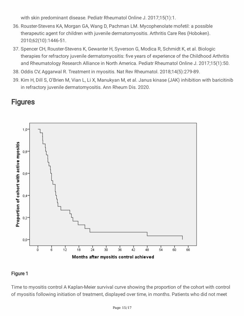
Figure 1
Time to myositis control A Kaplan-Meier survival curve showing the proportion of the cohort with controlof myositis following initiation of treatment, displayed over time, in months. Patients who did not meet
Page 16/17
the outcome were censored after the time of last follow-up. Myositis control was obtained at a median of7.1 months following initiation of treatment.
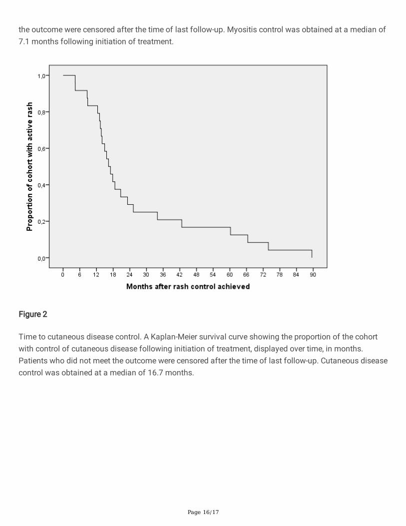
Figure 2
Time to cutaneous disease control. A Kaplan-Meier survival curve showing the proportion of the cohortwith control of cutaneous disease following initiation of treatment, displayed over time, in months.Patients who did not meet the outcome were censored after the time of last follow-up. Cutaneous diseasecontrol was obtained at a median of 16.7 months.
Page 17/17
Figure 3
Standardized height during course of illness and treatment. The effect of disease and treatment fromglucocorticoids on patients’ standardized height over years is shown by a random-effects mixed-modelanalysis. There was no statistically signi�cant difference between the standardized height at the time ofdiagnosis and standardized height at year �ve.