Embed Size (px)
Citation preview
SciPost Phys. 11, 007 (2021)
Field-induced magnetic states in geometrically frustrated SrEr2O4
N. Qureshi1, O. Fabelo1, P. Manuel2, D. D. Khalyavin2, E. Lhotel3,S.X.M. Riberolles1,4, G. Balakrishnan4 and O. A. Petrenko4?
1 Institut Laue-Langevin, 71 Avenue des Martyrs, CS 20156,38042 Grenoble Cedex 9, France
2 ISIS Facility, STFC Rutherford Appleton Laboratory, Chilton,Didcot, OX11 0QX, United Kingdom
3 Institut Néel CNRS, Université Grenoble Alpes, 38042 Grenoble, France4 Department of Physics, University of Warwick, Coventry CV4 7AL, United Kingdom
Abstract
We report an unusual in-field behaviour of SrEr2O4 for a magnetic field applied along twohigh-symmetry directions, the a and c axes. This geometrically frustrated magnet hoststwo crystallographically inequivalent Er ions, Er1 and Er2, that are both located on tri-angular zigzag ladders, but only one site, Er1, forms a long-range magnetic order at lowtemperatures in a zero field. We follow the sequence of peculiar field induced states inSrEr2O4 with detailed single-crystal magnetisation and neutron diffraction experiments.On application of an external field along the c axis, the long-range antiferromagneticorder of the Er1 ions is rapidly destroyed and replaced, in fields between 2 and 5 kOe,by a state with shorter-range correlations. The change in correlation length coincideswith a fast increase in magnetisation during the metamagnetic transition above whicha long-range order is reestablished and maintained into the high fields. The high-fieldferromagnet-like order is characterised by significantly different magnetic moments onthe two Er sites, with the Er1 site dominating the magnetisation process. For the fieldapplied parallel to the a axis, in the field range of 4 to 12 kOe, the planes of diffuse mag-netic scattering observed in zero field due to the one-dimensional correlations betweenthe Er2 moments are replaced by much more localised but still diffuse features corre-sponding to the establishment of an up-up-down structure associated with a one-thirdmagnetisation plateau. Above 14 kOe, a ferromagnet-like high-field order is induced fol-lowing another phase transition. For this direction of the field, the Er2 moments dictatethe succession of transitions while the Er1 moments remain significantly less polarised.A complete field polarisation of both Er sites is not achieved even at 50 kOe for eitherfield direction, reflecting the strongly anisotropic nature of magnetisation process inSrEr2O4.
Copyright N. Qureshi et al.This work is licensed under the Creative CommonsAttribution 4.0 International License.Published by the SciPost Foundation.
Received 04-03-2021Accepted 02-07-2021Published 12-07-2021
Check forupdates
doi:10.21468/SciPostPhys.11.1.007
1
SciPost Phys. 11, 007 (2021)
Contents
1 Introduction 2
2 Experimental details 3
3 Experimental results 53.1 Field along the a axis 53.2 Field along the c axis 8
4 Discussions 12
5 Conclusion 13
A Nuclear structure refinement 14
B Neutron Diffraction Intensity maps 14B.0.1 Scattering plane (0kl), H ‖ a 15B.0.2 Scattering plane (hk0), H ‖ c 15
References 19
1 Introduction
It is not unusual for magnetic systems to demonstrate contrasting behaviour in magnetic fieldsapplied along different crystallographic directions. In fact, considerable anisotropy of mag-netisation is present in most magnets, however, it is rather unusual for a magnetic system toshow completely different physics governing the magnetisation process for the two field direc-tions. We report the results of the investigation of the magnetic properties of SrEr2O4 whichprovides a rare opportunity of observing two contrasting phenomena for the two orthogonalfield directions, a metamagnetic transition corresponding to a spin-flip for one direction and astabilisation of the up-up-down (UUD) structure, usually found in triangular antiferromagnets,for another. Most peculiarly the field-induced UUD state formed by one half of the magnetic Erions appears to be embedded in an ordered antiferromagnetic state formed by the remaininghalf.
SrEr2O4 belongs to the large family of rare-earth (RE) strontium oxides, SrRE2O4. In-terest in the low-temperature magnetic behaviour of this family was initially been sparkedby the paper of Karunadasa et al. [1]. Later studies revealed a wide variety of magnetic be-haviours depending on the choice of the RE ion, ranging from the absence of long-range orderin SrDy2O4 [2–5], the coexistence of two types of short-range order in SrHo2O4 [3, 6, 7], anda noncollinear antiferromagneticic structure in SrYb2O4 [8]. The most interesting results forSrDy2O4 have been found in an applied magnetic field, where the long-range order is stabilisedout of a short-ranged zero-field state [2,9–11]. A somewhat similar field-induced magnetisa-tion process (and perhaps the one closest to SrEr2O4 in character) has recently been reportedfor SrHo2O4 [12], although the local environments are significantly different for the RE mag-netic ions in these two compounds.
The key ingredient for the unconventional behaviour of the SrRE2O4 magnets is the pres-ence of two slightly different crystallographic environments for the RE ions in the orthorhom-
2
SciPost Phys. 11, 007 (2021)
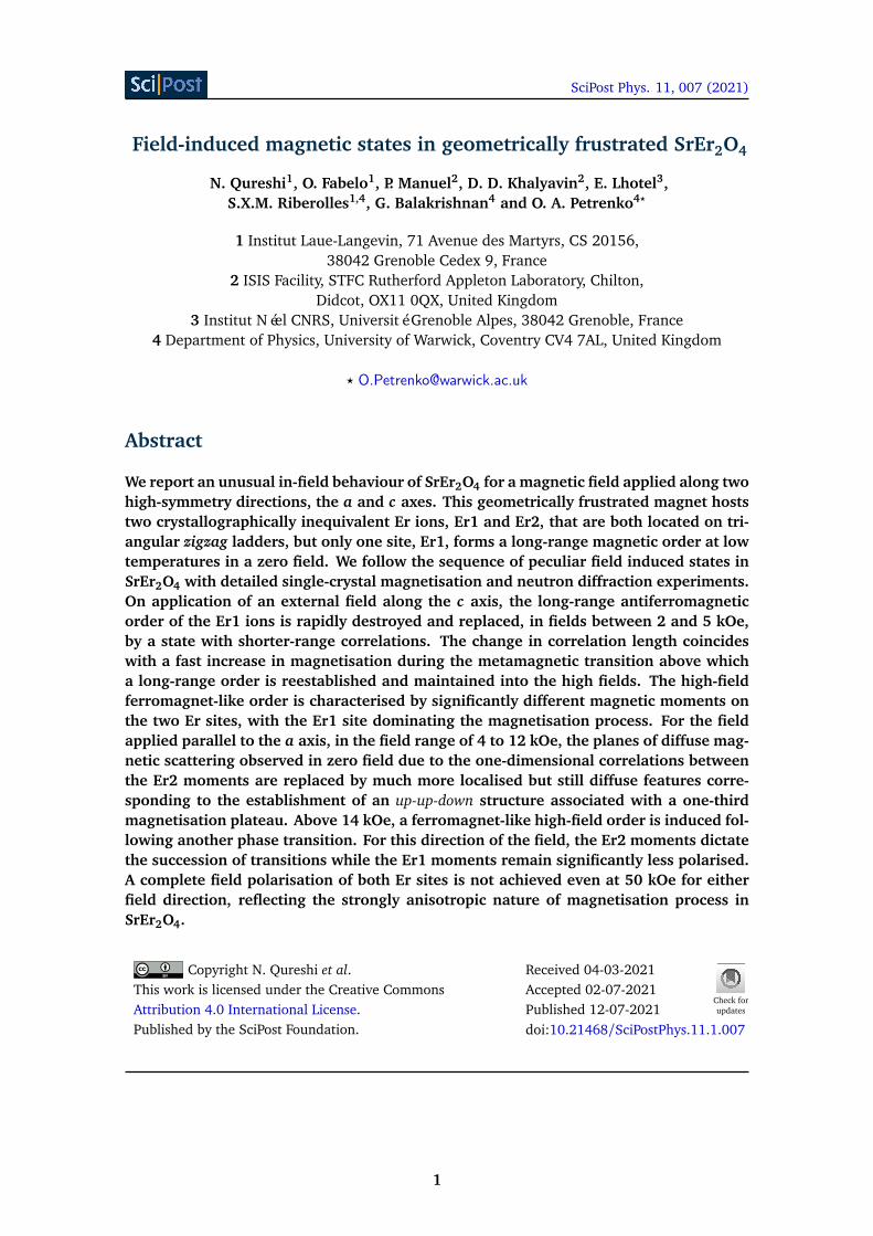
Figure 1: (a) Geometry of the zigzag ladders formed by the Er1 (blue) and Er2 (red)magnetic ions in SrEr2O4. (b) Long-range antiferromagnetic structure formed by theEr1 ions in zero field where the Er2 ions remain largely disordered (although they doparticipate in a formation of weakly correlated spin-chains). (c) Field induced UUDorder of the Er2 ions emerging in almost undistorted zero-field structure of the Er1ions for the field between 4 and 12 kOe applied along the a axis. (d) Field polarisedstructure for H ‖ c in which both Er1 and Er2 ions carry significant but still differentmagnetic moments.
bic unit cell (space group Pnam). The effects of crystal fields are such that the directions ofmagnetic easy axes (or easy planes) are often orthogonal for the two sites [3,13]. In SrEr2O4in particular, relatively small variations in the positions of six nearest-neighbour oxygen ionswhich form distorted octahedra around the Er ions on two sites results in the occurrence ofa strong Ising-like anisotropy with the easy-magnetisation direction along the c axis at theEr1 sites and along the a axis at the Er2 sites [13]. The RE ions on each site form triangu-lar zigzag ladders running along the c axis, while the distances between the ladders (bondsbetween the ions on different sites) are slightly longer, therefore making the interactions be-tween them weaker. Linked together, the zigzag ladders form a distorted honeycomb structurewhen viewed in the a-b plane, see Fig. 1a.
In SrEr2O4, the magnetic moments on the Er1 sites prefer to point along the c axis [13],and then below TN = 0.75 K, they form a fully ordered antiferromagnetic state characterisedby a q = 0 propagation vector as shown in Fig. 1b [14, 15]. The magnetic moments on theEr2 site prefer to point along the a axis [13] and participate in the formation of a short-rangeincommensurate structure, which consists of a collection of weakly-correlated antiferromag-netic chains [15]. The incommensurate chains are nearly antiferromagnetic, characterised bythe q = (0 0 1
2+δ) propagation vector with δ always small and varying between 0.02 and0.06 with temperature [15]. We show below that for the two cases considered in this paper,H ‖ a and H ‖ c, the magnetisation process is mostly governed by the rearrangements of themagnetic moments on the Er2 and Er1 sites, respectively, while the contributions to the mag-netisation from the other site are less significant. It is rare to find such a clear illustrations of
3
SciPost Phys. 11, 007 (2021)
two completely different fundamental processes, one associated with the field-induced selec-tion of a ground state from a degenerate manifold of states in a frustrated magnet [16–19]while another is governed by the peculiar physics of metamagnetic transitions [20], by simplychanging the direction of the applied field in the sample.
2 Experimental details
Single crystal samples of SrEr2O4, grown by the floating zone technique using an infrared im-age furnace as previously reported [21], were studied on the WISH neutron diffractometer [22]at the ISIS facility at the Rutherford Appleton Laboratory (United Kingdom) [23,24] as well ason the diffractometers D9 and D10 [25] at the Institut Laue-Langevin, Grenoble, France. OnWISH, a dilution refrigerator inside a vertical-field cryomagnet provided a base temperatureof 60 mK. The measurements were made in an applied field of up to 50 kOe. The samplewas attached to an oxygen free copper holder with either the a or c axis vertical defining thehorizontal (0kl) or (hk0) scattering planes. Although substantial coverage of the out-of-planescattering is typically available on WISH due to a continuous array of position-sensitive detec-tors, the cryomagnet employed limited the vertical aperture. In order to maximise the diffusesignal, we opted for rather large, irregular shaped samples of about 250 mm3 in volume.
For H ‖ c, the measurements were made in the temperature range from 60 mK to 0.8 K.Zero field measurements made at 15 K, i.e. well above TN were used to isolate the nuclearcomponent of the scattering allowing for the extraction of magnetic intensity by direct subtrac-tion. For integer (h k l), only the l = 0 reflections could be reached, however, a broad diffusescattering signal was observed in the (h k −1/2) plane. For H ‖ a, only the h = 0 reflectionscould be reached for integer values (h k l). For this direction of applied field, the experimentwas performed prior to the addition of the second detector bank on the WISH instrument.
The data collected on WISH have been treated with the Mantid software [26] whichtakes into account vanadium normalisation as well as a correction for absorption. The dif-fuse scattering features and their evolution with an applied field are best seen through thetwo-dimensional intensity maps in reciprocal space, particularly after the higher-temperaturebackground subtraction.
Measurements performed on the hot-neutron diffractometer D9 in zero field and four-circlegeometry were used to derive the precise crystal structure of SrEr2O4 including the extinctionparameters of the crystal used (see Appendix, Sec. A). A small cuboid sample of dimension1.1× 3.7× 2.9 mm3 along the a, b and c axes, respectively, was employed for these measure-ments in order to reduce the beam absorption and problems due to extinction and multiplescattering. The wavelength of the monochromatic beam was fixed at 0.84 Å and with thesample temperature at 20 K, 541 unique reflections were collected.
D9 was then operated with a cryomagnet in normal-beam geometry for further measure-ments at 1.5 K in a field of 20 kOe applied along the c axis in order to complement the WISHmeasurements. The wavelength was set at 0.51 Å and a set of 131 unique reflections in the ac-cessible reciprocal space was measured. Each reflection was measured twice, with and withoutapplied field in order to isolate the magnetic signal by direct subtraction.
An additional experiment was carried out using the D10 diffractometer using a dilutionrefrigerator and a vertical cryomagnet. The cuboid-shaped single-crystal sample of approxi-mate dimensions 2.5×1×3 mm3 along the principal crystallographic axes was glued to the tipof a Cu pin with the a axis vertical, i.e. parallel to the applied magnetic field. A wavelengthof 2.36 Å was selected from a pyrolytic graphite monochromator and the sample was cooledto the base temperature of 50 mK. Data sets of 151 unique integer reflections were collectedat 50 mK and at magnetic field values of 0, 6, 11 and 14 kOe, while a reduced data set of
4
SciPost Phys. 11, 007 (2021)
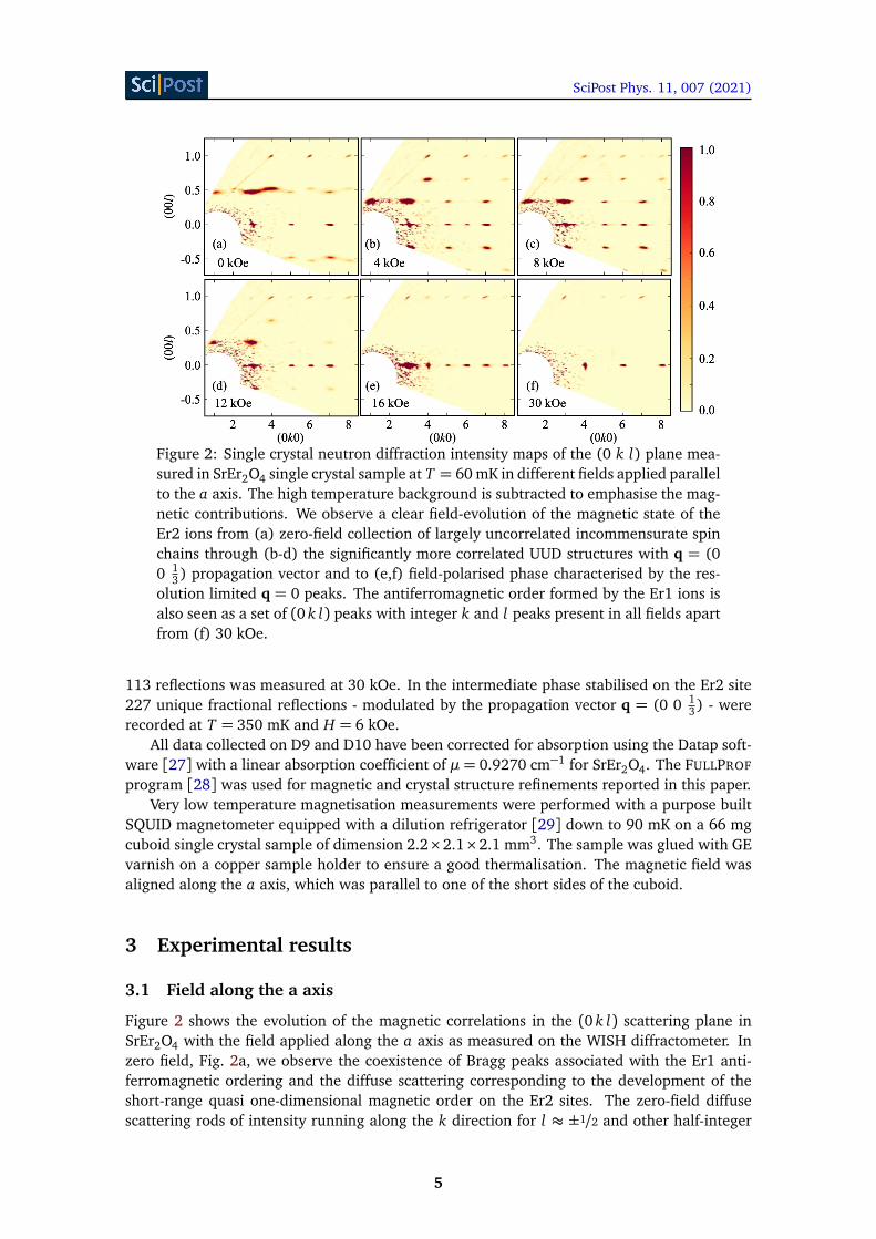
Figure 2: Single crystal neutron diffraction intensity maps of the (0 k l) plane mea-sured in SrEr2O4 single crystal sample at T = 60 mK in different fields applied parallelto the a axis. The high temperature background is subtracted to emphasise the mag-netic contributions. We observe a clear field-evolution of the magnetic state of theEr2 ions from (a) zero-field collection of largely uncorrelated incommensurate spinchains through (b-d) the significantly more correlated UUD structures with q = (00 1
3) propagation vector and to (e,f) field-polarised phase characterised by the res-olution limited q = 0 peaks. The antiferromagnetic order formed by the Er1 ions isalso seen as a set of (0 k l) peaks with integer k and l peaks present in all fields apartfrom (f) 30 kOe.
113 reflections was measured at 30 kOe. In the intermediate phase stabilised on the Er2 site227 unique fractional reflections - modulated by the propagation vector q = (0 0 1
3) - wererecorded at T = 350 mK and H = 6 kOe.
All data collected on D9 and D10 have been corrected for absorption using the Datap soft-ware [27] with a linear absorption coefficient of µ= 0.9270 cm−1 for SrEr2O4. The FULLPROF
program [28] was used for magnetic and crystal structure refinements reported in this paper.Very low temperature magnetisation measurements were performed with a purpose built
SQUID magnetometer equipped with a dilution refrigerator [29] down to 90 mK on a 66 mgcuboid single crystal sample of dimension 2.2×2.1×2.1 mm3. The sample was glued with GEvarnish on a copper sample holder to ensure a good thermalisation. The magnetic field wasaligned along the a axis, which was parallel to one of the short sides of the cuboid.
3 Experimental results
3.1 Field along the a axis
Figure 2 shows the evolution of the magnetic correlations in the (0 k l) scattering plane inSrEr2O4 with the field applied along the a axis as measured on the WISH diffractometer. Inzero field, Fig. 2a, we observe the coexistence of Bragg peaks associated with the Er1 anti-ferromagnetic ordering and the diffuse scattering corresponding to the development of theshort-range quasi one-dimensional magnetic order on the Er2 sites. The zero-field diffusescattering rods of intensity running along the k direction for l ≈ ±1/2 and other half-integer
5
SciPost Phys. 11, 007 (2021)
01234
0 5 1 0 1 5 2 00 . 0
0 . 2
0 . 4
0 . 6
M (µ B
/Er3+
ion)
T ( K ) 0 . 0 9 0 . 3 0 . 5 0 . 7 0 . 9 1 . 5 2 . 0
( a )
dM/dH
(µB/k
Oe Er
3+)
H ( k O e )
( b )
H / / a
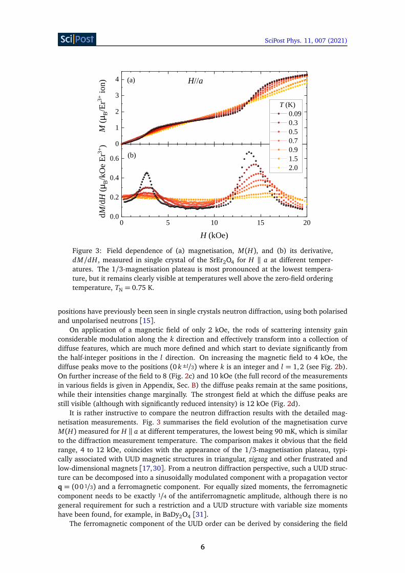
Figure 3: Field dependence of (a) magnetisation, M(H), and (b) its derivative,dM/dH, measured in single crystal of the SrEr2O4 for H ‖ a at different temper-atures. The 1/3-magnetisation plateau is most pronounced at the lowest tempera-ture, but it remains clearly visible at temperatures well above the zero-field orderingtemperature, TN = 0.75 K.
positions have previously been seen in single crystals neutron diffraction, using both polarisedand unpolarised neutrons [15].
On application of a magnetic field of only 2 kOe, the rods of scattering intensity gainconsiderable modulation along the k direction and effectively transform into a collection ofdiffuse features, which are much more defined and which start to deviate significantly fromthe half-integer positions in the l direction. On increasing the magnetic field to 4 kOe, thediffuse peaks move to the positions (0 k ±l/3) where k is an integer and l = 1,2 (see Fig. 2b).On further increase of the field to 8 (Fig. 2c) and 10 kOe (the full record of the measurementsin various fields is given in Appendix, Sec. B) the diffuse peaks remain at the same positions,while their intensities change marginally. The strongest field at which the diffuse peaks arestill visible (although with significantly reduced intensity) is 12 kOe (Fig. 2d).
It is rather instructive to compare the neutron diffraction results with the detailed mag-netisation measurements. Fig. 3 summarises the field evolution of the magnetisation curveM(H) measured for H ‖ a at different temperatures, the lowest being 90 mK, which is similarto the diffraction measurement temperature. The comparison makes it obvious that the fieldrange, 4 to 12 kOe, coincides with the appearance of the 1/3-magnetisation plateau, typi-cally associated with UUD magnetic structures in triangular, zigzag and other frustrated andlow-dimensional magnets [17,30]. From a neutron diffraction perspective, such a UUD struc-ture can be decomposed into a sinusoidally modulated component with a propagation vectorq = (00 1/3) and a ferromagnetic component. For equally sized moments, the ferromagneticcomponent needs to be exactly 1/4 of the antiferromagnetic amplitude, although there is nogeneral requirement for such a restriction and a UUD structure with variable size momentshave been found, for example, in BaDy2O4 [31].
The ferromagnetic component of the UUD order can be derived by considering the field
6
SciPost Phys. 11, 007 (2021)
02 0 04 0 0
02 0 04 0 06 0 0
0
5 0 0
1 0 0 0
( 0 6 0 ) ( a ) ( 0 3 0 ) ( 0 - 3 0 )
Integr
ated I
ntensi
ty (ar
b. unit
s)
( b )
( 0 3 1 / 3 ) ( 0 3 - 1 / 3 )
( c )
0 5 1 0 1 5 2 0 2 5 3 00
2
4
H ( k O e )
M (µ B
/Er3+
ion)
( d ) d2 M/dH
2
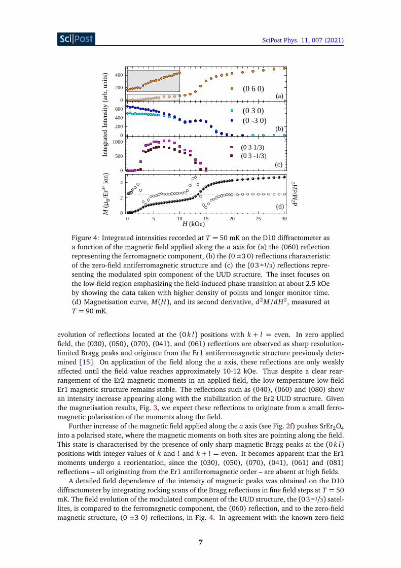
Figure 4: Integrated intensities recorded at T = 50 mK on the D10 diffractometer asa function of the magnetic field applied along the a axis for (a) the (060) reflectionrepresenting the ferromagnetic component, (b) the (0±3 0) reflections characteristicof the zero-field antiferromagnetic structure and (c) the (0 3±1/3) reflections repre-senting the modulated spin component of the UUD structure. The inset focuses onthe low-field region emphasizing the field-induced phase transition at about 2.5 kOeby showing the data taken with higher density of points and longer monitor time.(d) Magnetisation curve, M(H), and its second derivative, d2M/dH2, measured atT = 90 mK.
evolution of reflections located at the (0 k l) positions with k + l = even. In zero appliedfield, the (030), (050), (070), (041), and (061) reflections are observed as sharp resolution-limited Bragg peaks and originate from the Er1 antiferromagnetic structure previously deter-mined [15]. On application of the field along the a axis, these reflections are only weaklyaffected until the field value reaches approximately 10-12 kOe. Thus despite a clear rear-rangement of the Er2 magnetic moments in an applied field, the low-temperature low-fieldEr1 magnetic structure remains stable. The reflections such as (040), (060) and (080) showan intensity increase appearing along with the stabilization of the Er2 UUD structure. Giventhe magnetisation results, Fig. 3, we expect these reflections to originate from a small ferro-magnetic polarisation of the moments along the field.
Further increase of the magnetic field applied along the a axis (see Fig. 2f) pushes SrEr2O4into a polarised state, where the magnetic moments on both sites are pointing along the field.This state is characterised by the presence of only sharp magnetic Bragg peaks at the (0 k l)positions with integer values of k and l and k + l = even. It becomes apparent that the Er1moments undergo a reorientation, since the (030), (050), (070), (041), (061) and (081)reflections – all originating from the Er1 antiferromagnetic order – are absent at high fields.
A detailed field dependence of the intensity of magnetic peaks was obtained on the D10diffractometer by integrating rocking scans of the Bragg reflections in fine field steps at T = 50mK. The field evolution of the modulated component of the UUD structure, the (03±1/3) satel-lites, is compared to the ferromagnetic component, the (060) reflection, and to the zero-fieldmagnetic structure, (0 ±3 0) reflections, in Fig. 4. In agreement with the known zero-field
7
SciPost Phys. 11, 007 (2021)
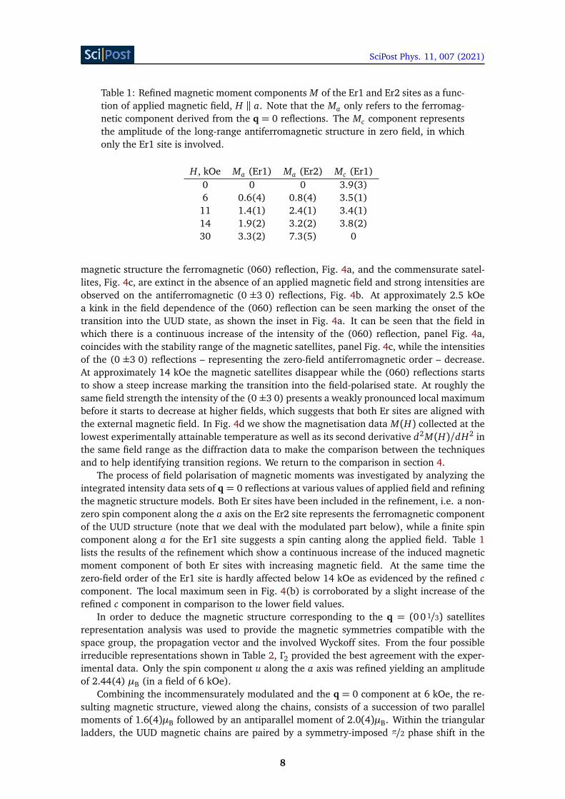
Table 1: Refined magnetic moment components M of the Er1 and Er2 sites as a func-tion of applied magnetic field, H ‖ a. Note that the Ma only refers to the ferromag-netic component derived from the q = 0 reflections. The Mc component representsthe amplitude of the long-range antiferromagnetic structure in zero field, in whichonly the Er1 site is involved.
H, kOe Ma (Er1) Ma (Er2) Mc (Er1)0 0 0 3.9(3)6 0.6(4) 0.8(4) 3.5(1)11 1.4(1) 2.4(1) 3.4(1)14 1.9(2) 3.2(2) 3.8(2)30 3.3(2) 7.3(5) 0
magnetic structure the ferromagnetic (060) reflection, Fig. 4a, and the commensurate satel-lites, Fig. 4c, are extinct in the absence of an applied magnetic field and strong intensities areobserved on the antiferromagnetic (0 ±3 0) reflections, Fig. 4b. At approximately 2.5 kOea kink in the field dependence of the (060) reflection can be seen marking the onset of thetransition into the UUD state, as shown the inset in Fig. 4a. It can be seen that the field inwhich there is a continuous increase of the intensity of the (060) reflection, panel Fig. 4a,coincides with the stability range of the magnetic satellites, panel Fig. 4c, while the intensitiesof the (0 ±3 0) reflections – representing the zero-field antiferromagnetic order – decrease.At approximately 14 kOe the magnetic satellites disappear while the (060) reflections startsto show a steep increase marking the transition into the field-polarised state. At roughly thesame field strength the intensity of the (0 ±3 0) presents a weakly pronounced local maximumbefore it starts to decrease at higher fields, which suggests that both Er sites are aligned withthe external magnetic field. In Fig. 4d we show the magnetisation data M(H) collected at thelowest experimentally attainable temperature as well as its second derivative d2M(H)/dH2 inthe same field range as the diffraction data to make the comparison between the techniquesand to help identifying transition regions. We return to the comparison in section 4.
The process of field polarisation of magnetic moments was investigated by analyzing theintegrated intensity data sets of q = 0 reflections at various values of applied field and refiningthe magnetic structure models. Both Er sites have been included in the refinement, i.e. a non-zero spin component along the a axis on the Er2 site represents the ferromagnetic componentof the UUD structure (note that we deal with the modulated part below), while a finite spincomponent along a for the Er1 site suggests a spin canting along the applied field. Table 1lists the results of the refinement which show a continuous increase of the induced magneticmoment component of both Er sites with increasing magnetic field. At the same time thezero-field order of the Er1 site is hardly affected below 14 kOe as evidenced by the refined ccomponent. The local maximum seen in Fig. 4(b) is corroborated by a slight increase of therefined c component in comparison to the lower field values.
In order to deduce the magnetic structure corresponding to the q = (0 0 1/3) satellitesrepresentation analysis was used to provide the magnetic symmetries compatible with thespace group, the propagation vector and the involved Wyckoff sites. From the four possibleirreducible representations shown in Table 2, Γ2 provided the best agreement with the exper-imental data. Only the spin component u along the a axis was refined yielding an amplitudeof 2.44(4) µB (in a field of 6 kOe).
Combining the incommensurately modulated and the q = 0 component at 6 kOe, the re-sulting magnetic structure, viewed along the chains, consists of a succession of two parallelmoments of 1.6(4)µB followed by an antiparallel moment of 2.0(4)µB. Within the triangularladders, the UUD magnetic chains are paired by a symmetry-imposed π/2 phase shift in the
8
SciPost Phys. 11, 007 (2021)
Table 2: Basis vectors ψn (n = 1-4) of the irreducible representations Γn for the Erions m = 1-4 at given fractional coordinates (x , y , z) associated with a propagationvector q= (00 1/3). The components u, v and w connected to the spin Sm
Γnhave been
refined according to their constraints (an overline indicates a negative number). Thephase factor φ = exp(−2πiqr) results from the translation r = (0 0 1/2) associatedwith the screw axis parallel to the c axis or r= (0 1/2 1/2) connected to the glide planeperpendicular to the a axis.
Atom m Position ψ1 ψ2 ψ3 ψ4
1
xy
1/4
uvw
uvw
uvw
uvw
2
xy
3/4
φ ·
uvw
φ ·
uvw
φ ·
uvw
φ ·
uvw
3
x + 1/2
y + 1/23/4
φ ·
uvw
φ ·
uvw
φ ·
uvw
φ ·
uvw
4
x + 1/2
y + 1/21/4
uvw
uvw
uvw
uvw
magnetic moment sequence, see Fig. 1c. We believe that this structure is largely responsiblefor the appearance of a one-third magnetisation plateau shown in Fig. 3.
3.2 Field along the c axis
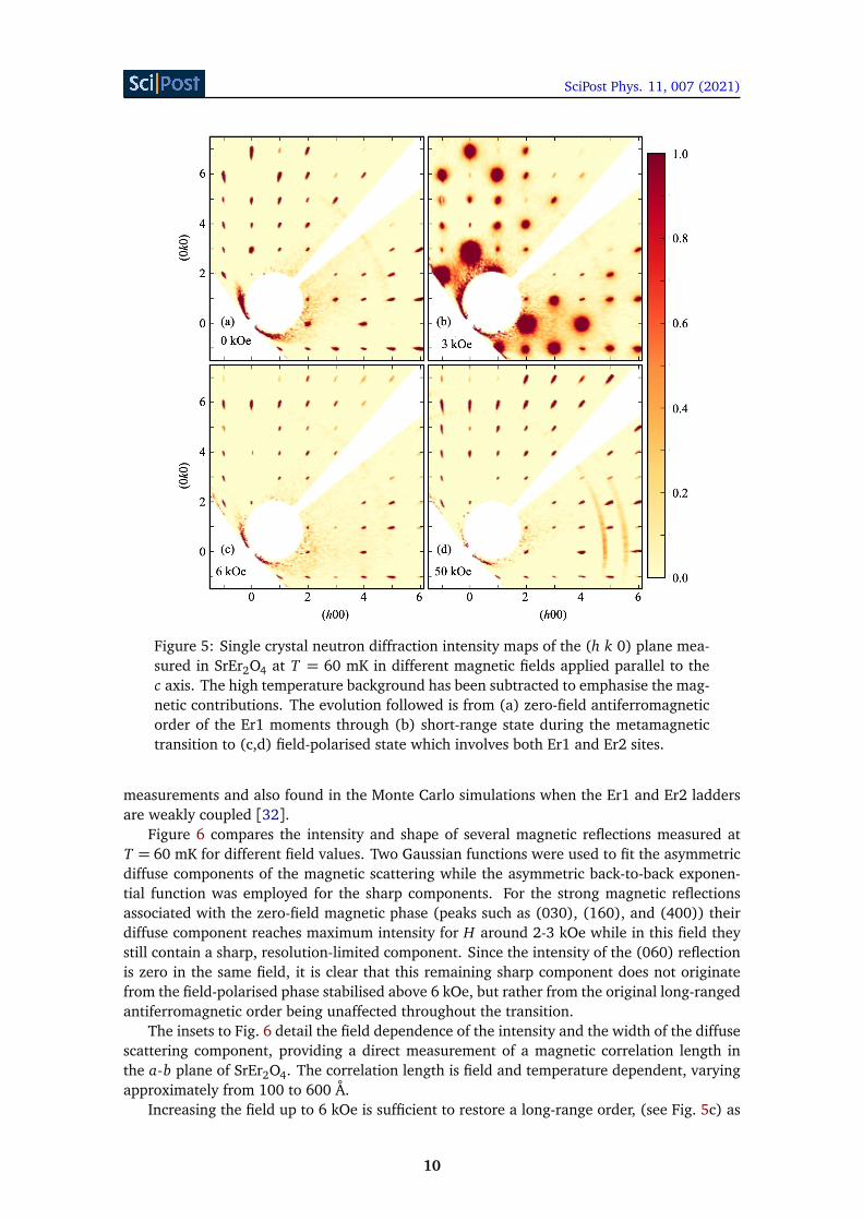
Figure 5 captures the main result of the experiment with H ‖ c on the WISH diffractometer.In zero field [see Fig. 5(a)], the diffraction pattern in the (h k 0) scattering plane consists ofsharp, resolution-limited Bragg peaks representing the q = 0 magnetic order of the Er1 sitein full agreement with the previous powder [14] and single crystal [15] neutron diffractionresults. Our further single-crystal data collected on the same instrument under similar condi-tions, H = 0 kOe, T ≈ 0.6 K, (not shown) confirm the previously established structure deducedfrom powder neutron diffraction [14]: a parallel alignment of the magnetic moments on Er1sites forming the chains running along the c axis with nearest-neighbour chains paired an-tiferromagnetically and only a short-range antiferromagnetic order on the Er2 sites with themoments along the a axis.
Application of a moderate field of 3 kOe (see Fig. 5b) causes significant decrease of theintensity and almost the disappearance of the sharp Bragg peaks, which are replaced by broaddiffuse scattering features. The broad peaks are centred around the same q = 0 positions asthe sharp peaks suggesting that the correlation length of the antiferromagnetic state becomeslimited. The region of fields where diffuse scattering dominates the pattern, 2 to 5 kOe, co-incides with an abrupt increase of magnetisation during a metamagnetic transition [32]. Theshape of a peculiar lozenge pattern formed by the diffuse scattering features in these fieldsis practically identical to the high-temperature (T > TN = 0.75 K) signal seen in zero-field
9
SciPost Phys. 11, 007 (2021)
Figure 5: Single crystal neutron diffraction intensity maps of the (h k 0) plane mea-sured in SrEr2O4 at T = 60 mK in different magnetic fields applied parallel to thec axis. The high temperature background has been subtracted to emphasise the mag-netic contributions. The evolution followed is from (a) zero-field antiferromagneticorder of the Er1 moments through (b) short-range state during the metamagnetictransition to (c,d) field-polarised state which involves both Er1 and Er2 sites.
measurements and also found in the Monte Carlo simulations when the Er1 and Er2 laddersare weakly coupled [32].
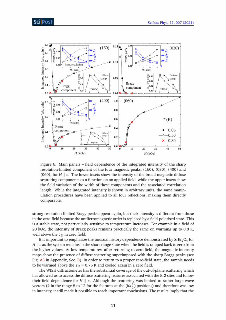
Figure 6 compares the intensity and shape of several magnetic reflections measured atT = 60 mK for different field values. Two Gaussian functions were used to fit the asymmetricdiffuse components of the magnetic scattering while the asymmetric back-to-back exponen-tial function was employed for the sharp components. For the strong magnetic reflectionsassociated with the zero-field magnetic phase (peaks such as (030), (160), and (400)) theirdiffuse component reaches maximum intensity for H around 2-3 kOe while in this field theystill contain a sharp, resolution-limited component. Since the intensity of the (060) reflectionis zero in the same field, it is clear that this remaining sharp component does not originatefrom the field-polarised phase stabilised above 6 kOe, but rather from the original long-rangedantiferromagnetic order being unaffected throughout the transition.
The insets to Fig. 6 detail the field dependence of the intensity and the width of the diffusescattering component, providing a direct measurement of a magnetic correlation length inthe a-b plane of SrEr2O4. The correlation length is field and temperature dependent, varyingapproximately from 100 to 600 Å.
Increasing the field up to 6 kOe is sufficient to restore a long-range order, (see Fig. 5c) as
10
SciPost Phys. 11, 007 (2021)
0 1 0 2 0 3 0 4 0 5 00 . 0
0 . 1
0 . 2
0 . 3
2 4 60 . 0
0 . 1
2 3 4 50 . 0 0
0 . 0 2
0 . 0 4
Integr
ated I
ntensi
ty (ar
b. unit
s)
H ( k O e )
( 4 0 0 )
B r a g gc o m p o n e n t
Int. (a
rb. un
its)
H ( k O e )
D i f f u s ec o m p .
FWHM
(Å-1 )
H ( k O e )0
2 0 0
4 0 0
Lengt
h (Å)
0 1 0 2 0 3 0 4 0 5 00 . 0
0 . 2
0 . 4
0 . 6
0 . 8
1 . 0 ( 0 6 0 )
H ( k O e )
T ( K )0 . 0 60 . 5 00 . 8 0
0 . 0 0
0 . 0 5
0 . 1 0
0 . 1 5
2 4 60 . 0 00 . 0 20 . 0 40 . 0 6
2 3 4 50 . 0 0
0 . 0 2
0 . 0 4 ( 0 3 0 )
B r a g gc o m p o n e n t Int
. (arb.
units)
H ( k O e )
D i f f u s ec o m p .
FWHM
(Å-1 )
H ( k O e )02 0 04 0 06 0 0
Lengt
h (Å)
0 . 0
0 . 1
0 . 2
0 . 3
0 . 4
0 . 5
0 . 6
2 4 60 . 0
0 . 1
0 . 2
2 3 4 50 . 0 0
0 . 0 2
0 . 0 4
Integr
ated I
ntensi
ty (ar
b. unit
s) ( 1 6 0 )
B r a g gc o m p o n e n t Int
. (arb.
units)
H ( k O e )
D i f f u s ec o m p .
FWHM
(Å-1 )
H ( k O e )I n s t r . r e s o l u t i o n 0
1 0 02 0 03 0 0
Lengt
h (Å)
Figure 6: Main panels – field dependence of the integrated intensity of the sharpresolution-limited component of the four magnetic peaks, (160), (030), (400) and(060), for H ‖ c. The lower insets show the intensity of the broad magnetic diffusescattering components as a function on an applied field, while the upper insets showthe field variation of the width of these components and the associated correlationlength. While the integrated intensity is shown in arbitrary units, the same manip-ulation procedures have been applied to all four reflections, making them directlycomparable.
strong resolution-limited Bragg peaks appear again, but their intensity is different from thosein the zero-field because the antiferromagnetic order is replaced by a field-polarised state. Thisis a stable state, not particularly sensitive to temperature increases. For example in a field of20 kOe, the intensity of Bragg peaks remains practically the same on warming up to 0.8 K,well above the TN in zero field.
It is important to emphasize the unusual history dependence demonstrated by SrEr2O4 forH ‖ c as the system remains in the short-range state when the field is ramped back to zero fromthe higher values. At low temperatures, after returning to zero field, the magnetic intensitymaps show the presence of diffuse scattering superimposed with the sharp Bragg peaks (seeFig. A5 in Appendix, Sec. B). In order to return to a proper zero-field state, the sample needsto be warmed above the TN = 0.75 K and cooled again in a zero field.
The WISH diffractometer has the substantial coverage of the out-of-plane scattering whichhas allowed us to access the diffuse scattering features associated with the Er2 sites and followtheir field dependence for H ‖ c. Although the scattering was limited to rather large wavevectors (k in the range 8 to 12 for the features at the (hk 1
2) positions) and therefore was lowin intensity, it still made it possible to reach important conclusions. The results imply that the
11
SciPost Phys. 11, 007 (2021)
structure formed by the Er2 moments is not very sensitive to the lower values of the externalfield applied along the c axis, the intensity drops rapidly above the metamagnetic transition, itstill remains present in 10 kOe and completely disappears in 20 kOe (see Fig. A6 in Appendix,Sec. B). This observation is consistent with a picture of a gradual polarisation of the Er2 sitesby the applied transverse field.
In order to extract the magnetic moments on both Er sites in the field polarised state weanalysed the monochromatic data set from the D9 diffractometer collected at T = 1.5 K andH = 20 kOe. Each reflection was measured twice, with and without applied magnetic field toallow for the isolation of the magnetic component of the scattering. The refinement returnsthe values of 5.4(1) and 1.9(1) µB for the moments on Er1 and Er2 sites, respectively, if aferromagnetic model is presumed on both Er sites. The average magnetic moment is then3.65 µB per Er ion for this value of a field applied along the c axis in a good agreement withthe previous magnetisation measurements [14,32].
4 Discussions
The experimental findings for SrEr2O4 in magnetic field applied along the c axis draw imme-diate comparisons with the properties of metamagnets, the antiferromagnets which in appliedfield undergo a phase transition into a state with a relatively large magnetic moment and whichhave been extensively investigated in the past, both experimentally and theoretically [20,33].In metamagnets, quite often the transition from an antiferromagnetic ground state to a field-polarised state (labeled as paramagnetic) is through an intermediate so called mixed phase inwhich domains of the antiferromagnetic and paramagnetic phases coexist. The origin of themixed phase is the presence of the demagnetizing field which forces a first-order transitionto be spread out over a range of applied fields [34]. Neutron diffraction measurements (see,for example, [35–37]) played a pivotal role in establishing the detailed magnetic phase di-agrams of metamagnetic materials, including discontinuous transitions and tricritical points,however, we could not find in published literature any neutron diffraction measurements ofthe magnetic correlation length in a mixed phase (similar to those shown in Fig. 6). Perhapsthis is not entirely surprising, as most of the early neutron scattering experiments were per-formed several decades ago, when the neutron instruments were lacking in both resolutionand reciprocal space coverage compared to the state-of-the-art diffractometers.
One feature that clearly distinguishes SrEr2O4 from other metamagnets is the presence ofthe second sublattice which does not participate directly in the metamagnetic transition butstill influences significantly the paramagnetic phase. In this respect the state near TN and in lowfields is exposed to a weak staggered field from weakly correlated chains of magnetic momentson the Er2 sites, while the state near the saturation field at low temperatures is exposed to asignificantly stronger field of the Er2 moments polarised along the field. It would be beneficialto explore in detail the magnetic H − T phase diagram of SrEr2O4 using, for example, heatcapacity or ultrasound velocity measurements which have been successfully used for mappingout phase transitions in similar compounds [2,9].
Remarkably, the picture of broad diffuse-scattering peaks in the (h k 0) plane characterisingthe intermediate state of SrEr2O4 is identical to what is observed in SrHo2O4 in zero field [6].Despite almost indistinguishable magnetisation curves for H ‖ c in these two compounds [32],the sequence of the field induced phases is rather different. One could envisage two possiblereasons for such a difference. The first one is to do with a possible splitting of the quasidou-blet states of the Ho3+ non-Kramers ions by crystalline electric fields (as briefly mentionedin Ref. [13]). Alternatively, in view of what we observed for SrEr2O4, it is possible that themixed state in SrHo2O4 is so wide that it extends from practically zero field all the way to the
12
SciPost Phys. 11, 007 (2021)
saturation field.With the demagnetisation effects being significant, ideally one has to correct for them by
subtracting the demagnetising field from the applied field. This procedure is straightforwardfor ellipsoidal samples for which the demagnetising factor depends only on sample shape,but is much more involved for non-ellipsoidal samples [38]. Faced with the necessity to usecuboid samples because of the low-temperature thermalisation requirements we report theuncorrected data. From previous magnetisation measurements [32] the demagnetising fieldwas found always to be less than 5% of the applied field. It is also worth pointing out thatour earlier measurements performed on the PRISMA instrument (technical details are given inRef. [15]) using differently shaped samples returned results identical to those reported here.
The experimental results presented in the previous sections strongly suggest that a fulldescription of the magnetic properties of SrEr2O4, would require a theoretical model whichapart from the exchange and dipolar interactions includes finite magnetic anisotropy term(s)allowing for the deviations of the magnetic moment away from their easy-axis orientationsin an applied magnetic field. However, it is still very useful to consider a simplified modelconsisting of two Ising-type sublattices on the two Er sites. Finding the general ground statesolutions for such a model in an applied field is an interesting and complex problem in its ownright [39], but one prediction for such a model seems to be especially relevant for SrEr2O4. Theprediction is that for H ‖ a, the zero-field phase cannot directly transform into the UUD stateand therefore an intermediate phase should exist. In this respect, an intriguing proposition isthat the observed change in magnetisation rate in a transition regime between zero field andmagnetisation plateau (see Fig. 3) marks the stability range of this additional phase. For lowerfields (less than 1.2 kOe) the slope of the magnetisation curve is 0.16 µB/kOe, on the plateau(between 5.5 and 10 kOe) it is 0.09 µB/kOe while in a transition region it is 0.39 µB/kOe.Within the Ising model [39], the predicted intermediate phase is characterised by the splittingof the Er1 site ladders into the two different types, one with the periodicity 2 along the c axis(UDUD state) and another with periodicity 3 (UUD state). The ratio of the number of theUDUD to the UUD ladders is three to one, therefore the overall magnetisation is fixed at 1
8 ofthe saturation magnetisation for the Er1 site. In the experiment, however, the magnetisationrapidly increases with field, perhaps a more realistic representation of the system is in fact afield-dependent ratio of the UDUD and UUD ladders. In a field of 2 kOe, neutron diffractiondetects diffuse peaks at the positions (0 k 1
2±δ)with δ = 0.05 (see Fig. A1 in Appendix, Sec. B).This observation is also consistent with the idea of a gradual evolution from the zero-field statecharacterised by δ = 0.02 to the UUD state with δ = 1
6 through a field-dependent mixture ofthe UDUD to the UUD ladders.
In terms of theoretical modelling, the one-dimensional ANNNI model was previously usedto describe the magnetic properties of SrHo2O4 and SrDy2O4 [7,11]. The model captures sev-eral features (including the UUD state for longitudinal fields) and allows for the introduction ofthe effective exchange constants, however, being minimalistic in nature, it ignores the interac-tions between the ladders and therefore excludes the possibility of the complex intermediatephases discussed above. While the magnetisation curves look similar for the magnetic fieldapplied along the easy-axis in SrHo2O4, SrDy2O4 and SrEr2O4 [32], the long-range magneticorder in the RE1 sublattice is established only in SrEr2O4, making it a unique case.
In many respects the behaviour of SrEr2O4 is similar to what is found in spin ice materials,including the presence of magnetic disorder down to the lowest temperatures and the unusualfield-induced transitions (such as three-dimensional Kasteleyn transition for H ‖ [100] [40]).For H ‖ [110] the spin ice breaks into magnetic chains [41] characterised by the appearanceof metastable states and unusually slow dynamics, as well as non-ergodicity (in common withwhat is observed in SrEr2O4 for H ‖ c).
The analogy with the low-T behaviour of spin ice materials might extend even further.
13
SciPost Phys. 11, 007 (2021)
An interesting general analogy could be drawn here with the phenomenon of moment frag-mentation [42–44] which gives rise to a coexistence of spin liquid and ordered states in otherfrustrated magnets. The SrRE2O4 systems often demonstrate a coexistence of ordered anddisordered magnetic components (which reside on different RE sites) and if the concept offragmentation is potentially extendable to them then the key question must be about quantumor classical origin of fragmentation.
5 Conclusion
The field evolution of the low-temperature magnetic structure of SrEr2O4 was studied bysingle-crystal neutron diffraction for the two directions of the applied magnetic field, H ‖ aand H ‖ c as well as by low-temperature magnetisation measurements for H ‖ a. Reflecting thehighly anisotropic Ising-like nature of the compound which contains magnetic Er ions on twodifferent crystallographic sites, Er1 and Er2, the field-induced transitions for H ‖ a are mostlyto do with the rearrangements of the magnetic moments on the Er2 sites, while for H ‖ c, themoments on the Er1 sites mainly determine the value and the properties of the metamagnetictransition. For H ‖ a, the applied field induces a transition to an extended, but still not fullylong-range ordered UUD state of the Er2 sites, which remains stable in a significantly widerange of the fields, 4 to 12 kOe, before changing to the state with all Er2 moments parallel tothe field. For H ‖ c, the applied field initially destroys the long-range antiferromagnetic orderof the Er1 sites and induces a much shorter-range order during the metamagnetic transition at2 kOe prior to establishing a long-range ferromagnetic-like state of the Er1 moments at 6 kOe.
The development of a detailed theoretical model is now required to fully appreciate thefascinating sequence of field-induced magnetic phases found in SrEr2O4.
Acknowledgements
We are grateful to B.Z. Malkin and Yu.I. Dublenych for numerous discussions of the magneticproperties of the SrRE2O4 compounds, to T.J. Hayes for the help with sample preparationsand initial neutron diffraction measurements as well as to S.T. Bramwell and M.R. Lees forcareful reading the manuscript and helpful suggestions. We would also like to acknowledgethe expertise and dedication of the low-temperature groups at both ISIS and Institut Laue-Langevin. The work at the University of Warwick was supported by EPSRC through grantsEP/I007210/1, EP/M028771/1, and EP/T005963/1.
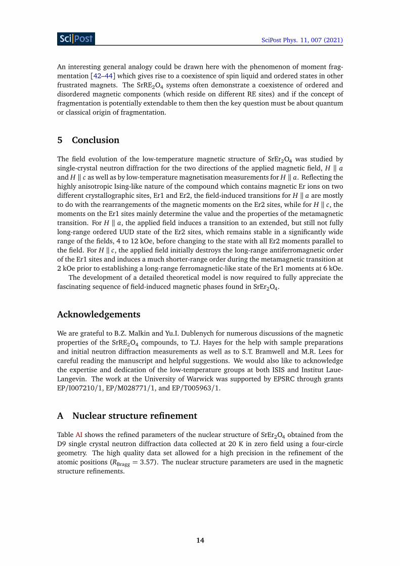
A Nuclear structure refinement
Table AI shows the refined parameters of the nuclear structure of SrEr2O4 obtained from theD9 single crystal neutron diffraction data collected at 20 K in zero field using a four-circlegeometry. The high quality data set allowed for a high precision in the refinement of theatomic positions (RBragg = 3.57). The nuclear structure parameters are used in the magneticstructure refinements.
14
SciPost Phys. 11, 007 (2021)
Table AI: Refined ionic positions of SrEr2O4 obtained from neutron diffractiondata measured at 20 K. The dimensions of the orthorhombic (Pnam) unit cell area = 10.0189 Å, b = 11.8611(9) Å and c = 3.3893 Å. All the ions occupy the 4c Wyck-off sites with general positions (x , y, 0.25).
Atom x ySr 0.7523(1) 0.6500(3)
Er1 0.4225(2) 0.11Er2 0.4232(8) 0.6120(9)O1 0.2119(5) 0.1745(9)O2 0.1262(3) 0.4799(6)O3 0.5146(2) 0.7836(1)O4 0.4250(1) 0.4222(3)
B Neutron Diffraction Intensity maps
Intensity maps have been collected on the WISH time-of-flight diffractometer at ISIS for a sin-gle sample orientation with H ‖ a and for the two different sample positions with H ‖ c. In allcases the high-temperature background was subtracted to emphasize the magnetic contribu-tions.
B.0.1 Scattering plane (0kl), H ‖ a
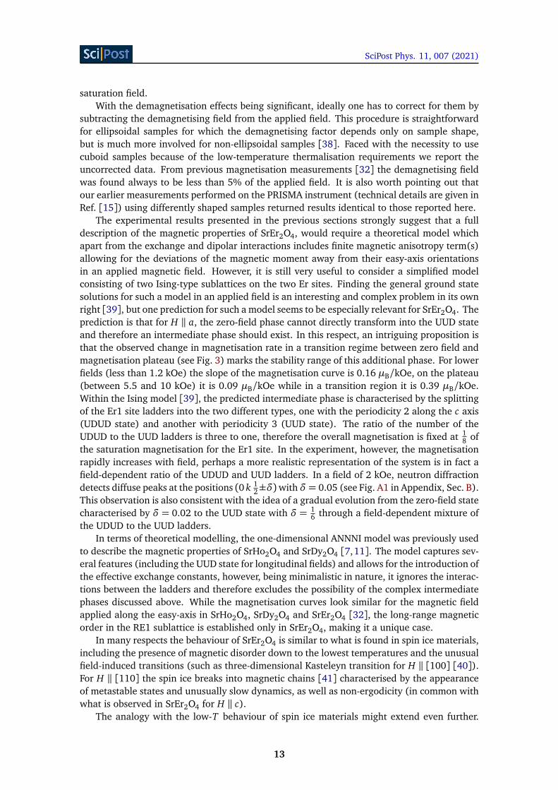
Figure A1: Neutron diffraction intensity maps (background subtracted) of the (0kl)plane measured on the WISH instrument at T = 60 mK in different fields appliedparallel to the a axis.
Fig. A1 gives the detailed record of the field dependence of the magnetic diffraction inten-sity in the (0kl) scattering plane for the field applied along the a axis. No significant history
15
SciPost Phys. 11, 007 (2021)
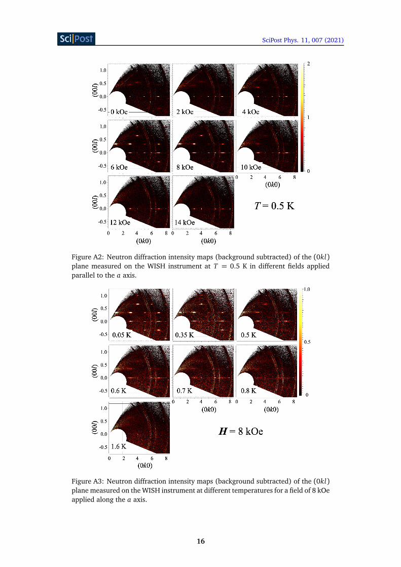
Figure A2: Neutron diffraction intensity maps (background subtracted) of the (0kl)plane measured on the WISH instrument at T = 0.5 K in different fields appliedparallel to the a axis.
Figure A3: Neutron diffraction intensity maps (background subtracted) of the (0kl)plane measured on the WISH instrument at different temperatures for a field of 8 kOeapplied along the a axis.
16
SciPost Phys. 11, 007 (2021)
dependence has been found during the measurements, the results were largely reproduciblefor increasing and decreasing fields for several runs.
Fig. A2 shows the field dependence taken at T = 0.5 K. Compared to the base temperature(60 mK), an overall decrease in intensity is visible for all fields.
Fig. A3 follows the temperature dependence of the magnetic diffraction intensity in the(0kl) scattering plane for the field H = 8 kOe applied along the a axis. The measurementsindicate a gradual reduction of the intensity of the diffuse features at the positions (0 k ±l/3)corresponding to the UUD magnetic structure.
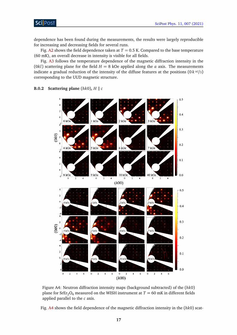
B.0.2 Scattering plane (hk0), H ‖ c
Figure A4: Neutron diffraction intensity maps (background subtracted) of the (hk0)plane for SrEr2O4 measured on the WISH instrument at T = 60 mK in different fieldsapplied parallel to the c axis.
Fig. A4 shows the field dependence of the magnetic diffraction intensity in the (hk0) scat-
17

SciPost Phys. 11, 007 (2021)
tering plane for the field applied along the c axis. Two sets of the intensity maps shown werecollected for the two sample positions (different by a rotation around the vertical c axis of27 degrees), allowing for access to different sections of the reciprocal space.
18
SciPost Phys. 11, 007 (2021)
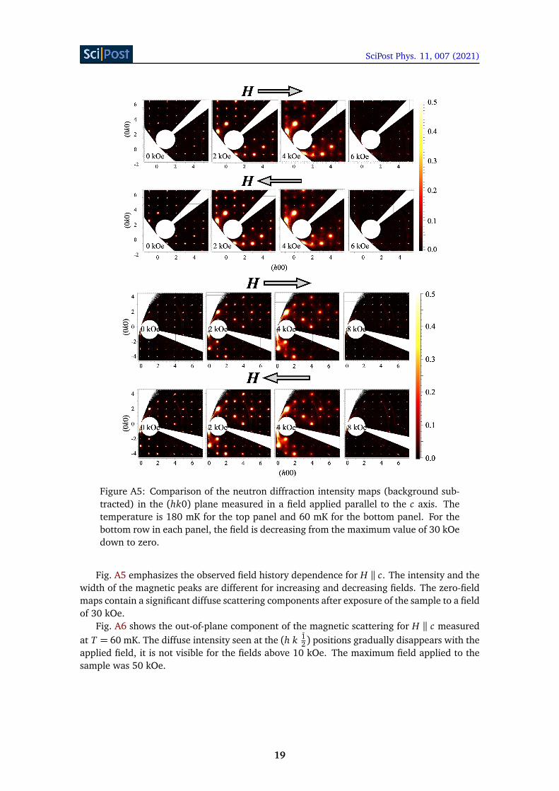
Figure A5: Comparison of the neutron diffraction intensity maps (background sub-tracted) in the (hk0) plane measured in a field applied parallel to the c axis. Thetemperature is 180 mK for the top panel and 60 mK for the bottom panel. For thebottom row in each panel, the field is decreasing from the maximum value of 30 kOedown to zero.
Fig. A5 emphasizes the observed field history dependence for H ‖ c. The intensity and thewidth of the magnetic peaks are different for increasing and decreasing fields. The zero-fieldmaps contain a significant diffuse scattering components after exposure of the sample to a fieldof 30 kOe.
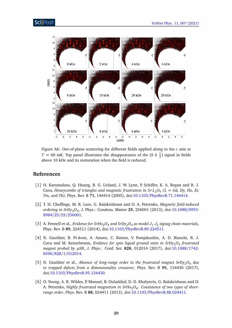
Fig. A6 shows the out-of-plane component of the magnetic scattering for H ‖ c measuredat T = 60 mK. The diffuse intensity seen at the (h k 1
2) positions gradually disappears with theapplied field, it is not visible for the fields above 10 kOe. The maximum field applied to thesample was 50 kOe.
19
SciPost Phys. 11, 007 (2021)
Figure A6: Out-of-plane scattering for different fields applied along to the c axis atT = 60 mK. Top panel illustrates the disappearance of the (h k 1
2) signal in fieldsabove 10 kOe and its restoration when the field is reduced.
References
[1] H. Karunadasa, Q. Huang, B. G. Ueland, J. W. Lynn, P. Schiffer, K. A. Regan and R. J.Cava, Honeycombs of triangles and magnetic frustration in Sr L2O4 (L = Gd, Dy, Ho, Er,Tm, and Yb), Phys. Rev. B 71, 144414 (2005), doi:10.1103/PhysRevB.71.144414.
[2] T. H. Cheffings, M. R. Lees, G. Balakrishnan and O. A. Petrenko, Magnetic field-inducedordering in SrDy2O4, J. Phys.: Condens. Matter 25, 256001 (2013), doi:10.1088/0953-8984/25/25/256001.
[3] A. Fennell et al., Evidence for SrHo2O4 and SrDy2O4 as model J1-J2 zigzag chain materials,Phys. Rev. B 89, 224511 (2014), doi:10.1103/PhysRevB.89.224511.
[4] N. Gauthier, B. Prévost, A. Amato, C. Baines, V. Pomjakushin, A. D. Bianchi, R. J.Cava and M. Kenzelmann, Evidence for spin liquid ground state in SrDy2O4 frustratedmagnet probed by µSR, J. Phys.: Conf. Ser. 828, 012014 (2017), doi:10.1088/1742-6596/828/1/012014.
[5] N. Gauthier et al., Absence of long-range order in the frustrated magnet SrDy2O4 dueto trapped defects from a dimensionality crossover, Phys. Rev. B 95, 134430 (2017),doi:10.1103/PhysRevB.95.134430.
[6] O. Young, A. R. Wildes, P. Manuel, B. Ouladdiaf, D. D. Khalyavin, G. Balakrishnan and O.A. Petrenko, Highly frustrated magnetism in SrHo2O4: Coexistence of two types of short-range order, Phys. Rev. B 88, 024411 (2013), doi:10.1103/PhysRevB.88.024411.
20
SciPost Phys. 11, 007 (2021)
[7] J.-J. Wen et al., Disorder from order among anisotropic next-nearest-neighbor Ising spinchains in SrHo2O4, Phys. Rev. B 91, 054424 (2015), doi:10.1103/PhysRevB.91.054424.
[8] D. L. Quintero-Castro et al., Coexistence of long- and short-range magneticorder in the frustrated magnet SrYb2O4, Phys. Rev. B 86, 064203 (2012),doi:10.1103/PhysRevB.86.064203.
[9] C. Bidaud, O. Simard, G. Quirion, B. Prévost, S. Daneau, A. D. Bianchi, H. A. Dabkowskaand J. A. Quilliam, Dimensionality and irreversibility of field-induced transitions inSrDy2O4, Phys. Rev. B 93, 060404 (2016), doi:10.1103/PhysRevB.93.060404.
[10] O. A. Petrenko, O. Young, D. Brunt, G. Balakrishnan, P. Manuel, D. D. Khalyavin and C.Ritter, Evolution of spin correlations in SrDy2O4 in an applied magnetic field, Phys. Rev. B95, 104442 (2017), doi:10.1103/PhysRevB.95.104442.
[11] N. Gauthier et al., Field dependence of the magnetic correlations of the frustrated magnetSrDy2O4, Phys. Rev. B 95, 184436 (2017), doi:10.1103/PhysRevB.95.184436.
[12] O. Young, G. Balakrishnan, P. Manuel, D. Khalyavin, A. Wildes and O. Petrenko,Field-induced transitions in highly frustrated SrHo2O4, Crystals 9, 488 (2019),doi:10.3390/cryst9100488.
[13] B. Z. Malkin et al., Magnetic and spectral properties of the multisublat-tice oxides SrY2O4:Er3+ and SrEr2O4, Phys. Rev. B 92, 094415 (2015),doi:10.1103/PhysRevB.92.094415.
[14] O. A. Petrenko, G. Balakrishnan, N. R. Wilson, S. de Brion, E. Suard and L. C.Chapon, Low-temperature magnetic ordering in SrEr2O4, Phys. Rev. B 78, 184410 (2008),doi:10.1103/PhysRevB.78.184410.
[15] T. J. Hayes, G. Balakrishnan, P. P. Deen, P. Manuel, L. C. Chapon and O. A. Petrenko,Coexistence of the long-range and short-range magnetic order components in SrEr2O4, Phys.Rev. B 84, 174435 (2011), doi:10.1103/PhysRevB.84.174435.
[16] D. H. Lee, J. D. Joannopoulos, J. W. Negele and D. P. Landau, Discrete-symmetry breakingand novel critical phenomena in an antiferromagnetic planar (XY) model in two dimensions,Phys. Rev. Lett. 52, 433 (1984), doi:10.1103/PhysRevLett.52.433.
[17] A. V. Chubukov and D. I. Golosov, Quantum theory of an antiferromagnet on a triangularlattice in a magnetic field, J. Phys.: Condens. Matter 3, 69 (1991), doi:10.1088/0953-8984/3/1/005.
[18] M. E. Zhitomirsky, A. Honecker and O. A. Petrenko, Field induced order-ing in highly frustrated antiferromagnets, Phys. Rev. Lett. 85, 3269 (2000),doi:10.1103/PhysRevLett.85.3269.
[19] C. Lacroix, P. Mendels and F. Mila, eds., Introduction to frustrated magnetism, Springer-Verlag, Berlin, ISBN 9783642105883 (2011), doi:10.1007/978-3-642-10589-0.
[20] E. Stryjewski and N. Giordano, Metamagnetism, Adv. Phys. 26, 487 (1977),doi:10.1080/00018737700101433.
[21] G. Balakrishnan, T. J. Hayes, O. A. Petrenko and D. McK Paul, High quality single crystalsof the SrR2O4 family of frustrated magnets, J. Phys.: Condens. Matter 21, 012202 (2008),doi:10.1088/0953-8984/21/1/012202.
21
SciPost Phys. 11, 007 (2021)
[22] L. C. Chapon et al., Wish: The new powder and single crystal magneticdiffractometer on the second target station, Neutron News 22, 22 (2011),doi:10.1080/10448632.2011.569650.
[23] O. A. Petrenko, G. Balakrishnan, T. Hayes, P. Manuel and L. Chapon, Neutron diffrac-tion study of highly frustrated SrEr2O4, STFC ISIS Neutron and Muon Source (2011),doi:10.5286/ISIS.E.24086063.
[24] O. A. Petrenko, G. Balakrishnan, S. X. M. Riberolles and P. Manuel, Melting the long-rangemagnetic order in SrEr2O4 with an applied magnetic field, STFC ISIS Neutron and MuonSource (2016), doi:10.5286/ISIS.E.RB1610402.
[25] N. Qureshi, G. Balakrishnan, M. Ciomaga Hatnean, O. Petrenko and S. Riberolles, Studyof the field induced magnetic phases of SrEr2O4 for H//a, Institut Laue-Langevin (ILL)(2018), doi:10.5291/ILL-DATA.5-41-974.
[26] J. Taylor, O. Arnold, J. Bilheaux, A. Buts, S. Campbell, M. Doucet, N. Draper, R. Fowler,M. Gigg and V. Lynch, Mantid, a high performance framework for reduction and analysisof neutron scattering data, Bull. Am. Phys. Soc. 57 (2012).
[27] P. Coppens, Crystallographic computing, Munksgaard International Booksellers and Pub-lishers Ltd, Copenhaguen, 255 (1979).
[28] J. Rodríguez-Carvajal, Recent advances in magnetic structure determination by neu-tron powder diffraction, Physica B: Cond. Matter 192, 55 (1993), doi:10.1016/0921-4526(93)90108-I.
[29] C. Paulsen, Introduction to physical techniques in molecular magnetism: Structural andmacroscopic techniques - Yesa 1999, Servicio de Publicaciones de la Universidad deZaragoza, Zaragoza (2001).
[30] F. Heidrich-Meisner, I. A. Sergienko, A. E. Feiguin and E. R. Dagotto, Universal emergenceof the one-third plateau in the magnetization process of frustrated quantum spin chains,Phys. Rev. B 75, 064413 (2007), doi:10.1103/PhysRevB.75.064413.
[31] D. D. Khalyavin, P. Manuel, M. Ciomaga Hatnean and O. A. Petrenko, Fragile ground stateand rigid field-induced structures in the zigzag ladder compound BaDy2O4, Phys. Rev. B103, 134434 (2021), doi:10.1103/PhysRevB.103.134434.
[32] T. J. Hayes, O. Young, G. Balakrishnan and O. A. Petrenko, Magnetisation Studies of Ge-ometrically Frustrated Antiferromagnets SrLn2O4, with Ln = Er, Dy, and Ho, J. Phys. Soc.Jpn. 81, 024708 (2012), doi:10.1143/JPSJ.81.024708.
[33] J. M. Kincaid and E. G. D. Cohen, Phase diagrams of liquid helium mixtures and metam-agnets: Experiment and mean field theory, Phys. Rep. 22, 57 (1975), doi:10.1016/0370-1573(75)90005-8.
[34] A. F. G. Wyatt, Magnetization of dysprosium aluminium garnet, J. Phys. C: Solid StatePhys. 1, 684 (1968), doi:10.1088/0022-3719/1/3/317.
[35] W. Schneider and H. Weitzel, Neutron diffraction determination of the magnetic phases ofFeCl2·H2O, Solid State Commun. 13, 303 (1973), doi:10.1016/0038-1098(73)90596-6.
[36] R. J. Birgeneau, G. Shirane, M. Blume and W. C. Koehler, Tricritical-point phase diagramin FeCl2, Phys. Rev. Lett. 33, 1098 (1974), doi:10.1103/PhysRevLett.33.1098.
22

SciPost Phys. 11, 007 (2021)
[37] N. Schibuya, K. Knorr, H. Dachs, M. Steiner and B. M. Wanklyn, Neutron scatteringinvestigation of the tricritical point in DyPO4, Solid State Commun. 17, 1305 (1975),doi:10.1016/0038-1098(75)90692-4.
[38] M. Twengström, L. Bovo, O. A. Petrenko, S. T. Bramwell and P. Henelius, LiHoF4:Cuboidal demagnetizing factor in an Ising ferromagnet, Phys. Rev. B 102, 144426 (2020),doi:10.1103/PhysRevB.102.144426.
[39] Yu. I. Dublenych and O. A. Petrenko, An Ising model on a 3D honeycomb zigzag-ladderlattice: a solution to the ground-state problem and application to the SrRE2O4 and BaRE2O4magnets, (2020), arXiv:2008.06445.
[40] L. D. C. Jaubert, J. T. Chalker, P. C. W. Holdsworth and R. Moessner, Three-dimensionalKasteleyn transition: Spin ice in a [100] field, Phys. Rev. Lett. 100, 067207 (2008),doi:10.1103/PhysRevLett.100.067207.
[41] T. Fennell, O. A. Petrenko, B. Fåk, J. S. Gardner, S. T. Bramwell and B. Ouladdiaf, Neutronscattering studies of the spin ices Ho2Ti2O7 and Dy2Ti2O7 in applied magnetic field, Phys.Rev. B 72, 224411 (2005), doi:10.1103/PhysRevB.72.224411.
[42] M. E. Brooks-Bartlett, S. T. Banks, L. D. C. Jaubert, A. Harman-Clarke and P. C. W.Holdsworth, Magnetic-moment fragmentation and monopole crystallization, Phys. Rev. X4, 011007 (2014), doi:10.1103/PhysRevX.4.011007.
[43] S. Petit et al., Observation of magnetic fragmentation in spin ice, Nat. Phys. 12, 746 (2016),doi:10.1038/nphys3710.
[44] E. Lhotel, L. D. C. Jaubert and P. C. W. Holdsworth, Fragmentation in frustrated magnets:A review, J. Low Temp. Phys. 201, 710 (2020), doi:10.1007/s10909-020-02521-3.
23


