Embed Size (px)
Citation preview

www.elsevier.com/locate/biochemsysecoBiochemical Systematics and Ecology 34 (2006) 575e584
Flavonoid glycosides from Egyptian species of the tribeAsclepiadeae (Apocynaceae, subfamily Asclepiadoideae)
Samia Heneidak a,b, Renee J. Grayer a,*, Geoffrey C. Kite a, Monique S.J. Simmonds a
a Royal Botanic Gardens, Kew, Richmond, Surrey TW9 3AB, UKb Department of Biological Sciences, Faculty of Education at Suez, Suez Canal University, Egypt
Received 5 May 2005; accepted 2 March 2006
Abstract
The flavonoids of 11 Egyptian species of the tribe Asclepiadeae (Apocynaceae, subfamily Asclepiadoideae) were studied:Pentatropis nivalis, Pleurostelma schimperi (subtribe Astephaninae), Glossonema boveanum, Solenostemma arghel (subtribeGlossonematinae), Cynanchum acutum, Oxystelma esculentum (subtribe Metastelmatinae), Calotropis procera, Gomphocarpusfruticosus, Gomphocarpus sinaicus, Pergularia tomentosa and Pergularia daemia (subtribe Asclepiadinae). These 11 specieswere found to produce flavonol glycosides. In addition, flavonol sulphates and disulphates were found in a specimen of P. nivalis.The flavonoids may provide useful taxonomic characters at several levels of classification.� 2006 Elsevier Ltd. All rights reserved.
Keywords: Asclepiadeae; Apocynaceae; Asclepiadoideae; Flavonoids; Chemosystematics
1. Introduction
It has been generally recognised that the families Asclepiadaceae and Apocynaceae are very closely related.Originally they were separated because of the presence of very specialised flower structures (complex translatorsand pollinia) in the former family, which were thought to be absent from the latter. However, extensive morphologicalstudies have shown that these flower characters exhibit a continuum between the extremes of the two families (Endressand Bruyns, 2000) and that five gradual stages of specialisation of floral structures associated with pollination occurwithin these groups, so that division into two is rather arbitrary (Goyder, 1999). The division into two groups is alsonot supported when a wider selection of morphological and other characters is considered. Chemically, both groupsare characterised by the presence of cardenolides (cardiac glycosides) and biogenetically related pregnane glycosides,and triterpenes (Hegnauer, 1964, 1989). These compounds are present in latex cells in the plants (Trease and Evans,1983). In the last decade, a number of molecular studies have shown that the various different groups within the Ascle-piadaceae nest in the Apocynaceae sensu stricto, and that this family is paraphyletic if the Asclepiadaceae are notincluded (Judd et al., 1994, 1999; Sennblad and Bremer, 1996; Endress et al., 1996; The Angiosperm PhylogenyGroup, 1998). For all these reasons, the Asclepiadaceae have recently been subsumed with the Apocynaceae (Endress
* Corresponding author. Tel.: þ44 20 8332 5312; fax: þ44 20 8332 5310.
E-mail address: [email protected] (R.J. Grayer).
0305-1978/$ - see front matter � 2006 Elsevier Ltd. All rights reserved.
doi:10.1016/j.bse.2006.03.001

576 S. Heneidak et al. / Biochemical Systematics and Ecology 34 (2006) 575e584
and Bruyns, 2000), and the family sensu lato now comprises five subfamilies, three from the former Asclepiadaceae(Asclepiadoideae, Secamonoideae and Periplocoideae) and two from the Apocynaceae s. str. (Rauvolfioideae andApocynoideae). Three tribes are recognised within subfamily Asclepiadoideae: Marsdenieae, Ceropegieae andAsclepiadeae (Endress and Bruyns, 2000).
As part of a biosystematic study of the Asclepiadoideae and Periplocoideae in Egypt (Heneidak, 2001), wesurveyed the flavonoids in all 11 native Egyptian species of the tribe Asclepiadeae, to see if flavonoid profiles couldprovide useful taxonomic characters in this group. Previously, mainly flavonol O-glycosides have been reported fromthe former Asclepiadaceae (Hegnauer, 1989), but also some flavone O- and C-glycosides, e.g. from species of Hoya(Baas et al., 1981). A few flavonol glycosides have already been described from some of the species investigated forthe present study (see below), but three of the species (Pentatropis nivalis, Pleurostelma schimperi and Glossonemaboveanum) have not yet been analysed phytochemically as far as we are aware. Kaempferol and kaempferol mono-and diglycosides have been reported from Solenostemma arghel (Khaled et al., 1974; Khalid et al., 1992) and inaddition the 3-O-glucuronide, 3-O-rutinoside, 7,40-O-diglucoside and 3,40-O-diglucoside of kaempferol by Michael(1998). Ebeid (1989) isolated quercetin and quercetin 3-O-galactoside from Cynanchum acutum; the latter compoundwas also reported from the same plant by Abou-Zeid et al. (2001) and quercetin plus quercetin 3-O-glucoside byAwaad (2000). According to Gibbs (1974), Pergularia species are characterised by the production of quercetin3-O-galactoside. Sarg et al. (1993) reported quercetin and rutin from Gomphocarpus sinaicus and Komissarenkoet al. (1997) kaempferol, quercetin and their rutinosides from Gomphocarpus fruticosus. Sen et al. (1992) isolatedisorhamnetin 3-O-rutinoside, isorhamnetin 3-O-glucoside and isorhamnetin 3-O-[2-O-b-D-galactopyranosyl-6-O-a-L-rhamnopyranosyl]-b-D-glucopyranoside from Calotropis gigantea, a species closely related to Calotropis procera,whereas Gibbs (1974) reported quercetin 3-O-galactoside and rutin from species of Calotropis.
2. Materials and methods
2.1. Plant material
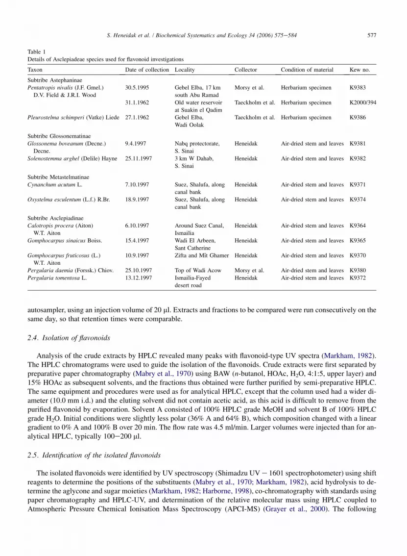
Fresh materials of the species were collected from the wild in Egypt, and all the species studied were natives. Thesamples were air-dried and a voucher specimen for each species has been deposited in the Herbarium, Royal BotanicGardens (RBG), Kew (K) or Suez Canal University, Egypt. Some species were also grown in a glasshouse at RBG,Kew. The studied plants are listed in Table 1.
2.2. Extraction
For a comparative analysis of the flavonoids, small extracts were prepared from the stems of the plants; 200 mg ofpowdered stem was transferred to a test tube and 5 ml of 80% aqueous MeOH was added. The mixture was quicklybrought to the boil in a hot water bath. After boiling for about 2 min, the mixture was cooled and left to extract for24 h. The extract was then filtered, evaporated to dryness, and taken up in 3 ml of 70% EtOH. About 20 ml of this crudeextract was used for analysis by HPLC with diode array detection. For the isolation of flavonoids, larger extracts (from10 to 20 g plant material) were prepared by using the above procedure.
2.3. Analytical HPLC with diode array detection
All samples were filtered through a nylon Acrodisc 13 syringe filter (Gelman Science; pore size 0.45 mm) beforeHPLC analysis, and all solvents used were HPLC grade and filtered for maximum purity. The HPLC system used forthe analysis of crude extracts, purified flavonoids and isolation of compounds consisted of a Waters LC 600 pump and996 photodiode array detector controlled by Millennium software. A Merck LiChrospher 100RP-18 column (5 mm)was used, 4.0 mm (i.d.)� 250 mm. A gradient of two solvents, 2% HOAc in H2O (A) and MeOH, HOAc, H2O, 18:1:1(B), was employed for elution of the compounds. Initial conditions were 75% A, 25% B, with a linear gradient reach-ing A¼ 0% at t¼ 20 min. This was followed by isocratic elution with 100% B until t¼ 23 min, after which time theprogramme returned to the initial solvent composition. A flow rate of 1.0 ml/min and column temperature of 30 � weremaintained during the HPLC analysis. For the analytical HPLC, all samples were injected by means of an
577S. Heneidak et al. / Biochemical Systematics and Ecology 34 (2006) 575e584
autosampler, using an injection volume of 20 ml. Extracts and fractions to be compared were run consecutively on thesame day, so that retention times were comparable.
2.4. Isolation of flavonoids
Analysis of the crude extracts by HPLC revealed many peaks with flavonoid-type UV spectra (Markham, 1982).The HPLC chromatograms were used to guide the isolation of the flavonoids. Crude extracts were first separated bypreparative paper chromatography (Mabry et al., 1970) using BAW (n-butanol, HOAc, H2O, 4:1:5, upper layer) and15% HOAc as subsequent solvents, and the fractions thus obtained were further purified by semi-preparative HPLC.The same equipment and procedures were used as for analytical HPLC, except that the column used had a wider di-ameter (10.0 mm i.d.) and the eluting solvent did not contain acetic acid, as this acid is difficult to remove from thepurified flavonoid by evaporation. Solvent A consisted of 100% HPLC grade MeOH and solvent B of 100% HPLCgrade H2O. Initial conditions were slightly less polar (36% A and 64% B), which composition changed with a lineargradient to 0% A and 100% B over 20 min. The flow rate was 4.5 ml/min. Larger volumes were injected than for an-alytical HPLC, typically 100e200 ml.
2.5. Identification of the isolated flavonoids
The isolated flavonoids were identified by UV spectroscopy (Shimadzu UV e 1601 spectrophotometer) using shiftreagents to determine the positions of the substituents (Mabry et al., 1970; Markham, 1982), acid hydrolysis to de-termine the aglycone and sugar moieties (Markham, 1982; Harborne, 1998), co-chromatography with standards usingpaper chromatography and HPLC-UV, and determination of the relative molecular mass using HPLC coupled toAtmospheric Pressure Chemical Ionisation Mass Spectroscopy (APCI-MS) (Grayer et al., 2000). The following
Table 1
Details of Asclepiadeae species used for flavonoid investigations
Taxon Date of collection Locality Collector Condition of material Kew no.
Subtribe Astephaninae
Pentatropis nivalis (J.F. Gmel.)
D.V. Field & J.R.I. Wood
30.5.1995 Gebel Elba, 17 km
south Abu Ramad
Morsy et al. Herbarium specimen K9383
31.1.1962 Old water reservoir
at Suakin el Qadim
Taeckholm et al. Herbarium specimen K2000/394
Pleurostelma schimperi (Vatke) Liede 27.1.1962 Gebel Elba,
Wadi Oolak
Taeckholm et al. Herbarium specimen K9386
Subtribe Glossonematinae
Glossonema boveanum (Decne.)
Decne.
9.4.1997 Nabq protectorate,
S. Sinai
Heneidak Air-dried stem and leaves K9381
Solenostemma arghel (Delile) Hayne 25.11.1997 3 km W Dahab,
S. Sinai
Heneidak Air-dried stem and leaves K9382
Subtribe Metastelmatinae
Cynanchum acutum L. 7.10.1997 Suez, Shalufa, along
canal bank
Heneidak Air-dried stem and leaves K9371
Oxystelma esculentum (L.f.) R.Br. 18.9.1997 Suez, Shalufa, along
canal bank
Heneidak Air-dried stem and leaves K9374
Subtribe Asclepiadinae
Calotropis procera (Aiton)
W.T. Aiton
6.10.1997 Around Suez Canal,
Ismailia
Heneidak Air-dried stem and leaves K9364
Gomphocarpus sinaicus Boiss. 15.4.1997 Wadi El Arbeen,
Sant Catherine
Heneidak Air-dried stem and leaves K9365
Gomphocarpus fruticosus (L.)
W.T. Aiton
10.9.1997 Zifta and Mıt Ghamer Heneidak Air-dried stem and leaves K9370
Pergularia daemia (Forssk.) Chiov. 25.10.1997 Top of Wadi Acow Morsy et al. Air-dried stem and leaves K9380
Pergularia tomentosa L. 13.12.1997 Ismailia-Fayed
desert road
Heneidak Air-dried stem and leaves K9372

578 S. Heneidak et al. / Biochemical Systematics and Ecology 34 (2006) 575e584
flavonoid standards were available for comparison during the study: quercetin 3-O-rutinoside, quercetin 3-O-glucoside, quercetin 3-O-galactoside, quercetin 3-O-xyloside, quercetin 3-O-sulphate, kaempferol 3-O-rutinoside,kaempferol 3-O-neohesperidoside, kaempferol 3-O-glucoside, isorhamnetin 3-O-rutinoside, isorhamnetin 3-O-glucoside, kaempferol, and quercetin. All standards were obtained from Apin Chemicals, Abingdon, UK.
2.6. HPLC with APCI-MS
The HPLC used the same pumps and column as described for analytical HPLC in Section 2.3 and a similar solventsystem, except that the solvents contained a lower concentration of acetic acid (1% HOAc in H2O and 1% HOAc inMeOH). Positive ion or negative ion APCI-MS of the column eluate was recorded over m/z 125e1200 using anion-trap mass spectrometer (Thermo-Finnigan LCQ Classic). The following source setting were used: vaporisertemperature, 550 �C; sheath and auxiliary nitrogen gas flows, 80 and 10 units, respectively; needle voltage (automat-ically adjusted by the instrument), ca. �4 kV; heated capillary temperature, 150 �C. Source and octapole voltageswere tuned on [MþH]þ of quercetin. Product ion spectra obtained by MS2 were recorded using an ion isolation widthof 3 amu and a normalised collision energy of 45%. The UV absorbance (at 335 nm) of the eluate from the HPLCcolumn was also monitored with a single wavelength UV detector prior to entry into the mass spectrometer.
3. Results and discussion
3.1. Identification of the flavonoid glycosides
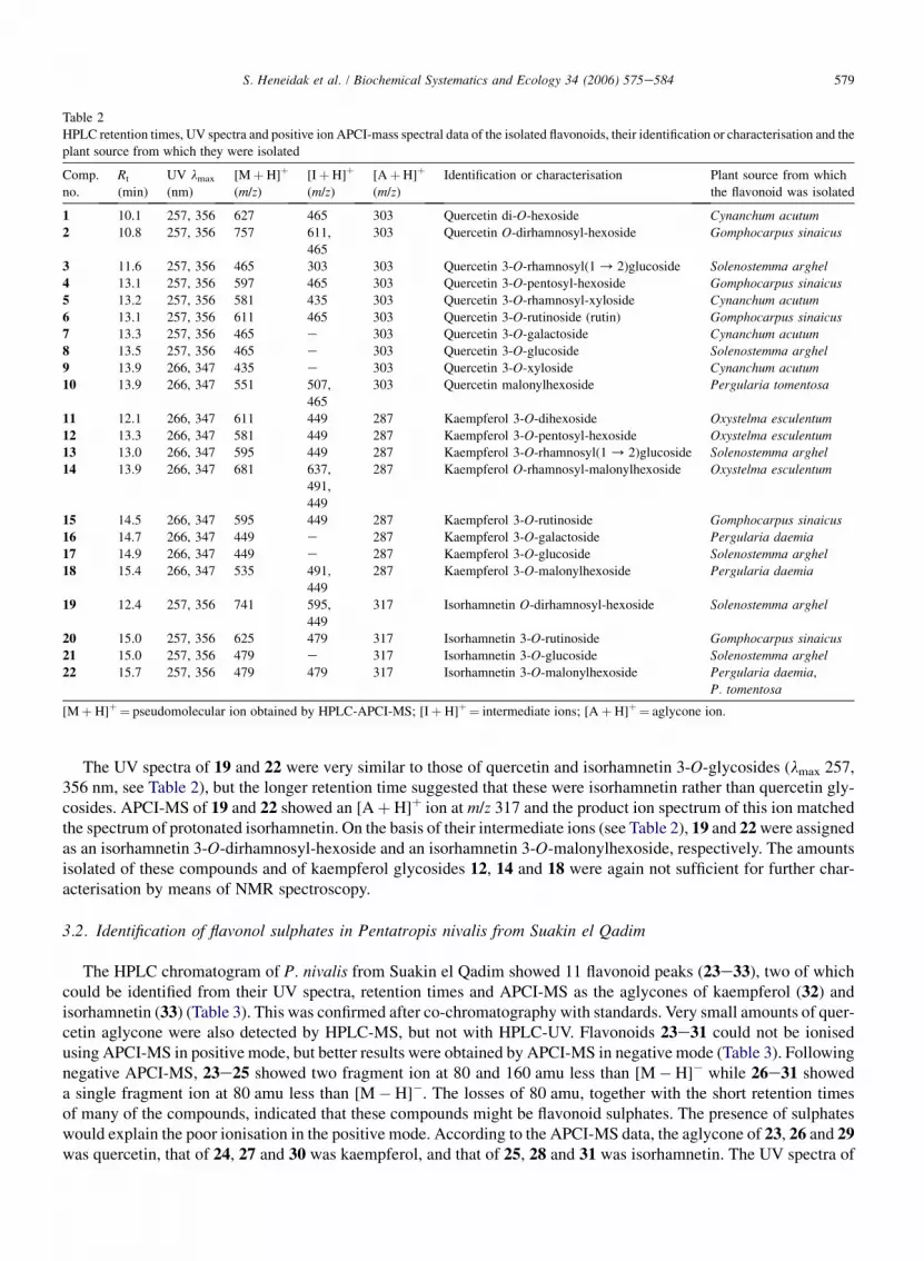
A total of 22 flavonoid glycosides were identified or characterised from the species Asclepiadoideae investigated.Table 2 shows the HPLC retention times of these compounds, their UV lmax (nm), APCI-MS data, the identification orcharacterisation of each flavonoid glycoside and the plant source from which they were obtained. Ten flavonoidmonoglycosides and common diglycosides were isolated and identified by a combination of UV spectroscopy,mass spectrometry (see below) and identification of aglycone and sugar moieties after acid hydrolysis. Standardswere available for all these compounds apart from 16 to confirm the identifications. The identified compoundswere quercetin 3-O-rutinoside (rutin) (6), quercetin 3-O-galactoside (7), quercetin 3-O-glucoside (8), quercetin 3-O-xyloside (9), kaempferol 3-O-neohesperidoside (13), kaempferol 3-O-rutinoside (15), kaempferol 3-O-galactoside(16), kaempferol 3-O-glucoside (17), isorhamnetin 3-O-rutinoside (20) and isorhamnetin 3-O-glucoside (21).
The remaining 12 flavonoids were characterised from UV spectra and APCI-MS data obtained online followingHPLC of purified compounds and in the case of 3 and 5 by identification of the acid hydrolysis products. The UVspectra of compounds 1e5 and 10 suggested that they were quercetin 3-O-glycosides (lmax 257, 356 nm, see Table2). The aglycone ion [AþH]þ at m/z 303 confirmed this assumption (Mr of quercetin is 302). Moreover, the production spectrum of [AþH]þmatched the spectrum of protonated quercetin. MS/MS of [MþH]þ of 1, 3, 4 and 5 showedthem to be diglycosides while 2 was a triglycoside. The primary sugar was a hexose in 1e4 (intermediate ion at[(AþH)þ 162]þ) and a pentose in 5 (intermediate ion at [(AþH)þ 132]þ). Hydrolysis of 5 yielded rhamnose,xylose and quercetin, thus 5 is a quercetin 3-O-rhamnosyl-xyloside. The additional hydrolysis data for 3 showed itto be a quercetin 3-O-rhamnosylglucoside. As 3 had a retention time of 1.5 min less than quercetin 3-O-rhamnosyl(1 / 6)glucoside (¼quercetin 3-O-rutinoside, 6) it is likely to be quercetin 3-O-rhamnosyl(1 / 2)gluco-side (¼quercetin 3-O-neohesperidoside) since the retention time of kaempferol 3-O-neohesperidoside (13) was also1.5 min shorter than that of the corresponding rutinoside (15). Also, 3 occurred in species that produced kaempferol3-O-neohesperidoside (S. arghel and Oxystelma esculentum). APCI-MS of the quercetin 3-O-glycoside 10 suggestedthat this compound was acylated with malonic acid. The [MþH]þ ion of 10 at m/z 551 was accompanied by ions 44and 86 amu less. Grayer et al. (2000) showed that these serial losses of 44 and 42 amu are characteristic of malonicacid as an acyl group. Therefore, 10 was assigned as a quercetin 3-O-malonylhexoside. The amounts isolated ofcompounds 1e5 and 10 were too small for NMR spectroscopy.
The UV spectra of 12, 14 and 18 suggested they were kaempferol 3-O-glycosides (lmax 266, 347 nm, the same asthose of kaempferol 3-O-glycosides 13, 15, 16 and 17, see Table 2). The product ion spectrum of [AþH]þ at m/z 287(Table 2) matched the spectrum of protonated kaempferol. The number of sugar moieties, nature of the primary sugarand presence of malonyl groups were determined from APCI-MS data as above and assignments are given in Table 2.
579S. Heneidak et al. / Biochemical Systematics and Ecology 34 (2006) 575e584
The UV spectra of 19 and 22 were very similar to those of quercetin and isorhamnetin 3-O-glycosides (lmax 257,356 nm, see Table 2), but the longer retention time suggested that these were isorhamnetin rather than quercetin gly-cosides. APCI-MS of 19 and 22 showed an [AþH]þ ion at m/z 317 and the product ion spectrum of this ion matchedthe spectrum of protonated isorhamnetin. On the basis of their intermediate ions (see Table 2), 19 and 22 were assignedas an isorhamnetin 3-O-dirhamnosyl-hexoside and an isorhamnetin 3-O-malonylhexoside, respectively. The amountsisolated of these compounds and of kaempferol glycosides 12, 14 and 18 were again not sufficient for further char-acterisation by means of NMR spectroscopy.
3.2. Identification of flavonol sulphates in Pentatropis nivalis from Suakin el Qadim
The HPLC chromatogram of P. nivalis from Suakin el Qadim showed 11 flavonoid peaks (23e33), two of whichcould be identified from their UV spectra, retention times and APCI-MS as the aglycones of kaempferol (32) andisorhamnetin (33) (Table 3). This was confirmed after co-chromatography with standards. Very small amounts of quer-cetin aglycone were also detected by HPLC-MS, but not with HPLC-UV. Flavonoids 23e31 could not be ionisedusing APCI-MS in positive mode, but better results were obtained by APCI-MS in negative mode (Table 3). Followingnegative APCI-MS, 23e25 showed two fragment ion at 80 and 160 amu less than [M�H]� while 26e31 showeda single fragment ion at 80 amu less than [M�H]�. The losses of 80 amu, together with the short retention timesof many of the compounds, indicated that these compounds might be flavonoid sulphates. The presence of sulphateswould explain the poor ionisation in the positive mode. According to the APCI-MS data, the aglycone of 23, 26 and 29was quercetin, that of 24, 27 and 30 was kaempferol, and that of 25, 28 and 31 was isorhamnetin. The UV spectra of
Table 2
HPLC retention times, UV spectra and positive ion APCI-mass spectral data of the isolated flavonoids, their identification or characterisation and the
plant source from which they were isolated
Comp.
no.
Rt
(min)
UV lmax
(nm)
[MþH]þ
(m/z)
[IþH]þ
(m/z)
[AþH]þ
(m/z)
Identification or characterisation Plant source from which
the flavonoid was isolated
1 10.1 257, 356 627 465 303 Quercetin di-O-hexoside Cynanchum acutum
2 10.8 257, 356 757 611,
465
303 Quercetin O-dirhamnosyl-hexoside Gomphocarpus sinaicus
3 11.6 257, 356 465 303 303 Quercetin 3-O-rhamnosyl(1 / 2)glucoside Solenostemma arghel
4 13.1 257, 356 597 465 303 Quercetin 3-O-pentosyl-hexoside Gomphocarpus sinaicus
5 13.2 257, 356 581 435 303 Quercetin 3-O-rhamnosyl-xyloside Cynanchum acutum
6 13.1 257, 356 611 465 303 Quercetin 3-O-rutinoside (rutin) Gomphocarpus sinaicus7 13.3 257, 356 465 e 303 Quercetin 3-O-galactoside Cynanchum acutum
8 13.5 257, 356 465 e 303 Quercetin 3-O-glucoside Solenostemma arghel
9 13.9 266, 347 435 e 303 Quercetin 3-O-xyloside Cynanchum acutum10 13.9 266, 347 551 507,
465
303 Quercetin malonylhexoside Pergularia tomentosa
11 12.1 266, 347 611 449 287 Kaempferol 3-O-dihexoside Oxystelma esculentum
12 13.3 266, 347 581 449 287 Kaempferol 3-O-pentosyl-hexoside Oxystelma esculentum13 13.0 266, 347 595 449 287 Kaempferol 3-O-rhamnosyl(1 / 2)glucoside Solenostemma arghel
14 13.9 266, 347 681 637,
491,
449
287 Kaempferol O-rhamnosyl-malonylhexoside Oxystelma esculentum
15 14.5 266, 347 595 449 287 Kaempferol 3-O-rutinoside Gomphocarpus sinaicus
16 14.7 266, 347 449 e 287 Kaempferol 3-O-galactoside Pergularia daemia
17 14.9 266, 347 449 e 287 Kaempferol 3-O-glucoside Solenostemma arghel
18 15.4 266, 347 535 491,
449
287 Kaempferol 3-O-malonylhexoside Pergularia daemia
19 12.4 257, 356 741 595,
449
317 Isorhamnetin O-dirhamnosyl-hexoside Solenostemma arghel
20 15.0 257, 356 625 479 317 Isorhamnetin 3-O-rutinoside Gomphocarpus sinaicus
21 15.0 257, 356 479 e 317 Isorhamnetin 3-O-glucoside Solenostemma arghel
22 15.7 257, 356 479 479 317 Isorhamnetin 3-O-malonylhexoside Pergularia daemia,
P. tomentosa
[MþH]þ¼ pseudomolecular ion obtained by HPLC-APCI-MS; [IþH]þ¼ intermediate ions; [AþH]þ ¼ aglycone ion.

580 S. Heneidak et al. / Biochemical Systematics and Ecology 34 (2006) 575e584
26, 27 and 28 were those of quercetin, kaempferol and isorhamnetin conjugated at the 3-hydroxyl, so that thesecompounds were tentatively identified as the 3-O-monosulphates of quercetin, kaempferol and isorhamnetin, respec-tively. The identification of 26 was confirmed by co-chromatography with a standard. No standards were available forcomparison with the other flavonol sulphates. The UV spectra of 30 and 31 suggested they were kaempferol and iso-rhamnetin conjugated at the 7-hydroxyl (Mabry et al., 1970), so that these compounds were tentatively identified asthe 7-O-monosulphates of kaempferol and isorhamnetin. The UV spectrum of 29 could not be obtained because thiscompound co-eluted with 27 which was present in much larger amounts. However, on the basis of its APCI-MS 29could be a quercetin monosulphate, and it also had the expected retention of quercetin 7-O-monosulphate, so the com-pound was tentatively identified as such. The disulphates 23e25 showed [A�H]� at m/z values expected for quer-cetin, kaempferol and isorhamnetin, respectively. Their very short retention times suggested that they were substitutedin both the 3- and 7-positions. Flavonols conjugated in the 7-position were found in the same plant, which are oftenbreakdown products of flavonols conjugated in both the 3- and 7-positions. Therefore, 23e25 were tentatively iden-tified as the 3,7-di-O-sulphates of quercetin, kaempferol and isorhamnetin, respectively.
3.3. Distribution of the flavonoids found in species of the Asclepiadeae
The distribution of flavonoids identified or tentatively identified from species in the tribe Asclepiadeae is given inTable 4. Below these results are summarised and discussed.
3.3.1. Subtribe Astephaninae: Pleurostelma schimperi and Pentatropis nivalisP. schimperi contained the 3-glucosides of quercetin (8) and kaempferol (17) and some minor unidentified
flavonoids, which could not be isolated due to lack of material.The flavonoid profile of one of the specimens studied for P. nivalis (Old water reservoir at Suakin el Qadim,
collected in 1962) differed from that of the remaining Asclepiadoideae studied by the production of flavonol sulphates(23e31) rather than flavonol glycosides. Free flavonol aglycones (32 and 33) were also found in the extract from thisplant, but these could have been derived from the 3-O-sulphates, which hydrolyse easily. Flavonoid sulphates havebeen found in more than 250 species belonging to ca. 20 monocot and dicot families, which are not all closely relatedtaxonomically. They are often found in plants such as P. nivalis that grow in wet places, especially salt marshes, so thatpresence of flavonoid sulphates is thought to be an ecological feature rather than an indication of phylogeneticrelationships (Barron et al., 1988). The other specimen studied of P. nivalis (Gebel Elba, 17 km south Abu Ramad,collected in 1995) contained flavonol 3-O-glycosides (isorhamnetin 3-O-glucoside (21) and a number of unidentifiedminor compounds) instead of flavonol sulphates, and thus shows the usual flavonoid profile of the tribe Asclepiadeae.It would be interesting to determine whether this infraspecific variation in P. nivalis is genetically fixed or whether theproduction of flavonol sulphates instead of glycosides in some specimens is simply a response to environmentalconditions.
Table 3
Retention times, UVand negative ion APCI-mass spectra, and tentative identifications of flavonoid sulphates from Pentatropis nivalis (from Suakin
el Qadim)
No. Rt (min) UV lmax (nm) [M�H]� [I�H]� [A�H]� Tentative identification
23 ca. 3.0 e 461 381 301 Quercetin 3,7-disulphate
24 3.76 266, 343 445 365 285 Kaempferol 3,7-disulphate
25 ca. 4.7 e 475 395 315 Isorhamnetin 3,7-disulphate
26 9.57 252, 352 381 e 301 Quercetin 3-sulphate
27 11.30 266, 343 365 e 285 Kaempferol 3-sulphate
28 12.09 257, 352 395 e 315 Isorhamnetin 3-sulphate
29 11.30 e 381 e 301 Quercetin 7-sulphate
30 12.89 247, 266, 370 365 e 285 Kaempferol 7-sulphate
31 13.36 252, 370 395 e 315 Isorhamnetin 7-sulphate
32 19.08 266, 365 285 e Kaempferol aglycone
33 19.44 252, 370 315 e Isorhamnetin aglycone

Table 4
Distribution of flavono
Subtribe S pferol glycosidesb Isorhamnetin glyc.c
12 13 14 15 16 17 18 19 20 21 22
Astephaninae P
P #
P þ
Glossonematinae G þ þS # þ þ
Metastelmatinae C
O þ # þ þ þ
Asclepiadinae P þ þ # þ þP þ þ # þ þG þ þ þG þ þ þC # þ
þ Means that the flavoa Quercetin glycosi ; 4. 3-O-pentosyl-hexoside; 5. 3-O-rhamnosyl-xyloside; 6. 3-O-rutinoside;
7. 3-O-galactoside; 8.b Kaempferol glycos e; 14. O-rhamnosyl-malonylhexoside; 15. 3-O-rutinoside; 16. 3-O-galactoside;
17. 3-O-glucoside; 18.c Isorhamnetin glyco ylhexoside.
58
1S.
Heneidak
etal.
/B
iochemical
Systematics
andE
cology34
(2006)575e
584
ids 1e22 in Egyptian species of Asclepiadeae
pecies Quercetin glycosidesa Kaem
1 2 3 4 5 6 7 8 9 10 11
entatropis nivalis 1962
entatropis nivalis 1995
leurostelma schimperi #
lossonema boveanum # þ þolenostemma arghel þ þ
ynanchum acutum þ þ þ þ þ #
xystelma esculentum þ þ þ þ þ
ergularia daemia þ þ þergularia tomentosa þ þ þomphocarpus sinaicus þ þ # þ þomphocarpus fruticosus þ þ # þ þalotropis procera
noid was detected; # means that it was the major flavonoid in the specimen investigated.
des: 1. di-O-hexoside; 2. O-dirhamnosyl-hexoside; 3. 3-O-rhamnosyl(1 / 2)glucoside
3-O-glucoside; 9. 3-O-xyloside; 10. malonylhexoside.
ides: 11. 3-O-dihexoside; 12. 3-O-pentosyl-hexoside; 13. 3-O-rhamnosyl(1 / 2)glucosid
3-O-malonylhexoside.
sides: 19. O-dirhamnosylhexoside; 20. 3-O-rutinoside; 21. 3-O-glucoside; 22. 3-O-malon

582 S. Heneidak et al. / Biochemical Systematics and Ecology 34 (2006) 575e584
3.3.2. Subtribe Glossonematinae: Glossonema boveanum and Solenostemma arghelWe detected 3-O-galactoside, 3-O-glucoside and 3-O-rutinoside of quercetin (6, 7 and 8) and 3-O-neohesperido-
side and 3-O-rutinoside of kaempferol (13 and 15) in G. boveanum. S. arghel shared the presence of quercetin 3-O-glucoside (8) and kaempferol 3-O-neohesperidoside (13) with G. boveanum, but in addition we found the 3-O-gluco-sides of kaempferol and isorhamnetin (17 and 21) and the 3-O-neohesperidoside of quercetin (3) in S. arghel. Previousinvestigations of S. arghel reported kaempferol and unspecified kaempferol mono- and diglycosides (Khaled et al.,1974; Khalid et al., 1992), whereas the flavonoid profile reported by Michael (1998) was rather different from theone we found, since they detected the 3-O-glucuronide, 3-O-rutinoside, 7,40-O-diglucoside and 3,40-O-diglucosideof kaempferol in this species. This means that S. arghel may also show infraspecific variation in flavonoid characters,and in the future we hope to investigate this further using specimens from different localities.
3.3.3. Subtribe Metastelmatinae: Cynanchum acutum and Oxystelma esculentumThe Egyptian specimens investigated for C. acutum and O. esculentum (subtribe Metastelmatinae) produced a large
number of flavonol 3-O-glycosides, but they only shared three flavonoids, the 3-O-neohesperidoside, 3-O-galactoside and 3-O-glucoside of quercetin (3, 7 and 8, respectively). C. acutum contained mainly quercetinglycosides, whereas O. esculentum in addition produced many kaempferol glycosides. Quercetin 3-O-galactoside(7) had been isolated before from C. acutum by Ebeid (1989) and Abou-Zeid et al. (2001), whereas quercetin 3-O-glucoside (8) had been reported from this species before by Awaad (2000), so that their results are in agreementwith ours.
3.3.4. Subtribe Asclepiadeae
3.3.4.1. Pergularia daemia and Pergularia tomentosaThe extracts of Pergularia daemia and Pergularia tomentosa yielded the same flavonol glycosides, including the
3-O-galactosides and 3-O-glucosides of quercetin and kaempferol (7, 8, 16 and 17), and malonylhexosides of quer-cetin, kaempferol and isorhamnetin (10, 18, 22). However, these compounds were present in different concentrationsand proportions in the two species, so that the profiles could be distinguished. In P. daemia, kaempferol glycosideswere present in the largest amounts, whereas in P. tomentosa there was a higher content of quercetin glycosides.
3.3.4.2. Gomphocarpus sinaicus and Gomphocarpus fruticosusThe extracts of the two Gomphocarpus species, G. sinaicus and G. fruticosus, again showed very similar flavonoid
profiles, which consisted mainly of quercetin glycosides (2, 4, 6, 7, 8). In both species quercetin 3-O-rutinoside (rutin,6) was the major flavonoid, and the 3-O-rutinoside of kaempferol (15) and isorhamnetin (20) were also present. Therewere quantitative differences in flavonoids between the two species. Rutin (6) had been isolated before fromG. sinaicus by Sarg et al. (1993) and the rutinosides of both quercetin and kaempferol (6 and 15, respectively)from G. fruticosus (Komissarenko et al., 1997).
3.3.4.3. Calotropis proceraThe 3-O-rutinoside of kaempferol (15) was the major flavonoid in C. procera, and the 3-O-rutinoside of isorham-
netin (20) was also present. The latter flavonoid had already been isolated from a closely related species, C. gigantea(Sen et al., 1992).
3.4. Taxonomic implications
Arguably the best way to use chemical features as characters for taxonomic classification is to take their biosyn-thesis into account. For example, the biosynthesis of flavonols and flavones takes place via different routes (Heller andForkmann, 1994), and therefore the presence of flavone glycosides is a different character from the presence of fla-vonol glycosides. The place of attachment of the sugars to the flavonoid aglycone is an important character, as forinstance glycosylation to the 3-hydroxyl requires different enzymes than glycosylation to the 7-hydroxyl. Further-more, each sugar (glucose, galactose, rhamnose, xylose, etc.) has its own transferase enzyme to attach it to a flavonoid(Heller and Forkmann, 1988), and different enzymes are also needed for the biosynthesis of di- or triglycosides, es-pecially when the sugars are linked to different hydroxyl groups. For instance, the biosynthesis of a neohesperidoside

583S. Heneidak et al. / Biochemical Systematics and Ecology 34 (2006) 575e584
(¼rhamnosyl[1 / 2]glucoside) requires a different enzyme from that of a rutinoside (¼rhamnosyl[1 / 6]glucoside).Finally, different acyl groups also require different transferases to attach them to the sugars of flavonoid glycosides.For example, malonic acid needs a different enzyme from a hydroxycinnamic acid (Heller and Forkmann, 1994).Therefore, the following characters can be used: the presence of neohesperidose as a sugar (3, 13), rutinose as a sugar(6, 15, 20), a pentose (xylose or arabinose) as a sugar (4, 5, 12), triglycosylation (2, 19), malonic acid as an acyl group(10, 14, 18, 22), and flavonoid sulphates (23e31).
The results of the present flavonoid analysis could be used to identify Egyptian species of Asclepiadeae and providemarkers at several levels of the taxonomic classification of this tribe. Firstly, at the species level, all species could bedistinguished on the basis of their profiles of flavonoids, so that in principle these profiles could be used as fingerprintsfor the identification of the species. However, in several species there was evidence of infraspecific variation of fla-vonoids, so that more specimens will have to be examined to see how constant or variable the flavonoid profiles are ineach species. Several closely related species were found to have very similar profiles, e.g. P. daemia and P. tomentosa,and also G. sinaicus and G. fruticosus. This indicates that in Pergularia and Gomphocarpus, which contain only a fewspecies each, profiles of flavonoids may be characteristic at the generic level. However, in both Pergularia andGomphocarpus there were quantitative differences in flavonoids between the two related species in each genus, sothat they still could be distinguished. The presence of flavonol glycosides acylated with malonic acid (10, 18 and22) and of flavonol triglycosides (2 and 19) appear to be a characteristic feature for the species of Pergularia inves-tigated, although malonylated glycosides were not found exclusively in this genus.
At the subtribal level, all Egyptian species of the tribes Glossonematinae and Metastelmatinae investigated werecharacterised by the presence of flavonol 3-O-neohesperidosides (3 and/or 13), which were not found in Egyptianmembers of the Astephaninae and Asclepiadinae. Furthermore, the Glossonematinae could be distinguished fromthe Metastelmatinae by the fact that rutinosides (6, 15, 20) were present in the former subtribe, but absent fromthe latter.
Flavonoid profiles may also provide markers at the tribal level in the Asclepiadoideae, because the overall flavo-noid profiles found in another tribe, the Ceropegieae, were very different from those found in the tribe Asclepiadeae.In the Ceropegieae, all the species appeared to produce unusual flavone O-glycosides and sometimes flavoneC-glycosides in the leaves in addition to flavonol O-glycosides (Heneidak, 2001), whereas in the leaves of Egyptianspecies of the Asclepiadeae we found exclusively flavonol O-glycosides. However, Rizk et al. (1990) reported flavoneC-glycosides from the fruits of Glossonema edule (Asclepiadeae), but the chemistry of leaves and fruits of the samespecies is often very different, so that the leaves of G. edule may contain only flavonol O-glycosides. If production offlavone O-glycosides is a more advanced feature than production of flavonol O-glycosides as suggested by Harborne(1967), the Asclepiadeae are a less advanced group than the Ceropegieae.
Acknowledgements
We would like to thank Prof. Ahmed Mursi Ahmed for helping to collect Pentatropis nivalis and Pergularia daemiafrom Gebel Elba, Egypt; the staff of the Lower Nursery (Royal Botanic Gardens, Kew) for growing the fresh material;and Dr. David Goyder (Royal Botanic Gardens, Kew) for providing valuable references.
References
Abou-Zeid, A.H.S., Ibrahim, N.A., Sammour, E.A., 2001. Phytochemical, insecticidal and molluscicidal investigations of the aerial parts of
Cynanchum acutum L. Bull. Fac. Pharm. Cairo Univ. 39, 235e245.
Angiosperm Phylogeny Group (APG), 1998. An ordinal classification for the families of flowering plants. Ann. Mo. Bot. Gard. 85, 531e553.
Awaad, A.S., 2000. Phytochemical investigation and biological activities of Cynanchum acutum growing in Egypt. Bull. Fac. Pharm. Cairo Univ.
38, 153e162.
Baas, W.J., Warnaar, F., Niemann, G.J., 1981. Investigations on Hoya species. VI. Latex composition and leaf phenolics and their taxonomic sig-
nificance. Acta Bot. Neerl. 30, 257e263.
Barron, D., Varin, L., Ibrahim, R.K., Harborne, J.B., Williams, C.A., 1988. Sulphated flavonoids e an update. Phytochemistry 27, 2375e2395.
Ebeid, K.A., 1989. A pharmacognostical study of certain plants belonging to the family Asclepiadaceae. Thesis, M.Sc. Pharm. Sci., Fac. Pharm.,
Mansoura Univ.
Endress, M.E., Bruyns, P.V., 2000. A revised classification of the Apocynaceae s.l. Bot. Rev. 66, 1e56.
Endress, M.E., Sennblad, B., Nilsson, S., Civeyrel, L., Chase, M., Huysmans, S., Grafstrom, E., Bremer, B., 1996. A phylogenetic analysis of
Apocynaceae s. str. and some related taxa in Gentianales. A multidisciplinary approach. Opera Bot. Belg. 7, 59e102.

584 S. Heneidak et al. / Biochemical Systematics and Ecology 34 (2006) 575e584
Gibbs, R.D., 1974. Chemotaxonomy of Flowering Plants, vol. 3. Mc Gill-Queen’s University Press, Montreal/London.
Goyder, D.J., 1999. The Asclepiadaceae e a figment of our imagination. In: Timberlake, J., Kativu, S. (Eds.), African Plants: Biodiversity,
Taxonomy and Uses. Royal Botanic Gardens, Kew, pp. 309e317.
Grayer, R.J., Kite, G.C., Abou-Zaid, M., Archer, L.J., 2000. The application of atmospheric pressure chemical ionization liquid chromatography
mass-spectrometry in the chemotaxonomic study of flavonoids: characterization of flavonoids from Ocimum gratissimum var. gratissimum.
Phytochem. Anal. 11, 257e267.
Harborne, J.B., 1967. Comparative Biochemistry of the Flavonoids. Academic Press, London.
Harborne, J.B., 1998. Phytochemical Methods, third ed. Chapman & Hall, London.
Hegnauer, R., 1964. Chemotaxonomie der Pflanzen, vol. 3. Birkhauser Verlag, Basel, pp. 199e223.
Hegnauer, R., 1989. Chemotaxonomie der Pflanzen, vol. 8. Birkhauser Verlag, Basel, pp. 84e96.
Heller, W., Forkmann, G., 1988. Biosynthesis. In: Harborne, J.B. (Ed.), The Flavonoids. Advances in Research Since 1980. Chapman and Hall,
London, pp. 399e425.
Heller, W., Forkmann, G., 1994. Biosynthesis of flavonoids. In: Harborne, J.B. (Ed.), The Flavonoids. Advances in Research Since 1986. Chapman
and Hall, London, pp. 499e535.
Heneidak, S., 2001. Biosystematic studies on the species of subfamilies Asclepiadoideae and Periplocoideae (family Apocynaceae sensu lato) in
Egypt. Ph.D. thesis, Suez Canal University, Egypt.
Judd, W.S., Sanders, R.W., Donoghue, M.J., 1994. Angiosperm family pairs, preliminary phylogenetic analyses. Harv. Pap. Bot. 5, 1e51.
Judd, W.S., Campbell, C.S., Kellogg, E.A., Stevens, P.F., 1999. Plant Systematics (A Phylogenetic Approach). Sinauer Associates, Inc., Sunder-
land, Massachusetts, USA, 366 pp.
Khaled, S.A., Szendrei, K., Novak, I., 1974. Sudanese plants. 1. Solenostemma arghel. Herba Hung. 13, 33e35.
Khalid, S.A., Kalaflla, E.B., Mohamed, O.Y., 1992. The flavonoids of Solenostemma argel and their antispasmodic activity. Planta Med. 58 (Suppl.
1), A651.
Komissarenko, A.N., Komissarenko, S.N., Chernobai, V.T., 1997. Cardenolides, coumarins and flavonoids of Gomphocarpus fruticosus (L.) Ait.
Fil. Rastit. Resur. 33, 29e41.
Mabry, T.J., Markham, K.R., Thomas, M.B., 1970. The Systematic Identification of Flavonoids. Springer Verlag, Berlin/New York.
Michael, H.N., 1998. A new flavonol glycoside from Solenostemma argel leaves. Asian J. Chem. 10, 1038e1040.
Markham, K.R., 1982. Techniques of Flavonoid Identification. Academic Press, London.
Rizk, A.M., Hammouda, F.M., Ismail, S.I., Hassan, N.M., El-Missiry, M.M., Ahmad, F.A., 1990. Constituents of plants growing in Qatar. Part
XIX. Flavonoids of Glossonema edule N.E.Br. Plant Foods Hum. Nutr. 40, 1e3 (Dordrecht, Netherlands).
Sarg, T., El-Domiaty, M., Abd El-Aziz, E., Abou-Hashem, M., 1993. Chemical constituents of Gomphocarpus sinaicus (Asclepiadaceae). Egypt J.
Pharm. Sci. 34, 577e585.
Sen, S., Sahu, N.P., Mahato, S.B., 1992. Flavonol glycosides from Calotropis gigantea. Phytochemistry 31, 2919e2921.
Sennblad, B., Bremer, B., 1996. The familial relationships of Apocynaceae and Asclepiadaceae evaluated with rbcL data. Plant Syst. Evol. 202,
153e175.
Trease, G.E., Evans, W.C., 1983. Pharmacognosy. Baillere, Tindall, Oxford, pp. 171, 212, 215.


