Embed Size (px)
Citation preview
N
Ra
Mb
a
A
R
R
2
A
P
K
H
P
C
P
A
A
1
Htptntocoaposf
M
0d
s t e r o i d s 7 2 ( 2 0 0 7 ) 881–891
avai lab le at www.sc iencedi rec t .com
journa l homepage: www.e lsev ier .com/ locate /s tero ids
ew calogenin glycosides from Hoodia gordonii
ahul S. Pawara, Yatin J. Shuklab, Ikhlas A. Khana,b,∗
National Center for Natural Products Research, Research Institute of Pharmaceutical Sciences, University of Mississippi, University,S 38677, USADepartment of Pharmacognosy, School of Pharmacy, University of Mississippi, University, MS 38677, USA
r t i c l e i n f o
rticle history:
eceived 26 April 2007
eceived in revised form
2 July 2007
ccepted 23 July 2007
ublished on line 27 July 2007
eywords:
oodia gordonii
a b s t r a c t
Ten new pregnane glycosides (1, 3–11) were isolated from organic extracts of aerial parts of
Hoodia gordonii, which is sold as an appetite suppressant herbal supplement. The aglycone
was identified as calogenin, based on the spectroscopic data of products obtained upon
chemical and enzymatic degradation of parent glycoside. The structures of the glycosides
were established by chemical degradation studies and extensive spectroscopic techniques
that included one-dimensional and two-dimensional NMR.
© 2007 Elsevier Inc. All rights reserved.
regnane
alogenin
57AS3
sclepiadaceae
ppetite suppressant
. Introduction
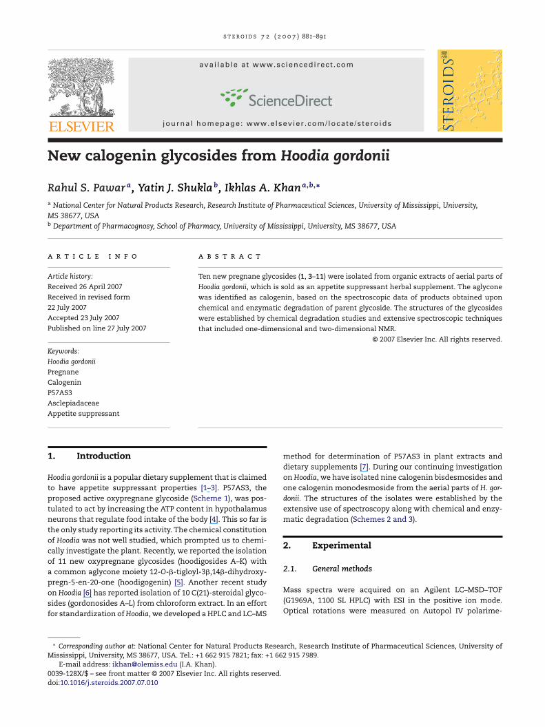
oodia gordonii is a popular dietary supplement that is claimedo have appetite suppressant properties [1–3]. P57AS3, theroposed active oxypregnane glycoside (Scheme 1), was pos-ulated to act by increasing the ATP content in hypothalamuseurons that regulate food intake of the body [4]. This so far ishe only study reporting its activity. The chemical constitutionf Hoodia was not well studied, which prompted us to chemi-ally investigate the plant. Recently, we reported the isolationf 11 new oxypregnane glycosides (hoodigosides A–K) withcommon aglycone moiety 12-O-�-tigloyl-3�,14�-dihydroxy-regn-5-en-20-one (hoodigogenin) [5]. Another recent study
n Hoodia [6] has reported isolation of 10 C(21)-steroidal glyco-ides (gordonosides A–L) from chloroform extract. In an effortor standardization of Hoodia, we developed a HPLC and LC–MS∗ Corresponding author at: National Center for Natural Products Reseaississippi, University, MS 38677, USA. Tel.: +1 662 915 7821; fax: +1 662
E-mail address: [email protected] (I.A. Khan).039-128X/$ – see front matter © 2007 Elsevier Inc. All rights reserved.oi:10.1016/j.steroids.2007.07.010
method for determination of P57AS3 in plant extracts anddietary supplements [7]. During our continuing investigationon Hoodia, we have isolated nine calogenin bisdesmosides andone calogenin monodesmoside from the aerial parts of H. gor-donii. The structures of the isolates were established by theextensive use of spectroscopy along with chemical and enzy-matic degradation (Schemes 2 and 3).
2. Experimental
2.1. General methods
rch, Research Institute of Pharmaceutical Sciences, University of915 7989.
Mass spectra were acquired on an Agilent LC–MSD–TOF(G1969A, 1100 SL HPLC) with ESI in the positive ion mode.Optical rotations were measured on Autopol IV polarime-
882 s t e r o i d s 7 2 ( 2 0
25
Scheme 1 – Structures of P57AS3 and its aglycone.
ter and specific rotations are expressed as 10◦ g−1 cm2. TheNMR spectra were recorded on Varian AS400 and AS600 NMRspectrometers. Proton and carbon chemical shift values arerelative to internal standard TMS and were acquired in C5D5Nand CDCl3, or CH3OD–CDCl3. Column chromatography wascarried out on silica gel (JT Baker, 40 �m for flash chro-matography) and reversed phase C18 silica gel (Polarbond, JTBaker). TLC analyses were carried out on silica gel 60 F254
plates (Merck, Germany) using CHCl3/MeOH/H2O (70:30:0.5)and C18 reversed phase silica TLC plates (Analtech, USA) withMeOH/H2O (70:30). Compounds were visualized by sprayingwith anisaldehde–H2SO4 reagent followed by heating at 105 ◦Cfor 1–2 min. d-Thevetose and d-glucose were obtained fromSigma–Aldrich (USA).
2.2. Plant material
Powdered plant material of H. gordonii, purchased online, wasauthenticated by Dr. Vaishali Joshi by comparing with anauthentic sample of H. gordonii provided by Missouri Botan-ical Garden Missouri, USA. Further, the HPLC fingerprint ofmethanol extract matched with that of the authentic sam-ple of H. gordonii. A voucher specimen (Voucher No. 2799) hasbeen deposited in the repository of National Center for NaturalProduct Research.
2.3. Extraction and isolation
Four hundred and sixty-three grams of plant material wasextracted by percolation with a mixture of equal amountsof CHCl3/MeOH (4 × 1 L). These extracts were combined and
Scheme
0 7 ) 881–891
concentrated to obtain a thick mass (78 g). The extract wassubjected to vacuum liquid chromatography (VLC) on silicagel by eluting with hexane (2 L), CHCl3 (3 L), CHCl3/MeOH (3 L),and MeOH (2 L).
The CHCl3/MeOH fraction was chromatographed on C18column, using gradients of MeOH/H2O (60:40) to (90:10). Sim-ilar fractions were pooled to generate five sub-fractions.Fraction 5 yielded compound 4 (82 mg) after repeated sepa-ration on silica gel columns using isocratic solvent systemsof CHCl3/MeOH (97:3) and CHCl3/MeOH (95:5). The fractionobtained by elution with methanol (36 g) was then subjected toVLC on silica gel by eluting with gradients of CHCl3/MeOH/H2Oto obtain nine sub-fractions. The sub-fraction obtained byelution with CHCl3/MeOH/H2O (90:20:0.6) was further chro-matographed on C18 silica gel by eluting with MeOH–H2Oto obtain compounds 5 (142 mg) and 10 (56 mg). The sub-fraction obtained by elution with CHCl3/MeOH/H2O (90:30:1)was similarly chromatographed on C18 silica gel by elutingwith MeOH–H2O to obtain 6 (32 mg), 7 (75 mg), 8 (38 mg), 9(68 mg) and 11 (62 mg). Similarly, the sub-fraction obtained onelution with CHCl3/MeOH/H2O (90:40:1) by chromatography onC18 gave a major compound 1 (2.1 g). Finally, the sub-fractionobtained by elution with CHCl3/MeOH/H2O (60:40:1), whensimilarly chromatographed on C18 silica gel by eluting withMeOH–H2O afforded 3 (308 mg).
Hoodigoside L (1): White amorphous powder: [˛]25D − 16.0
(MeOH, c0.3); IR �NaClmax : 3392, 2933, 1713, 1648, 1067 cm−1; 1H and
13C NMR data Tables 2 and 3; HR–ESIMS [M+Na]+ m/z 1229.5897(calcd for C58H94O26Na, 1229.5925).
Hoodigoside V (2): White amorphous powder: [˛]25D − 12.0
(MeOH, c0.23); IR �NaClmax : 3398, 2930, 1713, 1646, 1071 cm−1; 1H
and 13C NMR data Tables 2 and 3; HR–ESIMS [M+Na]+ m/z905.4882 (calcd for C46H74O16Na, 905.4869).
Hoodigoside M (3): White amorphous powder: [˛]25D − 14.7
(MeOH, c0.3); IR �NaClmax : 3390, 2933, 1641, 1066 cm−1; 1H and 13C
NMR data Tables 2 and 3; HR–ESIMS [M+Na]+ m/z 1147.5547(calcd for C53H88O25Na, 1147.5506).
Hoodigoside N (4): White amorphous powder: [˛]25D − 12.0
(MeOH, c0.3); IR �NaClmax : 3404, 2934, 1643, 1068 cm−1; 1H and 13C
NMR data Tables 2 and 3; HR–ESIMS [M+Na]+ m/z 661.3938(calcd for C35H58O10Na, 661.3922).
Hoodigoside O (5): White amorphous powder: [˛]25D − 12.4
(MeOH, c0.29); IR �NaClmax : 3389, 2934, 1710, 1068 cm−1; 1H and 13C
NMR data Tables 2 and 3; HR–ESIMS [M+Na]+ m/z 1067.5391(calcd for C52H84O21Na, 1067.5397).
Hoodigoside P (6): White amorphous powder: [˛]D − 9.7(MeOH, c0.3); �NaCl
max : 3393, 2932, 1711, 1650, 1060 cm−1; 1H and13C NMR data Tables 4 and 5; HR–ESIMS [M+Na]+ m/z 1213.6015(calcd for C58H94O25Na, 1213.5976).
2
s t e r o i d s 7 2 ( 2 0 0 7 ) 881–891 883
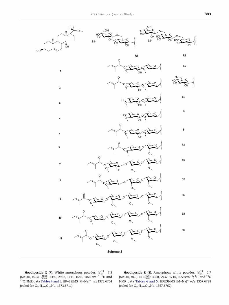
Scheme 3
(1
(
Hoodigoside Q (7): White amorphous powder: [˛]25D − 7.3
MeOH, c0.3); �NaClmax : 3395, 2932, 1711, 1646, 1076 cm−1; 1H and
3C NMR data Tables 4 and 5; HR–ESIMS [M+Na]+ m/z 1373.6764calcd for C65H106O29Na, 1373.6711).
Hoodigoside R (8): Amorphous white powder: [˛]25D − 2.7
(MeOH, c0.3); IR �NaClmax : 3368, 2932, 1710, 1059 cm−1; 1H and 13C
NMR data Tables 4 and 5; HRESI–MS [M+Na]+ m/z 1357.6788(calcd for C65H106O28Na, 1357.6762).
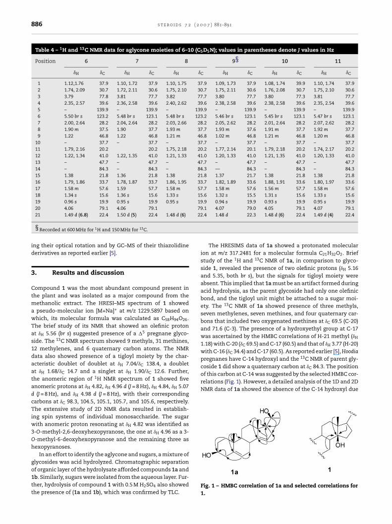
884 s t e r o i d s 7 2 ( 2 0
Table 1 – 1H (400 MHz) and 13C NMR (100 MHz) of 1a(CH3OD–CDCl3, 3:1) and 1b (C5D5N); values inparentheses denote J values in Hz
1a 1b
ıH ıC ıH ıC
1 1.02, 1.83 37.5 1.10, 1.83 37.92 1.45, 1.74 31.5 1.78, 2.05 32.83 3.42 m 71.6 3.82 m 71.54 2.12, 2.21 42.1 2.56, 2.60 43.75 – 140.5 – 141.56 5.35 br s 121.7 5.41 br s 121.37 1.89, 2.17 30.2 2.10, 2.56 30.48 1.78 31.2 2.18 31.49 0.93 50.4 0.98 50.5
10 – 37.3 – 37.511 1.42, 1.52 21.7 1.42, 1.51 22.012 1.21, 1.89 41.2 1.02, 1.86 41.413 – 46.5 – 46.614 – 154.3 – 154.215 5.16 br s 118.9 5.25 br s 119.616 2.12, 2.34 34.5 2.56, 2.98 34.517 1.66 dd 60.5 2.05 60.718 0.87 s 17.0 0.95 s 17.219 0.97 s 19.4 1.03 s 19.620 3.77 m 69.5 4.13 m 80.321 1.18 d (6) 23.7 1.56 d (6) 23.8
Glc1′ Glc2′ 5.00 d (8) 106.43′ 4.03 76.04′ 4.27 78.95′ 4.26 72.16′ 4.01 78.6
4.40 dd (12,6), 4.56 br d (12) 63.3
Table 2 – 1H and 13C NMR data for aglycone moieties of 1–5 (C5D
Position 1 2
ıH ıC ıH ıC ıH
1 1.11, 1.79 37.9 1.13, 1.74 37.9 1.13,2 1.75, 2.14 30.6 1.74, 2.14 30.6 1.75,3 3.83 m 78.0 3.85 m 78.0 3.834 2.42, 2.59 39.6 2.43, 2.60 39.6 2.43,5 – 139.9 – 140.0 –6 5.56 br s 123.2 5.58 br s 123.2 5.587 2.01, 2.65 28.2 2.04, 2.68 28.3 2.04,8 1.90 m 37.7 1.85 m 37.7 1.879 1.23 m 46.8 1.25 m 46.8 1.24
10 – 37.7 – 37.7 –11 1.37, 1.79 20.1 1.37, 1.76 20.1 1.37,12 1.20, 1.31 41.0 1.25, 1.36 41.0 1.24,13 – 47.7 – 47.8 –14 – 84.3 – 84.3 –15 1.38 21.8 1.42 21.8 1.4116 1.85, 1.95 33.6 1.83, 1.96 33.7 1.84,17 1.59 m 57.6 1.64 m 57.6 1.6218 1.35 s 15.5 1.42 s 15.6 1.3719 0.98 s 19.9 1.00 s 19.9 1.0020 4.04 m 79.1 4.15 78.3 4.0421 1.51 d (6) 22.3 1.45 d (6) 22.1 1.52
§Recorded at 600 MHz for 1H and 150 MHz for 13C.
0 7 ) 881–891
Hoodigoside S (9): Amorphous white powder: [˛]25D − 3.3
(MeOH, c0.3); IR �NaClmax : 3445, 2940, 1714, 1057 cm−1; 1H and 13C
NMR data Tables 4 and 5; HRESI–MS [M+Na]+ m/z 1501.7573(calcd for C72H118O31Na, 1501.7549).
Hoodigoside T (10): Amorphous white powder: [˛]25D − 0.1
(MeOH, c0.3); IR �NaClmax : 3412, 2934, 1710, 1092 cm−1 1H and 13C
NMR data Tables 4 and 5; HRESI–MS [M+Na]+ m/z 1339.7090(calcd for C66H108O26Na 1339.7021).
Hoodigoside U (11): Amorphous white powder: [˛]25D − 5.3
(MeOH, c0.3); IR �NaClmax : 3386, 2927, 1711, 1092 cm−1; 1H and 13C
NMR data Tables 4 and 5; HR–ESIMS [M+Na]+ m/z 1501.7574(calcd for C72H118O31Na, 1501.7549).
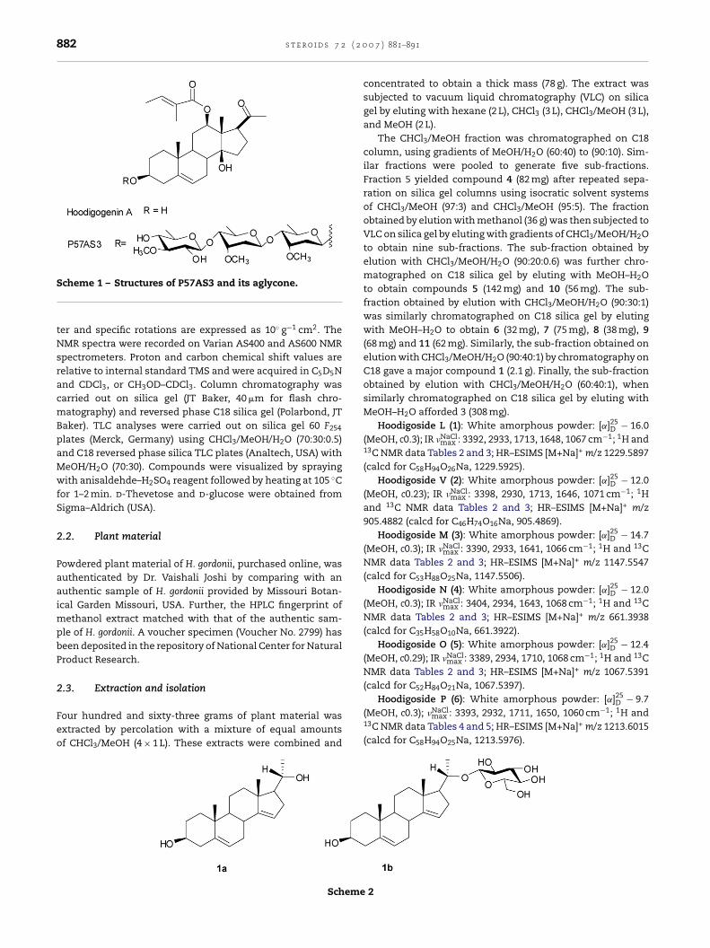
2.4. Acid hydrolysis of glycoside mixture
Mixture of glycosides (8.0 g) was hydrolyzed with 0.5 M H2SO4
in 50% 1,4-dioxane at 95 ◦C for 6 h. The organic layer was driedand subjected to column chromatography on silica gel by elut-ing with gradients of CHCl3 and MeOH to obtain 1a and 1b.
1a: White amorphous powder: [˛]25D − 14.0 (CHCl3/MeOH
1:1, c0.3); IR �NaClmax : 3260, 2962, 1438, 1030 cm−1; 1H and 13C
NMR data Table 1; HR–ESIMS [M+H]+ m/z 317.2481 (calcd for,C21H33O2, 317.2475).
1b: White amorphous powder; [˛]25D − 9.0 (MeOH, c0.27);
IR �NaClmax : 3380, 2932, 1636, 1076 cm−1; 1H and 13C NMR
data Table 1; HR–ESIMS [M+Na]+ m/z 501.2825 (calcd forC27H42O7Na, 501.2822).
2.5. Acid hydrolysis of compounds
Five milligrams of each compound was subjected to acidhydrolysis by heating at 95 ◦C with 0.05 N HCl in 50% 1,4-
5N, *CDCl3); values in parentheses denote J values in Hz
3§ 4* 5§ıC ıH ıC ıH ıC
1.75 37.9 1.28, 1.42 38.9 1.15, 1.77 37.92.13 30.6 1.54, 2.18 33.2 1.70, 2.10 30.6
m 77.9 3.77 m 77.5 3.81 m 78.02.61 39.6 2.20, 2.32 39.6 2.41, 2.58 39.6
139.9 – 139.6 – 139.9br s 123.2 5.37 br s 122.1 5.55 br s 123.22.67 28.2 2.17, 2.26 27.1 2.03, 2.67 28.1
m 37.7 2.32 36.3 1.90 m 37.7m 46.8 1.21 46.5 1.22 m 46.8
37.7 – 37.3 – 37.71.77 20.1 1.42, 1.85 21 1.37, 1.74 20.11.32 41.0 1.17, 1.74 37.6 1.20, 1.34 41.0
47.7 – 47.6 – 47.784.3 – 85.2 – 84.421.8 1.74, 1.85 37.1 1.40 21.8
1.99 33.6 1.23, 1.95 29.7 1.82, 1.99 33.7m 57.6 1.60 56.6 1.58 m 57.7s 15.5 1.03 s 14.8 1.34 s 15.6s 19.9 0.97 s 19.3 0.98 s 19.9
79.0 4.03 q (7) 65.4 4.06 m 79.1d (5) 22.3 1.10 d (6) 22.3 1.51 d (6) 22.4
s t e r o i d s 7 2 ( 2 0 0 7 ) 881–891 885
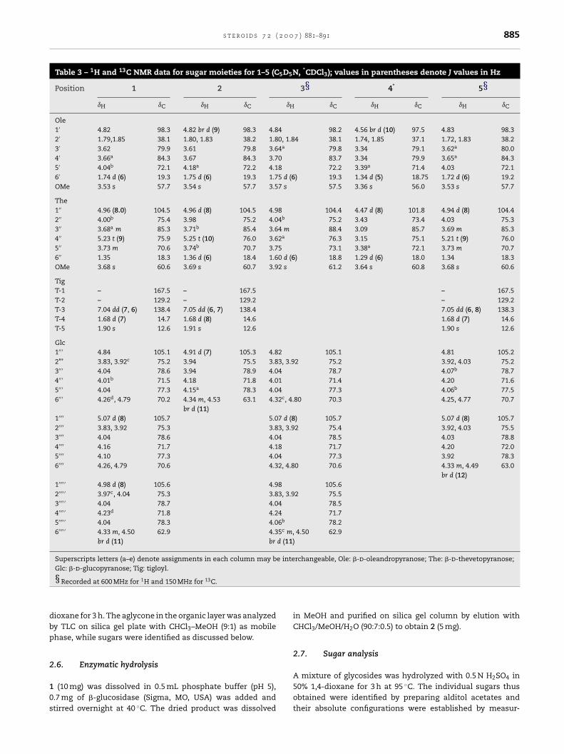
Table 3 – 1H and 13C NMR data for sugar moieties for 1–5 (C5D5N, *CDCl3); values in parentheses denote J values in Hz
Position 1 2 3§ 4* 5§ıH ıC ıH ıC ıH ıC ıH ıC ıH ıC
Ole1′ 4.82 98.3 4.82 br d (9) 98.3 4.84 98.2 4.56 br d (10) 97.5 4.83 98.32′ 1.79,1.85 38.1 1.80, 1.83 38.2 1.80, 1.84 38.1 1.74, 1.85 37.1 1.72, 1.83 38.23′ 3.62 79.9 3.61 79.8 3.64a 79.8 3.34 79.1 3.62a 80.04′ 3.66a 84.3 3.67 84.3 3.70 83.7 3.34 79.9 3.65a 84.35′ 4.04b 72.1 4.18a 72.2 4.18 72.2 3.39a 71.4 4.03 72.16′ 1.74 d (6) 19.3 1.75 d (6) 19.3 1.75 d (6) 19.3 1.34 d (5) 18.75 1.72 d (6) 19.2OMe 3.53 s 57.7 3.54 s 57.7 3.57 s 57.5 3.36 s 56.0 3.53 s 57.7
The1′′ 4.96 (8.0) 104.5 4.96 d (8) 104.5 4.98 104.4 4.47 d (8) 101.8 4.94 d (8) 104.42′′ 4.00b 75.4 3.98 75.2 4.04b 75.2 3.43 73.4 4.03 75.33′′ 3.68a m 85.3 3.71b 85.4 3.64 m 88.4 3.09 85.7 3.69 m 85.34′′ 5.23 t (9) 75.9 5.25 t (10) 76.0 3.62a 76.3 3.15 75.1 5.21 t (9) 76.05′′ 3.73 m 70.6 3.74b 70.7 3.75 73.1 3.38a 72.1 3.73 m 70.76′′ 1.35 18.3 1.36 d (6) 18.4 1.60 d (6) 18.8 1.29 d (6) 18.0 1.34 18.3OMe 3.68 s 60.6 3.69 s 60.7 3.92 s 61.2 3.64 s 60.8 3.68 s 60.6
TigT-1 – 167.5 – 167.5 – 167.5T-2 – 129.2 – 129.2 – 129.2T-3 7.04 dd (7, 6) 138.4 7.05 dd (6, 7) 138.4 7.05 dd (6, 8) 138.3T-4 1.68 d (7) 14.7 1.68 d (8) 14.6 1.68 d (7) 14.6T-5 1.90 s 12.6 1.91 s 12.6 1.90 s 12.6
Glc1′′ ′ 4.84 105.1 4.91 d (7) 105.3 4.82 105.1 4.81 105.22”’ 3.83, 3.92c 75.2 3.94 75.5 3.83, 3.92 75.2 3.92, 4.03 75.23′′ ′ 4.04 78.6 3.94 78.9 4.04 78.7 4.07b 78.74′′ ′ 4.01b 71.5 4.18 71.8 4.01 71.4 4.20 71.65′′ ′ 4.04 77.3 4.15a 78.3 4.04 77.3 4.06b 77.56′′ ′ 4.26d, 4.79 70.2 4.34 m, 4.53
br d (11)63.1 4.32c, 4.80 70.3 4.25, 4.77 70.7
1′′′′ 5.07 d (8) 105.7 5.07 d (8) 105.7 5.07 d (8) 105.72′′′′ 3.83, 3.92 75.3 3.83, 3.92 75.4 3.92, 4.03 75.53′′′′ 4.04 78.6 4.04 78.5 4.03 78.84′′′′ 4.16 71.7 4.18 71.7 4.20 72.05′′′′ 4.10 77.3 4.04 77.3 3.92 78.36′′′′ 4.26, 4.79 70.6 4.32, 4.80 70.6 4.33 m, 4.49
br d (12)63.0
1′′′′ ′ 4.98 d (8) 105.6 4.98 105.62′′′′ ′ 3.97c, 4.04 75.3 3.83, 3.92 75.53′′′′ ′ 4.04 78.7 4.04 78.54′′′′ ′ 4.23d 71.8 4.24 71.75′′′′ ′ 4.04 78.3 4.06b 78.26′′′′ ′ 4.33 m, 4.50
br d (11)62.9 4.35c m, 4.50
br d (11)62.9
Superscripts letters (a–e) denote assignments in each column may be interchangeable, Ole: �-d-oleandropyranose; The: �-d-thevetopyranose;
dbp
2
10s
Glc: �-d-glucopyranose; Tig: tigloyl.
§Recorded at 600 MHz for 1H and 150 MHz for 13C.
ioxane for 3 h. The aglycone in the organic layer was analyzedy TLC on silica gel plate with CHCl3–MeOH (9:1) as mobilehase, while sugars were identified as discussed below.
.6. Enzymatic hydrolysis
(10 mg) was dissolved in 0.5 mL phosphate buffer (pH 5),.7 mg of �-glucosidase (Sigma, MO, USA) was added andtirred overnight at 40 ◦C. The dried product was dissolved
in MeOH and purified on silica gel column by elution withCHCl3/MeOH/H2O (90:7:0.5) to obtain 2 (5 mg).
2.7. Sugar analysis
A mixture of glycosides was hydrolyzed with 0.5 N H2SO4 in50% 1,4-dioxane for 3 h at 95 ◦C. The individual sugars thusobtained were identified by preparing alditol acetates andtheir absolute configurations were established by measur-
886 s t e r o i d s 7 2 ( 2 0 0 7 ) 881–891
Table 4 – 1H and 13C NMR data for aglycone moieties of 6–10 (C5D5N); values in parentheses denote J values in Hz
Position 6 7 8 9§ 10 11
ıH ıC ıH ıC ıH ıC ıH ıC ıH ıC ıH ıC
1 1.12,1.76 37.9 1.10, 1.72 37.9 1.10, 1.75 37.9 1.09, 1.73 37.9 1.08, 1.74 39.9 1.10, 1.74 37.92 1.74, 2.09 30.7 1.72, 2.11 30.6 1.75, 2.10 30.7 1.75, 2.11 30.6 1.76, 2.08 30.7 1.75, 2.10 30.63 3.79 77.8 3.81 77.7 3.82 77.7 3.80 77.7 3.80 77.3 3.81 77.74 2.35, 2.57 39.6 2.36, 2.58 39.6 2.40, 2.62 39.6 2.38, 2.58 39.6 2.38, 2.58 39.6 2.35, 2.54 39.65 – 139.9 – 139.9 – 139.9 – 139.9 – 139.9 – 139.96 5.50 br s 123.2 5.48 br s 123.1 5.48 br s 123.2 5.46 br s 123.1 5.45 br s 123.1 5.47 br s 123.17 2.00, 2.64 28.2 2.04, 2.64 28.2 2.03, 2.66 28.2 2.05, 2.62 28.2 2.01, 2.64 28.2 2.07, 2.62 28.28 1.90 m 37.5 1.90 37.7 1.93 m 37.7 1.93 m 37.6 1.91 m 37.7 1.92 m 37.79 1.22 46.8 1.22 46.8 1.21 m 46.8 1.02 m 46.8 1.21 m 46.8 1.20 m 46.8
10 – 37.7 – 37.7 – 37.7 – 37.7 – 37.7 – 37.711 1.79, 2.16 20.2 20.2 1.75, 2.18 20.2 1.77, 2.14 20.1 1.79, 2.18 20.2 1.74, 2.17 20.212 1.22, 1.34 41.0 1.22, 1.35 41.0 1.21, 1.33 41.0 1.20, 1.33 41.0 1.21, 1.35 41.0 1.20, 1.33 41.013 – 47.7 – 47.7 – 47.7 – 47.7 – 47.7 – 47.714 – 84.3 – 84.3 – 84.3 –– 84.3 – 84.3 – 84.315 1.38 21.8 1.36 21.8 1.38 21.8 1.37 21.7 1.38 21.8 1.38 21.816 1.79, 1.86 33.7 1.78, 1.87 33.7 1.86, 1.95 33.7 1.82, 1.89 33.6 1.88, 1.91 33.6 1.80, 1.97 33.617 1.58 m 57.6 1.59 57.7 1.58 m 57.7 1.58 m 57.6 1.56 m 57.7 1.58 m 57.618 1.34 s 15.6 1.36 s 15.6 1.33 s 15.6 1.32 s 15.5 1.31 s 15.6 1.33 s 15.619 0.96 s 19.9 0.95 s 19.9 0.95 s 19.9 0.94 s 19.9 0.93 s 19.9 0.95 s 19.920 4.06 79.1 4.06 79.1 79.1 4.07 79.0 4.05 79.1 4.07 79.1
22
C
of this carbon at C-14 was suggested by the selected HMBC cor-relations (Fig. 1). However, a detailed analysis of the 1D and 2DNMR data of 1a showed the absence of the C-14 hydroxyl due
21 1.49 d (6.8) 22.4 1.50 d (5) 22.4 1.48 d (6)
§Recorded at 600 MHz for 1H and 150 MHz for 13C.
ing their optical rotation and by GC–MS of their thiazolidinederivatives as reported earlier [5].
3. Results and discussion
Compound 1 was the most abundant compound present inthe plant and was isolated as a major compound from themethanolic extract. The HRESI–MS spectrum of 1 showeda pseudo-molecular ion [M+Na]+ at m/z 1229.5897 based onwhich, its molecular formula was calculated as C58H94O26.The brief study of its NMR that showed an olefinic protonat ıH 5.56 (br s) suggested presence of a �5 pregnane glyco-side. The 13C NMR spectrum showed 9 methyls, 31 methines,12 methylenes, and 6 quaternary carbon atoms. The NMRdata also showed presence of a tigloyl moiety by the char-acteristic doublet of doublet at ıH 7.04/ıC 138.4, a doubletat ıH 1.68/ıC 14.7 and a singlet at ıH 1.90/ıC 12.6. Further,the anomeric region of 1H NMR spectrum of 1 showed fiveanomeric protons at ıH 4.82, ıH 4.96 d (J = 8 Hz), ıH 4.84, ıH 5.07d (J = 8 Hz), and ıH 4.98 d (J = 8 Hz), with their correspondingcarbons at ıC 98.3, 104.5, 105.1, 105.7, and 105.6, respectively.The extensive study of 2D NMR data resulted in establish-ing spin systems of individual monosaccharide. The sugarwith anomeric proton resonating at ıH 4.82 was identified as3-O-methyl-2,6-deoxyhexopyranose, the one at ıH 4.96 as a 3-O-methyl-6-deoxyhexopyranose and the remaining three ashexopyranoses.
In an effort to identify the aglycone and sugars, a mixture ofglycosides was acid hydrolyzed. Chromatographic separation
of organic layer of the hydrolysate afforded compounds 1a and1b. Similarly, sugars were isolated from the aqueous layer. Fur-ther, hydrolysis of compound 1 with 0.5 M H2SO4 also showedthe presence of (1a and 1b), which was confirmed by TLC..4 1.48 d 22.3 1.48 d (6) 22.4 1.49 d (4) 22.4
The HRESIMS data of 1a showed a protonated molecularion at m/z 317.2481 for a molecular formula C21H32O2. Briefstudy of the 1H and 13C NMR of 1a, in comparison to glyco-side 1, revealed the presence of two olefinic protons (ıH 5.16and 5.35, both br s), but the signals for tigloyl moiety wereabsent. This implied that 1a must be an artifact formed duringacid hydrolysis, as the parent glycoside had only one olefinicbond, and the tigloyl unit might be attached to a sugar moi-ety. The 13C NMR of 1a showed presence of three methyls,seven methylenes, seven methines, and four quaternary car-bons that included two oxygenated methines at ıC 69.5 (C-20)and 71.6 (C-3). The presence of a hydroxyethyl group at C-17was ascertained by the HMBC correlations of H-21 methyl (ıH
1.18) with C-20 (ıC 69.5) and C-17 (60.5) and that of ıH 3.77 (H-20)with C-16 (ıC 34.4) and C-17 (60.5). As reported earlier [5], Hoodiapregnanes have C-14 hydroxyl and the 13C NMR of parent gly-coside 1 did show a quaternary carbon at ı 84.3. The position
Fig. 1 – HMBC correlation of 1a and selected correlations for1.
st
er
oid
s7
2(2
00
7)
881–891887
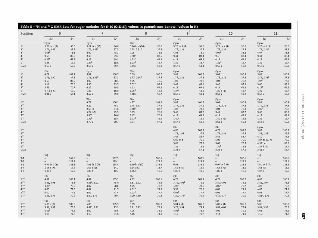
Table 5 – 1H and 13C NMR data for sugar moieties for 6–10 (C5D5N); values in parentheses denote J values in Hz
Position 6 7 8 9§ 10 11
ıH ıC ıH ıC ıH ıC ıH ıC ıH ıC ıH ıC
Cym Cym Cym Cym Cym Cym1′ 5.28 br d (8) 96.6 5.27 br d (10) 96.6 5.26 br d (10) 96.6 5.26 br d (8) 96.6 5.25 br d (8) 96.6 5.27 br d (9) 96.62′ 1.76, 2.32 37.5 1.76, 2.33a 37.5 1.75, 2.31a 37.3 1.77, 2.31 37.5 1.76, 2.31 37.5 1.79, 2.31a 37.23′ 4.05a 78.1 4.02 78.3 4.05 78.6 4.03 78.0 4.04a 78.2 4.07 78.84′ 3.53 83.9 3.48 83.7 3.50b 83.6 3.41 83.4 3.4 83.4 3.41 83.45′ 4.22b 69.2 4.22 69.3 4.23c 69.3 4.18 69.2 4.19 69.2 4.15 69.36′ 1.34 18.8 1.36b 18.8 1.33d 18.7 1.32 18.7 1.31b 18.7 1.32 18.7OMe 3.59 s 59.2 3.56 s 59.1 3.63 s 59.1 3.57 s 59.1 3.56 s 59.2 3.58 s 59.2
Ole Cym Cym Cym Cym Cym1′′ 4.78 102.2 5.09 100.7 5.09 100.7 5.09 100.7 5.08 100.8 5.09 100.82′′ 1.76, 1.90 37.5 1.76, 2.36a 37.3 1.77, 2.34a 37.5 1.77, 2.31 37.3 1.76, 2.31 37.3 1.79, 2.33a 37.33′′ 3.53 83.9 4.02 78.3 4.05 78.6 4.03 78.2 4.04 78.2 4.07b 77.34′′ 5.11 76.6 3.56 83.5 3.52b 83.8 3.48 83.5 3.46 83.5 3.46 83.55′′ 3.63 70.7 4.22 69.5 4.23 69.2 4.18 69.2 4.19 69.2 4.15c 69.26′′ 1. 44 d (6) 19.0 1.38 18.9 1.35d 18.8 1.37a 18.8 1.35 d (4) 18.7 1.32 18.7OMe 3.34 s 57.1 3.62 s 59.2 3.40 s 58.7 3.60 s 59.1 3.59 s 59.2 3.60 s 59.2
The Ole Cym Cym Cym1′′ ′ 4.78 106.3 4.77 102.2 5.09 100.7 5.08 100.8 5.09 100.82′′ ′ 4.22 75.3 1.75, 1.93 37.5 1.77, 2.31 37.2 1.76, 2.31 37.2 1.79, 2.33 37.63′′ ′ 3.68 m 84.8 3.48b 83.7 4.03 78.2 4.04 78.3 4.09b 78.64′′ ′ 5.22 t (9) 75.3 5.09 76.6 3.48 83.7 3.49 83.7 3.48 83.75′′ ′ 3.80c 70.6 3.67 70.8 4.18 69.3 4.19 69.3 4.15 69.26′′ ′ 1.35b 18.4 1.33d 18.9 1.40a 18.9 1.40 d (6) 18.8 1.32 18.7OMe 3.70 s 60.7 3.34 57.1 3.57 s 59.3 3.61 s 59.3 3.63 s 59.2
Ole Ole Cym1′′′′ 4.80 102.2 4.78 102.2 5.09 100.82′′′′ 1.73, 1.93 37.6 1.76, 2.31 37.5 1.82, 2.35 36.53′′′′ 3.48 83.7 3.46 83.7 4.10 78.34′′′′ 5.09 br d 76.6 5.08 76.6 4.87 dd (2, 7) 76.05′′′′ 3.65 70.8 3.65 70.8 4.32d m 68.56′′′′ 1.32 18.4 1.32b 18.4 1.37 d (5) 18.9OMe 3.34 s 57.1 3.33 s 57.1 3.41 s 58.7
Tig Tig Tig Tig Tig TigT-1 – 167.6 – 167.5 – 167.5 – 167.6 – 167.6 – 167.5T-2 – 129.2 – 129.1 – 129.2 – 129.2 – 129.2 – 129.1T-3 6.99 br d (8) 138.3 7.03 br d (7) 138.4 6.99 br d (7) 138.3 6.98 138.2 6.97 br d (6) 138.3 7.00 br d (7) 138.6T-4 1.64 d (7) 14.6 1.68 d (6) 14.7 1.64 d (7) 14.7 1.65 d (6) 14.6 1.63 d (6) 14.6 1.64 (5) 14.6T-5 1.86 s 12.6 1.86 s 12.7 1.86 s 12.6 1.86 s 12.6 1.85 s 12.6 1.87 s 12.5
Glc Glc Glc Glc Glc Glc1′′′′ ′ 4.81 105.1 4.81 105.2 4.82 105.1 4.78 105.1 4.75 105.2 4.83 105.22′′′′ ′ 3.81, 3.98 75.3 3.81c, 3.92 75.0 3.81, 3.92 75.3 3.79, 3.94b 75.2 3.80, 4.01 75.2 3.81, 3.97 75.33′′′′ ′ 4.06a 78.6 4.02 78.6 4.05 78.7 4.03d 78.6 4.05a 78.7 4.03 78.74′′′′ ′ 4.00 71.5 4.02 71.5 4.01e 71.5 3.99 71.5 4.05 71.5 4.03 71.15′′′′ ′ 4.00 77.3 4.02 77.4 4.03e 77.7 4.01d 77.7 4.01 77.7 4.03 77.76′′′′ ′ 4.28, 4.78 70.3 4.30, 4.78 70.4 4.29, 4.80 70.3 4.26, 4.78c 70.3 4.19, 4.77 70.6 4.24e, 4.78 70.3
Glc Glc Glc Glc Glc Glc1′′′′′′ 5.06 d (8) 105.8 5.06 105.9 5.09 105.8 5.04 d (8) 105.7 5.03 d (8) 105.7 5.06 105.92′′′′′′ 3.81, 3.98 75.3 3.81c, 3.92 75.3 3.81, 3.92 75.5 3.79, 3.94 75.4 3.88, 4.01 75.4 3.81, 3.97 75.53′′′′′′ 4.06a 78.6 4.02 78.6 4.05 78.1 4.03d 78.6 4.05 79.1 4.03 78.64′′′′′′ 4.17 71.7 4.17 71.8 4.16 71.8 4.13 71.7 4.14 71.9 4.16c 71.7
888 s t e r o i d s 7 2 ( 2 0
Tabl
e5
–(C
onti
nu
ed)
Posi
tion
67
89§
1011
ı Hı C
ı Hı C
ı Hı C
ı Hı C
ı Hı C
ı Hı C
5′′′′′
′4.
0677
.34.
0277
.44.
1077
.74.
01d
77.7
3.94
77.4
4.03
77.3
6′′′′′
′4.
24b
,4.7
870
.64.
30,4
.78
70.6
4.23
c,4
.80
70.6
4.26
,4.7
5c70
.84.
32m
4.47
brd
(12)
63.0
4.23
e,4
.81
70.6
Glc
Glc
Glc
Glc
Glc
1′′′′′
′′4.
97d
(8)
105.
74.
97d
(7.2
)10
5.7
4.95
d(8
)10
5.7
4.94
d(7
)10
5.6
4.97
d(8
)10
5.7
2′′′′′
′′3.
90,4
.02
75.4
3.92
,4.0
275
.53.
81,3
.92
75.9
3.96
b,3
.99
75.8
3.95
,4.0
375
.93′
′′′′′′
4.06
a78
.74.
0278
.84.
0978
.14.
0378
.64.
0378
.64′
′′′′′′
4.22
b71
.84.
2271
.84.
22c
71.7
4.18
71.9
4.25
e71
.55′
′′′′′′
4.06
78.3
4.06
78.3
4.05
78.3
4.10
78.3
4.03
78.2
6′′′′′
′′4.
37m
,4.4
8br
d(1
1)62
.94.
34m
4.49
brd
(10)
62.9
4.34
m,4
.47
brd
(10)
62.9
4.32
m,4
.45
brd
(10)
63.0
4.35
dm
,4.4
9br
d(1
1)62
.9
Sup
ersc
rip
tsle
tter
s(a
–e)d
enot
eas
sign
men
tsin
each
colu
mn
may
bein
terc
han
geab
le,C
ym:�
-d-c
ymar
opyr
anos
eO
le:�
-d-o
lean
dro
pyr
anos
e;T
he:
�-d
-th
evet
opyr
anos
e;G
lc:�
-d-g
luco
pyr
anos
e;T
ig:t
iglo
yl
§ Rec
ord
edat
600
MH
zfo
r1H
and
150
MH
zfo
r13
C.
0 7 ) 881–891
to formation of a double bond between C-14 and C-15. Thiswas confirmed by the HMBC correlations of H-18 methyl (ıH
0.87) with C-14, C-17, C-13, and C-12 and those of the olefinicproton H-15 (ıH 5.16) with C-14, C-17, C-13, and C-16. Thus,the primary structure of 1a was determined to be pregn-5-14-diene-3,20-diol. The NOESY correlations between H-18 andH-20 and between H-21 and H-16, H-12 were observed. Further,to determine the absolute configuration at C-20, we preparedthe Mosher esters in a NMR tube by reaction of 1a with (R)and (S)-MTPACl [8]. The calculated ı(S–R) values +0.02 and −0.10for H-18 and H-21 protons, respectively, confirmed the 17Sand 20S configuration, i.e. 1a is pregna-5,14-diene-3�,20�-diol.This molecule was reported in 1955 by Heusser et al. [9]; how-ever, this is the first report of detailed spectroscopic study of1a. Therefore, it was inferred that 1a was formed by the lossof a water molecule at C-14 and the aglycone present in theglycoside was calogenin (pregn-5-ene-3�,14�,20�-triol). Fur-ther, the chemical shift values for the aglycone of compound1 matched with those reported for calogenin bisdesmosides[10].
The hydrolysis product 1b showed a mass of m/z 501.2825that corresponded to a molecular formula of C27H42O7, whichwas 162 amu (a hexose unit) greater than 1a. Its 1H and 13CNMR signals for steroid skeleton matched closely to 1a, butthe attachment of a glucose unit at C-20 was evident by shiftin the resonance of C-20 carbon (ıC 80.3). This observation wasfurther confirmed by the HMBC correlation of H-20 (ıH 4.13)with C-1′ (ıC 106.4). On repeated efforts, we could only isolate1a but not the genuine aglycone calogenin.
On comparison of the 13C NMR resonances for C-3 andC-20 of glycoside 1 with those of 1a, the observed glycosyla-tion shifts indicated bisdesmosidic nature of 1. The sugarsobtained on acid hydrolysis were identified as d-oleandrose,d-thevetose, and d-glucose by GC–MS of their alditol acetates.The attachment of oleandrose to aglycone and thevetose tooleandrose was evident by the HMBC correlations of H-1′ (ıH
4.82) with C-3 (ıC 78.0) and H-1′′ (ıH 4.96) with C-4′ (ıC 84.3).The H-4′′ of thevetose was observed to be shifted downfieldto ıH 5.23/ıC 75.9 and showed long-range correlation withıC 167.5. This observation proved the attachment of tigloylmoiety to thevetose at C-4′′. Thus, the sugar chain at C-3 wascharacterized as �-(4-O-tigloyl)-d-thevetopyranosyl-(1 → 4)-�-d-oleandropyranosyl unit. Out of the three glucose units, twohad their methylene carbons shifted downfield (ıC 70.2 and70.6) as compared to the third (ıC 62.9). The HMBC correlationsof H-1′′′′ ′ (ıH 4.98) with C-6′′′′ (ıC 70.6) and H-1′′′′ (ıH 5.07) with C-6′′ ′ (ıC 70.2), and H-1′′ ′ (ıH 4.84) to C-20 (ıC 79.1) established thelinkage as �-d-glucopyranosyl-(1 → 6)-�-d-glucopyranosyl-(1 → 6) �-d-glucopyranose. Thus, hoodigoside L (1) wascharacterized as calogenin-20-O-�-d-glucopyranosyl-(1 → 6)-�-d-glucopyranosyl-(1 → 6)-�-d-glucopyranosyl-3-O-�-(4-O-tigloyl)-d-thevetopyranosyl)-(1 → 4)-�-d-oleandropyranoside.
In an attempt to obtain genuine aglycone, and to consol-idate the structure, 1 was subjected to enzymatic hydrolysiswith �-glucosidase. The purified product (2) showed a sodiatedmolecular ion at m/z 905.4882 which confirmed its molec-
ular formula as C46H74O16. Its proton NMR showed threeanomeric protons at ıH 4.82, 4.96, and 4.91 d (J = 7 Hz) that cor-responded to anomeric carbons resonating at ıC 98.3, 104.5,and 105.3, respectively. The NMR data for aglycone portion
2 0 0
atiatp
aqa1bIici1o(57o(�
NC1
eww611
49tatws�
mCpsnaa1wadhdTttH
s t e r o i d s 7 2 (
nd sugars remained similar to 1, but the signals for thewo (1 → 6) attached glucoses to the inner glucose were miss-ng. Thus, structure of hoodigoside V (2) was establisheds calogenin-20-O-�-d-glucopyranosyl-3-O-�-(4-O-tigloyl)-d-hevetopyranosyl)-(1 → 4)-�-d-oleandropyranoside, a prosa-ogenin obtained on enzymatic hydrolysis of 1.
The molecular formula for compound 3 was confirmeds C53H88O25 by HRESIMS, where it showed a prominentuasi-molecular ion [M+Na]+ at m/z 1147.5547. The aglyconend sugars were found to be similar to those obtained from. Its 1H and 13C NMR showed a close resemblance to 1,ut signals for the tigloyl moiety were found to be missing.n its 1H NMR, H-4′′ resonated at ıH 3.62, instead of ıH 5.23n 1 and did not show chemical shifts in the neighboringarbon atoms suggesting it to be the terminal sugar. Thus,t was clearly established that compound 3 is compound
devoid of the tigloyl group. Following HMBC correlationsf H-1′ (ıH 4.84) with C-3 (ıC 77.9), H-1′′ (ıH 4.98) with C-4′
ıC 83.7), H-1′′′′ ′ (ıH 4.98) with C-6′′′′ (ıC 70.6) and H-1′′′′ (ıH
.07) with C-1′′ ′ (ıC 70.3), and H-1′′ ′ (ıH 4.82) with C-20 (ıC
9.0) were observed to corroborate the chemical structuref hoodigoside M (3) as calogenin-20-O-�-d-glucopyranosyl-
1 → 6)-�-d-glucopyranosyl-(1 → 6)-�-d-glucopyranosyl-3-O--d-thevetopyranosyl-(1 → 4)-�-d-oleandropyranoside.
The HRESIMS, m/z 661.3938 for [M+Na]+, together with 13CMR spectrum was used to establish a molecular formula,
35H58O10 for compound 4. Preliminary observations in theH and 13C NMR data showed similarity to those of 3. How-ver, the absence of tigloyl moiety in the sugar chain at C-3as evident as well as the absence of the sugar chain at C-20,hich was confirmed by the marked upfield shift for C-20 (ıc
5.4) as compared to ıc 79.0 for C-20 of 3. Similar to compound, the acid hydrolysis of 4 indicated calogenin as the aglycone.H NMR of 4 showed two doublets for anomeric protons at ıH
.56 (J = 10 Hz) and ıH 4.47 (J = 8 Hz), which corresponded to ıc7.5 and 101.8 respectively. Detailed study of 2D NMR spec-roscopic data confirmed chemical structures of the sugarss d-oleandropyranose and d-thevetopyranose. HMBC correla-ion of ıH 1′ (ıH 4.56) with C-3 (ıC 77.5) and that of ıH 1′′ (ıH 4.47)ith C-4′ (ıC 79.9) helped establish the sugar sequence. Thus,
tructure of hoodigoside N (4) was deduced as calogenin-3-O--d-thevetopyranosyl-(1 → 4)-�-d-oleandropyranoside.
Compound 5 showed a quasi-molecular ion [M+Na]+ at/z 1067.5391 that corresponded to a molecular formula of
52H84O21. The acid hydrolysis yielded 1a that suggested theresence of calogenin as aglycone. Its 13C NMR spectrumhowed 9 methyls, 26 methines, 11 methylenes, and 6 quater-ary carbon atoms. The proton NMR showed presence of fivenomeric signals at ıH 4.83, 4.94 d (J = 8.0 Hz), 5.07 d (J = 8 Hz),nd 4.81 which corresponded to anomeric carbons at ıC 98.3,04.4, 105.7, and 105.2 observed in its 13C NMR. The sugarsith anomeric protons at ıH 4.83 and 4.97 were identified
s 3-O-methyl-2,6-deoxyhexopyranose and 3-O-methyl-6-eoxyhexopyranose, respectively, while the remaining two asexopyranoses. These sugars were identified as d-oleandrose,-thevetose and d-glucose by GC–MS of the acid hydrolysate.
he NMR data also showed presence of a tigloyl moiety andhe sugar linkage at C-3 was found to be superimposableo that present in 1, which was corroborated by followingMBC correlations: H-1′′ (ıH 4.94) with C-4′ (ıC 84.3), H-1′ (ıH
7 ) 881–891 889
4.83) with C-3 (ıC 78.0). The C-6 methylenes of two glucoseunits appeared at ıC 70.7 and 63.0 indicating a (1 → 6) linkagebetween them. The HMBC correlations of H-1′′′′ (ıH 4.81)with C-6′′ ′ (ıC 70.7) and that of H-20 (ıH 4.06) with C-1′′ ′ (ıC
105.7) confirmed the attachment of a �-d-glucopyranosyl-(1 → 6)-ˇ-d-glucopyranoside to C-20 of calogenin. Thus,hoodigoside O (5) was characterized as calogenin-20-O-�-d-glucopyranosyl-(1 → 6)-�-d-glucopyranosyl-3-O-�-(4-O-tigloyl)-d-thevetopyranosyl-(1 → 4)-�-d-oleandropyranoside.
Compound (6) showed a quasi-molecular ion peak atm/z 1213.6015 and with conjunction with the 13C NMR dataits molecular formula was assigned as C58H94O25. It wasidentified as a calogenin glycoside, as it produced 1a uponacid hydrolysis. The proton NMR showed five anomericprotons at ıH 5.28 br d (J = 8 Hz), 4.78, 4.81, 5.06 d (J = 8 Hz),and 4.97 d (J = 8 Hz) that corresponded to anomeric carbonsat ıC 96.6, 102.2, 105.1, 105.8, and 105.7. On determiningthe spin systems of individual sugars, the presence of two3-O-methyl-2,6-deoxyhexopyranose and three hexoses unitswere evident. The 3-O-methyl-2,6-deoxyhexopyranose sugarswere identified as d-cymarose and d-oleandrose while thehexoses as d-glucose. The shift values for three glucoseunits were superimposable to those recorded for compound(1), thus confirming the �-d-glucopyranosyl-(1 → 6)-�-d-glucopyranosyl-(1 → 6) ˇ-d-glucopyranose unit at C-20. Ascompared to previous compounds, the anomeric carbon offirst sugar resonated at ıC 96.6, which is a characteristic valuefor cymarose attached to C-3. This was further confirmed bythe HMBC correlation of H-1′ (ıH 5.28) with C-3 (ıC 77.8). Fur-ther H-4′′ of cymarose showed HMBC correlation with C-1′′ ′ ofoleandrose. The H-4′′ of oleandrose appeared at ıH/ıC 5.11/76.6and showed HMBC correlation with carbonyl carbon of thetigloyl unit (ıc 167.6), this suggested that the tigloyl unit wasattached to C-4 of oleandrose. Therefore, Hoodigoside P (6)was identified as calogenin-20-O-�-d-glucopyranosyl-(1 → 6)-�-d-glucopyranosyl-(1 → 6)-�-d-glucopyranosyl-3-O-�-(4-O-tigloyl)-d-oleandropyranosyl-(1 → 4)-�-d-cymaropyranoside.
Compound 7 showed a sodiated molecular ion at m/z1373.6764 that corresponded to a molecular formula ofC65H106O29. It was identified as a calogenin glycoside, asit produced 1a upon acid hydrolysis. The presence of sixsugars was evident as its 1H NMR showed signals for sixanomeric protons at ıH 5.27 br d (J = 10 Hz), 5.09, 4.78, 4.81,5.06, and 4.97 d (J = 7 Hz) that corresponded to carbon reso-nances at ıC 96.6, 100.7, 106.3, 105.2, 105.9, and 105.7. Thedata also clearly showed presence of a tigloyl moiety. Byestablishing their spin systems, the sugars were identifiedas two 3-O-methyl-2,6-deoxyhexopyranose, one 3-O-methyl-6-deoxyhexopyranose, and three hexoses. These sugarswere identified as d-cymarose, d-thevetose, and d-glucoseduring the hydrolytic studies. The resonance of anomericcarbon of the first sugar at ıC 96.6, was characteristic tocymarose at C-3, and was confirmed by the HMBC cor-relation of H-1′ (ıH 5.27) with C-3 (ıC 77.7). The H-1′′ (ıH
5.09) of second cymarose showed long-range correlationwith C-4′ (ıC 83.7) and the anomeric proton of thevetose
′′ ′′ ′
(ıH 4.78) with C-4 (ıC 83.5). Further, H-4 of thevetoseappeared at ıH/ıC 5.22/75.3 and showed HMBC correlationwith carbonyl of tigloyl at ıC 167.5. Thus, the sugar chain atC-3 was established as �-(4-O-tigloyl)-d-thevetopyranosyl-
( 2 0
890 s t e r o i d s 7 2(1 → 4)-�-d-cymaropyranosyl-(1 → 4)-�-d-cymaropyranose.The remaining signals for the three glucose units were foundto be consistent with those found in 1, i.e. �-d-glucopyranosyl-(1 → 6)-�-d-glucopyranosyl-(1 → 6)-�-d-glucopyranose. Thus,the complete structure of hoodigoside Q (7) was establishedas calogenin-20-O-�-d-glucopyranosyl-(1 → 6)-�-d-glucopyra-nosyl-(1 → 6)-�-d-glucopyranosyl-3-O-ˇ-(4-O-tigloyl)-d-theve-topyranosyl-(1 → 4)-�-d-cymaropyranosyl-(1 → 4)-�-d-cyma-ropyranoside.
Compound 8 showed a pseudo-molecular ion in the pos-itive HRESIMS at m/z 1357.6788 and in conjunction with 13CNMR data, the molecular formula was assigned as C65H106O28,which was one oxygen atom less than that of 7. The presenceof calogenin was determined by the hydrolysis studies. The1H NMR showed presence of six anomeric protons at ıH 5.26 brd (J = 10 Hz), 5.09, 4.77, 4.82, 5.09, and 4.85 d (J = 8 Hz) with theircorresponding carbons at ıC 96.6, 100.7, 102.2, 105.1, 105.8, and105.7. On establishing the spin systems of individual sugarsfrom the NMR data, they were identified as three 3-O-methyl-2,6-deoxyhexopyranoses and three hexoses. The hydrolyticstudy suggested presence of d-cymarose, d-oleandrose, andd-glucose. On comparing the NMR data with that of 7, itwas evident that, last of the three sugars at C-3, thevetose,was replaced by an oleandrose in 8. This observation wasfurther corroborated by the HMBC correlations of H-1′ (ıH
5.26) with C-3 (ıC 77.7), H-1′′ (ıH 5.09) with C-4′ (ıC 83.6), andH-1′′ ′ (ıH 4.77) with C-4′′ (ıC 83.8). The H-4′′ ′ of oleandroseappeared at ıH/ıC 5.09/76.6 and showed HMBC correlationwith carbonyl carbon of tigloyl group (ıc167.5). Further, theNMR data for the three glucose chains at C-20 was found to beidentical to that of compound 7. The structure of hoodigosideR (8) was thus, calogenin-20-O-�-d-glucopyranosyl-(1 → 6)-�-d-glucopyranosyl-(1 → 6)-�-d-glucopyranosyl-3-O-�–(4-O-tigloyl)-d-oleandropyranosyl-(1 → 4)-�-d-cymaropyranosyl-(1 → 4)-�-d-cymaropyranoside.
A molecular formula for 9 was established as C72H118O31
as it showed a sodiated molecular ion at m/z 1501.7573,which was 144 amu greater than 8 indicating an additional3-O-methyl-2,6-deoxyhexopyranose unit. It gave 1a on acidhydrolysis suggesting it to be a calogenin glycoside. Thepresence of seven sugars was evident by the resonances ofanomeric protons in 1H NMR at ıH 5.26 br d (J = 8 Hz), 5.09for two protons, 4.80, 4.78, 5.04, and 4.94 d (J = 7 Hz), whichcorresponded to anomeric carbons at ıC 96.6, 100.7 for twocarbons, 102.2, 105.1, 105.7, and 105.6. A detailed analysisof 1D and 2D NMR data showed that sugars with anomericcarbons at ıC 96.6, 100.7, 100.7, and 102.2 were 3-O-methyl-2,6-deoxy-hexopyranoses, while those at ıC 105.2, 105.3 and105.7 as hexopyranoses. Further, three of the 3-O-methyl-2,6-deoxy hexopyranose were identified as d-cymarose, theother as d-oleandrose and the three hexoses as d-glucose, byhydrolytic studies. Thus, it was evident that as compared to 8,compound 9 had one additional cymarose unit. Further HMBCcorrelations of H-1′ (ıH 5.26) with C-3 (ıC 77.7), H-1′′ (ıH 5.09)with C-4′ (ıC 83.4), H-1′′ ′ (ıH 5.09) with C-4′′ (ıC 83.5), and H-1′′′′
(ıH 4.80) with C-4′′ ′ (ıC 83.7) were observed. The shift value of′′′′
H-4 at ıH/ıC 5.09/76.6 and its HMBC correlation with carbonylcarbon of tigloyl moiety (ıc 167.6) proved their attachment.Hence, the sugar sequence at C-3 was established as 3-O-�-(4-O-tigloyl)-d-oleandropyranosyl-(1 → 4)-�-d-cymaropyranosyl-
0 7 ) 881–891
(1 → 4)-�-d-cymaropyranosyl-(1 → 4)-�-d-cymaropyranose.Further, the NMR data of sugar chain at C-20 was consis-tent to that of compound 8. Thus, hoodigoside S (9) wascharacterized as calogenin-20-O-�-d-glucopyranosyl-(1 → 6)-�-d-glucopyranosyl-(1 → 6)-�-d-glucopyranosyl-3-O-�-(4-O-tigloyl)-d-oleandropyranosyl-(1 → 4)-�-d-cymaropyranosyl-(1 → 4)-�-d-cymaropyranosyl-(1 → 4)-�-d-cymaropyranoside.
Compound 10 showed a quasi-molecular ion at m/z1339.7090 which confirmed the molecular formula asC66H108O26. The acid hydrolysis indicated presence ofcalogenin as the aglycone. The 1H NMR showed six anomericprotons at ıH 5.25 br d (J = 8 Hz), 5.08 for two protons, 4.78,4.75, and 5.03 d (J = 8 Hz) that corresponded to anomericcarbons at ıC 96.6, 100.8 for two carbons, 102.2, 105.2, and105.7 respectively. With the identical 1D and 2D NMR datafor the sugars at C-3 as recorded for 9, it was evident that 10too has a 3-O-�–(4-O-tigloyl)-d-oleandropyranosyl-(1 → 4)-�-d-cymaropyranosyl-(1 → 4)-�-d-cymaropyranosyl-(1 → 4)-�-d-cymaropyranosyl chain at C-3. However, compound 10 hadonly two glucose units as compared to three in 9. The twoglucose units were attached to each other by a (1 → 6) linkageand to the C-20, as evident by the HMBC correlation of H-1′′′′′′
(ıH 5.03) with C-6′′′′ ′ (ıC 70.6) and H-1′′′′ ′ (ıH 4.75) with C-20 (ıC
78.3). This was identical to the C-20 sugar chain in compound5. Thus, hoodigoside T (10) was characterized as calogenin-20-O-�-d-glucopyranosyl-(1 → 6)-�-d-glucopyranosyl-3-O-�-(4-O-tigloyl)-d-oleandropyranosyl-(1 → 4)-�-d-cymaropyranosyl-(1 → 4)-�-d-cymaropyranosyl-(1 → 4)-�-d-cymaropyranoside.
A molecular formula of C72H118O31 for compound 11 wasevident from the HRESIMS where it showed a sodiated molec-ular ion at m/z 1501.7574. This molecular formula was similarto compound 9 and the acid hydrolysis also indicated it as acalogenin glycoside. Its NMR data also showed presence of3-O-methyl-2,6-deoxy hexopyranose and hexose sugars. Theproton NMR showed the anomeric resonances at ıH 5.27 br d(J = 9 Hz), ıH 5.09 for three protons, 4.83, 5.06, and 4.97 d (J = 8 Hz)with corresponding carbon resonances at ıC 96.6, 100.8 forthree carbons, 105.2, 105.9, and 105.7, respectively. Thesesugars were identified as cymarose and glucose by hydrolyticstudy. The 2D NMR data when compared to 9 showed that thefour cymarose units were attached to each other by (1 → 4)linkage. Further, H-4′′′′ of the terminal cymarose resonatedat ıH/ıC 4.87/76.0 and showed HMBC correlation with thecarbonyl carbon of tigloyl. Thus, the sugar chain at C-3was identified as ˇ-(4-O-tigloyl)-d-cymaropyranosyl-(1 → 4)-�-d-cymaropyranosyl-(1 → 4)-�-d-cymaropyranosyl-(1 → 4)-�-d-cymaropyranose. The NMR data for the three glucoseunits was superimposable to that in 9. Thus, hoodigosideU (11) is calogenin-20-O-�-d-glucopyranosyl-(1 → 6)-�-d-glucopyranosyl-(1 → 6)-�-d-glucopyranosyl-3-O-�-(4-O-tigloyl)-d-cymaropyranosyl-(1 → 4)-�-d-cymaropyranosyl-(1 → 4)-�-d-cymaropyranosyl-(1 → 4)-�-d-cymaropyranoside.
The calogenin glycosides isolated during the present studypresent a new chemotype of compounds from H. gordonii. Calo-genin is not specific to the genus Hoodia; but is common inother Asclepiadaceae members like Hemidesmus indicus [11,12],
Leptadenia reticulata [13], Oxystelma esculentum [14], Periplocacalophylla [15], and Caralluma russeliana [10]. Thus, calogeninbeing a common aglycone found in many of the plants belong-ing to Asclepiadaceae family, these compounds may not be
2 0 0
uluMlh
A
PmAtmamAma
r
oligoglycoside from Oxystelma esculentum. Phytochemistry
s t e r o i d s 7 2 (
nique markers for identification of Hoodia species. Neverthe-ess, hoodigogenin and calogenin glycosides together can besed for effective identification and quality control of Hoodia.oreover, it will be worthwhile to study and compare the bio-
ogical activity of Hoodia compounds containing calogenin andoodigogenin skeletons.
cknowledgements
art of the research was funded by “Botanical dietary supple-ents: Science-Base for Authentication” of US food and Drugdministration Grant No FD-U-002071. The authors would like
o thank Missouri Botanical Garden, USA for authentic plantaterial and Dr. Vaishali Joshi for plant identification. Authors
lso thank Dr. Bharathi Avula for her kind help in acquiring theass data and Mr. Frank Wiggers for NMR data at 600 MHz.
uthors are also grateful to Dr. Xing-Cong Li for reviewing theanuscript. Y.J.S. is thankful to NCNPR for graduate research
ssistantship.
e f e r e n c e s
[1] Anonymous. Hoodia: lose weight without feeling hungry?Consumer Rep 2006:49.
[2] Habeck M. A succulent cure to end obesity. Drug DiscovToday 2002;7:280–1.
[3] Van Heerden FR, Vleggaar R, Horak RM, Learmonth RA,Maharaj V, Whittal RD. Inventor; (CSIR, South Africa),
Steroidal glycosides, methods for their production andpreparation pharmaceutical compositions containing them,and their use as appetite suppressants. WO 98/46243; 1998.[4] MacLean DB, Luo L-G. Increased ATP content/production inthe hypothalamus may be a signal for energy-sensing of
7 ) 881–891 891
satiety: studies of the anorectic mechanism of a plantsteroidal glycoside. Brain Res 2004;1020:1–11.
[5] Pawar RS, Shukla YJ, Khan SI, Avula B, Khan IA. Newoxypregnane glycosides from appetite suppressant herbalsupplement. Hoodia gordonii Steroid 2007;72:524–34.
[6] Dall’acqua S, Innocenti G. Steroidal glycosides from Hoodiagordonii. Steroids 2007;72:559–68.
[7] Avula B, Wang Y-H, Pawar RS, Shukla YJ, Schaneberg B, KhanIA. Determination of the appetite suppressant P57 in Hoodiagordonii plant extracts and dietary supplements by liquidchromatography/electrospray ionization mass spectrometry(LC–MSD–TOF) and LC-UV methods. J AOAC Int2006;89:606–11.
[8] Su B-N, Park EJ, Mbwambo ZH, Santarsiero BD, Mesecar AD,Fong HHS, et al. New chemical constituents of Euphorbiaquinquecostata and absolute configuration assignment by aconvenient Mosher ester procedure carried out in NMRtubes. J Nat Prod 2002;65:1278–82.
[9] Heusser H, Roth M, Rohr O, Anliker R. Steroids and sexhormones. CCVI. The reduction of ring-D dienes of thesteroid series. Helv Chim Acta 1955;38:1178–85.
[10] Al-Yahya MA-A, Abdel-Sattar E, Guittet E. Pregnaneglycosides from Caralluma russeliana. J Nat Prod2000;63:1451–3.
[11] Deepak D, Srivastava S, Khare A. Pregnane glycosides fromHemidesmus indicus. Phytochemistry 1996;44:145–51.
[12] Sigler P, Saksena R, Deepak D, Khare A. C21 steroidalglycosides from Hemidesmus indicus. Phytochemistry2000;54:983–7.
[13] Srivastav S, Deepak D, Khare A. Three novel pregnaneglycosides from Leptadenia reticulata Wight and Arn.Tetrahedron 1994;50:789–98.
[14] Trivedi R, Khare A, Khare MP. A pregnane ester
1989;28:1211–3.[15] Siciliano T, Bader A, De Tommasi N, Morelli I, Braca A.
Sulfated pregnane glycosides from Periploca graeca. J NatProd 2005;68:1164–8.


![[Stimulation and inhibition of the sodium pump by cardiac glycosides]](https://img.pdfslide.net/doc/110x75/635fe76d87d94554380e1932/stimulation-and-inhibition-of-the-sodium-pump-by-cardiac-glycosides.jpg)


