Embed Size (px)
Citation preview

Fluorescently Labeled Methyl-Beta-Cyclodextrin EntersIntestinal Epithelial Caco-2 Cells by Fluid-PhaseEndocytosisFerenc Fenyvesi1*., Katalin Reti-Nagy1., Zsolt Bacso2, Zsuzsanna Gutay-Toth2, Milo Malanga3,
Eva Fenyvesi3, Lajos Szente3, Judit Varadi1, Zoltan Ujhelyi1, Palma Feher1, Gabor Szabo2,
Miklos Vecsernyes1, Ildiko Bacskay1
1Department of Pharmaceutical Technology, University of Debrecen, Debrecen, Hungary, 2Department of Biophysics and Cell Biology, University of Debrecen, Debrecen,
Hungary, 3Cyclolab Cyclodextrin R&D Laboratory Ltd., Budapest, Hungary
Abstract
Cyclodextrins are widely used excipients for increasing the bioavailability of poorly water-soluble drugs. Their effect on drugabsorption in the gastrointestinal tract is explained by their solubility- and permeability-enhancement. The aims of thisstudy were to investigate penetration properties of fluorescently labeled randomly methylated-beta-cyclodextrin (FITC-RAMEB) on Caco-2 cell layer and examine the cellular entry of cyclodextrins on intestinal cells. The permeability of FITC-RAMEB through Caco-2 monolayers was very limited. Using this compound in 0.05 mM concentration the permeabilitycoefficient was 3.3561.2961028 cm/s and its permeability did not change in the presence of 5 mM randomly methylated-beta-cyclodextrin. Despite of the low permeability, cellular accumulation of FITC-RAMEB in cytoplasmic vesicles wassignificant and showed strong time and concentration dependence, similar to the characteristics of the macropinocytosismarker Lucifer Yellow. The internalization process was fully inhibited at 0uC and it was drastically reduced at 37uC applyingrottlerin, an inhibitor of macropinocytosis. Notably, FITC-RAMEB colocalized with the early endosome organizer Rab5a.These results have revealed that FITC-RAMEB is able to enter intestinal epithelial cells by fluid-phase endocytosis from theapical side. This mechanism can be an additional process which helps to overcome the intestinal barrier and contributes tothe bioavailability enhancement of cyclodextrins.
Citation: Fenyvesi F, Reti-Nagy K, Bacso Z, Gutay-Toth Z, Malanga M, et al. (2014) Fluorescently Labeled Methyl-Beta-Cyclodextrin Enters Intestinal EpithelialCaco-2 Cells by Fluid-Phase Endocytosis. PLoS ONE 9(1): e84856. doi:10.1371/journal.pone.0084856
Editor: Zoltan Rakonczay, University of Szeged, Hungary
Received August 30, 2013; Accepted November 19, 2013; Published January 8, 2014
Copyright: � 2014 Fenyvesi et al. This is an open-access article distributed under the terms of the Creative Commons Attribution License, which permitsunrestricted use, distribution, and reproduction in any medium, provided the original author and source are credited.
Funding: This research was supported by the European Union and the State of Hungary, co-financed by the European Social Fund in the framework of TAMOP4.2.4. A/2-11-1-2012-0001 National Excellence Program. This work was supported by the following grants: TAMOP-4.2.2.A- 11/1/KONV-2012-0025, OTKA 72762 andOTKA-101337. The preparation of FITC-RAMEB was supported by Marie Curie Program # 237962 CYCLON (FP7-PEOPLEITN-2008). The funders had no role in studydesign, data collection and analysis, decision to publish, or preparation of the manuscript.
Competing Interests: The authors declare that Milo Malanga, Eva Fenyvesi and Lajos Szente are employed by Cyclolab Cyclodextrin Research and DevelopmentLaboratory Ltd. This does not alter the authors’ adherence to all the PLOS ONE policies on sharing data and materials.
* E-mail: [email protected]
. These authors contributed equally to this work.
Introduction
Cyclodextrins are water-soluble cyclic oligosaccharides with
hydrophilic outer surface and hydrophobic inner cavity. Their
chemical structure enables them to form inclusion complexes with
lipophilic molecules in aqueous solutions leading to the increment
of aqueous solubility of guest molecules. The complex formation
ability of cyclodextrins is utilized mainly in pharmaceutical
industry for the formulation of water insoluble or poorly soluble
drugs of Class II and Class IV of the Biopharmaceutics
Classification System (BCS). Solubility- and absorption-enhancing
effects of cyclodextrins lead to higher bioavailability of intestinal
formulations, and complex formation can increase the stability of
active substances [1] [2]. Several cyclodextrin derivatives were
synthesized to improve the complexation efficacy and decrease
toxicity. Lipophilic cyclodextrins such as methylated cyclodextrins
(e.g. randomly methylated b-cyclodextrin) and hydrophilic cyclo-
dextrins like hydroxypropyl derivatives (e.g. 2-hydroxypropyl-b-
cyclodextrin) are distinguished, even if their solubility in water is
high [3]. Besides the pharmaceutical applications, b-cyclodextrinsare also used in cell biology research for the removal of cholesterol
from cell membrane [4] and to study the role of cholesterol on
cellular functions. In the case of b-cyclodextrins a relationship
could be identified among the substituents of the cyclodextrin ring,
cholesterol solubilization, hemolytic activity and cytotoxicity [5].
Membrane cholesterol extraction can induce several cellular
effects. The activity of membrane transporters, such as P-
glycoprotein is sensitive to the presence of cholesterol [6,7,8].
The disruption of cholesterol rich membrane rafts alters the
integrity of tight junctions and barrier functions of cell layers
[9,10]. These effects can also increase the permeability and
absorption of drug molecules from the intestine. On the other
hand membrane cholesterol depletion with high cyclodextrin
concentration inhibits endocytotic processes [11,12] and increases
exocytosis [13].
PLOS ONE | www.plosone.org 1 January 2014 | Volume 9 | Issue 1 | e84856

The chemical structure, number of hydrogen donors and
acceptors, relatively high molecular weight (.1000 Da) and the
hydrophilicity of cyclodextrins predict that these molecules are not
able to permeate biological membranes and have poor absorption
[14]; only lipophilic cyclodextrins are considered to be absorbed
from the gastrointestinal tract to some extent [3]. In general, only
the free form of drug, which dissociates from the cyclodextrin
complex, is thought to be absorbed. According to this mechanism
cyclodextrin delivers the drug to the surface of cell membrane, the
drug molecule penetrates into the lipophilic membrane, but after
delivery the cyclodextrin remains extracellular [3]. Interestingly
in vivo studies showed that relatively high amount of hydro-
xypropyl-b-cyclodextrin and dimethyl-b-cyclodextrin were ab-
sorbed via rectum of rats and excreted into the urine, suggesting
that not only the free form of drugs, but also cyclodextrin
complexes may be absorbable through the rectal mucosa [15].
Although cyclodextrins most likely cannot permeate the cell
membrane by diffusion, recent findings revealed that they are able
to enter cells. Methyl-b-cyclodextrin-dextran conjugates and
hydroxypropyl-b-cyclodextrin were found to enter cells by
endocytosis, as they reduced intracellular cholesterol accumulation
in Niemann-Pick type C mutant cells acting at the level of
endocytotic organelles inside the cells [16]. Intracellular accumu-
lation of the fluorescent mono-4-(N-6-deoxy-6-amino-b-cyclodex-trin)-7-nitrobenzofuran (NBD-b-CD) was also detected in HepG2
and SK-MEL-24 cells, and endocytosis as a possible mechanism
for the transmembrane passage of NBD-b-CD was suggested [17].
Macropinocytosis of amphiphilic cationic cyclodextrin transfection
complexes were also observed in Caco-2 intestinal epithelial cells
[12], and clathrin-dependent endocytosis of a fluorescent methyl-
b-cyclodextrin by HeLa cells was demonstrated [18].
These results raise the possibility that cyclodextrin molecules not
only increase the solubility of poorly soluble drugs and act as
permeation enhancers in the intestine, but are able to enter
intestinal cells by the endocytotic pathway. This mechanism, the
intracellular route and fate of cyclodextrins have not been
investigated on intestinal epithelial cells yet, although transcytosis
is known in the case of intestinal epithelial Caco-2 cells [19]. There
is also limited information about the permeability of cyclodextrins
on Caco-2 monolayers.
In the present study our aim was to examine the interaction of
the fluorescently labeled randomly methylated b-cyclodextrin(FITC-RAMEB) with Caco-2 colon cell layer and examine the
cellular uptake of cyclodextrins on intestinal epithelial cells.
Materials and Methods
Randomly-methylated b-cyclodextrin (RAMEB) was purchased
from Wacker Chemie (Munich, Germany). 6-monodeoxy-6-
mono[(5/6)-fluoresceinylthioureido]-RAMEB (FITC-RAMEB)
(DS= 1 for FITC, DS , 12 for methyl) was the product of
CycloLab Ltd(Budapest, Hungary). FITC-RAMEB was prepared
by reacting methylated amino-cyclodextrin with fluorescein-5(6)-
isothiocyanate as described elsewhere [18]. By this reaction FITC
was covalently coupled to RAMEB. CellMask Deep Red plasma
membrane stain and CellLightH Early Endosomes-RFP *BacMam
2.0* was from Invitrogen (Budapest, Hungary). All other reagents
were purchased from Sigma-Aldrich (Budapest, Hungary).
Caco-2 Cell CultureCaco-2 cell line originates from the European Collection of Cell
Cultures (ECACC UK). Caco-2 cells were cultured in Dulbecco’s
modified Eagle’s medium (DMEM) supplemented with 10% heat-
inactivated foetal bovine serum, 1% non essential amino acid and
1% penicillin-streptomycin solution at 37uC in an incubator
containing 5% CO2. The passage number of the cells was between
25 and 40.
For permeability experiments and release studies, Caco-2 cells
were seeded at density of 200,000 cells/well on TranswellH(Corning Costar, USA) polycarbonate filters (pore size 0.4 mm,
surface area 1.12 cm2). Culture medium was replaced with fresh
medium every two or three days in the inserts. Monolayers were
used for the experiments between 20 and 35 days after seeding.
The formation of functional epithelial layers was monitored by the
development of transepithelial electrical resistance (TEER) and
measured with a Millicell–ERS voltohmmeter (Millipore, USA). In
permeability experiments TEER values were also measured at the
beginning and at the end of sampling to check monolayer integrity
and follow the effects of cyclodextrin treatments.
Transepithelial RAMEB Permeability MeasurementsIn permeability measurements two different cyclodextrin
solutions were used for the treatments: 0.05 mM FITC-RAMEB
(FR) solution and 5.0 mM RAMEB solution containing 0.05 mM
FITC-RAMEB (FRR). The solvent was Hank’s Balanced Salt
Solution (HBSS). Caco-2 monolayers were washed twice and pre-
incubated with HBSS for 20 minutes at 37uC and then incubated
apically with FR or FRR solutions for 2 hours at 37uC. Samples
were collected from the basolateral side at 60 and 120 minutes and
the volume was replenished with HBSS. The monolayers were
washed five times with HBSS and cells were lysed with 1% Triton
X-100 (TX-100) (Roche Diagnostics GmbH (Mannheim, Ger-
many). The permeated amount and the FITC-RAMEB content of
the cell lysates were determined by FLUOstar Optima microplate
reader (BMG LABTECH, Offenburg, Germany) at 492 nm
excitation and 520 nm emission wavelength.
FITC-RAMEB permeation rates across the monolayers were
determined from the concentration values. With the formula
below the apparent permeability coefficients were calculated:
Papp ~dQ
dt|
1
C0|A
Papp: apparent permeability coefficient (cm/s)
dQ/dt: permeability rate of substances (mol/s)
C0: initial concentration of the substances in the apical chamber
(mol/ml)
A: surface area of the membrane (cm2).
FITC-RAMEB Uptake Studies of Caco-2 MonolayersIn this experiment Caco-2 cells were seeded in black 96 well
plates at the density of 104 cells/well. After 7 days the cells were
washed twice with HBSS and incubated with FR or FRR solutions
at 37uC for 5-, 10-, 30-, 60- or 120 minutes. After the treatment
cells were washed four times with ice cold HBSS, kept on ice and
fixed with 3% paraformaldehyde solution (37uC, 15 min).
Fluorescence intensities of the samples were measured with
FLUOstar Optima microplate reader. After the measurement
49,6-diamidino-2-phenylindole dihydrochloride (DAPI), at
300 nM final concentration was added to each well and incubated
for further 15 minutes. DAPI was measured at 355 nm excitation
and 485 nm emission wavelengths. This dye was used to
normalize FITC-RAMEB for DAPI fluorescence intensities.
Cyclodextrin Enters Caco-2 Cells by Endocytosis
PLOS ONE | www.plosone.org 2 January 2014 | Volume 9 | Issue 1 | e84856

FITC-RAMEB Release Studies on Caco-2 MonolayersThe first part of this experiment was the same like permeability
measurements, except that the apical chamber contained 0.5 mM
FITC-RAMEB solution. At the end of the 120 minutes long
incubation, inserts were washed five times with ice cold HBSS and
divided into two groups. The monolayers of the control group
were lysed with 1% Triton X-100 solution and FITC-RAMEB
contents were determined with FLUOstar Optima microplate
reader. The other group of the inserts was incubated in HBSS at
37uC for another 120 minutes. During the second incubation
samples were collected from apical and basolateral chambers at
10-, 30-, 60- and 120 minutes and the released amount of FITC-
RAMEB was measured. After incubation these monolayers were
also washed twice with HBSS, lysed and the FITC-RAMEB
content of the cell layers was determined. The rate of the release
was expressed as the percentage of FITC-RAMEB content of the
monolayers of control group.
Confocal MicroscopyFor microscopic investigations 80,000 cells/well were seeded on
round glass cover-slips in 12-well plates. 24 hours later cells were
treated with CellLightH Early Endosomes-RFP *BacMam 2.0* at
density of 30 particles per cell and incubated for further 48 hours
in cell culture medium. Then samples were washed twice with
HBSS and treated with FR or FRR solutions for 30 minutes at
37uC. To completely remove FITC-RAMEB, cells were washed
eight times with ice cold HBSS and samples were stained with
1 mg/ml solution of CellMask Deep Red plasma membrane stain
for 5 minutes at 37uC. After washing cells twice with HBSS and
fixing them with 3% paraformaldehyde solution, cell nuclei were
stained with DAPI (300 nM). In some experiments Caco-2
monolayers were treated applying the same protocol for plasma
membrane and cell nucleus staining but in these samples Early
Endosomes-RFP was not used. In experiments performed in
TranswellH, the insert membranes were excised and placed on
slides. Confocal microscopy measurements and analyses were
carried out by a Zeiss LSM 510 META (Jena, Germany) confocal
microscope. To eliminate spectral cross talk samples were
illuminated with three different excitations subsequently using
multi-track mode (UV lines: 351.1 nm and 363.8 nm of an Ar-ion
laser, these two lines were used simultaneously; blue line: 488 nm
of another Ar-ion laser; and red line: 633 nm of a He-Ne laser).
Emissions above 420 nm, above 505 nm and above 650 nm were
detected subsequently in three channels with the META detector,
respectively. For confocal imaging pinhole size was set to 1 Airy
unit.
Flow CytometryFlow cytometric experiments were used to verify endocytosis of
FITC-RAMEB by Caco-2 cells. For these experiments cells were
trypsinized, washed twice with HBSS and resuspended at 16106
cells/ml concentration. Cells were incubated with FITC-RAMEB,
Lucifer Yellow (LY) or calcein AM solutions in different
concentrations for 30 minutes at 37uC or at 0uC. Dyes were used
in the following concentration ranges: FITC-RAMEB from 0 to
500 mM, calcein AM from 0 to 1 mM, and LY from 0 to 960 mM.
At the end of the treatments cells were washed three times with ice
cold HBSS and kept on ice until measurements. Propidium-iodide
was added to the cells at the concentration of 2 mg/ml to recognize
dead cells. In uptake inhibition experiments cells were pre-
incubated with 10 mM rottlerin for 45 minutes before adding
FITC-RAMEB or LY. Cells were analyzed by five-laser BD
FACSaria II flow cytometer (BD Biosciences, San Jose, CA). In the
case of FITC-RAMEB and calcein AM staining, cells were
illuminated with 488 nm laser line, while for LY staining with the
more optimal 445 nm. In all previous cases fluorescence emission
was detected via 502 nm long pass dichroic mirror and 530/
30 nm band pass filter. Single cell events were recognized using
both the area and width of the forward-scattered light and side-
scattered light signals. Viable cells were gated in according to their
low intensity propidium iodine fluorescence excited at 561 nm and
detected via 590 nm long pass filter.
Statistical AnalysisFor statistical analysis SigmaStat softver (version 3.1; SPSS Inc.)
was used. Data are presented as means 6 SD. Comparison of two
groups was performed by unpaired or paired t-test, while
comparison of more than two groups was performed using
ANOVA. Differences were considered significant at p,0.05.
Results
Transepithelial FITC-RAMEB Permeability in Caco-2 CellMonolayersIn order to investigate the permeability of the fluorescent
derivative of RAMEB through the intestinal epithelial barrier we
applied Caco-2 monolayers. Two cyclodextrin solutions, 0.05 mM
FITC-RAMEB (FR) and 5.0 mM RAMEB solution containing
0.05 mM FITC-RAMEB (FRR) were used. The permeability of
FITC-RAMEB was determined in both cases and the results were
expressed in apparent permeability values (Papp). The apparent
permeability of FITC-RAMEB was very low both in FR and FRR
treatments, 3.3561.2961028 and 4.2361.4661028 cm/s, respec-
tively. There was no significant difference between these two
average permeability values (n = 9 for FR and n= 6 for FRR
treatments, p.0.05), indicating that 5 mM RAMEB co-treatment
had no effect on the permeability of FITC-RAMEB and the
integrity of the monolayer.
The integrity of monolayers was tested by measuring transepi-
thelial electrical resistance (TEER). The TEER values did not
decrease significantly after the cyclodextrin treatments (p.0.05)
(Fig. 1).
At the end of the permeability measurements cell layers were
washed thoroughly with HBSS and lysed with 1% TX-100. The
fluorescence of cell lysates were measured with FLUOstar Optima
microplate reader. The fluorescence of FR and FRR treated
samples were significantly higher than the untreated monolayers
(p,0.001), indicating, that Caco-2 cell layers accumulated
fluorescently-labeled RAMEB (Fig. 2). FRR, containing 5 mM
RAMEB did not change the accumulation of FITC-RAMEB in
the cell layers (p.0.05).
FITC-RAMEB Accumulation in Caco-2 MonolayersThe time dependence of FITC-RAMEB accumulation was
measured in 96-well plates with a microplate reader. Caco-2 cells
were treated with FR and FRR solutions for 5-, 10-, 30-, 60- or
120 minutes. No difference could be seen between FR and FRR
treatment up to 120 minutes. The accumulated amount of FITC-
RAMEB increased during the 120 minutes of the experiment. The
rate of uptake was fast during the first 5–10 minutes and slower in
the remaining period (Fig. 3).
Release of Accumulated FITC-RAMEB from Caco-2MonolayersIn FITC-RAMEB release studies Caco-2 monolayers were
treated with 0.5 mM FITC-RAMEB solution for 120 minutes,
washed five times with ice-cold HBSS and the fluorescence of
Cyclodextrin Enters Caco-2 Cells by Endocytosis
PLOS ONE | www.plosone.org 3 January 2014 | Volume 9 | Issue 1 | e84856

control group of monolayers was determined and considered as
100%. The second group of monolayers was kept for further 120
minutes in fresh HBSS at 37uC and the released amount of FITC-
RAMEB in apical and basal chambers was determined as a
function of time. FITC-RAMEB appeared rapidly in both
chambers. About 85% of initial fluorescence was released into
the apical chamber within the first hour, while only about 7.4% of
the accumulated FITC-RAMEB was released into the basal
chamber from monolayers after 2 hours of incubation (Fig. 4). At
the same time the fluorescence of the monolayers decreased
drastically, from 10068.8% to 8.961.5%.
FITC-RAMEB Internalization in Undifferentiated andDifferentiated Caco-2 Cells and Colocalization with Rab5aEarly Endosome MarkerThe accumulation of FITC-RAMEB in Caco-2 cells and
monolayers was visualized by confocal laser scanning microscopy.
In undifferentiated Caco-2 cells, FITC-RAMEB could be detected
on CLSM images as small bright particles, located in the
cytoplasm (Fig. 5). In the mid-sections the cyclodextrin-loaded
granules were found under the cell membrane and near the cell
nuclei.
To examine the FITC-RAMEB uptake of differentiated Caco-2
cells, monolayers grown on TranswellH inserts were used. Fig. 6
shows that FITC-RAMEB is able to enter the Caco-2 monolayer
and it is localized in granules within the cytoplasm. There was no
Figure 1. Transepithelial electric resistance (TEER) of Caco-2 monolayers before and after 120 minutes permeability experiments.Cell layers were treated with 0.05 mM FITC-RAMEB (FR) alone or in the presence of 5 mM RAMEB (FRR). Untreated monolayers were kept in HBSS.Values are expressed as means 6 SD, n = 9 for FR, n = 6 for FRR treatment and n= 6 for untreated samples. There were no significant differencesbetween TEER values before and after the treatments (p.0.05) and among the groups (p.0.05).doi:10.1371/journal.pone.0084856.g001
Figure 2. Accumulation of FITC-RAMEB in Caco-2 monolayers after 120 minutes permeability experiments. Caco-2 monolayers weretreated with 0.05 mM FITC-RAMEB (FR) alone or in combination with 5 mM RAMEB (FRR) for 120 minutes. Monolayers were washed and thefluorescence intensity of the accumulated FITC-RAMEB was determined with FLUOstar Optima microplate reader. Presented values are means 6 SD,n = 7 for FR, n = 4 for FRR treatment and n=5 for untreated samples. FR and FRR treatments increased significantly the fluorescence of monolayerscompared to the untreated control (p,0.001).doi:10.1371/journal.pone.0084856.g002
Cyclodextrin Enters Caco-2 Cells by Endocytosis
PLOS ONE | www.plosone.org 4 January 2014 | Volume 9 | Issue 1 | e84856

difference between cellular uptakes of FITC-RAMEB after FR or
FRR treatments on the confocal images.
These observations suggested that cyclodextrin molecules enter
the cells by endocytosis. To confirm this hypothesis, we
investigated the colocalization of FITC-RAMEB with the small
GTPase Rab5, which is a key determinant of early endosomes
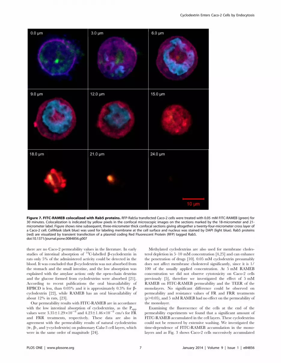
[20]. Caco-2 cells were transiently transfected with a plasmid
coding for a red fluorescent protein tagged Rab5a GTPase fusion
protein (RFP-Rab5a) that was strongly expressed in the cell
membrane. As Fig. 7 shows, FITC-RAMEB colocalizes with RFP-
Rab5a. Colocalization is marked by yellow pixels. Pearson’s
correlation coefficients were calculated and they were between
0.55 and 0.78 after 30 minutes incubation, indicating that the
entry of RAMEB into the cytoplasm and the formation of early
endosomes are associated. High degree of colocalization could be
observed after 2 minutes of incubation, and after 30 minutes
colocalization could be still detected indicating, that endocytosis
functioned continously (see also Figure S1 and S2.).
Internalization of FITC-RAMEB and its Inhibition by theFluid Phase Endocytosis Inhibitor RottlerinInternalization of FITC-RAMEB was also investigated by flow
cytometry. Caco-2 cell suspensions were treated by FITC-
RAMEB, the macropinocytosis marker Lucifer Yellow and the
lipophilic membrane permeability marker calcein-AM, both at
37uC and 0uC. Major differences could be seen between the
uptake of hydrophilic and lipophilic molecules (Fig. 8). Intracel-
lular accumulation of calcein was dependent on dye concentration,
but was independent of temperature. At the same time, both
FITC-RAMEB and Lucifer Yellow uptake increased as a function
of the dye concentration, but it was inhibited at 0uC.
Figure 3. Kinetics of FITC-RAMEB uptake in Caco-2 monolayers. Cell monolayers were treated with 0.05 mM FITC-RAMEB (FR) alone or incombination with 5 mM RAMEB (FRR) and in different time points the incubation was stopped. After washing cells were fixed with 3%paraformaldehyde solution and the accumulated FITC-RAMEB was determined by microplate reader. Cell nuclei were labeled with DAPI andfluorescence intensities of FITC-RAMEB were normalized for DAPI fluorescence intensities. Values are expressed as means 6 SD, n = 3 for FR and FRRtreatments.doi:10.1371/journal.pone.0084856.g003
Figure 4. Release of FITC-RAMEB from Caco-2 monolayers. Treatment with 0.5 mM FITC-RAMEB was carried out on TranswellH inserts for 120minutes, then the monolayers were washed and FITC-RAMEB release was followed in the apical and basolateral chambers during the next 120minutes. FITC fluorescence intensities were determined in the control group of the samples after the first 120 minutes and considered as 100%accumulation. The rate of release in the second group was compared to this value. Values are means 6 SD, n = 4.doi:10.1371/journal.pone.0084856.g004
Cyclodextrin Enters Caco-2 Cells by Endocytosis
PLOS ONE | www.plosone.org 5 January 2014 | Volume 9 | Issue 1 | e84856

Rottlerin, a macropinocytosis inhibitor decreased significantly
both FITC-RAMEB and Lucifer Yellow internalization in Caco-2
cells. Although the inhibition was not complete, the extent of
inhibition of FITC-RAMEB uptake was similar to that of Lucifer
Yellow (Fig. 9).
Discussion
In the present study we investigated the permeability and
cellular uptake of the fluorescent methyl-b-cyclodextrin in
intestinal Caco-2 cells. The available data regarding the absorp-
tion and oral bioavailability of cyclodextrins is very limited and
Figure 5. Confocal images of undifferentiated Caco-2 cells. Cells were treated with the solution of 0.05 mM FITC-RAMEB and 5 mM RAMEB(FRR). FITC-RAMEB (green) is localized in small vesicles (white arrows) under the CellMask labeled cell membrane (red) or in larger vesicles near theDAPI stained cell nucleus (light blue). Aggregated particles of FITC-RAMEB can be also seen outside the cell membrane (A). Nine consecutive confocalsections of a cluster of cells were recorded (B). Each section is one and half micrometer thick. FITC-RAMEB (green) is located in cytoplasmic granules.The granular bright particles are observed inside the cell membrane and outside of the cell nuclei.doi:10.1371/journal.pone.0084856.g005
Figure 6. Cyclodextrin enters differentiated Caco-2 cells of a high resistance Caco-2 cell layer. A confluent layer was treated with0.05 mM FITC-RAMEB and imaged by confocal microscope in twelve two-micrometer thick sections, of which six are demonstrated in the panels onthe left side (A–F). On the right, one middle section of the image shows the top view of the cell layer (H) at the level indicated by blue lines in sidesections. Upper (G) and right (I) side images are appropriate sections from perpendicular directions at green and at red lines. Crosshair (green and redlines at the long white arrow) set to an intense FITC-RAMEB (green) granule (indicated by arrows), which is located at the nuclear (light blue DAPIstain) level of cells. Several smaller FITC-RAMEB green granules can be seen below the cell membrane marked by CellMask (dark blue).doi:10.1371/journal.pone.0084856.g006
Cyclodextrin Enters Caco-2 Cells by Endocytosis
PLOS ONE | www.plosone.org 6 January 2014 | Volume 9 | Issue 1 | e84856
there are no Caco-2 permeability values in the literature. In early
studies of intestinal absorption of 14C-labelled b-cyclodextrin in
rats only 5% of the administered activity could be detected in the
blood. It was concluded that b-cyclodextrin was not absorbed from
the stomach and the small intestine, and the low absorption was
explained with the amylase action: only the open-chain dextrins
and the glucose formed from cyclodextrins were absorbed [21].
According to recent publications the oral bioavailability of
HPBCD is less, than 0.03% and it is approximately 0.3% for b-cyclodextrin [22], while RAMEB has an oral bioavailability of
about 12% in rats, [23].
Our permeability results with FITC-RAMEB are in accordance
with the low intestinal absorption of cyclodextrins, as the Pappvalues were 3.3561.2961028 and 4.2361.4661028 cm/s for FR
and FRR treatments, respectively. These data are also in
agreement with the permeability results of natural cyclodextrins
(a-, b-, and c-cyclodextrin) on pulmonary Calu-3 cell layers, which
were in the same order of magnitude [24].
Methylated cyclodextrins are also used for membrane choles-
terol depletion in 5–10 mM concentration [4,25] and can enhance
the penetration of drugs [10]. 0.05 mM cyclodextrin presumably
does not affect membrane cholesterol significantly, since it is 1/
100 of the usually applied concentration. At 5 mM RAMEB
concentration we did not observe cytotoxicity on Caco-2 cells
previously [5], therefore we investigated the effect of 5 mM
RAMEB on FITC-RAMEB permeability and the TEER of the
monolayers. No significant difference could be observed on
permeability and resistance values of FR and FRR treatments
(p.0.05), and 5 mM RAMEB had no effect on the permeability of
the monolayer.
Examining the fluorescence of the cells at the end of the
permeability experiments we found that a significant amount of
FITC-RAMEB accumulated in the cell layers. These cyclodextrins
could not be removed by extensive washing. We investigated the
time-dependence of FITC-RAMEB accumulation in the mono-
layers and as Fig. 3 shows Caco-2 cells successively accumulated
Figure 7. FITC-RAMEB colocalized with Rab5 proteins. RFP-Rab5a transfected Caco-2 cells were treated with 0.05 mM FITC-RAMEB (green) for30 minutes. Colocalization is indicated by yellow pixels in the confocal microscopic images on the sections marked by the 18-micrometer and 21-micrometer label. Figure shows nine subsequent, three-micrometer thick confocal sections giving altogether a twenty-four-micrometer cross layer ofa Caco-2 cell. CellMask (dark blue) was used for labeling membrane at the cell surface and nucleus was stained by DAPI (light blue). Rab5 proteins(red) are visualized by transient transfection of a plasmid coding Red Fluorescent Protein (RFP) tagged Rab5.doi:10.1371/journal.pone.0084856.g007
Cyclodextrin Enters Caco-2 Cells by Endocytosis
PLOS ONE | www.plosone.org 7 January 2014 | Volume 9 | Issue 1 | e84856

both FR and FRR up to 120 min of the experiment. To reveal the
fate of the accumulated FITC-RAMEB we loaded the cells with
0.5 mM FITC-RAMEB solution, using 10 time higher concen-
tration than in permeability studies. This resulted in 10 time
higher accumulation, but the permeability of FITC-RAMEB did
not increase (2.2860.3461028 cm/s). After 120 minutes, the
release of the accumulated cyclodextrins was followed both in
apical and basolateral directions. Interestingly FITC-RAMEB
appeared in both the apical and basal chambers, but the majority
of the accumulated cyclodextrin was released to the apical
Figure 8. Cellular uptake of calcein-AM (A), FITC-RAMEB (B) and Lucifer Yellow (C) as a function of ligand concentration. Cells weretreated at 37uC and 0uC and the cellular fluorescence was determined by flow cytometry, after excluding dead cells with propidium iodide.(Graphsshow results of a representative experiment).doi:10.1371/journal.pone.0084856.g008
Cyclodextrin Enters Caco-2 Cells by Endocytosis
PLOS ONE | www.plosone.org 8 January 2014 | Volume 9 | Issue 1 | e84856

direction. Only 7.4% of the accumulated FITC-RAMEB reached
the basal chamber.
These results indicated that although the intestinal Caco-2
monolayer is an almost impermeable barrier for the cyclodextrin
molecules, the cells are able to take up cyclodextrins from solutions
with a mechanism different from simple diffusion. Studies on
Calu-3 monolayers suggested that cyclodextrins traverse these
monolayers by paracellular route, although transcytosis could not
be excluded [24]. Recent publications revealed that certain cell
types are able to internalize cyclodextrins by endocytosis
[16,17,18]; therefore we investigated the intracellular localization
of FITC-RAMEB by confocal microscopy. Fig. 5 and 6 show that
FITC-RAMEB is able to enter into the cytoplasm of both
undifferentiated and differentiated Caco-2 cells. Since the labeled
cyclodextrin was localized in vesicles in the cytoplasm, the
possibility of endocytosis was investigated hereafter. In the cell
membrane RFP-Rab5a fusion protein and FITC-RAMEB showed
colocalization. Rab5 is a key organizer of early endosomes, but it
cannot be detected in late endosomes [20]. In our confocal
microscopy images Rab5 and FITC-RAMEB did not exhibit
colocalization in vesicles in deeper layers. The colocalization of
RFP-Rab5a and FITC-RAMEB suggests that endosome forma-
tion is involved in the initiation of cyclodextrin internalization.
Endocytosis has two major routes, phagocytosis and pinocytosis
or fluid-phase uptake. Fluid-phase endocytosis, which requires the
cargo molecules to be dissolved, can be subdivided into
macropinocytosis, clathrin-mediated, caveolin-mediated and cla-
thrin- and caveolin-independent endocytosis [26]. The widely used
marker of macropinocytosis is Lucifer Yellow [27,28,29]. Flow
cytometry analyses revealed that in Caco-2 cells Lucifer Yellow
was internalized in a concentration dependent manner and its
uptake could be inhibited at 0uC [28,29]. FITC-RAMEB showed
similar cellular uptake: at 37uC accumulated in the cells as a
function of concentration, while at 0uC FITC-RAMEB uptake was
diminished. On the other hand lipophilic calcein AM showed the
same cellular accumulation at 0uC and 37uC, as it rapidly
permeated the lipid membrane [30], and the intracellular
accumulation was not inhibited by cooling. The macropinocytosis
inhibitor rottlerin [27] had similar inhibitory effect on FITC-
RAMEB and LY accumulation. These results indicate, that in
Caco-2 cells macropinocytosis is involved in the entry of FITC-
RAMEB. It also explains why the majority of the accumulated
FITC-RAMEB was released to the apical direction. It was
demonstrated in human epidermoid A431 cells, that macropino-
somes recycle their content to the cell surface [31]. It seems that
the same mechanism could be observed in Caco-2 cells, as the
mechanism of internalization was macropinocytosis, the majority
of accumulated cyclodextrin was guided to the apical cell surface.
Nevertheless, the total recycling of the internalized cyclodextrin
molecules took at least one hour, which means that this process
prolongs the contact between cylodextrins or cyclodextrin-drug
complexes and the membrane of macropinosomes. It is important
to note, that other endocytotic mechanisms should be also taken
into consideration. Previous studies implicated fluid-phase endo-
cytosis and clathrin-dependent endocytosis [16,17,18] for the
mechanism of cyclodextrin internalization. Nevertheless, phago-
cytosis could be also a possibility for cyclodextrin internalization in
concentrated cyclodextrin solutions. It is reported that at high
concentrations, natural b-cyclodextrin [32] and the fluorescent
tetraamino rhodaminyl hydroxypropyl-b-cyclodextrin [33] form
large, nano-sized aggregates in water. However, the substitution of
OH groups with methyl groups on the cyclodextrin ring inhibits
the aggregation of RAMEB and at 12 mM no aggregation was
observed [32]. In this study 0.05 mM FITC-RAMEB was applied
alone or in combination with 5 mM RAMEB, which is 40–240
times lower cyclodextrin concentration than what was found to
form aggregates above, thus phagocytosis can be excluded from
among the possible mechanisms of cyclodextrin uptake.
In summary, our results on Caco-2 cells are in accordance with
earlier findings, the cellular internalization of water soluble FITC-
RAMEB is governed by fluid phase endocytosis in intestinal Caco-
2 cells. It is hard to predict the quantitative importance of this
mechanism. Even if permeability data are suitable to value the
extent of absorption of cyclodextrins, it is difficult to quantify the
amount of continuously internalized and released cyclodextrins
with this setup of the model. The intestinal absorptive surface area
relative to the volume of the gut is much bigger than the surface
area of Caco-2 monolayers and on the other hand the peristaltic
movement should be also considered. Thus the extent of
internalization can be much higher in vivo, even if cyclodextrins
are released back to the lumen of the gut and as the process can be
continuous along the small intestine its efficiency can be much
higher.
Figure 9. Effect of 10 mM rottlerin on the cellular uptake of FITC-RAMEB and Lucifer Yellow. Caco-2 cells were pre-incubated for 45minutes with rottlerin and the internalization of the fluorescent molecules was detected by flow cytometer. (n = 3, p,0.01).doi:10.1371/journal.pone.0084856.g009
Cyclodextrin Enters Caco-2 Cells by Endocytosis
PLOS ONE | www.plosone.org 9 January 2014 | Volume 9 | Issue 1 | e84856

Conclusions
Cyclodextrins are used to increase solubility, bioavailability and
stability of poorly water-soluble drugs. Our results demonstrate for
the first time that randomly methylated-b-cyclodextrins can enter
into intestinal epithelial cells by endocytosis. This process can
contribute to the enhancement of the intestinal delivery and
bioavailability of drugs by cyclodextrins in several ways. It can
help to overcome the intestinal membrane barrier, the endosome
formation increases the contact surface area between the
cyclodextrin-drug complexes and the cell membrane and prolongs
the retention time of cyclodextrins in the epithelial cells. Since this
study has demonstrated the role of macropinocytosis in the uptake
of methylated-b-cyclodextrin in intestinal cells, this mechanism
merits further investigations in connection with drug absorption
mediated by cyclodextrins.
Supporting Information
Figure S1 Colocalization of RFP-Rab5a with FITC-RAMEB, cell nucleus and cell membrane. RAMEB shows
high degree of colocalization with Rab5a immediately below the
cell surface membrane of two connected cells at 2 minutes
incubation (R= 0.93). Caco2 cells attached to surface of coverslip
were transiently transfected by Rab5a (red) tagged by red
fluorescent protein (RFP), treated by Fitc-RAMEB (green) for 2
minutes, and fixed. Before imaging surface membrane and nuclei
were labeled by CellMask (dark blue) and DAPI (cyan),
respectively, for 5 minutes. Panel A shows colocalization of Fitc-
RAMEB and RFP-Rab5a; panel B indicates colocalization of
DAPI and RFP-Rab5a as negative control (R=20.39) and panel
C specifies colocalization of surface membrane (CellMask) and
RFP-Rab5a as positive control (R= 0.95) at a confocal image
section crossing RAMEB granules (white areas in panel A right
side, section thickness is 1.5 micrometer). Right panels show two
channel images of signals tested for colocalization (bar is 10
micrometer), while corresponding left panels show two parameter
histograms of signals of the two channels. White areas in right side
images were chosen by setting channel signals above thresholds
indicated by red signs on scales of corresponding left side two
parameter histograms. R indicates Pearson correlation coefficients
calculated in images at location of the white areas. In two
parameter histograms the highest colocalization between tested
channels would be indicated by a 45u diagonal line correspondingto R=1 (in left panels of A and C R is close to this value), while a
135u diagonal line would indicate a negative correlation (left panel
of B).
(TIF)
Figure S2 Colocalization of FITC-RAMEB with RFP-Rab5a in the function of the time. Colocalization of RAMEB
and Rab5a was monitored in time during the endocytosis process.
The highest average colocalization (0.7660.01) was measured at 2
minutes after initiation of the endocytosis at 37uC. In later time
points, at 5, 10, 20 and 30 minutes R was dropped to a lower but
still significant value (R=0.5–0.6). R, Pearson correlation
coefficient was measured in region of interests (ROI) set to those
locations where RAMEB granules were observed in confocal
sections (one section was 1.5 micrometer thick). Pattern of the
intracellular localization of colocalized molecules also changed in
time. At 2 minutes colocalization was either dispersed in the
surface membrane of cell or in the cytoplasm close to surface
membrane. At later time points RAMEB granules moved closer to
cell nuclei with lower, but still significant R for Rab5a
colocalization (means6SD).
(TIF)
Author Contributions
Conceived and designed the experiments: FF KRN ZB GS MV.
Performed the experiments: FF KRN ZB ZGT JV ZU PF IB. Analyzed
the data: FF KRN ZB. Contributed reagents/materials/analysis tools: MM
EF LS. Wrote the paper: FF KRN ZB EF GS.
References
1. Szejtli J (1998) Introduction and General Overview of Cyclodextrin Chemistry.
Chem Rev 98: 1743–1754.
2. Loftsson T, Brewster ME (1996) Pharmaceutical applications of cyclodextrins. 1.Drug solubilization and stabilization. J Pharm Sci 85: 1017–1025.
3. Loftsson T, Jarho P, Masson M, Jarvinen T (2005) Cyclodextrins in drugdelivery. Expert Opin Drug Deliv 2: 335–351.
4. Kilsdonk EP, Yancey PG, Stoudt GW, Bangerter FW, Johnson WJ, et al. (1995)
Cellular cholesterol efflux mediated by cyclodextrins. J Biol Chem 270: 17250–17256.
5. Kiss T, Fenyvesi F, Bacskay I, Varadi J, Fenyvesi E, et al. (2010) Evaluation of
the cytotoxicity of beta-cyclodextrin derivatives: evidence for the role of
cholesterol extraction. Eur J Pharm Sci 40: 376–380.
6. Garrigues A, Escargueil AE, Orlowski S (2002) The multidrug transporter, P-glycoprotein, actively mediates cholesterol redistribution in the cell membrane.
Proc Natl Acad Sci U S A 99: 10347–10352.
7. Fenyvesi F, Fenyvesi E, Szente L, Goda K, Bacso Z, et al. (2008) P-glycoprotein
inhibition by membrane cholesterol modulation. European Journal ofPharmaceutical Sciences 34: 236–242.
8. Bacso Z, Nagy H, Goda K, Bene L, Fenyvesi F, et al. (2004) Raft and
cytoskeleton associations of an ABC transporter: P-glycoprotein. CytometryPart A 61A: 105–116.
9. Lambert D, O’Neill CA, Padfield PJ (2005) Depletion of Caco-2 cell cholesteroldisrupts barrier function by altering the detergent solubility and distribution of
specific tight-junction proteins. Biochem J 387: 553–560.
10. Deli MA (2009) Potential use of tight junction modulators to reversibly openmembranous barriers and improve drug delivery. Biochim Biophys Acta 1788:
892–910.
11. Zuhorn IS, Kalicharan R, Hoekstra D (2002) Lipoplex-mediated transfection of
mammalian cells occurs through the cholesterol-dependent clathrin-mediatedpathway of endocytosis. J Biol Chem 277: 18021–18028.
12. MJ ON, Guo J, Byrne C, Darcy R, O’Driscoll CM (2011) Mechanistic studies on
the uptake and intracellular trafficking of novel cyclodextrin transfection
complexes by intestinal epithelial cells. Int J Pharm 413: 174–183.
13. Chen FW, Li C, Ioannou YA (2010) Cyclodextrin induces calcium-dependent
lysosomal exocytosis. PLoS One 5: e15054.
14. Lipinski CA, Lombardo F, Dominy BW, Feeney PJ (2001) Experimental and
computational approaches to estimate solubility and permeability in drug
discovery and development settings. Adv Drug Deliv Rev 46: 3–26.
15. Matsuda H, Arima H (1999) Cyclodextrins in transdermal and rectal delivery.
Adv Drug Deliv Rev 36: 81–99.
16. Rosenbaum AI, Zhang G, Warren JD, Maxfield FR (2010) Endocytosis of beta-
cyclodextrins is responsible for cholesterol reduction in Niemann-Pick type C
mutant cells. Proc Natl Acad Sci U S A 107: 5477–5482.
17. Wei H, Zheng W, Diakur J, Wiebe LI (2011) Confocal laser scanning
microscopy (CLSM) based evidence for cell permeation by mono-4-(N-6-deoxy-
6-amino-beta-cyclodextrin)-7-nitrobenzofuran (NBD-beta-CyD). Int J Pharm
403: 15–22.
18. Plazzo AP, Hofer CT, Jicsinszky L, Fenyvesi E, Szente L, et al. (2012) Uptake of
a fluorescent methyl-beta-cyclodextrin via clathrin-dependent endocytosis.
Chem Phys Lipids 165: 505–511.
19. Artursson P, Palm K, Luthman K (2001) Caco-2 monolayers in experimental
and theoretical predictions of drug transport. Adv Drug Deliv Rev 46: 27–43.
20. Rink J, Ghigo E, Kalaidzidis Y, Zerial M (2005) Rab conversion as a mechanism
of progression from early to late endosomes. Cell 122: 735–749.
21. Szejtli J, Gerloczy A, Fonagy A (1980) Intestinal absorption of 14C-labelled beta-
cyclodextrin in rats. Arzneimittelforschung 30: 808–810.
22. Kurkov SV, Loftsson T (2012) Cyclodextrins. Int J Pharm.
23. Loftsson T, Brewster ME (2011) Pharmaceutical applications of cyclodextrins:
effects on drug permeation through biological membranes. J Pharm Pharmacol
63: 1119–1135.
24. Matilainen L, Toropainen T, Vihola H, Hirvonen J, Jarvinen T, et al. (2008) In
vitro toxicity and permeation of cyclodextrins in Calu-3 cells. J Control Release
126: 10–16.
25. Fenyvesi F, Fenyvesi E, Szente L, Goda K, Bacso Z, et al. (2008) P-glycoprotein
inhibition by membrane cholesterol modulation. Eur J Pharm Sci 34: 236–242.
Cyclodextrin Enters Caco-2 Cells by Endocytosis
PLOS ONE | www.plosone.org 10 January 2014 | Volume 9 | Issue 1 | e84856

26. Conner SD, Schmid SL (2003) Regulated portals of entry into the cell. Nature
422: 37–44.27. Sarkar K, Kruhlak MJ, Erlandsen SL, Shaw S (2005) Selective inhibition by
rottlerin of macropinocytosis in monocyte-derived dendritic cells. Immunology
116: 513–524.28. Swanson JA, Yirinec BD, Silverstein SC (1985) Phorbol Esters and Horseradish-
Peroxidase Stimulate Pinocytosis and Redirect the Flow of Pinocytosed Fluid inMacrophages. J Cell Biol 100: 851–859.
29. Sallusto F, Cella M, Danieli C, Lanzavecchia A (1995) Dendritic cells use
macropinocytosis and the mannose receptor to concentrate macromolecules inthe major histocompatibility complex class II compartment: downregulation by
cytokines and bacterial products. J Exp Med 182: 389–400.
30. Homolya L, Hollo Z, Germann UA, Pastan I, Gottesman MM, et al. (1993)
Fluorescent cellular indicators are extruded by the multidrug resistance protein.
J Biol Chem 268: 21493–21496.
31. Hewlett LJ, Prescott AR, Watts C (1994) The coated pit and macropinocytic
pathways serve distinct endosome populations. J Cell Biol 124: 689–703.
32. Gonzalez-Gaitano G, Rodriguez P, Isasi JR, Fuentes M, Tardajos G, et al.
(2002) The aggregation of cyclodextrins as studied by photon correlation
spectroscopy. J Incl Phenom Macrocycl Chem 44: 101–105.
33. Puskas I SM, Malanga M, Szente L (2013) Characterization and control of the
aggregation behavior of cyclodextrins. J Incl Phenom Macrocycl Chem 75: 269–
276.
Cyclodextrin Enters Caco-2 Cells by Endocytosis
PLOS ONE | www.plosone.org 11 January 2014 | Volume 9 | Issue 1 | e84856


