Embed Size (px)
Citation preview

Fronto-parietal regulation of media violenceexposure in adolescents: a multi-method studyMaren Strenziok,1,2,* Frank Krueger,1,3,* Gopikrishna Deshpande,4 Rhoshel K. Lenroot,5 Elke van der Meer,2
and Jordan Grafman1
1National Institute of Neurological Disorders and Stroke, National Institutes of Health, 10 Center Drive, Bethesda, MD 20892, USA,2Department of Cognitive Psychology, Humboldt University, Berlin, Rudower Chaussee 18, 12489 Berlin, Germany, 3Department of
Molecular Neuroscience, George Mason University, 4400 University Drive, Fairfax, VA 22030, USA, 4Department of Biomedical
Engineering, Emory University and Georgia Institute of Technology, 313 Ferst Drive, Atlanta, GA 30332, USA, and 5National Institute of
Mental Health, National Institutes of Health, 10 Center Drive, Bethesda, MD 20892, USA
Adolescents spend a significant part of their leisure time watching TV programs and movies that portray violence. It is unknown,however, how the extent of violent media use and the severity of aggression displayed affect adolescents� brain function. Weinvestigated skin conductance responses, brain activation and functional brain connectivity to media violence in healthy ado-lescents. In an event-related functional magnetic resonance imaging experiment, subjects repeatedly viewed normed videos thatdisplayed different degrees of aggressive behavior. We found a downward linear adaptation in skin conductance responses withincreasing aggression and desensitization towards more aggressive videos. Our results further revealed adaptation in afronto-parietal network including the left lateral orbitofrontal cortex (lOFC), right precuneus and bilateral inferior parietal lobules,again showing downward linear adaptations and desensitization towards more aggressive videos. Granger causality mappinganalyses revealed attenuation in the left lOFC, indicating that activation during viewing aggressive media is driven by input fromparietal regions that decreased over time, for more aggressive videos. We conclude that aggressive media activates an emo-tion–attention network that has the capability to blunt emotional responses through reduced attention with repeated viewingof aggressive media contents, which may restrict the linking of the consequences of aggression with an emotional response,and therefore potentially promotes aggressive attitudes and behavior.
Keywords: aggression; violence; functional magnetic resonance imaging; skin conductance response; Granger causality mapping
INTRODUCTIONAdolescents spend a significant part of their leisure time
watching TV programs and movies that portray violence
(Yoon and Somers, 2003). For example, it has been shown
that �70% of 14-year-olds (and 39% of 10-year-olds) saw at
least one of 51 extremely violent movies, and amongst the
most popular were I Know What You Did Last Summer,
Scream II and Die Hard (Sargent et al., 2002). Extensive
research and media coverage have linked school shootings
(Anderson et al., 2007, p. 3), real-life replications of
video-game contents (Crowley, 2008) and general aggression
to the exposure to extremely violent media (Anderson and
Bushman, 2001, 2002). Although it has been suggested that
individuals become more aggressive (Huesmann, 2007) and
desensitized (Funk, 2005) to real-life violence after the
repeated consumption of violent media programs
(American Academy of Pediatrics, 2001), little is known
about how the extent of violent media use and the severity
of aggression displayed affect adolescents’ brains.
During adolescence, developmental changes occur in
brain morphology and function, particularly in the pre-
frontal cortex (Blakemore, 2008; Giedd, 2008). One of the
neural systems that undergo significant changes during ado-
lescence (Spear, 2000) and that has been intimately linked to
the processing of the incentive value of stimuli is the dopa-
minergic system (Schultz, 1998). Dopamine neurons in the
orbitofrontal cortex (OFC) and subcortical regions encode
the emotional value of primary (e.g. food, touch, smell) and
secondary (e.g. money) reinforcers enabling the individual to
shape goal-directed social and emotional behavior in
response to changing external contingencies (Kringelbach
and Rolls, 2004; Rolls and Grabenhorst, 2008). In adoles-
cents, compared to adults and children, lateral OFC
(lOFC) activation has been observed to be enhanced
and reward-driven learning slowed, in the context of
monetary reward value manipulations, indicating that
adolescents’ lOFC development may be protracted
(Galvan et al., 2006).
Whereas the medial OFC has been shown to modulate ag-
gression by exerting inhibitory control over aggressive
Received 9 September 2009; Accepted 6 August 2010
The authors thank Dr Dimitrios Kapogiannis and Dr Edward Huey for performing the neurological exam-
inations on our subjects. This research was funded by the intramural research program of the National
Institutes of Health, National Institute of Neurological Disorders and Stroke.
Correspondence should be addressed to Jordan Grafman, Cognitive Neuroscience Section, National Institute
of Neurological Disorders and Stroke, National Institutes of Health, 10 Center Drive, Building 10, Room 7D43,
Bethesda, MD 20892, MSC1440, USA. E-mail: [email protected]
*These authors contributed equally to this work.
doi:10.1093/scan/nsq079 SCAN (2010) 1of11
Published by Oxford University Press 2010. For Permissions, please email: [email protected]
Social Cognitive and Affective Neuroscience Advance Access published October 18, 2010 by guest on O
ctober 26, 2010scan.oxfordjournals.org
Dow
nloaded from

impulses in adults and adolescents (Damasio et al., 1994;
Grafman et al., 1996; Anderson et al., 1999; Blair and
Cipolotti, 2000; Pietrini et al., 2000; Strenziok et al., 2009),
there is evidence that the lOFC, through its ability to adapt to
external stimuli, is involved in increasing or decreasing the
likelihood of aggressive behavior when aggressive cues are
present (Blair, 2004). Two functional neuroimaging studies
revealed that reduced activation in the lOFC is associated with
exposure to aggressive media in adults. Repeated exposure to
violence portrayed in movies was associated with a decrease in
activation in the lOFC over the period of the experiment
(Kelly et al., 2007). In the same study, reduced lOFC activa-
tion towards violent movies was associated with more
reactive-affective aggression, indicating that the lOFC may
be involved in increasing the likelihood of aggression through
dysfunctional emotion regulation. Furthermore, during play-
ing of a violent first-person shooter video game, the lOFC was
recruited less in experienced male gamers than during
non-violent virtual actions in the same game (Mathiak and
Weber, 2006). This study further revealed activation changes
in the rostral and dorsal anterior cingulate cortices, parietal
regions including the precuneus (Prec), angular gyrus, intra-
parietal sulcus, and temporoparietal junction, amygdala,
parahippocampus, insula, and cerebellum during violent ac-
tions, indicating the contribution of networks that regulate
visual-spatial attention, sensori-motor function, and moni-
toring of cognitive and affective processes.
It has been proposed that repeated exposure to violent
media causes emotional desensitization to subsequent ag-
gressive stimuli (Funk et al., 2004). A neurophysiological
study investigating the P300 amplitude of the event-related
brain potential has shown lower cortical responses towards
violent images in violent compared to non-violent video
game players, indicating desensitization towards violent sti-
muli in individuals who have been exposed to media vio-
lence (Bartholow et al., 2006). Because the P300 amplitude
has been hypothesized to reflect the activation of the aversive
motivational system (Cacioppo et al., 1993), reduced brain
activation during exposure to violent stimuli may indicate
reduced aversive emotional responses to these stimuli. The
process of emotional blunting to arousing events is also
associated with a reduced sympathetic skin conductance re-
sponse (SCR) to violent movies and portrayals of real life
aggression in children who were previously exposed to vio-
lent media (Cline et al., 1973; Thomas et al., 1977).
The present study was designed to explore the association
between violent media exposure, SCR responses, and activa-
tion changes in the lOFC in normally developing male ado-
lescents 14–17 years of age. Adolescence is a time period that
is sensitive to the adverse effects of violent media because
portrayals of aggression are more appealing and pleasurable
to youth, as evidenced by self-report (Benenson et al., 2007),
identification with antisocial characters is more likely
(Konijn et al., 2007), and parental control is low (Cheng
et al., 2004), whereas the opportunities to gain access to
violent media are abundant. Due to a number of interacting
factors such as adrenarche, gonadarche, cortical synaptic
pruning, antisocial peer pressure, family conflict and diffi-
culties in school, particularly male adolescents have an
increased risk for aggressive behavior and some develop life-
long antisocial and violent behavior patterns (Moffitt and
Caspi, 2001; Kirsh, 2003; Blakemore, 2008). We used func-
tional magnetic resonance imaging (fMRI) to investigate
brain activation changes during observed aggression and
Granger causality mapping (GCM) to investigate effective
connectivity (connectivity network and strength of connect-
ivity) in an media aggression network (Deshpande et al.,
2008, 2009). In an event-related fMRI design, 22 healthy
adolescents repeatedly viewed normed videos that displayed
different degrees of realistic, age-appropriate aggression. In
addition, SCRs were sampled throughout the acquisition of
fMRI images to monitor autonomic changes associated with
viewing aggressive videos.
Among others, two phenomena of neural activity during
emotion processing in the lOFC, that are relevant in the
context of the current study, have been reported: (i) a de-
crease in lOFC activity to stimuli that are applied repeatedly
and (ii) an association between lOFC activation magnitude
and stimulus magnitude (O’Doherty et al., 2001;
Kringelbach et al., 2003). Based on these findings, we pre-
dicted emotional desensitization after repeated exposure to
aggressive videos of varying aggression levels as evidenced by
reduced SCRs and decreased activation in the lOFC over
time. With increased aggression in the videos, we predicted
a decline in lOFC activation based on previous neuroima-
ging evidence that showed decreased OFC function asso-
ciated with an increase in aggression (Pietrini, 2000). If
activation changes in the lOFC reflect changes in the emo-
tional value of stimuli that motivate behavior, and if re-
peated exposure to aggressive media stimuli is associated
with emotional desensitization to those stimuli, then re-
peated exposure to aggressive media should be associated
with reduced activation levels in the lOFC over time. In
our connectivity analysis, we predicted a central role of the
lOFC in a media aggression network that subserves emotion
modulation and associated activation in structures that regu-
late visual-spatial input, attentional demands and familiarity
of recurring events.
METHODSSubjectsTwenty-two healthy male adolescents (mean� s.d.; years of
age: 15.9� 1.1, range 14–17, years of education: 9.9� 1.1,
range 8–12), who had no history of psychiatric or neuro-
logical illness, participated for financial compensation. All
were native English speakers, had normal or corrected-
to-normal vision, and were right-handed (Edinburgh
Inventory, laterality quotient: 90.8� 14.2, range 50–100)
(Oldfield, 1971) (see Supplementary Data, for trait aggres-
sion and exposure to violence scores). Parents gave written
2 of11 SCAN (2010) M. Strenziok et al.
by guest on October 26, 2010
scan.oxfordjournals.orgD
ownloaded from

informed consent and adolescents gave their written assent
for the procedures that were approved by the National
Institute of Neurological Disorders and Stroke Institutional
Review Board.
Stimuli
From commercially available DVDs, 114 videos (each 4 s
long) were retrieved that contained real scenes of aggression
(e.g. fist fights, street brawls and stadium violence). In a
pre-study, another group of 22 age- and education-matched
males rated the videos for aggression and excitement (see
Supplementary Data). Based on the ratings (mean� s.e.m.),
a total of 60 videos in three sets of 20 videos that differed in
the level of aggression (F2,38¼ 6924.14, P < 0.001; low:
14.91� 1.83; mild: 46.40� 2.17; moderate: 73.44� 1.72)
but not in their levels of excitement (F2,38¼ 1.24,
P¼ 0.293; low: 50.67� 2.37; mild: 47.80� 3.23; moderate:
53.77� 1.67) were selected for the fMRI study.
ProcedureAll subjects were seen twice for the study. During visit 1,
subjects were screened for psychiatric and neurological con-
ditions, handedness, trait aggression, and exposure to vio-
lence in the media and community. During visit 2, subjects
underwent the fMRI procedures. Prior to scanning, subjects
rated their emotional state using a 9-point rating scale ver-
sion of the Self-Assessment Manikin (Lang et al., 1993) to
assess their emotional status and were trained on the fMRI
task on a separate set of stimuli.
During scanning, stimulus presentation was controlled
by a computer with SuperLab Pro software (Cedrus
Corporation, San Pedro, CA). Subjects were given a response
pad with two buttons on which they placed their right index
and middle fingers. They were asked to view and judge mute
videos. At the beginning of each trial, a plus sign was pre-
sented (0.5 s) on the screen followed by a video that was
shown (4 s, video viewing phase). Then, a decision screen
appeared (3.5 s) to cue subjects to decide whether the
video they just saw was more or less aggressive than the
one that they saw in the previous trial by pressing one of
two assigned response buttons (decision phase). Stimulus
presentation was event-related and trials were interspersed
with jittered interstimulus intervals optimized following an
exponential schedule using Optseq2 software (Freesurfer,
Massachusetts General Hospital, Boston, MA). Three runs
of about nine minutes each were employed. During each
run, all 60 videos were presented randomly among runs
and subjects. Subjects were asked to make their decisions
as quickly as possible and response times and decisions
were recorded. Immediately after scanning, 1 day, and
2 weeks later adolescents completed an emotional assessment
to assess their mood state after viewing the videos (see
Supplementary Data).
Data acquisitionfMRI dataImaging data were collected using a 3T General Electric (GE
Medical Systems, Waukesha, WI) MRI scanner equipped
with an eight-channel head coil. Anatomical scans were per-
formed using a T1-weighted 3D MP-RAGE sequence (TR
9 ms, TE 4 ms, flip angle 128, FoV 256 mm, matrix size
256� 256, thickness 1.2 mm, in-plane resolution
0.8594� 0.8594 mm2). Functional images were acquired
using a T2*-weighted 2D gradient EPI sequence (TR 2 s,
TE 23 ms, flip angle 908, 30 slices, thickness 3 mm, in-plane
resolution 3.75� 3.75 mm2, FoV 240 mm). In each run, 320
volume images were taken parallel to the AC–PC line. The
first five volumes were discarded to allow for T1 equilibra-
tion effects.
Skin conductance responsesSCRs were sampled at 80 Hz throughout MRI scanning using
PSYLAB equipment (Contact Precision Instruments Inc.,
Boston, MA). A constant voltage of 0.5 V was applied to
the middle and ring fingers of the non-dominant hand
through a pair of MRI compatible surface gel cup electrodes
(Ag/AgCl, 6 mm diameter; Biopac, Goleta, CA, model
TSD203). Note that three subjects had to be excluded from
the analysis of SCR data due to equipment failure (n¼ 1)
and non-responding (n¼ 2).
Data analysesSkin conductance responsesSCR samples were first z-transformed (Boucsein, 1992) to
normalize possible baseline differences across aggression
levels and subjects. Stimulus related changes were identified
by extracting the maximum amplitude of the SCR during
each video viewing phase. Afterwards, a 3� 3 analysis of
variance (ANOVA) on those maximum measurements was
performed with Aggression (low, mild, moderate) and Time
(T1, T2, T3) as within-subject factors. Because both factors
had more than two levels, Mauchly’s test of sphericity was
applied to determine whether the correlation between vari-
ables was the same. If the estimated chi value of Mauchly’s
test was significant, then the assumption behind the normal
within-subjects ANOVA was violated and Wilks’ Lambda
multivariate statistics were reported. Furthermore, we com-
puted adaptation factors (AFs) for each aggression level
defined as maximum SCR measurement differences between
extreme time points (T3–T1). Finally, a one-way ANOVA
trend analysis on the AFs was computed with aggression
(low, mild, moderate) as a within-subject factor to deter-
mine whether the subjects’ SCRs adapted in a linear fashion,
from low to mild to moderate aggression levels. In those
cases where AFs were positive, we defined the adaptation
as sensitization, whereas negative AFs are referred to as de-
sensitization. (See Supplementary Data for behavioral data
analysis).
Media violence exposure in adolescents SCAN (2010) 3 of11
by guest on October 26, 2010
scan.oxfordjournals.orgD
ownloaded from

fMRI datafMRI data analyses were performed using BrainVoyager QX
(Brain Innovation, Maastricht, The Netherlands) and struc-
tural and functional data were preprocessed (see
Supplementary Data). Functional data were co-registered
with the individual’s 3D anatomical images and then reas-
sembled into 3� 3� 3 mm3 isotropic voxels. A GLM cor-
rected for first-order serial correlation was applied (Friston
et al., 1999). Random-effects analyses were performed to
explore brain regions that were associated with viewing ag-
gressive videos. The GLM model consisted of four regressors:
video viewing phase (n¼ 3: low, mildly and moderately ag-
gressive videos) and decision phase (n¼ 1). Regressor time
courses were adjusted for the hemodynamic response delay
by convolution with a double-gamma hemodynamic
response function (Buchel et al., 1998).
Multiple regression analyses were performed independent-
ly for the time course of each individual voxel. After com-
puting parameter estimates for all predictors, using the
ANCOVA random-effects analysis tool in BrainVoyager
QX, a 3� 3 ANOVA on those parameter estimates was per-
formed with Aggression (low, mild, moderate) and Time
(T1, T2, T3) as within-subject factors (see Supplementary
Data for design). Activations were reported in a whole
brain analysis using an FDR with a threshold of
q(FDR) < 0.05 (corrected) (Genovese et al., 2002) and a clus-
ter size threshold of 270 mm3.
To analyze the interaction effect in more detail, we derived
the parameter estimates of the beforehand identified
Aggression�Time interaction effect for each of the aggres-
sion and time levels. Mean values of parameter estimates
were derived from each region after identifying the peaks
of activation and surrounding voxels encompassing
125 mm3. Afterwards, we computed AFs for each aggression
level defined as differences in parameter estimates between
extreme time points (T3–T1). Furthermore, one-way
ANOVA trend analyses on AFs were computed with aggres-
sion (low, mild, moderate) as a within-subject factor to de-
termine whether subjects’ brain activations adapted in a
linear fashion, from low to mild to moderate aggression
levels. In those cases where AFs were positive, we defined
the adaptation as sensitization, whereas negative AFs are
referred to as desensitization.
Multivariate Granger causality analysisEffective connectivity was implemented using a multivariate
GCM analysis (Kaminski et al., 2001) based on a multivariate
vector autoregressive (MVAR) model capable of capturing
the simultaneous directional influences between multiple
regions (Kus et al., 2004). For the connectivity analysis,
we derived the parameter estimates from the peak voxel
activations of the beforehand identified significant
Aggression�Time interaction effect for each of the aggres-
sion and time levels because we were only interested in those
regions that changed depending on the aggression level and
repeated viewing of videos for the subsequent GCM analysis.
The ROIs were constrained to the center of activation within
each region and to a maximum size of 5� 5� 5 mm3. The
entire time series of BOLD signal intensities from the
selected ROIs were averaged across voxels, then normalized
across runs and subjects, and finally collapsed across all runs
and subjects to obtain a single time series per region
(Deshpande et al., 2008, 2009).
A first order MVAR model was fit based on the time series
of selected regions and a directed transfer function (DTF)
matrix was obtained (Kus et al., 2004). The DTF matrix was
weighted by the partial coherence between the selected re-
gions in order to emphasize direct connections (Deshpande
et al., 2008, 2009). The order of the model was determined
using Akaike’s Information Criterion (Akaike, 1974). The
DTF is based on the principle of Granger causality but is
rendered in a multivariate framework and therefore can ef-
fectively model the inherently multivariate nature of neur-
onal networks (Blinowska et al., 2004). This method has
been validated previously using simulations (Kus et al.,
2004) and applied successfully to electrophysiological
(Ding et al., 2000; Kaminski et al., 2001; Korzeniewska
et al., 2003 Blinowska et al., 2004; Kus et al., 2004) and
fMRI data measuring the BOLD response (Stilla et al.,
2007; Deshpande et al., 2008, 2009). The DTF analysis
matrix consists of a set of directional path weights describing
the strength of mutual impact (in arbitrary units) from each
region to each of the other regions. Surrogate null distribu-
tions were used to assess the significance of the path weights
(P < 0.05) (Deshpande et al., 2008). Since the selected re-
gions survived multiple comparison corrections in the initial
fMRI analyses, no further significance correction was applied
(Stilla et al., 2007).
To determine dominant directional influences between
regions (i.e. whether differences between reciprocal paths
were significant), differences between the reciprocal paths
were calculated using surrogate data to generate a corres-
ponding null distribution and compared it with the analo-
gous value obtained from real fMRI data. The fractional area
under the null distribution for the analogous value obtained
from real fMRI data gives the probability of the null hypoth-
esis being true, which is essentially the P-value of the signifi-
cance of the dominant directional influence.
Furthermore, to determine whether regions were either
predominantly driving other regions or being driven by
other regions (or, alternatively, a given region may be
driven as much as it is driving the other regions), we
computed input-output ratios for each region (see
Supplementary Data for computations).
CorrelationsWe correlated the AFs of SCRs, AFs of brain activation and
connectivity of brain activation with self-reported measures
of violent media exposure and trait aggression. On these
latter measures, we asked subjects to rate how much of the
4 of11 SCAN (2010) M. Strenziok et al.
by guest on October 26, 2010
scan.oxfordjournals.orgD
ownloaded from

media they used show fighting, crime, guns, war scenes or
similar content on a scale from one to five (1¼ none or very
little, 5¼ all or almost all), including TV programs/movies,
video games, books/journals, music, and websites.
Furthermore, we administrated the Aggression
Questionnaire (AQ) (Buss and Warren, 2000) to assess phys-
ical aggression, verbal aggression, anger, hostility and indir-
ect aggression. On this questionnaire, subjects were asked to
rate on a 5-point rating scale (1¼ not at all like me,
5¼ completely like me) how they interact with other
people using 34 statements.
RESULTSSkin conductance responsesWe investigated whether SCRs changed while viewing videos
with different levels of aggression over time. The 3
Aggression (low, mild, moderate)� 3 Time (T1, T2, T3)
ANOVA revealed no main effect of Aggression
(F2,36¼ 0.47, P¼ 0.614) but a significant main effect of
Time (F2,36¼ 4.38, P < 0.021), indicating that SCRs dimin-
ished over time. In addition, there was a significant
Aggression�Time interaction effect, indicating that SCRs
changed differently for the aggression levels over time
(F4,15¼ 4.84, P < 0.010; note that Wilks’ Lambda multivari-
ate statistics were reported because of a significant chi value
in the Mauchly’s test of Sphericity �2(9)¼ 17.07, P < 0.048).
To address this change in SCRs over time, we calculated AFs
of SCRs for each aggression level and submitted them to a
planned follow-up ANOVA. Results showed that SCRs
adapted significantly with aggression level (F2,36¼ 3.68,
P < 0.036) (Figure 1). Specifically, the trend analysis to de-
termine whether the subjects’ SCRs adapted in a linear fash-
ion revealed a significant downward linear trend
(Flin1,18¼ 9.04, P < 0.008), indicating that changes in SCRs
over time decreased linearly from low to mild to moderate
aggression levels. For the mildly and moderately aggres-
sive videos, SCR adaptation was negative, indicating
desensitization towards these videos (see Supplementary
Data and Figure S1 for behavioral results).
Brain activationWe also assessed brain activation patterns that were asso-
ciated with viewing videos that displayed different levels of
aggression with repeated exposure over time. The 3
Aggression (low, mild, moderate)� 3 Time (T1, T2, T3)
ANOVA revealed a significant main activation effect of
Aggression in the bilateral lOFC (left, BA 47; right, BA 45)
in addition to a fronto-parieto-temporo-occipital network
including the left inferior frontal gyrus (BA 9), rostral anter-
ior cingulate cortex (BA 32), right posterior cingulate cortex
(BA 23), bilateral middle temporal gyri (left, BA 39; right, BA
37) and bilateral middle occipital gyri (BA 19) [F2,42¼ 5.80,
q(FDR) < 0.05] (Figure S2, Supplementary Table S1A).
Furthermore, a significant main activation effect of Time
was associated with a fronto-temporo-parietal network
including the bilateral middle frontal gyri (BA 10), right
Prec (BA 31), right lingual gyrus (BA 18) and left middle
temporal gyrus (BA 37) [F2,42¼ 7.59, q(FDR) < 0.05] (Figure
S3, Table S1B). Finally, there was a significant
Aggression�Time interaction activation effect in the left
lOFC (BA 10) (Figure 2A) in addition to the right Prec
(BA 31) and bilateral inferior parietal lobules (left IPL, BA
39; right ILP, BA 7) [F4,84¼ 5.16, q(FDR) < 0.05]
(Supplementary Figure S4 and Table S1C).
To address the changes over time, we calculated the AFs of
lOFC, Prec, left ILP and right IPL activation (parameter es-
timates) for each aggression level and submitted them to
planned follow-up ANOVAs. Results showed that responses
in all four brain regions adapted significantly with aggression
level (lOFC, F2,42¼ 20.22, Figure 2B; Prec, F2,42¼ 25.67; left
ILP, F2,42¼ 12.77; right ILP, F2,42¼ 14.87, Ps < 0.001, Figure
S5). Specifically, linear trend analyses to determine whether
subjects’ brain responses adapted in a linear fashion revealed
significant downward linear trends in all four regions (lOFC,
-0.25
-0.20
-0.15
-0.10
-0.05
0.00
0.05
0.10
0.15
Low Mild Moderate
SC
R A
dapt
atio
n
SensitizationDesensitization
Fig. 1 SCR adaptation. Adaptation factors are shown for low, mildly and moderately aggressive videos. Results revealed a linear downward trend in SCR adaptation withincreasing aggression in the videos. SCR adaptation was positive (sensitization) for the low aggressive videos and negative (desensitization) for the mildly and moderatelyaggressive videos.
Media violence exposure in adolescents SCAN (2010) 5 of11
by guest on October 26, 2010
scan.oxfordjournals.orgD
ownloaded from

F1,21¼ 40.38; Prec, F1,21¼ 36.22; left ILP, F1,21¼ 20.92; right
ILP, F1,21¼ 24.15, Ps < 0.001), indicating that brain activa-
tion changes over time decreased linearly from low to mild
to moderate aggression levels. In the lOFC and right IPL,
adaptation was negative for the moderately aggressive videos,
indicating desensitization towards the most aggressive videos
in our study.
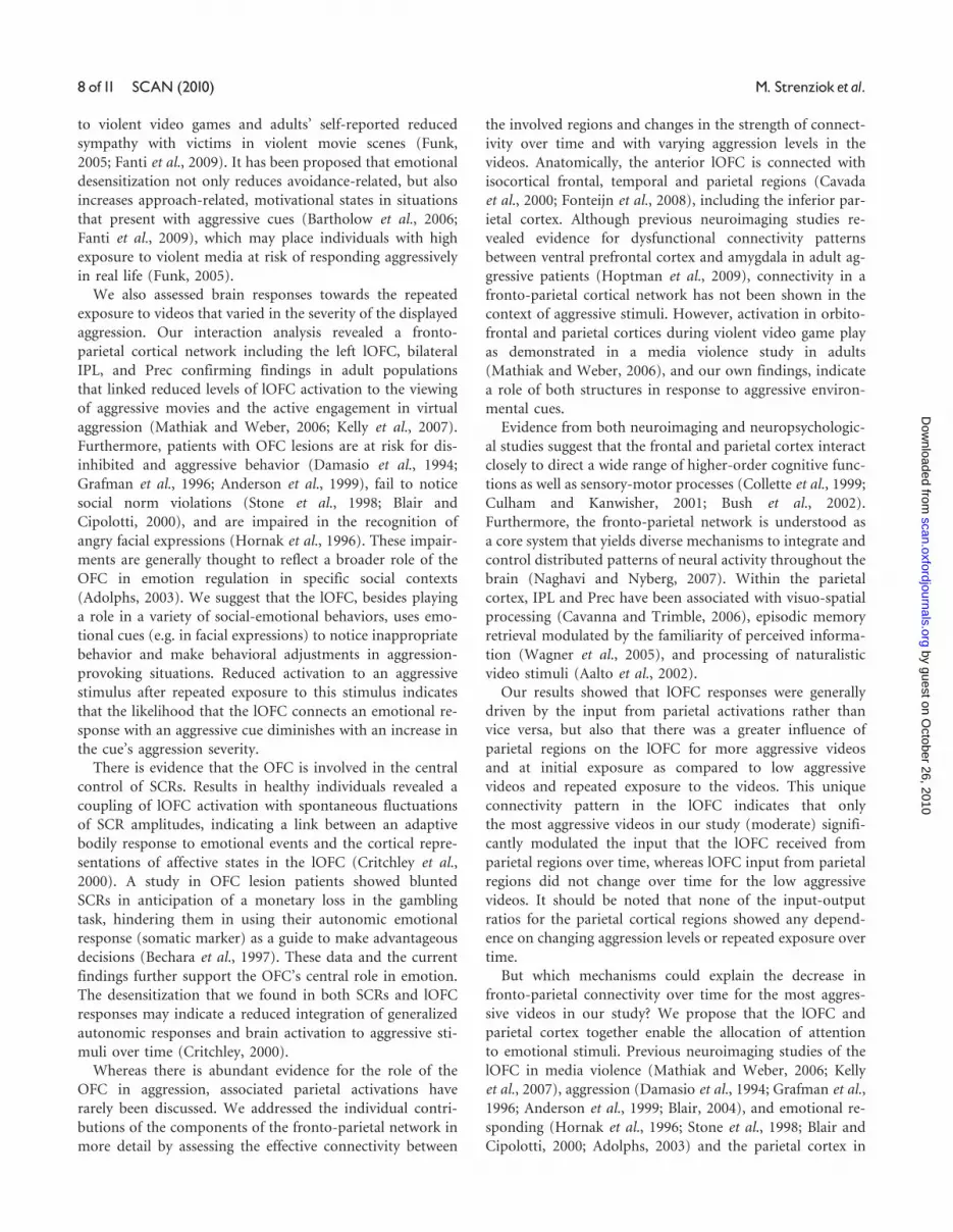
Multivariate GCM analysisFirst, using GCM analyses, we identified the effective con-
nectivity patterns among brain regions that showed an
Aggression�Time interaction effect (lOFC, Prec, left IPL
and right IPL) (Figure 3A, Supplementary Table S2). The
Prec and lOFC were reciprocally connected with both
the left and right IPL, but were not directly connected
(Figure 3A).
Second, the dominant directional influences for the recip-
rocal connections in the Granger causality network were
determined. Two dominant directional pathways were
found that gave input to the lOFC, indicating that in this
experiment the lOFC is mainly driven by other regions: (i)
inter-hemispherically, from the Prec via the right IPL
(P[Prec!R IPL]¼ 0.032) to the lOFC (P[R IPL!
lOFC]¼ 0.023) and (ii) intra-hemispherically, from the left
IPL to the lOFC (P[L IPS! L lOFC]¼ 0.012) (Figure 3B).
Finally, the input-output ratios for each region were deter-
mined and their differences between low to moderately ag-
gressive videos and changes from time T1 to T3 investigated
(Figure 3C). For the lOFC, the surrogate test revealed a sig-
nificant main input-output ratio effect of Aggression
(P < 0.009), indicating that low aggressive videos produced
a significantly lower input drive into the lOFC compared to
moderately aggressive videos (the most aggressive videos in
our study). Furthermore, a significant main input-output
ratio effect of Time (P < 0.021) was revealed, indicating
that the drive into the lOFC significantly decreased over
time. Finally, the surrogate test revealed a significant inter-
action input-output ratio effect of Aggression (low vs
moderate)�Time (T1 vs T3) (P < 0.009), indicating that
the drive into the lOFC significantly decreased over time
only for the moderately aggressive videos (P < 0.001) but
not for the low aggressive videos (P¼ 0.062). The surrogate
tests for the three other regions revealed no Aggression,
Time, nor Aggression�Time effects.
CorrelationsWe found a negative correlation between the AFs of the
SCRs and scores on the media violence exposure scale
(r¼ –0.47, P < 0.05) and AFs of the SCRs and scores on
the violent video games exposure scale (r¼ –0.46,
P < 0.05), indicating that individuals with the most exposure
to violent media show the lowest adaptation values (become
desensitized). No correlations between SCRs and lOFC acti-
vation, including its connectivity with the parietal cortical
regions, were found.
DISCUSSIONIn the present study, we measured neurophysiological re-
sponses while adolescents repeatedly viewed videos that
varied in the severity of aggression displayed. First, we mea-
sured SCRs to explore emotional arousal associated with
viewing aggressive media as an autonomic correlate of de-
sensitization. Second, we investigated brain activation in the
media aggression network that depended on the repeated
exposure to aggressive videos that differ in aggression sever-
ity. Finally, we investigated effective connectivity in the iden-
tified fronto-parietal cortical network for the interaction of
repeated viewing of aggressive videos with varying levels of
aggression. Our results show SCR desensitization associated
with viewing aggressive videos and underline the important
5.16
16.00
F4,84z=-5
lOFC
-0.30
-0.20
-0.10
0.00
0.10
0.20
0.30
0.40
0.50
Low Mild Moderate
Left
LOF
C A
dapt
atio
n
SensitizationDesensitization
BA
Fig. 2 Brain activation. LOFC activation and adaptation are displayed. (A) Left lOFC activation associated with viewing aggressive videos. We found activation changes in the leftlOFC (BA 10; x, y, z coordinates, –27, 47, –5) towards repeatedly viewed videos that portray varying levels of aggression. (B) Left lOFC adaptation. Adaptation factors are shownfor low, mildly and moderately aggressive videos. Results revealed a linear downward trend in lOFC adaptation with increasing aggression in the videos. Left lOFC adaptation waspositive (sensitization) for the low and mildly aggressive videos and negative (desensitization) for the moderately aggressive videos.
6 of11 SCAN (2010) M. Strenziok et al.
by guest on October 26, 2010
scan.oxfordjournals.orgD
ownloaded from

role of the left lOFC in the emotional representation of
media-related aggression with important time and aggres-
sion level dependent inputs from the parietal cortex thought
to modulate visual-spatial attention and event familiarity.
Our SCR data analysis revealed an adaptation over time,
showing a linear decrease in SCRs with increasing aggression
in the videos. For the mildly and moderately aggressive
videos in our study, we found SCR desensitization, indicat-
ing that autonomic responses diminished over time when
adolescents were exposed to more aggressive videos.
Furthermore, subjects who had the most exposure to violent
media in their daily life showed the greatest desensitization.
Our results are in accordance with previous studies that also
revealed reduced sympathetic skin conductance responses to
violent movies and portrayals of real life aggression in chil-
dren who were previously exposed to violent media (Cline
et al., 1973; Thomas et al., 1977). Furthermore, emotional
desensitization had been associated with children’s exposure
L lOFC
R IPL
L IPL
R PreC
L lOFC
R IPL
L IPL
R PreC
0.00470.039
A
B
C
Fig. 3 Multivariate GCM analyses. Effective connectivity network and strengths are displayed. (A) Granger causality network for aggression. The Prec and lOFC were reciprocallyconnected with both the left and right IPL, but were not directly connected. A pseudo-color code was used to indicate the path weights of all connections between ROIs.(B) Granger causality network for dominant directional influences. Two dominant directional pathways were found that gave input to the lOFC, indicating that this region ismainly driven by other ROIs: inter-hemispherically, from the Prec via the right IPL (Prec! R IPL) to the lOFC (R IPL! lOFC) and intra-hemispherically, from the left IPL to thelOFC (L IPS! L lOFC). The color scales in A and B are weighted by the P-values of the corresponding paths. (C) Input–output ratio for ROI activations. Only for left lOFC,the input–output ratio decreased significantly over time for the moderately aggressive videos, but not for the low aggressive videos.
Media violence exposure in adolescents SCAN (2010) 7 of11
by guest on October 26, 2010
scan.oxfordjournals.orgD
ownloaded from
to violent video games and adults’ self-reported reduced
sympathy with victims in violent movie scenes (Funk,
2005; Fanti et al., 2009). It has been proposed that emotional
desensitization not only reduces avoidance-related, but also
increases approach-related, motivational states in situations
that present with aggressive cues (Bartholow et al., 2006;
Fanti et al., 2009), which may place individuals with high
exposure to violent media at risk of responding aggressively
in real life (Funk, 2005).
We also assessed brain responses towards the repeated
exposure to videos that varied in the severity of the displayed
aggression. Our interaction analysis revealed a fronto-
parietal cortical network including the left lOFC, bilateral
IPL, and Prec confirming findings in adult populations
that linked reduced levels of lOFC activation to the viewing
of aggressive movies and the active engagement in virtual
aggression (Mathiak and Weber, 2006; Kelly et al., 2007).
Furthermore, patients with OFC lesions are at risk for dis-
inhibited and aggressive behavior (Damasio et al., 1994;
Grafman et al., 1996; Anderson et al., 1999), fail to notice
social norm violations (Stone et al., 1998; Blair and
Cipolotti, 2000), and are impaired in the recognition of
angry facial expressions (Hornak et al., 1996). These impair-
ments are generally thought to reflect a broader role of the
OFC in emotion regulation in specific social contexts
(Adolphs, 2003). We suggest that the lOFC, besides playing
a role in a variety of social-emotional behaviors, uses emo-
tional cues (e.g. in facial expressions) to notice inappropriate
behavior and make behavioral adjustments in aggression-
provoking situations. Reduced activation to an aggressive
stimulus after repeated exposure to this stimulus indicates
that the likelihood that the lOFC connects an emotional re-
sponse with an aggressive cue diminishes with an increase in
the cue’s aggression severity.
There is evidence that the OFC is involved in the central
control of SCRs. Results in healthy individuals revealed a
coupling of lOFC activation with spontaneous fluctuations
of SCR amplitudes, indicating a link between an adaptive
bodily response to emotional events and the cortical repre-
sentations of affective states in the lOFC (Critchley et al.,
2000). A study in OFC lesion patients showed blunted
SCRs in anticipation of a monetary loss in the gambling
task, hindering them in using their autonomic emotional
response (somatic marker) as a guide to make advantageous
decisions (Bechara et al., 1997). These data and the current
findings further support the OFC’s central role in emotion.
The desensitization that we found in both SCRs and lOFC
responses may indicate a reduced integration of generalized
autonomic responses and brain activation to aggressive sti-
muli over time (Critchley, 2000).
Whereas there is abundant evidence for the role of the
OFC in aggression, associated parietal activations have
rarely been discussed. We addressed the individual contri-
butions of the components of the fronto-parietal network in
more detail by assessing the effective connectivity between
the involved regions and changes in the strength of connect-
ivity over time and with varying aggression levels in the
videos. Anatomically, the anterior lOFC is connected with
isocortical frontal, temporal and parietal regions (Cavada
et al., 2000; Fonteijn et al., 2008), including the inferior par-
ietal cortex. Although previous neuroimaging studies re-
vealed evidence for dysfunctional connectivity patterns
between ventral prefrontal cortex and amygdala in adult ag-
gressive patients (Hoptman et al., 2009), connectivity in a
fronto-parietal cortical network has not been shown in the
context of aggressive stimuli. However, activation in orbito-
frontal and parietal cortices during violent video game play
as demonstrated in a media violence study in adults
(Mathiak and Weber, 2006), and our own findings, indicate
a role of both structures in response to aggressive environ-
mental cues.
Evidence from both neuroimaging and neuropsychologic-
al studies suggest that the frontal and parietal cortex interact
closely to direct a wide range of higher-order cognitive func-
tions as well as sensory-motor processes (Collette et al., 1999;
Culham and Kanwisher, 2001; Bush et al., 2002).
Furthermore, the fronto-parietal network is understood as
a core system that yields diverse mechanisms to integrate and
control distributed patterns of neural activity throughout the
brain (Naghavi and Nyberg, 2007). Within the parietal
cortex, IPL and Prec have been associated with visuo-spatial
processing (Cavanna and Trimble, 2006), episodic memory
retrieval modulated by the familiarity of perceived informa-
tion (Wagner et al., 2005), and processing of naturalistic
video stimuli (Aalto et al., 2002).
Our results showed that lOFC responses were generally
driven by the input from parietal activations rather than
vice versa, but also that there was a greater influence of
parietal regions on the lOFC for more aggressive videos
and at initial exposure as compared to low aggressive
videos and repeated exposure to the videos. This unique
connectivity pattern in the lOFC indicates that only
the most aggressive videos in our study (moderate) signifi-
cantly modulated the input that the lOFC received from
parietal regions over time, whereas lOFC input from parietal
regions did not change over time for the low aggressive
videos. It should be noted that none of the input-output
ratios for the parietal cortical regions showed any depend-
ence on changing aggression levels or repeated exposure over
time.
But which mechanisms could explain the decrease in
fronto-parietal connectivity over time for the most aggres-
sive videos in our study? We propose that the lOFC and
parietal cortex together enable the allocation of attention
to emotional stimuli. Previous neuroimaging studies of the
lOFC in media violence (Mathiak and Weber, 2006; Kelly
et al., 2007), aggression (Damasio et al., 1994; Grafman et al.,
1996; Anderson et al., 1999; Blair, 2004), and emotional re-
sponding (Hornak et al., 1996; Stone et al., 1998; Blair and
Cipolotti, 2000; Adolphs, 2003) and the parietal cortex in
8 of11 SCAN (2010) M. Strenziok et al.
by guest on October 26, 2010
scan.oxfordjournals.orgD
ownloaded from

attention to visuo-spatial input (Cavanna and Trimble,
2006) and familiarity/novelty of stimuli (Daffner et al.,
2003) support this proposal. It has been suggested that a
key role for the lOFC is to detect social norm violations,
including aggressive behavior, and that it guides social be-
havior through its ability to flexibly respond to environmen-
tal changes (Berthoz et al., 2002; Blair, 2004; King et al.,
2006). In this role, it may determine the emotional-
motivational relevance of aggressive behavior, shown in the
videos in our study, for guidance of social behavior in future
situations that present with similar aggressive cues.
Activation in the IPL is frequently attributed to attentional
processes (LaBar et al., 1999; Mesulam, 1999; Szczepanski
et al., 2010). Furthermore, the parietal cortex, particularly
the Prec, may play a role in updating one’s internal model of
the environment to take into account novel events.
Activation changes in this region have been shown to be
reduced when familiar events are presented (Daffner et al.,
2003; Wagner et al., 2005). Whereas the reduced IPL input to
the lOFC over time in our study may account for an attenu-
ation of attention to emotionally relevant videos, decreased
Prec input into the lOFC through the IPL may reflect famil-
iarity with repeatedly viewed stimuli. The decreased influ-
ence of the precuneus and IPL on lOFC activation may have
reduced the emotional-motivational relevance of the video
content, indicated by lOFC desensitization.
Adolescence is a time of increased psychosocial challenges
including antisocial peer pressure and difficulties at school
and in the family (Kirsh, 2003). Adolescents show a greater
interest in and an increased incidence rate of aggressive be-
havior (Moffitt and Caspi, 2001; Benenson et al., 2007;
Konijn et al., 2007). In addition, previous neuroimaging re-
search has shown that monetary reward processing in the
lOFC is altered in adolescents, as compared to adults and
children, and large rewards are perceived most desirable,
whereas small and medium rewards did not cause differen-
tial behavioral responses (Galvan et al., 2006). Altered mo-
tivational values of aggressive media, such as sensation
seeking, may explain media choices that adolescents make
for their everyday entertainment. A decrease in emotional
responsiveness towards more aggressive media, as shown in
the present study, might result in seeking a bigger variety of
those media to elicit similar reward effects and may also
impose an increased risk for aggressive attitudes and behav-
ior in real life.
Many questions about central and peripheral physiological
responses towards aggression, the link between both, and
connectivity patterns of brain structures associated with ag-
gression remain unanswered to date. For example, different
aggression modes, such as observed vs executed aggression,
may affect the direction and location of brain activation
differently. In addition, aggression-related changes in other
physiological parameters such as cardiac response and res-
piration not only showed mixed results while measured
during exposure to violent media, but also may interact
differently with brain responses (Bradley et al., 2001;
Kuniecki et al., 2003; Gomez et al., 2005). A limitation of
the current study is that it only included male subjects. The
incidence rate of aggression in females, even in female teen-
agers that are exposed to some of the same biopsychosocial
challenges as male adolescents, is low and raises the question
of what brain mechanisms and autonomic differences are
associated with this gender difference. This information
will be invaluable for defining neurobehavioral treatment
goals for aggressive individuals.
In summary, we have shown desensitization in SCRs and
left lOFC activation towards repeatedly viewed videos that
portray considerable aggression. We propose that exposure
to aggressive media results in a blunting of emotional re-
sponses, which in turn may prevent the connection of con-
sequences of aggression with an appropriate emotional
response, and therefore may increase the likelihood that ag-
gression is seen as acceptable behavior. It remains unknown,
however, whether individuals with elevated levels of aggres-
sion may be at particular risk for altered desensitization pat-
terns towards media violence, proviolent attitudes, and the
acceptance of real-world violence as normal social behavior
(Fanti et al., 2009). We also demonstrated that left lOFC
activation during viewing aggressive videos is driven by
input from parietal cortical regions, particularly during an
initial exposure to videos with a considerable level of aggres-
sion. These findings indicate that the left lOFC uses aggres-
sion cues to determine behavior consequences of, and to
make a behavioral adjustment in, an aggression-provoking
situation, whereas parietal regions reflect a more general
contribution to the integration and control of information.
SUPPLEMENTARY DATASupplementary data are available at SCAN online.
REFERENCESAalto, S., Naatanen, P., Wallius, E., et al. (2002). Neuroanatomical substrata
of amusement and sadness: a PET activation study using film stimuli.
Neuroreport, 13, 67–73.
Adolphs, R. (2003). Cognitive neuroscience of human social behaviour.
Nature Reviews Neuroscience, 4, 165–78.
Akaike, H. (1974). A new look at the statistical model identification. IEEE
Transactions on Automatic Control, 19, 716–23.
American Academy of Pediatrics, Committee on Public Education (2001).
Media violence. Pediatrics, 108, 1222–6.
Anderson, C.A., Bushman, B.J. (2001). Effects of violent video games on
aggressive behavior, aggressive cognition, aggressive affect, physiological
arousal, and prosocial behavior: a meta-analytic review of the scientific
literature. Psychological Science, 12, 353–9.
Anderson, C.A., Bushman, B.J. (2002). Psychology. The effects of media
violence on society. Science, 295, 2377–9.
Anderson, C.A., Gentile, D.A., Buckley, K.E. (2007). Violent Video Game
Effects on Children and Adolescents. Theory, Research, and Public Policy.
New York, NY: Oxford University Press.
Anderson, S.W., Bechara, A., Damasio, H., Tranel, D., Damasio, A.R.
(1999). Impairment of social and moral behavior related to early
damage in human prefrontal cortex. Nature Neuroscience, 2, 1032–7.
Bartholow, B., Bushman, B., Sestir, M. (2006). Chronic violent video
game exposure and desensitization to violence: behavioral and
Media violence exposure in adolescents SCAN (2010) 9 of11
by guest on October 26, 2010
scan.oxfordjournals.orgD
ownloaded from

event-related brain potential data. Journal of Experimental Social
Psychology, 42, 5329.
Bechara, A., Damasio, H., Tranel, D., Damasio, A.R. (1997). Deciding ad-
vantageously before knowing the advantageous strategy. Science, 275,
1293–5.
Benenson, J.F., Carder, H.P., Geib-Cole, S.J. (2007). The development of
boys’ preferential pleasure in physical aggression. Aggressive Behavior, 33,
1–13.
Berthoz, S., Armony, J.L., Blair, R.J., Dolan, R.J. (2002). An fMRI study of
intentional and unintentional (embarrassing) violations of social norms.
Brain, 125, 1696–708.
Blair, R.J. (2004). The roles of orbital frontal cortex in the modulation of
antisocial behavior. Brain and Cognition, 55, 198–208.
Blair, R.J., Cipolotti, L. (2000). Impaired social response reversal. A case of
‘acquired sociopathy’. Brain, 123, 1122–41.
Blakemore, S.J. (2008). The social brain in adolescence. Nature Review
Neuroscience, 9, 267–77.
Blinowska, K.J., Kus, R., Kaminski, M. (2004). Granger causality and infor-
mation flow in multivariate processes. Physical Review. E, Statistical,
Nonlinear, and Soft Matter Physics, 70, 050902.
Boucsein, W. (1992). Electrodermal Activity. New York, NY: Plenum Press.
Bradley, M.M., Codispoti, M., Cuthbert, B.N., Lang, P.J. (2001). Emotion
and motivation I: defensive and appetitive reactions in picture processing.
Emotion, 1, 276–98.
Buchel, C., Holmes, A.P., Rees, G., Friston, K.J. (1998). Characterizing
stimulus-response functions using nonlinear regressors in parametric
fMRI experiments. Neuroimage, 8, 140–8.
Bush, G., Vogt, B.A., Holmes, J., et al. (2002). Dorsal anterior
cingulate cortex: a role in reward-based decision making. Proceedings of
the National Academy of Sciences of United States of America, 99, 523–8.
Buss, A.H., Warren, W.L. (2000). The Aggression Questionnaire. Los Angeles,
CA: Western Psychological Services.
Cacioppo, J., Crites, S., Gardner, G., Coles, M. (1993). If attitudes affect how
stimuli are processed, should they not affect the event-related brain po-
tential? Psychological Science, 4, 108–12.
Cavada, C., Company, T., Tejedor, J., Cruz-Rizzolo, R.J., Reinoso-Suarez, F.
(2000). The anatomical connections of the macaque monkey orbitofron-
tal cortex. A review. Cerebral Cortex, 10, 220–42.
Cavanna, A.E., Trimble, M.R. (2006). The precuneus: a review of its func-
tional anatomy and behavioural correlates. Brain, 129, 564–83.
Cheng, T.L., Brenner, R.A., Wright, J.L., Sachs, H.C., Moyer, P., Rao, M.R.
(2004). Children’s violent television viewing: are parents monitoring?
Pediatrics, 114, 94–9.
Cline, V.B., Croft, R.G., Courrier, S. (1973). Desensitization of children to tele-
vision violence. Journal of Personality and Social Psychology, 27, 360–5.
Collette, F., Salmon, E., Van der Linden, M., et al. (1999). Regional brain
activity during tasks devoted to the central executive of working memory.
Brain Research. Cognitive Brain Research, 7, 411–7.
Critchley, H.D., Elliott, R., Mathias, C.J., Dolan, R.J. (2000). Neural activity
relating to generation and representation of galvanic skin conductance
responses: a functional magnetic resonance imaging study. Journal of
Neuroscience, 20, 3033–40.
Crowley, K. (2008). Video Villains Come to Life. New York, NY: New York
Times.
Culham, J.C., Kanwisher, N.G. (2001). Neuroimaging of cognitive functions
in human parietal cortex. Current Opinion in Neurobiology, 11, 157–63.
Daffner, K.R., Scinto, L.F., Weitzman, A.M., et al. (2003). Frontal and par-
ietal components of a cerebral network mediating voluntary attention to
novel events. Journal of Cognitive Neuroscience, 15, 294–313.
Damasio, H., Grabowski, T., Frank, R., Galaburda, A.M., Damasio, A.R.
(1994). The return of Phineas Gage: clues about the brain from the
skull of a famous patient. Science, 264, 1102–5.
Deshpande, G., Hu, X., Stilla, R., Sathian, K. (2008). Effective con-
nectivity during haptic perception: a study using Granger causality
analysis of functional magnetic resonance imaging data. Neuroimage,
40, 1807–14.
Deshpande, G., Laconte, S., James, G.A., Peltier, S., Hu, X. (2009).
Multivariate Granger causality analysis of fMRI data. Human Brain
Mapping, 30, 1361–73.
Ding, M., Bressler, S.L., Yang, W., Liang, H. (2000). Short-window spectral
analysis of cortical event-related potentials by adaptive multivariate auto-
regressive modeling: data preprocessing, model validation, and variability
assessment. Biological Cybernetics, 83, 35–45.
Fanti, K.A., Vanman, E., Henrich, C.C., Avraamides, M.N. (2009).
Desensitization to media violence over a short period of time.
Aggressive Behavior, 35, 179–87.
Fonteijn, H.M., Norris, D.G., Verstraten, F.A. (2008). Exploring the ana-
tomical basis of effective connectivity models with DTI-based fiber trac-
tography. International Journal Of Biomedical Imaging, 423192.
Friston, K.J., Holmes, A.P., Worsley, K.J. (1999). How many subjects con-
stitute a study? Neuroimage, 10, 1–5.
Funk, J.B. (2005). Children’s exposure to violent video games and desensi-
tization to violence. Child & Adolescent Psychiatric Clinics of North
America, 14, 387–404.
Funk, J.B., Baldacci, H.B., Pasold, T., Baumgardner, J. (2004). Violence
exposure in real-life, video games, television, movies, and the internet:
is there desensitization? Journal of Adolescence, 27, 23–39.
Galvan, A., Hare, T.A., Parra, C.E., et al. (2006). Earlier development of the
accumbens relative to orbitofrontal cortex might underlie risk-taking
behavior in adolescents. J Neurosci, 26, 6885–92.
Genovese, C.R., Lazar, N.A., Nichols, T. (2002). Thresholding of statistical
maps in functional neuroimaging using the false discovery rate.
Neuroimage, 15, 870–8.
Giedd, J.N. (2008). The teen brain: insights from neuroimaging. Journal of
Adolescent Health, 42, 335–43.
Gomez, P., Zimmermann, P., Guttormsen-Schar, S., Danuser, B. (2005).
Respiratory responses associated with affective processing of film stimuli.
Biological Psychology, 68, 223–35.
Grafman, J., Schwab, K., Warden, D., Pridgen, A., Brown, H.R.,
Salazar, A.M. (1996). Frontal lobe injuries, violence, and aggression: a
report of the Vietnam Head Injury Study. Neurology, 46, 1231–8.
Hoptman, M.J., D’Angelo, D., Catalano, D., et al. (2009).
Amygdalofrontal functional disconnectivity and aggression in schizo-
phrenia. Schizophr Bull. Advance online access at http://
schizophreniabulletin.oxfordjournals.org/cgi/reprint/sbp012v2.
Hornak, J., Rolls, E.T., Wade, D. (1996). Face and voice expression
identification in patients with emotional and behavioural
changes following ventral frontal lobe damage. Neuropsychologia, 34,
247–61.
Huesmann, L.R. (2007). The impact of electronic media violence: scientific
theory and research. Journal of Adolescent Health, 41, S6–13.
Kaminski, M., Ding, M., Truccolo, W.A., Bressler, S.L. (2001). Evaluating
causal relations in neural systems: granger causality, directed transfer
function and statistical assessment of significance. Biological Cybernetics,
85, 145–57.
Kelly, C.R., Grinband, J., Hirsch, J. (2007). Repeated exposure to media
violence is associated with diminished response in an inhibitory fronto-
limbic network. PLoS ONE, 2, e1268.
King, J.A., Blair, R.J., Mitchell, D.G., Dolan, R.J., Burgess, N. (2006). Doing
the right thing: a common neural circuit for appropriate violent or com-
passionate behavior. Neuroimage, 30, 1069–76.
Kirsh, S.J. (2003). The effects of violent video games on adolescents. The
overlooked influence of development. Aggression and Violent Behavior, 8,
377–89.
Konijn, E.A., Bijvank, M.N., Bushman, B.J. (2007). I wish I were a war-
rior: the role of wishful identification in the effects of violent video
games on aggression in adolescent boys. Developmental Psychology, 43,
1038–44.
Korzeniewska, A., Manczak, M., Kaminski, M., Blinowska, K.J., Kasicki, S.
(2003). Determination of information flow direction among brain struc-
tures by a modified directed transfer function (dDTF) method. J Neurosci
Methods, 125, 195–207.
10 of11 SCAN (2010) M. Strenziok et al.
by guest on October 26, 2010
scan.oxfordjournals.orgD
ownloaded from

Kringelbach, M.L., O’Doherty, J., Rolls, E.T., Andrews, C. (2003). Activation
of the human orbitofrontal cortex to a liquid food stimulus is correlated
with its subjective pleasantness. Cerebral Cortex, 13, 1064–71.
Kringelbach, M.L., Rolls, E.T. (2004). The functional neuroanatomy of the
human orbitofrontal cortex: evidence from neuroimaging and neuro-
psychology. Progress in Neurobiology, 72, 341–72.
Kuniecki, M., Urbanik, A., Sobiecka, B., Kozub, J., Binder, M. (2003).
Central control of heart rate changes during visual affective processing
as revealed by fMRI. Acta Neurobiologiae Experimentalis (Wars), 63, 39–48.
Kus, R., Kaminski, M., Blinowska, K.J. (2004). Determination of EEG
activity propagation: pair-wise versus multichannel estimate. IEEE
Transactions on Biomedical Engineering, 51, 1501–10.
LaBar, K.S., Gitelman, D.R., Parrish, T.B., Mesulam, M. (1999).
Neuroanatomic overlap of working memory and spatial attention net-
works: a functional MRI comparison within subjects. Neuroimage, 10,
695–704.
Lang, P.J., Greenwald, M.K., Bradley, M.M., Hamm, A.O. (1993). Looking
at pictures: affective, facial, visceral, and behavioral reactions.
Psychophysiology, 30, 261–73.
Mathiak, K., Weber, R. (2006). Toward brain correlates of natural behavior:
fMRI during violent video games. Human Brain Mapping, 27, 948–56.
Mesulam, M.M. (1999). Spatial attention and neglect: parietal, frontal and
cingulate contributions to the mental representation and attentional tar-
geting of salient extrapersonal events. Philos Trans R Soc Lond B Biol Sci,
354, 1325–46.
Moffitt, T.E., Caspi, A. (2001). Childhood predictors differentiate
life-course persistent and adolescence-limited antisocial pathways
among males and females. Development and Psychopathology, 13, 355–75.
Naghavi, H.R., Nyberg, L. (2007). Integrative action in the fronto-parietal
network: a cure for a scattered mind. Behavioral and Brain Sciences, 30,
135–87.
O’Doherty, J., Kringelbach, M.L., Rolls, E.T., Hornak, J., Andrews, C.
(2001). Abstract reward and punishment representations in the human
orbitofrontal cortex. Nature Neuroscience, 4, 95–102.
Oldfield, R.C. (1971). The assessment and analysis of handedness: the
Edinburgh inventory. Neuropsychologia, 9, 97–113.
Pietrini, P., Guazzelli, M., Basso, G., Jaffe, K., Grafman, J. (2000). Neural
correlates of imaginal aggressive behavior assessed by positron emission
tomography in healthy subjects. American Journal of Psychiatry, 157,
1772–81.
Rolls, E.T., Grabenhorst, F. (2008). The orbitofrontal cortex and beyond:
From affect to decision-making. Progress in Neurobiology, 86, 216–44.
Sargent, J.D., Heatherton, T.F., Ahrens, M.B., Dalton, M.A., Tickle, J.J.,
Beach, M.L. (2002). Adolescent exposure to extremely violent movies.
The Journal of Adolescent Health, 31, 449–54.
Schultz, W. (1998). Predictive reward signal of dopamine neurons. Journal
of Neurophysiology, 80, 1–27.
Spear, L.P. (2000). The adolescent brain and age-related behavioral mani-
festations. Neuroscience Biobehavior Review, 24, 417–63.
Stilla, R., Deshpande, G., LaConte, S., Hu, X., Sathian, K. (2007).
Posteromedial parietal cortical activity and inputs predict tactile spatial
acuity. Jornal of Neuroscience, 27, 11091–102.
Stone, V.E., Baron-Cohen, S., Knight, R.T. (1998). Frontal lobe contribu-
tions to theory of mind. Journal of Cognitive Neuroscience, 10, 640–56.
Strenziok, M., Krueger, F., Heinecke, A., et al. (2009). Developmental effects
of aggressive behavior in male adolescents assessed with structural and
functional brain imaging. Social Cognitive and Affective Neuroscience,
online advance access at http://scan.oxfordjournals.org/content/early/
2009/09/21/scan.nsp036.full.pdfþhtml.
Szczepanski, S.M., Konen, C.S., Kastner, S. (2010). Mechanisms of spatial
attention control in frontal and parietal cortex. J Neurosci, 30, 148–60.
Thomas, M.H., Horton, R.W., Lippincott, E.C., Drabman, R.S. (1977).
Desensitization to portrayals of real-life aggression as a function of ex-
posure to television violence. Journal of Personality and Social Psychology,
35, 450–8.
Wagner, A.D., Shannon, B.J., Kahn, I., Buckner, R.L. (2005). Parietal lobe
contributions to episodic memory retrieval. Trends in Cognitive Science, 9,
445–53.
Yoon, J.S., Somers, C.L. (2003). Aggressive content of high school students’
TV viewing. Psychological Reports, 93, 949–53.
Media violence exposure in adolescents SCAN (2010) 11of11
by guest on October 26, 2010
scan.oxfordjournals.orgD
ownloaded from


