Embed Size (px)
Citation preview

, 20130047, published 12 August 2013368 2013 Phil. Trans. R. Soc. B Elisabeth A. Herniou and Jean-Michel DrezenKarine Musset, Jérome Lesobre, Patricia Lenoble, Catherine Dupuy, Dawn Gundersen-Rindal, Annie Bézier, Faustine Louis, Séverine Jancek, Georges Periquet, Julien Thézé, Gabor Gyapay, evolutionary dynamics of bracoviruses
: insights into theCotesia congregataparasitoid wasp Functional endogenous viral elements in the genome of the
Supplementary data
ml http://rstb.royalsocietypublishing.org/content/suppl/2013/07/31/rstb.2013.0047.DC1.ht
"Data Supplement"
References
http://rstb.royalsocietypublishing.org/content/368/1626/20130047.full.html#related-urls Article cited in:
http://rstb.royalsocietypublishing.org/content/368/1626/20130047.full.html#ref-list-1
This article cites 45 articles, 21 of which can be accessed free
Subject collections
(60 articles)genomics � (622 articles)evolution �
Articles on similar topics can be found in the following collections
Email alerting service hereright-hand corner of the article or click Receive free email alerts when new articles cite this article - sign up in the box at the top
http://rstb.royalsocietypublishing.org/subscriptions go to: Phil. Trans. R. Soc. BTo subscribe to
on August 13, 2013rstb.royalsocietypublishing.orgDownloaded from

on August 13, 2013rstb.royalsocietypublishing.orgDownloaded from
rstb.royalsocietypublishing.org
ResearchCite this article: Bezier A, Louis F, Jancek S,
Periquet G, Theze J, Gyapay G, Musset K,
Lesobre J, Lenoble P, Dupuy C, Gundersen-
Rindal D, Herniou EA, Drezen J-M. 2013
Functional endogenous viral elements in the
genome of the parasitoid wasp Cotesia
congregata: insights into the evolutionary
dynamics of bracoviruses. Phil Trans R Soc B
368: 20130047.
http://dx.doi.org/10.1098/rstb.2013.0047
One contribution of 13 to a Theme Issue
‘Paleovirology: insights from the genomic fossil
record’.
Subject Areas:genomics, evolution
Keywords:polydnavirus, bracovirus, parasitoid wasp,
obligatory mutualism, comparative genomics
Author for correspondence:Jean-Michel Drezen
e-mail: [email protected]
& 2013 The Author(s) Published by the Royal Society. All rights reserved.
†Present address: Laboratoire Ecologie et
Biologie des Interactions, CNRS UMR7267,
Universite de Poitiers, 40 Avenue du Recteur
Pineau, 86022 Poitiers Cedex, France.
Electronic supplementary material is available
at http://dx.doi.org/10.1098/rstb.2013.0047 or
via http://rstb.royalsocietypublishing.org.
Functional endogenous viral elements inthe genome of the parasitoid waspCotesia congregata: insights into theevolutionary dynamics of bracoviruses
Annie Bezier1, Faustine Louis1, Severine Jancek1, Georges Periquet1,Julien Theze1, Gabor Gyapay2, Karine Musset1, Jerome Lesobre1,†,Patricia Lenoble2, Catherine Dupuy1, Dawn Gundersen-Rindal3,Elisabeth A. Herniou1 and Jean-Michel Drezen1
1Institut de Recherche sur la Biologie de l’Insecte, CNRS UMR 7261, Universite Francois Rabelais,Parc de Grandmont, 37200 Tours, France2Commissariat a l’Energie Atomique, Genoscope (Centre National de Sequencage), 2 rue Gaston Cremieux,CP 5706, 91057 Evry Cedex, France3US Department of Agriculture, Agricultural Research Service, Invasive Insect Biocontrol and Behavior Laboratory,10300 Baltimore Avenue, Building 011A BARC-WEST, Beltsville, MD 20705, USA
Bracoviruses represent the most complex endogenous viral elements (EVEs)
described to date. Nudiviral genes have been hosted within parasitoid wasp
genomes since approximately 100 Ma. They play a crucial role in the wasp
life cycle as they produce bracovirus particles, which are injected into para-
sitized lepidopteran hosts during wasp oviposition. Bracovirus particles
encapsidate multiple dsDNA circles encoding virulence genes. Their expression
in parasitized caterpillars is essential for wasp parasitism success. Here, we
report on the genomic organization of the proviral segments (i.e. master
sequences used to produce the encapsidated dsDNA circles) present in the
Cotesia congregata parasitoid wasp genome. The provirus is composed of a
macrolocus, comprising two-thirds of the proviral segments and of seven
dispersed loci, each containing one to three segments. Comparative genomic
analyses with closely related species gave insights into the evolutionary
dynamics of bracovirus genomes. Conserved synteny in the different wasp gen-
omes showed the orthology of the proviral macrolocus across different species.
The nudiviral gene odv-e66-like1 is conserved within the macrolocus, suggesting
an ancient co-localization of the nudiviral genome and bracovirus proviral seg-
ments. By contrast, the evolution of proviral segments within the macrolocus
has involved a series of lineage-specific duplications.
1. IntroductionBracoviruses (BVs) are symbiotic viruses associated with tens of thousands of
braconid wasp species [1]. They have atypical virus life cycles that require
two separate host species. The primary hosts are parasitoid wasps, in which
the virus particles are produced. The secondary host are the lepidopteran
larvae parasitized by the wasp, in which the virus is expressed in infected
cells (reviewed in [1]). BV particles are produced from endogenous viral
elements (EVEs) integrated in the wasp genomes, and contain multiple
dsDNA circular molecules. BVs are produced in specialized cells of the wasp
ovaries and constitute the major component of the fluid injected with the
eggs into the parasitized caterpillar host during wasp oviposition. The wasps
use BVs as gene-transfer agents to express virulence factors that manipulate
the immune defences of the lepidopteran host [2]. BVs are essential for the sur-
vival and development of the wasp eggs and larvae, which would otherwise be

rstb.royalsocietypublishing.orgPhilTransR
SocB368:20130047
2
on August 13, 2013rstb.royalsocietypublishing.orgDownloaded from
encapsulated in a cellular sheath of haemocytes and killed by
the potent immune system of the caterpillar hosts.
Bracovirus-associated wasps form a monophyletic group,
which evolved approximately 100 million years ago (Ma)
[3]. Their common ancestor integrated in its germline the
genome of a virus belonging to nudiviruses: a sister group
of the Baculoviridae [4,5]. All BVs associated with contempor-
ary wasps originated from this unique evolutionary event:
the capture of a nudivirus genome. Half of the nudiviral
genes identified within the genome of the braconid wasp
Cotesia congregata are still localized in a 17 kb region referred
to as the nudiviral cluster. This EVE corresponds to the major
remnant of the nudivirus genome captured by the wasp
ancestor [4,6,7], whereas other nudiviral genes have been dis-
persed in the wasp genome. Nudiviral genes encode the viral
RNA polymerase, BV particle structural components and
envelope proteins [6,8,9]. However, they are not packaged
in BV particles, which instead contain multiple dsDNA circu-
lar molecules, called ‘circles’. BV circles are produced from
‘proviral segments’. They encode virulence factors involved
in the manipulation of the host [10] and contain conserved
regulatory sequences (termed direct repeat junctions, DRJs)
involved in their production. As no nudiviral genes are pres-
ent in the DNA of the particles, BVs cannot replicate in
parasitized caterpillars, such as free viruses would do. Conse-
quently, the BV genomes (nudiviral EVEs and proviral
segments) are exclusively transmitted vertically as parts of
the wasp genome.
Co-options of single EVE genes by cells in order to per-
form specific physiological functions have been described.
For example, different mammalian lineages have independ-
ently acquired retroviral genes that are known to be
involved in placental development [11]. In the case of BVs,
parasitoid wasps have co-opted a nudiviral genome to
ensure virus particle production. The BV particles and the
DNA they enclose act together to ensure wasp survival in
the host. This essential functional role has protected BV
genome sequences from the mutation load generally incurred
by non-functional EVEs [1,5].
Previous studies using molecular approaches [12,13] and
in situ hybridization on wasp chromosomes [14] showed that
the proviral segments analysed were clustered together. This
led to the hypothesis that all proviral segments might be organ-
ized in the wasp genome in tandem arrays constituting a
macrolocus [14,15]. However, a more complex picture emerged
from the first extensive genomic analysis of proviral segments
in two wasp species belonging to the genus Glyptapanteles(G. indiensis and G. flavicoxis). A large majority of proviral
segments (75% corresponding to 21 segments) were indeed
located in a single region within the wasp genome constituting
the so-called macrolocus. However, contrary to the prediction,
seven segments were dispersed in five localizations (desig-
nated as dispersed loci) [16]. Moreover, although proviral
segments and nudiviral genes are believed to originate from
the ancestral nudivirus genome, no physical link could be
identified between them at the time.
Here, we present an extensive analysis of the C. congregata
bracovirus (CcBV) proviral segments found within the wasp
genome based on the BAC inserts sequencing approach.
Five new proviral segments were identified, which together
with de novo annotation of all proviral segments led to an
increase in the total number of predicted genes in CcBV par-
ticles. We also performed extensive analyses of DRJ
regulatory sequences involved in circle production. Moreover,
we identified, for the first time, a physical link between a nudi-
viral gene and the proviral segments. To determine whether
BV organization was evolutionarily conserved or whether viral
sequences were mobile in the wasp genome, we compared the
results obtained on C. congregata with data from the closely
related Cotesia sesamiae and from the Glyptapanteles spp. [16].
The comparisons highlighted the striking conservation of bra-
covirus genomic organization over the approximately 17 Myr
period since the separation of both genera. Most proviral seg-
ments were localized at homologous positions in all four
parasitoid wasp genomes. By contrast, the evolution of proviral
loci contents involved numerous rearrangements. In particular,
the macrolocus was shaped by successive large lineage-specific
duplications, each creating a series of new circles encoding
similar genes.
2. Material and methods(a) Insects and DNA extractionThe gregarious wasp C. congregata (Braconidae) was reared under
laboratory conditions on host larvae, Manduca sexta (Sphingidae)
maintained on artificial diet at 278C, under a 16 L : 8 D photoperiod
[17]. Virus particles were purified from 200 C. congregata ovaries by
SpinX filtration (Costar, France), and the DNA packaged in the par-
ticles was extracted as previously described [17]. Genomic DNA
used for PCR approaches was extracted from over 80 wasps
(50 mg) using the Easy-DNA kit (Invitrogen, France).
(b) Isolation of proviral and flanking sequences withinthe wasp genome
High molecular weight DNA suitable for BAC library prep-
aration was extracted from C. congregata larvae nuclei in
agarose plugs and partially digested with HindIII. DNA frag-
ments of selected size (50 kb) isolated using pulsed field gel
electrophoresis were cloned into the pBeloBAC11 vector [18].
Clones (18 432) were selected and spotted onto nylon membranes
in duplicate. The filters were then screened by hybridization in
high stringency conditions using specific 35-mer oligonucleo-
tide probes (GC% . 50) designed based on each previously
sequenced viral circle [19]. Positive clones were further con-
firmed by PCR using primers located in a different part of the
circles in order to provide high screening specificity.
Three successive steps of chromosome walking were per-
formed to extend proviral segment flanking regions. Most of the
macrolocus sequence was obtained from overlapping BAC inserts.
The gap between the proviral locus 1 and 2 (of the macrolocus)
was filled using a PCR approach, and primers designed based
on the alignments of conserved wasp genes from Glyptapantelesspp. and C. sesamiae present in this region. Sequencing of overlap-
ping PCR fragments was also used for assembly verifications.
Primer sequences are reported in the electronic supplementary
material: tables S1 and S2 show how each piece of genomic
sequence (BAC and PCR fragments) was obtained and used in
the assembly. For amplification of fragments under 3 kb, a
35-cycle PCR was performed (948C, 60 s; 58 or 608C, 60 s; 728C,
120 or 240 s; depending on fragment length) using 50 ng of wasp
genomic DNA, 30 pmol of each primer, 2.5 mM MgCl2, 0.2 mM
dNTP and one unit of Goldstar Taq polymerase (Eurogentec,
France). Larger fragments were obtained using long-range PCR
of 35 cycles (20 min extension plus 15 s added at each cycle from
the 20th cycle), performed using 50–250 ng of wasp genomic
DNA, 20 pmol of each primer, 0.4 mM dNTP and one unit of LA
Taq polymerase (Takara, France).

PL3
PL4
PL5
PL6
PL7
PL8
macrolocus
dispersed loci
PL9
sense segment
reverse segment
pseudo-segment
PL1 & PL2
(a) CcBV
PL1 and PL2
1537
1 100 000 200 000 300 000 500 000 600 000 700 000400 000
218
ps34
nested segments
* * * * * * * * *
* * * * *
* * * **
* *
* * * * * * * * ** *
* * *
* * * *3227 2428
30
20/33 36
5 18
25 3513223 6
3227 242830 33 365 1825
35
1326
26
2319 20 29 39 22
16
31
1017
7
1
11 1412
4
15
conserved wasp genes
Glyptapanteles species syntenic orthologous genes
conserved nudiviral odv-e66-like1 gene*
**
Cotesia congregata
Cotesia sesamiaeGlyptapanteles indiensis
Glyptapanteles flavicoxis
17 Mamacrolocus(b) CsBV
} }
proviral intersegment DNA
wasp DNA
wasp DNA gap
* * * * * * *
* *
Figure 1. Structural organization of proviral loci within (a) Cotesia congregata and (b) Cotesia sesamiae. Proviral segments are represented as black or red arrowsdepending on their orientation. CcBV proviral segments have been given the same number as their corresponding circles packaged in virus particles, whereas CsBVsegments were numbered based on their CcBV homologues, except for CsBV S20/33 and S37 specific for CsBV. Only partial sequences of CsBV S25, S5 and S18 couldbe identified from available data. Loci were named based on those previously characterized in G. indiensis and G. flavicoxis [16] except for PL8 and PL9 (specific forC. congregata). The small tree on the right is a schematic of phylogenetic relationships between wasp species indicating the estimated time since the separation ofCotesia and Glyptapanteles lineages. Note the wasp genes in flanking sequences that are conserved in orthologous positions in Glyptapanteles spp. ( purple stars) andthe gene of the nudiviral machinery involved in particle production (green star) within the macrolocus. Scale is expressed in basepairs. For detailed analysis ofproviral loci flanking region synteny, see figure 2, table 2 and electronic supplementary material, S5.
rstb.royalsocietypublishing.orgPhilTransR
SocB368:20130047
3
on August 13, 2013rstb.royalsocietypublishing.orgDownloaded from
(c) Sequence assembly, proviral segments identificationand circle junction PCR
Thirty-four C. congregata BAC inserts and 13 PCR fragment
sequences were assembled (see the electronic supplementary
material, table S2) and annotated. Proviral segments were iden-
tified by comparison with circle sequences [19] and by the
MEME/MAST program suite [20], which allowed extensive
search of conserved segment extremities (DRJ) that have been
shown to terminate bracovirus proviral segments [12]. The genu-
ine presence of newly identified circles in the particles (S16, S24,
S27, S28 and S29) was assessed by circle junction PCR tests, as
each proviral segment extremity is joined in the circle. These
PCRs were performed using 50 ng of DNA extracted from puri-
fied virus particles and primers designed in opposite orientation
at the extremities of proviral segments, allowing fragment ampli-
fication from circles (see the electronic supplementary material,
table S3).
The end of the CcBV macrolocus (figure 1) contains unusually
short spacers separating segments in the same orientation (see the
electronic supplementary material, table S4), and we hypothesized
that this could interfere with circle production. The occurrence of
larger circles containing the sequence of smaller circles (a feature
previously described as ‘nesting’ in symbiotic viruses associated
with ichneumonid wasps [21,22]) was assessed by 35-cycle PCR
using primers in opposite orientation designed at the extremities
of the putative composite proviral segment (3F and 29R for S29/
3, 27F and 28R for S28/27, 24F and 32R for S32/24; see the elec-
tronic supplementary material, table S3). All PCR fragments
(circle junction PCR and nesting) were purified and sequenced to
confirm amplification accuracy.
Cotesia sesamiae sequences were retrieved from NCBI
(EF710626–EF710643) and assembled with GENEIOUS PRO assembly
software [23]. NEWBLER MAPPER [24] was used to map viral circle
sequences onto wasp genomic sequences [25].
(d) Annotation and direct repeat junction regulatorysequences analysis
For both Cotesia spp., gene predictions were performed using a
combination of FGENESH and FGENESHþ software from the
SoftBerry platform with the Apis mellifera training set (http://
linux1.softberry.com/all.htm) and from the EMBL-EBI platform
using Wise2 algorithms (http://www.ebi.ac.uk/Tools/Wise2/
index.html). Four criteria were used to guide the annotation
choice: (i) orthologous gene prediction in previously published
BVs or insect sequences, (ii) clustering based on conserved
domains, (iii) intron/exon structure prediction in other genes
of the family, and (iv) mRNA sequences reported in the litera-
ture. Final annotation was conducted using the ARTEMIS
software [26]. CcBV-annotated sequences have been deposited
at EMBL (accession numbers HF586472–HF586480), and annota-
tion was added to C. sesamiae sequences (EF710626–EF710643
[25]). Sequence coding density was measured as the ratio
between the number of bases in coding DNA sequences (CDS)
over the total number of bases.
For DRJ analyses, approximately 200 bp surrounding the DRJ
highly conserved core were analysed. Sequences upstream of S10
and downstream of S4 (containing S10 50DRJ and S4 30DRJ,
respectively) were lacking and a former 30DRJ from S28 (S28*;
figure 7) was used. Thus, a total of 34 50DRJ, 35 30DRJ and 35
circle junction sequences were aligned using MULTIALIN
(http://multalin.toulouse.inra.fr/multalin). Consensus motifs
were generated using the MEME program suite [27] and visual-
ized with WEBLOGO [28]. Proviral segment clustering for the

Table 1. Cotesia congregata conserved wasp genes in proviral flankingregions (for more details, see electronic supplementary material, table S5).No C. congregata wasp gene has been identified in PL3 and PL7 flankingregions. Gene locus tags are displayed for G. indiensis or G. flavicoxis whennot available. Cs, C. sesamiae; G. spp., Glyptapanteles spp. þ, Present; n.a.
rstb.royals
4
on August 13, 2013rstb.royalsocietypublishing.orgDownloaded from
50 and 30DRJ proviral sequences was performed using maximum
likelihood on the Phylogeny platform (http://www.phylogeny.
fr/version2_cgi/alacarte.cgi) with PhyML v. 3.0, SH-like test
and the most adapted substitution model (GTR for the 50DRJ
dataset and HYK85 for the 30DRJ dataset).
not available.
regionlocustag
genename Cs G. spp.
PL1 50 003 nt5-like1 n.a. GIP_L1_050
30 036 CcPL1.036 þ GIP_L1_060
037 nmt þ GIP_L7_700
038 hyal þ GIP_L7_710
039 hyal-like þ GIP_L8_010
040 odv-e66- þ GIP_L8_020
ocietypublishing.orgPhilTransR
SocB368
(e) Comparative genomic analysesGlyptapanteles spp. sequences were retrieved from NCBI (accession
numbers AC191960 and EF710652–EF710658 for G. indiensis and
EF710644–EF710650 for G. flavicoxis) and concatenated to allow
macroloci dot plot analyses (543 890 bp for GiBV and 554 319 bp
for GfBV). Comparisons of regions within the C. congregata macro-
locus and between C. congregata and Glyptapanteles macroloci were
performed using MULTIALIN, MAFFT v. 6.8.11 (http://mafft.cbrc.
jp/alignment/server/index.html) and DIALIGN-TX (http://dia
lign-tx.gobics.de/submission?type=dna), and the series of BLASTN
tools available at NCBI. The graphical tool WEBACT (http://
www.webact.org/WebACT/home) was used to display results.
like1a
PL2 50 001 nt5-like2 þ GIP_L8_080
002 nt5-like3 þ GIP_L8_090
003 nt5-like4 þ GIP_L8_100
30 179 CcPL2.179 n.a. GIP_L6_040
180 CcPL2.180 n.a. GIP_L6_030
PL4 50 001 CcPL4.001 þ GFP_L4_260
002 CcPL4.002 þ GIP_L4_010
30 008 chits þ GIP_L4_170
009 slit1 þ GIP_L4_180
010 iqca þ GIP_L4_190
PL5 50 001 mtsa n.a. GIP_L5_030
30 013 kif3 n.a. GIP_L5_140
014 prpc n.a. GIP_L5_150
015 pka-C1 n.a. GIP_L5_160
016 ros n.a. GIP_L5_170
PL6 30 028 ari n.a. GIP_L2_110aConserved nudiviral gene.
:20130047
3. Results(a) Global proviral segment organization is conservedGenomic analyses of BV proviral regions from closely relatedparasitoid wasp species G. indiensis and G. flavicoxis have pre-
viously shown that 75% of BV proviral segments were
localized within an approximately 550 kb long macrolocus
[16,29]. This macrolocus comprised two regions named PL1
and PL2 (for proviral locus 1 and 2) separated by a region
containing wasp genes. In addition, several proviral loci, con-
taining one or two proviral segments, were found dispersed
in the wasp genome [16]. Recently, circles were reported to
integrate in vivo into parasitized host DNA [30], and sequences
resembling reintegrated circles were identified within the
genome of the wasp C. sesamiae [31]. This raised the question
as to whether proviral segments stayed integrated at conserved
loci or were mobile within the wasp genome. To understand
how BVs evolve within the wasp genome, we characterized the
proviral sequences of C. congregata and C. sesamiae, which
belong to a genus that separated from Glyptapanteles approxi-
mately 17 Ma [3]. We assembled CcBV proviral segments and
their flanking regions from C. congregata DNA using genomic
BAC libraries, chromosome walking and sequencing of overlap-
ping PCR fragments. Altogether, over 1.2 Mb of C. congregatachromosomal regions, including CcBV proviral segments, was
annotated (figure 1a). In parallel, non-annotated sequences
available for C. sesamiae were characterized (figure 1b).
CcBV proviral segments were generally clustered and
separated by spacers of variable length (114 bp to greater
than 10 kb; see electronic supplementary material, table S4).
As found in Glyptapanteles [29], a single region corresponding
to the macrolocus contained the majority of proviral segments
(68%). This macrolocus was larger in C. congregata (approx.
700 kb) but had a similar organization with two parts (PL1
and PL2) linked by a region containing wasp genes. The
other proviral segments were dispersed in seven distinct
loci (PL3–PL9) each comprising one to three segments
(figure 1a). The flanking regions of the proviral loci encoded
either wasp genes or remnants of mobile elements. Wasp
genes present in the flanking regions were highly conserved
between Glyptapanteles and Cotesia spp. (table 1 and electronic
supplementary material, S5) indicating that the proviral seg-
ments are inserted in homologous genomic regions in these
species. We identified that the macrolocus (PL1–PL2) and
three isolated loci (PL4, PL5 and PL6) were orthologous
within the wasp genomes of G. flavicoxis, G. indiensis,C. sesamiae and C. congregata. Most proviral segments have
therefore remained at the same localization in braconid wasp
genomes since the separation of the Cotesia and Glyptapanteleslineages approximately 17 Ma. By contrast, some proviral loci
appeared to be lineage-specific, such as PL8 and PL9 within
C. congregata (for PL3 and PL7, we obtained limited and incon-
clusive data on flanking regions).
To date, no nudiviral genes involved in the production of
particle components have been found in the genomic regions
containing the proviral segments [4]. Analyses of these regions
in Glyptapanteles and Cotesia spp. revealed that the odv-e66-like1nudiviral gene encoding a bracovirus particle component [8]
was localized between PL1 and PL2 within the macrolocus
(figures 1 and 2). For C. congregata, this region was obtained
by sequencing overlapping fragments isolated by PCR with
primers designed based on conserved Glyptapanteles and
C. sesamiae genes (figure 2 and table 1; electronic supplemen-
tary material, S5). We thus showed the macrolocus is an EVE

nudiviral odv-e66-like1
wasp DNA PL2PL1
730 740 750 760 770 780 790 800 810Gf
–229
091
–230
113
–232
029
–235
279
–242
586
–243
704
–245
407
–248
362
–257
959
–261
612
–264
082
–264
641
–265
471
–269
119
–269
917
–276
127
–279
403
690 700 710 10 20 30 40 50 60 70 80 90 100Gi
–197
431
–198
525
–200
371
–779
4
–13
675
–14
573
–15
010
–25
401
–30
954
–40
813
–43
061
–44
269
–50
560
–54
771
–58
017
–38
595
–60
004
–64
224
–66
276
–67
558
–562
9–6
568
–12
392
–18
448
–18
955
–24
331
–25
398
–25
781
–26
393
–27
052
–33
588
–39
810
–337
8
–669
2–1
1 59
4
–14
041
–17
220
–19
414
–23
933
Cs 037 038 039 040 041 042 043 001 002 003
001 002 003Cc
–115
297
–116
100
–138
329
–118
010
–128
781
–129
607
–131
855
–588
9
–10
034
–15
442
–17
906
–21
988
–25
718
–31
945
036 037 038
036
–121
777
–112
039
–138
820
–144
249
odv-e66-like-1
8
8
5
5 040
–146
312
9
9
20
20–8
478
–213
125
nc
nc
nc
–257
464
–250
121
BAC PCR fragments BAC
–10
112
–399
1
–22
247
–219
744
–220
354
–222
489
–732
0
–11
468
–18
830
–19
489
Figure 2. Synteny in wasp genes-containing region joining PL1 to PL2 (macrolocus). This region includes the conserved nudiviral odv-e66-like1 gene. Genes areindicated by squares and numbers are those given in GenBank. Their positions on the DNA sequences (following the numbering in GenBank) are indicated above thesquares. Gene synteny is highlighted in purple and the nudiviral gene is coloured green. Interruptions in the black lines indicate gaps in the sequence (non-over-lapping BACs). White areas correspond to non-homologous sequence or to a lack of data for one species. Proviral segments flanking this region corresponding to theextremities of PL1 and PL2 are shown in red, with arrows indicating their orientation. CcBV sequences were obtained either from overlapping BAC sequencing or PCRfragments as indicated below. Cc, C. congregata; Cs, C. sesamiae; Gi, G. indiensis and Gf, G. flavicoxis; nc, non-coding sequences. CsPL1 and region containing waspgenes (accession number EF710629); CsPL2 (EF710635); GiPL1 (AC191960); GiPL2 (EF710657); GfPL1 (EF710644) and GfPL2 (EF710648).
rstb.royalsocietypublishing.orgPhilTransR
SocB368:20130047
5
on August 13, 2013rstb.royalsocietypublishing.orgDownloaded from
composed of both nudiviral and proviral segment sequences,
which were already present at this chromosomal location
before the separation of the Glyptapanteles and Cotesia lineages.
(b) New Cotesia congregata bracovirus segments wereidentified within Cotesia congregata bracovirus PL2
In addition to the 30 circles previously reported [19], we were
able to predict five new CcBV circles from PL2 proviral seg-
ments (S16, S24, S27, S28 and S29) that correspond to
duplicated copies of previously reported circles. Specific PCR
assays (circle junction PCR) confirmed their presence in BV
particles. Thus, the CcBV packaged genome is an assortment
of 35 different circles. Unexpectedly, we also detected larger
molecules made of two smaller segments from PL2 (S29 þS3, S28 þ S27 and S32 þ S24; figure 1a). The ‘nesting’ of small
circles within large circles shown in ichnoviruses [21,22]
therefore also exists in BVs.
In silico de novo annotation predicted more packaged genes
than previously reported [19], with now 222 CDS, 29 putative
pseudo-genes and 11 remnants from mobile elements iden-
tified within CcBV proviral segments (figure 3). Bracovirus
genomes feature numerous gene families: 183 CcBV genes
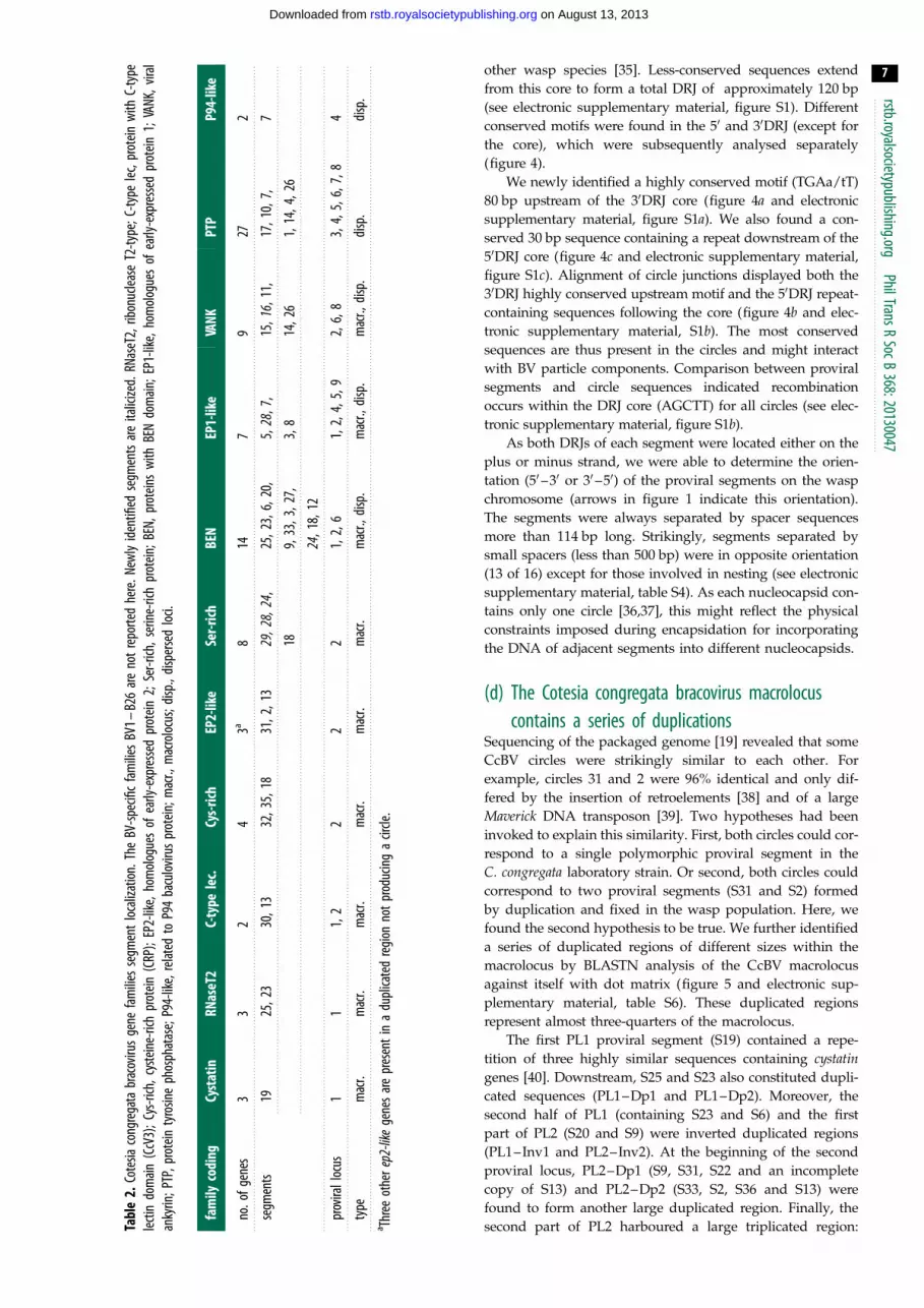
and 26 predicted pseudo-genes belong to 37 families (table 2
and figure 3). Seven of these gene families encoded proteins
containing eukaryotic-conserved domains (PTP, VANK,
cystatin, RNaseT2, BEN, Cys-rich, C-type lectin), one family
codes for a P94-like baculovirus protein and 29 families are
specific to BVs (EP1-like, EP2-like, Ser-rich and BV families
1–26). In contrast to nudiviral genes that do not contain
introns, 60% of genes present in proviral segments were pre-
dicted to contain introns, like cellular genes (see also [19,29]).
(c) Proviral segment extremities are conservedAll proviral segments from all BVs analysed to date are termin-
ated by direct repeats at both extremities, termed DRJs
[12,13,16,29,30,32]. Bracovirus circles contain a unique sequence
(circle junction) produced from a recombination event between
these DRJs [12,33]. Site-specific tyrosine recombinases iden-
tified in the nudiviral machinery (VLF-1a or VLF-1b) were
proposed to perform this recombination [7] based on functional
homology with the homologous baculovirus protein VLF-1
[34]. VLF-1 has been demonstrated to be a nucleocapsid com-
ponent and we therefore hypothesize that a VLF-1 complex
could bind DRJs terminating a segment and resolve the circles,
following encapsidation of BV DNA [7].
We performed comprehensive sequence analyses of
CcBV segment extremities and of their corresponding circle
junctions (figure 4). The alignments led to the identifica-
tion of a perfectly conserved 5 bp direct sequence motif
(AGCTT), which constitutes the DRJ core also found in the

0 10 20 30 40 50 kbp
other BV conserved families
EP1-like
cystatin
Cys-rich (crp)
VANK
PTP
Duffy-like
histone H4
CrV1-like
RNaseT2
p494-like
non-coding sequence
C-type lectin (CcV3)
Ser-rich
BEN
P94-like
mobile elements
26
1
3
5
11
1 5 26 26 5
15 2 11
7 7 7
19
(a)
(b)
8
6
8 6
6 gene content
gene distribution
EP2-like
unknown
2 5
2
19 2 2 2
19 14 3 11
2 5
16 16
macrolocus:
12
12
20 20
12
6 8
8 6
8 6
1925
302365
209
312233
236
1329
32827
15
3224
3518
16
PL1
PL2
PL3
PL4
PL5
PL6
PL7
PL8
PL9
1710
7
1
12
11ps34
14
4
26
8
21
6 6 22 24 6 6 13 9 8 6 23 6 6 10 9 9 6 219
8 17 18
6 22 8 24 6 13 9 8 6 23 6 6 10 9 6 21 9
6 6 8 24 6 66 8 20 23 6 6 6 10 9 21
8
8 4 4 6
3 14 6 6
8 17 18
dispersed loci:
11
5 5 5
25 3 25 15 2
7 7 7 7
Figure 3. Gene content of CcBV proviral segments within (a) macrolocus and (b) dispersed loci displayed by coloured boxes (macrolocus, 260 genes; dispersed loci,62 genes). CcBV contains 37 gene families: seven encode proteins with described conserved domains (cystatin, RNaseT2, C-type lectin, Cys-rich, BEN, VANK and ptp)representing approximately 23.5% of the genes and one encoded a baculovirus homologue protein ( p94-like). Twenty-nine gene families representing 57% of thegenes encode proteins of unknown function conserved in BVs associated with wasps of the Cotesia and Glyptapanteles genera (Ser-rich, EP2-like, EP1-like and BV1 –BV26 represented by grey boxes, with the number identifying the family indicated above the boxes). Some other previously identified BV gene families are onlyrepresented by one member in CcBV (CrV1, histone H4, Duffy and p494). Other genes of unknown function are unique (approx. 14.5%) and some coding DNAsequences are identified as remnants of genes from mobile elements (approx. 4.25%). Note that the ptp gene family constitutes the major part of the genes fromisolated loci (see gene distribution pie charts on the right). Unlike in GiBV, no ptp genes are found in the CcBV macrolocus. Other genes such as cystatin, RNaseT2,C-type lectin or cys-rich are found only in the macrolocus, which contains a majority of BV specific genes (table 2). ps34 shown by a dashed line corresponds to aformer proviral segment mutated in the 30DRJ core homologous to CvBV S30 (accession number HQ009553) but no longer producing a circle.
rstb.royalsocietypublishing.orgPhilTransR
SocB368:20130047
6
on August 13, 2013rstb.royalsocietypublishing.orgDownloaded from
Tabl
e2.
Cote
siaco
ngre
gata
brac
oviru
sge
nefa
milie
sse
gmen
tlo
caliz
ation
.The
BV-sp
ecifi
cfa
milie
sBV
1–B2
6ar
eno
trep
orte
dhe
re.N
ewly
iden
tified
segm
ents
are
italic
ized.
RNas
eT2,
ribon
uclea
seT2
-type
;C-ty
pelec
,pro
tein
with
C-ty
pelec
tindo
main
(CcV
3);C
ys-ri
ch,c
yste
ine-
rich
prot
ein(C
RP);
EP2-
like,
hom
olog
ues
ofea
rly-e
xpre
ssed
prot
ein2;
Ser-r
ich,s
erin
e-ric
hpr
otein
;BEN
,pro
tein
sw
ithBE
Ndo
main
;EP1
-like
,hom
olog
ues
ofea
rly-e
xpre
ssed
prot
ein1;
VANK
,vira
lan
kyrin
;PTP
,pro
tein
tyro
sine
phos
phat
ase;
P94-
like,
relat
edto
P94
bacu
loviru
spr
otein
;mac
r.,m
acro
locu
s;di
sp.,
disp
erse
dlo
ci.
fam
ilyco
ding
Cyst
atin
RNas
eT2
C-ty
pele
c.Cy
s-ric
hEP
2-lik
eSe
r-rich
BEN
EP1-
like
VANK
PTP
P94-
like
no.o
fgen
es3
32
43a
814
79
272
segm
ents
1925
,23
30,1
332
,35,
1831
,2,1
329
,28,
24,
25,2
3,6,
20,
5,28
,7,
15,1
6,11
,17
,10,
7,7
189,
33,3
,27,
3,8
14,2
61,
14,4
,26
24,1
8,12
prov
irall
ocus
11
1,2
22
21,
2,6
1,2,
4,5,
92,
6,8
3,4,
5,6,
7,8
4
type
mac
r.m
acr.
mac
r.m
acr.
mac
r.m
acr.
mac
r.,di
sp.
mac
r.,di
sp.
mac
r.,di
sp.
disp
.di
sp.
a Thre
eot
here
p2-li
kege
nes
are
pres
enti
na
dupl
icate
dre
gion
notp
rodu
cing
acir
cle.
rstb.royalsocietypublishing.orgPhilTransR
SocB368:20130047
7
on August 13, 2013rstb.royalsocietypublishing.orgDownloaded from
other wasp species [35]. Less-conserved sequences extend
from this core to form a total DRJ of approximately 120 bp
(see electronic supplementary material, figure S1). Different
conserved motifs were found in the 50 and 30DRJ (except for
the core), which were subsequently analysed separately
(figure 4).
We newly identified a highly conserved motif (TGAa/tT)
80 bp upstream of the 30DRJ core (figure 4a and electronic
supplementary material, figure S1a). We also found a con-
served 30 bp sequence containing a repeat downstream of the
50DRJ core (figure 4c and electronic supplementary material,
figure S1c). Alignment of circle junctions displayed both the
30DRJ highly conserved upstream motif and the 50DRJ repeat-
containing sequences following the core (figure 4b and elec-
tronic supplementary material, S1b). The most conserved
sequences are thus present in the circles and might interact
with BV particle components. Comparison between proviral
segments and circle sequences indicated recombination
occurs within the DRJ core (AGCTT) for all circles (see elec-
tronic supplementary material, figure S1b).
As both DRJs of each segment were located either on the
plus or minus strand, we were able to determine the orien-
tation (50 –30 or 30 –50) of the proviral segments on the wasp
chromosome (arrows in figure 1 indicate this orientation).
The segments were always separated by spacer sequences
more than 114 bp long. Strikingly, segments separated by
small spacers (less than 500 bp) were in opposite orientation
(13 of 16) except for those involved in nesting (see electronic
supplementary material, table S4). As each nucleocapsid con-
tains only one circle [36,37], this might reflect the physical
constraints imposed during encapsidation for incorporating
the DNA of adjacent segments into different nucleocapsids.
(d) The Cotesia congregata bracovirus macrolocuscontains a series of duplications
Sequencing of the packaged genome [19] revealed that some
CcBV circles were strikingly similar to each other. For
example, circles 31 and 2 were 96% identical and only dif-
fered by the insertion of retroelements [38] and of a large
Maverick DNA transposon [39]. Two hypotheses had been
invoked to explain this similarity. First, both circles could cor-
respond to a single polymorphic proviral segment in the
C. congregata laboratory strain. Or second, both circles could
correspond to two proviral segments (S31 and S2) formed
by duplication and fixed in the wasp population. Here, we
found the second hypothesis to be true. We further identified
a series of duplicated regions of different sizes within the
macrolocus by BLASTN analysis of the CcBV macrolocus
against itself with dot matrix (figure 5 and electronic sup-
plementary material, table S6). These duplicated regions
represent almost three-quarters of the macrolocus.
The first PL1 proviral segment (S19) contained a repe-
tition of three highly similar sequences containing cystatingenes [40]. Downstream, S25 and S23 also constituted dupli-
cated sequences (PL1–Dp1 and PL1–Dp2). Moreover, the
second half of PL1 (containing S23 and S6) and the first
part of PL2 (S20 and S9) were inverted duplicated regions
(PL1–Inv1 and PL2–Inv2). At the beginning of the second
proviral locus, PL2–Dp1 (S9, S31, S22 and an incomplete
copy of S13) and PL2–Dp2 (S33, S2, S36 and S13) were
found to form another large duplicated region. Finally, the
second part of PL2 harboured a large triplicated region:

(a) 3¢DRJ proviral motif
(b) circle junction motif
(c) 5¢DRJ proviral motif
2
1bits
0–8
0–2
0–1
9–1
8–1
7–1
6–1
5–1
4–1
3–1
2–1
1–1
0 –9 –8 –7 –6 –5 –4 –3 –2 –1 0 1 2 3 4 5 6 7 8 9 10 11 12 13 14 15 16 17 18 19 20 21 22 23 24 25 26 27 28 29 30 31 32 33 34 35 36 37 38 39
–20
–19
–18
–17
–16
–15
–14
–13
–12
–11
–10 –9 –8 –7 –6 –5 –4 –3 –2 –1 0 1 2 3 4 5 6 7 8 9 10 11 12 13 14 15 16 17 18 19 20 21 22 23 24 25 26 27 28 29 30 31 32 33 34 35 36 37 38 39
–20
–19
–18
–17
–16
–15
–14
–13
–12
–11
–10 –9 –8 –7 –6 –5 –4 –3 –2 –1 0 1 2 3 4 5 6 7 8 9 10 11 12 13 14 15 16 17 18 19 20 21 22 23 24 25 26 27 28 29 30 31 32 33 34 35 36 37 38 39
–79
–78
–77
–76
–75
–74
–73
–72
–71
–70
–69
–68
–67
–66
–65
–64
–63
–62
–61
–60
–59
–58
–57
–56
–55
–54
–53
–52
–51
–50
–49
–48
–47
–46
–45
–44
–43
–42
–41
–40
–39
–38
–37
–36
–35
–34
–33
–32
–31
–30
–29
–28
–27
–26
–25
–24
–23
–22
–21
–80
–79
–78
–77
–76
–75
–74
–73
–72
–71
–70
–69
–68
–67
–66
–65
–64
–63
–62
–61
–60
–59
–58
–57
–56
–55
–54
–53
–52
–51
–50
–49
–48
–47
–46
–45
–44
–43
–42
–41
–40
–39
–38
–37
–36
–35
–34
–33
–32
–31
–30
–29
–28
–27
–26
–25
–24
–23
–22
–21
5¢
2
1bits
05¢
2
1bits
05¢
2
1bits
05¢
2
1bits
05¢
3¢
3¢
3¢
3¢
3¢
weblogo.berkeley.edu
weblogo.berkeley.edu
weblogo.berkeley.edu
Figure 4. DRJ sequence motifs within C. congregata proviral segments and CcBV circles visualized using WEBLOGO. Each logo consists of stacks of bases, with one stackfor each position in the sequence. The height of the stack at a position indicates the sequence conservation, whereas the height of a base indicates the relativefrequency of this base at this position. Note that the circle junction sequence (b) corresponds to a recombined form of the two DRJs within the perfectly conservedDRJ core shown in the black box. Sequences characterizing (a) 30DRJ and (c) 50DRJs are circled in black. A 30 bp sequence containing a 13 bp repeat(TTtnAatantGAAyaaAAatnntGAwcAaa) following the 50DRJ core was found to be conserved, whereas the sequence following the core in 30DRJ was smaller(TTcnAATTgt). A highly conserved motif (TGAa/tT) was also identified 80 bp upstream of the 30DRJ core. These graphical representations were generated fromindependent alignments of 34 50DRJ, 35 30DRJ and 35 circle junction sequences (see electronic supplementary material, figure S1).
rstb.royalsocietypublishing.orgPhilTransR
SocB368:20130047
8
on August 13, 2013rstb.royalsocietypublishing.orgDownloaded from

TE
2519 6 5 20 9 31 22 33 2 3630 13 29 3 28 27 15 32 24 35 18 1623
PL2-Tr1
PL2Inv2
PL1Inv1
PL1Dp1
PL1Dp2
PL1 PL2
100 200 300 400 500 600
200
400
600
PL2-Tr2
PL2-Tr3
PL2-Dp1
PL2-Dp2
Figure 5. Similarity matrix of the C. congregata proviral macrolocus compared with itself. The main diagonal represents sequence alignment with itself; dotted lines(grey) identify duplicated regions within the sequence analysed. Those parallel to the diagonal correspond to duplicated regions in the same orientation; thoseantiparallel correspond to inverted sequences (striped arrows). Scale is expressed in kilobase pair. Relative positions of proviral loci 1 and 2 and of each CcBV proviralsegment forming the macrolocus are indicated below the dot plot matrix. PL1, proviral locus 1; PL2, proviral locus 2; TE, transposable element. Grey boxes: dupli-cation PL1 – Dp1/PL1 – Dp2 within PL1; striped boxes: inverted duplication PL1 – Inv1/PL2 – Inv2; light grey boxes: duplication PL2 – Dp1/PL2 – Dp2; dark grey boxes:triplication PL2 – Tr1/PL2 – Tr2/PL2 – Tr3. Nucleotide positions of duplication extremities are indicated in the electronic supplementary material, table S6.
rstb.royalsocietypublishing.orgPhilTransR
SocB368:20130047
9
on August 13, 2013rstb.royalsocietypublishing.orgDownloaded from
PL2–Tr1 (S29, S3), PL2–Tr2 (S28, S27 and S15) and PL2–Tr3
(S32, S24, S35 and S18).
The history of viral segment production was also inferred
through 50DRJ and 30DRJ maximum-likelihood phylogenies
(figure 6). For 16 segments present in the duplicated regions,
both DRJs evolved in co-phylogeny (figure 6), but for other
segments, 50 and 30DRJs had different histories (figure 6),
which indicated the formation of mosaic segments (as
described in figure 7).
(e) Macrolocus evolution involved duplicationsAs BV sequences share a common origin, duplication histories
can be tentatively reconstituted by comparing duplicated
regions in related species using parsimonious interpretations.
To identify the most recent rearrangements, we have analysed
the C. sesamiae proviral segments available in GenBank. BAC
inserts corresponding to PL1 and PL2 were identified corres-
ponding to a large part of the CsBV macrolocus (figure 1b).
Proviral segment boundaries were identified (DRJs), and
CsBV segment gene contents were annotated [25]. Orthology
between both Cotesia spp. was readily identified, because
gene order was mostly conserved. CsBV segments were there-
fore annotated and numbered based on the CcBV orthologues
(figure 1a) except for CsBV S20/33, corresponding to a fusion
of CcBV S20 and S33 specific for C. sesamiae (in C. vestalis BV,
the segments are separated [41]), and for CsBV S37, which is
dismantled in CcBV (figure 7).
Comparison with Glyptapanteles spp. gave insights into
evolutionary events dating back to 17 Ma. The macrolocus
structure (PL1 region containing wasp genes PL2) was also
conserved between Glyptapanteles and Cotesia spp., but the
number of segments and their gene content were different.
However, in many cases, it was still possible to trace evo-
lutionary relationships between segments of the different
genera based on conserved homologous DRJ sequences and
of nucleotide similarities remaining between homologous
segments. The comparison of CcBV and GiBV macroloci is
shown in table 3. Strikingly, homologous segments were in
the same orientation even if gene content was not conserved.
The evolutionary dynamics of macrolocus content has
involved duplications that could be traced back to (i) before
the separation of the Cotesia and Glyptapanteles lineages
(17 Ma), (ii) before the separation of C. congregata and
C. sesamiae, or (iii) after this separation (figure 8). Before the
separation of Cotesia and Glyptapanteles lineages, the proviral
form was already organized in a macrolocus composed of
two proviral regions separated by several wasp genes (figure
8c). Two duplications present in the ancestor of the four BVs
remain detectable: PL1–Dp1, PL1–Dp2, PL2–Tr2 and PL2–
Tr3. Specific events occurred in the Glyptapanteles lineage: (i)
the formation of 1p–2p–3p, (ii) that of 17p and 18p, associated
with the capture of sugar transporter genes from the wasp
genome [16] and (iii) the formation of 20p, by re-integration
of a dispersed proviral segment in the macrolocus [31]
(figure 8d ). Between 17 Ma and the separation of Cotesia spp.,
an inverted duplication of PL1 sequences (PL1–Inv1) occurred
in the PL2 anterior part (PL2–Inv2) of the Cotesia lineage
(figure 8b). Finally, in the lineage leading to C. congregata,
two main events occurred. A complex duplication in the

S22S36
S17S1S6
S4S26
S20S15
S35S16
S7S3S24
S27S18
S11S14S12S2S31S19
S5S8S21
S29S32S28
S13S25
S23S9S33
S30
80
6474
81
90
85
72
73
54
59
99
65
92
7664
96
80
8392
62
88
0.4
S2S31
S9S33S23
S25S18S32
S10S17
S1S26
S16S29
S28*S3
S27S24
S6S20
S7S11
S13S30
S19S14
S22S36
S12S5
S8S21
S28S15
S35
77
94
9886
96
64
88
8099
76
8697
10082
7196
71
90
9076
83
98
77
88
9685
99
91
0.7
8765
7773
77
71
73
(a) (b)
PL2-Dp1/2
PL2-Dp1/2
PL1-Dp2PL1-Dp1
PL2-Tr2/3
PL2-Dp1/2
PL2-Tr1/2/3
M/PL9
Figure 6. Proviral segment clustering based on (a) 50 and (b) 30DRJ sequences. The trees were obtained from maximum-likelihood phylogenetic inferences based onthe alignments of approximately 200 bp sequences of 50 and 30DRJs including the DRJ core (‘extended DRJs’). Only SH-like branch values above 50 are indicated.Thick branches highlight 50DRJs in co-phylogeny with the 30DRJ of the same segment, produced by complete duplication of proviral segments including their DRJs(duplicated regions containing the DRJs are indicated on the right). The stars indicated the three S28 DRJs. In this segment, the ancestral 30DRJ S28* (still functionalin C. sesamiae) was replaced by a new 30DRJ recruited during the rearrangement that produced PL2-Tr1. This resulted in a mosaic S28 segment (figure 7).
rstb.royalsocietypublishing.orgPhilTransR
SocB368:20130047
10
on August 13, 2013rstb.royalsocietypublishing.orgDownloaded from
posterior part of PL2 produced PL2–Tr1 (figure 7). Another
duplication also occurred upstream in PL2, leading to the for-
mation of PL2–Dp1 and PL2–Dp2 (figure 8a). The later
duplication occurred relatively recently as judged from the
very high similarity between the duplicated regions.
( f ) Cotesia congregata bracovirus has two specificdispersed loci
In C. congregata, the seven dispersed proviral loci each con-
tained only one to three segments. However, duplications,
such as mirror duplications (S17/S10 on PL3 and S8/S21
on PL9), have also occurred in these regions. Most dispersed
segments encode ptp genes, which are involved in complex
functional interactions with the caterpillar hosts [31]. It is
noteworthy that S1 (PL5), S7 (PL4), S17–S10 (PL3) and S26
(PL8) in addition to ptp genes have a common 30DRJ with
a one base deletion before the DRJ core (see electronic sup-
plementary material, figure S1a). This mutation is also
found in GiBV segments 20p (PL2) and 25p (PL5), and in
Microplitis demolitor BV (MdBV) segments H, J and M,
and could reflect their common origin.
The DRJs of the PL9 segments (S8 and S21) are closely
related to those of PL1 S5 (figures 1a and 6; electronic sup-
plementary material, figure S1). PL9 segments are separated
from the macrolocus but are known to localize on the short
arm of chromosome 5, like the macrolocus [14]. Therefore,
PL9 segments could originate from a duplication of S5 fol-
lowed by a translocation. The unique segment within PL8
(S26) appears to correspond to the integration of a sequence
originally present in locus PL4 at a new localization in
the wasp genome (see electronic supplementary material,
figure S2). No PL8 or PL9 homologues have been identified
in Glyptapanteles spp. Therefore, PL8 and PL9 are new proviral
loci and could constitute an exception to proviral sequences
stability in the wasp genome (unless they have yet to be
isolated in Glyptapanteles spp.).
4. DiscussionIn this study, we report the characterization of bracovirus
proviral segments in the genomes of the wasps C. congregataand C. sesamiae. Comparative genomics with Glyptapanteles
proviruses gave further insights into the evolutionary history
of BVs. The presence of common hymenopteran genes in
flanking regions of most proviral sites indicated that the local-
izations of bracovirus segments in the wasp genomes have
remained the same since the separation of the Cotesia and
Glyptapanteles lineages approximately 17 Ma. The proviral

13 37 28 3227 15
*ep1-like6 3¢DRJ*
(c) CsBV
(a) CcBV
(b) ancestral macrolocus13 37 28 3227 15
*ep1-like6 3¢DRJ* bv8
*
dis37
gene lossin the C. sesamiae lineage
*
duplication and inversionin the C. congregata lineage
*
dis37dis3713 28 3227 1529 3
*
ep1-like63¢DRJ*3¢DRJ*
bv8bv8¢
PL2–Tr1 PL2–Tr2
DRJ proviral segment gene
Figure 7. A proposed parsimonious scenario for the complex rearrangement that may have produced PL2-Tr1 based on the analysis of duplications among Cotesiaspp. (a) Cotesia congregata and (c) C. sesamiae macrolocus sequences were used to infer the putative organization of this region in their common ancestor (b). In thelineage leading to C. sesamiae, the bv8 gene was lost (or this gene was acquired specifically in the C. congregata lineage). In the lineage leading to C. congregata, acomplex rearrangement occurred resulting in inversion and duplication of proviral segment sequences. Inversion: the segment S37 was inverted and its ep1-like6gene and regular 30DRJ (black triangle) were incorporated into an enlarged S28. The regular 30DRJ became that of S28, replacing the former S28 DRJ readilyidentified by its particular sequence (30DRJ*, grey triangle). Duplication: the region encompassing S28, S27 and a part of S15 was duplicated and insertedwithin S37 that was dismantled (dis37). It should be noted that inversion, duplication and dismantlement might have been produced by a single complex rearrange-ment caused by errors during replication (fork stalling and template switching model). DRJs are indicated by white triangles to delimit the segments.
rstb.royalsocietypublishing.orgPhilTransR
SocB368:20130047
11
on August 13, 2013rstb.royalsocietypublishing.orgDownloaded from
sequences were organized in a macrolocus comprising over
two-thirds of the proviral sequences completed by seven
smaller dispersed loci, each with one to three segments.
The macrolocus comprised two proviral loci (PL1 and PL2)
joined by a region containing wasp genes.
In C. congregata, the dispersed PL4 and PL9 loci and the
macrolocus had previously been visualized on the short arm
of chromosome 5 by in situ hybridization [14]. Homologues
of wasp genes either flanking or within the macrolocus and
PL4 (see electronic supplementary material, table S5) belong
to the same linkage group in the genome of A. mellifera [42].
This might suggest PL4 and PL9 were originally a part of the
macrolocus, from which they were later separated by chromo-
somal rearrangements. By contrast, homologues of PL8 wasp
genes were located in a different linkage group in A. melliferaand Nasonia vitripennis genomes, implying that this proviral
locus might be located on a different chromosome.
The identification of homologous sequences to all proviral
loci of Glyptapanteles spp. suggests data concerning the CcBV
proviral segment is relatively complete. However, because the
BAC clones were identified by hybridization to sequences
from previously known segments, we cannot exclude that
other proviral segments in dispersed loci have remained
unidentified. Most of the packaged circles of C. vestalis
bracovirus (CvBV), recently sequenced using a high-throughput
approach [41], have CcBV homologues (including the newly
identified CcBV circles) except CvBV C19, C31, C32 and C35
that could be specific for CvBV or still missing from our analysis.
However, CvBV C35 lacks the DRJ that are characteristic of BV
circles. Furthermore, CvBV C35 encodes a helicase and shows
high nucleotide similarity with different arthropod genomes,
suggesting it could correspond to a mobile element.
We found one nudiviral gene within CcBV proviral locus:
the odv-e66-like1 gene, which had been shown to encode a par-
ticle component of Chelonus inanitus BV (CiBV) [8]. We
showed that this gene is present within the macrolocus of the
four species examined (figure 2). We suppose this reflects the
ancient presence of the nudiviral genome and proviral macro-
locus at the same locus. Forthcoming C. congregata whole
genome sequencing should reveal whether the nudiviral clus-
ter is localized on chromosome 5, like the macrolocus.
No nudiviral genes are present in the packaged genome;
however, the DRJs terminating the BV proviral segments are
probably a component of the nudiviral machinery. Indeed,
based on what is known for baculovirus replication, BV DRJs
involved in the dsDNA circle production could constitute the
binding sites of a nudiviral site-specific recombinase that
would ensure the excision of the circles from larger amplified
molecules during DNA encapsidation [7]. In baculoviruses,
the VLF-1 protein is a tyrosine recombinase localized at one

Tabl
e3.
Corre
spon
denc
ebe
twee
nCc
BVan
dGi
BVse
gmen
tsw
ithin
the
mac
rolo
cus.
Corre
spon
denc
eswe
rees
tabl
ished
acco
rdin
gto
DRJ
sequ
ence
san
dge
neco
nten
tby
analy
ses
ofho
mol
ogou
sse
quen
ces
dete
cted
byTB
LAST
X.Se
gmen
tor
ienta
tion
isdi
splay
ed(.
sens
e.an
d,
reve
rse,
).Sim
ilarit
yis
give
nby
the
perce
ntag
eof
hom
olog
ous
sequ
ence
sco
mm
onbe
twee
nth
eco
rresp
ondi
ngCc
BVan
dGi
BVse
gmen
ts:10
%,
*,
35%
,35
%,
**,
45%
,**
*.
45%
;inv
,inv
erse
dse
quen
ce;8
,,c
onse
rved
DRJ
inre
desig
ned
segm
ent;
—,r
edes
igne
dse
gmen
t;n.
a.ab
sent
;dis3
7,di
sman
tled
segm
ent
37.$
,in
that
last
case
the
perce
ntag
eof
com
mon
hom
olog
ous
sequ
ence
sis
low(1
3%)
betw
een
CcBV
and
GiBV
,but
high
(46%
)bet
ween
the
inta
ctS3
7pr
esen
tin
CsBV
and
the
9pfro
mGi
BV.
mac
rolo
cus
hom
olog
ous
segm
ents
CcBV
PL1
8,19
,8
.25
.,
30,
.23
..
6.,
5,
GiBV
PL1
8,1p
,.
2p.
,3p
,8
.4p
.,
5p,
.6p
..
7p.
,8p
,
simila
rity
rang
e*
***
**
***
***
***
CcBV
PL2
—,
20,
,33
/(9),
——
.2/
(31)
.,
36/(2
2),
.13
.
GiBV
PL1in
v.
8p.
,7p
,,
6p,
.5p
.,
4p,
.3p
.,
2p,
.1p
.
simila
rity
rang
e**
***
**
**
CcBV
PL2
.di
s37.
,28
,,
27,
.15
.,
32,
,24
,.
35.
8,18
n.a.
n.a.
,8
n.a.
.16
.
GiBV
PL2
.9p
.,
10p ,
,11
p,.
12p.
,13
p,,
14p,
.15
p.8,
16p,
,17
p,.
18p.
,19
p,8
.20
p..
21p.
simila
rity
rang
e*$
****
**
****
***
***
*** rstb.royalsocietypublishing.org
PhilTransRSocB
368:20130047
12
on August 13, 2013rstb.royalsocietypublishing.orgDownloaded from
extremity of the nucleocapsid. VLF-1 is involved in the resolu-
tion of the baculovirus DNA molecules, which are amplified as
genome concatemers during replication [34] and separated as
individual genome monomers during encapsidation. Two
nudiviral genes related to vlf-1 homologues were expressed
in braconid wasp ovaries and could encode the DRJ binding
recombinase [7,9].
The DRJ sequences were conserved in all BVs studied
(CcBV, CsBV, GiBV, GfBV and MdBV). In CiBV, some vari-
ations occurred on the first and last bases of the core DRJ
(consensus (A/c)GCT(T/c)), the 13 bp repeat after the 50DRJ
core was absent, and the sequence downstream of the 30DRJ
core showed variation with the CcBV consensus (CiBV consen-
sus: TCnAATt). This could reflect the greater phylogenetic
distance of Chelonus inanitus (belonging to Cheloninae sub-
family) compared with the relatively closely related Cotesia,Glyptapanteles and Microplitis spp. (all classified in the Micro-
gastrinae subfamily). Still, the high conservation of DRJ
motifs underlines their deep phylogenetic relationships going
back to the common origin of BVs. In the ancestral nudivirus,
a DRJ-related sequence might have been used to separate
genome concatemers produced during viral DNA replication
such as in baculoviruses. Strong selective constraints linked
to the interaction with the nudiviral machinery may have led
to high DRJ sequence conservation in comparison with other
proviral sequences.
Proviral segments were oriented on the wasp chromosome
as both DRJs of a segment are always located either on the
positive or negative strand. Proviral segments were separated
by spacer sequences more than 114 bp long that are not
packaged in the particles [43], and surprisingly segments
separated by short spacers (less than 500 bp) were often in
opposite directions. This particular organization could derive
from physical constraints as adjacent segments are amplified
together in the same molecule [43] before their separation
and circularization by recombination of the DRJs, which is
likely coupled with viral DNA entry into the nucleocapsids.
When spacers are short, opposite orientation of segments
may permit capsid access to the amplified DNA molecule
from opposite sides, whereas segments in the same orientation
will induce capsid competition for space. According to this
hypothesis, adjacent segments in the same orientation and sep-
arated by small spacers could fail to be resolved (figure 1a, blue
lines). We were indeed able to observe this phenomenon result-
ing in large circles containing the sequences of two segments in
the particles, a situation similar to circle ‘nesting’ observed in
ichnoviruses [44]. Given that the length of BV nucleocapsids
is correlated with the size of the dsDNA molecule they contain
[36,37], a large range of DNA circle sizes can be encapsidated
thus allowing nesting.
As proviral loci of different species are orthologous, it is
possible to compare their content and to reconstruct their his-
tory. Stability of proviral segment localizations within wasp
genomes contrasted sharply with the evolutionary dynamics
of their content that has involved duplications. The macrolocus
evolved mainly by duplications within segments (S19) or com-
prising one (PL1-Dp1/PL1-Dp2) to a series of segments (PL2
triplication). Duplication boundaries do not correspond to
those of the segments (see electronic supplementary material,
table S6). Thus, in most cases, duplications do not appear to
have been produced by a viral mechanism (circle re-integration
for example). Gene amplifications have been reported to be
involved in mosquito resistance to insecticides, but the

Dp1 Tr1 Tr2 Tr3Dp1 Dp2Dp2
PL1 PL2
odv
20 139 31 3322 2 36525 2319 30 6 28 32 1629 1835153 27 24
odv
5 20/33 3725 23 13 28 18230 6 36 27 15 32 3524 16
odv
(c) ancestral macrolocus
(b) CsBV
(a) CcBV
(d ) GBV9p 15p 21p16p 20p1p 4p 6p 12p10p 13p8p3p2p 5p 7p 11p 14p 17p18p19p
odv
Figure 8. A proposed parsimonious scenario for macrolocus proviral genome evolution in Cotesia and Glyptapanteles lineages based on the analysis of duplicationsamong wasp species. Sequences from C. congregata (a), C. sesamiae (b), G. indiensis and G. flavicoxis (d ) were used to infer the putative organization of an ancestralmacrolocus (c) containing two proviral regions that would have existed before the separation of the Cotesia and Glyptapanteles lineages over 17 Ma. In the lineageleading to Glyptapanteles spp., new segments 1p – 2p – 3p, 17p, 18p and 20p (re-integrated) were formed. In the lineage leading to Cotesia spp., inverted dupli-cations of PL1 sequences (hashed boxes) resulted in modifications of the anterior PL2 region. Subsequent rearrangements in the lineage leading to C. sesamiae leadto the fusion of segments S20 and S33 (S20/33) and the loss of segment S15 gene content. In the lineage leading to C. congregata, duplication in the posteriorregion of PL2 produced PL2 – Tr1 and a larger S28. In addition, duplications upstream of PL2 lead to the formation of PL2 – Dp1 and PL2 – Dp2. Names and boxcolours for duplications and inversion are the same as in figure 5.
rstb.royalsocietypublishing.orgPhilTransR
SocB368:20130047
13
on August 13, 2013rstb.royalsocietypublishing.orgDownloaded from
duplications have not been sequenced, and the mechanism of
their production is unknown [45]. In human genetic diseases,
complex rearrangements involving duplications have been
sequenced and attributed to DNA replication errors [46].
According to this model, unusual genome architecture, such
as the presence of repetitive sequences, may confuse the
DNA replication machinery. This results in replication
fork stalling, causing the DNA polymerase to switch from
one template to another. Template switching can occur several
times forwards or backwards on the molecule used as master
sequence before replication resumes on the original DNA
template. This could potentially simultaneously cause both
inversions and duplications, such as those described for
PL2–Tr1 (figure 7). DRJs, being repeated sequences, could be
involved in such a mechanism.
Beyond the hypothetical mechanism leading to their pro-
duction, the reason duplications are evolutionarily conserved
is probably because of the antagonistic coevolution between
caterpillar hosts and parasitoid wasps, which resulted in com-
plex evolutionary arms races [1,25,47]. The packaged genes
expressed in infected caterpillar tissues produce virulence
factors involved in manipulating host physiology and
altering host immunity or development [48,49]. The selection
of particular beneficial alleles and accelerated mutation
accumulation in duplicated genes should provide new weap-
ons against host targets [25], and we found the triplicated
cystatin genes (S19) to undergo strong positive selection [50].
In this context, the localization of most proviral sequences
within a macrolocus might have been maintained because it
readily allowed the production of new circles. Indeed, the
large duplications resulted in new proviral sequences with
two DRJs that are very likely to produce new circles, whereas
re-integrated circles were found to contain a single DRJ and
to be dysfunctional [31].
Acknowledgements. We thank Cindy Menoret for taking care of theinsects and Germain Chevignon for helpful discussion. The Cotesiacongregata proviral locus study was performed as a part of the pro-jects Evparasitoid and Paratoxose supported by the agencies ANRand CNRS (GDR 2153, GDR 2157 and IFR 136) with the collaborationof the Genoscope (Evry, France) and the European Research Councilstarting grant GENOVIR (205206). This work has been carried outwith the technical support of the Genomics Department (PPFASB) - University F. Rabelais (Tours).
References
1. Herniou EA, Huguet E, Theze J, Bezier A, Periquet G,Drezen J-M. 2013 When parasitic wasps hijackedviruses: genomic and functional evolution ofpolydnaviruses. Phil. Trans. R. Soc. B 368,20130051. (doi:10.1098/rstb.2013.0051)
2. Dupuy C, Huguet E, Drezen J-M. 2006 Unfoldingthe evolutionary story of polydnaviruses.
Virus Res. 117, 81 – 89. (doi:10.1016/j.virusres.2006.01.001)
3. Murphy N, Banks JC, Whitfield JB, Austin AD.2008 Phylogeny of the parasitic microgastroidsubfamilies (Hymenoptera: Braconidae) based onsequence data from seven genes, with an improvedtime estimate of the origin of the lineage. Mol.
Phylogenet. Evol. 47, 378 – 395. (doi:10.1016/j.ympev.2008.01.022)
4. Bezier A et al. 2009 Polydnaviruses of braconidwasps derive from an ancestral nudivirus. Science323, 926 – 930. (doi:10.1126/science.1166788)
5. Theze J, Bezier A, Periquet G, Drezen J-M, HerniouEA. 2011 Paleozoic origin of insect large dsDNA

rstb.royalsocietypublishing.orgPhilTransR
SocB368:20130047
14
on August 13, 2013rstb.royalsocietypublishing.orgDownloaded from
viruses. Proc. Natl Acad. Sci. USA 108, 15 931 –15 935. (doi:10.1073/pnas.1105580108)
6. Bezier A, Herbiniere J, Lanzrein B, Drezen J-M. 2009Polydnavirus hidden face: the genes producing virusparticles of parasitic wasps. J. Invertebr. Pathol. 101,194 – 203. (doi:10.1016/j.jip.2009.04.006.)
7. Drezen J-M, Herniou EA, Bezier A. 2012 Evolutionaryprogenitors of bracoviruses. In Parasitoid virusessymbionts and pathogens (eds NE Beckage, J-MDrezen), pp. 15– 31. San Diego, CA: CRC Press-Elsevier.
8. Wetterwald C et al. 2010 Identification of bracovirusparticle proteins and analysis of their transcriptlevels at the stage of virion formation. J. Gen. Virol.91, 2610 – 2619. (doi:10.1099/vir.0.022699-0)
9. Burke GR, Strand MR. 2012 Deep sequencingidentifies viral and wasp genes with potential rolesin replication of Microplitis demolitor bracovirus.J. Virol. 86, 3293 – 3306. (doi:10.1128/JVI.06434-11)
10. Dupuy C, Gundersen-Rindal D, Cusson M. 2012Genomic and replication of polydnaviruses. InParasitoid viruses symbionts and pathogens (edsNE Beckage, J-M Drezen), pp. 47 – 61. San Diego,CA: CRC Press-Elsevier.
11. Cornelis G, Heidmann O, Bernard-Stoecklin S,Reynaud K, Veron G, Mulot B, Dupressoir A,Heidmann T. 2012 Ancestral capture of syncytin-Car1, a fusogenic endogenous retroviral envelopegene involved in placentation and conserved inCarnivora. Proc. Natl Acad. Sci. USA 109,E432 – E441. (doi:10.1073/pnas.1115346109)
12. Savary S, Beckage NE, Tan F, Periquet G, DrezenJ-M. 1997 Excision of the polydnaviruschromosomal integrated EP1 sequence of theparasitoid wasp Cotesia congregata (Braconidae,Microgastrinae) at potential recombinase bindingsites. J. Gen. Virol. 78, 3125 – 3134.
13. Annaheim M, Lanzrein B. 2007 Genomeorganization of the Chelonus inanitus polydnavirus:excision sites, spacers and abundance of proviraland excised segments. J. Gen. Virol. 88, 450 – 457.(doi:10.1099/vir.0.82396-0)
14. Belle E, Beckage NE, Rousselet J, Poirie M,Lemeunier F, Drezen J-M. 2002 Visualization ofpolydnavirus sequences in a parasitoid waspchromosome. J. Virol. 76, 5793 – 5796. (doi:10.1128/JVI.76.11.5793-5796.2002)
15. Pasquier-Barre F, Dupuy C, Huguet E, Monteiro F,Moreau A, Poirie M, Drezen J-M. 2002 Polydnavirusreplication: the EP1 segment of the parasitoid waspCotesia congregata is amplified within a largerprecursor molecule. J. Gen. Virol. 83, 2035 – 2045.
16. Desjardins CA et al. 2008 Comparative genomics ofmutualistic viruses of Glyptapanteles parasiticwasps. Genome Biol. 9, R183. (doi:10.1186/gb-2008-9-12-r183)
17. Beckage NE, Tan F, Schleifer KW, Lane RD, CherubinLL. 1994 Characterization and biological effects ofCotesia congregata polydnavirus on host larvae ofthe tobacco hornworm, Manduca sexta. Arch. InsectBiochem. Physiol. 26, 165 – 195. (doi:10.1002/arch.940260209)
18. Kim UJ, Birren BW, Slepak T, Mancino V, Boysen C,Kang HL, Simon MI, Shizuya H. 1996 Construction
and characterization of a human bacterial artificialchromosome library. Genomics 34, 213 – 218.(doi:10.1006/geno.1996.0268)
19. Espagne E, Dupuy C, Huguet E, Cattolico L, ProvostB, Martins N, Poirie M, Periquet G, Drezen J-M.2004 Genome sequence of a polydnavirus: insightsinto symbiotic virus evolution. Science 306,286 – 289. (doi:10.1126/science.1103066)
20. Bailey TL, Boden M, Buske FA, Frith M, Grant CE,Clementi L, Ren J, Li WW, Noble WS. 2009 MEMEsuite: tools for motif discovery and searching.Nucleic Acids Res. 37, W202 – 208. (doi:10.1093/nar/gkp335)
21. Xu D, Stoltz DB. 1993 Polydnavirus genomesegment families in the ichneumonidparasitoid Hyposoter fugitivus. J. Virol. 67,1340 – 1349.
22. Cui L, Webb BA. 1997 Homologous sequences in theCampoletis sonorensis polydnavirus genome areimplicated in replication and nesting of the Wsegment family. J. Virol. 71, 8504 – 8513.
23. Kearse M et al. 2012 GENEIOUS basic: an integratedand extendable desktop software platform for theorganization and analysis of sequence data.Bioinformatics 28, 1647 – 1649. (doi:10.1093/bioinformatics/bts199)
24. Margulies M et al. 2005 Genome sequencing inmicrofabricated high-density picolitre reactors.Nature 437, 376 – 380. (doi:10.1038/nature03959)
25. Jancek S et al. 2013 Adaptive selection onbracovirus genomes drives the specialization ofCotesia parasitoid wasps. PLoS ONE 8, e64432.(doi:10.1371/journal.pone.0064432)
26. Rutherford K, Parkhill J, Crook J, Horsnell T, Rice P,Rajandream MA, Barrell B. 2000 Artemis: sequencevisualization and annotation. Bioinformatics 16,944 – 945. (doi:10.1093/bioinformatics/16.10.944)
27. Bailey TL, Elkan C. 1994 Fitting a mixturemodel by expectation maximization to discovermotifs in biopolymers. Proc. Int. Conf. Intell.Syst. Mol. Biol. 2, 28 – 36. (doi:10.1093/bioinformatics/16.10.944)
28. Crooks GE, Hon G, Chandonia J-M, Brenner SE. 2004WEBLOGO: a sequence logo generator. Genome Res.14, 1188 – 1190. (doi:10.101/gr.849004)
29. Desjardins CA et al. 2007 Structure and evolution ofa proviral locus of Glyptapanteles indiensisbracovirus. BMC Microbiol. 7, 61. (doi:10.1186/1471-2180-7-61)
30. Beck MH, Zhang S, Bitra K, Burke GR, Strand MR.2011 The encapsidated genome of Microplitisdemolitor bracovirus integrates into the hostPseudoplusia includens. J. Virol. 85, 11 685 – 11 696.(doi:10.1128/JVI.05726-11)
31. Serbielle C, Dupas S, Perdereau E, Hericourt F,Dupuy C, Huguet E, Drezen J-M. 2012 Evolutionarymechanisms driving the evolution of a largepolydnavirus gene family coding for protein tyrosinephosphatases. BMC Evol. Biol. 12, 253. (doi:10.1186/1471-2148-12-253)
32. Gruber A, Stettler P, Heiniger P, Schumperli D,Lanzrein B. 1996 Polydnavirus DNA of the braconidwasp Chelonus inanitus is integrated in the wasp
genome and excised only in later pupal and adultstages of the female. J. Gen. Virol. 77, 2873 – 2879.(doi:10.1099/0022-1317-77-11-2873)
33. Savary S, Drezen J-M, Tan F, Beckage NE, PeriquetG. 1999 The excision of polydnavirus sequencesfrom the genome of the wasp Cotesia congregata(Braconidae, Microgastrinae) is developmentallyregulated but not strictly restricted to the ovaries inthe adult. Insect Mol. Biol. 8, 319 – 327. (doi:10.1046/j.1365-2583.1999.83130.x)
34. Vanarsdall AL, Okano K, Rohrmann GF. 2006Characterization of the role of very late expressionfactor 1 in baculovirus capsid structure and DNAprocessing. J. Virol. 80, 1724 – 1733. (doi:10.1128/JVI.80.4.1724-1733.2006)
35. Gundersden-Rindal D, Dupuy C, Huguet E, DrezenJ-M. 2013 Parasitoid polydnaviruses: evolution,pathology and applications. Biocontrol Sci. Technol.23, 1 – 61. (doi:10.1080/09583157.2012.731497)
36. Albrecht U, Wyler T, Pfister-Wilhelm R, Gruber A,Stettler P, Heiniger P, Kurt E, Schumperli D, Lanzrein B.1994 Polydnavirus of the parasitic wasp Chelonusinanitus (Braconidae): characterization, genomeorganization and time point of replication. J. Gen. Virol.75, 3353 – 3363. (doi:10.1099/0022-1317-75-12-3353)
37. Beck MH, Inman RB, Strand MR. 2007 Microplitisdemolitor bracovirus genome segments vary inabundance and are individually packaged in virions.Virology 359, 179 – 189. (doi:10.1016/j.virol.2006.09.002)
38. Drezen J-M, Bezier A, Lesobre J, Huguet E, CattolicoL, Periquet G, Dupuy C. 2006 The few virus-likegenes of Cotesia congregata bracovirus. Arch. InsectBiochem. Physiol. 61, 110 – 122. (doi:10.1002/arch.20108)
39. Dupuy C, Periquet G, Serbielle C, Bezier A, Louis F,Drezen J-M. 2011 Transfer of a chromosomalMaverick to endogenous bracovirus in a parasitoidwasp. Genetica 139, 489 – 496. (doi:10.1007/s10709-011-9569-x)
40. Espagne E et al. 2005 A virus essential for insecthost-parasite interactions encodes cystatins. J. Virol.79, 9765 – 9776. (doi:10.1128/JVI.79.15.9765-9776.2005)
41. Chen YF, Gao F, Ye XQ, Wei SJ, Shi M, Zheng HJ,Chen XX. 2011 Deep sequencing of Cotesia vestalisbracovirus reveals the complexity of a polydnavirusgenome. Virology 414, 42 – 50. (doi:10.1016/j.virol.2011.03.009)
42. The Honeybee Genome Sequencing Consortium.2006 Insights into social insects from the genomeof the honeybee Apis mellifera. Nature 443,931 – 949. (doi:10.1038/nature05260)
43. Louis F, Bezier A, Periquet P, Ferras C, Drezen J-M,Dupuy C. In press. Bracovirus DNAs and the nudivralcluster involved in particle production are amplifiedin several replication units. J. Virol. (doi:10.1128/JVI.00886-13)
44. Kroemer JA, Webb BA. 2004 Polydnavirus genes andgenomes: emerging gene families and new insightsinto polydnavirus replication. Annu. Rev. Entomol.49, 431 – 456. (doi:10.1146/annurev.ento.49.072103.120132)

rstb.royalsocietypublishing.or
15
on August 13, 2013rstb.royalsocietypublishing.orgDownloaded from
45. Alout H, Labbe P, Berthomieu A, Pasteur N, Weill M.2009 Multiple duplications of the rare ace-1mutation F290 V in Culex pipiens naturalpopulations. Insect Biochem. Mol. Biol. 39,884 – 891. (doi:10.1016/j.ibmb.2009.10.005)
46. Lee JA, Carvalho CM, Lupski JR. 2007 A DNAreplication mechanism for generating nonrecurrentrearrangements associated with genomicdisorders. Cell 131, 1235 – 1247. (doi:10.1016/j.cell.2007.11.037)
47. Dupas S, Dubuffet A, Carton Y, Poirie M. 2009Local, geographic and phylogenetic scales ofcoevolution in Drosophila-parasitoid interactions.Adv. Parasitol. 70, 281 – 295. (doi:10.1016/S0065-308X(09)70011-9)
48. Strand MR. 2012 Polydnavirus gene products thatinteract with the host immune system. In Parasitoidviruses symbionts and pathogens (eds NE Beckage,J-M Drezen), pp. 149 – 161. San Diego, CA: CRCPress-Elsevier.
49. Beckage NE. 2012 Polydnaviruses as endocrineregulators. In Parasitoid viruses symbionts andpathogens (eds NE Beckage, J-M Drezen),pp. 163 – 168. San Diego, CA: CRC Press-Elsevier.
50. Serbielle C, Chowdhury S, Pichon S, Dupas S, Lesobre J,Purisima EO, Drezen J-M, Huguet E. 2008 Viral cystatinevolution and three-dimensional structure modelling:a case of directional selection acting on a viral proteininvolved in a host – parasitoid interaction. BMC Biol. 6,38. (doi:10.1186/1741-7007-6-38)
gP
hilTransRSocB368:20130047


