Embed Size (px)
Citation preview
JOURNALOFNEARINFRAREDSPECTROSCOPY
75
ISSN: 0967-0335 © IM Publications LLP 2012
doi: 10.1255/jnirs.969 All rights reserved
Cognitive neuroscience is a multidisciplinary fi eld focused on
the exploration of the neural substrates underlying cognitive
functions; it originated in the early 1980s from the connection
between neuroscience and cognitive science although over
the years it has constantly been enriched by an increasing
interaction with several other disciplines,1 such as neuro-
physiology, neuroanatomy, neuropsychology, psychophysiology
and computational modelling. Nowadays, cognitive neuro-
science represents a prominent fi eld in the investigation of
the human brain. Due to its multidisciplinary nature, cognitive
neuroscience adopts several investigation methods, such as
lesion studies, multi-unit and single-cell recording; never-
theless, the most remarkable progress in understanding the
relationship between brain and cognition has been made with
functional brain imaging methods.
Before the advent of brain imaging, the association between
brain regions and cognitive functions was mainly provided by
clinical neuropsychological investigations of brain-damaged
patients and post-mortem examination. When brain imaging
was introduced, cognitive scientists were given the chance
to investigate the human brain in a wide variety of actions,
from perception to higher order mental activities. With brain
Review
Functional near infrared optical imaging in cognitive neuroscience: an introductory review
Simone Cutini,a,* Sara Basso Moroa and Silvia Biscontib
aDepartment of General Psychology, University of Padova, Padova, Italy. E-mail: [email protected]
bDepartment of Health Sciences, University of L’Aquila, L’Aquila, Italy
Cognitive neuroscience is a multidisciplinary fi eld focused on the exploration of the neural substrates underlying cognitive functions;
the most remarkable progress in understanding the relationship between brain and cognition has been made with functional brain
imaging. Functional near infrared (fNIR) spectroscopy is a non-invasive brain imaging technique that measures the variation of oxygen-
ated and deoxygenated haemoglobin at high temporal resolution. Stemming from the fi rst pioneering experiments, the use of fNIR spec-
troscopy in cognitive neuroscience has constantly increased. Here, we present a brief review of the fNIR spectroscopy investigations in
the cognitive neuroscience fi eld. The topics discussed encompass the classical issues in cognitive neuroscience, such as the exploration
of the neural correlates of vision, language, memory, attention and executive functions. Other relevant research topics are introduced
in order to show the strengths and the limitations of fNIR spectroscopy, as well as its potential in the biomedical fi eld. This review is
intended to provide a general view of the wide variety of optical imaging applications in the fi eld of cognitive neuroscience. The increas-
ing body of studies and the constant technical improvement suggest that fNIR spectroscopy is a versatile and promising instrument to
investigate the neural correlates of human cognition.
Keywords: cognitive neuroscience, near infrared spectroscopy, fNIRS, optical imaging, cognition, psychology, human brain mapping
Introduction
S. Cutini, S. Basso Moro and S. Bisconti, J. Near Infrared Spectrosc. 20, 75–92 (2012)
Received: 19 August 2011 ■ Revised: 28 November 2011 ■ Accepted: 30 November 2011 ■ Publication: 6 March 2012
Special Issue on Medical Applications
76 Functional NIR Optical Imaging in Cognitive Neuroscience
imaging, scientists started to address not only which brain
areas were necessary for a specifi c cognitive function, but
also the neural circuitry involved in a particular mental
activity (e.g. when reading a word2). The wide variety of brain
imaging methods available can provide different measure-
ments of the neural correlates of cognitive processes: for
instance, some methods record the magnetic (magnetoen-
cephalography) or electrical (electroencephalography, EEG)
fl uctuations occurring in relation to neural activity, while other
methods, such as functional magnetic resonance imaging
(fMRI) and functional near infrared spectroscopy (fNIR
spectroscopy, or fNIRS) measure local changes in cerebral
haemodynamic activity that can be used to infer information
on the underlying neural activity (Figure 1). In this respect,
the term “neurovascular coupling”3–5 indicates the well-
known link (both in terms of space and amplitude) between
neural activity and the local change in cerebral blood fl ow
(CBF). Since neither glucose nor oxygen reserves are present
in neurons, an increase in neural activity is accompanied by
an increase in regional CBF, providing additional glucose and
oxygen to the area of active neurons. Given that the increase
in CBF is signifi cantly higher than the corresponding oxygen
consumption, the resulting effect leads to the haemodynamic
response (Figure 1): a net increase in the amount of oxygen-
ated haemoglobin (HbO) and a net decrease of deoxygen-
ated haemoglobin (HbR). Such an over-supply of oxygen,
generated by CBF, is the basis of the blood-oxygenation
level dependent (BOLD) signal used in fMRI,6,7 which broadly
corresponds to the HbR response.8
Among all the brain imaging techniques, fMRI is consid-
ered as the “gold standard” technique for non-invasive func-
tional mapping of the human brain. Nevertheless, the use
of fNIR optical imaging in the cognitive neuroscience fi eld
has increased signifi cantly in recent years. Similar to fMRI,
fNIR spectroscopy monitors haemodynamic changes in the
brain;9 however, unlike the BOLD signal of fMRI, which derives
contrast from the paramagnetic properties of HbR, brain optical
imaging is based on: (i) high near infrared (650–1000 nm) light
propagation into scattering tissues and (ii) absorption by the
main chromophores (HbO and HbR of the red blood cells).
Using light sources with two or more different wavelengths
(e.g. 690 nm and 830 nm), fNIR spectroscopy can simultane-
ously record HbO and HbR [as well as total haemoglobin (HbT)
given by the sum of HbO and HbR] concentration changes with
high temporal resolution (typically > 10 Hz). Cortical haemody-
namic activity can be recorded by locating a set of optodes (i.e.
optical fi bres) over the scalp: sources emit the near infrared
light which is conveyed to the scalp by means of optical fi bres,
while other optical fi bres collect the photons emerging from
the scalp and convey them to be measured by near infrared
detectors. Each source–detector pair is considered a meas-
urement point (i.e. channel). Multi-channel fNIR spectroscopy,
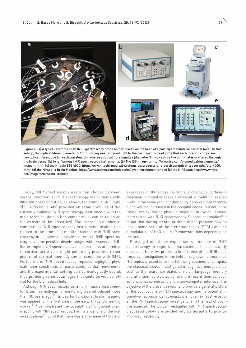
obtained by using arrays of multiple optodes (sources and
detectors) placed on the scalp [Figure 2(a)], represents the
current standard technology. Some typical instruments are
shown in Figure 2(b).
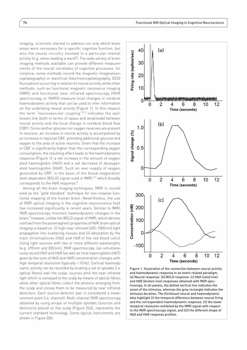
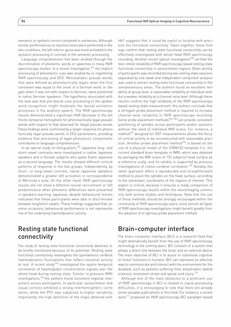
Figure 1. Illustration of the connection between neural activity
and haemodynamic response in an event-related paradigm.
(a) Neural response. (b) BOLD response. (c) HbO (solid line)
and HbR (broken line) responses obtained with fNIR spec-
troscopy. In all panels, the dotted vertical line indicates the
onset of the stimulus, whereas the grey rectangle indicates the
stimulus duration. The (fi ctitious) neural and haemodynamic
data highlight (i) the temporal difference between neural fi ring
and the correspondent haemodynamic response, (ii) the lower
temporal resolution exhibited by the fMRI signal with respect
to the fNIR spectroscopy signal, and (iii) the different shape of
HbO and HbR response profi les.
40
(a)
(b)
(c)
S. Cutini, S. Basso Moro and S. Bisconti, J. Near Infrared Spectrosc. 20, 75–92 (2012) 77
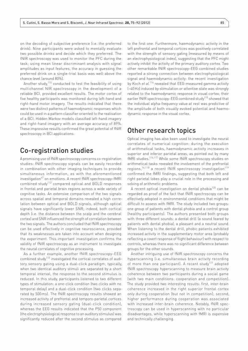
Today, fNIR spectroscopy users can choose between
several commercial fNIR spectroscopy instruments with
different characteristics, as shown, for example, in Figure
2(b). A recent study9 provided an exhaustive list of the
currently available fNIR spectroscopy instruments with the
main technical details (the complete list can be found on
the website of the industries). The increasing number of
commercial fNIR spectroscopy instruments available is
related to the promising results obtained with fNIR spec-
troscopy in cognitive neuroscience: even if fNIR spectros-
copy has some peculiar disadvantages with respect to fMRI
(for example, fNIR spectroscopy measurements are limited
to cortical activity), it might potentially provide a richer
picture of cortical haemodynamics compared with fMRI.
Furthermore, fNIR spectroscopy imposes negligible phys-
ical/motor constraints on participants, so that movements
and the experimental setting can be ecologically sound,
thus providing some advantages that could be very benefi -
cial for the biomedical fi eld.
Although NIR spectroscopy as a non-invasive instrument
for brain haemodynamic monitoring was introduced more
than 30 years ago,10 its use for functional brain mapping
was applied for the fi rst time in the early 1990s: pioneering
works11–14 demonstrated the possibility of functional brain
mapping with NIR spectroscopy. For instance, one of the fi rst
investigations11 found that there was an increase of HbO and
a decrease in HbR across the frontal and occipital cortices in
response to cognitive tasks and visual stimulation, respec-
tively. In the same year, another study14 showed that cerebral
blood volume increased in the occipital cortex (but not in the
frontal cortex) during photic stimulation in fi ve adult volun-
teers tested with fNIR spectroscopy. Subsequent studies15,16
found that during mental arithmetic and problem solving
tasks, some parts of the prefrontal cortex (PFC) exhibited
a modulation of HbO and HbR concentrations depending on
the task.
Starting from these experiments, the use of fNIR
spectroscopy in cognitive neuroscience has constantly
increased. Here, we present a brief review of the fNIR spec-
troscopy investigations in the fi eld of cognitive neuroscience.
The topics presented in the following sections encompass
the classical issues investigated in cognitive neuroscience,
such as the neural correlates of vision, language, memory
and attention, as well as some more recent themes, such
as functional connectivity and brain–computer interface. The
objective of the present review is to provide a general picture
of the applications of fNIR spectroscopy and its potential in
cognitive neuroscience (obviously, it is not an exhaustive list of
all the fNIR spectroscopy investigations in the fi eld of cogni-
tive science). The topics investigated with fNIR spectroscopy
discussed below are divided into paragraphs to provide
improved readability.
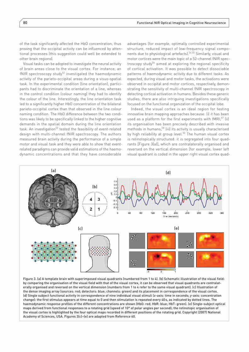
Figure 2. (a) A typical example of an fNIR spectroscopy probe holder placed on the head of a participant (bilateral parietal lobe). In this
set-up, thin optical fi bres (diameter 0.4 mm) convey near infrared light to the participant’s head (note that each location comprises
two optical fi bres, one for each wavelength), whereas optical fi bre bundles (diameter 3 mm) capture the light that is scattered through
the brain tissue. (b) to (e) Various fNIR spectroscopy instruments. (b) The ISS Imagent: http://www.iss.com/biomedical/instruments/
imagent.html; (c) the Hitachi ETG 4000: http://www.hitachi-medical-systems.eu/products-and-services/optical-topography/etg-4000.
html; (d) the Nirsoptix Brain Monitor: http://www.techen.com/index.cfm?event=brainmonitor and (e) the NIRScout: http://www.nirx.
net/imagers/nirscout-xtended.
78 Functional NIR Optical Imaging in Cognitive Neuroscience
Executive functionsExecutive functions include a broad category of cognitive
operations that drive complex behaviour, such as selection of
the appropriate action, inhibition of inappropriate actions or
irrelevant information, planning, confl ict resolution, cognitive
flexibility; the neural correlates of executive functions are
thought to be located in the PFC.17 One of the fi rst studies
that investigated the field of the executive functions with
fNIR spectroscopy18 aimed at detecting the small metabolic
changes in the frontal cortex when performing cognitive tasks,
as already observed in previous studies.11,13,16 The authors
adopted the continuous performance test (a repetitive task
where participants must respond to targets and inhibit the
response to non-targets) to examine the involvement of the
frontal brain regions with 2-channel fNIR spectroscopy. The
main result they observed was a difference in the course of
HbR, but not HbO, between the right and left hemisphere. In
contrast to left frontal areas, in which HbO and HbR increased
at the beginning and decreased at the end with the same
curve, right frontal areas showed a signifi cantly smaller initial
increase of HbR followed by a decrease on baseline level.
Another study19 introduced a motor control task within a
response inhibition paradigm, investigating brain oxygenation
of PFC with a 24-channel fNIR spectroscopy instrument. In
this experiment, participants were asked to perform a “go/
no-go” task in which the “no-go condition” was the experi-
mental condition and the “go condition” was the control condi-
tion. A signifi cantly higher increase in HbO in the most inferior
part of the PFC was found by analysis for the “no-go condi-
tion” in comparison with the “go condition”. In addition to
the go/no-go task,20 inhibitory processes21 also have been
investigated with fNIR spectroscopy by using the stop signal
task,22 which is widely adopted to assess motor inhibition
performance and is considered more demanding than a go/
no-go paradigm.23 A recent study24 investigated the involve-
ment of PFC in such a task. In some trials, participants were
asked to reconfi gure their (already initiated) response, thus
providing a different response from that already programmed.
The results were very interesting. First, the authors localised
the neural correlates of response inhibition in the inferior PFC;
second, they found a strong increase in the right PFC during
successful inhibition of already initiated responses, as well
as a bilateral increase of activity in PFC when the inhibition
was unsuccessful. These studies, together, bring evidence
in favour of the feasibility of functional fNIR spectroscopy to
measure the changes in cortical blood oxygenation due to the
inhibition of prepotent and/or already initiated responses.
Other trends in research confi rm the appropriateness of
fNIR spectroscopy to assess executive functions. The Stroop
effect25 is considered a prominent behavioral demonstration of
cognitive confl ict processing. In Stroop tasks, a stimulus with at
least two dimensions is presented. Participants are instructed
to give a behavioural response on the basis of the task-rele-
vant stimulus dimension and neglect the other dimension. In
the incongruent condition of Stroop tasks, in each trial the
task-relevant stimulus dimension prompts a certain behav-
ioural response while the task-irrelevant stimulus dimen-
sion prompts a different behavioural response. In contrast,
in the congruent condition, the task-relevant and task-irrel-
evant stimulus dimensions prompt the same behavioural
response. Interestingly, there has been a massive investiga-
tion of the Stroop effect with optical imaging (which has been
briefly reviewed in a recent work26). For instance, one27 of
those investigations showed how the interference during the
incongruent trials of a colour–word matching Stroop task
causes stronger bilateral brain activity in the lateral PFC. The
increase of HbO and the decrease of HbR were signifi cantly
higher in the incongruent condition with respect to the neutral
condition in the superior-lateral PFC. Another study28 that
investigated the frontal oxygenation in response to a Stroop
colour–word task was conducted utilising multi-channel fNIR
spectroscopy. In incongruent trials, a specifi c increase in HbO
and HbT in the left inferior-frontal brain areas was observed,
although the haemodynamic modulation of superior-frontal
areas due to Stroop interference was less pronounced in
comparison with previous results.27
Interestingly, a seminal fNIR spectroscopy study29
comparing young versus older people during Stroop task has
shown that the haemodynamic activity of the lateral PFC is
signifi cantly infl uenced by age. It is also worth mentioning
that some studies have focused on the Stroop task in clinical
populations such as patients with cerebral microangiopathy30
or children with attention defi cit disorder.31 Notably, fNIR
spectroscopy has been successfully used in clinical popula-
tions also with other paradigms, highlighting the potential
use of fNIR spectroscopy in the medical area. Patients that
are hard to test with other neuroimaging techniques, such
as impulsive children,32 patients with Parkinson’s disease or
patients with dementia33 have been successfully tested with
fNIR spectroscopy.
Several other experimental paradigms concerning executive
functions have been used with fNIR spectroscopy. For instance,
in a recent fNIR spectro scopy investigation,34 the PFC of the
participants was monitored using a computerised version of
the Trail Making Test; during the performance, the researchers
observed a bilateral frontal increase in HbO and a decrease
in HbR. In another optical imaging study,35 concentration
changes of HbO and HbR were monitored during the digit
span task using multi-channel fNIR spectroscopy. The digit
span task is widely used in neuro psychological research and
clinical evaluation: it involves remembering a set of numbers
in the standard order (digit span forward) or in the reverse
order (digit span backward). While in the digit span forward
no signifi cant difference was observed in the concentrations
of HbO during the performing phase compared to the resting
phase, the digit span backward performance caused a signifi -
cant increase in HbO with respect to the rest interval. Thus, the
digit span backward task required greater PFC activation than
the digit span forward. The results replicated those carried
out earlier with other imaging techniques36 and are consistent
with a previous fNIR spectroscopy study,37 which described an
S. Cutini, S. Basso Moro and S. Bisconti, J. Near Infrared Spectrosc. 20, 75–92 (2012) 79
activation of the right dorso-lateral prefrontal cortex (DLPFC)
when the digit span backward task was performed.
Another fNIR spectroscopy experiment38 aimed at isolating
the neural correlates of task-switching.39 In a typical task-
switching paradigm, subjects are instructed to repeat the
same task over a variable number of trials and switch to a
different task at some point during the trial sequence. Each
task often involves a rapid response and a reaction time is
usually recorded. The finding of interest is that the mean
reaction times in switch trials are longer than those in
repetition trials. Such a difference is attributed to the task-
set reconfi guration,39 which consists of enabling a different
response set and adjusting response criteria. There is no
substantial agreement about the neural correlates of task-
set reconfi guration: although most brain imaging studies on
task-switching found a co-activation of two key regions [the
DLPFC and the superior frontal gyrus (sFG)], an infl uential
fMRI study40 investigated the possible functional dissociation
between these two regions. According to this account, both
the DLPFC and sFG would be engaged while carrying out a
task-switching paradigm, but only the sFG would be determi-
nant for switching between tasks per se: whereas the DLPFC
would be primarily involved in the maintenance and coor-
dination of the different stimulus–response mapping rules
in working memory, the sFG would be more directly related
to reconfi guring the cognitive parameters demanded by the
execution of a specifi c task upon detection of a stimulus feature
associated with the task-switch. The authors performed a
sophisticated fMRI investigation, showing that these two
regions are interdependent but functionally separable. The
aforementioned fNIR spectroscopy experiment38 was aimed
at providing additional evidence with regard to the functional
dissociation of these two regions. In order to exclude poten-
tial confounds, the task-switching paradigm was designed
with an equal number of switch and repetition trials and with
an unpredictable occurrence of switch trials. The results of
the optical investigation indicated that both the DLPFC and
sFG were actively engaged during the execution of the task-
switching paradigm but, most importantly, only the activity of
the sFG was signifi cantly higher in switch trials with respect to
repetition trials.
EmotionsIn recent years, the neural correlates of emotion in
humans have been intensely investigated. The amygdala is
a sub cortical structure which is well known to be crucially
involved in emotion processing; nevertheless, several studies
investigated whether emotion might also be related to cortical
activity. For instance, an fMRI study revealed that the PFC is
involved in emotional regulation,41 showing a broad hemi-
spherical asymmetry between left and right PFC for positive
and negative emotions, respectively (for details see a recent
review42). An fNIR spectroscopy investigation43 used 2-channel
fNIR spectroscopy to measure the brain activity of the left and
right medial PFC, while participants were presented with a set
of pictures. The paradigm had two conditions in which the self-
monitoring requirement was manipulated: low self-monitoring
(i.e. just look at the emotional pictures presented) or high self-
monitoring (i.e. try to feel like the emotion expressed by the
stimulus presentation). The results showed that the assign-
ment type infl uenced the blood oxygenation of the frontal lobe:
when the task required more self-monitoring processes, there
were signifi cantly higher HbO concentrations in the frontal left
hemisphere, especially when highly emotional pictures were
viewed. This result suggests that self-monitoring processes
are tightly involved in certain aspects of emotional induc-
tion and might contribute to frontal activation. It should
also be noted that the frontal cortex is tightly involved in the
maintenance of attentional demands of a task44 and action
monitoring.45 The relationship between prefrontal activation
and processing of emotional stimuli was investigated in a
recent fNIR spectroscopy experiment,46 which revealed that
prefrontal activation is present both in emotion induction and
emotion regulation. Notably, the effect was located in the left
PFC and it was limited to HbR (with no HbO difference).
Another fNIR spectroscopy investigation47 examined the
infl uence of emotional content on the occipital cortex during
visual stimulation. Interestingly, emotionally positive and
negative stimuli produced a larger decrease of HbR in specifi c
areas of the occipital cortex in comparison with the decrease
induced by neutral stimuli. A recent fNIR spectroscopy study48
showed that the haemodynamic response peak latency of the
visual cortex is modulated by the emotional valence of the
stimuli. Specifi cally, the processing of positive pictures caused
reduced haemodynamic response peak latency with respect to
negative pictures. Besides the theoretical implications of these
results, it is worth noting that fNIR spectroscopy was able to
capture a temporal difference that might be hardly detectable
with fMRI, confirming that the higher temporal resolution
of fNIR spectroscopy can be effectively used under the right
circumstances and with the appropriate objectives. Another
recent fNIR spectroscopy study49 examined the modulation
of auditory cortex by emotional auditory stimuli, fi nding that
pleasant and unpleasant sounds produced stronger auditory
cortex activity as compared to neutral sounds; the results
suggest that human auditory cortex activity is modulated by
complex emotional sounds.
VisionThe neural correlates of vision have been extensively investi-
gated with fNIR spectroscopy since the fi rst optical imaging
experiments (see Introduction) and nowadays the activity of the
occipital lobe continues to be investigated in optical studies.50
A recent fNIR spectroscopy study of the visual cortex51 was
aimed at dissociating the task-related activity from the effect
of attentional load, in order to exclude a potential confound in
the interpretation of the results. Not surprisingly, the authors
found that the amount of attention paid during the execution
80 Functional NIR Optical Imaging in Cognitive Neuroscience
of the task signifi cantly affected the HbO concentration, thus
proving that the occipital activity can be infl uenced by atten-
tional processes (this suggestion could well be extended to
other brain regions).
Visual tasks can be adopted to investigate the neural activity
of brain areas close to the visual cortex. For instance, an
fNIR spectroscopy study52 investigated the haemodynamic
activity of the parieto-occipital areas during a visuo-spatial
task. In the experimental condition (line orientation), partici-
pants had to discriminate the orientation of a line, whereas
in the control condition (colour naming) they had to identify
the colour of the line. Interestingly, the line orientation task
led to a signifi cantly higher HbO concentration of the bilateral
parieto-occipital cortex than that observed in the line colour
naming condition. The HbO difference between the two condi-
tions was likely to be specifi cally linked to the higher cognitive
demands in the spatial domain during the line orientation
task. An investigation53 tested the feasibility of event-related
design with multi-channel fNIR spectroscopy. The authors
measured brain activity during the performance of a simple
motor and visual task and they were able to show that event-
related paradigms can provide valid estimations of the haemo-
dynamic concentrations and that they have considerable
advantages (for example, optimally controlled experimental
structure, reduced impact of low-frequency signal compo-
nents due to physiological artefacts).54,55 Similarly, visual and
motor cortices were the main topic of a 52-channel fNIR spec-
troscopy study56 aimed at exploring the regional specifi city
of cortical activation. It was possible to detect dissociable
patterns of haemodynamic activity due to different tasks. As
expected, during visual and motor tasks, the activations were
observed in occipital and motor cortices, respectively, demon-
strating the sensitivity of multi-channel fNIR spectroscopy in
detecting cortical activation in humans. Besides these generic
studies, there are also intriguing investigations specifi cally
focused on the functional organization of the occipital lobe.
Indeed, the visual cortex is an ideal region for testing
innovative brain mapping approaches because: (i) it has been
used as a platform for the fi rst experiments with fMRI;57 (ii)
its organisation has been precisely described with invasive
methods in humans;58 (iii) its activity is usually characterised
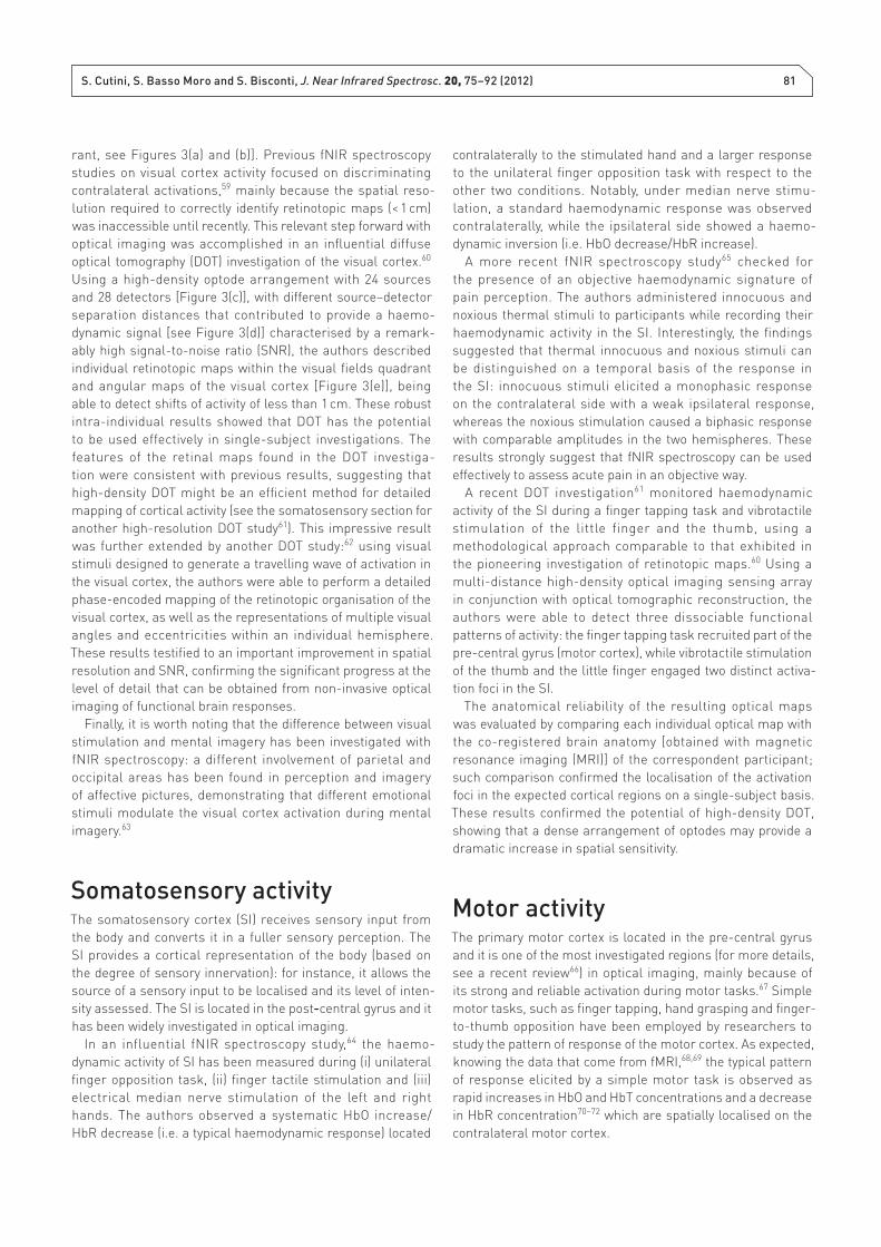
by high reliability at group level.50 The human visual cortex
is retinotopically structured: it is segregated into four quad-
rants [Figure 3(a)], which are contralaterally organised and
reversed on the vertical dimension [for example, lower left
visual quadrant is coded in the upper right visual cortex quad-
Figure 3. (a) A template brain with superimposed visual quadrants (numbered from 1 to 4). (b) Schematic illustration of the visual fi eld:
by comparing the organisation of the visual fi eld with that of the visual cortex, it can be observed that visual quadrants are contralat-
erally organised and reversed on the vertical dimension (numbers from 1 to 4 refer to the same visual quadrant). (c) Illustration of
the dense imaging array (sources: red; detectors: blue; channels: green) and its placement in correspondence of the visual cortex.
(d) Single subject functional activity in correspondence of nine individual visual stimuli (x-axis: time in seconds; y-axis: concentration
change): the fi rst stimulus appears at time equal to 0 and then stimulation is repeated every 40 s, as indicated by dotted lines. The
haemodynamic response profi les of the different concentrations are shown (HbO: red; HbR: blue; HbT: green). (e) Single-subject optical
maps derived from functional responses to a rotating grid (speed of 10° of polar angles per second); the retinotopic organisation of
the visual cortex is highlighted by the four optical maps recorded in different positions of the rotating grid. Copyright (2007) National
Academy of Sciences, USA. Figures 3(c)–(e) are adapted from Reference 60.
(a)
(b)
(c) (e)
(d)
S. Cutini, S. Basso Moro and S. Bisconti, J. Near Infrared Spectrosc. 20, 75–92 (2012) 81
rant, see Figures 3(a) and (b)]. Previous fNIR spectroscopy
studies on visual cortex activity focused on discriminating
contralateral activations,59 mainly because the spatial reso-
lution required to correctly identify retinotopic maps (< 1 cm)
was inaccessible until recently. This relevant step forward with
optical imaging was accomplished in an infl uential diffuse
optical tomography (DOT) investigation of the visual cortex.60
Using a high-density optode arrangement with 24 sources
and 28 detectors [Figure 3(c)], with different source–detector
separation distances that contributed to provide a haemo-
dynamic signal [see Figure 3(d)] characterised by a remark-
ably high signal-to-noise ratio (SNR), the authors described
individual retinotopic maps within the visual fi elds quadrant
and angular maps of the visual cortex [Figure 3(e)], being
able to detect shifts of activity of less than 1 cm. These robust
intra- individual results showed that DOT has the potential
to be used effectively in single-subject investigations. The
features of the retinal maps found in the DOT investiga-
tion were consistent with previous results, suggesting that
high-density DOT might be an effi cient method for detailed
mapping of cortical activity (see the somato sensory section for
another high-resolution DOT study61). This impressive result
was further extended by another DOT study:62 using visual
stimuli designed to generate a travelling wave of activation in
the visual cortex, the authors were able to perform a detailed
phase-encoded mapping of the retinotopic organisation of the
visual cortex, as well as the representations of multiple visual
angles and eccentricities within an individual hemisphere.
These results testifi ed to an important improvement in spatial
resolution and SNR, confi rming the signifi cant progress at the
level of detail that can be obtained from non-invasive optical
imaging of functional brain responses.
Finally, it is worth noting that the difference between visual
stimulation and mental imagery has been investigated with
fNIR spectroscopy: a different involvement of parietal and
occipital areas has been found in perception and imagery
of affective pictures, demonstrating that different emotional
stimuli modulate the visual cortex activation during mental
imagery.63
Somatosensory activityThe somatosensory cortex (SI) receives sensory input from
the body and converts it in a fuller sensory perception. The
SI provides a cortical representation of the body (based on
the degree of sensory innervation): for instance, it allows the
source of a sensory input to be localised and its level of inten-
sity assessed. The SI is located in the post-central gyrus and it
has been widely investigated in optical imaging.
In an influential fNIR spectroscopy study,64 the haemo-
dynamic activity of SI has been measured during (i) uni lateral
fi nger opposition task, (ii) fi nger tactile stimulation and (iii)
electrical median nerve stimulation of the left and right
hands. The authors observed a systematic HbO increase/
HbR decrease (i.e. a typical haemodynamic response) located
contralaterally to the stimulated hand and a larger response
to the unilateral fi nger opposition task with respect to the
other two conditions. Notably, under median nerve stimu-
lation, a standard haemodynamic response was observed
contralaterally, while the ipsilateral side showed a haemo-
dynamic inversion (i.e. HbO decrease/HbR increase).
A more recent fNIR spectroscopy study65 checked for
the presence of an objective haemodynamic signature of
pain perception. The authors administered innocuous and
noxious thermal stimuli to participants while recording their
haemodynamic activity in the SI. Interestingly, the fi ndings
suggested that thermal innocuous and noxious stimuli can
be distinguished on a temporal basis of the response in
the SI: innocuous stimuli elicited a monophasic response
on the contra lateral side with a weak ipsilateral response,
whereas the noxious stimulation caused a biphasic response
with comparable amplitudes in the two hemispheres. These
results strongly suggest that fNIR spectroscopy can be used
effectively to assess acute pain in an objective way.
A recent DOT investigation61 monitored haemodynamic
activity of the SI during a fi nger tapping task and vibrotactile
stimulation of the little finger and the thumb, using a
methodological approach comparable to that exhibited in
the pioneering investigation of retinotopic maps.60 Using a
multi-distance high-density optical imaging sensing array
in conjunction with optical tomographic reconstruction, the
authors were able to detect three dissociable functional
patterns of activity: the fi nger tapping task recruited part of the
pre-central gyrus (motor cortex), while vibrotactile stimulation
of the thumb and the little fi nger engaged two distinct activa-
tion foci in the SI.
The anatomical reliability of the resulting optical maps
was evaluated by comparing each individual optical map with
the co-registered brain anatomy [obtained with magnetic
resonance imaging (MRI)] of the correspondent participant;
such comparison confi rmed the localisation of the activation
foci in the expected cortical regions on a single-subject basis.
These results confi rmed the potential of high-density DOT,
showing that a dense arrangement of optodes may provide a
dramatic increase in spatial sensitivity.
Motor activityThe primary motor cortex is located in the pre-central gyrus
and it is one of the most investigated regions (for more details,
see a recent review66) in optical imaging, mainly because of
its strong and reliable activation during motor tasks.67 Simple
motor tasks, such as fi nger tapping, hand grasping and fi nger-
to-thumb opposition have been employed by researchers to
study the pattern of response of the motor cortex. As expected,
knowing the data that come from fMRI,68,69 the typical pattern
of response elicited by a simple motor task is observed as
rapid increases in HbO and HbT concentrations and a decrease
in HbR concentration70–72 which are spatially localised on the
contralateral motor cortex.
82 Functional NIR Optical Imaging in Cognitive Neuroscience
For instance, a recent fNIR spectroscopy study73 used a
fi nger tapping task to investigate the pattern and the time
course variations of brain activity across the task periods.
During haemodynamic recording of frontal, temporal and
parietal areas, participants were required to lean their thumb
sequentially (i.e. from the index fi nger to the little fi nger and
then in reversed order) on each finger as fast as possible
while respecting the sequence order. Generally, fi nger tapping
caused a higher haemodynamic response in the contralateral
hemisphere, with HbO increase in a broad area surrounding
the motor cortex and a HbR decrease in a more restricted
area. Furthermore, the authors attempted to perform a
functional segregation of the different brain regions based
also on the time course variation across the task: according
to them, it was possible to observe three partially overlapping
anatomical clusters related to execution, sensory monitoring
and maintenance of fi nger tapping.
Another fNIR spectroscopy study74 investigated the
influence of motor task complexity on the cortical activity.
Participants were asked to perform different fi nger tapping
tasks: unimanual simple and complex tasks and bimanual
tasks. Simple and complex right hand tasks caused the largest
HbO increase, followed by bimanual and left hand tasks, thus
showing a signifi cant task infl uence on motor cortex activity.
Note also that other optical investigations revealed that the
haemo dynamic response in the motor cortex is strongly
modulated by tapping frequency75 and intensity.76
Memory and attentionThe activation of the PFC in memory tasks has been widely
examined with fNIR spectroscopy. For instance, a typical pattern
characterised by an increase in HbO and a decrease in HbR was
observed during a word memory task.77 A sophisticated fNIR
spectroscopy study78 investigated the haemodynamic activity
during the execution of a visual n-back task. This paradigm
recruits both memory and selective attention: while observing
a sequence of stimuli, the participant has to indicate whether
the current stimulus matches the one presented from n steps
earlier in the sequence (task diffi culty can be manipulated by
changing the value of n). In this optical study, the activity of the
PFC was investigated while performing the aforementioned
n-back task, with sequentially presented task-relevant and
task-irrelevant faces. Participants were instructed to press
a response button whenever a presented face of the relevant
category (i.e. a white-bordered face) matched the previously
presented face of the same category (pointed out by the
black arrow above one picture). Relevant stimuli activated
the middle frontal/pre-central cortices bilaterally and the left
post-central cortex, probably in relation to a verbal rehearsal
strategy to maintain the features of the relevant stimuli.
Conversely, irrelevant stimuli activated superior, middle and
inferior parts of the right PFC, consistently with the selective
inhibition needed to properly perform the task. Thus, the
results indicated that the prefrontal activity during working
memory tasks refl ected processes of maintenance, selection
and inhibition of information, as well as attentional monitoring.
The PFC has also been studied in relation to information
encoding and retrieval, which are known to involve partially
overlapping neural circuits.79 The results of a recent fNIR
spectroscopy investigation on taste encoding and retrieval80
indicated that haemodynamic activity during taste retrieval
was signifi cantly stronger than that observed during encoding
in the bilateral frontopolar and dorsolateral prefrontal regions,
particularly in the right hemisphere. These results are broadly
consistent with the hemispheric encoding/retrieval asymmetry
theory.81 In parallel to memory, attention has been extensively
investigated with fNIR spectroscopy. An optical investigation82
measured the haemodynamic changes of the PFC during
a continuous performance test. Participants were asked to
react to infrequent letters appearing on a screen but not to
frequent distractors. An increase in HbO and HbR was found
in the DLPFC, but the lack of a suitable control task limited the
interpretation of the results. Although the neural substrates
of visuo-spatial attention have been widely investigated, it
is not clear whether the attentional resources required for
each visual hemifi eld are functionally separated between the
cerebral hemispheres. An fNIR spectroscopy investigation83
recorded brain activity during a visuo-spatial task. Interestingly,
an increase in attentional load produced a greater increase
in brain activity in the case of the left visual hemifi eld than
in the case of the right visual hemifield. This asymmetry
was observed in all the examined brain areas, including the
bilateral occipital and parietal cortices; the strongest activa-
tions were seen in posterior parietal cortex, including the
intraparietal sulcus. Those results have been interpreted as
an effect of the asymmetry of inhibitory interactions between
the hemispheres.
Interestingly, the same regions are involved in specific
memory tasks. Indeed, a recent fNIR spectroscopy study84
investigated the neural correlates of visual short-term
memory; previous neuro imaging studies attempting to isolate
the neural substrate of visual short-term memory in humans
have concentrated on the posterior parietal cortex; in partic-
ular, classical fMRI studies have revealed that the activity
recorded in posterior parietal regions increases with the
number of objects encoded in visual short-term memory,85
but only up to its capacity limits, levelling off thereafter. In
the optical investigation, the authors adopted a spatially cued
variant of the change–detection task to record haemodynamic
responses to unilaterally encoded objects; participants were
shown an arrow head pointing to the left or to the right side of
the screen. The offset of the arrow was followed by a memory
array, which was composed of either four or eight coloured
squares, evenly distributed to the left/right of fixation (i.e.
either two on each side, or four on each side). Participants
were instructed to maintain their gaze at fi xation and memo-
rise the colour of the squares on the side of the memory
array cued by the arrow head. Following the memory array
offset, participants were shown another array of coloured
squares in the same positions as occupied by the squares of
S. Cutini, S. Basso Moro and S. Bisconti, J. Near Infrared Spectrosc. 20, 75–92 (2012) 83
the memory array. Participants had to indicate (with button
press) whether a change in colour had occurred or not.
This task provides very different results for EEG and fMRI:
whereas electrophysiological data86 show that maintenance
of unilaterally encoded objects elicits a contralateral nega-
tive activity (proportional to the number of objects retained in
visual short-term memory), fMRI results indicate a modula-
tion of the BOLD response in relation to the number of memo-
rised elements in the parietal lobes of both hemispheres.87
Similarly to fMRI results, the optical imaging investigation
revealed a memory-related increase in HbO concentration
located bilaterally in the posterior parietal cortex, even though
objects had to be encoded uni laterally in the absence of eye
movements. Interestingly, the high similarity of such results
with those obtained with fMRI87 suggests that EEG and fMRI/
fNIR spectroscopy techniques might reveal partially distinct
neural signatures of the mechanisms supporting visual short-
term memory.
Language productionLanguage has been extensively investigated with optical
imaging, thus this section and the following one are meant to
provide general information on the use of fNIR spectroscopy
in the language fi eld (for a more extensive description of the
literature, see a recent review88). Many fNIR spectroscopy
studies are focused on the neural correlates of language during
the generation of words beginning with a certain letter, in the
letter version, or including the same category, in the semantic
version. An optical study89 investigated the effects of age and
gender on brain activation during a letter verbal fl uency task
(VFT). The results indicated an effect of age: elderly participants
showed less activity in the left DLPFC than younger ones and no
lateralisation was seen in these participants; on the other hand,
there was no effect of gender (although a previous study90 found
a higher frontal and temporal activity in males than in females).
In another study,91 the short- and long-term reliability of
brain activity was evaluated with a test–retest phonological
VFT. The participants were asked to repeat the same task
after 3 and 53 weeks from the fi rst and second data collec-
tion, respectively. In both sessions, increases in HbO and
decreases in HbR were observed in the inferior DLPFC and in
part of temporal cortex; nevertheless, the authors observed
a smaller haemodynamic response in the second session.
Analysis of the brain activation in the time course revealed an
adequate reliability of fNIR spectroscopy for repeated meas-
urements of the cortical activity at group level, even though
a weak retest reliability was found at single subject and
single channel levels. The fNIR spectroscopy was employed
in another study92 to investigate the lateralisation and the
functional connectivity of the frontal cortex in response to
a VFT and jaw movement. First, the results demonstrated a
signifi cant difference in the activation between VFT and jaw
movement in the PFC. Furthermore, a bilateral activation and
a symmetrical connectivity of PFC were present in both tasks,
although the laterality index calculated on the anterior frontal
cortex showed a left hemispheric predominance; likewise,
the connectivity analysis showed an asymmetrical neural
recruitment of the same region during the VFT.
Other fNIR spectroscopy studies used word generation tasks
in the determination of language hemispheric dominance
as an alternative to the invasive Wada test93–95 revealing the
fNIR spectroscopy potential in the presurgical exploration
of language. In order to investigate brain activity during a
language-related task, the authors of an fNIR spectroscopy
investigation96 compared a covert visual object naming task
(which required subjects to name pictures without oro-facial
movements) to two motor tasks: fi nger opposition and tongue
movement. Although the HbO increase was similar in the
three tasks, HbR changes were larger in the anterior inferior
frontal area during covert visual object naming tasks with
respect to tongue movement. Moreover, a greater decrease in
HbR in the posterior frontal regions during fi nger opposition
was observed in comparison to the other two tasks.
Overt picture naming has been used in an fNIR spectroscopy
experiment on bilinguals97 to investigate the brain mechanisms
that allow them to correctly use two languages without confu-
sion. The authors compared the use of signed and spoken
language in rapid alternation from bilingual to monolingual
mode during an overt picture naming task; although bilin-
guals showed an accurate performance in both conditions,
a greater activation of left posterior temporal regions was
observed when bilinguals executed the task in bilingual mode
than in monolingual mode, indicating a role of these areas in
the bilingual language switching ability. Effects of the use of
a second language were evaluated in an fNIR spectroscopy
study98 on sentence generation in native Dutch individuals
who were profi cient in English. The protocol required an overt
translation of visually presented sentences; the sentences
could have been presented in either of the two languages
separately or alternately. The involvement of the left frontal
cortex, which includes the Broca’s area, was evidenced by an
increase in HbO and a smaller decrease in HbR in all conditions
although no differences were detected when subjects trans-
lated the sentences separately or in alternation.
Language representation has been investigated by fNIR
spectroscopy during speech production to understand brain
activity during social interactions;99 the authors monitored
an ordinary face-to-face conversation, assuming that autistic
traits in typically developed participants were correlated to the
amplitude of haemodynamic signals. Interestingly, the results
indicated a negative correlation between autistic traits and the
activity of the left superior temporal sulcus region of males
(but no correlation was found for the PFC).
Language comprehensionLanguage processing has been investigated in a fNIR
spectroscopy study100 on syntactic and semantic decision
tasks. Participants were asked to identify the presence of
84 Functional NIR Optical Imaging in Cognitive Neuroscience
semantic or syntactic errors contained in sentences. Although
similar performances or reaction times were performed in the
two conditions, the left inferior gyrus was more activated in the
syntactic processing in contrast to the semantic processing.
Language comprehension has been studied through the
discrimination of phonems, words or speeches in many fNIR
spectroscopy studies. In a recent investigation,101 the implicit
processing of phonotactic cues was studied by co-registering
fNIR spectroscopy and EEG. Monosyllabic pseudo-words,
that were defined as phonotactically legals when the first
consonant was equal to the onset of a German word, or ille-
gals when it was not with respect to German, were presented
to native German speakers. The hypothesis associated with
the task was that pre-lexical cues processing in the spoken
word recognition might modulate the lexical activation
processes in the auditory speech. The fNIR spectroscopy
results demonstrated a signifi cant HbR decrease in the left
fronto-temporal hemisphere for phonotactically legal pseudo-
words with respect to that observed for illegal pseudo-words.
These fi ndings were confi rmed by a larger response for phono-
tactically legal pseudo-words in EEG parameters, providing
evidence that processing of legal phonotactic components
contributes to language comprehension.
In an optical study on bilingualism,102 Japanese long- and
short-vowel contrasts were investigated in native Japanese
speakers and in Korean subjects who spoke fl uent Japanese
as a second language. The results showed different cortical
patterns of response in the two groups. Independently, by
short- or long-vowel contrast, native Japanese speakers
demonstrated a greater left activation in correspondence
of Wernicke’s area. On the other hand, fNIR spectroscopy
results did not show a different neural recruitment or left
pre dominance when phonemic differences were presented
in speakers learning Japanese, despite behavioural results
indicated that these participants were able to discriminate
between long/short vowels. These fi ndings suggested that, on
some occasions, behavioural performance is not representa-
tive of the underlying haemodynamic activity.
Resting state functional
connectivity
The study of resting state functional connectivity deserves to
be briefl y mentioned because of its potential. Resting state
functional connectivity investigates the spontaneous cerebral
haemodynamic fluctuations that reflect neuronal activity
at rest. A recent study103 investigated the spatio–temporal
correlation of haemoglobin concentration signals over the
whole head during resting state. Similar to previous fMRI
investigations,104 the authors found consistent regional inter-
actions across participants. In particular, sensorimotor and
visual cortices exhibited a strong interhemispheric corre-
lation, while the PFC was subjected to higher variability.
Importantly, the high definition of the maps obtained with
HbT suggests that it could be useful to localise with preci-
sion the functional connectivity. Taken together, these fi nd-
ings confi rm that resting state functional connectivity can be
effectively investigated with whole head fNIR spectroscopy
recording. Another recent optical investigation105 verifi ed the
test–retest reliability of fNIR spectroscopy-based resting state
functional connectivity in sensorimotor regions. Brain activity
of participants was recorded during two resting state sessions
separated by one week and independent component analysis
was used to extract resting state functional connectivity in the
somatosensory areas. The authors found an excellent reli-
ability at group level, a reasonable reliability at individual level
but a weaker reliability at a channel-wise level. Although these
results confi rm the high reliability of the fNIR spectroscopy-
based resting state measurement, the authors conclude that
a stringent probe placement method is required to increase
channel-wise reliability in fNIR spectroscopy recording.
Some probe placement methods106–108 can provide consistent
positioning of optodes across participants and/or sessions
without the need of individual MRI scans. For instance, a
method107 designed for DOT measurements allows the focus
of cortical activity to be reconstructed with reasonable preci-
sion. Another probe placement method106 is based on the
use of a physical model of the ICBM152 template (i.e. the
current standard brain template in fMRI, which was obtained
by averaging the MRI scans of 152 subjects) head surface as
a reference scalp, and its validity is supported by previous
investigations of cranio-cerebral correlation.109 Notably, the
latter approach offers a reproducible and straightforward
method to place the optodes on the head surface, according
to the stereotaxic coordinates of the regions of interest. This
aspect is critical, because it ensures a ready comparison of
fNIR spectroscopy results within the neuroimaging commu-
nity, both across studies and techniques. Note that the use
of those methods should be strongly encouraged within the
community of fNIR spectroscopy users, since almost all types
of fNIR spectroscopy investigations might benefi t greatly from
the adoption of a rigorous probe placement method.
Brain–computer interfaceThe brain–computer interface (BCI) is a research fi eld that
might dramatically benefi t from the use of fNIR spectroscopy
technology in the coming years. BCI consists of a system that
allows a direct link between the brain and an external device.
The main objective of BCI is to assist or substitute cognitive
or motor functions in humans. BCI can represent an effective
way to communicate and interact with the environment for the
disabled, such as patients suffering from amyotrophic lateral
sclerosis, brainstem stroke and spinal cord injury.110
Although one of the main obstacles to a proficient use
of fNIR spectroscopy in BCI is related to signal processing
diffi culties, it is encouraging to note that there are already
some valuable publications in this fi eld. For instance, a recent
work111 proposed an fNIR spectroscopy-BCI paradigm based
S. Cutini, S. Basso Moro and S. Bisconti, J. Near Infrared Spectrosc. 20, 75–92 (2012) 85
on the decoding of subjective preference (i.e. the preferred
drink). Nine participants were asked to mentally evaluate
two possible drinks and decide which they preferred. The
fNIR spectroscopy was used to monitor the PFC during the
task; using mean linear discriminant analysis with signal
amplitudes as input features, the accuracy in guessing the
preferred drink on a single-trial basis was well above the
chance level (around 80%).
Another study,110 conducted to test the feasibility of using
multichannel NIR spectroscopy in the development of a
reliable BCI, provided excellent results. The motor cortex of
fi ve healthy participants was monitored during left-hand and
right-hand motor imagery. The results indicated that there
were two distinct patterns of haemodynamic responses which
could be used in a pattern classifi er oriented to the realisation
of a BCI. Hidden Markov models classifi ed left-hand imagery
and right-hand imagery with an average accuracy of 89%.
These impressive results confi rmed the great potential of fNIR
spectroscopy in BCI applications.
Co-registration studiesA promising use of fNIR spectroscopy concerns co- registration
studies: fNIR spectroscopy signals can be easily recorded
in combination with other imaging techniques to provide
simultaneous information, as with the aforementioned
investigation47 on emotions. A recent fNIR spectroscopy–fMRI
combined study112 compared optical and BOLD responses
in frontal and parietal brain regions across a wide variety of
cognitive tasks. An extensive comparison of the two signals
across spatial and temporal domains revealed a high corre-
lation between optical and BOLD signals, although optical
signals have signifi cantly lower SNR; indeed, both cortical
depth (i.e. the distance between the scalp and the cerebral
cortex) and SNR infl uenced the strength of correlation between
the two signals. The authors concluded that fNIR spectroscopy
can be used effectively in cognitive neuroscience, provided
that its weaknesses are taken into account when designing
the experiment. This important investigation confirms the
validity of fNIR spectroscopy as an instrument to investigate
the neural correlates of cognitive processing.
As a further example, another fNIR spectroscopy–EEG
combined study113 investigated the cortical correlates of audi-
tory sensory gating using a dual-click paradigm; typically,
when two identical auditory stimuli are separated by a short
temporal interval, the response to the second stimulus is
reduced. In this study, participants listened to two different
types of stimulation: a one-click condition (two clicks with no
temporal delay) and a dual-click condition (two clicks sepa-
rated by 500 ms). The fNIR spectroscopy results showed an
increased activity of prefrontal and temporo-parietal cortices
during increased sensory gating (dual-click condition),
whereas the EEG results indicated that the P50 component
(the electrophysiological response to an auditory stimulus) was
signifi cantly reduced after the second stimulus as compared
to the fi rst one. Furthermore, haemo dynamic activity in the
left prefrontal and temporal cortices was positively correlated
with the strength of sensory gating (measured by means of
an electrophysiological index), suggesting that the PFC might
actively inhibit the activity of the primary auditory cortex. Two
other remarkable fNIR spectroscopy–EEG combined studies
reported a strong connection between electrophysiological
signal and haemodynamic activity: the recent investigation
by Koch et al.114 revealed that EEG-measured gamma activity
(~40 Hz) induced by stimulation or attentive state was strongly
related to the haemodynamic response in visual cortex; their
earlier fNIR spectroscopy–EEG combined study115 showed that
the individual alpha-frequency value at rest was predictive of
the amplitude of both visually evoked potential and haemo-
dynamic response in the visual cortex.
Other research topicsOptical imaging has also been used to investigate the neural
correlates of numerical cognition: during the execution
of arithmetical tasks, haemodynamic activity increases in
superior and inferior parietal areas, as pointed out by many
fMRI studies.116,117 While some fNIR spectroscopy studies on
arithmetical tasks revealed the involvement of the prefrontal
regions,16,118 a recent fNIR spectroscopy investigation119
confi rmed the fMRI fi ndings, suggesting that both left and
right parietal lobes play a crucial role in the processing and
solving of arithmetic problems.
A recent optical investigation on dental phobia120 can be
regarded as proof of the fact that fNIR spectroscopy can be
effectively adopted in environmental conditions that might be
diffi cult to assess with fMRI. The study included two groups:
one group of patients with dental phobia and a control group
(healthy participants). The authors presented both groups
with three different sounds: a dental drill (a sound feared by
patients with dental phobia), a pleasant and a neutral sound.
When listening to the dental drill, phobic patients exhibited
increased activity in the supplementary motor area (probably
refl ecting a covert response of fl ight behaviour) with respect to
controls, whereas there was no signifi cant difference between
groups for the other sounds.
Another intriguing use of fNIR spectroscopy concerns the
hyperscanning (i.e. simultaneous brain activity recording
of more than one participant). A recent study121 adopted
fNIR spectroscopy hyperscanning to measure brain activity
coherence between two participants during a social game
(with two main conditions: cooperation and competition).
The study provided two interesting results: fi rst, inter-brain
coherence increased in the right superior frontal cortex
only during cooperation (but not in competition); second,
higher performance during cooperation was associated
with increased inter-brain coherence. Notably, fNIR spec-
troscopy can be used in hyperscanning with no particular
disadvantages, while hyperscanning with fMRI is expensive
and technically challenging.
86 Functional NIR Optical Imaging in Cognitive Neuroscience
Finally, fNIR spectroscopy can also be used effectively in
imaging genetics, a discipline that investigates the impact of
genes on brain functioning (by conjunctively using neuroimaging
and genotyping techniques). For instance, a recent fNIR spec-
troscopy investigation122 revealed that a gene variant of cycloox-
ygenase-1 infl uences the neurovascular coupling in the visual
cortex: the authors found that the cyclooxygenase-1 genotype-
dependent enzymatic function was associated with a strongly
reduced amplitude of the haemodynamic response. As well
as the implications of this study, it should be noted that the
advantage of using fNIR spectroscopy in imaging genetics is its
low-cost application (a large sample size of subjects is vital for
imaging genetics studies).
ConclusionsFunctional near infrared optical imaging has some relevant
disadvantages compared with other neuroimaging techniques:
for instance, it is blind to subcortical activity and anatomical
information must be obtained with other techniques or inferred
with specific methods, while fMRI can measure haemo-
dynamic activity of the entire brain and detailed anatomical
information can be obtained within the same session of func-
tional recording with MRI.
On the other hand, fNIR spectroscopy has also some
undeniable advantages: unlike fMRI, it can be used with no
problem on infants; similarly, it does not produce instru-
mental noise, allowing the execution of linguistic tasks that
require subtle acoustic features of words or conversations to
be distinguished.123 Furthermore, fNIR spectroscopy has been
promoted in a number of fi elds in which fMRI is limited due
to the constraints induced by the scanning environment. In
addition, the experimental measurements may be recorded
in a more comfortable and natural environment,124 providing
a marked advantage when testing groups of clinical popula-
tions (which might not be conveniently examined with other
techniques).
Nevertheless, even without taking into consideration such
advantages, it appears clear that fNIR spectroscopy is often
used even in those research fi elds where it does not imme-
diately provide an apparent advantage over other techniques.
Indeed, the heterogeneity of the researches mentioned in the
present review testifi es to the fact that fNIR spectroscopy can
be successfully applied in almost all cognitive neuroscience
issues. In some cases, it can provide converging evidence in
regard to a specifi c cognitive theory, by confi rming previous
results obtained with different brain imaging techniques. Most
importantly, in some other cases, the information obtained
with fNIR spectroscopy can provide novel fi ndings that could
hardly be obtained with other brain imaging techniques. Thus,
the use of fNIR spectroscopy in cognitive neuroscience should
be directed towards those topics that allow a full capitalisation
of the peculiar characteristics of optical imaging (for example,
the simultaneous recording of HbO, HbR and HbT with high
temporal resolution).
Finally, it is evident that functional near infrared optical
imaging is constantly evolving from a technical point of view:
the main proof of such progress is the fact that optical data are
beginning to become meaningful at a single subject level,60
although further development is needed to allow reliable single
case studies in most of the research fi elds. Nevertheless, the
high SNR and the spatial resolution that can be achieved
with recent optical imaging methods,60,61 the successful test–
retest assessments of reliability50,105 and the advanced probe
placement techniques106,107 should be viewed as encouraging
elements that might be able to trigger the progress in single
subject studies in the next few years. When considering the
additional advantages of fNIR spectroscopy in testing clinical
populations, it is reasonable to hypothesise that the biomed-
ical fi eld might radically benefi t from the progress of fNIR
spectroscopy towards its applicability in single case studies of
clinical patients.
AcknowledgementsThe authors would like to thank Professor M. Ferrari for his
helpful suggestions.
References1. M.S. Gazzaniga, R.B. Ivry and G.R. Mangun, Cognitive
Neuroscience: The Biology of the Mind. W.W. Norton &
Company, Inc., New York, USA (2009).
2. S.E. Petersen, P.T. Fox, M.I. Posner, M. Mintun and M.E.
Raichle, “Positron emission tomographic studies of the
cortical anatomy of single-word processing”, Nature
331, 585 (1988). doi: 10.1038/331585a0
3. J. Berwick, D. Johnston, M. Jones, J. Martindale,
P. Redgrave, N. McLoughlin, I. Schiessl and J.E.W.
Mayhew, “Neurovascular coupling investigated with
two-dimensional optical imaging spectroscopy in rat
whisker barrel cortex”, Eur. J. Neurosci. 22, 1655 (2005).
doi: 10.1111/j.1460-9568.2005.04347.x
4. J. Berwick, D. Johnston, M. Jones, J. Martindale,
C. Martin, A.J. Kennerley, P. Redgrave and J.E.W.
Mayhew, “Fine detail of neurovascular coupling
revealed by spatiotemporal analysis of the hemody-
namic response to single whisker stimulation in rat
barrel cortex”, J. Neurophysiol. 99, 787 (2008). doi:
10.1152/jn.00658.2007
5. A. Villringer and U. Dirnagl, “Coupling of brain
activity and cerebral blood flow: Basis of functional
neuro imaging”, Cerebrovasc. Brain Metab. Rev. 7, 240
(1995).
6. P.A. Bandettini, A. Jesmanowicz, E.C. Wong and
J.S. Hyde, “Processing strategies for time-course
data sets in functional MRI of the human brain”,
Magn. Reson. Med. 30, 161 (1993). doi: 10.1002/
mrm.1910300204
S. Cutini, S. Basso Moro and S. Bisconti, J. Near Infrared Spectrosc. 20, 75–92 (2012) 87
7. S. Ogawa, D.W. Tank, R. Menon, J.M. Ellermann,
S.G. Kim, H. Merkle and K. Ugurbil, “Intrinsic signal
changes accompanying sensory stimulation: functional
brain mapping with magnetic resonance imaging”,
Proc. Natl. Acad. Sci. USA 89, 5951 (1992). doi: 10.1073/
pnas.89.13.5951
8. N.K. Logothetis, J. Pauls, M. Augath, T. Trinath and A.
Oeltermann, “Neurophysiological investigation of the
basis of the fMRI signal”, Nature 412, 150 (2001). doi:
10.1038/35084005
9. C.E. Elwell and C.E. Cooper, “Making light work:
Illuminating the future of biomedical optics”, Philos.
Transact. Roy. Soc. A Math. Phys. Eng. Sci. 369, 4358
(2011). doi: 10.1098/rsta.2011.0302
10. F. Jobsis, “Noninvasive, infrared monitoring of cerebral
and myocardial oxygen suffi ciency and circulatory
parameters”, Science 198, 1264 (1977). doi: 10.1126/sci-
ence.929199
11. A. Villringer, J. Planck, C. Hock, L. Schleinkofer and
U. Dirnagl, “Near infrared spectroscopy (NIRS):
A new tool to study hemodynamic changes dur-
ing activation of brain function in human adults”,
Neurosci. Lett. 154, 101 (1993). doi: 10.1016/0304-
3940(93)90181-J
12. B. Chance, Z. Zhuang, C. Unah, C. Alter and L. Lipton,
“Cognition-activated low-frequency modulation of light
absorption in human brain”, Proc. Natl. Acad. Sci. USA
90, 3770 (1993). doi: 10.1073/pnas.90.8.3770
13. Y. Hoshi and M. Tamura, “Dynamic multichannel near-
infrared optical imaging of human brain activity”, J. Appl.
Physiol. 75, 1842 (1993).
14. T. Kato, A. Kamei, S. Takashima and T. Ozaki, “Human
visual cortical function during photic stimulation
monitoring by means of near-infrared spectroscopy”,
J. Cereb. Blood Flow Metab. 13, 516 (1993). doi: 10.1038/
jcbfm.1993.66
15. Y. Hoshi, H. Onoe, Y. Watanabe, J. Andersson, M.
Bergstrom, A. Lilja, B. Langstrom and M. Tamura,
“Non-synchronous behavior of neuronal activity, oxi-
dative metabolism and blood supply during mental
tasks in man”, Neurosci. Lett. 172, 129 (1994). doi:
10.1016/0304-3940(94)90679-3
16. Y. Hoshi and M. Tamura, “Near-infrared optical detec-
tion of sequential brain activation in the prefrontal cor-
tex during mental tasks”, NeuroImage 5, 292 (1997). doi:
10.1006/nimg.1997.0270
17. E.K. Miller and J.D. Cohen, “An integrative theory of
prefrontal cortex function”, Annu. Rev. Neurosci. 24, 167
(2001). doi: 10.1146/annurev.neuro.24.1.167
18. A.J. Fallgatter and W.K. Strik, “Frontal brain activation
during the Wisconsin Card Sorting Test assessed with
two-channel near-infrared spectroscopy”, Eur. Arch.
Psychiatry Clin. Neurosci. 248, 245 (1998). doi: 10.1007/
s004060050045
19. M.J. Herrmann, M.M. Plichta, A.-C. Ehlis and A.J.
Fallgatter, “Optical topography during a Go-NoGo task
assessed with multi-channel near-infrared spectros-
copy”, Behav. Brain Res. 160, 135 (2005). doi: 10.1016/j.
bbr.2004.11.032
20. E.A. Drewe, “Go–no go learning after frontal lobe
lesions in humans”, Cortex 11, 8 (1975).
21. G.D. Logan, “On the ability to inhibit thought and action:
A users’ guide to the stop signal paradigm”, in Inhibitory
Processes in Attention Memory and Language, Ed by D.
Dagenbach and T.H. Carr. Academic Press, New York,
USA, p. 189 (1994).
22. S. Konishi, K. Nakajima, I. Uchida, K. Sekihara and Y.
Miyashita, “No-go dominant brain activity in human
inferior prefrontal cortex revealed by functional mag-
netic resonance imaging”, Eur. J. Neurosci. 10, 1209
(1998). doi: 10.1046/j.1460-9568.1998.00167.x
23. K. Rubia, T. Russell, S. Overmeyer, M.J. Brammer,
E.T. Bullmore, T. Sharma, A. Simmons, S.C. Williams,
V. Giampietro, C.M. Andrew and E. Taylor, “Mapping
motor inhibition: Conjunctive brain activations
across different versions of go/no-go and stop
tasks”, NeuroImage 13, 250 (2001). doi: 10.1006/
nimg.2000.0685
24. M. Boecker, M.M. Buecheler, M.L. Schroeter and S.
Gauggel, “Prefrontal brain activation during stop-signal
response inhibition: an event-related functional near-
infrared spectroscopy study”, Behav. Brain Res. 176, 259
(2007). doi: 10.1016/j.bbr.2006.10.009
25. B.S. Peterson, M.J. Kane, G.M. Alexander, C. Lacadie,
P. Skudlarski, H.C. Leung, J. May and J.C. Gore, “An
event-related functional MRI study comparing inter-
ference effects in the Simon and Stroop tasks”, Brain
Res. Cogn. Brain Res. 13, 427 (2002). doi: 10.1016/S0926-
6410(02)00054-X
26. K. Ciftçi, B. Sankur, Y.P. Kahya and A. Akin, “Multilevel
statistical inference from functional near-infrared
spectroscopy data during Stroop interference”, IEEE
Trans. Biomed. Eng. 55, 2212 (2008). doi: 10.1109/
TBME.2008.923918
27. M.L. Schroeter, S. Zysset, T. Kupka, F. Kruggel and D.
Yves von Cramon, “Near-infrared spectroscopy can
detect brain activity during a color-word matching
Stroop task in an event-related design”, Hum. Brain Map.
17, 61 (2002). doi: 10.1002/hbm.10052
28. A.-C. Ehlis, M.J. Herrmann, A. Wagener and A.J.
Fallgatter, “Multi-channel near-infrared spectroscopy
detects specifi c inferior-frontal activation during incon-
gruent Stroop trials”, Biol. Psychol. 69, 315 (2005). doi:
10.1016/j.biopsycho.2004.09.003
29. M.L. Schroeter, S. Zysset, F. Kruggel and D. Yves
von Cramon, “Age dependency of the hemodynamic
response as measured by functional near-infrared
spectroscopy”, NeuroImage 19, 555 (2003). doi: 10.1016/
S1053-8119(03)00155-1
30. M.L. Schroeter, S. Cutini, M.M. Wahl, R. Scheid and D.
Yves von Cramon, “Neurovascular coupling is impaired
in cerebral microangiopathy—An event-related Stroop
88 Functional NIR Optical Imaging in Cognitive Neuroscience
study”, NeuroImage 34, 26 (2007). doi: 10.1016/j.neuro-
image.2006.09.001
31. S. Jourdan Moser, S. Cutini, P. Weber and M.L.
Schroeter, “Right prefrontal brain activation due to
Stroop interference is altered in attention-defi cit
hyperactivity disorder—A functional near-infrared
spectroscopy study”, Psychiatry Res. 173, 190 (2009). doi:
10.1016/j.pscychresns.2008.10.003
32. M. Schecklmann, M. Romanos, F. Bretscher, M.M.
Plichta, A. Warnke and A.J. Fallgatter, “Prefrontal
oxygenation during working memory in ADHD”, J.
Psychiatry Res. 44, 621 (2010).
33. M.M. Richter, M.J. Herrmann, A.-C. Ehlis, M.M. Plichta
and A.J. Fallgatter, “Brain activation in elderly people
with and without dementia: Infl uences of gender and
medication”, World J. Biol. Psychiatry 8, 23 (2007). doi:
10.1080/15622970600960132
34. M. Kubo, C. Shoshi, T. Kitawaki, R. Takemoto, K.
Kinugasa, H. Yoshida, C. Honda and M. Okamoto,
“Increase in prefrontal cortex blood fl ow during the
computer version trail making test”, Neuropsychobiology
58, 200 (2008). doi: 10.1159/000201717
35. H. Kaneko, T. Yoshikawa, K. Nomura, H. Ito, H.
Yamauchi, M. Ogura and S. Honjo, “Hemodynamic
changes in the prefrontal cortex during digit
span task: A near-infrared spectroscopy
study”, Neuropsychobiology 63, 59 (2011). doi:
10.1159/000323446
36. B.K. Gerton, T.T. Brown, A. Meyer-Lindenberg, P. Kohn,
J.L. Holt, R.K. Olsen and K.F. Berman, “Shared and
distinct neurophysiological components of the digits
forward and backward tasks as revealed by functional
neuroimaging”, Neuropsychologia 42, 1781 (2004). doi:
10.1016/j.neuropsychologia.2004.04.023
37. Y. Hoshi, I. Oda, Y. Wada, Y. Ito, Y. Yamashita, M.Oda, K.
Ohta, Y. Yamada and M. Tamura, “Visuospatial imagery
is a fruitful strategy for the digit span backward task:
A study with near-infrared optical tomography”, Brain
Res. Cogn. Brain Res. 9, 339 (2000). doi: 10.1016/S0926-
6410(00)00006-9
38. S. Cutini, P. Scatturin, E. Menon, P.S. Bisiacchi, L.
Gamberini, M. Zorzi and R. Dell’Acqua, “Selective acti-
vation of the superior frontal gyrus in task-switching:
An event-related fNIRS study”, NeuroImage 42, 945
(2008). doi: 10.1016/j.neuroimage.2008.05.013
39. S. Monsell, “Task switching”, Trends Cogn. Sci. 7, 134
(2003). doi: 10.1016/S1364-6613(03)00028-7
40. E.A. Crone, C. Wendelken, S.E. Donohue and S.A.
Bunge, “Neural evidence for dissociable components
of task-switching”, Cereb. Cortex 16, 475 (2006). doi:
10.1093/cercor/bhi127
41. R.J. Davidson and W. Irwin, “The functional neuro-
anatomy of emotion and affective style”, Trends Cogn.
Sci. 3, 11 (1999). doi: 10.1016/S1364-6613(98)01265-0
42. F. Dolcos, A.D. Iordan and S. Dolcos, “Neural correlates
of emotion–cognition interactions: A review of evidence
from brain imaging investigations”, J. Cogn. Psychol. 23,
669 (2011). doi: 10.1080/20445911.2011.594433
43. M.J. Herrmann, A.-C. Ehlis and A.J. Fallgatter,
“Prefrontal activation through task requirements of
emotional induction measured with NIRS”, Biol. Psychol.
64, 255 (2003). doi: 10.1016/S0301-0511(03)00095-4
44. A.W. MacDonald, J.D. Cohen, V.A. Stenger and C.S.
Carter, “Dissociating the role of the dorsolateral
prefrontal and anterior cingulate cortex in cognitive
control”, Science 288, 1835 (2000). doi: 10.1126/sci-
ence.288.5472.1835
45. P. Luu, T. Flaisch and D.M. Tucker, “Medial frontal cor-
tex in action monitoring”, J. Neurosci. 20, 464 (2000).
46. E. Glotzbach, A. Mühlberger, K. Gschwendtner, A.J.
Fallgatter, P. Pauli and M.J. Herrmann, “Prefrontal
brain activation during emotional processing:
A functional near infrared spectroscopy study
(fNIRS)”, Open Neuroimag. J. 5, 33 (2011). doi:
10.2174/1874440001105010033
47. M.J. Herrmann, T. Huter, M.M. Plichta, A.-C. Ehlis,
G.W. Alpers, A. Mühlberger and A.J. Fallgatter,
“Enhancement of activity of the primary visual cortex
during processing of emotional stimuli as measured
with event-related functional near-infrared spectros-
copy and event-related potentials”, Hum. Brain Map. 29,
28 (2008). doi: 10.1002/hbm.20368
48. L. Minati, C.L. Jones, M.A. Gray, N. Medford, N.A.
Harrison and H.D. Critchley, “Emotional modulation
of visual cortex activity: A functional near-infrared
spectroscopy study”, Neuroreport 20, 1344 (2009). doi:
10.1097/WNR.0b013e328330c751
49. M.M. Plichta, A.B.M. Gerdes, G.W. Alpers, W. Harnisch,
S. Brill, M.J. Wieser and A.J. Fallgatter, “Auditory
cortex activation is modulated by emotion: A func-
tional near-infrared spectroscopy (fNIRS) study”,
NeuroImage 55, 1200 (2011). doi: 10.1016/j.neuroim-
age.2011.01.011
50. M.M. Plichta, M.J. Herrmann, C.G. Baehne, A.-C. Ehlis,
M.M. Richter, P. Pauli and J. Fallgatter, “Event-related
functional near-infrared spectroscopy (fNIRS): Are the
measurements reliable?”, NeuroImage 31, 116 (2006).
doi: 10.1016/j.neuroimage.2005.12.008
51. H. Kojima and T. Suzuki, “Hemodynamic change in
occipital lobe during visual search: Visual attention
allocation measured with NIRS”, Neuropsychologia
48, 349 (2010). doi: 10.1016/j.neuropsycholo-
gia.2009.09.028
52. M.J. Herrmann, A.-C. Ehlis, A. Wagener, C.P. Jacob and
A.J. Fallgatter, “Near-infrared optical topography to
assess activation of the parietal cortex during a visuo-
spatial task”, Neuropsychologia 43, 1713 (2005). doi:
10.1016/j.neuropsychologia.2005.02.011
53. G. Jasdzewski, G. Strangman, J. Wagner, K.K. Kwong,
R.A. Poldrack and D.A. Boas, “Differences in the
hemodynamic response to event-related motor and
visual paradigms as measured by near-infrared
S. Cutini, S. Basso Moro and S. Bisconti, J. Near Infrared Spectrosc. 20, 75–92 (2012) 89
spectroscopy”, NeuroImage 20, 479 (2003). doi: 10.1016/
S1053-8119(03)00311-2
54. M.A. Franceschini, V. Toronov, M. Filiaci, E. Gratton and
S. Fantini, “On-line optical imaging of the human brain
with 160-ms temporal resolution”, Opt. Express 6, 49
(2000). doi: 10.1364/OE.6.000049
55. M.A. Franceschini, D.K. Joseph, T.J. Huppert, S.G.
Diamond and D.A. Boas, “Diffuse optical imaging of
the whole head”, J. Biomed. Opt. 11, 054007 (2006). doi:
10.1117/1.2363365
56. M.M. Plichta, M.J. Herrmann, A.-C. Ehlis, C.G. Baehne,
M.M. Richter and A.J. Fallgatter, “Event-related visual
versus blocked motor task: detection of specifi c corti-
cal activation patterns with functional near-infrared
spectroscopy”, Neuropsychobiology 53, 77 (2006). doi:
10.1159/000091723
57. S. Engel, G.H. Glover and B.A. Wandell, “Retinotopic
organization in human visual cortex and the spatial pre-
cision of functional MRI”, Cereb. Cortex 7, 181 (1997). doi:
10.1093/cercor/7.2.181
58. G.F.A. Harding, C. Degg, S.J. Anderson, I. Holliday, F.
Fylan, G. Barnes and J. Bedford, “Topographic map-
ping of the pattern onset evoked magnetic response
to stimulation of different portions of the visual fi eld”,
Int. J. Psychophysiol. 16, 175 (1994). doi: 10.1016/0167-
8760(89)90044-5
59. W. Colier, V. Quaresima, R. Wenzel, M.C. van der Sluijs,
B. Oeseburg, M. Ferrari and A. Villringer, “Simultaneous
near-infrared spectroscopy monitoring of left and right
occipital areas reveals contra-lateral hemodynamic
changes upon hemi-fi eld paradigm”, Vision Res. 41, 97
(2001). doi: 10.1016/S0042-6989(00)00219-4
60. B.W. Zeff, B.R. White, H. Dehghani, B.L. Schlaggar and
J.P. Culver, “Retinotopic mapping of adult human visual
cortex with high-density diffuse optical tomography”,
Proc. Natl. Acad. Sci. USA 104, 12169 (2007). doi: 10.1073/
pnas.0611266104
61. S.P. Koch, C. Habermehl, J. Mehnert, C.H. Schmitz, S.
Holtze, A. Villringer, J. Steinbrink and H. Obrig, “High-
resolution optical functional mapping of the human
somatosensory cortex”, Front. Neuroenergetics 2, 12
(2010). doi: 10.3389/fnene.2010.00012
62. B.R. White and J.P. Culver, “Phase-encoded retinotopy
as an evaluation of diffuse optical neuroimaging”,
NeuroImage 49, 568 (2010). doi: 10.1016/j.neuroim-
age.2009.07.023
63. A. Köchel, M.M. Plichta, A. Schäfer, V. Leutgeb,
W. Scharmüller, A.J. Fallgatter and A. Schienle,
“Affective perception and imagery: A NIRS study”, Int.
J. Psychophysiol. 80, 192 (2011). doi: 10.1016/j.ijpsy-
cho.2011.03.006
64. M.A. Franceschini, S. Fantini, J.H. Thompson, J.P.
Culver and D.A. Boas, “Hemodynamic evoked response
of the sensorimotor cortex measured noninvasively with
near-infrared optical imaging”, Psychophysiology 40, 548
(2003). doi: 10.1111/1469-8986.00057
65. L. Becerra, W. Harris, M. Grant, E. George, D.A. Boas
and D. Borsook, “Diffuse optical tomography activa-
tion in the somatosensory cortex: specifi c activation by
painful vs. non-painful thermal stimuli”, PloS One 4, 11
(2009). doi: 10.1371/journal.pone.0008016
66. D.R. Leff, F. Orihuela-Espina, C.E. Elwell, T. Athanasiou,
D.T. Delpy, A.W. Darzi and G.-Z. Yang, “Assessment of
the cerebral cortex during motor task behaviours in
adults: a systematic review of functional near infrared
spectroscopy (fNIRS) studies”, NeuroImage 54, 2922
(2011). doi: 10.1016/j.neuroimage.2010.10.058
67. M.M. Plichta, M.J. Herrmann, C.G. Baehne, A.-C. Ehlis,
M.M. Richter, P. Pauli and A.J. Fallgatter, “Event-related
functional near-infrared spectroscopy (fNIRS) based on
craniocerebral correlations: reproducibility of activa-
tion?”, Hum. Brain Map. 28, 733 (2007).
68. T.J. Huppert, R.D. Hoge, S.G. Diamond, M.A.
Franceschini and D.A. Boas, “A temporal comparison
of BOLD, ASL, and NIRS hemodynamic responses to
motor stimuli in adult humans”, NeuroImage 29, 368
(2006). doi: 10.1016/j.neuroimage.2005.08.065
69. V. Toronov, S. Walker, R. Gupta, J.H. Choi, E. Gratton, D.
Hueber and A. Webb, “The roles of changes in deoxy-
hemoglobin concentration and regional cerebral blood
volume in the fMRI BOLD signal”, NeuroImage 19, 1521
(2003). doi: 10.1016/S1053-8119(03)00152-6
70. E. Watanabe, Y. Yamashita, A. Maki, Y. Ito and H.
Koizumi, “Non-invasive functional mapping with
multi-channel near infra-red spectroscopic topog-
raphy in humans”, Neurosci. Lett. 205, 41 (1996). doi:
10.1016/0304-3940(96)12376-4
71. M. Wolf, U. Wolf, V. Toronov, A. Michalos, L.A.
Paunescu, J.H. Choi and E. Gratton, “Different time
evolution of oxyhemoglobin and deoxyhemoglobin
concentration changes in the visual and motor cor-
tices during functional stimulation: A near-infrared
spectroscopy study”, NeuroImage 16, 704 (2002). doi:
10.1006/nimg.2002.1128
72. V. Toronov, A. Webb, J.H. Choi, M. Wolf, A. Michalos, E.
Gratton and D. Hueber, “Investigation of human brain
hemodynamics by simultaneous near-infrared spec-
troscopy and functional magnetic resonance imaging”,
Med. Phys. 28, 521 (2001). doi: 10.1118/1.1354627
73. T. Sato, M. Ito, T. Suto, M. Kameyama, M. Suda, Y.
Yamagishi, A. Ohshima, T. Uehara, M. Fukuda and M.
Mikuni, “Time courses of brain activation and their
implications for function: a multichannel near-infrared
spectroscopy study during fi nger tapping”, Neurosci.
Res. 58, 297 (2007). doi: 10.1016/j.neures.2007.03.014
74. L. Holper, M. Biallas and M. Wolf, “Task complexity
relates to activation of cortical motor areas during uni-
and bimanual performance: a functional NIRS study”,
NeuroImage 46, 1105 (2009). doi: 10.1016/j.neuroim-
age.2009.03.027
75. N. Kuboyama, T. Nabetani, K. Shibuya, K. Machida and
T. Ogaki, “Relationship between cerebral activity and
90 Functional NIR Optical Imaging in Cognitive Neuroscience
movement frequency of maximal fi nger tapping”, J.
Physiol. Anthropol. Appl. Hum. Sci. 24, 201 (2005). doi:
10.2114/jpa.24.201
76. M. Shibusawa, T. Takeda, K. Nakajima, H. Jun, S.
Sekiguchi, K. Ishigami and K. Sakatani, “Functional
near-infrared spectroscopy study on primary motor
and somatosensory cortex response to biting and fi n-
ger clenching”, Adv. Exp. Med. Biol. 662, 485 (2010). doi:
10.1007/978-1-4419-1241-1_70
77. M. Matsui, K. Tanaka, M. Yonezawa and M. Kurachi,
“Activation of the prefrontal cortex during memory
learning: near-infrared spectroscopy study”, Psychiatry
Clin. Neurosci. 61, 31 (2007). doi: 10.1111/j.1440-
1819.2007.01607.x
78. T. Schreppel, J. Egetemeir, M. Schecklmann, M.M.
Plichta, P. Pauli, H. Ellgring, A.J. Fallgatter and
M.J. Herrmann, “Activation of the prefrontal cor-
tex in working memory and interference resolu-
tion processes assessed with near-infrared spec-
troscopy”, Neuropsychobiology 57, 188 (2008). doi:
10.1159/000147473
79. M.D. Rugg, J.D. Johnson, H. Park and M.R. Uncapher,
“Encoding–retrieval overlap in human episodic
memory: a functional neuroimaging perspective”,
Prog. Brain Res. 169, 339 (2008). doi: 10.1016/S0079-
6123(07)00021-0
80. M. Okamoto, Y. Wada, Y. Yamaguchi, Y. Kyutoku, L.
Clowney, A.K. Singh and I. Dan, “Process-specifi c
prefrontal contributions to episodic encoding
and retrieval of tastes: a functional NIRS study”,
NeuroImage 54, 1578 (2011). doi: 10.1016/j.neuroim-
age.2010.08.016
81. E. Tulving, S. Kapur, F.I. Craik, M. Moscovitch and S.
Houle, “Hemispheric encoding/retrieval asymmetry
in episodic memory: Positron emission tomography
fi ndings”, Proc. Natl. Acad. Sci. USA 91, 2016 (1994). doi:
10.1073/pnas.91.6.2016
82. M. Toichi, R.L. Findling, Y. Kubota, J.R. Calabrese,
M. Wiznitzer, N.K. McNamara and K. Yamamoto,
“Hemodynamic differences in the activation of the pre-
frontal cortex: attention vs. higher cognitive process-
ing”, Neuropsychologia 42, 698 (2004). doi: 10.1016/j.
neuropsychologia.2003.08.012
83. M. Harasawa and S. Shioiri, “Asymmetrical brain activ-
ity induced by voluntary spatial attention depends on
the visual hemifi eld: A functional near-infrared spec-
troscopy study”, Brain Cogn. 75, 292 (2011). doi: 10.1016/j.
bandc.2011.01.006
84. S. Cutini, F. Scarpa, P. Scatturin, P. Jolicœur, P.
Pluchino, M. Zorzi and R. Dell’acqua, “A hemodynamic
correlate of lateralized visual short-term memories”,
Neuropsychologia 49, 1611 (2011). doi: 10.1016/j.neu-
ropsychologia.2010.12.009
85. J.J. Todd and R. Marois, “Capacity limit of visual short-
term memory in human posterior parietal cortex”,
Nature 428, 751 (2004). doi: 10.1038/nature02466
86. E.K. Vogel and M.G. Machizawa, “Neural activity predicts
individual differences in visual working memory capac-
ity”, Nature 428, 748 (2004). doi: 10.1038/nature02447
87. N. Robitaille, R. Marois, J. Todd, S. Grimault, D. Cheyne
and P. Jolicoeur, “Distinguishing between lateralized
and nonlateralized brain activity associated with visual
short-term memory: fMRI, MEG, and EEG evidence
from the same observers”, NeuroImage 53, 1334 (2010).
doi: 10.1016/j.neuroimage.2010.07.027
88. V. Quaresima, S. Bisconti and M. Ferrari, “A brief review
on the use of functional near-infrared spectroscopy
(fNIRS) for language imaging studies in human new-
borns and adults”, Brain Lang. in press (2011). doi:
10.1016/j.bandl.2011.03.009
89. M.J. Herrmann, A. Walter, A.-C. Ehlis and A.J.
Fallgatter, “Cerebral oxygenation changes in the
prefrontal cortex: Effects of age and gender”,
Neurobiol. Aging 27, 888 (2006). doi: 10.1016/j.neurobio-
laging.2005.04.013
90. M. Kameyama, M. Fukuda, T. Uehara and M. Mikuni,
“Sex and age dependencies of cerebral blood volume
changes during cognitive activation: A multichannel
near-infrared spectroscopy study”, NeuroImage 22,
1715 (2004). doi: 10.1016/j.neuroimage.2004.03.050
91. M. Schecklmann, A.-C. Ehlis, M.M. Plichta and A.J.
Fallgatter, “Functional near-infrared spectroscopy: A
long-term reliable tool for measuring brain activity
during verbal fl uency”, NeuroImage 43, 147 (2008). doi:
10.1016/j.neuroimage.2008.06.032
92. U. Chaudhary, M. Hall, J. DeCerce, G. Rey and A.
Godavarty, “Frontal activation and connectivity using
near-infrared spectroscopy: verbal fl uency language
study”, Brain Res. Bull. 84, 197 (2011). doi: 10.1016/j.
brainresbull.2011.01.002
93. A. Gallagher, M. Thériault, E. Maclin, K. Low, G.
Gratton, M. Fabiani, L. Gagnon, K. Valois, I. Rouleau,
H.C. Sauerwein, L. Carmant, D.K. Nguyen, A. Lortie, F.
Lepore, R. Béland and M. Lassonde, “Near-infrared
spectroscopy as an alternative to the Wada test for
language mapping in children, adults and special popu-
lations”, Epileptic Disord. 9, 241 (2007). doi: 10.1684/
epd.2007.0118
94. T. Ota, K. Kamada, K. Kawai, M. Yumoto, S. Aoki and
N. Saito, “Refi ned analysis of complex language
representations by non-invasive neuro imaging
techniques”, Br. J. Neurosurg. 25, 197 (2011). doi:
10.3109/02688697.2010.505986
95. N.F. Watson, C. Dodrill, D. Farrell, M.D. Holmes and
J.W. Miller, “Determination of language dominance with
near-infrared spectroscopy: comparison with the intra-
carotid amobarbital procedure”, Seizure 13, 399 (2004).
doi: 10.1016/j.seizure.2003.09.008
96. A.F. Cannestra, I. Wartenburger, H. Obrig, A. Villringer
and A.W. Toga, “Functional assessment of Broca’s
area using near infrared spectroscopy in humans”,
Neuroreport 14, 1961 (2003).
S. Cutini, S. Basso Moro and S. Bisconti, J. Near Infrared Spectrosc. 20, 75–92 (2012) 91
97. I. Kovelman, M.H. Shalinsky, K.S. White, S.N. Schmitt,
M.S. Berens, N. Paymer and L.-A. Petitto, “Dual lan-
guage use in sign–speech bimodal bilinguals: fNIRS
brain-imaging evidence”, Brain Lang. 109, 112 (2008).
doi: 10.1016/j.bandl.2008.09.008
98. V. Quaresima, M. Ferrari, M.C.P. van der Sluijs, J.
Menssen and W.N.J.M. Coller, “Lateral frontal cortex
oxygenation changes during translation and language
switching revealed by non-invasive near-infrared
multi-point measurements”, Brain Res. Bull. 59, 235
(2002). doi: 10.1016/S0361-9230(02)00871-7
99. M. Suda, Y. Takei, Y. Aoyama, K. Narita, T. Sato, M.
Fukuda and M. Mikuni, “Frontopolar activation during
face-to-face conversation: an in situ study using near-
infrared spectroscopy”, Neuropsychologia 48, 441 (2010).
doi: 10.1016/j.neuropsychologia.2009.09.036
100. Y. Noguchi, T. Takeuchi and K.L. Sakai, “Lateralized
activation in the inferior frontal cortex during syntactic
processing: event-related optical topography study”,
Hum. Brain Map. 17, 89 (2002). doi: 10.1002/hbm.10050
101. S. Rossi, I.B. Jürgenson, A. Hanulíková, S. Telkemeyer,
I. Wartenburger and H. Obrig, “Implicit processing of
phonotactic cues: evidence from electrophysiological
and vascular responses”, J. Cogn. Neurosci. 23, 1752
(2011). doi: 10.1162/jocn.2010.21547
102. Y. Minagawa-Kawai, K. Mori and Y. Sato, “Different brain
strategies underlie the categorical perception of foreign
and native phonemes”, J. Cogn. Neurosci. 17, 1376 (2005).
doi: 10.1162/0898929054985482
103. R.C. Mesquita, M.A. Franceschini and D.A. Boas,
“Resting state functional connectivity of the whole head
with near-infrared spectroscopy”, Biomed. Opt. Express
1, 324 (2010). doi: 10.1364/BOE.1.000324
104. J.S. Damoiseaux, S.A.R.B. Rombouts, F. Barkhof, P.
Scheltens, C.J. Stam, S.M. Smith and C.F. Beckmann,
“Consistent resting-state networks across healthy sub-
jects”, Proc. Natl. Acad. Sci. USA 103, 13848 (2006). doi:
10.1073/pnas.0601417103
105. H. Zhang, L. Duan, Y.-J. Zhang, C.-M. Lu, H. Liu and C.-Z.
Zhu, “Test–retest assessment of independent compo-
nent analysis–derived resting–state functional connec-
tivity based on functional near-infrared spectroscopy”,
NeuroImage 55, 607 (2011).
106. S. Cutini, P. Scatturin and M. Zorzi, “A new method
based on ICBM152 head surface for probe placement
in multichannel fNIRS”, NeuroImage 54, 919 (2011). doi:
10.1016/j.neuroimage.2010.09.030
107. A. Custo, D.A. Boas, D. Tsuzuki, I. Dan, R. Mesquita,
B. Fischl, W.E.L. Grimson and W. Wells, “Anatomical
atlas-guided diffuse optical tomography of brain activa-
tion”, NeuroImage 49, 561 (2010). doi: 10.1016/j.neuroim-
age.2009.07.033
108. D. Tsuzuki, V. Jurcak, A.K. Singh, M. Okamoto, E.
Watanabe and I. Dan, “Virtual spatial registration of
stand-alone fNIRS data to MNI space”, NeuroImage 34,
1506 (2007). doi: 10.1016/j.neuroimage.2006.10.043
109. M. Okamoto, H. Dan, K. Sakamoto, K. Takeo, K. Shimizu,
S. Kohno, I. Oda, S. Isobe, T. Suzuki, K. Kohyama and
I. Dan, “Three-dimensional probabilistic anatomical
cranio–cerebral correlation via the international 10–20
system oriented for transcranial functional brain map-
ping”, NeuroImage 21, 99 (2004). doi: 10.1016/j.neuroim-
age.2003.08.026
110. R. Sitaram, H. Zhang, C. Guan, M. Thulasidas, Y. Hoshi,
A. Ishikawa, K. Shimizu and N. Birbaumer, “Temporal
classifi cation of multichannel near-infrared spectros-
copy signals of motor imagery for developing a brain-
computer interface”, NeuroImage 34, 1416 (2007). doi:
10.1016/j.neuroimage.2006.11.005
111. S. Luu and T. Chau, “Decoding subjective preference
from single-trial near-infrared spectroscopy signals”,
J. Neural Eng. 6, 016003 (2009). doi: 10.1088/1741-
2560/6/1/016003
112. X. Cui, S. Bray, D.M. Bryant, G.H. Glover and A.L. Reiss,
“A quantitative comparison of NIRS and fMRI across
multiple cognitive tasks”, NeuroImage 54, 2808 (2010).
doi: 10.1016/j.neuroimage.2010.10.069
113. A.-C. Ehlis, T.M. Ringel, M.M. Plichta, M.M. Richter,
M.J. Herrmann and A.J. Fallgatter, “Cortical corre-
lates of auditory sensory gating: A simultaneous near-
infrared spectroscopy event-related potential study”,
Neuroscience 159, 1032 (2009). doi: 10.1016/j.neurosci-
ence.2009.01.015
114. S.P. Koch, P. Werner, J. Steinbrink, P. Fries and H. Obrig,
“Stimulus-induced and state-dependent sustained
gamma activity is tightly coupled to the hemodynamic
response in humans”, J. Neurosci. 29, 13962 (2009). doi:
10.1523/JNEUROSCI.1402-09.2009
115. S.P. Koch, S. Koendgen, R. Bourayou, J. Steinbrink and
H. Obrig, “Individual alpha-frequency correlates with
amplitude of visual evoked potential and hemodynamic
response”, NeuroImage 41, 233 (2008). doi: 10.1016/j.
neuroimage.2008.02.018
116. J. Kong, C. Wang, K. Kwong, M. Vangel, E. Chua and R.
Gollub, “The neural substrate of arithmetic operations
and procedure complexity”, Brain Res. Cogn. Brain Res.
22, 397 (2005). doi: 10.1016/j.cogbrainres.2004.09.011
117. M. Piazza, P. Pinel, D. Le Bihan and S. Dehaene, “A
magnitude code common to numerosities and number
symbols in human intraparietal cortex”, Neuron 53, 293
(2007). doi: 10.1016/j.neuron.2006.11.022
118. M. Tanida, K. Sakatani, R. Takano and K. Tagai,
“Relation between asymmetry of prefrontal cortex
activities and the autonomic nervous system during
a mental arithmetic task: near infrared spectroscopy
study”, Neurosci. Lett. 369, 69 (2004). doi: 10.1016/j.neu-
let.2004.07.076
119. M.M. Richter, K.C. Zierhut, T. Dresler, M.M. Plichta, A.-C.
Ehlis, K. Reiss, R. Pekrun and A.J. Fallgatter, “Changes
in cortical blood oxygenation during arithmetical tasks
measured by near-infrared spectroscopy”, J. Neural
Transm. 116, 267 (2009). doi: 10.1007/s00702-008-0168-7

92 Functional NIR Optical Imaging in Cognitive Neuroscience
120. A. Köchel, M.M. Plichta, A. Schäfer, F. Schöngassner,
A.J. Fallgatter and A. Schienle, “Auditory symp-
tom provocation in dental phobia: A near-infrared
spectroscopy study”, Neurosci. Lett. 503, 48 (2011). doi:
10.1016/j.neulet.2011.07.058
121. X. Cui, D.M. Bryant and A.L. Reiss, “NIRS-based hyper-
scanning reveals increased interpersonal coherence in
superior frontal cortex during cooperation”, NeuroImage
59, 2430 (2012). doi: 10.1016/j.neuroimage.2011.09.003
122. T. Hahn, S. Heinzel, M.M. Plichta, A. Reif, K.-P. Lesch
and A.J. Fallgatter, “Neurovascular coupling in the
human visual cortex is modulated by cyclooxygenase-1
(COX-1) gene variant”, Cereb. Cortex 21, 1659 (2011). doi:
10.1093/cercor/bhq236
123. H. Obrig, S. Rossi, S. Telkemeyer and I. Wartenburger,
“From acoustic segmentation to language processing:
evidence from optical imaging”, Front. Neuroenergetics
2, 13 (2010). doi: 10.3389/fnene.2010.00013
124. A.C. Dieler, S.V. Tupak and A.J. Fallgatter, “Functional
near-infrared spectroscopy for the assessment of
speech related tasks”, Brain Lang. in press (2011). doi:
10.1016/j.bandl.2011.03.005


