Embed Size (px)
Citation preview

European Polymer Journal xxx (2015) xxx–xxx
Contents lists available at ScienceDirect
European Polymer Journal
journal homepage: www.elsevier .com/locate /europol j
Gelation characteristics, physico-mechanical propertiesand degradation kinetics of micellar hydrogels
http://dx.doi.org/10.1016/j.eurpolymj.2015.04.0280014-3057/� 2015 Elsevier Ltd. All rights reserved.
⇑ Corresponding author at: Chemical and Biomedical Engineering, Swearingen Engineering Center, Rm 2C11, University of South Carolina, Colu29208, USA.
E-mail address: [email protected] (E. Jabbari).
Please cite this article in press as: S. Moeinzadeh, E. Jabbari, Gelation characteristics, physico-mechanical properties and degradationics of micellar hydrogels, Eur. Polym. J. (2015), http://dx.doi.org/10.1016/j.eurpolymj.2015.04.028
Seyedsina Moeinzadeh, Esmaiel Jabbari ⇑Biomimetic Materials and Tissue Engineering Laboratory, Department of Chemical Engineering, University of South Carolina, Columbia, SC 29208, USA
a r t i c l e i n f o a b s t r a c t
Article history:Received 29 January 2015Received in revised form 18 April 2015Accepted 20 April 2015Available online xxxx
Keywords:Micellar hydrogelAliphatic hydroxy acid chain extensionCell encapsulationGelationDegradationElasticity
Due to their high water content and diffusivity of nutrients and biomolecules, hydrogelsare very attractive as a matrix for growth factor immobilization and in situ delivery of cellsto the site of regeneration in tissue engineering. The formation of micellar structures at thenanoscale in hydrogels alters the spatial distribution of the reactive groups and affectsthe rate and extent of crosslinking and mechanical properties of the hydrogel. Further,the degradation rate of a hydrogel is strongly affected by the proximity of water moleculesto the hydrolytically degradable segments at the nanoscale. The objective of this review isto summarize the unique properties of micellar hydrogels with a focus on our previouswork on star polyethylene glycol (PEG) macromonomers chain extended with short alipha-tic hydroxy acid (HA) segments (SPEXA hydrogels). Micellar SPEXA hydrogels have fastergelation rates and higher compressive moduli compared to their non-micellar counterpart.Owing to their micellar structure, SPEXA hydrogels have a wide range of degradation ratesfrom a few days to many months as opposed to non-degradable PEG gels while both gelspossess similar water contents. Furthermore, the viability and differentiation of mesenchy-mal stem cells (MSCs) is enhanced when the cells are encapsulated in degradable micellarSPEXA gels compared with those cells encapsulated in non-micellar PEG gels.
� 2015 Elsevier Ltd. All rights reserved.
1. Introduction
Hydrogels are hydrophilic polymeric networks that retain a significant fraction of water in the equilibrium state withoutdissolving. Owing to their high water content and high permeability to small nutrient molecules and large proteins, hydro-gels are used as a carrier for delivery of cells to the site of regeneration in cell based therapies and tissue regeneration [1–5].In that approach after injection and in situ hardening, the gel is gradually degraded to provide new volume for neo-tissueformation and replacement by the patient’s own tissue [2]. Natural hydrogels like collagen, chitosan, and alginate as wellas synthetic polyethylene glycol (PEG) and polypeptide gels are used as a carrier in stem cell delivery in regenerative med-icine [6–13]. Neural stem cells (NSCs) encapsulated in alginate gels differentiated into neuronal lineages only in gels with anelastic modulus similar to that of brain tissue (100–1000 Pa) [14]. Likewise natural and synthetic hydrogels like PEG [15],collagen [16], chitosan [17], mixture of PEG and agarose [18], and mixture of hyaluronic acid and chitosan [19] have beenused as a matrix for cell delivery in cartilage regeneration. Tissue engineered constructs require composite, multi-phasic,micro-patterned gels with a wide range of elasticity and degradability to support neurogenesis, vascularization, and
mbia, SC
kinet-

2 S. Moeinzadeh, E. Jabbari / European Polymer Journal xxx (2015) xxx–xxx
structural stability. As an example, osteogenesis requires a highly elastic and slowly degrading matrix whereas a compliantfast-degrading matrix is essential for vasculogenesis [20–22]. Aside from biocompatibility, hydrogels used in cell deliveryshould have fast gelation kinetics to reduce the exposure of cells to reactive macromers and low molecular weight initiators,provide a wide range of elasticity, and degrade concurrent with tissue formation [6]. In that regard, synthetic macromono-mers and more specifically the inert non-immunogenic PEG macromonomers generate hydrogels with a wide range of elas-ticity and stiffness [23,24] and the extent of interaction and adhesion of the encapsulated cells with the matrix can becontrolled by conjugation of integrin- and heparin-binding peptides to the gel [25,26]. However, most synthetic hydrogelslike PEG, polyvinyl alcohol (PVA), polyacrylamide (PAM), and poly(hydroxyethyl methacrylate) (PHEMA) are non-degradableand their use as a cell delivery matrix is limited by their persistence at the site of delivery, thus limiting the rate of tissueregeneration [27–29]. Co-polymerization of hydrophilic macromers with degradable hydrophobic monomers generatesmacromonomers which form micellar structures in aqueous solution [30–32]. These micellar structures affect the proximityof water molecules to the hydrolytically degradable segments of the copolymer chains at the nanoscale leading to a notice-able change in gelation kinetics, elasticity, and degradation of the hydrogel [32]. In this work we review the unique proper-ties of micellar hydrogels specifically those based on star polyethylene glycol (PEG) macromonomers chain extended withshort aliphatic hydroxy acid (HA) segments (SPEXA hydrogels) with respect to water–copolymer interaction, water content,gelation kinetics, elasticity, degradation, and cell–matrix interaction.
2. Physically versus covalently bonded micellar gels
It is well known that surfactant-like amphiphilic diblock copolymers such as PEG–polypropylene oxide (PEG–PEO), PEG–polylactide (PEG–PLA), and PEG–poly(lactide-co-glycolide) (PEG–PLGA) form micelles in aqueous solution at low concentra-tions and undergo physical gelation at high concentrations [33,34]. Hydrogel formation by diblock copolymers is attributedto the packing of micelles into a crystal-like macro-lattice with body-centered cubic symmetry and the interpenetration ofpolymer chains in the corona of the neighboring micelles [33]. A–B–A triblock copolymers are known to form stimuli-re-sponsive micellar gels in aqueous solution [35,36]. Typically in micelle forming A–B–A copolymers, one of the ‘‘A’’ or ‘‘B’’blocks is hydrophilic and the other block is hydrophobic or becomes hydrophobic in response to an external stimulus liketemperature, pH, ionic strength or enzyme concentration [32,37–39]. When the ‘‘A’’ block is permanently hydrophilic, A–B–A copolymers form star-like micelles at low concentrations [36]. At high concentrations (>20 wt%), a hydrogel is formedthrough the packing of micelles into an ordered phase of the permanently hydrophilic ‘‘A’’ blocks [35,40]. Conversely, whenthe ‘‘B’’ block is permanently hydrophilic, gelation takes place by bridge formation between the micelles [38,41]. In that case,aggregation of hydrophobic ‘‘A’’ blocks forms the core of flower-like micelles when the concentration of block copolymersexceeds the critical micelle concentration (CSC). The hydrophilic ‘‘B’’ blocks with a loop conformation form the corona ofthe flower-like micelles at low concentrations [32]. With increasing polymer concentration, the density of micelles increases,the average density between micelles decreases and some of the loop forming ‘‘B’’ blocks transform to inter-micellar bridges[32]. The density of bridges and the bridge/loop ratio increases with increasing the macromer concentration [42]. A transientmicellar network, crosslinked physically by inter-micellar bridges, is formed when the polymer concentration exceeds thepercolation threshold [32]. In addition to A–B–A block copolymers, the A–B–C block copolymers have been used for the syn-thesis of stimuli-responsive micellar gels. For example, pH and temperature-responsive gels are formed in aqueous solutionsof polystyrene-b-poly(2-vinylpyridine)-b-poly(ethylene oxide) (PS–PVP–PEG) at a macromer concentration of 8 wt% [43].
In addition to the above block copolymers, several peptides and peptide conjugated macromers have been shown to formphysically bonded micellar hydrogels. For example, peptides with alternating Arginine–Alanine–Aspartate (RAD) residueswith a total of 16 amino acids, (RADA)4 and (RARADADA)2, self-assemble to form fibrous gels with a b-sheet structure at pep-tide concentrations of 1–10 mg/mL [44]. Similarly, fibrous gels are formed in the aqueous solutions of b-sheet forming(AEAEAKAK)2 peptides with alternating alanine (A), glutamic acid (E) and lysine (K) residues in the concentration rangeof 0.1–1 wt% [45,46]. Peptide amphiphiles (PA) with a hydrophilic head group conjugated to a hydrophobic alkyl tail groupare used to synthesize micellar gels [47–49]. For example, PAs with a head composed of 4 consecutive cysteines, 3 glycines, aserine and a segment of arginine–glycine–aspartic acid (RGD) and an alkyl tail of 16 carbon atoms formed nanofibrous micel-lar gels with decreasing pH to below 4 [47]. Long peptides (�230 amino acids) with a-helical end blocks and hydrophilicmiddle blocks are shown to form pH and temperature-sensitive micellar gels due to coiled-coil aggregation of the terminalblocks [50].
The crosslinks (bridges) in physically bonded micellar gels have a finite residence time within the micelles depending onthe hydrophobicity of the micelles’ core and bridging blocks. Therefore, the physically bonded gels are dynamic at the molec-ular scale and mechanically soft at the macro-scale [32,38]. Physically bonded micellar gels can be mechanically reinforcedby incorporation of covalent bonds within the micelles. The confinement of the crosslinking reaction to the micellar phaseimparts special properties to the hydrogel with respect to gelation kinetics, elasticity, water content, and cell–matrix inter-actions in cell encapsulation [32]. To test that, we synthesized a series of degradable covalently crosslinkable micelle-form-ing macromonomers by chain extension of star 4-arm PEG macromers with short aliphatic hydroxy acid (HA) segmentsincluding L-lactide (L), glycolide (G) and e-caprolactone (C) followed by termination of the arms with a reactive acrylate(Ac) group (SPEXA macromonomer with X = L, G or C) (Fig. 1) [30,31,51–53]. The SPELA, SPEGA and SPECA macromonomersare hereafter denoted by L, G and C, respectively. The star PEG acrylate without chain extension with HA is denoted by ‘‘w/o
Please cite this article in press as: S. Moeinzadeh, E. Jabbari, Gelation characteristics, physico-mechanical properties and degradation kinet-ics of micellar hydrogels, Eur. Polym. J. (2015), http://dx.doi.org/10.1016/j.eurpolymj.2015.04.028

Fig. 1. Schematic representation for the formation of a uniformly crosslinked gel (top) versus a micellar gel (bottom). Green, red and blue colors representPEG, acrylate (Ac) and aliphatic hydroxy acid (HA), respectively. Star PEG acrylate macromonomers were soluble in aqueous solution in the absence of HA,crosslinked under UV irradiation and formed a uniform network (top). SPEXA macromonomers formed a transient micellar network in aqueous solution dueto aggregation of the hydrophobic HA segments and Ac groups. The Ac groups within the micelles’ core crosslinked under UV irradiation which transformedthe transient micellar network to a permanent covalent-crosslinked micellar gel. (For interpretation of the references to color in this figure legend, thereader is referred to the web version of this article.)
S. Moeinzadeh, E. Jabbari / European Polymer Journal xxx (2015) xxx–xxx 3
HA’’. The length of the hydrophobic HA segment on each arm was relatively short (<5 HA monomers per arm) for the macro-monomer to be soluble in aqueous solution [30,52]. Dissipative Particle Dynamics (DPD) simulations demonstrated that thehydrophobic segments of SPEXA macromonomers aggregated to form micelles in the hydrogel precursor solution [30,31,52].The hydrophobic HA segments and Ac units formed the micelles’ core whereas the hydrophilic PEG segments formed thecorona (Fig. 1) [30,31,52]. The size, aggregation number (number of macromonomers per micelle) and number density ofmicelles depended on the number of HA monomers per arm (m). According to the simulation results, core radius of the
G, L and C micelles increased from 0, 9 and 11 ÅA0
to 22, 23 and 24 ÅA0
, respectively, when m increased from 1 to 4 [30]. TheG macromonomers did not form micelles when m was 1 whereas the more hydrophobic L and C macromonomers formedmicellar structures even with just one monomer per macromonomer arm [30]. In addition, the average aggregation numberof C micelles increased from 4 to 19 when m increased from 1 to 4 which was the highest aggregation number among thethree macromonomers. The G macromonomer had the lowest aggregation number in the range of 0–14 as m increased from1 to 4 [30]. A similar trend is reported for linear PEG–PLA copolymers with respect to micelle core size and aggregation num-ber with increasing HA segment length [54]. The simulation results also showed that the SPEXA macromonomer concentra-tion (in 5–30 wt% range) had an insignificant effect on size of the micelles [31].
3. Gelation kinetics and viscoelastic properties of micellar gels
The gelation kinetics and viscoelastic properties of physically or covalently crosslinked micellar gels was evaluated byrheometry. The frequency sweep tests on physically crosslinked micellar gels at temperatures slightly above the sol–geltransition demonstrated that the micellar gel precursor solutions maintained a viscous response at low frequencies andan elastic response at high frequencies [38]. The shear storage (G0) and loss (G00) moduli increased with different power
low dependencies (G0 � f 2; G00 � f ) at low frequencies and intersected at f G0¼G00 [38]. The rate of increase of G0 and G00 became
slower and relatively independent of frequency at frequencies higher than f G0¼G00 where G0 > G00 [38]. The residence time of aninter-micellar bridge (s) is given by [38]
Pleaseics of
s ¼ 12pf G0¼G00
ð1Þ
At time scales well below s or at temperatures well above the sol–gel transition, the transient bridges act as permanentcrosslinks and contribute to the gel’s elasticity [32,38]. Since s increases with the extent of hydrophobicity and length of thehydrophobic block, the stability of the transient micellar network increases with the above-mentioned factors of the macro-monomer [32,38].
The gelation kinetics of covalently crosslinked micellar gels depend on several factors including the concentration anddistribution of crosslinkable groups and initiator concentration [30,31,52,53]. The gelation time of L macromonomers with
cite this article in press as: S. Moeinzadeh, E. Jabbari, Gelation characteristics, physico-mechanical properties and degradation kinet-micellar hydrogels, Eur. Polym. J. (2015), http://dx.doi.org/10.1016/j.eurpolymj.2015.04.028

4 S. Moeinzadeh, E. Jabbari / European Polymer Journal xxx (2015) xxx–xxx
respect to the concentration of UV photo-initiator is shown in Fig. 2a. As the initiator concentration was increased from 0.08to 0.80 wt%, gelation time of L macromonomers decreased from 200 ± 9 to 42 ± 2 s [52], respectively. A decrease in the gela-tion time with increasing photo-initiator concentration was attributed to an increase in the propagation rate of crosslinkingby [55]
Fig. 2.molecugelationshown(e) (adaversion
Pleaseics of
RP ¼ KP½AC� /eI0d½I�Kt
� �1=2
ð2Þ
where KP and Kt are the rate constants for chain propagation and termination, respectively, [AC] is the concentration of unre-acted acrylates, / is the initiation efficiency, e is the molar extinction coefficient, I0 is the intensity of incident radiation, d isthe sample thickness, and [I] is the photo-initiator concentration. According to Eq. (2), the propagation rate of crosslinkingfor L macromonomers increased with increasing initiator concentration which led to a decrease in gelation time (Fig. 2a). Therate of photo-activation of acrylates depended on the proximity of initiator molecules to acrylate groups. The inset in Fig. 2ashows a simulated distribution of photo-initiator beads (pink color) within the core of L micelles (brown and red colors rep-resent lactide and acrylate beads, respectively). The simulation images indicate that 98% of the photo-initiator beads parti-tioned into the hydrophobic core of the micelles in the proximity of acrylates. Therefore, the crosslinking reaction wasconfined to the micellar phase in the SPEXA gel precursor solutions. The gelation time of SPEXA gels with respect to the con-centration of reactive acrylate groups is shown in Fig. 2b. The gelation time of G hydrogels decreased from 128 to 60 s withincreasing acrylate concentration from 0.02 to 0.13 mol/L, respectively (Fig. 2b). The gelation time of L and C gels rangedfrom 64 to 28 s and from 77 to 30 s, respectively, which were significantly lower than the G gel. A decrease in gelation timewith increasing the concentration of acrylates was attributed to an increase in the rate of propagation reaction (see Eq. (2)).
(a) Effect of initiator concentration on the gelation time of L hydrogel (20 wt%, m = 1.7), the inset in (a) shows the simulated distribution of initiatorles in the hydrophobic core of a micelle. The effect of acrylate concentration (b) and number of monomers per macromonomer arm (c) on the
time of SPEXA hydrogels. Micelle formation in SPEXA gel precursor solution (20 wt%) and localization of Ac groups within the micelles’ core isin (d). G, L and C monomers are shown by blue, orange and purple beads, respectively. The chemical structure of Ac, G, L and C groups are shown inpted with permission from Refs. [30,51,52]). (For interpretation of the references to color in this figure legend, the reader is referred to the webof this article.)
cite this article in press as: S. Moeinzadeh, E. Jabbari, Gelation characteristics, physico-mechanical properties and degradation kinet-micellar hydrogels, Eur. Polym. J. (2015), http://dx.doi.org/10.1016/j.eurpolymj.2015.04.028
S. Moeinzadeh, E. Jabbari / European Polymer Journal xxx (2015) xxx–xxx 5
Based on simulation results, the propensity for micelle formation decreased and the fraction of acrylate groups in the aque-ous solution (free from the micelle core) increased when HA type was changed from C to L and G [30]. As a result, a signif-icant part of the crosslinking reaction occurred in the aqueous phase for the less hydrophobic G macromonomers whereas asignificant part of the crosslinking reaction occurred in the micellar phase for the L and C macromonomers. Therefore, the Land C micellar gels had a faster crosslinking rate and shorter gelation time compared to the G gels [30]. The slightly lowergelation time of the L gel compared to that of C gel was attributed to the more branched structure of the L monomer than C,hence higher residence time of acrylate end groups within the micelle’s cores and faster crosslinking reaction for the Lmacromonomers compared with C [56]. The effect of number of HA monomers per macromonomer (m) on the gelation timeof SPEXA macromonomers is shown in Fig. 2c. The gelation time of G, L and C macromonomers decreased from 150 s to 61,28 and 34 s, respectively, with increasing m from 0 to 3 [30]. The initial sharp decrease in gelation time of SPEXA macromo-nomers was attributed to a change in the distribution of reactive Ac groups concurrent with micelle formation. According tosimulation results (Fig. 2d), the Ac beads (the chemical structures of the beads are shown in Fig. 2e) were uniformly dis-tributed in the aqueous solution in the absence of HA segments (red beads in Fig. 2d-w/o HA) whereas the Ac beads werepositioned in the core of the micelles concurrent with micelle formation in the G, L and C gel precursor solutions(Fig. 2d). The Ac groups were localized within the micelles’ core which decreased gelation time with increasing m for allthree HA types (Fig. 2c). The simulation results also showed that the proximity of Ac groups in the SPEXA gel precursor solu-tion increased with changing the HA type from G to L or C (at the same m) concurrent with the formation of larger micelles.These results were consistent with the shorter experimental gelation times for the L and C macromonomers compared to G[30].
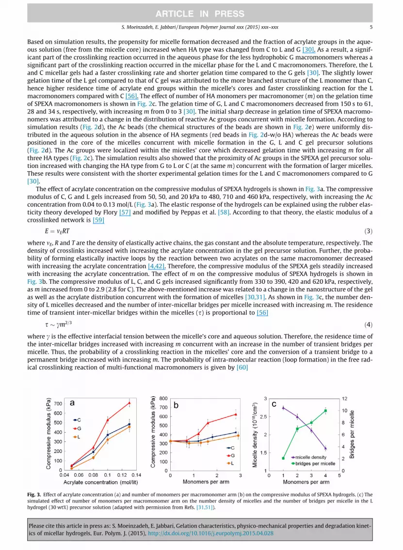
The effect of acrylate concentration on the compressive modulus of SPEXA hydrogels is shown in Fig. 3a. The compressivemodulus of C, G and L gels increased from 50, 50, and 20 kPa to 480, 710 and 460 kPa, respectively, with increasing the Acconcentration from 0.04 to 0.13 mol/L (Fig. 3a). The elastic response of the hydrogels can be explained using the rubber elas-ticity theory developed by Flory [57] and modified by Peppas et al. [58]. According to that theory, the elastic modulus of acrosslinked network is [59]
Fig. 3.simulathydrog
Pleaseics of
E ¼ mERT ð3Þ
where mE, R and T are the density of elastically active chains, the gas constant and the absolute temperature, respectively. Thedensity of crosslinks increased with increasing the acrylate concentration in the gel precursor solution. Further, the proba-bility of forming elastically inactive loops by the reaction between two acrylates on the same macromonomer decreasedwith increasing the acrylate concentration [4,42]. Therefore, the compressive modulus of the SPEXA gels steadily increasedwith increasing the acrylate concentration. The effect of m on the compressive modulus of SPEXA hydrogels is shown inFig. 3b. The compressive modulus of L, C, and G gels increased significantly from 330 to 390, 420 and 620 kPa, respectively,as m increased from 0 to 2.9 (2.8 for C). The above-mentioned increase was related to a change in the nanostructure of the gelas well as the acrylate distribution concurrent with the formation of micelles [30,31]. As shown in Fig. 3c, the number den-sity of L micelles decreased and the number of inter-micellar bridges per micelle increased with increasing m. The residencetime of transient inter-micellar bridges within the micelles (s) is proportional to [56]
s � cm2=3 ð4Þ
where c is the effective interfacial tension between the micelle’s core and aqueous solution. Therefore, the residence time ofthe inter-micellar bridges increased with increasing m concurrent with an increase in the number of transient bridges permicelle. Thus, the probability of a crosslinking reaction in the micelles’ core and the conversion of a transient bridge to apermanent bridge increased with increasing m. The probability of intra-molecular reaction (loop formation) in the free rad-ical crosslinking reaction of multi-functional macromonomers is given by [60]
Effect of acrylate concentration (a) and number of monomers per macromonomer arm (b) on the compressive modulus of SPEXA hydrogels. (c) Theed effect of number of monomers per macromonomer arm on the number density of micelles and the number of bridges per micelle in the Lel (30 wt%) precursor solution (adapted with permission from Refs. [31,51]).
cite this article in press as: S. Moeinzadeh, E. Jabbari, Gelation characteristics, physico-mechanical properties and degradation kinet-micellar hydrogels, Eur. Polym. J. (2015), http://dx.doi.org/10.1016/j.eurpolymj.2015.04.028

Fig. 4.the wa
6 S. Moeinzadeh, E. Jabbari / European Polymer Journal xxx (2015) xxx–xxx
Pleaseics of
W ¼ 1� 1
exp A½AC�r0 l2
� � ð5Þ
where [AC] is the local concentration of acrylates, r0 is the average distance between the double bonds on macromonomers, lis the statistical length of a repeating unit and A is a constant (A ¼ 3=4pNA where NA is Avogadro’s number). According to Eq.(5), the probability of loop formation decreased with increasing m due to an increase in the local concentration of acrylates asdescribed earlier. Altogether, the net effect of increasing m in the SPEXA macromonomer is an increase in the probability offormation of permanent inter-micellar bridges and a decrease in the probability of formation of elastically inactive loops.Therefore based on the theory of rubber elasticity, the compressive modulus of SPEXA gels increased with increasing m(Fig. 3b).
4. Swelling of micellar gels
The degree of swelling (or water content) of a hydrogel is typically controlled by two opposing forces, namely the ther-modynamic force of mixing between the polymer and water and the elastic force of extending polymer chains [57]. The forceof mixing tends to increase the water content of the gel by attractive interactions between water molecules and the networkchains [57]. The elastic force of extending polymer chains on the other hand tends to decrease the water content of the geldue to a change in polymer chain conformation from an entropically more favorable random coil to a less favorable extendedconformation. Several factors including molecular weight, hydrophobicity, functionality and flexibility of the macromono-mers, degree of crosslinking, nanostructure of the gel and temperature affect hydrogel swelling [61,62]. Specifically, thehydrogel swelling decreases with increasing hydrophobicity of the chains and crosslink density by influencing the forceof mixing and the elastic force of the chains, respectively [58]. The effect of acrylate group concentration on the swellingof micellar SPEXA gels is shown in Fig. 4a. The swelling ratio of C, L and G gels decreased from 710% to 300%, 730% to340% and 830% to 430%, respectively, when the acrylate concentration increased from 0.02 to 0.13 mol/L [51]. A decreasein the swelling ratio of SPEXA gels with increasing acrylate concentration was attributed to an increase in crosslink density.The swelling of hydrogels decreased slightly concurrent with increasing the hydrophobicity of HA segments from G to L andC at the same acrylate concentration. The effect of m on the water content of SPEXA gels is shown in Fig. 4b. The bulk watercontent of SPEXA gels ranged between 78% and 83% and m did not have a statistically significant effect on the water contentof hydrogels. The data in Fig. 4b implies that the chain extension of star PEG macromonomers with short HA segments, dueto the formation of micellar structures at the nanoscale, did not negatively affect the water content of SPEXA hydrogels.
5. Degradation of micellar gels
Although hydrogels provide enormous flexibility in controlling the cell microenvironment, their use in regenerative med-icine is limited by their persistence in the site of regeneration [6]. Therefore, hydrogels for tissue engineering applicationsshould be degradable with a rate corresponding to that of ECM production and remodeling [6]. The role of matrix degrada-tion on the fate of encapsulated cells has been investigated previously. C2C12 mouse myoblast cells encapsulated in adegradable alginate gel had lower proliferation and higher extent of myotube formation compared to those encapsulatedin a non-degradable gel [63]. Similarly, MSCs encapsulated in a non-degradable hyaluronic acid gel underwent adipogenicdifferentiation whereas those encapsulated in a degradable gel differentiated to the osteogenic lineage [64]. The rate ofhydrogel degradation can also affect tissue morphogenesis. For instance, blood vessel formation and angiogenesis requirea relatively fast (few days) degrading gel whereas mineralization and osteogenesis require a relatively slow (few weeks)degrading gel [20–22]. Therefore, there is a need to develop hydrogels with tunable degradation in order to regenerate com-plex tissues with many cell types.
Synthetic hydrogels can be made degradable with incorporation of enzymatic, hydrolytic or photolytically degradablesegments in the macromonomer chains or crosslinkers [65]. Copolymerization of non-degradable hydrophilic macromers
(a) Effect of acrylate concentration on the swelling ratio of SPEXA hydrogels (m = 1.7). (b) Effect of number of monomers per macromonomer arm onter content of SPEXA hydrogels (20 wt%) (adapted with permission from Refs. [30,51]).
cite this article in press as: S. Moeinzadeh, E. Jabbari, Gelation characteristics, physico-mechanical properties and degradation kinet-micellar hydrogels, Eur. Polym. J. (2015), http://dx.doi.org/10.1016/j.eurpolymj.2015.04.028

S. Moeinzadeh, E. Jabbari / European Polymer Journal xxx (2015) xxx–xxx 7
with degradable lactide and glycolide blocks has been used to impart degradability and control the water content of PEGhydrogels [4,66] but solubility of the copolymer in the aqueous gel precursor solution for cell encapsulation decreased dra-matically with increasing the length of lactide blocks [53]. Hydrogels synthesized from PEG and e-caprolactone co-polymersare shown to be hydrolytically degradable, but the degradation rate is limited by the hydrophobicity and phase separation ofe-caprolactone segments in solution [67]. In addition to poly(aliphatic hydroxy acids), other hydrolytically degradable poly-mers including poly(ester amides) [68], polyphosphoesters [69,70], poly(amino-ester urethanes) [71] are used to synthesizedegradable hydrogels. Photodegradable hydrogels are synthesized by incorporation of nitrobenzyl ether-derived moieties inPEG based hydrogels [72]. Remarkably, the SPEXA macromonomers chain extended with short HA segments generate micel-lar hydrogels with a wide range of degradation rates [30,51,52]. Owing to the micellization of hydrophobic segments, thedegradation of SPEXA gels can be tuned to a particular application from a few days to a few weeks, few months, and manymonths by changing hydrophobicity and length of the HA segments [30,51,52]. The mass losses of SPEXA hydrogels in a nar-row range of m values (1.6 6m 6 1.8) are compared in Fig. 5a. While the PEG hydrogel without HA (purple curve in Fig. 5a)had only 6% mass loss after 6 weeks of incubation, the C hydrogel lost 20% mass in 6 weeks and the G and L hydrogels com-pletely degraded in 3 days and 5 weeks, respectively. Based on simulation results, the differences in hydrophobicity of HAmonomers and number of hydrolytically-degradable ester groups per HA monomer contributed to the measured wide rangeof degradation rates for SPEXA hydrogels (Fig. 5a) [30]. The G and L macromonomers had 2 ester groups per HA monomerwhereas the C macromonomer had one ester group. In addition, the hydrophilicity of HA monomers which affected theirproximity to water molecules, increased from C to L and G (Fig. 5c). As a result, the G hydrogel with 2 ester groups perHA monomer and highest proximity of ester moieties to water molecules had the highest degradation rate whereas the Chydrogel with one ester group per HA monomer and ester moieties furthest away from water molecules had the lowestdegradation rate (Fig. 5a). The effect of m on mass loss of the L hydrogel was bimodal as shown in Fig. 5b. The mass lossof 20 wt% L gels increased from 6 to 37, 80% and 100% after 28 days of incubation when m increased from 0 to 0.8, 1.7and 2.9, respectively. Then, the mass loss decreased from 100% to 87% with increasing m from 2.9 to 3.7. The bimodal effectof m on mass loss of SPEXA gels was attributed to the formation of large micelles for m > 3 with reduced proximity of watermolecules to ester groups in the micelles’ core (Fig. 5b). The formation of micelles did not significantly affect the bulk watercontent of the SPEXA gels (Fig. 4b). However, the hydrophobic core of the micelles repelled water molecules, whichdecreased their proximity to the ester groups, leading to a reduction in the rate of degradation of SPEXA gels at high m valuesand a transition from surface (controlled by the number of ester groups) to bulk (dominated by the water content ofmicelles) degradation [30].
Fig. 5. (a) Effect of HA type on the mass remaining of SPEXA hydrogels (20 wt%, m = 1.7). (b) Effect of number of L monomers per macromonomer arm onthe mass remaining of L hydrogels after 28 days. The insets in (b) are molecular dynamic simulations of the structure of L (m = 1.7 and 3.7) micelles withgreen, brown and red beads representing ethylene oxide (EO), lactide and acrylate repeat units in the macromonomer, respectively. (c) Effect of degradableHA monomer type on the distribution of water beads around the micelles’ core. G, L, C, Ac, and water units are shown by blue, orange, purple, red, and lightblue beads, respectively, and EO beads are not shown for clarity (adapted with permission from Refs. [30,52]). (For interpretation of the references to colorin this figure legend, the reader is referred to the web version of this article.)
Please cite this article in press as: S. Moeinzadeh, E. Jabbari, Gelation characteristics, physico-mechanical properties and degradation kinet-ics of micellar hydrogels, Eur. Polym. J. (2015), http://dx.doi.org/10.1016/j.eurpolymj.2015.04.028

Fig. 6. (a) Molecular dynamic simulation of the effect of number of degradable lactide monomers per SPEXA macromonomer arm on the fraction of initiatormolecules in the aqueous solution of L hydrogel. (b) ALP activity and (c) calcium content of the MSCs encapsulated in SPEXA hydrogels with incubation timein osteogenic medium. A ‘‘star’’ indicates a statically significant difference (p < 0.05) between the test group and all other groups at the same time point(adapted with permission from Ref. [30]).
8 S. Moeinzadeh, E. Jabbari / European Polymer Journal xxx (2015) xxx–xxx
6. Cell–matrix interactions in micellar gels
Cell–matrix interactions within a cell-laden hydrogel play a central role in regulating cell function [73,74]. In the naturalextracellular matrix (ECM), cell adhesive proteins such as laminin and fibronectin bind to integrin cell surface receptors toregulate cell adhesion, migration, and differentiation [73,75,76]. Further, soluble proteins or tethered growth factors, presentin the ECM, modulate proliferation, migration and differentiation of the cells [77]. Therefore, synthetic hydrogels should bemodified with bioactive ligands for optimal cell–matrix interaction [78]. In that regard, the viability of human MSCs was sig-nificantly higher in a PEG gel conjugated with cell-adhesive arginine–glycine–aspartic acid (RGD) peptide as compared withthat with no peptide [79]. Human MSCs seeded on PEG hydrogels modified with RGD and an osteoinductive BMP-2 protein-derived KIPKASSVPTELSAISTLYL peptide showed 5- and 12-fold increase in ALP activity and calcium content after 14 and21 days of incubation, respectively [25]. We have previously shown that the viability of MSCs encapsulated in the micellarPEG gels and their differentiation to the osteogenic lineage are significantly enhanced by the incorporation of RGD peptideand BMP-2 protein or peptide in the hydrogel matrix [52,80]. In addition, the micellar nature of the gel affects cell viabilityand function. The simulated fraction of initiator molecules (the fraction not partitioned to the micelles’ core) in the L hydro-gel precursor solution decreased from 100% to 7.4%, 3.3% and 2% as m increased from 0 to 1, 2 and 3, respectively (Fig. 6a).The partition of initiator molecules to the core limited the gelation reaction to the micelle phase, thus reducing the cytotoxiceffect of low molecular weight initiator molecules on the encapsulated cells. Cell culture experiments showed that the via-bility of human MSCs after 2 days of encapsulation in the micellar L and C gels was significantly higher than those encap-sulated in the non-micellar PEG gel [30]. Since the water content of micellar gels was similar to the non-micellar gel (w/oHA gel in Fig. 4b) and the toxic effect of the initiator was significantly lower, the viability of MSCs encapsulated in the micel-lar PEG was higher than the non-micellar gels. The osteogenic differentiation of human MSCs encapsulated in the SPEXA gels(20 wt% and m = 1.7) was evaluated by measuring alkaline phosphatase (ALP) activity (Fig. 6b) and calcium content (Fig. 6c)with incubation in osteogenic medium over 28 days. The G gel due to its relatively fast degradation was not used for MSCencapsulation. The ALP activity of MSCs encapsulated in the L, C and w/o HA gels increased from day 7 to 14 and thendecreased from day 14 to 28. The peak in ALP activity corresponded to the initiation of osteogenesis as previously reported[25,31]. The ALP activity of MSCs encapsulated in the degradable L or C hydrogels at day 14 was significantly higher thanthose encapsulated in the non-degradable w/o HA gel. MSCs encapsulated in the L hydrogel had a higher ALP activity after14 days compared to those in the C hydrogel. The calcium content of the encapsulated MSCs had an increasing trend withtime over 28 days of incubation. The calcium content of MSCs encapsulated in the L gel, with �80% mass loss after 28 days,was significantly higher than those in the C gel with �20% mass loss. The calcium content of MSCs encapsulated in the L or Cgel was higher than those in the non-degradable w/o HA gel after 28 days of incubation. The micellar SPEXA gels supportedviability and differentiation of human MSCs to the osteogenic lineage. The SPEXA gels with high water content, tunabledegradation, low gelation time, and adjustable stiffness can be used for differentiation of stem cells to other cell types as softand fast-degrading SPEXA gels support vasculogenic differentiation of co-encapsulated MSCs and endothelial progenitorcells (EPCs) [51].
7. Conclusion
The formation of micellar structures in the aqueous solution of acrylated star PEG maromonomers chain extended withshort aliphatic hydroxy acid (HA) segments (SPEXA) decreased gelation time and exposure time of encapsulated cells to thetoxic polymerization photo-initiator. As a result of micelle formation, the degradation rate of SPEXA hydrogels was tunablefrom a few days to a few weeks, a few months, and many months by changing the HA monomer or varying the length of HA
Please cite this article in press as: S. Moeinzadeh, E. Jabbari, Gelation characteristics, physico-mechanical properties and degradation kinet-ics of micellar hydrogels, Eur. Polym. J. (2015), http://dx.doi.org/10.1016/j.eurpolymj.2015.04.028

S. Moeinzadeh, E. Jabbari / European Polymer Journal xxx (2015) xxx–xxx 9
segment while maintaining a relatively constant water content. The compressive modulus of the micellar SPEXA hydrogelsranged from 5 to 700 kPa. The viability and osteogenic differentiation of encapsulated human MSCs was significantly higherin the micellar SPEXA hydrogels compared to the non-micellar PEG gel. Micellar hydrogels with a wide range of physico-me-chanical properties are potentially useful as cell carriers in regeneration of living tissues from the relatively soft nerve andvascular tissues to stiff skeletal tissues.
Acknowledgments
This work was supported by research grants to E. Jabbari from the National Science Foundation under Award NumbersDMR1049381, IIP-1357109, and CBET1403545. Research reported in this publication was supported by the NationalInstitute of Arthritis and Musculoskeletal and Skin Diseases of the National Institutes of Health under Award NumberAR063745. The content is solely the responsibility of the authors and does not necessarily represent the official views ofthe National Institutes of Health. This work was also supported by the Arbeitsgemeinschaft Fur Osteosynthesefragen (AO)Foundation under Grant Number C10-44J, and the University of South Carolina VP Office for Research under GrantNumber 15510-E414.
References
[1] Y.H. Cheng, S.H. Yang, W.Y. Su, Y.C. Chen, K.C. Yang, W.T.K. Cheng, S.C. Wu, F.H. Lin, Thermosensitive chitosan–gelatin–glycerol phosphate hydrogels asa cell carrier for nucleus pulposus regeneration: an in vitro study, Tissue Eng. Part A 16 (2010) 695–703.
[2] A.S. Sarvestani, X.Z. He, E. Jabbari, Viscoelastic characterization and modeling of gelation kinetics of injectable in situ cross-linkable poly(lactide-co-ethylene oxide-co-fumarate) hydrogels, Biomacromolecules 8 (2007) 406–415.
[3] N.A. Peppas, S.R. Lustig, Solute diffusion in hydrophilic network structures, in: Hydrogels in Medicine and Pharmacy. I. Fundamentals, CRC Press, BocaRaton, FL, 2004.
[4] A.S. Sarvestani, W. Xu, X. He, E. Jabbari, Gelation and degradation characteristics of in situ photo-crosslinked poly(L-lactide-co-ethylene oxide-co-fumarate) hydrogels, Polymer 48 (2007) 7113–7120.
[5] L.A. Zhao, M.D. Weir, H.H.K. Xu, An injectable calcium phosphate–alginate hydrogel-umbilical cord mesenchymal stem cell paste for bone tissueengineering, Biomaterials 31 (2010) 6502–6510.
[6] M.C. Cushing, K.S. Anseth, Hydrogel cell cultures, Science 316 (2007) 1133–1134.[7] R.A. Perez, M. Kim, T.H. Kim, J.H. Kim, J.H. Lee, J.H. Park, J.C. Knowles, H.W. Kim, Utilizing core–shell fibrous collagen–alginate hydrogel cell delivery
system for bone tissue engineering, Tissue Eng. Part A 20 (2014) 103–114.[8] E. Hesse, T.E. Hefferan, J.E. Tarara, C. Haasper, R. Meller, C. Krettek, L.C. Lu, M.J. Yaszemski, Collagen type I hydrogel allows migration, proliferation, and
osteogenic differentiation of rat bone marrow stromal cells, J. Biomed. Mater. Res., Part A 94A (2010) 442–449.[9] L.M. Wang, R.R. Rao, J.P. Stegemann, Delivery of mesenchymal stem cells in chitosan/collagen microbeads for orthopedic tissue repair, Cells Tissues
Organs 197 (2013) 333–343.[10] N. Huebsch, P.R. Arany, A.S. Mao, D. Shvartsman, O.A. Ali, S.A. Bencherif, J. Rivera-Feliciano, D.J. Mooney, Harnessing traction-mediated manipulation of
the cell/matrix interface to control stem-cell fate, Nat. Mater. 9 (2010) 518–526.[11] Y. Chen, X.H. Pang, C.M. Dong, Dual stimuli-responsive supramolecular polypeptide-based hydrogel and reverse micellar hydrogel mediated by host–
guest chemistry, Adv. Funct. Mater. 20 (2010) 579–586.[12] A.M. Jonker, D.W.P.M. Lowik, J.C.M. van Hest, Peptide- and protein-based hydrogels, Chem. Mater. 24 (2012) 759–773.[13] M.H. Yao, J. Yang, M.S. Du, J.T. Song, Y. Yu, W. Chen, Y.D. Zhao, B. Liu, Polypeptide-engineered physical hydrogels designed from the coiled-coil region of
cartilage oligomeric matrix protein for three-dimensional cell culture, J. Mater. Chem. B 2 (2014) 3123–3132.[14] A. Banerjee, M. Arha, S. Choudhary, R.S. Ashton, S.R. Bhatia, D.V. Schaffer, R.S. Kane, The influence of hydrogel modulus on the proliferation and
differentiation of encapsulated neural stem cells, Biomaterials 30 (2009) 4695–4699.[15] J.J. Roberts, S.J. Bryant, Comparison of photopolymerizable thiol-ene PEG and acrylate-based PEG hydrogels for cartilage development, Biomaterials 34
(2013) 9969–9979.[16] T. Yuan, L. Zhang, K.F. Li, H.S. Fan, Y.J. Fan, J. Liang, X.D. Zhang, Collagen hydrogel as an immunomodulatory scaffold in cartilage tissue engineering, J.
Biomed. Mater. Res., Part B 102 (2014) 337–344.[17] F. Mirahmadi, M. Tafazzoli-Shadpour, M.A. Shokrgozar, S. Bonakdar, Enhanced mechanical properties of thermosensitive chitosan hydrogel by silk
fibers for cartilage tissue engineering, Mater. Sci. Eng. C: Mater. Biol. Appl. 33 (2013) 4786–4794.[18] B.J. DeKosky, N.H. Dormer, G.C. Ingavle, C.H. Roatch, J. Lomakin, M.S. Detamore, S.H. Gehrke, Hierarchically designed agarose and poly(ethylene glycol)
interpenetrating network hydrogels for cartilage tissue engineering, Tissue Eng. Part C: Methods 16 (2010) 1533–1542.[19] H. Park, B. Choi, J.L. Hu, M. Lee, Injectable chitosan hyaluronic acid hydrogels for cartilage tissue engineering, Acta Biomater. 9 (2013) 4779–4786.[20] K. Chatterjee, S. Lin-Gibson, W.E. Wallace, S.H. Parekh, Y.J. Lee, M.T. Cicerone, M.F. Young, C.G. Simon, The effect of 3D hydrogel scaffold modulus on
osteoblast differentiation and mineralization revealed by combinatorial screening, Biomaterials 31 (2010) 5051–5062.[21] J.A. Henderson, X. He, E. Jabbari, Concurrent differentiation of marrow stromal cells to osteogenic and vasculogenic lineages, Macromol. Biosci. 8
(2008) 499–507.[22] Y.C. Chen, R.Z. Lin, H. Qi, Y.Z. Yang, H.J. Bae, J.M. Melero-Martin, A. Khademhosseini, Functional human vascular network generated in
photocrosslinkable gelatin methacrylate hydrogels, Adv. Funct. Mater. 22 (2012) 2027–2039.[23] T. Karimi, D. Barati, O. Karaman, S. Moeinzadeh, E. Jabbari, A developmentally inspired combined mechanical and biochemical signaling approach on
zonal lineage commitment of mesenchymal stem cells in articular cartilage regeneration, Integr. Biol. 7 (2014) 112–127.[24] X. Yang, S.K. Sarvestani, S. Moeinzadeh, X. He, E. Jabbari, Three-dimensional engineered matrix to study cancer stem cells and tumorsphere formation:
effect of matrix modulus, Tissue Eng. Part A 19 (2013) 669–684.[25] X. He, J. Ma, E. Jabbari, Effect of grafting RGD and BMP-2 protein-derived peptides to a hydrogel substrate on osteogenic differentiation of marrow
stromal cells, Langmuir 24 (2008) 12508–12516.[26] X. He, X. Yang, E. Jabbari, Combined effect of osteopontin and BMP-2 derived peptides grafted to an adhesive hydrogel on osteogenic and vasculogenic
differentiation of marrow stromal cells, Langmuir 28 (2012) 5387–5397.[27] D.B. An, T.H. Kim, J.H. Lee, S.H. Oh, Evaluation of stem cell differentiation on polyvinyl alcohol/hyaluronic acid hydrogel with stiffness gradient, J. Tissue
Eng. Regen. Med. 8 (2014) 346–347.[28] P. Guo, Y.S. Yuan, F.L. Chi, Biomimetic alginate/polyacrylamide porous scaffold supports human mesenchymal stem cell proliferation and
chondrogenesis, Mater. Sci. Eng. C: Mater. Biol. Appl. 42 (2014) 622–628.[29] L. Sun, D. Li, U.D. Hemraz, H. Fenniri, T.J. Webster, Self-assembled rosette nanotubes and poly(2-hydroxyethyl methacrylate) hydrogels promote skin
cell functions, J. Biomed. Mater. Res., Part A 102 (2014) 3446–3451.
Please cite this article in press as: S. Moeinzadeh, E. Jabbari, Gelation characteristics, physico-mechanical properties and degradation kinet-ics of micellar hydrogels, Eur. Polym. J. (2015), http://dx.doi.org/10.1016/j.eurpolymj.2015.04.028

10 S. Moeinzadeh, E. Jabbari / European Polymer Journal xxx (2015) xxx–xxx
[30] S. Moeinzadeh, D. Barati, S.K. Sarvestani, O. Karaman, E. Jabbari, Nanostructure formation and transition from surface to bulk degradation inpolyethylene glycol gels chain-extended with short hydroxy acid segments, Biomacromolecules 14 (2013) 2917–2928.
[31] S. Moeinzadeh, E. Jabbari, Mesoscale simulation of the effect of a lactide segment on the nanostructure of star poly(ethylene glycol-co-lactide)-acrylatemacromonomers in aqueous solution, J. Phys. Chem. B 116 (2012) 1536–1543.
[32] S. Moeinzadeh, E. Jabbari, Nanostructure formation in hydrogels, in: B. Bhushan et al. (Eds.), Handbook of Nanomaterials Properties, Springer, Berlin,2014, pp. 285–297.
[33] E.S. Gil, S.M. Hudson, Stimuli-reponsive polymers and their bioconjugates, Prog. Polym. Sci. 29 (2004) 1173–1222.[34] B. Jeong, Y.H. Bae, D.S. Lee, S.W. Kim, Biodegradable block copolymers as injectable drug-delivery systems, Nature 388 (1997) 860–862.[35] C.T. Huynh, M.K. Nguyen, D.S. Lee, Injectable block copolymer hydrogels: achievements and future challenges for biomedical applications,
Macromolecules 44 (2011) 6629–6636.[36] G. Riess, Micellization of block copolymers, Prog. Polym. Sci. 28 (2003) 1107–1170.[37] D.M. Henn, R.A.E. Wright, J.W. Woodcock, B. Hu, B. Zhao, Tertiary-amine-containing thermo- and pH-sensitive hydrophilic ABA triblock copolymers:
effect of different tertiary amines on thermally induced sol–gel transitions, Langmuir 30 (2014) 2541–2550.[38] T.G. O’Lenick, N.X. Jin, J.W. Woodcock, B. Zhao, Rheological properties of aqueous micellar gels of a thermo- and pH-sensitive ABA triblock copolymer, J.
Phys. Chem. B 115 (2011) 2870–2881.[39] J.W. Woodcock, X.G. Jiang, R.A.E. Wright, B. Zhao, Enzyme-induced formation of thermoreversible micellar gels from aqueous solutions of
multiresponsive hydrophilic ABA triblock copolymers, Macromolecules 44 (2011) 5764–5775.[40] D. Cohn, G. Lando, A. Sosnik, S. Garty, A. Levi, PEO–PPO–PEO-based poly(ether ester urethane)s as degradable reverse thermo-responsive multiblock
copolymers, Biomaterials 27 (2006) 1718–1727.[41] H.K. Kim, W.S. Shim, S.E. Kim, K.H. Lee, E. Kang, J.H. Kim, K. Kim, I.C. Kwon, D.S. Lee, Injectable in situ-forming pH/thermo-sensitive hydrogel for bone
tissue engineering, Tissue Eng. Part A 15 (2009) 923–933.[42] N. Sanson, J. Rieger, Synthesis of nanogels/microgels by conventional and controlled radical crosslinking copolymerization, Polym. Chem. 1 (2010)
965–977.[43] N. Willet, J.F. Gohy, L.C. Lei, M. Heinrich, L. Auvray, S. Varshney, R. Jerome, B. Leyh, Fast multiresponsive micellar gels from a smart ABC triblock
copolymer, Angew. Chem., Int. Ed. 46 (2007) 7988–7992.[44] T.C. Holmes, S. de Lacalle, X. Su, G.S. Liu, A. Rich, S.G. Zhang, Extensive neurite outgrowth and active synapse formation on self-assembling peptide
scaffolds, Proc. Natl. Acad. Sci. U. S. A. 97 (2000) 6728–6733.[45] J. Kisiday, M. Jin, B. Kurz, H. Hung, C. Semino, S. Zhang, A.J. Grodzinsky, Self-assembling peptide hydrogel fosters chondrocyte extracellular matrix
production and cell division: implications for cartilage tissue repair, Proc. Natl. Acad. Sci. U. S. A. 99 (2002) 9996–10001.[46] S.G. Zhang, T.C. Holmes, C.M. Dipersio, R.O. Hynes, X. Su, A. Rich, Self-complementary oligopeptide matrices support mammalian-cell attachment,
Biomaterials 16 (1995) 1385–1393.[47] J.D. Hartgerink, E. Beniash, S.I. Stupp, Self-assembly and mineralization of peptide-amphiphile nanofibers, Science 294 (2001) 1684–1688.[48] T.H. Larsen, M.C. Branco, K. Rajagopal, J.P. Schneider, E.M. Furst, Sequence-dependent gelation kinetics of beta-hairpin peptide hydrogels,
Macromolecules 42 (2009) 8443–8450.[49] S.M. Zhang, M.A. Greenfield, A. Mata, L.C. Palmer, R. Bitton, J.R. Mantei, C. Aparicio, M.O. de la Cruz, S.I. Stupp, A self-assembly pathway to aligned
monodomain gels, Nat. Mater. 9 (2010) 594–601.[50] W.A. Petka, J.L. Harden, K.P. McGrath, D. Wirtz, D.A. Tirrell, Reversible hydrogels from self-assembling artificial proteins, Science 281 (1998) 389–392.[51] D. Barati, S. Moeinzadeh, O. Karaman, E. Jabbari, Time dependence of material properties of polyethylene glycol hydrogels chain extended with short
hydroxy acid segments, Polymer 55 (2014) 3894–3904.[52] S. Moeinzadeh, D. Barati, X. He, E. Jabbari, Gelation characteristics and osteogenic differentiation of stromal cells in inert hydrolytically degradable
micellar polyethylene glycol hydrogels, Biomacromolecules 13 (2012) 2073–2086.[53] S. Moeinzadeh, S.N. Khorasani, J. Ma, X. He, E. Jabbari, Synthesis and gelation characteristics of photo-crosslinkable star poly(ethylene oxide-co-lactide-
glycolide acrylate) macromonomers, Polymer 52 (2011) 3887–3896.[54] P. Posocco, M. Fermeglia, S. Pricl, Morphology prediction of block copolymers for drug delivery by mesoscale simulations, J. Mater. Chem. 20 (2010)
7742–7753.[55] G. Odian, Principles of Polymerization, John Wiley, New York, 1981.[56] T. Nicolai, O. Colombani, C. Chassenieux, Dynamic polymeric micelles versus frozen nanoparticles formed by block copolymers, Soft Matter 6 (2010)
3111–3118.[57] P.J. Flory, Principles of Polymer Chemistry, Cornell University Press, New York, 1953.[58] N.A. Peppas, P. Bures, W. Leobandung, H. Ichikawa, Hydrogels in pharmaceutical formulations, Eur. J. Pharm. Biopharm. 50 (2000) 27–46.[59] L.G. Cima, S.T. Lopina, Network structures of radiation-crosslinked star polymer gels, Macromolecules 28 (1995) 6787–6794.[60] J.E. Elliott, C.N. Bowman, Kinetics of primary cyclization reactions in cross-linked polymers: an analytical and numerical approach to heterogeneity in
network formation, Macromolecules 32 (1999) 8621–8628.[61] F. Ganji, S. Vasheghani-Farahani, E. Vasheghani-Farahani, Theoretical description of hydrogel swelling: a review, Iran. Polym. J. 19 (2010) 375–398.[62] V.S. Sukumar, S.T. Lopina, Network model for the swelling properties of end-linked linear and star poly(ethylene oxide) hydrogels, Macromolecules 35
(2002) 10189–10192.[63] T. Boontheekul, E.E. Hill, H.J. Kong, D.J. Mooney, Regulating myoblast phenotype through controlled gel stiffness and degradation, Tissue Eng. 13 (2007)
1431–1442.[64] S. Khetan, M. Guvendiren, W.R. Legant, D.M. Cohen, C.S. Chen, J.A. Burdick, Degradation-mediated cellular traction directs stem cell fate in covalently
crosslinked three-dimensional hydrogels, Nat. Mater. 12 (2013) 458–465.[65] P.M. Kharkar, K.L. Kiick, A.M. Kloxin, Designing degradable hydrogels for orthogonal control of cell microenvironments, Chem. Soc. Rev. 42 (2013)
7335–7372.[66] Y. Nakayama, K. Okuda, K. Takamizawa, A. Nakayama, Preparation of well-defined poly(ether-ester) macromers: photogelation and biodegradability,
Acta Biomater. 7 (2011) 1496–1503.[67] C.Y. Ko, C.Y. Yang, S.R. Yang, K.L. Ku, C.K. Tsao, D.C.C. Chuang, I.M. Chu, M.H. Cheng, Cartilage formation through alterations of amphiphilicity of
poly(ethylene glycol)–poly(caprolactone) copolymer hydrogels, RSC Adv. 3 (2013) 25769–25779.[68] A. Rodriguez-Galan, L. Franco, J. Puiggali, Degradable poly(ester amide)s for biomedical applications, Polymer 3 (2011) 65–99.[69] J.L. He, M.Z. Zhang, P.H. Ni, Rapidly in situ forming polyphosphoester-based hydrogels for injectable drug delivery carriers, Soft Matter 8 (2012) 6033–
6038.[70] Y.C. Wang, W.J. Lee, S.P. Ju, Modeling of the polyethylene and poly(L-lactide) triblock copolymer: a dissipative particle dynamics study, J. Chem. Phys.
131 (2009) 124901.[71] Y.T. Zheng, C.L. He, C.T. Huynh, D.S. Lee, Biodegradable pH- and temperature-sensitive multiblock copolymer hydrogels based on poly(amino-ester
urethane)s, Macromol. Res. 18 (2010) 974–980.[72] A.M. Kloxin, A.M. Kasko, C.N. Salinas, K.S. Anseth, Photodegradable hydrogels for dynamic tuning of physical and chemical properties, Science 324
(2009) 59–63.[73] E. Cukierman, R. Pankov, K.M. Yamada, Cell interactions with three-dimensional matrices, Curr. Opin. Cell Biol. 14 (2002) 633–639.[74] M.J. Dalby, N. Gadegaard, R.O.C. Oreffo, Harnessing nanotopography and integrin–matrix interactions to influence stem cell fate, Nat. Mater. 13 (2014)
558–569.[75] T. Gloe, U. Pohl, Laminin binding conveys mechanosensing in endothelial cells, News Physiol. Sci. 17 (2002) 166–169.
Please cite this article in press as: S. Moeinzadeh, E. Jabbari, Gelation characteristics, physico-mechanical properties and degradation kinet-ics of micellar hydrogels, Eur. Polym. J. (2015), http://dx.doi.org/10.1016/j.eurpolymj.2015.04.028

S. Moeinzadeh, E. Jabbari / European Polymer Journal xxx (2015) xxx–xxx 11
[76] A.E. Aplin, A. Howe, S.K. Alahari, R.L. Juliani, Signal transduction and signal modulation by cell adhesion receptors: the role of integrins, cadherins,immunoglobulin-cell adhesion molecules, and selectins, Pharmacol. Rev. 50 (1998) 197–263.
[77] A.K.A. Silva, C. Richard, M. Bessodes, D. Scherman, O.W. Merten, Growth factor delivery approaches in hydrogels, Biomacromolecules 10 (2009) 9–18.[78] J.M. Zhu, Bioactive modification of poly(ethylene glycol) hydrogels for tissue engineering, Biomaterials 31 (2010) 4639–4656.[79] S.Q. Liu, Q.A. Tian, L. Wang, J.L. Hedrick, J.H.P. Hui, Y.Y. Yang, P.L.R. Ee, Injectable biodegradable poly(ethylene glycol)/RGD peptide hybrid hydrogels for
in vitro chondrogenesis of human mesenchymal stem cells, Macromol. Rapid Commun. 31 (2010) 1148–1154.[80] S. Moeinzadeh, D. Barati, S.K. Sarvestani, T. Karimi, E. Jabbari, Experimental and computational investigation of the effect of hydrophobicity on
aggregation and osteoinductive potential of BMP-2 derived peptide in a hydrogel matrix, Tissue Eng. Part A 21 (2014) 134–146.
Please cite this article in press as: S. Moeinzadeh, E. Jabbari, Gelation characteristics, physico-mechanical properties and degradation kinet-ics of micellar hydrogels, Eur. Polym. J. (2015), http://dx.doi.org/10.1016/j.eurpolymj.2015.04.028


