Embed Size (px)
Citation preview

i
Genetic and evolutionary dynamics of avian influenza A virus
in wild birds
by Jessica Benkaroun
A thesis submitted to the School of Graduate Studies
in partial fulfillment of the requirements for the degree of
Doctor of Philosophy
Department of Biology, Faculty of Science
Memorial University of Newfoundland
St. John’s, Newfoundland and Labrador, Canada
September 2018

ii
Abstract
Influenza A virus (IAV) is the prototype of the family Orthomyxoviridae, a
group of segmented, negative-sense, single-stranded RNA viruses. The virus
circulates in wild bird species but does not usually cause severe disease in these
hosts. However, highly pathogenic forms exist and have caused numerous deaths in
wild and farmed birds. The eastern coast of Canada represents an interesting
location for the study of IAVs in their natural reservoir as it has a large number of
bird breeding colonies and migratory bird connections with the mainland of North
America and Eurasia. Previous research on IAV ecology and transmission has shown
that migratory birds in this region move the virus around the globe and contribute
an important facet to IAV dynamics. My thesis focuses on the study of the virus
genetics and evolutionary dynamics in different wild bird species. By applying high-
throughput next-generation sequencing technologies, I characterized complete IAV
genomes from different wild bird species from Newfoundland and Labrador and
conducted in-depth analyses of the virus genomic structure. My study revealed that
the structure of the virus genome is conserved among similar avian hosts. I also
demonstrated though experimental mutation studies that a change of host can
causes major changes in the viral genome. I also explored evolutionary patterns in
the viral genomic non-coding regions (NCRs), and found that variation in the NCR
sequences is correlated with the original host species and geographic origin. Finally, I
analyzed IAVs from Laridae family hosts (gulls and terns) and demonstrated that

iii
these hosts are important for the transmission of IAVs around the globe and to other
hosts and participate in the generation of pandemic viruses. Overall, my results
contribute to give a better understanding on the evolution and geographic patterns
of influenza A viruses in their natural hosts.
iv
Acknowledgments
First of all, I would like to extend my gratitude to my supervisors, Dr. Andrew
Lang and Dr. Hugh Whitney, for giving me the opportunity to take part and guide me
through this thesis project. I thank you for your support that allowed me to complete
this work.
I would like to thank the members of my thesis committee, Dr. Sherri Christian, and
Dr. Kensuke Hirasawa, for their valuable help in bringing this project to fruition.
Many thanks to all members of Andrew’s lab who gave me precious help and
support. A deep thank you to Marc Grüll, Dr. Marta Canuti, and Purvikalyan Pallegar.
I am grateful to all MUN students I met during my thesis: Esteban, Elena, Paula, Boni,
Nathalia, Abdou, Daigo, Damiano, Anastasia, Oihane, Rosa, Pramod, Amrita, Carlos,
Fabio, Fritz, and Lizbeth.
Thank you to my friends from home: Magali, Ananda, Delphine, Olivier, Mako, Julien,
Stephane, and Yan.
An eternal gratitude to my parents and my little sisters for their listening and advice.
Thank you to my husband’s family for their support: Jesus, Irene, Amparo, Rosa,
Nubia, David, and Viviana. Gracias por estar en nuestros vidas.
At last, a deep and grateful thank you to my love, my companion, you have been
admirable and deeply supportive throughout this journey. I love you John Ceballos.

v
Je dédie cette thèse à mes parents, Christine Waille et Didier Benkaroun, à mes sœurs
Julie Waille et Rachel Waille. Je vous remercie de tout mon cœur pour votre soutien
inconditionnel tout au long de ce séjour.
My thesis project was supported by funding from the Newfoundland and Labrador
Research & Development Corporation, Environment and Climate Change Canada,
and the Newfoundland and Labrador Forestry and Agrifoods Agency. I received a
fellowship from the Memorial University School of Graduate Studies and the
Newfoundland and Labrador Forestry and Agrifoods Agency.
vi
Table of Contents
Abstract .................................................................................................................... ii
Acknowledgments .................................................................................................... iv
Table of Contents ..................................................................................................... vi
List of Figures ............................................................................................................xi
List of Abbreviations and Symbols ........................................................................... xiv
List of Appendices.................................................................................................... xvi
Co-authorship Statement ........................................................................................ xix
Chapter 1: Introduction and Overview ..................................................................... 1
1.1 The viral family Orthomyxoviridae .................................................................... 1
1.2 Influenza A virus structure and genomic organization ...................................... 4
1.2.1 The virus structure ..................................................................................... 4
1.2.2 The virus genome ...................................................................................... 7
1.3 Influenza A virus life cycle ................................................................................ 9
1.4 Influenza A virus reservoir and host transmission ........................................... 12
1.4.1 IAV in birds .............................................................................................. 12
1.4.2 IAV inter-species transmission ................................................................. 14
1.4.3 IAV in swine ............................................................................................. 16
1.4.4 IAV in humans .......................................................................................... 16
1.4.5 IAV in marine mammals ........................................................................... 17
1.5 Influenza A virus diversity and evolutionary mechanisms ............................... 17

vii
1.5.1 Random mutations and gene reassortments ............................................ 18
1.5.2 Other mutational mechanisms ................................................................. 20
1.5.3 The role of the host immune system ........................................................ 20
1.6 IAV in Newfoundland and Labrador, Canada .................................................. 21
1.7 Thesis Aim and Outline ................................................................................... 21
1.8 References ..................................................................................................... 24
Chapter 2: Evaluation of influenza A virus quasispecies populations in different
avian reservoir hosts ............................................................................................... 41
Abstract ............................................................................................................... 41
2.1 Introduction ................................................................................................... 42
2.2 Materials and Methods .................................................................................. 44
2.2.1 Viruses and RNA isolation ........................................................................ 44
2.2.2 Deep sequencing workflow of IAV genomes ............................................ 45
2.2.3 IAV genome assembly and variant calling ................................................ 46
2.2.4 Statistical analyses ................................................................................... 47
2.3 Results ........................................................................................................... 47
2.3.1 Analysis of IAV quasispecies populations shed by different avian reservoir
hosts................................................................................................................. 47
2.3.1.1 Gull IAV quasispecies structure ......................................................... 47
2.3.1.2 Murre IAV quasispecies structure ..................................................... 54
2.3.1.3 Duck IAV quasispecies structure ....................................................... 59

viii
2.3.2 Effect of replication in a different host on the virus quasispecies ............. 64
2.3.2.1 Mutational analysis of the duck viral genome ................................... 66
2.3.2.2 Mutational analysis of the duck protein sequences .......................... 70
2.4 Discussion ...................................................................................................... 72
2.5 References ..................................................................................................... 76
Chapter 3: Analysis of the Variability in the Non-Coding Regions of Influenza A
Viruses .................................................................................................................... 81
Abstract ............................................................................................................... 81
3.1 Introduction ................................................................................................... 82
3.2. Materials and Methods ................................................................................. 84
3.2.1. NCR sequence determination ................................................................. 84
3.2.2 Sequence analyses ................................................................................... 85
3.3 Results and discussion .................................................................................... 86
3.3.1 Characterization of NCRs from different wild bird viruses ........................ 86
3.3.2 The variability in segment-specific NCR sequences differs among segments
......................................................................................................................... 89
3.3.3 Patterns of variability within NCRs can be explained by viral host species
and geographic origins ..................................................................................... 93
3.4 Conclusions .................................................................................................. 102
3.5 References ................................................................................................... 102
Chapter 4: Analysis of influenza A viruses from gulls: An evaluation of inter-
regional movements and interactions with other avian and mammalian influenza A
viruses ................................................................................................................... 108

ix
Abstract ............................................................................................................. 108
4.1 Introduction ................................................................................................. 109
4.2 Results and Discussion ................................................................................. 111
4.2.1 Ecogenetic aspects of gull IAVs .............................................................. 111
4.2.2 Ecogeographic aspects of gull IAVs......................................................... 119
4.2.3 Clinical and subclinical IAV infections of gulls ......................................... 121
4.2.4 Inter-species transmission of gull IAVs ................................................... 128
4.2.5 The role of gulls as contributors to the transmission of HPAI viruses...... 137
4.2.6 The role of terns in global IAV transmission, and relationships with gull
IAVs ................................................................................................................ 138
4.2.7 Ecobiological aspects of gull IAVs ........................................................... 140
4.3 Concluding remarks ...................................................................................... 141
4.4 References ................................................................................................... 143
Chapter 5: Summary and conclusions ................................................................... 177
5.1 IAVs quasispecies population structure in wild birds ................................... 177
5.2 Effect of replication in a different host on the IAV genome ......................... 178
5.3 Analysis of IAV genomic non-coding regions ................................................ 179
5.4 The role of gulls and terns in IAV transmission ............................................ 180
5.5 Conclusion and future perspectives ............................................................. 180
5.6 References .................................................................................................. 181
Appendices ............................................................................................................ 187

x
List of Tables
Table 2.1: Genomic comparison of three gull H13N6 viruses. .................................. 48
Table 2.2: Genomic comparison of three murre H1N2 viruses. ................................ 54
Table 2.3: Genomic comparison of three duck viruses ............................................. 60
Table 4.1: Experimental infections of gulls with avian strains................................. 124
Table 4.2: Experimental infections of avian and mammalian hosts with gull strains
.............................................................................................................................. 126
Table 4.3: Gull virus binding assays on avian and human tissue ............................. 128

xi
List of Figures
Figure 1.1: Orthomyxoviridae genera phylogeny. ...................................................... 1
Figure 1.2: Influenza A virus structure........................................................................ 5
Figure 1.3: Influenza A segment organization. replication of the virus. ...................... 8
Figure 1.4: Influenza virus life cycle ......................................................................... 10
Figure 1.5: Influenza A subtype distribution across the virus’ reservoir and other
hosts ........................................................................................................................ 13
Figure 1.6: IAV antigenic shift and drift mechanisms ................................................ 18
Figure 1.7: Virus quasispecies formation. ................................................................. 21
Figure 2.1: Overall variant frequencies for the quasispecies of gull viruses ............. 49
Figure 2.2: Shared variant frequencies for pairwise comparisons of the three gull
viruses ..................................................................................................................... 50
Figure 2.3: Synonymous and non-synonymous substitution frequencies for all
segments of the gull viruses .................................................................................... 51
Figure 2.4: Comparison of the quasispecies structure of two gull viruses with the
same genotype ........................................................................................................ 53
Figure 2.5: Overall variant frequencies of the quasispecies of murre viruses .......... 55
Figure 2.6: Shared variant frequencies for pairwise comparisons of the three murre
viruses ..................................................................................................................... 56
Figure 2.7: Synonymous and non-synonymous substitution frequencies for all
segments of the murre viruses ................................................................................ 57

xii
Figure 2.8: Comparison of the quasispecies structure of two murre viruses with an
identical genotype. .................................................................................................. 59
Figure 2.9: Overall variant frequencies of the quasispecies of duck viruses .............. 61
Figure 2.10: Shared variant frequencies for pairwise comparisons of the three duck
viruses ..................................................................................................................... 62
Figure 2.11: Synonymous and non-synonymous substitution frequencies for all
segments of the duck viruses ................................................................................... 63
Figure 2.12: Comparison of the quasispecies structure of two duck viruses with
similar genotypes .................................................................................................... 64
Figure 2.13: Viral titer of the duck virus over the serial passages in puffin embryos . 66
Figure 2.14: Venn diagrams of the overall proportion of virus variants in the initial
swab sample (P0), the fifth passage (P5), and the last passage (P10) ....................... 67
Figure 2.15: Proportions of non-synonymous (dN) and synonymous (dS) substitutions
in the duck virus segments ...................................................................................... 69
Figure 2.16: Comparison of the HA amino acid changes from the fifth and last virus
passages .................................................................................................................. 71
Figure 2.17: Comparison of the NA amino acid changes from the fifth and last virus
passages .................................................................................................................. 73
Figure 3.1: 3’ NCRs of wild bird virus segments determined in this study ................. 88
Figure 3.2: 5’ NCRs of wild bird virus segments determined in this study ................. 89
Figure 3.3: Genetic diversity of 3’ and 5’ NCRs for different segments ..................... 92
Figure 3.4: Relationships among H9 3’ NCR and CDS regions for viruses from different
host species and geographic locations ..................................................................... 95
xiii
Figure 3.5: Relationships among H13 3’ NCR and CDS regions for viruses from
different host species and geographic locations ...................................................... 97
Figure 3.6: Relationships among H1 3’ NCR and CDS regions for viruses from different
host species and geographic locations ..................................................................... 99
Figure 3.7: Relationships among N6 3’ NCR and CDS regions for viruses from different
host species and geographic locations ................................................................... 101
Figure 3.8: Relationships among NP 3’ NCR and CDS regions for viruses from different
host species and geographic locations ................................................................... 102
Figure 3.9: Relationships among M 3’ NCR and CDS regions for viruses from different
host species and geographic locations ................................................................... 103
Figure 4.1: Distribution of IAV subtypes isolated from gulls ................................... 114
Figure 4.2: Time-clock Bayesian inference analysis of H13 nucleotide sequences from
North America and Eurasia .................................................................................... 115
Figure 4.3: Time-clock Bayesian inference analysis of H16 nucleotide sequences from
North America and Eurasia .................................................................................... 117
Figure 4.4: Putative geographic progression of gull virus genes that contributed to
the genesis of the Eurasian H16N3 gull virus, identified in Newfoundland, Canada 120
Figure 4.5: Phylogenetic analysis of gull H5 and H7 nucleotide sequences ............. 133

xiv
List of Abbreviations and Symbols
AIV: avian influenza virus
PB1: polymerase basic 1 protein
PB2: polymerase basic 2 protein
CDS: coding sequence
cRNA: complementary RNA
dN: non-synonymous substitution frequency
dN/dS: Ratio of the number of non-synonymous to synonymous substitutions
dS: synonymous substitution frequency
ER: endoplasmic reticulum
H’: Shannon diversity index
HA: hemagglutinin A
HEF: hemagglutinin-esterase-fusion glycoprotein
HPAI: highly pathogenic avian influenza virus
IAV: influenza A virus
IFN: interferon
ISP : ion sphere particles
LPAI: low pathogenic avian influenza virus
M1: matrix 1 protein
M2: matrix 2 protein
MDS: classical multidimensional analysis

xv
mRNA: messenger RNA
NA: neuraminidase A
NCR: non-coding region
NEP: nuclear export protein
NP: nucleoprotein
NS: non-structural gene
NS1: non-structural protein 1
PA: polymerase acidic protein
PCR: polymerase chain reaction
RACE: rapid amplification of cDNA ends
RBD: receptor binding site
RIG-I: retinoic acid-inducible gene I
RNP: ribonucleoprotein
RT-PCR: Reverse transcription polymerase chain reaction
TMRCA: time to most recent common ancestor
vRNA: viral RNA

xvi
List of Appendices
Supporting documents from chapter 2
Supplementary table 2.1: Viruses sequenced and analyzed ................................... 187
Supplementary table 2.2: Pairwise nucleotide sequence identities between the
different viruses .................................................................................................... 188
Supplementary table 2.3: Type of mutation present in the HA of the duck virus from
passage 5 ............................................................................................................... 191
Supplementary table 2.4: Type of mutation present in the HA of the duck virus from
passage 10 ............................................................................................................. 192
Supplementary table 2.5: Type of mutation present in the NA of the duck virus from
passage 5 ............................................................................................................... 193
Supplementary table 2.6: Type of mutation present in the NA of the duck virus from
passage 10 ............................................................................................................. 194
Supplementary figure 2.1: Average coverage for each segment from the IAV genomic
sequencing ............................................................................................................ 195
Supplementary figure 2.2: Relationships between segment shared variants and
sequence homology............................................................................................... 196

xvii
Supporting documents from chapter 3
Supplementary table 3.1: List of RACE primers used .............................................. 198
Supplementary table 3.2: Number of NCRs analyzed by host. ................................ 203
Supplementary figure 3.1: NS 3’ NCRs from different host species and locations ... 204
Supporting documents from chapter 4
Supplementary table 4.1: Pairwise genetic distance matrices comparing gene segments
of A/herring gull/Newfoundland/YH019/2010(H16N3) to segments from other related
viruses ................................................................................................................... 205
Supplementary table 4.2: Pairwise genetic distances for comparison of gull virus HA
gene segments to related segments from different hosts
.............................................................................................................................. 208
Supplementary table 4.3: Pairwise distances between tern virus sequences and viruses
from various hosts .................................................................................................. 214
Supplementary figure 4.1: Relationship of Alaskan Arctic tern viruses to gull viruses
from North America ................................................................................................ 217
Supplementary figure 4.2: Relationships of tern viruses to waterfowl viruses .............. 219

xviii
Other scientific publications
Structural insights into the HIV-1 minus-strand strong-stop DNA.
Yingying Chen, Ouerdia Maskri, Françoise Chaminade, Brigitte René, Jessica
Benkaroun, Julien Godet, Yves Mély, Olivier Mauffret, Philippe Fossé. The Journal of
Biological Chemistry, 291(7):3468-82, 2016
Perpetuation and reassortment of gull influenza A viruses in Atlantic North
America.
Huang Y., M. Wille, J. Benkaroun, H. Munro, A.L. Bond, D.A. Fifield D, G.J. Robertson,
D. Ojkic, H. Whitney, and A.S. Lang. Virology 456-457: 353-363, 2014
Emerging complexities of APOBEC3G action on immunity and viral fitness during
HIV infection and treatment.
Monajemi M, Woodworth CF, Benkaroun J, Grant M, Larijani M. Retrovirology,
30;9:35, 2012

xix
Co-authorship Statement
This thesis work was completed with the collaboration of: Dr. Andrew Lang
from the Department of Biology, at Memorial University of Newfoundland, Canada;
Dr. Hugh Whitney from the Newfoundland and Labrador Forestry and Agrifoods
Agency, Canada; Dr. Gregory Robertson from the Wildlife Research Division at
Environment and Climate Change Canada; and Dr. Dany Shoham from Begin-Sadat
Center for Strategic Studies in Bar-Ilan University, Israel.
Chapters 1, 2, and 5 were drafted by myself and revised in response to
comments from my supervisors, Dr. Andrew Lang and Dr. Hugh Whitney, and
supervisory committee members, Drs. Sherri Christian and Kensuke Hirasawa.
Chapter 3 is a version of a research article published in the journal Cogent
Biology (Jessica Benkaroun, Dany Shoham, Ashley N.K. Kroyer, Hugh Whitney and
Andrew S. Lang. Analysis of influenza A viruses from gulls: An evaluation of inter-
regional movements and interactions with other avian and mammalian influenza A
viruses. Cogent Biology (2016), 2: 1234957). I conducted all analyses in this research
paper with the help of Ashley N.K. Kroyer for retrieving nucleotide sequences and
performing phylogenetic analyses (M.Sc. student). Dr. Dany Shoham and I
participated in the writing of the article with the help of Drs. Andrew Lang and Hugh
Whitney.
Chapter 4 is a version of a research article published in the journal Veterinary
Sciences (Jessica Benkaroun, Gregory J. Robertson, Hugh Whitney, and Andrew S.

xx
Lang. Analysis of the variability in the non-coding regions of influenza A viruses.
Veterinary Sciences (2018), 5(3): 76). I was responsible for all analyses performed in
this article. I wrote the article with the help of Dr. Andrew Lang and with feedback
from co-authors Dr. Gregory J. Robertson and Dr. Hugh Whitney prior to publication.

1
Chapter 1: Introduction and Overview
1.1 The viral family Orthomyxoviridae
Influenza A virus (IAV) belongs to the family Orthomyxoviridae, a family of viruses
with segmented, negative-sense, single-stranded RNA genomes, that contains six
genera, Influenzavirus A, B, and C, Thogotovirus, Isavirus, and Quaranjavirus (Figure
1.1) (Baltimore 1971, Lefkowitz, Dempsey et al. 2018).
Figure 1.1: Orthomyxoviridae genera phylogeny. The phylogenetic tree was made
based on PB1 nucleotide sequences and shows the relationships among genera
within the family (Adapted from Lefkowitz, Dempsey et al. 2018, material free to
use).

2
The genus Isavirus only contains the virus species, Infectious salmon anemia
virus, which is restricted to fish and causes major disease outbreaks in farmed
salmon production (Mjaaland, Rimstad et al. 1997).
The genus Quaranjavirus contains two virus species, Quaranfil virus and
Johnston Atoll virus. These viruses were first isolated from ticks in Egypt and the
Johnston Atoll island in the Pacific ocean, respectively (Presti, Zhao et al. 2009). They
have subsequently been isolated from birds (Presti, Zhao et al. 2009). Only the
Quaranfil virus infects humans according to serological studies (Clifford, Thomas et
al. 1968, Baskerville and Lloyd 1976). No signs of disease have been observed in
humans or birds for either virus, but infection by those viruses causes high mortality
in mice (Clifford, Thomas et al. 1968, Baskerville and Lloyd 1976).
The genus Thogotovirus contains two virus species, Thogoto virus and Dhori
virus, which are both tick-borne viruses that can infect humans and other mammals.
Both viruses have been found in Africa, Europe, India, and North America (Williams,
Hoogstraal et al. 1973).
The genus Influenzavirus C contains viruses that infect humans, dogs, and
pigs (Guo, Jin et al. 1983, Moriuchi, Katsushima et al. 1991, Manuguerra and
Hannoun 1992). The virus was isolated in the late 1940s from a man with mild
clinical signs of disease (Taylor 1949). Only mild respiratory symptoms are associated
with this virus in humans (Moriuchi, Katsushima et al. 1991). Frequent interspecies
transmission occurs between human and pigs (Kimura, Abiko et al. 1997).
The genus Influenzavirus B contains viruses that have been isolated from
humans and seals (Hiromoto, Saito et al. 2000, Osterhaus, Rimmelzwaan et al. 2000).

3
Two distinct virus lineages also co-circulate in the human population. In humans, the
virus is associated with mild to severe respiratory disease, but rarely causes
mortality (Glezen, Schmier et al. 2013).
The genus Influenzavirus A harbours viruses that are known to have caused
the deadliest pandemic in humans which occurred in 1918 and is also called the
Spanish flu. Following this pandemic, the virus was isolated a decade later from pigs
in the early 1930s in North America (Shope 1931) and some years later from a
human patient (Smith, 1933 ). Multiple subtypes of the virus exist and it is classified
by two of its genes, HA and NA, which are important in infection and transmission.
There are 18 different HA types and 11 NA types, 16 HA and 9 NA have been identified
in birds, and 2 HA and 2 NA in bats (Webster, Bean et al. 1992, Tong, Li et al. 2012).
The virus is known to cause yearly outbreaks with more rare severe pandemics in the
human population and frequently causes outbreaks in the swine and the poultry
industries (Simonsen, Clarke et al. 1998, Capua and Marangon 2000, Horimoto and
Kawaoka 2001, Girard, Tam et al. 2010, Vincent, Awada et al. 2014).
In comparison to influenza A virus, both influenza B and C have unique
subtypes and evolve relatively slowly (Suzuki and Nei 2002, Bedford, Suchard et al.
2014). Their narrow genetic diversity likely accounts for their more limited host
range. Based on evolutionary studies, influenza C virus is more distant to A and B
(Suzuki and Nei 2002). Moreover, the influenza C genome contains 7 segments
whereas influenza A and B genomes are made of 8 segments. Both influenza A and B
viruses possess genes encoding both surface glycoproteins, hemagglutinin and
neuraminidase, while influenza C viruses possess one gene encoding for a protein

4
that harbours both hemagglutinin and neuraminidase functions, the hemagglutinin-
esterase-fusion glycoprotein (HEF) (Wang and Veit 2016).
1.2 Influenza A virus structure and genomic organization
1.2.1 The virus structure
IAV particles range from 80 to 120 nm in diameter and have a spherical capsid
surrounded by an envelope derived from the host cell cytoplasmic membrane
(Figure 1.2).

5
Figure 1.2: Influenza A virus structure (From Hulo, de Castro et al. 2011, permission
to use this material is granted by the Swiss Institute of Bioinformatics). The virus has
an outer membrane where the glycoproteins HA (yellow) and NA (green) and matrix
protein M2 (purple) are embedded. The matrix protein M1 underlays the membrane
(brown). Within the capsid is the nuclear export protein (NEP; orange) and the viral
genome made of 8 gene segments, with each bound to a polymerase complex and
nucleoproteins (NP).
The virus envelope harbours two types of surface glycoproteins: the
hemagglutinin A (HA) and neuraminidase A (NA). The HA protein (~60 kDa) is
encoded by one gene (~1.8 Kb), contains two domains, HA1 and HA2, and exists as a
trimer in the virus envelope (Wilson, Skehel, & Wiley, 1981). The HA1 domain is
located in the N-terminal region of the protein and is formed by a globular head that
contains receptor binding sites that interact with cellular sialic acid receptors to
allow the entry of the virus into the host cell (Edinger, Pohl et al. 2014). The HA2

6
domain is in the C-terminal part of the protein and contains a pH-sensitive fusion
domain that allows the release of the viral contents into the host cell following
conformational changes. A hydrophobic transmembrane region is also located in the
C-terminal region that anchors the protein in the membrane (Wilson, Skehel et al.
1981).
The NA protein (~60 kDa) is involved in virus exit from the host cell (Gamblin
and Skehel 2010). This glycoprotein is present as a tetramer on the virus envelope
(Varghese, Laver et al. 1983). The protein is encoded by one gene (~1.6 Kb) and
contains in the N-terminal portion a conserved hydrophobic transmembrane region
responsible for the protein anchoring into the viral envelope, and a stalk region that
varies in size that is thought to be related to the virus pathogenicity. A conserved
catalytic domain is located in the C-terminal region that forms the globular head of
the protein. This catalytic domain is responsible for the enzymatic activity of the
protein, which catalyzes the cleavage of glycosidic bonds from cellular sialic acid
receptors that are bound to HA proteins, to prevent the aggregation of new viral
particles on the surface of the host cell (Palese and Compans 1976).
Within the inner part of the virion are embedded matrix 1 protein (M1)
dimers and matrix 2 protein (M2) tetramers. The M1 and M2 proteins are encoded
on the same segment (~1 Kb) with their mRNAs generated by differential splicing
(Dubois, Terrier et al. 2014). The M1 protein (~28 kDa) has multiple functions. It is
responsible for maintaining the integrity of the virus particle, for importing the viral
genome into the cell nucleus, and for the virus budding from the host cell membrane
(Burleigh, Calder et al. 2005). The M2 protein (~11 Da) is a proton channel that is

7
responsible for altering the pH inside the virion once the virus is inside an endocytic
vesicle, which causes a conformational change in the HA protein that exposes its
fusion domain and results in fusion of the viral envelope with the vesicle membrane,
thereby releasing the virion’s contents within the host cell (Pinto, Holsinger et al.
1992).
The virion contains the nuclear export protein (NEP) (~13 kDa) that is
encoded by the non-structural (NS) segment (~0.9 Kb) and responsible for the viral
genome export from the nucleus of the host cell cytoplasm (Boulo, Akarsu et al.
2007). The NS segment also encodes an additional protein, produced by differential
splicing, the non-structural protein 1 (NS1) (~26 kDa) (Dubois, Terrier et al. 2014).
The NS1 protein has been described as an antagonist to the host type I interferon
(IFN) immune responses (Lin, Lan et al. 2007). Type I IFN responses correspond to
the production of IFNα and β proteins by the host and these display antiviral
properties (Weber and Haller 2007). Deletion studies on the NS segment have
confirmed its crucial role in preventing host IFN responses upon virus infection
(Garcia-Sastre, Egorov et al. 1998). Inhibition of the cellular retinoic acid-inducible
gene I (RIG-I) pathway, also involved in initiation of immune responses, by NS1 has
also been documented (Hale, Albrecht et al. 2010).
1.2.2 The virus genome
The influenza A virus genome contained in the virion is formed by 8 negative-
sense RNA segments that are coated by the nucleoprotein (NP) (~60 kDa) and bound
with a polymerase complex. The polymerase complex (~260 kDa) is formed by three

8
subunits, the polymerase acidic protein (PA), the polymerase basic 1 protein (PB1),
and polymerase basic 2 protein (PB2) (Figure 1.3) (Pflug, Guilligay et al. 2014).
Figure 1.3: Influenza A segment organization. The influenza A genome is composed
of 8 negative-sense gene segments, which vary in size from 2.3 Kb to 0.9 Kb. Each
coding region is flanked by non-coding regions (NCRs) at both extremities. The 3’
NCR is composed of 12 conserved nucleotides followed by a segment-specific region
of various lengths. The 5’ NCR is composed of 13 conserved nucleotides followed as
well by a segment-specific region of various lengths. Both NCRs have inverted partial
complementary sequences that allow formation of a panhandle-like structure and
that acts as the promoter for transcription. The polymerase complex made of PB2,
PB1, and PA proteins binds the promoter region to initiate the transcription and
replication of the virus.

9
This entire structure of RNA, NP and polymerase complex form the
ribonucleoprotein complex (RNP). Each genomic segment contains non-coding
regions (NCRs) at its extremities that are partially complementary and form a
panhandle structure (Hsu, Parvin et al. 1987). This structure constitutes the
promoter where the polymerase complex binds and initiates transcription and
replication. Additional accessory proteins with various functions have been
characterised that are encoded by alternative splicing or leaky scanning mechanisms
(Chen, Calvo et al. 2001, Wise, Foeglein et al. 2009, Wise, Hutchinson et al. 2012,
Muramoto, Noda et al. 2013, Yamayoshi, Watanabe et al. 2016).
1.3 Influenza A virus life cycle
The first step of the virus life cycle consists of the virus entry into the host cell
(Figure 1.4). The virus entry into the host cell is mediated by the viral glycoprotein
HA, which recognizes sialic acid receptors present on the host cell surface in a
specific manner. After receptor recognition, the interaction leads to the
internalization of the virus within the cytoplasm of the host cell in endosomal
vesicles. The next step consists of the release of the virus genome from endosomes
into the cytoplasm of the host cell. This step is triggered by the M2 protein, which
allows the influx of protons into the virion from the endosomes that contain the
virus, which acidify the virus capsid environment. This acidification activates a
conformational change in the HA protein that exposes its fusion domain, leading to
fusion of the endosome and envelope membranes to release the RNP complexes
into the cytoplasm (Stegmann 2000).

10
RNP complexes are imported into the nucleus with the help of the NEP protein.
In the nucleus, each segment is transcribed and replicated by the viral polymerase
complex. The virus genome is transcribed into positive-sense complementary RNA
(cRNA) followed by replication of negative-sense copies that are packaged into new
virus particles. In a concomitant manner, the viral gene segments are transcribed
into messenger RNA (mRNA) and converted into mature mRNA by a process of cap
snatching from cellular mRNA (Dias, Bouvier et al. 2009).
Viral mRNAs are exported from the nucleus to the cytoplasm to be translated
into proteins, with some processed through the host endoplasmic reticulum (ER) and
the golgi systems. New virions are formed by neo-synthesized structural viral
proteins M1, M2, HA, and NA. HA proteins are subjected to post-translational
modifications at the ER including glycosylation and proteolytic cleavage, which leads
to mature and functional HA proteins. HA proteins are cleaved into two domains,
HA1 and HA2, by specific cellular enzymes (Chen, Lee et al. 1998). The viral
polymerase complex along with the NP proteins and viral RNAs (vRNAs) are
packaged into new virus particles.
Mature infectious particles are released from the host cell by budding from the
cytoplasmic membrane. The viral glycoprotein NA is known to facilitate the exit of
the virus from the host cell by cleaving sialic acids bound to the HA proteins on
newly assembled virions.

11
Figure 1.4: Influenza virus life cycle (From Te Velthuis and Fodor 2016, permission to
use this material is granted). The virus is internalized in cellular endosomes after
receptor recognition with a host cell. The virus is released into the cell’s cytoplasm
and the viral ribonucleoproteins (vRNP) are imported into the nucleus. The viral
genome is replicated (via cRNP) and transcribed into mRNA. Both mRNA and vRNP
are exported to the cytoplasm. mRNAs are translated to produce the viral proteins.
The new vRNP are packaged into new virions along with viral proteins at the plasma
membrane.

12
1.4 Influenza A virus reservoir and host transmission
1.4.1 IAV in birds
Aquatic wild birds constitute the main reservoir of IAV. The avian orders
Anseriformes (waterfowl) and Charadriiformes (gull and shorebirds) form the
primary reservoir of the virus and carry the majority of IAV subtypes (Webster, Bean
et al. 1992). To date, 18 HA and 11 NA subtypes have been characterized. Among
those subtypes, H1 to H16 and N1 to N9 subtypes are known to circulate in birds,
(Webster, Bean et al. 1992, Olsen, Munster et al. 2006), while H17, H18, N10, and
N11 subtypes have been identified solely in bats (Figure 1.5) (Tong, Li et al. 2012,
Tong, Zhu et al. 2013).

13
Figure 1.5: Influenza A subtype distribution across the virus’ reservoir and other
hosts (Adapted from Long, Mistry et al. 2019, permission to use this material is
granted). 16 HA and 9 NA subtypes have been identified and circulate in wild birds.
Some subtypes are restricted and only identified in certain hosts such as the H3N8
and H7N7 subtypes that only circulate in horses or the H1-H3 subtypes in pigs.
IAV transmission in birds occurs through the fecal oral route via feces or from
the environment (Hinshaw, Webster et al. 1979, Webster, Bean et al. 1992). Studies
have revealed that IAV can reside in water for long periods of time, over 6 months,

14
and this could contribute to virus transmission and perpetuation (Stallknecht,
Kearney et al. 1990, Stallknecht, Shane et al. 1990). In wild birds, the virus replicates
mostly in the epithelial cells of the intestinal tract and infection is usually
asymptomatic or causes mild clinical symptoms, while in domestic Galliformes
(chicken, quail, and turkey) the virus is known to be more frequently associated with
high mortality rates due to systemic infections (Naeem and Hussain 1995, Shortridge
1999, Latorre-Margalef, Gunnarsson et al. 2009, Jourdain, Gunnarsson et al. 2010,
Pasick, Berhane et al. 2015). Systemic infection is thought to occur due to the
presence of multiple basic amino acids in the HA protein’s cleavage site (Nao,
Yamagishi et al. 2017). This causes a variety of enzymes to recognize and cleave HA
proteins at the cleavage site, which allows viral replication to spread to multiple
tissues and organs. To date, only the H5 and H7 subtypes have caused severe
mortality in poultry, while the other subtypes are mostly asymptomatic (Spalding
2009).
1.4.2 IAV inter-species transmission
Avian influenza virus (AIV) strains usually do not infect humans efficiently. Strong
barriers (viral and host factors) usually prevent the transmission of AIVs to humans
and other mammals (Ito and Kawaoka 2000). The host cell surface receptor that
allows the entry of the virus (sialic acid receptors) into the host cell is one of the
factors responsible for the species barrier. The HA protein of avian strains bind
preferentially to Siaα2,3Gal receptors that are mostly found on avian epithelial cells
while the HA protein of human strains have affinity for Siaα2,6Gal receptors found
on human epithelial cells (Couceiro, Paulson et al. 1993, Matrosovich, Matrosovich

15
et al. 2004). The host-virus specificity is also influenced by a difference in the
distribution of these cellular receptors. Humans also possess Siaα2,3Gal receptors
recognized by avian strains, but these are located in the lower respiratory tract and
in smaller quantities (Kumlin, Olofsson et al. 2008).
Historically, avian strains have been involved in pandemic outbreaks in humans.
The human H2N2 virus that caused a deadly pandemic in 1957 in Asia is believed to
have originated from the exchange of genes between an avian H2N2 virus and a
circulating human H1N1 virus. Phylogenetic studies of gene sequences from H2N2
viruses isolated from humans during the outbreak showed that three genes, HA, NA,
and PB1, were related to avian strains from Eurasia. In contrast, the other genes
were associated with human H1N1 viruses circulating in the human population
(Kawaoka, Krauss et al. 1989, Schafer, Kawaoka et al. 1993).
AIV inter-species transmission also commonly occurs with swine. Swine harbour
both Siaα2,3Gal and Siaα2,6Gal receptors on their epithelial cells, so they support
the replication of avian and human strains and are considered as an intermediate
host involved in the generation of recombinant viruses through reassortment (Ito,
Couceiro et al. 1998, Brown 2001). Indeed, the human pandemic H1N1 virus from
2009 contained avian, swine and human virus genes (de Silva and Yasunaga 2011).
Characterization and phylogenetic analyses of IAVs isolated from whales have shown
that some of the virus genes are closely related to those from gulls, suggesting IAV
inter-species transmission (Groth, Lange et al. 2014).

16
1.4.3 IAV in swine
To date, three major subtypes are currently circulating in the swine
population: H1N1, H3N2, and H1N2. The first identification of IAV in swine occurred
in North America in the 1930s and is thought to have originated from the human
pandemic H1N1 virus from 1918 (Shope 1931, Reid and Taubenberger 2003). Later in
the 1930s, the same virus was isolated in Europe in swine (Blakemore F 1941). This
virus, also called classical swine virus, circulated in the swine population until it got
replaced by another H1N1 virus in the 1990s that has an Eurasian and avian origin
(Schultz, Fitch et al. 1991). The H3N2 subtype was first detected in the 1970s in the
swine population and was introduced from humans (Harkness, Schild et al. 1972).
The H1N2 virus subtype is the result of mixed infections between co-circulating
H1N1 and H3N2 viruses (Brown, Harris et al. 1998).
The role of swine in the generation of human pandemic outbreaks is thought
to be facilitated by the presence of two types of cellular sialic acid receptors,
Siaα2,6Gal and Siaα2,3Gal, present on the epithelial cell surface of the swine trachea
(Ito, Couceiro et al. 1998). Siaα2,6Gal are mostly found in mammalian host epithelial
tissues while Siaα2,3Gal are present in avian species’ tissues. The presence of both
receptors allows co-infections of viruses that have avian and mammalian origins,
which could result in the generation of new viruses with mixed-species origins and
this is known to have potential for pandemic virus generation.
1.4.4 IAV in humans
In humans, the virus mostly replicates in the respiratory tract (Taubenberger
and Morens 2008) and transmission usually occurs through direct contact with

17
infected people by aerosol droplet inhalation. Two major virus subtypes currently co-
circulate in the human population, H1N1 and H3N2 (Finkelman, Viboud et al. 2007).
The virus does not efficiently infect immunocompetent individuals, with pre-existing
immunity. It is typically mostly young children, elderly persons, and
immunocompromised individuals that are at risk of mortality from IAV infection
(Simonsen 1999).
1.4.5 IAV in marine mammals
Serological studies revealed the occurrence of IAV infections in marine
mammals such as seals and whales (Kida, Brown et al. 1982, Nielsen, Clavijo et al.
2001). Several mortality events associated with IAV in seals have also been reported
(Geraci, Staubin et al. 1982, Hinshaw, Bean et al. 1984). In all cases, the virus had an
avian origin and marine birds such as gulls have been associated with IAV
transmission to whales (Hinshaw, Bean et al. 1986, Groth, Lange et al. 2014).
1.5 Influenza A virus diversity and evolutionary mechanisms
IAVs evolve rapidly due to mutational and gene exchange mechanisms that
contribute to the generation of modifications within the virus genome. Two major
events create diversity and are responsible for the fast accumulation of changes
within the virus genome: antigenic drift, which corresponds to the introduction of
random mutations, and antigenic shift, which corresponds with the exchange of
virus genes between virus strains (Figure 1.6).

18
Figure 1.6: IAV antigenic shift and drift mechanisms (From Thi H. O. Nguyen 2016,
permission to use this material is granted). Antigenic drift corresponds to random
nucleotide mutations occurring within the virus gene segments. Antigenic shift
corresponds to the shuffling of gene segments of two viruses that could have
different subtypes. This leads to the formation of virus progenies with genes from
both parental viruses.
1.5.1 Random mutations and gene reassortments
Random point mutations appear because the viral RNA polymerase replicase
enzyme is error-prone and lacks proof-reading activity during the replication of the
viral RNA. The viral polymerase has a high error rate, estimated at around 10-5

19
mutations per genome replication (Drake, 1993), which creates diversity within the
virus genome. Gene exchange by reassortment occurs when two different viruses
infect the same cell. The co-infection can lead to an exchange of one or several gene
segments during assembly. The combination of these two events can enable the
virus to evolve and potentially gain the ability to infect new hosts. In some cases, the
infection of a new host can be responsible for deadly outbreaks. For instance, the
human H3N2 virus that caused a pandemic in humans in 1968 had acquired two
genes, HA and PB1, from an avian H3 virus strain circulating in birds (Wendel,
Rubbenstroth et al. 2015). Similarly, the human pandemic H2N2 virus, as mentioned
earlier, was a product of reassortment of avian and human viruses (Udayan Joseph
2015). The introduction of an HA gene segment from an avian host in humans seems
to have an important impact for inter-species transmissions of the virus. However,
the introduction of genes does not seem to be sufficient to explain the efficient
adaptation of new viruses. The H2N2 virus disappeared from the human population
after the pandemic, while other pandemic human viruses, such as the H3N2 virus,
are still endemic in the human population (Westgeest, Russell et al. 2014, Joseph,
Linster et al. 2015). Subsequent adaptive mutations in the IAV genome are certainly
required for viral establishment and circulation in a new host. Experimental
infections in ferrets, which are used as an animal model to mimic IAV infection in
humans, with H5 subtype viruses revealed the appearance of mutations within the
HA segment that enabled the virus to be efficiently transmitted among ferrets (Imai,
Watanabe et al. 2012).

20
1.5.2 Other mutational mechanisms
Homologous recombination occurs between similar gene segments of closely
related IAVs. For most research studies, it is considered a rare event in negative-
sense RNA viruses (Boni, Zhou et al. 2008) but other studies have shown its
involvement in the evolutionary dynamics of IAV (He, Xie et al. 2009) (He, Han et al.
2008, Hao 2011).
Heterologous recombination occurs between non-related gene segments,
and was observed with the NP segment of the virus A/seal/Mass/1/80 (H7N7) that
contains a region corresponding to the HA segment (Orlich, Gottwald et al. 1994).
This mutational insertion also increased the pathogenicity of the recombinant virus
in chickens.
1.5.3 The role of the host immune system
Selective pressure from the host immune system also contributes to the
virus’ evolution (Shao, Li et al. 2017). It can trigger the selection of pre-existing
mutants from a pool of related viruses, called quasispecies, and lead to the
emergence of viruses with different phenotypes such as a better replicative fitness,
or being able to recognise new entry cell receptors (Figure 1.7) (Domingo, 1998).

21
Figure 1.7: Virus quasispecies formation. A pool of related viruses is formed at each
round of replication that harbour different mutations within their genomes (From
Echeverria, 2015, permission to use this material is granted).
1.6 IAV in Newfoundland and Labrador, Canada
Newfoundland and Labrador is the most eastern province of Canada. The
island of Newfoundland is surrounded by the Atlantic Ocean in the east and the Gulf
of St. Lawrence in the west. In particular, the island of Newfoundland contains a
large number of ponds and lakes which are common breeding grounds of different
duck species such as American black ducks (Anas rubripes), mallards (Anas

22
platyrhynchos), and northern pintails (Anas acuta). During the summer period, many
different migratory birds inhabit the island for breeding. The Atlantic flyway overlaps
the province and brings migratory birds from the Americas and Eurasia. This creates
the potential for vast circulation of IAV by migratory birds around the globe and
facilitates the perpetuation and transmission of the virus among different species.
However, IAV is not homogenized across the globe. Geographic isolation of the virus
has caused it to evolve into distinct genetic lineages based on geographic origin.
These genetic lineages correspond to groups of viruses that share common genetic
composition. Because of this, a virus’ origin (host and geographic location) can be
retraced with evolutionary trees (Penny, Hendy et al. 1992). Evolutionary
reconstructions for IAV have shown that it groups into two major geographic genetic
lineages, a North American lineage and a Eurasian lineage (Olsen, Munster et al.
2006). Similarly, two major bird host group-specific viral lineages have evolved, avian
and gull (Olsen, Munster et al. 2006).
Previous research on the island of Newfoundland has shown that the virus
circulates yearly in the duck population (Huang, Wille et al. 2014, Long, Mistry et al.
2019). During a 4-year epidemiological study, a virus prevalence of 7.2% was
detected, principally in American black ducks in the autumn period. Genetic studies
of those viruses have revealed that they largely originate from a North American
lineage and from waterfowl hosts (Long, Mistry et al. 2019). Seabird species found in
Newfoundland and Labrador were also previously investigated (Wille, Huang et al.
2014, Thi H. O. Nguyen 2016). From the different seabirds investigated for the
presence of IAVs, most viruses were identified in common murres. These viruses

23
originated mostly from waterfowl and displayed a higher rate of gene exchanges
with viruses from Eurasia in comparison to the viruses that were found in ducks. This
could be attributed to their migration across continents. Viruses isolated from gull
species from Newfoundland and Labrador were also investigated and found to have
a low active infection prevalence of 1.8% but a high seroprevalence of 50% (Wille,
Huang et al. 2014). The role of gulls in moving the virus over long distances was also
shown.
1.7 Thesis Aim and Outline
In this thesis, I explore the genetic and evolutionary dynamics of IAVs across
different natural reservoir bird species. In chapter 2, I characterized and analysed the
genomic population structure and evolution of AIVs isolated from different wild birds
in Newfoundland and Labrador. To highlight variation and specificity that could be
involved in AIV evolution and transmission, I characterized and investigated the
genomic non-coding regions of different wild bird viruses in Chapter 3. Among wild
birds carrying AIVs, the Laridae (gulls and terns) are one important host group that
contributes to the dynamics of AIV transmission and evolution. In Chapter 4, I
analyzed the relatedness of a variety of viruses isolated from gulls and terns to those
from other hosts to highlight their role in virus transmission among host groups and
across large distances, and for being potential contributors to the generation of
pandemic viruses. In Chapter 5, I summarize my findings and suggest new research
perspectives.
24
1.8 References
Baltimore, D. (1971). "Expression of animal virus genomes." Bacteriological Reviews
35(3): 235-241.
Baskerville, A. and G. Lloyd (1976). "The pathogenesis and pathology of experimental
Quaranfil virus infection." British journal of experimental pathology 57(2): 152-156.
Bedford, T., M. A. Suchard, P. Lemey, G. Dudas, V. Gregory, A. J. Hay, J. W. McCauley,
C. A. Russell, D. J. Smith and A. Rambaut (2014). "Integrating influenza antigenic
dynamics with molecular evolution." Elife 3: e01914.
Blakemore F, G. A. (1941). "Swine Influenza in the British Isles." Proceedings of the
Royal Society of Medicine 34(9): 611-618.
Boni, M. F., Y. Zhou, J. K. Taubenberger and E. C. Holmes (2008). "Homologous
recombination is very rare or absent in human influenza A virus." Journal of Virology
82(10): 4807-4811.
Boulo, S., H. Akarsu, R. W. Ruigrok and F. Baudin (2007). "Nuclear traffic of influenza
virus proteins and ribonucleoprotein complexes." Virus Res 124(1-2): 12-21.
Brown, I. H. (2001). "The pig as an intermediate host for influenza A viruses between
birds and humans." International Congress Series 1219: 173-178.
Brown, I. H., P. A. Harris, J. W. McCauley and D. J. Alexander (1998). "Multiple
genetic reassortment of avian and human influenza A viruses in European pigs,
25
resulting in the emergence of an H1N2 virus of novel genotype." The Journal of
General Virology 79 ( Pt 12): 2947-2955.
Burleigh, L. M., L. J. Calder, J. J. Skehel and D. A. Steinhauer (2005). "Influenza a
viruses with mutations in the m1 helix six domain display a wide variety of
morphological phenotypes." Journal of Virology 79(2): 1262-1270.
Capua, I. and S. Marangon (2000). "The avian influenza epidemic in Italy, 1999-2000:
a review." Avian Pathology 29(4): 289-294.
Chen, J., K. H. Lee, D. A. Steinhauer, D. J. Stevens, J. J. Skehel and D. C. Wiley (1998).
"Structure of the hemagglutinin precursor cleavage site, a determinant of influenza
pathogenicity and the origin of the labile conformation." Cell 95(3): 409-417.
Chen, W., P. A. Calvo, D. Malide, J. Gibbs, U. Schubert, I. Bacik, S. Basta, R. O'Neill, J.
Schickli, P. Palese, P. Henklein, J. R. Bennink and J. W. Yewdell (2001). "A novel
influenza A virus mitochondrial protein that induces cell death." Nat Med 7(12):
1306-1312.
Clifford, C. M., L. A. Thomas, L. E. Hughes, G. M. Kohls and C. B. Philip (1968).
"Identification and comparison of two viruses isolated from ticks of the genus
Ornithodoros." The American Journal of Tropical Medicine and Hygiene 17(6): 881-
885.
Couceiro, J. N., J. C. Paulson and L. G. Baum (1993). "Influenza virus strains
selectively recognize sialyloligosaccharides on human respiratory epithelium; the

26
role of the host cell in selection of hemagglutinin receptor specificity." Virus Res
29(2): 155-165.
de Silva, U. C. and T. Yasunaga (2011). "Swine to human transmission of reassortants
of pandemic (H1N1) 2009 and endemic swine influenza viruses: Abstract." PLoS
Currents 3: RRN1285.
Dias, A., D. Bouvier, T. Crepin, A. A. McCarthy, D. J. Hart, F. Baudin, S. Cusack and R.
W. H. Ruigrok (2009). "The cap-snatching endonuclease of influenza virus
polymerase resides in the PA subunit." Nature 458(7240): 914-918.
Dubois, J., O. Terrier and M. Rosa-Calatrava (2014). "Influenza viruses and mRNA
splicing: doing more with less." MBio 5(3): e00070-00014.
Edinger, T. O., M. O. Pohl and S. Stertz (2014). "Entry of influenza A virus: host
factors and antiviral targets." Journal of General Virology 95: 263-277.
Finkelman, B. S., C. Viboud, K. Koelle, M. J. Ferrari, N. Bharti and B. T. Grenfell (2007).
"Global Patterns in Seasonal Activity of Influenza A/H3N2, A/H1N1, and B from 1997
to 2005: Viral Coexistence and Latitudinal Gradients." PLoS One 2(12): e1296.
Fodor, A. J. W. t. V. a. E. (2016). "Influenza virus RNA polymerase: insights into the
mechanisms of viral RNA synthesis." Nature reviews. Microbiology 14(8): 479-493.
Gamblin, S. J. and J. J. Skehel (2010). "Influenza Hemagglutinin and Neuraminidase
Membrane Glycoproteins." The Journal of Biological Chemistry 285(37): 28403-
28409.

27
Garcia-Sastre, A., A. Egorov, D. Matassov, S. Brandt, D. E. Levy, J. E. Durbin, P. Palese
and T. Muster (1998). "Influenza A virus lacking the NS1 gene replicates in
interferon-deficient systems." Virology 252(2): 324-330.
Geraci, J. R., D. J. Staubin, I. K. Barker, R. G. Webster, V. S. Hinshaw, W. J. Bean, H. L.
Ruhnke, J. H. Prescott, G. Early, A. S. Baker, S. Madoff and R. T. Schooley (1982).
"Mass mortality of harbor seals: pneumonia associated with influenza A virus."
Science 215(4536): 1129-1131.
Girard, M. P., J. S. Tam, O. M. Assossou and M. P. Kieny (2010). "The 2009 A (H1N1)
influenza virus pandemic: A review." Vaccine 28(31): 4895-4902.
Glezen, W. P., J. K. Schmier, C. M. Kuehn, K. J. Ryan and J. Oxford (2013). "The
Burden of Influenza B: A Structured Literature Review." American Journal of Public
Health 103(3): E43-E51.
Groth, M., J. Lange, P. Kanrai, S. Pleschka, C. Scholtissek, A. Krumbholz, M. Platzer, A.
Sauerbrei and R. Zell (2014). "The genome of an influenza virus from a pilot whale:
Relation to influenza viruses of gulls and marine mammals." Infection Genetics and
Evolution 24: 183-186.
Guo, Y. J., F. G. Jin, P. Wang, M. Wang and J. M. Zhu (1983). "Isolation of influenza C
virus from pigs and experimental infection of pigs with influenza C virus." The Journal
of General Virology 64 (Pt 1): 177-182.

28
Hale, B. G., R. A. Albrecht and A. García-Sastre (2010). "Innate immune evasion
strategies of influenza viruses." Future microbiology 5: 23.
Hao, W. (2011). "Evidence of intra-segmental homologous recombination in
influenza A virus." Gene 481(2): 57-64.
Harkness, J. W., G. C. Schild, P. H. Lamont and C. M. Brand (1972). "Studies on
relationships between human and porcine influenza: 1. Serological evidence of
infection in swine in Great Britain with an influenza A virus antigenically like human
Hong Kong/68 virus." Bulletin of the World Health Organization 46(6): 709-719.
He, C. Q., G. Z. Han, D. Wang, W. Liu, G. R. Li, X. P. Liu and N. Z. Ding (2008).
"Homologous recombination evidence in human and swine influenza A viruses."
Virology 380(1): 12-20.
He, C. Q., Z. X. Xie, G. Z. Han, J. B. Dong, D. Wang, J. B. Liu, L. Y. Ma, X. F. Tang, X. P.
Liu, Y. S. Pang and G. R. Li (2009). "Homologous recombination as an evolutionary
force in the avian influenza A virus." Molecular Biology and Evolution 26(1): 177-187.
Hinshaw, V. S., W. J. Bean, J. Geraci, P. Fiorelli, G. Early and R. G. Webster (1986).
"Characterization of two influenza A viruses from a pilot whale." Journal of Virology
58(2): 655-656.
Hinshaw, V. S., W. J. Bean, R. G. Webster, J. E. Rehg, P. Fiorelli, G. Early, J. R. Geraci
and D. J. St Aubin (1984). "Are seals frequently infected with avian influenza
viruses?" Journal of Virology 51(3): 863-865.

29
Hinshaw, V. S., R. G. Webster and B. Turner (1979). "Water-bone transmission of
influenza A viruses?" Intervirology 11(1): 66-68.
Hiromoto, Y., T. Saito, S. E. Lindstrom, Y. Li, R. Nerome, S. Sugita, M. Shinjoh and K.
Nerome (2000). "Phylogenetic analysis of the three polymerase genes (PB1, PB2 and
PA) of influenza B virus." The Journal of General Virology 81(Pt 4): 929-937.
Horimoto, T. and Y. Kawaoka (2001). "Pandemic threat posed by avian influenza A
viruses." Clinical Microbiology Reviews 14(1): 129-+.
Hsu, M. T., J. D. Parvin, S. Gupta, M. Krystal and P. Palese (1987). "Genomic RNAs of
influenza viruses are held in a circular conformation in virions and in infected cells by
a terminal panhandle." Proceedings of the National Academy of Sciences of the
United States of America 84(22): 8140-8144.
Huang, Y., G. J. Robertson, D. Ojkic, H. Whitney and A. S. Lang (2014). "Diverse inter-
continental and host lineage reassortant avian influenza A viruses in pelagic
seabirds." Infection, Genetics and Evolution : Journal of molecular epidemiology and
evolutionary genetics in infectious diseases 22: 103-111.
Huang, Y., M. Wille, J. Benkaroun, H. Munro, A. L. Bond, D. A. Fifield, G. J. Robertson,
D. Ojkic, H. Whitney and A. S. Lang (2014). "Perpetuation and reassortment of gull
influenza A viruses in Atlantic North America." Virology 456-457: 353-363.
Huang, Y., M. Wille, A. Dobbin, G. J. Robertson, P. Ryan, D. Ojkic, H. Whitney and A.
S. Lang (2013). "A 4-year study of avian influenza virus prevalence and subtype

30
diversity in ducks of Newfoundland, Canada." Canadian Journal of Microbiology
59(10): 701-708.
Huang, Y., M. Wille, A. Dobbin, N. M. Walzthoni, G. J. Robertson, D. Ojkic, H. Whitney
and A. S. Lang (2014). "Genetic structure of avian influenza viruses from ducks of the
Atlantic flyway of North America." PLoS One 9(1): e86999.
Hulo, C., E. de Castro, P. Masson, L. Bougueleret, A. Bairoch, I. Xenarios and P. Le
Mercier (2011). "ViralZone: a knowledge resource to understand virus diversity."
Nucleic Acids Research 39(Database issue): D576-582.
Imai, M., T. Watanabe, M. Hatta, S. C. Das, M. Ozawa, K. Shinya, G. Zhong, A. Hanson,
H. Katsura, S. Watanabe, C. Li, E. Kawakami, S. Yamada, M. Kiso, Y. Suzuki, E. A.
Maher, G. Neumann and Y. Kawaoka (2012). "Experimental adaptation of an
influenza H5 haemagglutinin (HA) confers respiratory droplet transmission to a
reassortant H5 HA/H1N1 virus in ferrets." Nature 486(7403): 420-428.
Ito, T., J. N. Couceiro, S. Kelm, L. G. Baum, S. Krauss, M. R. Castrucci, I. Donatelli, H.
Kida, J. C. Paulson, R. G. Webster and Y. Kawaoka (1998). "Molecular basis for the
generation in pigs of influenza A viruses with pandemic potential." Journal of
Virology 72(9): 7367-7373.
Ito, T. and Y. Kawaoka (2000). "Host-range barrier of influenza A viruses." Vet
Microbiol 74(1-2): 71-75.

31
Joseph, U., M. Linster, Y. Suzuki, S. Krauss, R. A. Halpin, D. Vijaykrishna, T. P. Fabrizio,
T. M. Bestebroer, S. Maurer-Stroh, R. J. Webby, D. E. Wentworth, R. A. Fouchier and
J. Bahl (2015). "Adaptation of pandemic H2N2 influenza A viruses in humans."
Journal of Virology 89(4): 2442-2447.
Jourdain, E., G. Gunnarsson, J. Wahlgren, N. Latorre-Margalef, C. Brojer, S. Sahlin, L.
Svensson, J. Waldenstrom, A. Lundkvist and B. Olsen (2010). "Influenza virus in a
natural host, the mallard: experimental infection data." PLoS One 5(1): e8935.
Kawaoka, Y., S. Krauss and R. G. Webster (1989). "Avian-to-human transmission of
the PB1 gene of influenza A viruses in the 1957 and 1968 pandemics." Journal of
Virology 63(11): 4603-4608.
Kida, H., L. E. Brown and R. G. Webster (1982). "Biological activity of monoclonal
antibodies to operationally defined antigenic regions on the hemagglutinin molecule
of A/Seal/Massachusetts/1 /80 (H7N7) influenza virus." Virology 122(1): 38-47.
Kimura, H., C. Abiko, G. Peng, Y. Muraki, K. Sugawara, S. Hongo, F. Kitame, K. Mizuta,
Y. Numazaki, H. Suzuki and K. Nakamura (1997). "Interspecies transmission of
influenza C virus between humans and pigs." Virus Res 48(1): 71-79.
Kumlin, U., S. Olofsson, K. Dimock and N. Arnberg (2008). "Sialic acid tissue
distribution and influenza virus tropism." Influenza and Other Respiratory Viruses
2(5): 147-154.

32
Latorre-Margalef, N., G. Gunnarsson, V. J. Munster, R. A. Fouchier, A. D. Osterhaus, J.
Elmberg, B. Olsen, A. Wallensten, P. D. Haemig, T. Fransson, L. Brudin and J.
Waldenstrom (2009). "Effects of influenza A virus infection on migrating mallard
ducks." Proceedings. Biological Sciences 276(1659): 1029-1036.
Lefkowitz, E. J., D. M. Dempsey, R. C. Hendrickson, R. J. Orton, S. G. Siddell and D. B.
Smith (2018). "Virus taxonomy: the database of the International Committee on
Taxonomy of Viruses (ICTV)." Nucleic Acids Research 46(D1): D708-d717.
Lin, D., J. Lan and Z. Zhang (2007). "Structure and function of the NS1 protein of
influenza A virus." Acta Biochimica et Biophysica Sinica 39(3): 155-162.
Long, J. S., Mistry, B., Haslam, S. M., Barclay, W. S. "Host and viral determinants of
influenza A virus species specificity." Nat Rev Microbiol 17(2): 67-81
Manuguerra, J. C. and C. Hannoun (1992). "Natural infection of dogs by influenza C
virus." Research in Virology 143(3): 199-204.
Matrosovich, M. N., T. Y. Matrosovich, T. Gray, N. A. Roberts and H. D. Klenk (2004).
"Human and avian influenza viruses target different cell types in cultures of human
airway epithelium." Proc Natl Acad Sci U S A 101(13): 4620-4624.
Mjaaland, S., E. Rimstad, K. Falk and B. H. Dannevig (1997). "Genomic
characterization of the virus causing infectious salmon anemia in Atlantic salmon
(Salmo salar L.): an orthomyxo-like virus in a teleost." Journal of Virology 71(10):
7681-7686.

33
Moriuchi, H., N. Katsushima, H. Nishimura, K. Nakamura and Y. Numazaki (1991).
"Community-acquired influenza C virus infection in children." The Journal of
Pediatrics 118(2): 235-238.
Muramoto, Y., T. Noda, E. Kawakami, R. Akkina and Y. Kawaoka (2013).
"Identification of Novel Influenza A Virus Proteins Translated from PA mRNA."
Journal of Virology 87(5): 2455.
Naeem, K. and M. Hussain (1995). "An outbreak of avian influenza in poultry in
Pakistan." Vet Rec 137(17): 439.
Nao, N., J. Yamagishi, H. Miyamoto, M. Igarashi, R. Manzoor, A. Ohnuma, Y. Tsuda,
W. Furuyama, A. Shigeno, M. Kajihara, N. Kishida, R. Yoshida and A. Takada (2017).
"Genetic Predisposition To Acquire a Polybasic Cleavage Site for Highly Pathogenic
Avian Influenza Virus Hemagglutinin." MBio 8(1).
Nielsen, O., A. Clavijo and J. A. Boughen (2001). "Serologic evidence of influenza A
infection in marine mammals of Arctic Canada." Journal of Wildlife Diseases 37(4):
820-825.
Olsen, B., V. J. Munster, A. Wallensten, J. Waldenstrom, A. Osterhaus and R. A. M.
Fouchier (2006). "Global patterns of influenza A virus in wild birds." Science
312(5772): 384-388.

34
Orlich, M., H. Gottwald and R. Rott (1994). "Nonhomologous recombination between
the hemagglutinin gene and the nucleoprotein gene of an influenza virus." Virology
204(1): 462-465.
Osterhaus, A. D., G. F. Rimmelzwaan, B. E. Martina, T. M. Bestebroer and R. A.
Fouchier (2000). "Influenza B virus in seals." Science 288(5468): 1051-1053.
Palese, P. and R. W. Compans (1976). "Inhibition of influenza virus replication in
tissue culture by 2-deoxy-2,3-dehydro-N-trifluoroacetylneuraminic acid (FANA):
mechanism of action." The Journal of General Virology 33(1): 159-163.
Pasick, J., Y. Berhane, T. Joseph, V. Bowes, T. Hisanaga, K. Handel and S.
Alexandersen (2015). "Reassortant Highly Pathogenic Influenza A H5N2 Virus
Containing Gene Segments Related to Eurasian H5N8 in British Columbia, Canada,
2014." Scientific Reports 5: 9484.
Penny, D., M. D. Hendy and M. A. Steel (1992). "Progress with methods for
constructing evolutionary trees." Trends in Ecology & Evolution 7(3): 73-79.
Pflug, A., D. Guilligay, S. Reich and S. Cusack (2014). "Structure of influenza A
polymerase bound to the viral RNA promoter." Nature 516(7531): 355-360.
Pinto, L. H., L. J. Holsinger and R. A. Lamb (1992). "Influenza virus M2 protein has ion
channel activity." Cell 69(3): 517-528.
Presti, R. M., G. Zhao, W. L. Beatty, K. A. Mihindukulasuriya, A. P. da Rosa, V. L.
Popov, R. B. Tesh, H. W. Virgin and D. Wang (2009). "Quaranfil, Johnston Atoll, and
35
Lake Chad viruses are novel members of the family Orthomyxoviridae." Journal of
Virology 83(22): 11599-11606.
Presti, R. M., G. Y. Zhao, W. L. Beatty, K. A. Mihindukulasuriya, A. da Rosa, V. L.
Popov, R. B. Tesh, H. W. Virgin and D. Wang (2009). "Quaranfil, Johnston Atoll, and
Lake Chad Viruses Are Novel Members of the Family Orthomyxoviridae." Journal of
Virology 83(22): 11599-11606.
Reid, A. H. and J. K. Taubenberger (2003). "The origin of the 1918 pandemic influenza
virus: a continuing enigma." The Journal of General Virology 84(Pt 9): 2285-2292.
Schafer, J. R., Y. Kawaoka, W. J. Bean, J. Suss, D. Senne and R. G. Webster (1993).
"Origin of the pandemic 1957 H2 influenza A virus and the persistence of its possible
progenitors in the avian reservoir." Virology 194(2): 781-788.
Schultz, U., W. M. Fitch, S. Ludwig, J. Mandler and C. Scholtissek (1991). "Evolution of
pig influenza viruses." Virology 183(1): 61-73.
Shao, W., X. Li, M. U. Goraya, S. Wang and J.-L. Chen (2017). "Evolution of Influenza A
Virus by Mutation and Re-Assortment." International Journal of Molecular Sciences
18(8): 1650.
Shope, R. E. (1931). "Swine Influenza. I. Experimental Transmission and Pathology."
The Journal of Experimental Medicine 54(3): 349-359.

36
Shors, T. (2013, 2018/8/28/). "H5N1 virus (bird flu) controversy." from
https://www.accessscience.com:443/content/h5n1-virus-bird-flu-
controversy/YB130019.
Shortridge, K. F. (1999). "Poultry and the influenza H5N1 outbreak in Hong Kong,
1997: abridged chronology and virus isolation." Vaccine 17 Suppl 1: S26-29.
Simonsen, L. (1999). "The global impact of influenza on morbidity and mortality."
Vaccine 17 Suppl 1: S3-10.
Simonsen, L., M. J. Clarke, L. B. Schonberger, N. H. Arden, N. J. Cox and K. Fukuda
(1998). "Pandemic versus epidemic influenza mortality: A pattern of changing age
distribution." Journal of Infectious Diseases 178(1): 53-60.
Spalding, M. (2009). "Diseases of Poultry, 12th Edition." Journal of Wildlife Diseases
45(1): 251-256.
Stallknecht, D. E., M. T. Kearney, S. M. Shane and P. J. Zwank (1990). "Effects of pH,
temperature, and salinity on persistence of avian influenza viruses in water" Avian
Diseases 34(2): 412-418.
Stallknecht, D. E., S. M. Shane, M. T. Kearney and P. J. Zwank (1990). "Persistence of
avian influenza viruses in water." Avian Diseases 34(2): 406-411.
Stegmann, T. (2000). "Membrane fusion mechanisms: the influenza hemagglutinin
paradigm and its implications for intracellular fusion." Traffic 1(8): 598-604.

37
Suzuki, Y. and M. Nei (2002). "Origin and evolution of influenza virus hemagglutinin
genes." Mol Biol Evol 19(4): 501-509.
Suzuki, Y. and M. Nei (2002). "Origin and Evolution of Influenza Virus Hemagglutinin
Genes." Molecular Biology and Evolution 19(4): 501-509.
Taubenberger, J. K. and D. M. Morens (2008). "The Pathology of Influenza Virus
Infections." Annual review of pathology 3: 499-522.
Taylor, R. M. (1949). "Studies on Survival of Influenza Virus Between Epidemics and
Antigenic Variants of the Virus." American Journal of Public Health and the Nations
Health 39(2): 171-178.
Thi H. O. Nguyen, Marios Koutsakos, Emma J. Grant, Peter C. Doherty, Katherine
Kedzierska. "Towards Future T Cell-Mediated Influenza Vaccines".Infectious Diseases
and Translational Medicine 2(1): 20-29
Tong, S. X., Y. Li, P. Rivailler, C. Conrardy, D. A. A. Castillo, L. M. Chen, S. Recuenco, J.
A. Ellison, C. T. Davis, I. A. York, A. S. Turmelle, D. Moran, S. Rogers, M. Shi, Y. Tao, M.
R. Weil, K. Tang, L. A. Rowe, S. Sammons, X. Y. Xu, M. Frace, K. A. Lindblade, N. J. Cox,
L. J. Anderson, C. E. Rupprecht and R. O. Donis (2012). "A distinct lineage of influenza
A virus from bats." Proceedings of the National Academy of Sciences of the United
States of America 109(11): 4269-4274.
Tong, S. X., X. Y. Zhu, Y. Li, M. Shi, J. Zhang, M. Bourgeois, H. Yang, X. F. Chen, S.
Recuenco, J. Gomez, L. M. Chen, A. Johnson, Y. Tao, C. Dreyfus, W. L. Yu, R. McBride,
38
P. J. Carney, A. T. Gilbert, J. Chang, Z. Guo, C. T. Davis, J. C. Paulson, J. Stevens, C. E.
Rupprecht, E. C. Holmes, I. A. Wilson and R. O. Donis (2013). "New World Bats
Harbor Diverse Influenza A Viruses." Plos Pathogens 9(10).
Udayan Joseph, M. L., Yuka Suzuki, Scott Krauss, Rebecca A. Halpin, Dhanasekaran
Vijaykrishna, Thomas P. Fabrizio, Theo M. Bestebroer, Sebastian Maurer-Stroh,
Richard J. Webby, David E. Wentworth, Ron A. M. Fouchier, Justin Bahl, Gavin J. D.
Smith and members of the CEIRS H2N2 Working Group (2015). "Adaptation of
Pandemic H2N2 Influenza A Viruses in Humans." Journal of Virology 89(4).
Varghese, J. N., W. G. Laver and P. M. Colman (1983). "Structure of the influenza
virus glycoprotein antigen neuraminidase at 2.9 A resolution." Nature 303(5912): 35-
40.
Vincent, A., L. Awada, I. Brown, H. Chen, F. Claes, G. Dauphin, R. Donis, M. Culhane,
K. Hamilton, N. Lewis, E. Mumford, T. Nguyen, S. Parchariyanon, J. Pasick, G. Pavade,
A. Pereda, M. Peiris, T. Saito, S. Swenson, K. Van Reeth, R. Webby, F. Wong and J.
Ciacci-Zanella (2014). "Review of Influenza A Virus in Swine Worldwide: A Call for
Increased Surveillance and Research." Zoonoses and Public Health 61(1): 4-17.
Wang, M. and M. Veit (2016). "Hemagglutinin-esterase-fusion (HEF) protein of
influenza C virus." Protein & Cell 7(1): 28-45.
Weber, F. and O. Haller (2007). "Viral suppression of the interferon system."
Biochimie 89(6-7): 836-842.

39
Webster, R. G., W. J. Bean, O. T. Gorman, T. M. Chambers and Y. Kawaoka (1992).
"Evolution and ecology of influenza A viruses." Microbiological Reviews 56(1): 152-
179.
Wendel, I., D. Rubbenstroth, J. Doedt, G. Kochs, J. Wilhelm, P. Staeheli, H. D. Klenk
and M. Matrosovich (2015). "The avian-origin PB1 gene segment facilitated
replication and transmissibility of the H3N2/1968 pandemic influenza virus." Journal
of Virology 89(8): 4170-4179.
Westgeest, K. B., C. A. Russell, X. Lin, M. I. Spronken, T. M. Bestebroer, J. Bahl, R. van
Beek, E. Skepner, R. A. Halpin, J. C. de Jong, G. F. Rimmelzwaan, A. D. Osterhaus, D. J.
Smith, D. E. Wentworth, R. A. Fouchier and M. de Graaf (2014). "Genomewide
analysis of reassortment and evolution of human influenza A(H3N2) viruses
circulating between 1968 and 2011." Journal of Virology 88(5): 2844-2857.
Wille, M., Y. Huang, G. J. Robertson, P. Ryan, S. I. Wilhelm, D. Fifield, A. L. Bond, A.
Granter, H. Munro, R. Buxton, I. L. Jones, M. G. Fitzsimmons, C. Burke, L. M.
Tranquilla, M. Rector, L. Takahashi, A. L. Kouwenberg, A. Storey, C. Walsh, A. Hedd,
W. A. Montevecchi, J. A. Runstadler, D. Ojkic, H. Whitney and A. S. Lang (2014).
"Evaluation of seabirds in Newfoundland and Labrador, Canada, as hosts of influenza
A viruses." J Wildl Dis 50(1): 98-103.
Williams, R. E., H. Hoogstraal, J. Casals, M. N. Kaiser and M. I. Moussa (1973).
"Isolation of Wanowrie, Thogoto, and Dhori viruses from Hyalomma ticks infesting
camels in Egypt." Journal of Medical Entomology 10(2): 143-146.

40
Wilson, I. A., J. J. Skehel and D. C. Wiley (1981). "Structure of the haemagglutinin
membrane glycoprotein of influenza virus at 3 A resolution." Nature 289(5796): 366-
373.
Wise, H. M., A. Foeglein, J. Sun, R. M. Dalton, S. Patel, W. Howard, E. C. Anderson, W.
S. Barclay and P. Digard (2009). "A complicated message: Identification of a novel
PB1-related protein translated from influenza A virus segment 2 mRNA." J Virol
83(16): 8021-8031.
Wise, H. M., E. C. Hutchinson, B. W. Jagger, A. D. Stuart, Z. H. Kang, N. Robb, L. M.
Schwartzman, J. C. Kash, E. Fodor, A. E. Firth, J. R. Gog, J. K. Taubenberger and P.
Digard (2012). "Identification of a novel splice variant form of the influenza A virus
M2 ion channel with an antigenically distinct ectodomain." PLoS Pathog 8(11):
e1002998.
Yamayoshi, S., M. Watanabe, H. Goto and Y. Kawaoka (2016). "Identification of a
Novel Viral Protein Expressed from the PB2 Segment of Influenza A Virus." Journal of
Virology 90(1): 444.

41
Chapter 2: Evaluation of influenza A virus quasispecies populations in
different avian reservoir hosts
Abstract
Influenza A viruses (IAVs) evolve rapidly through mutations, due to an error-
prone viral replicase enzyme, and through the process of reassortment during co-
infections. Mutations generate new, closely related virus variants that can have
different capacities of infection and replication. The pool of closely related variants
that are produced during an infection is known as a quasispecies. Wild birds are the
natural reservoir of IAVs and are known to carry the majority of IAV subtypes and
overall genetic diversity. Most avian IAVs have low pathogenicity in their natural
hosts and circulate without causing symptoms, but they are still of broad importance
due to their involvement in the generation of highly pathogenic viruses in domestic
poultry and viruses infecting humans. To better understand the genetic structure
and evolutionary dynamics of IAVs in their natural hosts, we performed a
comparative analysis of quasispecies populations shed by different wild birds. This
was done by performing high-throughput sequencing of IAVs present in paired
oropharyngeal/cloacal swab samples collected from three different groups of wild
bird hosts: seabirds, gulls and ducks. My analysis shows that most of the virus
population structure is conserved within the same bird host (i.e., among ducks, gulls,
or murres). However, comparison of the virus population structure for two different
hosts, such as ducks and gulls, shows more dissimilarities. Deep sequencing was also

42
applied during the serial passage of a virus isolated from a wild duck in Atlantic
Puffin embryos to investigate the effect of change in host species on viral evolution. I
found an overall increase of viral variants within the virus quasispecies in the fifth
and last virus passages analyzed compared to the initial pre-passage virus swab
sample. I also observed a rapid accumulation of non-synonymous mutations in the
HA and NA segments that could alter their function, and both of these genes showed
high dN/dS ratios, which indicates that they were under positive selection.
2.1 Introduction
Influenza A viruses (IAVs) are members of the viral family Orthomyxoviridae,
a group of RNA viruses with segmented, negative-sense, single-stranded RNA
genomes. Wild aquatic birds (waterfowl, gulls, and shorebirds) are considered the
predominant natural reservoir hosts of IAVs but other bird groups, such as seabirds,
also carry the virus (Webster, Bean et al. 1992). The virus evolves rapidly through the
exchange of genes (reassortment) during co-infections and via the accumulation of
mutations during the replication of the virus (Holland, Spindler et al. 1982, Yoon,
Webby et al. 2014). The evolution of the virus is affected by host immune pressure,
which can select for specific mutants within the virus quasispecies that escape host
immune responses. A viral quasispecies is formed by a “cloud” of related viruses
generated during the replication of the virus due to the error-prone nature of the
viral RNA polymerase (Domingo, Baranowski et al. 1998, Andino and Domingo 2015).
During infection of a new host, new immune pressures can then restructure the
dynamics of the virus quasispecies. This can lead to dramatic changes in the virus

43
population by causing extinction of the virus or mediating its maintenance if
adequate virus mutants are present and able to replicate within the new host
(Kuiken, Holmes et al. 2006). Evolutionary studies in avian hosts have mostly been
conducted with viruses isolated from ducks and chickens and these analyses were
mostly restricted to the HA and NA gene segments (Li, 2010, Dlugolenski, 2011).
These studies were also mostly focused on the main genomic composition of the
virus genome (i.e. the genome’s consensus sequence), not at the quasispecies level
considering all the virus mutants.
To date, limited information exists on IAV genetic diversity and evolution in
the natural host reservoirs at the quasispecies level. I have performed a comparative
in-depth analysis of the quasispecies population of IAVs shed by reservoir bird hosts.
The structure of the IAV populations was investigated in paired
oropharyngeal/cloacal swab samples from three avian host groups: seabirds,
represented by common murre (Uria aalge), a seabird species found in the Northern
Hemisphere; ducks, represented by American black duck (Anas rubripes), a duck
species commonly found in parts of North America, and a feral domestic duck; and
gulls, represented by American herring gull (Larus smithsonianus), which is widely
distributed in North America. The evolution and genetic structure of a duck virus was
also investigated experimentally by 10 serial passages of the virus in Atlantic puffin
(Fratercula arctica) egg embryos.

44
2.2 Materials and Methods
2.2.1 Viruses and RNA isolation
The bird swab samples used in this research were collected according to
guidelines of the Canadian Council on Animal Care (approved protocols 10-01-AL and
11-01-AL) from the Memorial University Institutional Animal Care Committee). Lab
work was performed under Memorial University Biosafety Certificate S-103. Puffin
eggs were collected from the Witless Bay Ecological Reserve under permits from the
Newfoundland and Labrador Department of Environment and Conservation.
The viruses used in this study originated from three different host groups
identified in Newfoundland and Labrador and are described in the Supplementary
table 2.1. Virus genotypes were previously determined based on phylogenetic
analyses of the segments (Huang, Wille et al. 2013, Huang, Robertson et al. 2014,
Huang, Wille et al. 2014). Viral quasispecies were characterized from paired
oropharyngeal and cloacal swabs. A duck virus (MW668) contained in a swab sample
was serially passaged 10 times via the allantoic route in embryonated bird eggs from
Atlantic puffin. Embryonated puffin eggs were chosen for this analysis due to their
availability in the province over the summer period. The presence and titer of the
virus was determined by hemagglutination assay. A 2-fold serial dilution of virus
sample was performed in 1X phosphate-buffered saline in 96-well plates. An equal
volume of 0.25 % chicken red blood cells was added to each well containing the
diluted virus sample, and the HA titer corresponding to the last dilution factor where
hemagglutination occurred was used to determine the virus titer. RNA was extracted
directly from 200 µL of the viral transport medium of the swab sample and from 200

45
µL of allantoic fluid containing viruses from the 10 serial passages, using the Trizol LS
reagent (Thermo Fisher Scientific). The swab sample had been initially tested for the
presence of IAV by QRT-PCR targeting the M gene (Huang, Wille et al. 2014).
2.2.2 Deep sequencing workflow of IAV genomes
Prior to sequencing, the viral RNA genome was converted into cDNA and
amplified in a one-step reaction by RT-PCR (SuperScript III One-Step RT-PCR System
with Platinum Taq High Fidelity DNA Polymerase, Thermo Fisher Scientific) with a
pair of universal primers that bind specifically to the non-coding regions of the virus,
MBTuni-12: 5′-ACGCGTGATCAGCAAAAGCAGG-3’ and MBTuni-13: 5′-
ACGCGTGATCAGTAGAAACAAGG-3’ (Hoffmann, Stech et al. 2001). This allows the
amplification of the complete viral genome. The construction of the sequencing
libraries was first performed with the Ion Xpress™ Plus gDNA Fragment Library
Preparation according to the manufacturer's instructions (Thermo Fisher Scientific).
For cost reasons, an in-house method was later developed and used for the virus
genomic library preparation (Verhoeven, Canuti et al. 2018). For both methods, the
quality (presence of primer-dimers, size of the library) and quantity of each library
was assessed and determined on a High Sensitivity DNA chip with the 2100
Bioanalyzer instrument (Agilent). Libraries were pooled in equimolar concentrations
at 100 pM. The pooled libraries were amplified onto Ion Sphere Particles (ISPs) by
emulsion PCR (emPCR). Coated ISPs were enriched with the Ion OneTouch 2
instrument to eliminate uncoated ISPs and loaded on a 314 chip and sequenced
using the Ion Torrent Personal Genome Machine (Thermo Fisher Scientific). The virus

46
genomes from GR256 and MW668 passage 5 were prepared with the in-house
method, while the rest of the viruses were prepared with the commercial kit
(Supplementary table 2.1).
2.2.3 IAV genome assembly and variant calling
Adapters and barcodes were removed from the sequencing reads prior to
analysis with the Torrent Suite software (Thermo Fisher Scientific). The reads from
each genomic library were quality-trimmed at the 5’ and 3’ extremities and de novo
assembled and mapped to an IAV reference genome with Geneious version 8
(Biomatters, New Zealand). Read quality was checked based on the PHRED score at
each base of the sequence, determined by the sequencer, which gives information
on the quality of each nucleotide sequenced. All IAV genomes showed an average
PHRED quality score of 30, which means that the accuracy of the sequencing at each
base is 99.9 %. Variant calling for each IAV genome was performed with the
Geneious software) with a minimum variant frequency of 25% within the total
number of reads and a maximum p-value cut-off of 10-6 to decrease the chance of
selecting variants that could result from sequencing errors. In this analysis, a lower p-
value means that the variation observed is less likely due to a sequencing error.
Homopolymer quality reduction was applied to account for possible issues from the
Ion Torrent sequencing because this instrument is known to be prone to errors in
homopolymeric regions. The sequencing coverage for each virus is provided in
Supplementary figure 2.1. Viruses with similar average coverages were chosen to
avoid potential bias due to coverage differences in the identification of virus
variants.

47
2.2.4 Statistical analyses
The dN/dS ratios were calculated according to the following formulas: 𝑑𝑁 =
−3𝐿𝑛(1 − ((4𝑁
3)/4) and 𝑑𝑆 = −3𝐿𝑛(1 − ((
4𝑆
3)/4) where N and S represent the
proportion of non-synonymous and synonymous substitutions, respectively. The
Shannon diversity index (H’) was calculated according to the formula: 𝐻′ =
− ∑ 𝑃𝑖 × log2 𝑃𝑖𝑛−1𝑖=0 , where Pi represents the frequency of each variant and n the 4
bases A, T, C, and G. GraphPad Prism 7 was used to generate data plots. A Kruskal-
Wallis test was performed to compare the distribution of variants among the same
bird host.
2.3 Results
2.3.1 Analysis of IAV quasispecies populations shed by different avian reservoir
hosts
2.3.1.1 Gull IAV quasispecies structure
In order to assess whether the quasispecies of IAVs were conserved within
the same host, I first compared the genomic structure of three H13N6 gull viruses,
two of which have an identical genotype; their genotypes were previously
determined based on phylogenetic analyses of the segments (Huang, Wille et al.
2014) (Table 2.1).

48
Table 2.1: Genomic comparison of three gull H13N6 viruses.
*Similar hosts or geographic origins are indicated by the same colours
+EA: Eurasian origin; ƾNA: North American origin
Among the viruses, GR654 and GR658 are genotype I while GR848 is
genotype K. The sequence identities among the three viruses for each segment are
given in Supplementary table 2.2. Even between the non-identical viruses, some of
the segments are the same in terms of host and geographic lineages. For instance,
the PB2 segment falls within a Eurasian gull clade for all three viruses, whereas the
PB1 segment of GR848 belongs to a North American avian lineage.
I first investigated the overall distribution of variant sites present within each
segment of the three gull viruses (Figure 2.1). Each variant corresponds to a single
mutation that causes a genetic change in the virus genome.

49
Figure 2.1: Overall variant frequencies for the quasispecies of gull viruses. Each
symbol represents the proportion of variants found in gull genomic segments.
Horizontal bars represent the mean of the proportion of variants present in the
three gull viruses.
Most of the segments had very similar variant proportions, except for PB1 and NP.
This difference in proportions seems to be correlated with the phylogenetic
relationships of the segments such that similar variant frequencies are detected in
more closely related segments. For instance, the PB1 and NP segments of virus
GR848 are from different phylogenetic lineages than the other two viruses (Table
2.1).

50
Analysis of the proportions of shared variants for all segments of the
different gull viruses revealed that higher proportions of identical variants were
detected in more closely related segments. Indeed, the overall shared variant
proportion of the two viruses with identical genotypes, GR654 and GR658, was
proportionally high. In contrast, the PB1 and NP segments from virus GR848 have
much lower shared variants compare to the other two viruses. This could be
explained by the fact that the PB1 and NP genes of this virus have different host and
geographic origins, respectively, compared to GR654 and GR658 (Figure 2.2, Table
2.1).
Figure 2.2: Shared variant frequencies for pairwise comparisons of the three gull
viruses. The proportion of identical virus variants present in the segments of the gull
viruses were compared pairwise, as indicated by the legend.

51
The types of variants within each gull virus quasispecies were investigated
Figure 2.3).
Figure 2.3: Synonymous and non-synonymous substitution frequencies for all
segments of the gull viruses. A) The proportions of non-synonymous substitutions
(dN) and synonymous substitutions (dS) were determined in each virus quasispecies
for all segments. B) The dN/dS ratio was determined for all segments.
I identified the synonymous (silent) and non-synonymous (resulting in change to the
encoded amino acid sequence) substitutions found in each segment and quantified
their proportions). The proportion of non-synonymous substitutions (dN) for each of

52
the segments of each virus shows that the highest numbers are detected for the HA
and NS segments (Figure 2.3A, Supplementary table 2.2). The NS segment of the
three gull viruses have a dN/dS ratio >1 which means that the sequence is under
positive selection. Positive selection favours the selection of variants, within the
virus quasipecies, that confer a replicative advantage for the virus. A ratio <1 implies
that the sequence is under negative selection. Negative selection causes the
elimination of variants within the virus quasipecies that are deleterious for the virus
replication. A ratio equal to 1 indicates that the gene is under neutral pressure (Jukes
and Cantor 1969) (Figure 2.3B).
Lastly, I compared the variants’ diversities, in terms of frequencies within the
virus quasispecies, of the two most similar gull viruses, GR654 and GR658 (Figure
2.4).

53
Figure 2.4: Comparison of the quasispecies structure of two gull viruses with the
same genotype. Shannon diversity index was applied to all variants present for each
segment of the two gull viruses, GR654 and GR658. Each dot represents how
frequent the variant is within the segment quasispecies. Horizontal bars represent
the mean of the variant diversity within each segment. The distributions of the
variants within each gull quasispecies were compared with a Kruskal-Wallis test
(P<0.0001). Dissimilar distribution in terms of variant frequencies between
compared segments are indicated by an asterisk above the plots (P value ≥ 0.0001).
No asterisk means the variant distributions are very similar for the gene compared.
This analysis was done using the Shannon diversity index, which is based on variant
frequencies.This gives a measure of how diverse the virus quasipecies is in terms of
variant frequencies. A Kruskal-Wallis test was used to test whether each virus’
population, per segment, is distributed in a similar manner. This showed that the

54
degree of variant diversity in terms of frequency is different for some segments
(Figure 2.4). Indeed, only the H13, N6, and NS segments had similar degrees of
diversity. This is in accordance with my earlier analysis indicated that they shared a
high number of identical variants (Figure 2.2). Therefore, two viruses with identical
genotypes can harbour the same variants but with differences in their frequencies
within the individual quasispecies populations.
2.3.1.2 Murre IAV quasispecies structure
Similar to what was done for the gull viruses, I then compared a set of three
murre viruses in which two, AB341 and AB319, have identical genotypes (Table 2.2).
Table 2.2: Genomic comparison of three murre H1N2 viruses
*Similar hosts or geographic origins are indicated by the same colours
+EA: Eurasian origin; ƾNA: North American origin

55
Analysis of the quasispecies structure of these viruses showed that they all
shared a similar proportion of variants for most segments, except for the PB2 and M
segments of AB327. Indeed, both of these genes have different phylogenetic
affiliations compared to the M segment of AB341 AND ab319 (Figure 2.5, Table 2.2).
Figure 2.5: Overall variant frequencies of the quasispecies of murre viruses. Each
symbol represents the proportion of variants found in each genomic segment.
Horizontal bars represent the mean of the proportions of variants present in the
three viruses.
Therefore, as seen with the gull viruses, the similarities in variant proportions in
murre viruses are correlated with the phylogenetic relatedness of the segments.

56
Investigation of the proportion of shared variants in the murre viruses
showed that a high number of identical variants were also present in genes that are
closely related phylogenetically In fact, the PB2 and M segments for AB327, which
are phylogenetically different, again showed a different pattern in this regard, and
had the least number of shared variants with the corresponding segments from the
other viruses (Figure 2.6).
Figure 2.6: Shared variant frequencies for pairwise comparisons of the three murre
viruses. The proportion of identical virus variants present in the segments of the
murre viruses were compared pairwise, as indicated by the legend.
A higher proportion of non-synonymous substitutions (dN) was observed in
the HA, NA, and NS segments of all murre viruses, and in the PB1 segment of only
the AB341 virus (Figure 2.7).

57
Figure 2.7: Synonymous and non-synonymous substitution frequencies for all
segments of the murre viruses. A) The proportions of non-synonymous substitutions
(dN) and synonymous substitutions (dS) were determined in each virus quasispecies
for all segments. B) The dN/dS ratio was determined for all segments.
Overall, synonymous substitutions (dS) were more common for all segments of the
three viruses (Figure 2.7A, Supplementary table 2.3). Contrary to the gull viruses, the
dN/dS ratios of the NS segment of all murre viruses are <1, which means that the
segment is under negative selection (Figure 2.7B).

58
The distribution of variants, in terms of frequencies within the quasispecies
of the two murre viruses with an identical genotype, AB341 and AB319, were similar
for some segments but not for all. Indeed, only the variant distributions of the H1,
M, and NS segments are conserved among the two murre viruses compared (Figure
2.8).
Figure 2.8: Comparison of the quasispecies structure of two murre viruses with an
identical genotype. Shannon diversity index was applied to all variants present for
each segment of the two murre viruses, AB341 and AB319. Each dot represents how
frequent the variant is within the segment quasispecies. Horizontal bars represent
the mean of the variant diversity within each segment. The distributions of the
variants within each murre quasispecies were compared with a Kruskal-Wallis test
(P<0.0001). Segments indicated with an asterisk do not have the same variant
distribution as in the other virus. No asterisk means the genes compared have very
similar variant distribution.

59
2.3.1.3 Duck IAV quasispecies structure
Finally, I investigated a set of duck viruses that have different genotypes but
display similarities in terms of host and geographic origins (Table 2.3).
Table 2.3: Genomic comparison of three duck viruses.
*Identical phylogenetic clades are indicated by the same colours.
Duck viruses with identical subtypes and genotypes were not available to
include in the analysis. Two duck viruses have the same H3N2 subtype, GR679 and
GR256, and were isolated from American black duck. The third virus, MW668, was
isolated from a feral domestic duck and has the H1N1 subtype. All three viruses have
gene segments that originate from the North American waterfowl/avian lineage
(Huang, Wille et al. 2014). Genotypes with identical first letters but followed with
different numbers indicates that the segments are within the same phylogenetic
clade but within different subclades (Huang, Wille et al. 2014).

60
Analysis of the proportion of variants for the three duck viruses showed that
similar proportions were found in segments with similar genotypes. For instance, the
PA segment of the virus GR679 has a different genotype compared to the other two
viruses and showed a different (higher) proportion of variants. Similarly, the MW668
virus that has a different HA subtype than the other two viruses also had a different
(lower) proportion of variants (Figure 2.9).
Figure 2.9: Overall variant frequencies of the quasispecies of duck viruses. Each
symbol represents the proportion of variants found in each genomic segment.
Horizontal bars represent the mean of the proportions of variants present in the
three viruses.
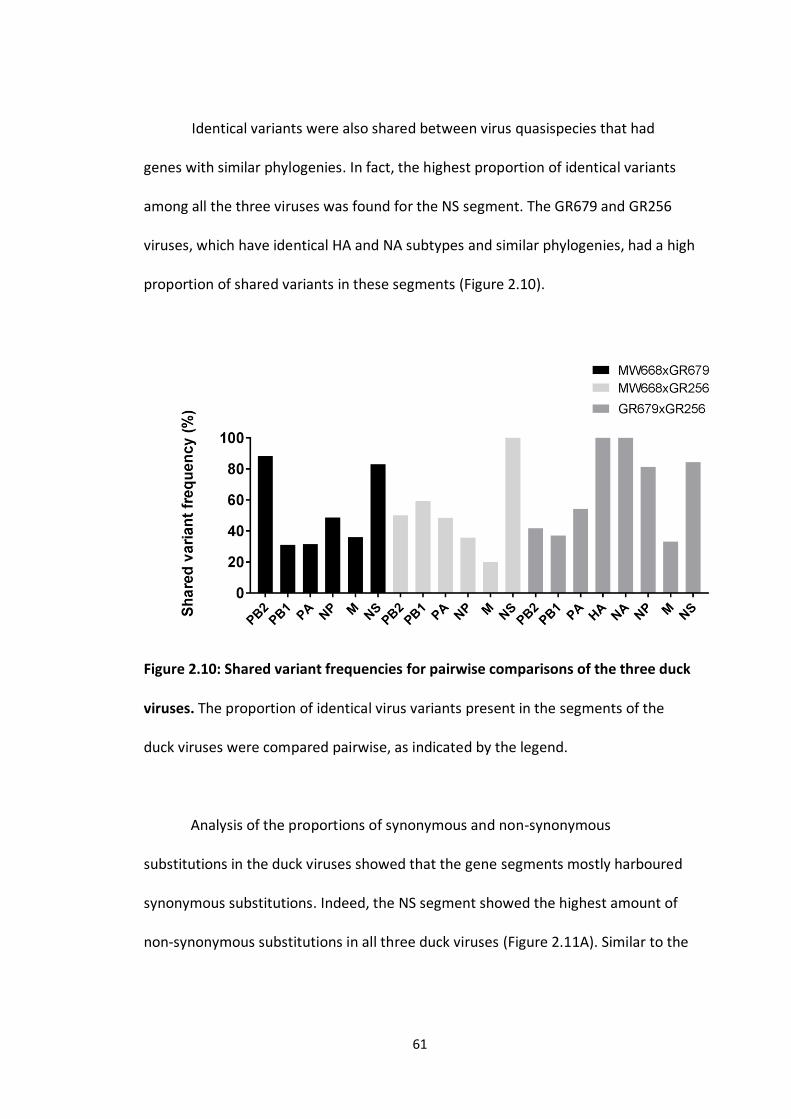
61
Identical variants were also shared between virus quasispecies that had
genes with similar phylogenies. In fact, the highest proportion of identical variants
among all the three viruses was found for the NS segment. The GR679 and GR256
viruses, which have identical HA and NA subtypes and similar phylogenies, had a high
proportion of shared variants in these segments (Figure 2.10).
Figure 2.10: Shared variant frequencies for pairwise comparisons of the three duck
viruses. The proportion of identical virus variants present in the segments of the
duck viruses were compared pairwise, as indicated by the legend.
Analysis of the proportions of synonymous and non-synonymous
substitutions in the duck viruses showed that the gene segments mostly harboured
synonymous substitutions. Indeed, the NS segment showed the highest amount of
non-synonymous substitutions in all three duck viruses (Figure 2.11A). Similar to the

62
murre viruses, none of the NS segment of all duck viruses have a dN/dS ratio>1
(Figure 2.11B).
Figure 2.11: Synonymous and non-synonymous substitution frequencies for all
segments of the duck viruses. A) The proportions of non-synonymous substitutions
(dN) and synonymous substitutions (dS) were determined in each virus quasispecies
for all segments. B) The dN/dS ratio was determined for all segments.
Variants present in the quasispecies of the two similar duck viruses, GR679
and GR256, shared some similar distributions in terms of frequency but only for the
H3, M, and NS segments (Figure 2.12).

63
Figure 2.12: Comparison of the quasispecies structure of two duck viruses with
similar genotypes. Shannon diversity index was applied to all variants present for
each segment of the two duck viruses, GR679 and GR256. Each dot represents how
frequent the variant is within the segment quasispecies. Horizontal bars represent
the overall variant diversity within each segment. The distributions of the variants
within each duck quasispecies were compared with a Kruskal-Wallis test (P<0.0001).
An asterisk above the plot represents significantly dissimilar distribution compared
to the corresponding segment of the other virus. No asterisk means the variant
distributions are very similar for the segments.
Overall, my analyses show that gull, murre or duck viruses with similar
genotypes display similar quasispecies structure in terms of shared variants and
proportions, but the amount of diversity among the variants, in terms of

64
frequencies, differ since some segments contain shared variants in higher
proportions than others.
2.3.2 Effect of replication in a different host on the virus quasispecies
Adaptation experiments were performed by infecting Atlantic puffin embryos
with a single IAV isolate from a wild duck through 10 consecutive passages. Due to
the limited availability of such eggs from wild birds, the serial passage was
performed over three consecutive years due to the availability of wild bird eggs and
allowance of the permits. The virus genome was deep-sequenced from the initial
swab sample (P0), the first (P1), the fifth (P5) and last passage (P10). Mutation
analyses were performed by comparing the viral genome deep sequenced and
translated computational protein sequences. The viral titer, determined by
hemagglutination assays for the 10 passages, decreased over the passages and
became undetectable in the last two passages (Figure 2.13).

65
Figure 2.13: Viral titer of the duck virus over the serial passages in puffin embryos.
The virus titer was determined by hemagglutination assay in the 10 samples
collected after the serial virus passages.
2.3.2.1 Mutational analysis of the duck viral genome
Analysis of the number of variants present in the initial swab sample (P0), the
fifth passage (P5), and the last passage in puffin embryos (P10) shows an overall
increase of the number of variants in the last virus passage for all gene segments
except for NS compared to the initial swab (P0) (Figure 2.14).

66
Figure 2.14: Venn diagrams of the overall number of virus variants in the initial
swab sample (P0), the fifth passage (P5), and the last passage (P10). A) The total
number of variants was compared between the initial swab sample (P0) and the fifth
passage (P5). B) The total number of variants was compared between the initial
swab sample (P0) and the last passage (P10).

67
From the overall number of virus variants presented in Figure 2.14, I then
quantified the proportions of synonymous (dS) and non-synonymous (dN)
substitutions in all segments of the initial swab sample (P0), the fifth passage (P5),
and the last virus passage (P10) were analyzed (Figure 2.15).
Figure 2.15: Proportions of non-synonymous (dN) and synonymous (dS)
substitutions in the duck virus segments. A) Both dN and dS were determined in the
initial swab sample (P0), the fifth passage (P5), and the last virus passage (P10) for all
segments. B) The dN/dS ratio was determined for all segments in P0, P5 and P10.

68
There was an increase in the proportion of non-synonymous substitutions in
the fifth and last virus passages for the HA and NA gene segments compared to P0
(Figure 2.15A). The proportion of non-synonymous substitutions was higher in P10
compared to P5. I next examined the direction of the evolution of the different gene
segments with the use of dN/dS ratios we previously described in section 2.3.1.1. To
resume, a dN/dS ratio >1 means that the sequence is under positive selection while a
ratio <1 implies that the sequence is under negative selection. A ratio equal to 1
indicates that the gene is under neutral pressure. Both the HA and NA genes had a
dN/dS ratio >1 in P5 and P10 meaning that both genes were under positive selective
pressure over the passage of the virus (Figure 2.15, B).
2.3.2.2 Mutational analysis of the duck protein sequences
To investigate the effects of non-synonymous mutations introduced in the HA
and NA proteins during the fifth and last virus passages, I analyzed their amino acid
sequences. I performed protein alignments with the sequences from P5 and P10 and
additional HA and NA proteins retrieved from the NCBI Influenza Virus Database in
order to identify more- and less-conserved regions. Analysis of the HA protein during
the different passages showed that amino acid changes were mostly being
accumulated, for both P5 and P10, in conserved regions of the cleavage site and
fusion domain of the protein. Also, some truncations and substitutions also
appeared in the central region of HA2 which could negatively interfere with the
protein functions (Figure 2.16).

69
70
Figure 2.16 Comparison of the HA amino acid changes from the fifth and last virus
passages. The fifth and last virus passages were aligned with other HA protein
sequences with MUSCLE implemented in Geneious 8. The HA amino acid (aa)
sequence is annotated above the alignment. The N-terminal part of the protein
contains a peptide signal (17 aa) responsible for membrane anchoring at the cell
surface. The remainder of the protein is divided into two domains, HA1 and HA2,
with a cleavage site (7 aa) located between the two domains. A fusion domain (23
aa) is located in the HA2 domain. A transmembrane domain (28 aa) is located in the
C-terminal part of the protein. Receptor binding sites (RBS) and antigenic sites are
also displayed on the annotation. The amino acid changes (substitutions and
deletions) on the fifth (P5 mutations) and last virus passage (P10 mutations) are
indicated according to the legend.
Analysis of the NA amino acid sequences from P5 and P10 showed an
accumulation of mostly substitutions in similar locations within the stalk and
catalytic domains of the protein (Figure 2.17).

71

72
Figure 2.17: Comparison of the NA amino acid changes from the fifth and last virus
passages. The fifth and last virus passages were aligned with other NA protein
sequences with MUSCLE implemented in Geneious. The NA amino acid (aa)
sequence is annotated and displayed above the alignment. The N-terminal part of
the protein contains a signal-anchoring domain (35 aa) followed by a stalk domain
(55 aa), and a catalytic domain (380 aa). The amino acid changes (substitutions and
frameshift) from P5 and P10 are indicated.
2.4 Discussion
Virus quasispecies are spectra of non-identical, but closely related viruses
that are released after a viral infection (Andino and Domingo 2015). The virus
quasispecies can change depending on the host environment (Schneider and
Roossinck 2001). IAV genetic studies are often focused on the consensus nucleotide
composition of the viral genome. Few research studies have undertaken
investigations on IAV genomic structure at the quasispecies level. Thus, I performed
an analysis of the genomic structure of viruses shed by three different bird hosts,
murres, gulls, and ducks to investigate whether the virus quasispecies could be
conserved within similar hosts. My analyses showed that viruses from the same bird
host display similar quasispecies patterns. This was also demonstrated in one study
when comparing the quasispecies of hepatitis C viruses within similar human
recipients (Laskus, Wilkinson et al. 2004, Asghar, Pettersson et al. 2017). In my study,
all viruses showed variant frequency levels that matched with other viruses
73
according to the corresponding phylogenetic relatedness of the viral segments. The
proportions of shared variants were also correlated with the phylogenetic
relationships of the segments such that similar genes in term of host and geographic
origins share similar proportions of variants. Analysing the association of shared
variant frequencies with the sequence homologies between segments
(Supplementary figure 2.2) showed a general correlation between the proportion of
shared variants and nucleotide sequence similarities. The pattern was most striking
for the gull viruses, with slightly broader distributions for the murre and duck
viruses. When comparing the variant diversities, as quantified by the Shannon
diversity index, of two viruses with similar genotypes I saw that some segments
harbour similar variant distributions but not all. This could be explained by the fact
that viruses that harbour similar genotypes have and maintain similar variants within
the same host. Although the quasispecies of viruses with similar genotypes share a
large number of variants, the proportion of those shared variants within each
quasispecies varied. The NS segments from the gull and murre viruses had a higher
proportion of non-synonymous substitutions compared to the duck viruses. These
differences might be explained by the fact that different bird host species carry
different allelic versions of the NS segment. The gull and murre viruses contained NS
allele A, which is found in bird and mammalian hosts, while the duck viruses in this
study contained allele B, which is typically only found in bird species (Ludwig, Schultz
et al. 1991). Differences in host genetic and phenotypic makeup could explain why
certain viral genes are transmitted or not in certain hosts. The host immunity could
act as a barrier for allowing the transmission of viral genes.

74
The effect of replication in a different host on the evolutionary adaptation of
a duck virus in Atlantic puffin embryos was studied experimentally. A decrease of
viral titer was observed during serial passage of the virus. An overall increase of
variants within the virus quasispecies was observed in the fifth and last virus
passages examined by deep sequencing, except for the NS segment. I would have
expected an increase in viral titer once the virus adapted to a new host. Hence,
additional passages could be required to see an increase of viral titers. Also, the
technique I used to titer the virus, hemagglutination, might lack sensitivity to detect
the virus at low concentrations. A sensitive QPCR assay to quantify the number of
viral genome copies could be performed in parallel. Lastly, the possible presence of
maternal antibodies in embryos from wild birds could restrict the virus replication
and also affect the viral titer I measured.
I analyzed the accumulated mutations over the different passages within the
amino acid sequences of both surface glycoproteins, HA and NA.
For the HA protein, most of the mutations were nucleotide substitutions that
changed the amino acid sequence at the cleavage site or fusion domain. The fusion
domain contains conserved amino acids that are necessary for the virus to be
released into the cell’s cytoplasm (Smrt, Draney et al. 2015). The HA cleavage site
allows maturation of HA protein to produce functional and replicative viruses
(Garten and Klenk 1983, Steinhauer 1999). A large number of mutations, nucleotide
substitutions and deletions, were also found in the central domain of HA2, and these
mutations could have an impact on the conformational changes during the fusion
process (Armstrong, Kushnir et al. 2000). Surprisingly, few mutations were found in
75
RBS regions that are involved in cellular receptor recognition (Sriwilaijaroen and
Suzuki 2012, Lazniewski, Dawson et al. 2017). A previous investigation of the
adaptation of wild viruses to new avian hosts showed that new mutations
accumulated in the HA1 domain of the HA protein and some of these mutations
were located in RBS regions (Li and Cardona 2010, Li, zu Dohna et al. 2010). Other
studies found similar findings to what I observed, with mutations in the HA cleavage
site and HA2 domain (Hossain, Hickman et al. 2008, Dlugolenski, Jones et al. 2011).
We could not directly compare mutations with those studies because the viruses do
not have the same HA subtype.
For the NA protein, several mutations, mostly substitutions but also one
frameshift, were found in the stalk and catalytic domains. A deletion within the stalk
region has been previously reported during experimental infections of wild bird
viruses in some poultry hosts, but not in wild bird hosts (Li and Cardona 2010,
Dlugolenski, Jones et al. 2011). The NA stalk deletion conferred a replicative
advantage of the virus in poultry hosts (Dlugolenski, 2011, Hossain, 2008, Pei, 2009).
Deep sequencing analysis of a wild bird virus genome before introduction into
poultry revealed the NA stalk deletion preexisted in the virus quasispecies (Croville,
Soubies et al. 2012). Thus, it seems that the introduction of mutations within the
stalk region of the NA protein I analyzed during the experimental assay could be the
result of an adaption process of the duck virus in the Atlantic puffin host.
To conclude, my analysis of the of different IAVs shed by wild bird hosts
shows a general conservation of overall quasispecies genomic structure for viruses
replicating in the same host groups. This suggests that similar virus variants are

76
favored within each host group, and these are likely those best adapted to that host
and which allow their effective transmission and preservation within the same host
species. The study of the adaptation of a wild duck virus to a different avian host,
conducted experimentally in bird embryos, showed major genetic changes in the HA
and NA proteins despite of an overall decrease of viral titers. In our study, regions
associated with the maturation of the HA and NA proteins appeared to be the most
frequent regions where mutations accumulate, and this is similar to observations
from other studies with different viruses and host groups. Despite giving insights on
the origin of viruses in term of host and geographic location, applying high-
throughput next-generation sequencing technologies also allows the dissection of
the virus genomic structure which gives a better understanding on how the virus
could evolve and be transmitted in different hosts. It allows us to foresee mutations
that could have occurred and have an impact of the virus function and evolution in
different hosts. This could also be a useful method to predict and detect emerging
pathogenic viruses in their natural reservoir that could be deadly to other hosts.
2.5 References
Andino, R. and E. Domingo (2015). "Viral quasispecies." Virology 479-480: 46-51.
Armstrong, R. T., A. S. Kushnir and J. M. White (2000). "The Transmembrane Domain of
Influenza Hemagglutinin Exhibits a Stringent Length Requirement to Support the Hemifusion
to Fusion Transition." The Journal of Cell Biology 151(2): 425-438.

77
Asghar, N., J. H. Pettersson, P. Dinnetz, A. Andreassen and M. Johansson (2017). "Deep
sequencing analysis of tick-borne encephalitis virus from questing ticks at natural foci
reveals similarities between quasispecies pools of the virus." The Journal of General Virology
98(3): 413-421.
Croville, G., S. M. Soubies, J. Barbieri, C. Klopp, J. Mariette, O. Bouchez, C. Camus-
Bouclainville and J. L. Guerin (2012). "Field monitoring of avian influenza viruses: whole-
genome sequencing and tracking of neuraminidase evolution using 454 pyrosequencing."
Journal of Clinical Microbiology 50(9): 2881-2887.
Dlugolenski, D., L. Jones, G. Saavedra, S. M. Tompkins, R. A. Tripp and E. Mundt (2011).
"Passage of low-pathogenic avian influenza (LPAI) viruses mediates rapid genetic adaptation
of a wild-bird isolate in poultry." Archives of Virology 156(4): 565-576.
Domingo, E., E. Baranowski, C. M. Ruiz-Jarabo, A. M. Martín-Hernández, J. C. Sáiz and C.
Escarmís (1998). "Quasispecies structure and persistence of RNA viruses." Emerging
Infectious Diseases 4(4): 521-527.
Garten, W. and H. D. Klenk (1983). "Characterization of the carboxypeptidase involved in the
proteolytic cleavage of the influenza haemagglutinin." The Journal of General Virology 64 (Pt
10): 2127-2137.
Hoffmann, E., J. Stech, Y. Guan, R. G. Webster and D. R. Perez (2001). "Universal primer set
for the full-length amplification of all influenza A viruses." Arch Virol 146(12): 2275-2289.
Holland, J., K. Spindler, F. Horodyski, E. Grabau, S. Nichol and S. VandePol (1982). "Rapid
evolution of RNA genomes." Science 215(4540): 1577-1585.

78
Hossain, M. J., D. Hickman and D. R. Perez (2008). "Evidence of Expanded Host Range and
Mammalian-Associated Genetic Changes in a Duck H9N2 Influenza Virus Following
Adaptation in Quail and Chickens." PLoS One 3(9): e3170.
Huang, Y., M. Wille, J. Benkaroun, H. Munro, A. L. Bond, D. A. Fifield, G. J. Robertson, D.
Ojkic, H. Whitney and A. S. Lang (2014). "Perpetuation and reassortment of gull influenza A
viruses in Atlantic North America." Virology 456-457: 353-363.
Huang, Y., M. Wille, A. Dobbin, N. M. Walzthoni, G. J. Robertson, D. Ojkic, H. Whitney and A.
S. Lang (2014). "Genetic structure of avian influenza viruses from ducks of the Atlantic flyway
of North America." PLoS One 9(1): e86999.
Jukes, T. H. and C. R. Cantor (1969). CHAPTER 24 - Evolution of Protein Molecules.
Mammalian Protein Metabolism. H. N. Munro, Academic Press: 21-132.
Kuiken, T., E. C. Holmes, J. McCauley, G. F. Rimmelzwaan, C. S. Williams and B. T. Grenfell
(2006). "Host species barriers to influenza virus infections." Science 312(5772): 394-397.
Laskus, T., J. Wilkinson, J. F. Gallegos-Orozco, M. Radkowski, D. M. Adair, M. Nowicki, E.
Operskalski, Z. Buskell, L. B. Seeff, H. Vargas and J. Rakela (2004). "Analysis of hepatitis C
virus quasispecies transmission and evolution in patients infected through blood
transfusion." Gastroenterology 127(3): 764-776.
Lazniewski, M., W. K. Dawson, T. Szczepinska and D. Plewczynski (2017). "The structural
variability of the influenza A hemagglutinin receptor-binding site." Brief Funct Genomics.
Li, J. and C. J. Cardona (2010). "Adaptation and transmission of a wild duck avian influenza
isolate in chickens." Avian Diseases 54(1 Suppl): 586-590.

79
Li, J., H. zu Dohna, N. L. Anchell, S. C. Adams, N. T. Dao, Z. Xing and C. J. Cardona (2010).
"Adaptation and transmission of a duck-origin avian influenza virus in poultry species." Virus
Research 147(1): 40-46.
Ludwig, S., U. Schultz, J. Mandler, W. M. Fitch and C. Scholtissek (1991). "Phylogenetic
relationship of the nonstructural (NS) genes of influenza A viruses." Virology 183(2): 566-
577.
Schneider, W. L. and M. J. Roossinck (2001). "Genetic Diversity in RNA Virus Quasispecies Is
Controlled by Host-Virus Interactions." Journal of Virology 75(14): 6566-6571.
Smrt, S. T., A. W. Draney and J. L. Lorieau (2015). "The Influenza Hemagglutinin Fusion
Domain Is an Amphipathic Helical Hairpin That Functions by Inducing Membrane Curvature."
The Journal of Biological Chemistry 290(1): 228-238.
Sriwilaijaroen, N. and Y. Suzuki (2012). "Molecular basis of the structure and function of H1
hemagglutinin of influenza virus." Proceedings of the Japan Academy. Series B, Physical and
Biological Sciences 88(6): 226-249.
Steinhauer, D. A. (1999). "Role of hemagglutinin cleavage for the pathogenicity of influenza
virus." Virology 258(1): 1-20.
Verhoeven, J. T. P., M. Canuti, H. J. Munro, S. C. Dufour and A. S. Lang (2018). "ViDiT-
CACTUS: an inexpensive and versatile library preparation and sequence analysis method for
virus discovery and other microbiology applications." Canadian Journal of Microbiology: 1-
13.
Webster, R. G., W. J. Bean, O. T. Gorman, T. M. Chambers and Y. Kawaoka (1992). "Evolution
and ecology of influenza A viruses." Microbiological Reviews 56(1): 152-179.

80
Yoon, S. W., R. J. Webby and R. G. Webster (2014). "Evolution and ecology of influenza A
viruses." Current topics in Microbiology and Immunology 385: 359-375.

81
Chapter 3: Analysis of the Variability in the Non-Coding Regions of
Influenza A Viruses
Abstract
The genomes of influenza A viruses (IAVs) comprise eight negative-sense single-
stranded RNA segments. In addition to the protein-coding region, each segment
possesses 5′ and 3′ non-coding regions (NCR) that are important for transcription,
replication and packaging. The NCRs contain both conserved and segment-specific
sequences, and the impacts of variability in the NCRs are not completely understood.
Full NCRs have been determined from some viruses, but a detailed analysis of
potential variability in these regions among viruses from different host groups and
locations has not been performed. To evaluate the degree of conservation in NCRs
among different viruses, we sequenced the NCRs of IAVs isolated from different wild
bird host groups (ducks, gulls and seabirds). We then extended our study to include
NCRs available from the National Center for Biotechnology Information (NCBI)
Influenza Virus Database, which allowed us to analyze a wider variety of host species
and more HA and NA subtypes. We found that the amount of variability within the
NCRs varies among segments, with the greatest variation found in the HA and NA
and the least in the M and NS segments. Overall, variability in NCR sequences was
correlated with the coding region phylogeny, suggesting vertical coevolution of the
(coding sequence) CDS and NCR regions.

82
3.1 Introduction
Influenza A viruses (IAVs) are most well known for their circulation in humans,
causing yearly seasonal epidemics and occasional pandemics, and for outbreaks in
poultry. However, the viral strains responsible for these infections represent only a
small portion of the total known IAV diversity, most of which is maintained in the
natural wild bird reservoir.
IAVs are members of the family Orthomyxoviridae and have segmented
negative-sense single-stranded RNA genomes. The viral genome is composed of 8
segments that encode at least 11 proteins, depending on the strain. Each segment
has non-coding regions (NCRs) at both ends. The 3’ NCR contains 12 conserved
nucleotides followed by a segment-specific region of variable length. The 5’ NCR
contains 13 conserved nucleotides and a segment-specific region of variable length.
The two conserved regions are partially complementary (Skehel and Hay 1978,
Robertson 1979, Desselberger, Racaniello et al. 1980) and form a panhandle-like
structure (Hsu, Parvin et al. 1987, Bergmann and Muster 1996, Kim, Fodor et al.
1997) involved in the transcription and replication steps of the viral life cycle (Seong
and Brownlee 1992, Fodor, Pritlove et al. 1994, Flick, Neumann et al. 1996).
Mutagenesis studies identified residues within the NCRs that are required for the
viral polymerase complex to bind and initiate transcription (Li and Palese 1992,
Fodor, Seong et al. 1993). However, a binding preference for the 5’ NCR was
demonstrated (Pritlove, Fodor et al. 1995). There is also a stretch of uridines at the 5’
end that is involved in the polyadenylation of viral mRNAs (Luo, Luytjes et al. 1991, Li
and Palese 1994, Pritlove, Poon et al. 1998, Poon, Pritlove et al. 1999). Further

83
studies have revealed the presence of some variability within the two NCRs at
different positions. There is variability in the fourth nucleotide (C or U) in the 3’ end
of all RNA segments (Robertson 1979), which affects the promoter and could play a
role in differential regulation of transcription and replication (Lee, Bae et al. 2003).
Non-conserved nucleotides located in the segment-specific regions have also been
shown to play an important role in viral replication (Zheng, Palese et al. 1996).
Mutagenesis studies have revealed the importance of NCRs for the incorporation of
the eight different gene segments during viral assembly (Fujii, Fujii et al. 2005, Gao
and Palese 2009, Zhao, Peng et al. 2014, Crescenzo-Chaigne, Barbezange et al. 2017).
These packaging signals can influence the reassortment of genes during co-infections
of different strains by preventing their incorporation during the virus genome
packaging (Gao and Palese 2009, White, Steel et al. 2017).
Amplification of complete IAV genomes with universal primers was initially
designed based on the conserved nature of NCRs (Hoffmann, Stech et al. 2001), but
amplification biases have been reported (Zhou, Donnelly et al. 2009, Widjaja, de
Vries et al. 2012). To date, a thorough analysis of IAV NCRs from different hosts and
across segments has not been performed. We hypothesized that the NCRs within
both the conserved and the segment-specific regions may vary according to the
original host species. To test our hypothesis, we characterized the true NCRs of IAV
genomes from different wild bird species by the rapid amplification of cDNA ends
(RACE) method and then expanded our analyses to include additional viral subtypes,
hosts and geographic origins.

84
3.2. Materials and Methods
3.2.1. NCR sequence determination
NCR sequences from previously identified and sequenced viruses originating
from three different wild bird host groups (ducks, gulls and seabirds) were
determined: A/domestic duck/Newfoundland/MW668/2010(H1N1), A/American
black duck/Newfoundland/GR679/2011(H3N2), A/herring
gull/Newfoundland/GR578/2011(H13N6), A/herring
gull/Newfoundland/GR848/2011(H13N6), A/common
murre/Newfoundland/AB318/2011(H1N2), and A/common
murre/Newfoundland/AB324/2011(H1N2). From these six viruses, we determined
92 NCR sequences, comprising 46 3’ and 46 5’ NCRs. When possible, viruses with the
same subtype were chosen for NCR characterization.
Viral genomic RNA was extracted from allantoic fluids of specific pathogen
free eggs inoculated with the different viruses using the Trizol reagent (Thermo
Fisher Scientific). Primers were designed to efficiently amplify the 3’ and 5’ NCRs of
the targeted viruses based on the full genomic sequences of viruses that were
available from previous studies (Supplementary table 3.1).
For the 3’ NCRs, genomic RNAs were polyadenylated with poly(A) polymerase (2 U
µL-1) (Thermo Fisher Scientific) in the presence of MnCl2 (2.5 mM) and ATP (1 mM)
in 1X reaction buffer and a final volume of 50 µL. Reactions were incubated at 37°C
for 30 min and the resulting RNA used as the input for the 3’ RACE System for Rapid
Amplification of cDNA Ends (Thermo Fisher Scientific). For the 5’ NCRs, the 5’ RACE
System for Rapid Amplification of cDNA Ends (Thermo Fisher Scientific) was used

85
according to the manufacturer’s recommendations. The resulting amplicons were
purified and sequenced using Sanger chemistry at The Center for Applied Genomics
(Toronto, Canada).
3.2.2 Sequence analyses
The 92 NCRs determined from our samples were compared with NCRs
retrieved from the influenza resource database
(http://www.ncbi.nlm.nih.gov/genomes/FLU/FLU.html), chosen to represent
different viral subtypes, host species, geographic origins, and collection dates. When
possible, all available or a high number of nucleotide sequences of complete gene
segments were downloaded from the database. The number of NCRs analyzed are
indicated in Figure 3.3. Sequence alignments were performed using MUSCLE,
implemented in MEGA version 6 (Tamura, Stecher et al. 2013). Multiple alignments
were then edited in Geneious version 8 (Kearse, Moir et al. 2012). The CDS regions of
segments that had complete NCRs were removed from the sequence, with only the
NCRs kept for further analysis. Complete segments without NCRs were removed
from the analysis.
Pairwise genetic distance matrices determined with the Maximum Composite
Likelihood method, generated by MEGA, were used for classical multidimensional
analysis (MDS) in R (R Development Core Team 2008). The viral segments were
assigned geographic origins based on nucleotide BLAST searches (Altschul, Gish et al.
1990) and neighbor-joining phylogenetic analyses in MEGA. High bootstrap values
are shown at the nodes on the trees. A small number of sequences was added in

86
each multiple alignment figures due to space limitation, but the exact number of
NCRs originally aligned with MUSCLE are indicated in section 3.3.2.
3.3 Results and Discussion
3.3.1 Characterization of NCRs from different wild bird viruses
Comparison of the 3’ and 5’ NCRs we determined with the RACE method showed
that the 12- and 13-nt conserved regions, respectively, contained no novel
substitutions in any of the different wild bird viruses (Figures 3.1 and 3.2), which
agrees with previous studies (Skehel and Hay 1978, Robertson 1979, Desselberger,
Racaniello et al. 1980).

87
Figure 3.1: 3’ NCRs of wild bird virus segments determined in this study. The
multiple alignment of 3’ NCRs was generated with MUSCLE, implemented in
Geneious version 8. The RNA sequences are displayed in the 3’ to 5’ orientation
corresponding to the packaged RNAs.

88
Figure 3.2: 5’ NCRs of wild bird virus segments determined in this study. The
multiple alignment of 5’ NCRs was generated with MUSCLE implemented in
Geneious version 8. The RNA sequences are displayed in the 3’ to 5’ orientation
corresponding to the packaged RNAs.
Comparison of the 3’ NCRs (Figure 3.1) showed that the 4th position in the 12-
nucleotide conserved region displays the expected variability, with the polymerase

89
gene segments, PB2, PB1 and PA, containing a cytosine residue while the other
segments contain a uridine residue, as previously observed (Robertson 1979, Lee, Bae
et al. 2003).
For the 3’ segment-specific regions (Figure 3.1), some of the segments (PB1, M
and NS) had identical sequences across all six viruses, but the others showed some
differences. The murre and gull PB2 and NP segments were identical whereas the
gull viruses differed from the others for these segments. The PA segment was
identical in the gull and duck viruses but differed from these in the murre viruses.
The HA sequences were identical within subtypes, even for the H1 viruses that came
from murres and a duck. Similarly, the NA sequences were identical within the same
subtype.
Comparison of the 5’ NCRs showed the conservation of a stretch of 5 or 6
uridine residues upstream of the 13-nt conserved region among the different wild
bird viruses (Figure 3.2). Similar to the 3’ NCRs, the segment-specific regions varied
among segments, with some of the individual segments’ NCRs conserved among the
six viruses and others varying among the viruses (Figure 3.2). All PB2, PA, M, and NS
segments’ NCRs were identical among the viruses. There was a single variation in the
PB1 segment, with the murre and one gull virus differing from the other gull virus
and the duck viruses. As observed for the 3’ NCRs, the HA and NA segments were the
same within subtypes, and the H1 and H2 segments were also identical.
3.3.2 The variability in segment-specific NCR sequences differs among segments
To further investigate the differences observed in the NCRs of our limited set
of six viruses, we expanded our analysis of NCR genetic diversity to include viral

90
sequences from a variety of host species and geographic origins retrieved from the
NCBI Influenza Virus Database (Supplementary Table 3.2). There is no apparent
relationship between the number of sequences analyzed and the NCR sequence
diversity (Figure 3.3).

91
Figure 3.3: Genetic diversity of 3’ and 5’ NCRs for different segments. (A) HA
segments of the different subtypes. (B) NA segments of the different subtypes. (C)
“Internal” protein-coding segments. The genetic diversity for each of the NCR
regions for the different segments was calculated as mean genetic distance for the
NCRs included in our analyses. The genetic distance was determined with the
Maximum Composite Likelihood method implemented in MEGA. The number above
each bar represents the number of sequences used to calculate the amount of
genetic diversity for each segment.

92
The greatest amount of variability in NCRs was observed in the HA segments
(Figure 3.3A), but there is variability among the segments and between 3’ and 5’
NCRs for some subtypes. The H4, H6, H7, H8, H10, H12, and H14 NCRs showed very
little variability overall. The H5, H9 and H13 subtypes show greater variability within
their 3 ’NCRs relative to their 5’ NCRs, whereas the H1, H2, H3, H11 and H16 were
equally variable at the two ends. The lower variability at the NCRs could be
attributed to a lack of host diversity or a bias of sampling.
The genetic diversity of different NA subtypes’ NCRs (Figure 3.3B), which
originated from a large number of host species and geographic locations, showed
that the N3 and N6 subtypes have more diversity at their 3’ ends while the N5, N8,
and N9 are more diverse at the 5’ ends. The N1 and N2 viruses showed very little
diversity, despite the large assortment of hosts from which viruses were included.
Matching the general pattern observed for IAV segment coding regions, the
NCRs of the remaining segments are overall less variable than found for HA and NA
(Figure 3.3C). The M and NS segments showed the lowest diversity, in agreement
with the slower evolution rates for these segments’ CDS [e.g. (Bahl, Vijaykrishna et
al. 2009, Qu, Zhang et al. 2011)]. These differences we have observed across many
viruses may not be too surprising given that a previous analysis of NCRs from a
limited dataset of human H3N2 viruses circulating worldwide since 1968 found that
NCR variability differed among the segments (Furuse and Oshitani 2011).
3.3.3 Patterns of variability within NCRs can be explained by viral host species and
geographic origins

93
To better understand the origin of the segment-specific NCR diversity, we
assessed whether the NCRs of viruses from different hosts and geographic origins
showed similar patterns of relationships as the respective coding regions (CDS). We
used a multidimensional scaling (MDS) analysis based on pairwise genetic distance
matrices for the NCR sequences, which allowed us to visualize the relationships
among NCRs relative to the viral hosts and geographic origins. The results of these
analyses were then compared to MDS analyses performed on the respective coding
regions. We focused this analysis on segments that showed greater NCR diversity:
H1, H9 and H13 for the hemagglutinin, N6 for the neuraminidase, and the NP
segment.
The relationships among the 3’ NCRs and CDS regions of a set of H9 segments
were compared (Figure 3.4).

94
Figure 3.4: Relationships among H9 3’ NCR and CDS regions for viruses from
different host species and geographic locations. The MDS scatter plots (A) are based
on the pairwise genetic distance matrices from the multiple alignments of complete
CDS regions and 3’NCRs. Each dot represents a virus, colored according to their
origins as indicated on the CDS-based phylogenetic tree (B). Alignments of the 3’

95
NCRs, shown corresponding to the genomic RNAs, from the viruses used in the
analyses are next to the CDS-based neighbor-joining tree for these viruses.
The 3’ NCRs and CDS regions showed similar clustering in the MDS analyses (Figure
3.4A), corresponding to their CDS phylogenetic clades (Figure 3.4B). The two North
American clade sequences grouped more closely and are more distant from the
Eurasian and North African sequences (Figure 3.4A). This matches the patterns for
the CDS phylogeny and 3’ NCR alignments (Figure 3.4B).
The same patterns were observed with the H13 subtype 3’ NCRs in the MDS
analyses (Figure 3.5A), and there was again a clear correspondence between the CDS
phylogeny and the NCR sequence relationships (Figure 3.5B).

96

97
Figure 3.5: Relationships among H13 3’ NCR and CDS regions for viruses from
different host species and geographic locations. The MDS scatter plots (A) are based
on the pairwise genetic distance matrices from the multiple alignments of complete
CDS regions and 3’NCRs. Each dot represents a virus, colored according to their
origins as indicated on the CDS-based phylogenetic tree (B). Alignments of the 3’
NCRs, shown corresponding to the genomic RNAs, from the viruses used in the
analyses are next to the CDS-based neighbor-joining tree for these viruses.
For the H1 subtype, the CDS phylogeny contains larger host-specific (swine,
human and avian) clades that separate into geographic-specific (Eurasian and North
American) clades (Figure 3.6).

98
Figure 3.6: Relationships among H1 3’ NCR and CDS regions for viruses from
different host species and geographic locations. Alignments of the 3’ NCRs, shown
corresponding to the genomic RNAs, from the viruses used in the analyses are next
to the CDS-based neighbor-joining tree for these viruses.
The correspondence between CDS phylogeny and 3’ NCR similarities was again found
for these H1 sequences (Figure 3.6). A similar pattern was also found for the N6 NA
(Figure 3.7) and NP sequences (Figure 3.8), where the CDS phylogenies also
determined the 3’ NCR relationships. This was true even when the viruses falling
within specific clades originated from different hosts (e.g. gulls and ducks) or

99
different continental origins (Eurasia and North America). Overall, these patterns
suggest vertical coevolution of the CDS and NCRs.
Figure 3.7: Relationships among N6 3’ NCR and CDS regions for viruses from
different host species and geographic locations. Alignments of the 3’ NCRs, shown
corresponding to the genomic RNAs, from the viruses used in the analyses are next
to the CDS-based neighbor-joining tree for these viruses. To facilitate the distinction
of viruses from different hosts, gull viruses are in green, duck viruses are in orange,
shorebird viruses are in purple, and chicken viruses are in brown.

100
Figure 3.8: Relationships among NP 3’ NCR and CDS regions for viruses from
different host species and geographic locations. Alignments of the 3’ NCRs, shown
corresponding to the genomic RNAs, from the viruses used in the analyses are next
to the CDS-based neighbor-joining tree for these viruses.
Lastly, we looked at the M and NS segments, which showed very low overall
NCR diversity (Figure 3.3C). These two segments share very similar 3’ NCRs among
different viruses, regardless of the host species or geographic origins (Figure 3.9,
Supplementary figure 3.1).

101
Figure 3.9: Relationships among M 3’ NCR and CDS regions for viruses from
different host species and geographic locations. Alignments of the 3’ NCRs, shown
corresponding to the genomic RNAs, from the viruses used in the analyses are next
to the CDS-based neighbor-joining tree for these viruses.
Some variations were found for the M segment, for a total of three different M 3’
NCR sequences (Figure 3.9), but they do not show the same correspondence to the
phylogeny of their CDS as observed for the other segments. Comparison of a subset
of NS NCRs from varied hosts and geographic locations found these all to be identical
(Supplementary figure 3.1). The conservation of the M and NS NCRs may be linked to
the slower evolutionary rate of the coding regions of these segments, as mentioned
earlier.

102
3.4 General conclusions
We first hypothesized that NCRs may vary according to the host species. Our
initial evaluation of the 3’ and 5’ NCRs from viruses isolated from different wild bird
hosts showed that the conserved regions are identical among different hosts, but the
segment-specific regions showed some variability among the viruses for some
segments. This prompted a larger analysis including more sequences, which showed
that the amount of variability in the NCRs varies among the different segments and
can also vary between the 3’ and 5’ ends, depending on the segment. The overall
variability is highest for the HA and NA segments. Examination of 3’ NCRs from HA
and NA subtypes with higher variability indicates that the relationships among NCR
sequences matches the coding region phylogenies, which generally follow patterns
based on the viral host species and geographic origins, and possibly reflects a lack of
recombination in these segments. The most conserved NCRs are found on the M and
NS segments, which also show the slowest coding sequence evolutionary rates. The
exact evolutionary pressures acting on the NCRs and consequences of sequence
changes in these regions remain to be fully defined.
3.5 References
Altschul, S. F., W. Gish, W. Miller, E. W. Myers and D. J. Lipman (1990). "Basic local
alignment search tool." Journal of Molecular Biology. 215(3): 403-410.

103
Bahl, J., D. Vijaykrishna, E. C. Holmes, G. J. Smith and Y. Guan (2009). "Gene flow and
competitive exclusion of avian influenza A virus in natural reservoir hosts." Virology
390(2): 289-297.
Bergmann, M. and T. Muster (1996). "Mutations in the nonconserved noncoding
sequences of the influenza A virus segments affect viral vRNA formation." Virus
Research 44(1): 23-31.
Crescenzo-Chaigne, B., C. V. Barbezange, S. Leandri, C. Roquin, C. Berthault and S.
van der Werf (2017). "Incorporation of the influenza A virus NA segment into virions
does not require cognate non-coding sequences." Scientific reports 7: 43462.
Desselberger, U., V. R. Racaniello, J. J. Zazra and P. Palese (1980). "The 3' and 5'-
terminal sequences of influenza A, B and C virus RNA segments are highly conserved
and show partial inverted complementarity." Gene 8(3): 315-328.
Flick, R., G. Neumann, E. Hoffmann, E. Neumeier and G. Hobom (1996). "Promoter
elements in the influenza vRNA terminal structure." Rna 2(10): 1046-1057.
Fodor, E., D. C. Pritlove and G. G. Brownlee (1994). "The influenza virus panhandle is
involved in the initiation of transcription." Journal of Virology 68(6): 4092-4096.
Fodor, E., B. L. Seong and G. G. Brownlee (1993). "Photochemical cross-linking of
influenza A polymerase to its virion RNA promoter defines a polymerase binding site
at residues 9 to 12 of the promoter." The Journal of General Virology 74 ( Pt 7):
1327-1333.

104
Fujii, K., Y. Fujii, T. Noda, Y. Muramoto, T. Watanabe, A. Takada, H. Goto, T. Horimoto
and Y. Kawaoka (2005). "Importance of both the coding and the segment-specific
noncoding regions of the influenza A virus NS segment for its efficient incorporation
into virions." Journal of Virology 79(6): 3766-3774.
Furuse, Y. and H. Oshitani (2011). "Evolution of the influenza A virus untranslated
regions." Infection, Genetics and Evolution 11(5): 1150-1154.
Gao, Q. and P. Palese (2009). "Rewiring the RNAs of influenza virus to prevent
reassortment." PNAS 106(37): 15891.
Hoffmann, E., J. Stech, Y. Guan, R. G. Webster and D. R. Perez (2001). "Universal
primer set for the full-length amplification of all influenza A viruses." Archives of
Virology 146(12): 2275-2289.
Hsu, M. T., J. D. Parvin, S. Gupta, M. Krystal and P. Palese (1987). "Genomic RNAs of
influenza viruses are held in a circular conformation in virions and in infected cells by
a terminal panhandle." PNAS 84(22): 8140-8144.
Kearse, M., R. Moir, A. Wilson, S. Stones-Havas, M. Cheung, S. Sturrock, S. Buxton, A.
Cooper, S. Markowitz, C. Duran, T. Thierer, B. Ashton, P. Meintjes and A. Drummond
(2012). "Geneious Basic: an integrated and extendable desktop software platform
for the organization and analysis of sequence data." Bioinformatics 28(12): 1647-
1649.
105
Kim, H. J., E. Fodor, G. G. Brownlee and B. L. Seong (1997). "Mutational analysis of
the RNA-fork model of the influenza A virus vRNA promoter in vivo." The Journal of
General Virology 78 ( Pt 2): 353-357.
Lee, M. K., S. H. Bae, C. J. Park, H. K. Cheong, C. Cheong and B. S. Choi (2003). "A
single-nucleotide natural variation (U4 to C4) in an influenza A virus promoter
exhibits a large structural change: implications for differential viral RNA synthesis by
RNA-dependent RNA polymerase." Nucleic Acids Research 31(4): 1216-1223.
Li, X. and P. Palese (1992). "Mutational analysis of the promoter required for
influenza virus virion RNA synthesis." Journal of Virology 66(7): 4331-4338.
Li, X. and P. Palese (1994). "Characterization of the polyadenylation signal of
influenza virus RNA." Journal of Virology 68(2): 1245-1249.
Luo, G. X., W. Luytjes, M. Enami and P. Palese (1991). "The polyadenylation signal of
influenza virus RNA involves a stretch of uridines followed by the RNA duplex of the
panhandle structure." Journal of Virology 65(6): 2861-2867.
Poon, L. L., D. C. Pritlove, E. Fodor and G. G. Brownlee (1999). "Direct evidence that
the poly(A) tail of influenza A virus mRNA is synthesized by reiterative copying of a U
track in the virion RNA template." Journal of Virology 73(4): 3473-3476.
Pritlove, D. C., E. Fodor, B. L. Seong and G. G. Brownlee (1995). "In vitro transcription
and polymerase binding studies of the termini of influenza A virus cRNA: evidence
for a cRNA panhandle." The Journal of General Virology 76 ( Pt 9): 2205-2213.

106
Pritlove, D. C., L. L. Poon, E. Fodor, J. Sharps and G. G. Brownlee (1998).
"Polyadenylation of influenza virus mRNA transcribed in vitro from model virion RNA
templates: requirement for 5' conserved sequences." Journal of Virology 72(2):
1280-1286.
Qu, Y., R. Zhang, P. Cui, G. Song, Z. Duan and F. Lei (2011). "Evolutionary genomics of
the pandemic 2009 H1N1 influenza viruses (pH1N 1v)." Virology Journal 8: 250.
R Development Core Team (2008). R: A Language and Environment for Statistical
Computing, R Foundation for Statistical Computing.
Robertson, J. S. (1979). "5' and 3' terminal nucleotide sequences of the RNA genome
segments of influenza virus." Nucleic Acids Research 6(12): 3745-3757.
Seong, B. L. and G. G. Brownlee (1992). "Nucleotides 9 to 11 of the influenza A virion
RNA promoter are crucial for activity in vitro." The Journal of General Virology 73 ( Pt
12): 3115-3124.
Skehel, J. J. and A. J. Hay (1978). "Nucleotide sequences at the 5' termini of influenza
virus RNAs and their transcripts." Nucleic Acids Research 5(4): 1207-1219.
Tamura, K., G. Stecher, D. Peterson, A. Filipski and S. Kumar (2013). "MEGA6:
Molecular Evolutionary Genetics Analysis Version 6.0." Molecular Biology and
Evolution 30(12): 2725-2729.

107
White, M. C., J. Steel and A. C. Lowen (2017). "Heterologous Packaging Signals on
Segment 4, but Not Segment 6 or Segment 8, Limit Influenza A Virus Reassortment."
Journal of Virology 91(11).
Widjaja, I., E. de Vries, P. J. M. Rottier and C. A. M. de Haan (2012). "Competition
between Influenza A Virus Genome Segments." PLOS One 7(10): e47529.
Zhao, L., Y. Peng, K. Zhou, M. Cao, J. Wang, X. Wang, T. Jiang and T. Deng (2014).
"New Insights into the Nonconserved Noncoding Region of the Subtype-Determinant
Hemagglutinin and Neuraminidase Segments of Influenza A Viruses." Journal of
Virology 88(19): 11493-11503.
Zheng, H., P. Palese and A. Garcia-Sastre (1996). "Nonconserved nucleotides at the 3'
and 5' ends of an influenza A virus RNA play an important role in viral RNA
replication." Virology 217(1): 242-251.
Zhou, B., M. E. Donnelly, D. T. Scholes, K. St George, M. Hatta, Y. Kawaoka and D. E.
Wentworth (2009). "Single-reaction genomic amplification accelerates sequencing
and vaccine production for classical and Swine origin human influenza a viruses."
Journal of Virology 83(19): 10309-10313.
108
Chapter 4: Analysis of influenza A viruses from gulls: An evaluation of
inter-regional movements and interactions with other avian and
mammalian influenza A viruses
In this chapter, the results and discussion sections are combined in one section and
the methodology of the different analyses are described in their corresponding
figure legends.
Abstract
Birds, including members of the families Anatidae (waterfowl) and Laridae
(gulls and terns), serve as the major reservoir of influenza A viruses (IAVs). The
ecogeographic contributions of gulls to global IAV dynamics, in terms of geographic
scale and virus movements, are important and are distinct from those of waterfowl.
Gulls primarily carry the H13 and H16 subtypes, yet can be infected by additional
subtypes. Also, gulls are frequently infected by IAVs that contain mixtures of genes
from different geographic phylogenetic lineages (e.g. North American and Eurasian).
The present analysis examines a variety of viruses isolated from gulls and terns
across the world that exhibit particularly high phylogenetic affinities to viruses found
in other hosts. This illustrates the potential for gulls to act as highly pathogenic virus
carriers, disseminators of viruses over long distances, and contributors in the genesis
of pandemic strains. The historical evolution of an entirely Eurasian gull virus isolated
in North America was also traced and indicates the Caspian Sea, in Southwestern

109
Asia, was an important area for the generation of this virus, and analysis of IAVs from
terns also points to this region as relevant for the generation of novel strains.
4.1 Introduction
Wild birds, including waterfowl, shorebirds and gulls, harbor the vast majority
of known influenza A virus (IAV) genetic diversity. Within these avian influenza IAVs,
there is clear distinction between Eurasian and North American gene pools (Olsen,
Munster et al. 2006) and there is increasing evidence of additional phylogeographic
lineages (Pereda, Uhart et al. 2008, Gonzalez-Reiche and Perez 2012, Hurt,
Vijaykrishna et al. 2014). However, bird movements lead to inter-regional
transmission of IAV genes and their establishment in new regions. Intercontinental
transmission of IAV genes from Eurasia to America has been frequently observed,
and isolation of reassortant viruses that contain gene segments of different
continental origins is particularly common from gulls in these regions, which is
presumably facilitated by gull movements across the oceans. The North Atlantic
Ocean is one location where gull movements between North America and Europe
have been documented (Wille, Robertson et al. 2011), and intercontinental IAV
reassortants have also been identified frequently (Echeverría, Moratorio et al. 2015).
Additionally, both entirely Eurasian and North American gull IAVs have been found in
Iceland (Dusek, Hallgrimsson et al. 2014), suggesting this location as important for
the mixing of viruses between regions. An equivalent position has also been
proposed for Greenland (Shoham and Rogers 2006). In North America, only two
wholly Eurasian avian influenza A viruses have been found to date. One was an
H16N3 virus from an American herring gull (Larus smithsonianus) in eastern Canada,
110
(Echeverría, Moratorio et al. 2015). The other was the highly pathogenic avian
influenza (HPAI) H5N8 virus that recently emerged in China, spread to South Korea,
Japan and Siberia, and is proposed to have been transmitted through the Beringia
region by migratory waterfowl into Pacific North America (Lee, Swayne et al. 2015).
This H5N8 arrival ended the long speculation about whether or not virulent Asian
avian strains, such as H5N1, could be transmitted into the Americas by migrating
birds. There is also a third case, involving H9N2 viruses from Alaska that were nearly
identical (>99%) to viruses in China and South Korea, which were proposed to be
carried to Alaska by migratory waterfowl (Ramey, Reeves et al. 2015). In this case,
however, the gene segments of the viruses showed mixed geographic origins.
In addition to geographic divisions in IAV genes, distinct host group lineages
are found. These include mainly the avian, gull, human, swine and equine lineages
(Olsen, Munster et al. 2006). These distinctions make it possible to recognize that
inter-species transmissions of IAVs between gulls and other hosts have occurred.
Although most IAVs from gulls contain gull-lineage gene segments, and most are of
the hemagglutinin (HA) subtypes H13 and H16, various viruses found in gulls contain
HA genes and other gene segments that do not appear to be gull-specific, but rather
fall within avian clades and are closely related to waterfowl viruses in phylogenetic
analyses (Wille, Robertson et al. 2011, Van Borm, Rosseel et al. 2012, Hall, TeSlaa et
al. 2013). The opportunities for these transmissions certainly exist, as gulls share
terrestrial, freshwater and marine habitats with other birds and mammals, and
therefore gulls could be important for moving IAVs amongst ecosystems and for
contributing to the transmission of highly pathogenic IAVs to other hosts.

111
The present analysis is an investigation of the potential for gulls to act as
carriers of highly pathogenic viruses, disseminators of viruses over long distances,
and contributors in the genesis of pandemic strains. This is done through
examination a variety of gull and tern viruses isolated from different locations that
exhibit particularly high phylogenetic affinities to viruses found in other hosts.
4.2 Results and Discussion
4.2.1 Ecogenetic aspects of gull IAVs
Avian IAVs constitute the major, and likely the primal, component within the
total IAV gene pool worldwide (Webster, Bean et al. 1992, Olsen, Munster et al.
2006). Distinct phylogeographic lineages, such as those representing Eurasia and
North America, can be identified within avian IAVs, mainly as a result of partial
segregation of migratory birds (Olsen, Munster et al. 2006). The first indication that
gulls might represent a distinct reservoir for IAVs came from surveillance work on
the Atlantic coast of North America, with the identification of a new HA antigen,
H13, distinct from the previous 12 HA antigens identified in other avian species
(Hinshaw, Air et al. 1982). Two decades later, an additional novel HA subtype, H16,
was identified and characterized from gulls in Sweden (Fouchier, Munster et al.
2005). Continued surveillance and research have shown that gulls are predominantly
infected by these H13 and H16 subtypes, and these subtypes are not often found
outside of gulls (other than in shorebirds) (Arnal, Vittecoq et al. 2015), (Figure 4.1).

112
Figure 4.1: Distribution of IAV subtypes isolated from gulls. The numbers of viruses
for which sequence data are available with the indicated subtypes isolated from gulls
in North America and Eurasia are shown. The circular representation shows the total
proportions when both regions are combined. Gull viruses were located by presence
of M gene segment sequences in the NCBI Influenza Virus Resource
(http://www.ncbi.nlm.nih.gov/genomes/FLU/FLU.html), as of September 2015.
We performed phylogenetic and time of most recent common ancestor
(TMRCA) analyses of H13 and H16 nucleotide sequences. These showed three
distinct clades for each subtype that were estimated to originate from a common
ancestor in the 1930s for the H13 lineage (Figure 4.2) and in the 1920s for the H16
lineage (Figure 4.3), similar to previous estimates (Worobey, Han et al. 2014). Both
trees show some segregation of sequences according to geographic origin as well as
clades that contain mixtures of viruses from both Eurasia and North America. These
gull lineage HA genes have been transferred to waterfowl, poultry, and marine

113
mammals but there is no evidence of persistence of these sequences in these other
host groups.

114
Figure 4.2: Evolutionary history of H13 nucleotide sequences from North America
and Eurasia. All H13 nucleotide sequences were downloaded from the NCBI
Influenza Virus Resource (Bao, Bolotov et al. 2008) and aligned using MUSCLE
integrated in MEGA6 (Tamura, Stecher et al. 2013). The aligned sequences were
quality-trimmed, resulting in an alignment of 1133 nts (corresponding to nt positions
465-1597). The maximum credibility tree was generated from the trimmed
alignment by the Bayesian inference method implemented in BEAST v1.8.0
(Drummond, Suchard et al. 2012) using the SRD06 substitution model with a strict
molecular clock and with the use of the GMRF Bayesian skyride coalescent prior
distribution. These parameters were chosen because they gave the best distribution
of posterior probabilities. A strict molecular clock was chosen as appropriate to
estimate the time of the most recent ancestors at each node because the sequences
are almost all from the same host species. The branches are colored according to the
host group: green, North American gulls; blue, Eurasian gulls; brown, poultry; dark
blue, marine mammals; dark pink, Eurasian waterfowl; orange, North American
waterfowl; purple, shorebirds and other wild birds excluding gulls, waterfowls, and
poultry. The posterior probabilities for the support of the branches are given at
major nodes. Photo credit: http://www.freeimages.com.

115

116
Figure 4.3: Evolutionary history of H16 nucleotide sequences from North America
and Eurasia. All complete H16 nucleotide sequences were downloaded from the
NCBI Influenza Virus Resource (Bao, Bolotov et al. 2008) and aligned using MUSCLE
integrated in MEGA6 (Tamura, Stecher et al. 2013). The aligned sequences were
quality-trimmed, resulting in an alignment of 1562 nts (corresponding to nt positions
94-1655). The maximum credibility tree was generated from the trimmed alignment
by the Bayesian inference method implemented in BEAST v1.8.0 (Drummond,
Suchard et al. 2012) using the SRD06 substitution model with a strict molecular clock
and with the use of the GMRF Bayesian skyride coalescent prior distribution. The
branches are colored according to the host group: green, North American gulls; blue,
Eurasian gulls; brown, poultry; dark pink, Eurasian waterfowl; orange, North
American waterfowl; purple, shorebirds and other wild birds excluding gulls and
waterfowl. The posterior probabilities for the support of the branches are given at
major nodes. Photo credit: http://www.freeimages.com.
Inter-hemispheric movements of avian IAV genes from Eurasia to America
have repeatedly been documented (Ip, Flint et al. 2008, Fries, Nolting et al. 2013,
Dusek, Hallgrimsson et al. 2014). However, less common observations of IAV
movements from America to Eurasia have been observed. Possible explanations for
this include a higher rate of infection in birds during movements from Eurasia to
America, differences in net avian biomass traveling in the two directions,
incompatibility of the Eurasian IAV gene pool with North American genes introduced
into Eurasia, or uneven virological monitoring. Regardless, the North American avian

117
IAV gene pool appears appreciably permissive for Eurasian genes, which are
frequently assimilated (Bahl, Vijaykrishna et al. 2009). This is exemplified by the case
of the H6 subtype, where it was shown that IAV movement from Eurasia to North
America led to the replacement of the North American H6 subtype, continent-wide,
within a decade of its introduction (Zu Dohna, Li et al. 2009). However, this is an
extreme case, and Eurasian gene introductions do not usually appear to be so
dramatic and successful. As mentioned above, there have been only two discoveries
of entirely Eurasian IAVs in North America, the H16N3 gull virus and the highly
pathogenic H5N8 virus. Examining the evolutionary histories of the entirely Eurasian
H16N3 gull virus’ genes reveals that the Caspian Sea appears to be a source for the
generation of this virus and others. Indeed, genetic analyses illustrate the
spatiotemporal distribution of this virus that traces its gene segment origins from
Astrakhan, in Russia, in 1983 to Ukraine in 2005, Scandinavia between 2006-2009,
then to Iceland and Eastern Canada in 2010 (Figure 4.4 and Supplementary table
4.1).

118
Figure 4.4: Putative geographic progression of gull virus genes that contributed to
the genesis of the Eurasian H16N3 gull virus, identified in Newfoundland, Canada.
The background map was downloaded from Mapbox Editor online tool
(https://www.mapbox.com/editor). The evolutionary history of the virus was
retraced based on the relationships of the virus genomic sequences with other
viruses by BLAST searches of the NCBI database. The pairwise genetic distances of
the virus genomic sequences to other related viruses are provided in Supplementary
Table 4.1.
Therefore, it appears that the Eurasian isolate from Eastern Canada has been
generated through the gradual accumulation of different genes (through
reassortment events) at different locations from 1983 to 2010, although we
recognize that there are limitations in the consistency of availability of data through
time and from all regions. This was presumably facilitated through the migration of
gulls across these regions and the spatiotemporal pattern suggests a predominantly
east to west movement and evolutionary trend. While only single gene segments
were found closely related to the Eurasian gull virus preceding Iceland, this recent
virus from Iceland showed a close relationship to the virus from Eastern Canada for
four gene segments (HA, PB2, PA, and NP). It is also notable that an H16 segment
similar to the North American virus was detected three years later in a duck in Japan,
which points towards an active inter-hemispheric gull-duck interface (Supplementary
table 4.1). Globally, the H16N3 North American gull virus is presently the first and
only whole IAV of which a trans-Atlantic ecophylogenetic course has been concretely
119
traced. Its origin and spatiotemporal phylogenetic history are analogous to the
apparent natural history of the H14 viruses that were originally isolated from a gull
and mallard (Anas platyrhynchos) in Astrakhan, and subsequently recovered 28 years
later in ducks in America (Fries, Nolting et al. 2013). The H14 subtype was originally
isolated in 1982 from both European herring gulls (Larus argentatus) and mallards
sampled in Central Asia along the northern shore of the Caspian Sea. Subsequent
identifications of H14 viruses have occurred solely in North and Central America
(USA and Guatemala) during 2010 to 2012 (Boyce, Schobel et al. 2013, Ramey,
Poulson et al. 2014). These viruses originated from sea ducks and dabbling ducks,
which are commonly associated with marine and freshwater habitats, respectively.
This unusual spatiotemporal profile might indicate a role played by gulls in inter-
hemispheric and marine-freshwater avifauna IAV transmission. The gap in H14 virus
isolation from 1982 to 2010 could be due to a lack of fortuitous sampling during
those periods and the same applies for the large gap between the identification of
gene segments similar to the North American gull virus H16N3. Regardless, the
overall movements of the involved host species are compatible with the viral
migration routes. Broadly, such analyses, to retrace the origin and evolution of a
virus, might be useful for detailed reconstruction of evolutionary processes in other
IAVs.
4.2.2 Ecogeographic aspects of gull IAVs
The global distribution of gulls is vast and nearly unlimited worldwide (Olsen
and Larsson 2004). They breed on every continent, from the margins of Antarctica to
the high Arctic. Many species breed in coastal colonies and although they are less

120
common on tropical islands, where terns are often very common, a few species do
inhabit islands such as the Galapagos and New Caledonia. There is considerable
variety within the group, and species may breed and feed in marine, freshwater
and/or terrestrial habitats. Most gull species migrate to warmer habitats during the
winter, but the extent to which they migrate varies among species. Some species
migrate long distances, such as Franklin’s gull (Leucophaeus pipixcan) that migrates
from Canada to wintering grounds in southern South America. Two examples
illustrating inter-hemispheric distribution are black-headed gull (Chroicocephalus
ridibundus) and laughing gull (Leucophaeus atricilla). The black-headed gull mostly
breeds in Europe and Asia, and most of the population is migratory, wintering
further south, but some birds in the milder westernmost areas of Europe are non-
migratory. Some birds also spend the winter in northeastern North America.
Additionally, black-headed gulls have an impressive ability to adapt to man-made
environments. The laughing gull is largely a species of North and South America
although it also occurs as a rare vagrant to Western Europe. Herring gulls exhibit
intercontinental distribution and connectivity and there is overlap in the ranges of
closely related species/subspecies within a larger species complex that includes
American herring gull, European herring gull and East Siberian gull (Larus vegae) as
members of a larger species complex (Sangster, Collinson et al. 2007). However, it is
likely that migratory connectivity exists among these related American, East Eurasian
and European populations, and this may serve as a platform for virus movements
across both the Pacific and Atlantic Oceans.

121
4.2.3 Clinical and subclinical IAV infections of gulls
Historically, IAVs have been classified as virulent in gulls and also in terns, in
only two natural episodes. These are the first wild bird IAV isolate, A/Tern/South
Africa/61(H5N3) (Becker 1966), and the Asian highly pathogenic H5N1 lineage that
infects many species of mammals and birds (Claas, Osterhaus et al. 1998, Subbarao,
Klimov et al. 1998, Chen, Smith et al. 2005, Amonsin, Songserm et al. 2007, Mushtaq,
Juan et al. 2008). However, an H13 virus circulating in a Canadian colony of dense-
breeding ring-billed gulls (Larus delawarensis) caused inflammation of the heart,
kidney, pancreas, and liver in chicks but no clinical symptoms (Velarde, Calvin et al.
2010). Details of experimental infection studies involving gulls or other hosts
infected with avian strains or with gull viruses are summarized in Tables 4.1-4.3, and
some details are expanded below.
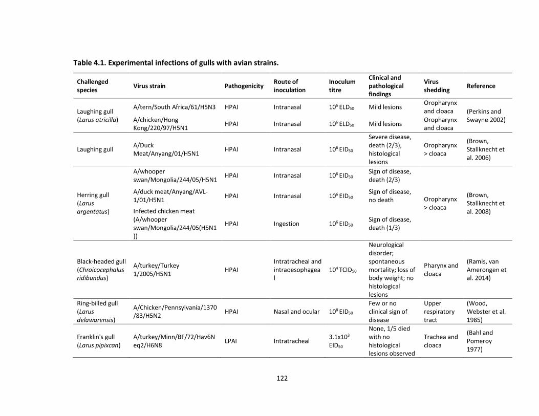
122
Table 4.1. Experimental infections of gulls with avian strains.
Challenged species
Virus strain Pathogenicity Route of inoculation
Inoculum titre
Clinical and pathological findings
Virus shedding
Reference
Laughing gull (Larus atricilla)
A/tern/South Africa/61/H5N3 HPAI Intranasal 106 ELD50 Mild lesions
Oropharynx and cloaca (Perkins and
Swayne 2002) A/chicken/Hong Kong/220/97/H5N1
HPAI Intranasal 106 ELD50 Mild lesions Oropharynx and cloaca
Laughing gull A/Duck Meat/Anyang/01/H5N1
HPAI Intranasal 106 EID50
Severe disease, death (2/3), histological lesions
Oropharynx > cloaca
(Brown, Stallknecht et al. 2006)
Herring gull (Larus argentatus)
A/whooper swan/Mongolia/244/05/H5N1
HPAI Intranasal 106 EID50 Sign of disease,
death (2/3)
Oropharynx > cloaca
(Brown, Stallknecht et al. 2008)
A/duck meat/Anyang/AVL-1/01/H5N1
HPAI Intranasal 106 EID50 Sign of disease, no death
Infected chicken meat (A/whooper swan/Mongolia/244/05(H5N1))
HPAI Ingestion 106 EID50 Sign of disease, death (1/3)
Black-headed gull (Chroicocephalus ridibundus)
A/turkey/Turkey 1/2005/H5N1
HPAI Intratracheal and intraoesophageal
104 TCID50
Neurological disorder; spontaneous mortality; loss of body weight; no histological lesions
Pharynx and cloaca
(Ramis, van Amerongen et al. 2014)
Ring-billed gull (Larus delawarensis)
A/Chicken/Pennsylvania/1370/83/H5N2
HPAI Nasal and ocular 108 EID50 Few or no clinical sign of disease
Upper respiratory tract
(Wood, Webster et al. 1985)
Franklin's gull (Larus pipixcan)
A/turkey/Minn/BF/72/Hav6Neq2/H6N8
LPAI Intratracheal 3.1x10³ EID50
None, 1/5 died with no histological lesions observed
Trachea and cloaca
(Bahl and Pomeroy 1977)

123
Ring-billed gull A/laughing gull/NJ/AI08-1460/2008/H13N9
LPAI Intranasal and intratracheal
104 EID50 No sign of disease
Oropharynx and cloaca
(Brown, Poulson et al. 2012)
Silver gull (Chroicocephalus novaehollandiae)
A/Eurasian coot/WA/2727/79/H6N2
LPAI Oropharyngeal 107.95 EID50 None Trachea and cloaca
(Curran, Robertson et al. 2013)
a ELD50 and EID50: amount of infectious virus that will cause the death of 50 % of inoculated embryonated eggs; TCID50: amount of infectious virus that will cause the infection of 50% of tissue culture.

124
Table 4.2. Experimental infections of avian and mammalian hosts with gull strains.
Challenged species
Virus strain Pathogenicity Route of inoculation
Inoculum titre
Clinical and pathological findings
Virus shedding Reference
White Leghorn chicken
A/black-tailed gull/Tottori/61/80/Hav1Neq1/H7N7
LPAI Intratracheal
107.8-108.3 EID50
No sign of disease Brain, lungs, liver, spleen,
(Otsuki, Kawaoka et al. 1982) Intraperitoneal 1/9 died
Kidneys, jejunum, and rectum
Duck A/ring-billed gull/Maryland/704/77/H13N6
LPAI Oral and intratracheal
108 EID50 No sign of disease No tracheal and cloacal shedding
(Hinshaw, Air et al. 1982) Chicken
A/ring-billed gull/Maryland/704/77/H13N6
LPAI Oral and intratracheal
108 EID50 No sign of disease No tracheal and cloacal shedding
Ferret A/gull/Massachusetts/26/1980/H13N6
LPAI Intranasal 106 EID50 No sign of disease Nasal shedding
Domestic Lohmann white chicken
A/herring gull/Norway/10_1623/2006/H16N3
LPAI Intranasal 106 EID50 No sign of disease Oropharynx (2/19) and nasal (1/19)
(Tonnessen, Valheim et al. 2011)
Turkey
A/ laughing gull /NJ/AI08-1460/2008/H13N9
LPAI Intranasal and intratracheal
107 EID50 No sign of disease Oropharynx (1/8) and cloaca
(Brown, Poulson et al. 2012) A/ring-billed
gull/MN/AI10-1708/2010/H13N6
LPAI Intranasal and intratracheal
107 EID50 No sign of disease None
Mallard (Anas platyrhynchos)
A/Gull/Ontario/680-6/2001/H13N6
LPAI Intratracheally and intraesophageally
108 EID50 No sign of disease, some lung tissue lesions observed
Limited pharyngeal, cloacal, and respiratory tissue
(Daoust, van de Bildt et al. 2013)
Mallard A/herring gull/Germany/R3309/07/H16N3
LPAI Oculo-nasal-oral 105 TCID50 No sign of disease None (Fereidouni, Harder et al. 2014)
Ferret A/gull/Delaware/428/2009(H1N1)
LPAI Airborne 106 EID50 Lethargy, fever, sneezing, coughing
Nasal (Kocer, Krauss et al. 2015)

125
a ELD50 and EID50: amount of infectious virus that will cause the death of 50 % of inoculated embryonated eggs; TCID50: amount of infectious virus that will cause the infection of 50% of tissue culture

126
Table 4.3. Gull virus binding assays on avian and human tissues (Lindskog, Ellström
et al. 2013).
Species’ tissue tested Virus strain Tissues bound
Franklin's gull (Leucophaeus pipixcan)
A/black-headed gull/Sweden/2/99/H16N3
Trachea, duodenum, ileum, ileocecal junction, colon
Herring gull (Larus argentatus)
A/black-headed gull/Sweden/2/99/H16N3
Duodenum, ileum, ileocecal junction, colon
Mallard (Anas platyrhynchos)
A/black-headed gull/Sweden/2/99/H16N3
Trachea
Human A/black-headed gull/Sweden/2/99/H16N3
Cornea, conjunctiva, nasopharynx, bronchus, pulmonary alveolus, salivary gland
Experimental infection with the A/chicken/Hong Kong/220/97(H5N1) strain
produced mild disease in laughing gulls (Perkins and Swayne 2002) whereas severe
disease and mortality were reported in laughing gull after infection with highly
pathogenic H5N1 strains from 2001 and 2006 (Brown, Stallknecht et al. 2006).
Another highly pathogenic H5 strain, A/Chicken/Penn./1370/83(H5N2), which
caused 80% mortality in chickens in a Pennsylvania outbreak, produced little to no
clinical signs in experimentally infected ring-billed gulls (Wood, Webster et al. 1985).
Franklin’s gulls experimentally infected with a turkey virus that was pathogenic for
turkeys did not develop signs of disease, yet were capable of shedding the virus (Bahl
and Pomeroy 1977). These studies suggest that gulls could be infected with virulent
strains from poultry and serve as asymptomatic carriers for subsequent virus
transmission. In another study, the virus A/laughing gull/NJ/AI08-1460/2008(H13N9)
was used to experimentally infect ring-billed gulls by the respiratory route, and high
susceptibility to the virus was observed without developing any clinical symptoms.

127
Most importantly, the virus was excreted via the oropharynx and cloaca for several
days (Brown, Poulson et al. 2012). Collectively, in vivo experimental infection studies
show fairly consistent clinical sensitivity of gulls to different highly pathogenic
viruses demonstrating signs of disease, and subclinical sensitivity to low
pathogenicity viruses with no observable signs of disease, with consistent evidence
of virus shedding.
Studies involving experimental infections of mammals with gull viruses have
been limited but several are of particular interest. In one study (Hinshaw, Air et al.
1982), the H13 gull virus A/gull/Massachusetts/26/1980(H13N6) was used in
experimental infection of ferrets, which are generally considered as the best animal
model for human influenza infection. When challenged intranasally, the ferrets
contracted the virus and shed it in nasal secretions. Additionally, attachment of the
strain A/black-headed gull/Sweden/2/99(H16N3) to human, gull and mallard tissues
was investigated using tissue microarrays and virus histochemistry, using mallard
and human viruses as references (Lindskog, Ellström et al. 2013). The H16N3 virus
attached more readily to the human respiratory tract than did the other avian IAVs,
and it could also bind to the human cornea and conjunctiva. The human virus,
A/Netherlands/213/03(H3N2), also attached to the trachea of both gull species
investigated, which demonstrates the presence of the first necessary characteristic
for respiratory transmission. It was consequently suggested that H16 viruses might
present a different cell tropism in humans and mallards than do other bird-origin
IAVs (Lindskog, Ellström et al. 2013). Elsewhere it was shown that several gull species
display α2,6-linked sialic acid receptors, to which human IAVs usually bind, on the

128
surface of their tracheal epithelium (Jourdain, van Riel et al. 2011). Those findings
indicate the potential for either direct or indirect gull-human IAV interface. Viruses
from gulls were also among those identified in wild birds as encoding proteins that
differed at relatively few amino acid positions from the 1918 pandemic strain
(Watanabe, Zhong et al. 2014), with gull viruses found that had <15 amino acid
differences in their PB2, PB1, PA, NP, M1 or NS1 proteins. A virus that contained
such segments identified in wild bird viruses showed higher pathogenicity than
typical avian viruses and was able to gain mammalian respiratory drop transmission
after acquiring as few as three substitutions in the HA protein (Watanabe, Zhong et
al. 2014). This supports the notion that the potential to generate a pandemic virus
exists within wild birds and, although the sequences used in the experimental study
were not from gull viruses, gulls could be a relevant part of this scenario.
4.2.4 Inter-species transmission of gull IAVs
Although the level of active infection found is much lower, seroprevalence for
IAV in gulls is high, reaching 45-92% (Brown, Luttrell et al. 2010, Velarde, Calvin et al.
2010, Toennessen, Germundsson et al. 2011, Echeverría, Moratorio et al. 2015). As
mentioned above, gulls also exhibit notable capacity in terms of inter-hemispheric
virus transmission (Perkins and Swayne 2002, Wille, Robertson et al. 2011, Wille,
Robertson et al. 2011, Ratanakorn, Wiratsudakul et al. 2012, Van Borm, Rosseel et al.
2012, Hall, TeSlaa et al. 2013, Echeverría, Moratorio et al. 2015). If gull-imported
Eurasian gene segments persist within the American avian gene pool, they could
subsequently be transmitted to other domestic hosts such as chickens, turkeys and
pigs. In consideration of experimental studies that frequently show sub-clinical
129
infections of gulls by highly pathogenic avian viruses, these hosts should be regarded
as candidates for inter-hemispheric transmission of highly pathogenic virus. The
following sections will evaluate whether gulls could play a major role in inter-species
and long-range transmission of IAVs and specific IAV genes and whether they could
be considered as contributors to the transmission of HPAI viruses.
4.2.4.1 The gull-poultry IAV interface
The first evidence of poultry infection by a gull IAV came from a study in
which H13N2 viruses were isolated in North America from subclinically infected
domestic turkey flocks living near accessible pond water and gulls (Sivanandan,
Halvorson et al. 1991). The H13 sequence of one of the resultant viruses,
A/turkey/MN/1012/1991(H13N2), is included within the North American gull AIV
lineage (Figure 4.2). These turkey H13N2 viruses did exhibit marked pathogenicity in
experimentally infected turkeys (Laudert, Sivanandan et al. 1993). However, various
H13 gull viruses used for experimental challenges of various poultry species
infrequently resulted in infection and appeared only subclinical (Table 4.2). There
have been no reports of H16 viruses infecting poultry. A low pathogenic H7N7 gull
strain was widely invasive and caused systemic, though sub-clinical, infection in
experimental studies with chickens (Otsuki, Kawaoka et al. 1982). Another low
pathogenic gull virus of the H7 subtype, A/Laughing gull/DE/42/06(H7N3), is believed
to have been naturally transmitted to chickens, with subsequent evolution to a
highly pathogenic form therein (Krauss, Obert et al. 2007). This virus was also fully
infectious, although mostly subclinically, in experimentally challenged chickens
(Krauss, Obert et al. 2007). Inter-species transmission between gulls and chickens

130
was also observed with A/brown-head gull/Thailand/vsmu-4/2008(H5N1), being
highly similar to two highly pathogenic chicken isolates identified from the same
region (Figure 4.5).

131

132
Figure 4.5: Phylogenetic analysis of gull H5 and H7 nucleotide sequences.
Sequences of H5 and H7 genes that showed the highest similarity in BLAST (Altschul,
Gish et al. 1990) searches to gull gene sequences were downloaded from NCBI and
aligned by MUSCLE with MEGA6 (Tamura, Stecher et al. 2013). The aligned
sequences were quality-trimmed, resulting in alignments of 1073 nts (corresponding
to nt positions 172-1242) and 1054 nts (corresponding to nt positions 32-1085) for
the H5 and H7 gene segments, respectively. The phylogenetic tree was inferred in
MEGA6 with the neighbor-joining method with 1000 bootstrap replications. High
bootstrap values are shown at the nodes on the trees. Evolutionary distances were
computed using the Maximum Composite Likelihood method and are represented by
the number of base substitutions per site (scale bar). The branches are colored
according to the host group: green, gulls; red, waterfowl from Eurasia; orange,
waterfowl from North America; pink, swine; brown, poultry; purple, shorebirds and
other wild birds excluding gulls and waterfowl; blue, humans and other mammals.
Further sampling in turkey and chicken flocks, especially where there are potential
interactions with gulls, would help confirm whether gulls could play a significant role
for IAV transmission to poultry.
4.2.4.2 The gull-waterfowl IAV interface
Sympatry exists between many gull species and waterfowl across freshwater
and, secondarily, marine habitats. In addition to the H14 viruses discussed above,
particularly high gene sequence identity can be found between various gull and duck
viruses (Figure 4.4 and 4.5, Supplementary table 4.2). These include both wild

133
(mostly mallard) and domestic duck viruses in America and Eurasia. These diverse
affinities substantiate an ongoing, reciprocal gull-waterfowl interface, worldwide,
which spans both natural ecosystems and farms containing domestic ducks.
Additionally, one study showed that H16N3 gull viruses isolated in Russia readily
replicated in respiratory and intestinal tissues in challenged ducks (Iamnikova,
Gambarian et al. 2009).
4.2.4.3 The gull-marine mammal IAV interface
The movement of IAV between gulls and mammals was identified for the first
time in 1981, when it was shown that all genomic segments from a seal H7N7 virus
were highly similar to viral gene segments from avian species, including gulls
(Webster, Hinshaw et al. 1981). Interestingly, the seal isolate replicated poorly in
avian species but well in mammals such as ferrets, pigs, and cats, which indicated
this isolate had probably accumulated mammalian-adaptive mutations. Phylogenetic
analysis of some seal and whale viruses has shown that these are frequently closely
related to gull and shorebird viruses (Groth, Lange et al. 2014). More specifically,
relatedness between NP gene segments of the strains
A/gull/Maryland/704/77(H13N6) and A/whale/Maine/328/84(H13N2) from the
Atlantic Coast of the USA was found (Mandler, Gorman et al. 1990). Similarly, the HA
gene segment from the same whale isolate falls within a North American gull H13
lineage, and presumably originated from gulls (Figure 4.2, Supplementary table 4.2).
High sequence identity was also found between the H5 gene segments from a gull
isolate, A/black_headed_gull/HK/12.1/2003(H5N1), and a human isolate,

134
A/Hong_Kong/213/2003(H5N1), both obtained in Asia (Figure 4.5, Supplementary
table 4.2).
4.2.4.4 The gull-swine IAV interface
Swine are considered one of the most important mixing vessels of IAV, as
genetic reassortment between avian and mammalian viruses occurs in this host.
Many of the viruses found in swine also contain genes that originated from human
and bird viruses. H1N1, H1N2, and H3N2 viruses are endemic in the swine
population worldwide (Brown 2000), which also represent the subtypes circulating in
the human population and the presence of those viruses or their genes within gulls
might be important in terms of transmission of viruses across distances and host
species. To date, several H1 viruses have been found in the gull population in North
America and they all fall within a North American lineage that mainly comprises
waterfowl viruses. Although no direct evidence of AIV transmission between gulls
and swine has been found to date, we identified gull viruses that do exhibit
phylogenetic affinities to swine viruses, in conjunction with related waterfowl and
poultry viruses. One swine virus from 2002,
A/swine/Saskatchewan/18789/02(H1N1), which was closely related to waterfowl
viruses (Karasin, West et al. 2004), was also close to two gull viruses obtained in
2002 from the United States for the HA gene segment (Figure 4.6).

135

136
Figure 4.6: Phylogenetic analysis of gull H1 and H3 nucleotide sequences.
Sequences of H1 and H3 genes that showed the highest similarity in BLAST (Altschul,
Gish et al. 1990) searches to gull gene segments were downloaded from NCBI and
aligned by MUSCLE with MEGA6 (Tamura, Stecher et al. 2013). The aligned
sequences were quality-trimmed, resulting in alignments of 1028 nts (corresponding
to nt positions 10-1037) and 641 nts (corresponding to nt positions 1049-1689) for
the H1 and H3 gene segments, respectively. The phylogenetic tree was inferred in
MEGA6 with the neighbor-joining method with 1000 bootstrap replications. High
bootstrap values are shown at the nodes on the trees. Evolutionary distances were
computed using the Maximum Composite Likelihood method and are represented by
the number of base substitutions per site (scale bar). The branches are colored
according to the host group: gulls, green; red, waterfowl from Eurasia; orange,
waterfowl from North America; swine, pink; brown, poultry; purple, shorebirds and
other wild birds excluding gulls and waterfowl.
The H3N2 viruses currently found in swine are believed to have originated
mostly from reassortment events involving avian and swine viruses or avian, swine
and human viruses (Brown 2000) and the triple reassortant virus has now become
predominant in the swine population (Richt, Lager et al. 2003). Sequence analysis of
H3 genes from gull viruses showed that two viruses from America and Asia,
A/herring gull/New Jersey/159/1990(H3N6) and A/herring
gull/Irkutsk/111/2005(H3), which belong to a Eurasian avian lineage, were related to
swine and poultry viruses from England and China (Figure 4.6). Additionally, H3 gene

137
segments from shorebirds related to A/herring gull/New Jersey/159/1990(H3N6)
and found in North America are falling within a Eurasian lineage, which further
illustrate the potential role of gulls in inter-hemispheric movement of IAVs (Bahl,
Krauss et al. 2013).
Investigating the evolutionary history and origin of some gull viruses has
shown that they exhibit close relationships to viruses from other hosts such as
poultry, swine and humans. These analyses also show that pathogenic strains are
closely related to some gull viruses, which shows their potential for being carriers
and probably disseminators of such strains.
4.2.5 The role of gulls as contributors to the transmission of HPAI viruses
Transmission of IAV from wild birds to poultry and swine is a major concern
for the agriculture industry and must also be considered with respect to the possible
generation of novel pandemic viruses. H5 and H7 subtypes have been commonly
associated with high mortality in poultry (Stech and Mettenleiter 2013). Analysis of
H5 sequences from gull viruses showed that most of them are closely related to
highly pathogenic H5N1 virus isolated from waterfowl, poultry, and swine (Figure
4.5). Specifically, the virus A/slaty-backed gull/Shandong/59/04(H5N1) is identical to
the avian virus A/babbler/Fujian/320/04(H5N1) and groups closely with a swine
strain, A/swine/Anhui/ca/2004(H5N1), with all these isolates from 2004. This shows
that there has been frequent inter-species transmission of this virus between avian
species, including gulls, and swine. Similarly, phylogenetic analysis of H7 sequences
138
from gulls showed that gull viruses from both North America and Eurasia are closely
related to viruses associated with lethal H7 outbreaks in poultry (Figure 4.5).
The movements of viruses amongst gulls, waterfowl, poultry, and mammals
indicate that gulls need to be considered as important contributors to IAV
evolutionary dynamics and ecology. It is also important to recognize the potential
contribution of gulls to the generation of pandemic viruses, which always carry gene
segments of bird origin, and could contribute to the emergence of pathogenic
strains.
4.2.6 The role of terns in global IAV transmission, and relationships with gull IAVs
Terns are the most closely related taxon to gulls; they exhibit shared
ecological features and can also be sympatric with gulls. It is also noteworthy that
the first isolate of IAV from wild birds was from terns, with A/tern/South
Africa/1961(H5N3) representing a highly pathogenic H5 virus that caused many
deaths among terns in South Africa (Becker 1966). Close relationships of viruses from
terns to those from other hosts can also be found. A low pathogenicity tern isolate,
A/tern/Potsdam/342-6/1979(H7N7), is similar to a virus that infected chickens in
Germany and subsequently accrued several mutations to become a highly
pathogenic virus that caused a severe outbreak in chickens (Rohm, Suss et al. 1996).
A connection from terns to marine mammals has also been previously documented
with the NP gene segment of the tern isolate A/tern/Turkmenistan/18/1972(H3N3)
closely related to the whale isolate A/whale/Pacific ocean/19/1976(H1N3) (Mandler,
Gorman et al. 1990).

139
Phylogenetic analyses with tern virus sequences showed high relatedness of
these viruses to viruses identified in gulls, ducks, turkeys and whales from around
the world (Supplementary figure 4.1 and 4.2, Supplementary table 4.3). Broadly,
these sequence comparisons reveal a series of IAVs isolated from two partially
sympatric and highly migratory tern species, the common tern (Sterna hirundo) and
the Arctic tern (Sterna paradisaea). In particular, the common tern isolate from
South Africa showed inter-hemispheric reassortment, as well as affinities to isolates
from turkey, seal, whale and another common tern strain (Supplementary figure
4.2). This isolate from Turkmenistan is one of a group of common tern viruses from a
region of Central Asia that show affinities to North American gull isolates, a
European mallard isolate, a tufted duck (Aythya fuligula) isolate from southern
Siberia and some duck isolates from Taiwan. The phylogenetic analysis of M gene
segments also showed significant homology between an Arctic tern virus (A/arctic
tern/Alaska/300/1975(H5N3)) and gull and turkey viruses from North America
(Supplementary figure 4.1). Finally, the three polymerase gene segments of the same
A/arctic tern/Alaska/300/1975(H5N3) isolate also exhibit affinities to these same
North American gull isolates, and the PB2 gene segment is also highly related to gull
isolates from Astrakhan, Asia (Supplementary figure 4.1). Overall, these analyses
show that terns also constitute an important IAV host. They share many
characteristics and display migratory connectivity with gulls, and they should also be
considered as contributors to the IAV dynamics, diversity and transmission to
different hosts.

140
4.2.7 Ecobiological aspects of gull IAVs
Two aspects of the ecobiology of gulls are particularly relevant for their role
in IAV dynamics. These concern their utilization of both marine and freshwater
habitats and their potential role in zoonotic transmission pathways. Gulls are
generalist feeders and eat both live-captured prey and opportunistically scavenge,
including on carcasses of birds and mammals. Presumably, one means by which gulls
could potentially contract IAVs from other species in aquatic habitats is by eating
IAV-infected dead birds or mammals. The utilization of both marine and freshwater
habitats, and interactions with different species in both environments, raises the
possibility for gulls to be exposed to viruses from both freshwater and marine
sources. The ecology of IAVs is fairly well understood in freshwater habitats, but this
has been explored less in marine environments. However, alongside gulls, additional
marine birds such as murres and puffins (members of the family Alcidae) and sea
ducks (family Anatidae, tribe Mergini) are known to host IAVs and be sympatric with
gulls (Ip, Flint et al. 2008). Although some IAVs do not appear very stable in elevated
salinity (Stallknecht, Kearney et al. 1990), marine birds drink seawater and it must be
presumed that effective fecal-oral transmission can take place in seawater. Viral
circulation within marine environments also involves seals and whales, which are
known to host avian-derived IAVs and apparently mostly those originating from gulls
and terns as our phylogenetic and phylogeographic findings concerning both gull and
tern viruses indicate relationships with viruses from marine mammals globally.
There is also an important zoonotic dimension for IAV circulation in gulls.
While the important role played by waterfowl as facilitators of the emergence and

141
dispersal of epizootic and pandemic strains is soundly established, gulls are both
cosmopolitan migrants and salient synanthropic birds adjacent to the duck-poultry-
swine-human IAV interface. It has previously been articulated that there is a crucial
epidemiological position occupied by gulls among wildlife, domestic birds as well as
humans (Arnal, Vittecoq et al. 2015), and the potential for gulls to act in transmission
of microorganisms from sewage plants or outflows to domestic animals has also
been described (Teale 2002). Natural interfaces between gull viruses and those from
ducks, turkeys, chickens and swine have been identified in the present analysis,
either in terms of interchangeability of certain gene segments, or whole viruses, and
IAV-related interactions of gulls with ducks, turkeys, chickens and mammals,
phylogenetically and ecologically, might contribute to emergence and spread of
epizootic and pandemic strains. It is also of note that a connection was found
between marine mammal and human infections (Lvov, Zhdanov et al. 1983, Ohishi,
Kishida et al. 2004, Belser, Bridges et al. 2009), and that the 2009 H1N1 human
pandemic strain naturally infected and spread, sub-clinically, among seals in
California (Goldstein, Mena et al. 2013). However, it is not known if gulls or terns
were directly involved in transmission of this virus.
4.3 Concluding remarks
Within the enormous spectrum of IAVs hosted by avian species, gull IAVs
constitute an important component, somewhat parallel to waterfowl viruses, of the
global ecophylogenetic system defining this protean virus. Gulls affect, both directly
and indirectly, the evolutionary dynamics of low and high pathogenicity avian viruses

142
and various mammalian IAVs worldwide. Analysis of the evolutionary history of an
entirely Eurasian virus isolated in North America allowed the reconstruction of the
gradual evolutionary process that generated this virus, starting in Central Asia,
moving northwest, and ending in Iceland, with subsequent transmission to Canada.
The origin of this virus appears to be associated with a region of Central Asia that
also appears to have given rise to an important H14 strain that involved both gull
and duck viruses. Such reverse ecophylogenetic analysis is useful for attaining a
broadened perspective of the evolutionary history of a given virus and can allow
identification of the presumed strains, genes, and hosts involved in the evolutionary
and spatiotemporal pathways that gave rise to a strain in question. Additional gull
viruses that were identified as phylogenetically interesting, in terms of their
relationships to pathogenic strains and viruses from other hosts, in this analysis were
also from the same Central Asian geographic region. A similar scenario was found
when analyzing a series of common tern viruses that were obtained in the same
general region of Central Asia, where those viruses exhibited clear affinities to gull,
duck and whale viruses. Our analyses of the tern viruses reveal a broad
phylogeographic contribution for these viruses beyond Central Asia, with gene
transactions extending to Europe, South Africa, North America and the Pacific Ocean.
The traits contributing to the importance of gulls as IAV hosts include their
cosmopolitan distributions, migratory behaviors, synanthropic associations with
humans, use of both marine and freshwater habitats, prevalence as a host of many
different IAVs of a large variety of host group origins (including farm animals), and
predominance of subclinical infections. As shown in the present study, the summary

143
of these properties defines an important IAV host group that needs to be considered
in the context of the perpetuation and emergence of enzootic, epizootic and
pandemic strain. A routine surveillance of the virus in migratory birds such as gulls
and terns could be conducted worldwide to foresee potential zoonotic transmission
of pathogenic strains to humans and domestic livestock.
4.4 References
Adams, S., Z. Xing, J. Li, K. Mendoza, D. Perez, K. Reed and C. Cardona (2013). "The
effect of avian influenza virus NS1 allele on virus replication and innate gene
expression in avian cells." Mol Immunol 56(4): 358-368.
Altschul, S. F., W. Gish, W. Miller, E. W. Myers and D. J. Lipman (1990). "Basic local
alignment search tool." Journal of Molecular Biology. 215(3): 403-410.
Altschul, S. F., W. Gish, W. Miller, E. W. Myers and D. J. Lipman (1990). "Basic local
alignment search tool." Journal of Molecular Biology 215: 403-410.
Amonsin, A., T. Songserm, S. Chutinimitkul, R. Jam-On, N. Sae-Heng, N. Pariyothorn,
S. Payungporn, A. Theamboonlers and Y. Poovorawan (2007). "Genetic analysis of
influenza A virus (H5N1) derived from domestic cat and dog in Thailand." Archives of
Virology 152: 1925-1933.
Andino, R. and E. Domingo (2015). "Viral quasispecies." Virology 479-480: 46-51.

144
Arnal, A., M. Vittecoq, J. Pearce-Duvet, M. Gauthier-Clerc, T. Boulinier and E.
Jourdain (2015). "Laridae: A neglected reservoir that could play a major role in avian
influenza virus epidemiological dynamics." Critical Reviews in Microbiology 41: 508-
519.
Asghar, N., J. H. Pettersson, P. Dinnetz, A. Andreassen and M. Johansson (2017).
"Deep sequencing analysis of tick-borne encephalitis virus from questing ticks at
natural foci reveals similarities between quasispecies pools of the virus." J Gen Virol
98(3): 413-421.
Bahl, A. K. and B. S. Pomeroy (1977). "Experimental exposure of Franklin's gulls
(Larvus pipixcan) and mallards (Anas platyrhynchos) to a turkey influenza A virus
A/Turkey/Minn/BF/72(Hv6Neq2)." Journal of Wildlife Diseases 13: 420-426.
Bahl, J., S. Krauss, D. Kuhnert, M. Fourment, G. Raven, S. P. Pryor, L. J. Niles, A.
Danner, D. Walker, I. H. Mendenhall, Y. C. Su, V. G. Dugan, R. A. Halpin, T. B.
Stockwell, R. J. Webby, D. E. Wentworth, A. J. Drummond, G. J. Smith and R. G.
Webster (2013). "Influenza a virus migration and persistence in North American wild
birds." PLoS Pathogens 9: e1003570.
Bahl, J., D. Vijaykrishna, E. C. Holmes, G. J. Smith and Y. Guan (2009). "Gene flow and
competitive exclusion of avian influenza A virus in natural reservoir hosts." Virology
390(2): 289-297.
Baltimore, D. (1971). "Expression of animal virus genomes." Bacteriological Reviews
35(3): 235-241.

145
Bao, Y., P. Bolotov, D. Dernovoy, B. Kiryutin, L. Zaslavsky, T. Tatusova, J. Ostell and D.
Lipman (2008). "The influenza Virus Resource at the National Center for
Biotechnology Information." Journal of Virology 82: 596-601.
Baskerville, A. and G. Lloyd (1976). "The pathogenesis and pathology of experimental
Quaranfil virus infection." British journal of experimental pathology 57(2): 152-156.
Becker, W. B. (1966). "The isolation and classification of Tern virus: influenza A-Tern
South Africa--1961." Journal of Hygiene 64: 309-320.
Bedford, T., M. A. Suchard, P. Lemey, G. Dudas, V. Gregory, A. J. Hay, J. W. McCauley,
C. A. Russell, D. J. Smith and A. Rambaut (2014). "Integrating influenza antigenic
dynamics with molecular evolution." Elife 3: e01914.
Belser, J. A., C. B. Bridges, J. M. Katz and T. M. Tumpey (2009). "Past, present, and
possible future human infection with influenza virus A subtype H7." Emerging
Infectious Diseases 15: 859-865.
Bergmann, M. and T. Muster (1996). "Mutations in the nonconserved noncoding
sequences of the influenza A virus segments affect viral vRNA formation." Virus
Research 44(1): 23-31.
Blakemore F, G. A. (1941). "Swine Influenza in the British Isles." Proceedings of the
Royal Society of Medicine 34(9): 611-618.

146
Boni, M. F., Y. Zhou, J. K. Taubenberger and E. C. Holmes (2008). "Homologous
recombination is very rare or absent in human influenza A virus." Journal of Virology
82(10): 4807-4811.
Boulo, S., H. Akarsu, R. W. Ruigrok and F. Baudin (2007). "Nuclear traffic of influenza
virus proteins and ribonucleoprotein complexes." Virus Res 124(1-2): 12-21.
Boyce, W. M., S. Schobel, V. G. Dugan, R. A. Halpin, X. Lin, D. E. Wentworth, L. L.
Lindsay, E. Mertens and M. Plancarte (2013). "Complete genome sequence of a
reassortant H14N2 avian influenza virus from California." Genome Announcements
1: e00543-00513.
Brown, I. H. (2000). "The epidemiology and evolution of influenza viruses in pigs."
Veterinary Microbiology 74: 29-46.
Brown, I. H. (2001). "The pig as an intermediate host for influenza A viruses between
birds and humans." International Congress Series 1219: 173-178.
Brown, I. H., P. A. Harris, J. W. McCauley and D. J. Alexander (1998). "Multiple
genetic reassortment of avian and human influenza A viruses in European pigs,
resulting in the emergence of an H1N2 virus of novel genotype." The Journal of
General Virology. 79 ( Pt 12): 2947-2955.
Brown, J., R. Poulson, D. Carter, C. Lebarbenchon, M. Pantin-Jackwood, E. Spackman,
E. Shepherd, M. Killian and D. Stallknecht (2012). "Susceptibility of avian species to

147
North American H13 low pathogenic avian influenza viruses." Avian Diseases 56:
969-975.
Brown, J. D., M. P. Luttrell, R. D. Berghaus, W. Kistler, S. P. Keeler, A. Howey, B.
Wilcox, J. Hall, L. Niles, A. Dey, G. Knutsen, K. Fritz and D. E. Stallknecht (2010).
"Prevalence of antibodies to type a influenza virus in wild avian species using two
serologic assays." Journal of Wildlife Diseases 46: 896-911.
Brown, J. D., D. E. Stallknecht, J. R. Beck, D. L. Suarez and D. E. Swayne (2006).
"Susceptibility of North American ducks and gulls to H5N1 highly pathogenic avian
influenza viruses." Emerging Infectious Diseases 12: 1663-1670.
Burleigh, L. M., L. J. Calder, J. J. Skehel and D. A. Steinhauer (2005). "Influenza a
viruses with mutations in the m1 helix six domain display a wide variety of
morphological phenotypes." Journal of Virology. 79(2): 1262-1270.
Capua, I. and S. Marangon (2000). "The avian influenza epidemic in Italy, 1999-2000:
a review." Avian Pathology 29(4): 289-294.
Chen, H., G. J. Smith, S. Y. Zhang, K. Qin, J. Wang, K. S. Li, R. G. Webster, J. S. Peiris
and Y. Guan (2005). "Avian flu: H5N1 virus outbreak in migratory waterfowl." Nature
436: 191-192.
Chen, J., K. H. Lee, D. A. Steinhauer, D. J. Stevens, J. J. Skehel and D. C. Wiley (1998).
"Structure of the hemagglutinin precursor cleavage site, a determinant of influenza
pathogenicity and the origin of the labile conformation." Cell 95(3): 409-417.

148
Claas, E. C., A. D. Osterhaus, R. van Beek, J. C. De Jong, G. F. Rimmelzwaan, D. A.
Senne, S. Krauss, K. F. Shortridge and R. G. Webster (1998). "Human influenza A
H5N1 virus related to a highly pathogenic avian influenza virus." Lancet 351: 472-
477.
Clifford, C. M., L. A. Thomas, L. E. Hughes, G. M. Kohls and C. B. Philip (1968).
"Identification and comparison of two viruses isolated from ticks of the genus
Ornithodoros." The American Journal of Tropical Medicine and Hygiene. 17(6): 881-
885.
Couceiro, J. N., J. C. Paulson and L. G. Baum (1993). "Influenza virus strains
selectively recognize sialyloligosaccharides on human respiratory epithelium; the
role of the host cell in selection of hemagglutinin receptor specificity." Virus Res
29(2): 155-165.
Crescenzo-Chaigne, B., C. V. Barbezange, S. Leandri, C. Roquin, C. Berthault and S.
van der Werf (2017). "Incorporation of the influenza A virus NA segment into virions
does not require cognate non-coding sequences." Scientific reports 7: 43462.
Croville, G., S. M. Soubies, J. Barbieri, C. Klopp, J. Mariette, O. Bouchez, C. Camus-
Bouclainville and J. L. Guerin (2012). "Field monitoring of avian influenza viruses:
whole-genome sequencing and tracking of neuraminidase evolution using 454
pyrosequencing." J Clin Microbiol 50(9): 2881-2887.

149
De Silva, U. C. and T. Yasunaga (2011). "Swine to human transmission of reassortants
of pandemic (H1N1) 2009 and endemic swine influenza viruses: Abstract." PLoS
Currents 3: RRN1285.
Desselberger, U., V. R. Racaniello, J. J. Zazra and P. Palese (1980). "The 3' and 5'-
terminal sequences of influenza A, B and C virus RNA segments are highly conserved
and show partial inverted complementarity." Gene 8(3): 315-328.
Dias, A., D. Bouvier, T. Crepin, A. A. McCarthy, D. J. Hart, F. Baudin, S. Cusack and R.
W. H. Ruigrok (2009). "The cap-snatching endonuclease of influenza virus
polymerase resides in the PA subunit." Nature 458(7240): 914-918.
Dlugolenski, D., L. Jones, G. Saavedra, S. M. Tompkins, R. A. Tripp and E. Mundt
(2011). "Passage of low-pathogenic avian influenza (LPAI) viruses mediates rapid
genetic adaptation of a wild-bird isolate in poultry." Arch Virol 156(4): 565-576.
Domingo, E., E. Baranowski, C. M. Ruiz-Jarabo, A. M. Martín-Hernández, J. C. Sáiz and
C. Escarmís (1998). "Quasispecies structure and persistence of RNA viruses."
Emerging Infectious Diseases 4(4): 521-527.
Drummond, A. J., M. A. Suchard, D. Xie and A. Rambaut (2012). "Bayesian
Phylogenetics with BEAUti and the BEAST 1.7." Molecular Biology and Evolution 29:
1969-1973.
Dubois, J., O. Terrier and M. Rosa-Calatrava (2014). "Influenza viruses and mRNA
splicing: doing more with less." MBio 5(3): e00070-00014.
150
Dusek, R. J., G. T. Hallgrimsson, H. S. Ip, J. E. Jónsson, S. Sreevatsan, S. W. Nashold, J.
L. Teslaa, S. Enomoto, R. A. Halpin, X. Lin, N. Fedorova, T. B. Stockwell, V. G. Dugan,
D. E. Wentworth and J. S. Hall (2014). "North Atlantic migratory bird flyways provide
routes for intercontinental movement of avian influenza viruses." PLoS One 9:
e92075.
Edinger, T. O., M. O. Pohl and S. Stertz (2014). "Entry of influenza A virus: host
factors and antiviral targets." Journal of General Virology 95: 263-277.
Finkelman, B. S., C. Viboud, K. Koelle, M. J. Ferrari, N. Bharti and B. T. Grenfell (2007).
"Global Patterns in Seasonal Activity of Influenza A/H3N2, A/H1N1, and B from 1997
to 2005: Viral Coexistence and Latitudinal Gradients." PLoS One 2(12): e1296.
Flick, R., G. Neumann, E. Hoffmann, E. Neumeier and G. Hobom (1996). "Promoter
elements in the influenza vRNA terminal structure." Rna 2(10): 1046-1057.
Fodor, A. J. W. t. V. a. E. (2016). "Influenza virus RNA polymerase: insights into the
mechanisms of viral RNA synthesis." Nature reviews. Microbiology 14(8): 479-493.
Fodor, E., D. C. Pritlove and G. G. Brownlee (1994). "The influenza virus panhandle is
involved in the initiation of transcription." Journal of Virology 68(6): 4092-4096.
Fodor, E., B. L. Seong and G. G. Brownlee (1993). "Photochemical cross-linking of
influenza A polymerase to its virion RNA promoter defines a polymerase binding site
at residues 9 to 12 of the promoter." The Journal of General Virology 74 ( Pt 7):
1327-1333.
151
Fouchier, R. A. M., V. Munster, A. Wallensten, T. M. Bestebroer, S. Herfst, D. Smith,
G. F. Rimmelzwaan, B. Olsen and A. D. M. E. Osterhaus (2005). "Characterization of a
novel influenza A virus hemagglutinin subtype (H16) obtained from black-headed
gulls." Journal of Virology 79: 2814-2822.
Fries, A. C., J. M. Nolting, A. Danner, R. G. Webster, A. S. Bowman, S. Krauss, R. D.
Slemons and Y. Wang (2013). "Evidence for the circulation and inter-hemispheric
movement of the H14 subtype influenza A virus." PLoS ONE 8: e59216.
Fujii, K., Y. Fujii, T. Noda, Y. Muramoto, T. Watanabe, A. Takada, H. Goto, T. Horimoto
and Y. Kawaoka (2005). "Importance of both the coding and the segment-specific
noncoding regions of the influenza A virus NS segment for its efficient incorporation
into virions." Journal of Virology 79(6): 3766-3774.
Furuse, Y. and H. Oshitani (2011). "Evolution of the influenza A virus untranslated
regions." Infection, Genetics and Evolution 11(5): 1150-1154.
Gamblin, S. J. and J. J. Skehel (2010). "Influenza Hemagglutinin and Neuraminidase
Membrane Glycoproteins." The Journal of Biological Chemistry 285(37): 28403-
28409.
Gao, Q. and P. Palese (2009). "Rewiring the RNAs of influenza virus to prevent
reassortment." PNAS 106(37): 15891.

152
Garcia-Sastre, A., A. Egorov, D. Matassov, S. Brandt, D. E. Levy, J. E. Durbin, P. Palese
and T. Muster (1998). "Influenza A virus lacking the NS1 gene replicates in
interferon-deficient systems." Virology 252(2): 324-330.
Garten, W. and H. D. Klenk (1983). "Characterization of the carboxypeptidase
involved in the proteolytic cleavage of the influenza haemagglutinin." J Gen Virol 64
(Pt 10): 2127-2137.
Geraci, J. R., D. J. Staubin, I. K. Barker, R. G. Webster, V. S. Hinshaw, W. J. Bean, H. L.
Ruhnke, J. H. Prescott, G. Early, A. S. Baker, S. Madoff and R. T. Schooley (1982).
"Mass mortality of harbor seals: pneumonia associated with influenza A virus."
Science 215(4536): 1129-1131.
Girard, M. P., J. S. Tam, O. M. Assossou and M. P. Kieny (2010). "The 2009 A (H1N1)
influenza virus pandemic: A review." Vaccine 28(31): 4895-4902.
Glezen, W. P., J. K. Schmier, C. M. Kuehn, K. J. Ryan and J. Oxford (2013). "The
Burden of Influenza B: A Structured Literature Review." American Journal of Public
Health 103(3): E43-E51.
Goldstein, T., I. Mena, S. J. Anthony, R. Medina, P. W. Robinson, D. J. Greig, D. P.
Costa, W. I. Lipkin, A. Garcia-Sastre and W. M. Boyce (2013). "Pandemic H1N1
influenza isolated from free-ranging Northern Elephant Seals in 2010 off the central
California coast." PLoS ONE 8: e62259.

153
Gonzalez-Reiche, A. S. and D. R. Perez (2012). "Where do avian influenza viruses
meet in the Americas?" Avian Diseases 56: 1025-1033.
Groth, M., J. Lange, P. Kanrai, S. Pleschka, C. Scholtissek, A. Krumbholz, M. Platzer, A.
Sauerbrei and R. Zell (2014). "The genome of an influenza virus from a pilot whale:
Relation to influenza viruses of gulls and marine mammals." Infection Genetics and
Evolution 24: 183-186.
Guo, Y. J., F. G. Jin, P. Wang, M. Wang and J. M. Zhu (1983). "Isolation of influenza C
virus from pigs and experimental infection of pigs with influenza C virus." The Journal
of General Virology. 64 (Pt 1): 177-182.
Hale, B. G., R. A. Albrecht and A. García-Sastre (2010). "Innate immune evasion
strategies of influenza viruses." Future microbiology 5: 23.
Hall, J. S., J. L. TeSlaa, S. W. Nashold, R. A. Halpin, T. Stockwell, E. Wentworth, V. G.
Dugan and H. S. Ip (2013). "Evolution of a reassortant North American gull influenza
virus lineage: drift, shift and stability." Virology Journal 10: 179.
Hao, W. (2011). "Evidence of intra-segmental homologous recombination in
influenza A virus." Gene 481(2): 57-64.
Harkness, J. W., G. C. Schild, P. H. Lamont and C. M. Brand (1972). "Studies on
relationships between human and porcine influenza: 1. Serological evidence of
infection in swine in Great Britain with an influenza A virus antigenically like human
Hong Kong/68 virus." Bulletin of the World Health Organization 46(6): 709-719.

154
He, C. Q., G. Z. Han, D. Wang, W. Liu, G. R. Li, X. P. Liu and N. Z. Ding (2008).
"Homologous recombination evidence in human and swine influenza A viruses."
Virology 380(1): 12-20.
He, C. Q., Z. X. Xie, G. Z. Han, J. B. Dong, D. Wang, J. B. Liu, L. Y. Ma, X. F. Tang, X. P.
Liu, Y. S. Pang and G. R. Li (2009). "Homologous recombination as an evolutionary
force in the avian influenza A virus." Molecular Biology and Evolution. 26(1): 177-
187.
Hinshaw, V. S., G. M. Air, A. J. Gibbs, L. Graves, B. Prescott and D. Karunakaran
(1982). "Antigenic and genetic characterization of a novel hemagglutinin subtype of
influenza A viruses from gulls." Journal of Virology 42: 865-872.
Hinshaw, V. S., W. J. Bean, J. Geraci, P. Fiorelli, G. Early and R. G. Webster (1986).
"Characterization of two influenza A viruses from a pilot whale." Journal of Virology
58(2): 655-656.
Hinshaw, V. S., W. J. Bean, R. G. Webster, J. E. Rehg, P. Fiorelli, G. Early, J. R. Geraci
and D. J. St Aubin (1984). "Are seals frequently infected with avian influenza
viruses?" Journal of Virology. 51(3): 863-865.
Hinshaw, V. S., R. G. Webster and B. Turner (1979). "Water-bone transmission of
influenza A viruses?" Intervirology 11(1): 66-68.

155
Hiromoto, Y., T. Saito, S. E. Lindstrom, Y. Li, R. Nerome, S. Sugita, M. Shinjoh and K.
Nerome (2000). "Phylogenetic analysis of the three polymerase genes (PB1, PB2 and
PA) of influenza B virus." The Journal of General Virology. 81(Pt 4): 929-937.
Hoffmann, E., J. Stech, Y. Guan, R. G. Webster and D. R. Perez (2001). "Universal
primer set for the full-length amplification of all influenza A viruses." Arch Virol
146(12): 2275-2289.
Hoffmann, E., J. Stech, Y. Guan, R. G. Webster and D. R. Perez (2001). "Universal
primer set for the full-length amplification of all influenza A viruses." Archives of
Virology 146(12): 2275-2289.
Holland, J., K. Spindler, F. Horodyski, E. Grabau, S. Nichol and S. VandePol (1982).
"Rapid evolution of RNA genomes." Science 215(4540): 1577-1585.
Horimoto, T. and Y. Kawaoka (2001). "Pandemic threat posed by avian influenza A
viruses." Clinical Microbiology Reviews 14(1): 129-+.
Hossain, M. J., D. Hickman and D. R. Perez (2008). "Evidence of Expanded Host Range
and Mammalian-Associated Genetic Changes in a Duck H9N2 Influenza Virus
Following Adaptation in Quail and Chickens." PLoS ONE 3(9): e3170.
Hsu, M. T., J. D. Parvin, S. Gupta, M. Krystal and P. Palese (1987). "Genomic RNAs of
influenza viruses are held in a circular conformation in virions and in infected cells by
a terminal panhandle." Proceedings of the National Academy of Sciences of the
United States of America 84(22): 8140-8144.

156
Hsu, M. T., J. D. Parvin, S. Gupta, M. Krystal and P. Palese (1987). "Genomic RNAs of
influenza viruses are held in a circular conformation in virions and in infected cells by
a terminal panhandle." PNAS 84(22): 8140-8144.
Huang, Y., G. J. Robertson, D. Ojkic, H. Whitney and A. S. Lang (2014). "Diverse inter-
continental and host lineage reassortant avian influenza A viruses in pelagic
seabirds." Infection, Genetics and Evolution : Journal of molecular epidemiology and
evolutionary genetics in infectious diseases. 22: 103-111.
Huang, Y., M. Wille, J. Benkaroun, H. Munro, A. L. Bond, D. A. Fifield, G. J. Robertson,
D. Ojkic, H. Whitney and A. S. Lang (2014). "Perpetuation and reassortment of gull
influenza A viruses in Atlantic North America." Virology 456-457: 353-363.
Huang, Y., M. Wille, A. Dobbin, G. J. Robertson, P. Ryan, D. Ojkic, H. Whitney and A.
S. Lang (2013). "A 4-year study of avian influenza virus prevalence and subtype
diversity in ducks of Newfoundland, Canada." Canadian Journal of Microbiology.
59(10): 701-708.
Huang, Y., M. Wille, A. Dobbin, N. M. Walzthoni, G. J. Robertson, D. Ojkic, H. Whitney
and A. S. Lang (2014). "Genetic structure of avian influenza viruses from ducks of the
Atlantic flyway of North America." PLoS One 9(1): e86999.
Hulo, C., E. de Castro, P. Masson, L. Bougueleret, A. Bairoch, I. Xenarios and P. Le
Mercier (2011). "ViralZone: a knowledge resource to understand virus diversity."
Nucleic Acids Research. 39(Database issue): D576-582.

157
Hurt, A. C., D. Vijaykrishna, J. Butler, C. Baas, S. Maurer-Stroh, M. C. Silva-de-la-
Fuente, G. Medina-Vogel, B. Olsen, A. Kelso, I. G. Barr and D. Gonzalez-Acuna (2014).
"Detection of evolutionarily distinct avian influenza A viruses in Antarctica." mBio 5:
e01098-01014.
Iamnikova, S. S., A. S. Gambarian, V. A. Aristova, K. L'Vov D, N. F. Lomakina, V.
Munster, P. Lexmond and R. A. Foucher (2009). "[A/H13 and A/H16 influenza viruses:
different lines of one precursors]." Voprosy virusologii 54: 10-18.
Imai, M., T. Watanabe, M. Hatta, S. C. Das, M. Ozawa, K. Shinya, G. Zhong, A. Hanson,
H. Katsura, S. Watanabe, C. Li, E. Kawakami, S. Yamada, M. Kiso, Y. Suzuki, E. A.
Maher, G. Neumann and Y. Kawaoka (2012). "Experimental adaptation of an
influenza H5 haemagglutinin (HA) confers respiratory droplet transmission to a
reassortant H5 HA/H1N1 virus in ferrets." Nature 486(7403): 420-428.
Ip, H. S., P. L. Flint, J. C. Franson, R. J. Dusek, D. V. Derksen, R. E. Gill, Jr., C. R. Ely, J.
M. Pearce, R. B. Lanctot, S. M. Matsuoka, D. B. Irons, J. B. Fischer, R. M. Oates, M. R.
Petersen, T. F. Fondell, D. A. Rocque, J. C. Pedersen and T. C. Rothe (2008).
"Prevalence of Influenza A viruses in wild migratory birds in Alaska: patterns of
variation in detection at a crossroads of intercontinental flyways." Virology Journal 5:
71.
Ito, T., J. N. Couceiro, S. Kelm, L. G. Baum, S. Krauss, M. R. Castrucci, I. Donatelli, H.
Kida, J. C. Paulson, R. G. Webster and Y. Kawaoka (1998). "Molecular basis for the

158
generation in pigs of influenza A viruses with pandemic potential." Journal of
Virology. 72(9): 7367-7373.
Ito, T. and Y. Kawaoka (2000). "Host-range barrier of influenza A viruses." Vet
Microbiol 74(1-2): 71-75.
Joseph, U., M. Linster, Y. Suzuki, S. Krauss, R. A. Halpin, D. Vijaykrishna, T. P. Fabrizio,
T. M. Bestebroer, S. Maurer-Stroh, R. J. Webby, D. E. Wentworth, R. A. Fouchier and
J. Bahl (2015). "Adaptation of pandemic H2N2 influenza A viruses in humans."
Journal of Virology 89(4): 2442-2447.
Jourdain, E., G. Gunnarsson, J. Wahlgren, N. Latorre-Margalef, C. Brojer, S. Sahlin, L.
Svensson, J. Waldenstrom, A. Lundkvist and B. Olsen (2010). "Influenza virus in a
natural host, the mallard: experimental infection data." PLoS One 5(1): e8935.
Jourdain, E., D. van Riel, V. J. Munster, T. Kuiken, J. Waldenstrom, B. Olsen and P.
Ellstrom (2011). "The pattern of influenza virus attachment varies among wild bird
species." PLoS ONE 6: e24155.
Karasin, A. I., K. West, S. Carman and C. W. Olsen (2004). "Characterization of avian
H3N3 and H1N1 influenza A viruses isolated from pigs in Canada." Journal of Clinical
Microbiology 42: 4349-4354.
Kawaoka, Y., S. Krauss and R. G. Webster (1989). "Avian-to-human transmission of
the PB1 gene of influenza A viruses in the 1957 and 1968 pandemics." Journal of
Virology. 63(11): 4603-4608.

159
Kearse, M., R. Moir, A. Wilson, S. Stones-Havas, M. Cheung, S. Sturrock, S. Buxton, A.
Cooper, S. Markowitz, C. Duran, T. Thierer, B. Ashton, P. Meintjes and A. Drummond
(2012). "Geneious Basic: an integrated and extendable desktop software platform
for the organization and analysis of sequence data." Bioinformatics 28(12): 1647-
1649.
Kida, H., L. E. Brown and R. G. Webster (1982). "Biological activity of monoclonal
antibodies to operationally defined antigenic regions on the hemagglutinin molecule
of A/Seal/Massachusetts/1 /80 (H7N7) influenza virus." Virology 122(1): 38-47.
Kim, H. J., E. Fodor, G. G. Brownlee and B. L. Seong (1997). "Mutational analysis of
the RNA-fork model of the influenza A virus vRNA promoter in vivo." The Journal of
General Virology 78 ( Pt 2): 353-357.
Kimura, H., C. Abiko, G. Peng, Y. Muraki, K. Sugawara, S. Hongo, F. Kitame, K. Mizuta,
Y. Numazaki, H. Suzuki and K. Nakamura (1997). "Interspecies transmission of
influenza C virus between humans and pigs." Virus Res 48(1): 71-79.
Krauss, S., C. A. Obert, J. Franks, D. Walker, K. Jones, P. Seiler, L. Niles, S. P. Pryor, J.
C. Obenauer, C. W. Naeve, L. Widjaja, R. J. Webby and R. G. Webster (2007).
"Influenza in migratory birds and evidence of limited intercontinental virus
exchange." PLoS Pathogens 3: e167.
Kuiken, T., E. C. Holmes, J. McCauley, G. F. Rimmelzwaan, C. S. Williams and B. T.
Grenfell (2006). "Host species barriers to influenza virus infections." Science
312(5772): 394-397.

160
Kumlin, U., S. Olofsson, K. Dimock and N. Arnberg (2008). "Sialic acid tissue
distribution and influenza virus tropism." Influenza and Other Respiratory Viruses
2(5): 147-154.
Lang, A. S., C. Lebarbenchon, A. M. Ramey, G. J. Robertson, J. Waldenstrom and M.
Wille (2016). "Assessing the Role of Seabirds in the Ecology of Influenza A Viruses."
Avian Dis 60(1 Suppl): 378-386.
Latorre-Margalef, N., G. Gunnarsson, V. J. Munster, R. A. Fouchier, A. D. Osterhaus, J.
Elmberg, B. Olsen, A. Wallensten, P. D. Haemig, T. Fransson, L. Brudin and J.
Waldenstrom (2009). "Effects of influenza A virus infection on migrating mallard
ducks." Proceedings. Biological Sciences. 276(1659): 1029-1036.
Laudert, E., V. Sivanandan, D. Halvorson, D. Shaw and R. G. Webster (1993).
"Biological and molecular characterization of H13N2 influenza type A viruses isolated
from turkeys and surface water." Avian Diseases 37: 793-799.
Lee, D.-H., D. E. Swayne, M. K. Torchetti, K. Winker, H. S. Ip and C.-S. Song (2015).
"Intercontinental spread of asian-origin H5N8 to North America through Beringia by
migratory birds." Journal of Virology 89: 6521-6524.
Lee, M. K., S. H. Bae, C. J. Park, H. K. Cheong, C. Cheong and B. S. Choi (2003). "A
single-nucleotide natural variation (U4 to C4) in an influenza A virus promoter
exhibits a large structural change: implications for differential viral RNA synthesis by
RNA-dependent RNA polymerase." Nucleic Acids Research 31(4): 1216-1223.

161
Lefkowitz, E. J., D. M. Dempsey, R. C. Hendrickson, R. J. Orton, S. G. Siddell and D. B.
Smith (2018). "Virus taxonomy: the database of the International Committee on
Taxonomy of Viruses (ICTV)." Nucleic Acids Research. 46(D1): D708-d717.
Li, J. and C. J. Cardona (2010). "Adaptation and transmission of a wild duck avian
influenza isolate in chickens." Avian Dis 54(1 Suppl): 586-590.
Li, J., H. zu Dohna, N. L. Anchell, S. C. Adams, N. T. Dao, Z. Xing and C. J. Cardona
(2010). "Adaptation and transmission of a duck-origin avian influenza virus in poultry
species." Virus Res 147(1): 40-46.
Li, X. and P. Palese (1992). "Mutational analysis of the promoter required for
influenza virus virion RNA synthesis." Journal of Virology 66(7): 4331-4338.
Li, X. and P. Palese (1994). "Characterization of the polyadenylation signal of
influenza virus RNA." Journal of Virology 68(2): 1245-1249.
Lin, D., J. Lan and Z. Zhang (2007). "Structure and function of the NS1 protein of
influenza A virus." Acta Biochimica et Biophysica Sinica. 39(3): 155-162.
Lindskog, C., P. Ellström, B. Olsen, F. Pontén, D. van Riel, V. J. Munster, D. González-
Acuña, T. Kuiken and E. Jourdain (2013). "European H16N3 gull influenza virus
attaches to the human respiratory tract and eye." PLoS ONE 8: e60757.
Ludwig, S., U. Schultz, J. Mandler, W. M. Fitch and C. Scholtissek (1991).
"Phylogenetic relationship of the nonstructural (NS) genes of influenza A viruses."
Virology 183(2): 566-577.

162
Luo, G. X., W. Luytjes, M. Enami and P. Palese (1991). "The polyadenylation signal of
influenza virus RNA involves a stretch of uridines followed by the RNA duplex of the
panhandle structure." Journal of Virology 65(6): 2861-2867.
Lvov, D. K., V. M. Zhdanov, F. Sadykhova, S. S. Yamnikova, V. A. Isachenko and E. A.
Vladimirtseva (1983). "Isolation of a natural recombinant of influenza A virus (H1N3)
from a sick child." Lancet 2: 1246-1247.
Mandler, J., O. T. Gorman, S. Ludwig, E. Schroeder, W. M. Fitch, R. G. Webster and C.
Scholtissek (1990). "Derivation of the nucleoproteins (NP) of influenza A viruses
isolated from marine mammals." Virology 176: 255-261.
Manuguerra, J. C. and C. Hannoun (1992). "Natural infection of dogs by influenza C
virus." Research in Virology. 143(3): 199-204.
Matrosovich, M. N., T. Y. Matrosovich, T. Gray, N. A. Roberts and H. D. Klenk (2004).
"Human and avian influenza viruses target different cell types in cultures of human
airway epithelium." Proc Natl Acad Sci U S A 101(13): 4620-4624.
Mjaaland, S., E. Rimstad, K. Falk and B. H. Dannevig (1997). "Genomic
characterization of the virus causing infectious salmon anemia in Atlantic salmon
(Salmo salar L.): an orthomyxo-like virus in a teleost." Journal of Virology 71(10):
7681-7686.
Moncla, L. H., G. Zhong, C. W. Nelson, J. M. Dinis, J. Mutschler, A. L. Hughes, T.
Watanabe, Y. Kawaoka and T. C. Friedrich (2016). "Selective bottlenecks shape

163
evolutionary pathways taken during mammalian adaptation of a 1918-like avian
influenza virus." Cell host & microbe 19(2): 169-180.
Moriuchi, H., N. Katsushima, H. Nishimura, K. Nakamura and Y. Numazaki (1991).
"Community-acquired influenza C virus infection in children." The Journal of
Pediatrics 118(2): 235-238.
Munir, M., S. Zohari, G. Metreveli, C. Baule, S. Belak and M. Berg (2011). "Alleles A
and B of non-structural protein 1 of avian influenza A viruses differentially inhibit
beta interferon production in human and mink lung cells." J Gen Virol 92(Pt 9): 2111-
2121.
Mushtaq, M. H., H. Juan, P. Jiang, Y. Li, T. Li, Y. Du and M. M. Mukhtar (2008).
"Complete genome analysis of a highly pathogenic H5N1 influenza A virus isolated
from a tiger in China." Archives of Virology 153: 1569-1574.
Naeem, K. and M. Hussain (1995). "An outbreak of avian influenza in poultry in
Pakistan." Vet Rec 137(17): 439.
Nao, N., J. Yamagishi, H. Miyamoto, M. Igarashi, R. Manzoor, A. Ohnuma, Y. Tsuda,
W. Furuyama, A. Shigeno, M. Kajihara, N. Kishida, R. Yoshida and A. Takada (2017).
"Genetic Predisposition To Acquire a Polybasic Cleavage Site for Highly Pathogenic
Avian Influenza Virus Hemagglutinin." MBio 8(1).

164
Nielsen, O., A. Clavijo and J. A. Boughen (2001). "Serologic evidence of influenza A
infection in marine mammals of Arctic Canada." Journal of Wildlife Diseases 37(4):
820-825.
Ohishi, K., N. Kishida, A. Ninomiya, H. Kida, Y. Takada, N. Miyazaki, A. N. Boltunov
and T. Maruyama (2004). "Antibodies to human-related H3 influenza A virus in Baikal
seals (Phoca sibirica) and ringed seals (Phoca hispida) in Russia." Microbiology and
Immunology 48: 905-909.
Olsen, B., V. J. Munster, A. Wallensten, J. Waldenstrom, A. Osterhaus and R. A. M.
Fouchier (2006). "Global patterns of influenza A virus in wild birds." Science
312(5772): 384-388.
Olsen, B., V. J. Munster, A. Wallensten, J. Waldenstrom, A. D. Osterhaus and R. A.
Fouchier (2006). "Global patterns of influenza A virus in wild birds." Science 312:
384-388.
Olsen, K. M. and H. Larsson (2004). Gulls of North America, Europe, and Asia.
Princeton, Princeton University Press.
Orlich, M., H. Gottwald and R. Rott (1994). "Nonhomologous recombination between
the hemagglutinin gene and the nucleoprotein gene of an influenza virus." Virology
204(1): 462-465.
Osterhaus, A. D., G. F. Rimmelzwaan, B. E. Martina, T. M. Bestebroer and R. A.
Fouchier (2000). "Influenza B virus in seals." Science 288(5468): 1051-1053.

165
Otsuki, K., Y. Kawaoka, T. Nakamura and M. Tsubokura (1982). "Pathogenicity for
chickens of avian influenza viruses isolated from whistling swans and a black-tailed
gull in Japan." Avian Diseases 26: 314-320.
Palese, P. and R. W. Compans (1976). "Inhibition of influenza virus replication in
tissue culture by 2-deoxy-2,3-dehydro-N-trifluoroacetylneuraminic acid (FANA):
mechanism of action." The Journal of General Virology. 33(1): 159-163.
Pasick, J., Y. Berhane, T. Joseph, V. Bowes, T. Hisanaga, K. Handel and S.
Alexandersen (2015). "Reassortant Highly Pathogenic Influenza A H5N2 Virus
Containing Gene Segments Related to Eurasian H5N8 in British Columbia, Canada,
2014." Scientific Reports 5: 9484.
Pereda, A. J., M. Uhart, A. A. Perez, M. E. Zaccagnini, L. La Sala, J. Decarre, A.
Goijman, L. Solari, R. Suarez, M. I. Craig, A. Vagnozzi, A. Rimondi, G. Konig, M. V.
Terrera, A. Kaloghlian, H. Song, E. M. Sorrell and D. R. Perez (2008). "Avian influenza
virus isolated in wild waterfowl in Argentina: evidence of a potentially unique
phylogenetic lineage in South America." Virology 378: 363-370.
Perkins, L. E. L. and D. E. Swayne (2002). "Susceptibility of laughing gulls (Larus
atricilla) to H5N1 and H5N3 highly pathogenic avian influenza viruses." Avian
Diseases 46: 877-885.
Pflug, A., D. Guilligay, S. Reich and S. Cusack (2014). "Structure of influenza A
polymerase bound to the viral RNA promoter." Nature 516(7531): 355-360.
166
Pinto, L. H., L. J. Holsinger and R. A. Lamb (1992). "Influenza virus M2 protein has ion
channel activity." Cell 69(3): 517-528.
Poon, L. L., D. C. Pritlove, E. Fodor and G. G. Brownlee (1999). "Direct evidence that
the poly(A) tail of influenza A virus mRNA is synthesized by reiterative copying of a U
track in the virion RNA template." Journal of Virology 73(4): 3473-3476.
Presti, R. M., G. Zhao, W. L. Beatty, K. A. Mihindukulasuriya, A. P. da Rosa, V. L.
Popov, R. B. Tesh, H. W. Virgin and D. Wang (2009). "Quaranfil, Johnston Atoll, and
Lake Chad viruses are novel members of the family Orthomyxoviridae." Journal of
Virology. 83(22): 11599-11606.
Presti, R. M., G. Y. Zhao, W. L. Beatty, K. A. Mihindukulasuriya, A. da Rosa, V. L.
Popov, R. B. Tesh, H. W. Virgin and D. Wang (2009). "Quaranfil, Johnston Atoll, and
Lake Chad Viruses Are Novel Members of the Family Orthomyxoviridae." Journal of
Virology 83(22): 11599-11606.
Pritlove, D. C., E. Fodor, B. L. Seong and G. G. Brownlee (1995). "In vitro transcription
and polymerase binding studies of the termini of influenza A virus cRNA: evidence
for a cRNA panhandle." The Journal of General Virology 76 ( Pt 9): 2205-2213.
Pritlove, D. C., L. L. Poon, E. Fodor, J. Sharps and G. G. Brownlee (1998).
"Polyadenylation of influenza virus mRNA transcribed in vitro from model virion RNA
templates: requirement for 5' conserved sequences." Journal of Virology 72(2):
1280-1286.

167
Qu, Y., R. Zhang, P. Cui, G. Song, Z. Duan and F. Lei (2011). "Evolutionary genomics of
the pandemic 2009 H1N1 influenza viruses (pH1N 1v)." Virology Journal 8: 250.
R Development Core Team (2008). R: A Language and Environment for Statistical
Computing, R Foundation for Statistical Computing.
Ramey, A. M., R. L. Poulson, A. S. Gonzalez-Reiche, D. R. Perez, D. E. Stallknecht and
J. D. Brown (2014). "Genomic characterization of H14 subtype influenza A viruses in
new world waterfowl and experimental infectivity in mallards (Anas platyrhynchos)."
PLoS ONE 9: e95620.
Ramey, A. M., A. B. Reeves, S. A. Sonsthagen, J. L. TeSlaa, S. Nashold, T. Donnelly, B.
Casler and J. S. Hall (2015). "Dispersal of H9N2 influenza A viruses between East Asia
and North America by wild birds." Virology 482: 79-83.
Ratanakorn, P., A. Wiratsudakul, W. Wiriyarat, K. Eiamampai, A. H. Farmer, R. G.
Webster, K. Chaichoune, S. Suwanpakdee, D. Pothieng and P. Puthavathana (2012).
"Satellite tracking on the flyways of Brown-Headed Gulls and their potential role in
the spread of highly pathogenic avian influenza H5N1 virus " PLoS ONE 7: e49939.
Reid, A. H. and J. K. Taubenberger (2003). "The origin of the 1918 pandemic influenza
virus: a continuing enigma." The Journal of General Virology. 84(Pt 9): 2285-2292.
Richt, J. A., K. M. Lager, B. H. Janke, R. D. Woods, R. G. Webster and R. J. Webby
(2003). "Pathogenic and antigenic properties of phylogenetically distinct reassortant

168
H3N2 swine influenza viruses cocirculating in the United States." Journal of Clinical
Microbiology 41: 3198-3205.
Robertson, J. S. (1979). "5' and 3' terminal nucleotide sequences of the RNA genome
segments of influenza virus." Nucleic Acids Research 6(12): 3745-3757.
Rohm, C., J. Suss, V. Pohle and R. G. Webster (1996). "Different hemagglutinin
cleavage site variants of H7N7 in an influenza outbreak in chickens in Leipzig,
Germany." Virology 218: 253-257.
Sangster, G., J. M. Collinson, A. G. Knox, D. T. Parkin and L. Svensson (2007).
"Taxonomic recommendations for British birds: Fourth report." Ibis 149: 853-857.
Schafer, J. R., Y. Kawaoka, W. J. Bean, J. Suss, D. Senne and R. G. Webster (1993).
"Origin of the pandemic 1957 H2 influenza A virus and the persistence of its possible
progenitors in the avian reservoir." Virology 194(2): 781-788.
Schneider, W. L. and M. J. Roossinck (2001). "Genetic Diversity in RNA Virus
Quasispecies Is Controlled by Host-Virus Interactions." Journal of Virology 75(14):
6566-6571.
Schultz, U., W. M. Fitch, S. Ludwig, J. Mandler and C. Scholtissek (1991). "Evolution of
pig influenza viruses." Virology 183(1): 61-73.
Seong, B. L. and G. G. Brownlee (1992). "Nucleotides 9 to 11 of the influenza A virion
RNA promoter are crucial for activity in vitro." The Journal of General Virology 73 ( Pt
12): 3115-3124.

169
Shao, W., X. Li, M. U. Goraya, S. Wang and J.-L. Chen (2017). "Evolution of Influenza A
Virus by Mutation and Re-Assortment." International Journal of Molecular Sciences
18(8): 1650.
Shoham, D. and S. Rogers (2006). "Greenland as a plausible springboard for trans-
Atlantic avian influenza spread." Medical Hypotheses 67: 1460-1461.
Shope, R. E. (1931). "Swine Influenza. I. Experimental Transmission and Pathology."
The Journal of Experimental Medicine. 54(3): 349-359.
Shors, T. (2013, 2018/8/28/). "H5N1 virus (bird flu) controversy." from
https://www.accessscience.com:443/content/h5n1-virus-bird-flu-
controversy/YB130019.
Shortridge, K. F. (1999). "Poultry and the influenza H5N1 outbreak in Hong Kong,
1997: abridged chronology and virus isolation." Vaccine 17 Suppl 1: S26-29.
Simonsen, L. (1999). "The global impact of influenza on morbidity and mortality."
Vaccine 17 Suppl 1: S3-10.
Simonsen, L., M. J. Clarke, L. B. Schonberger, N. H. Arden, N. J. Cox and K. Fukuda
(1998). "Pandemic versus epidemic influenza mortality: A pattern of changing age
distribution." Journal of Infectious Diseases 178(1): 53-60.
Sivanandan, V., D. A. Halvorson, E. Laudert, D. A. Senne and M. C. Kumar (1991).
"Isolation of H13N2 influenza A virus from turkeys and surface water." Avian
Diseases 35: 974-977.
170
Skehel, J. J. and A. J. Hay (1978). "Nucleotide sequences at the 5' termini of influenza
virus RNAs and their transcripts." Nucleic Acids Research 5(4): 1207-1219.
Smrt, S. T., A. W. Draney and J. L. Lorieau (2015). "The Influenza Hemagglutinin
Fusion Domain Is an Amphipathic Helical Hairpin That Functions by Inducing
Membrane Curvature." The Journal of Biological Chemistry 290(1): 228-238.
Sorrell, E. M. and D. R. Perez (2007). "Adaptation of influenza
A/Mallard/Potsdam/178-4/83 H2N2 virus in Japanese quail leads to infection and
transmission in chickens." Avian Dis 51(1 Suppl): 264-268.
Spalding, M. (2009). "Diseases of Poultry, 12th Edition." Journal of Wildlife Diseases
45(1): 251-256.
Stallknecht, D. E., M. T. Kearney, S. M. Shane and P. J. Zwank (1990). "Effects of pH,
temperature, and salinity on persistence of avian influenza viruses in water" Avian
Diseases 34(2): 412-418.
Stallknecht, D. E., M. T. Kearney, S. M. Shane and P. J. Zwank (1990). "Effects of pH,
temperature, and salinity on persistence of avian influenza viruses in water." Avian
Diseases 34: 412-418.
Stallknecht, D. E., S. M. Shane, M. T. Kearney and P. J. Zwank (1990). "Persistence of
avian influenza viruses in water." Avian Diseases 34(2): 406-411.
Stech, J. and T. C. Mettenleiter (2013). "Virulence determinants of high-pathogenic
avian influenza viruses in gallinaceous poultry." Future Virology 8: 459-468.

171
Stegmann, T. (2000). "Membrane fusion mechanisms: the influenza hemagglutinin
paradigm and its implications for intracellular fusion." Traffic 1(8): 598-604.
Steinhauer, D. A. (1999). "Role of hemagglutinin cleavage for the pathogenicity of
influenza virus." Virology 258(1): 1-20.
Subbarao, K., A. Klimov, J. Katz, H. Regnery, W. Lim, H. Hall, M. Perdue, D. Swayne, C.
Bender, J. Huang, M. Hemphill, T. Rowe, M. Shaw, X. Xu, K. Fukuda and N. Cox
(1998). "Characterization of an avian influenza A (H5N1) virus isolated from a child
with a fatal respiratory illness." Science 279: 393-396.
Suzuki, Y. (2006). "Natural selection on the influenza virus genome." Mol Biol Evol
23(10): 1902-1911.
Suzuki, Y. and M. Nei (2002). "Origin and Evolution of Influenza Virus Hemagglutinin
Genes." Molecular Biology and Evolution 19(4): 501-509.
Tamura, K., G. Stecher, D. Peterson, A. Filipski and S. Kumar (2013). "MEGA6:
Molecular Evolutionary Genetics Analysis Version 6.0." Molecular Biology and
Evolution 30(12): 2725-2729.
Taylor, R. M. (1949). "Studies on Survival of Influenza Virus Between Epidemics and
Antigenic Variants of the Virus." American Journal of Public Health and the Nations
Health 39(2): 171-178.
Teale, C. J. (2002). "Antimicrobial resistance and the food chain." Journal of Applied
Microbiology 92 Suppl: 85s-89s.

172
Toennessen, R., A. Germundsson, C. M. Jonassen, I. Haugen, K. Berg, R. T. Barrett and
E. Rimstad (2011). "Virological and serological surveillance for type A influenza in the
black-legged kittiwake (Rissa tridactyla)." Virology Journal 8: 21.
Tong, S. X., Y. Li, P. Rivailler, C. Conrardy, D. A. A. Castillo, L. M. Chen, S. Recuenco, J.
A. Ellison, C. T. Davis, I. A. York, A. S. Turmelle, D. Moran, S. Rogers, M. Shi, Y. Tao, M.
R. Weil, K. Tang, L. A. Rowe, S. Sammons, X. Y. Xu, M. Frace, K. A. Lindblade, N. J. Cox,
L. J. Anderson, C. E. Rupprecht and R. O. Donis (2012). "A distinct lineage of influenza
A virus from bats." Proceedings of the National Academy of Sciences of the United
States of America 109(11): 4269-4274.
Tong, S. X., X. Y. Zhu, Y. Li, M. Shi, J. Zhang, M. Bourgeois, H. Yang, X. F. Chen, S.
Recuenco, J. Gomez, L. M. Chen, A. Johnson, Y. Tao, C. Dreyfus, W. L. Yu, R. McBride,
P. J. Carney, A. T. Gilbert, J. Chang, Z. Guo, C. T. Davis, J. C. Paulson, J. Stevens, C. E.
Rupprecht, E. C. Holmes, I. A. Wilson and R. O. Donis (2013). "New World Bats
Harbor Diverse Influenza A Viruses." Plos Pathogens 9(10).
Van Borm, S., T. Rosseel, D. Vangeluwe, F. Vandenbussche, T. van den Berg and B.
Lambrecht (2012). "Phylogeographic analysis of avian influenza viruses isolated from
Charadriiformes in Belgium confirms intercontinental reassortment in gulls."
Archives of Virology 157: 1509-1522.
Varghese, J. N., W. G. Laver and P. M. Colman (1983). "Structure of the influenza
virus glycoprotein antigen neuraminidase at 2.9 A resolution." Nature 303(5912): 35-
40.
173
Velarde, R., S. E. Calvin, D. Ojkic, I. K. Barker and E. Nagy (2010). "Avian influenza
virus H13 circulating in ring-billed gulls (Larus delawarensis) in southern Ontario,
Canada." Avian Diseases 54: 411-419.
Verhoeven, J. T. P., M. Canuti, H. J. Munro, S. C. Dufour and A. S. Lang (2018). "ViDiT-
CACTUS: an inexpensive and versatile library preparation and sequence analysis
method for virus discovery and other microbiology applications." Canadian journal of
microbiology: 1-13.
Vincent, A., L. Awada, I. Brown, H. Chen, F. Claes, G. Dauphin, R. Donis, M. Culhane,
K. Hamilton, N. Lewis, E. Mumford, T. Nguyen, S. Parchariyanon, J. Pasick, G. Pavade,
A. Pereda, M. Peiris, T. Saito, S. Swenson, K. Van Reeth, R. Webby, F. Wong and J.
Ciacci-Zanella (2014). "Review of Influenza A Virus in Swine Worldwide: A Call for
Increased Surveillance and Research." Zoonoses and Public Health 61(1): 4-17.
Wang, M. and M. Veit (2016). "Hemagglutinin-esterase-fusion (HEF) protein of
influenza C virus." Protein & Cell 7(1): 28-45.
Watanabe, T., G. Zhong, C. A. Russell, N. Nakajima, M. Hatta, A. Hanson, R. McBride,
D. F. Burke, K. Takahashi, S. Fukuyama, Y. Tomita, E. A. Maher, S. Watanabe, M. Imai,
G. Neumann, H. Hasegawa, J. C. Paulson, D. J. Smith and Y. Kawaoka (2014).
"Circulating avian influenza viruses closely related to the 1918 virus have pandemic
potential." Cell Host & Microbe 15: 692-705.
Weber, F. and O. Haller (2007). "Viral suppression of the interferon system."
Biochimie 89(6-7): 836-842.

174
Webster, R. G., W. J. Bean, O. T. Gorman, T. M. Chambers and Y. Kawaoka (1992).
"Evolution and ecology of influenza A viruses." Microbiol Rev 56(1): 152-179.
Webster, R. G., V. S. Hinshaw, W. J. Bean, K. L. Van Wyke, J. R. Geraci, D. J. St. Aubin
and G. Petursson (1981). "Characterization of an influenza A virus from seals."
Virology 113: 712-724.
Wendel, I., D. Rubbenstroth, J. Doedt, G. Kochs, J. Wilhelm, P. Staeheli, H. D. Klenk
and M. Matrosovich (2015). "The avian-origin PB1 gene segment facilitated
replication and transmissibility of the H3N2/1968 pandemic influenza virus." Journal
of Virology. 89(8): 4170-4179.
Westgeest, K. B., C. A. Russell, X. Lin, M. I. Spronken, T. M. Bestebroer, J. Bahl, R. van
Beek, E. Skepner, R. A. Halpin, J. C. de Jong, G. F. Rimmelzwaan, A. D. Osterhaus, D. J.
Smith, D. E. Wentworth, R. A. Fouchier and M. de Graaf (2014). "Genomewide
analysis of reassortment and evolution of human influenza A(H3N2) viruses
circulating between 1968 and 2011." Journal of Virology. 88(5): 2844-2857.
White, M. C., J. Steel and A. C. Lowen (2017). "Heterologous Packaging Signals on
Segment 4, but Not Segment 6 or Segment 8, Limit Influenza A Virus Reassortment."
Journal of Virology 91(11).
Widjaja, I., E. de Vries, P. J. M. Rottier and C. A. M. de Haan (2012). "Competition
between Influenza A Virus Genome Segments." PLOS One 7(10): e47529.

175
Wille, M., G. J. Robertson, H. Whitney, M. A. Bishop, J. A. Runstadler and A. S. Lang
(2011). "Extensive geographic mosaicism in avian influenza viruses from gulls in the
northern hemisphere." PLoS ONE 6: e20664.
Wille, M., G. J. Robertson, H. Whitney, D. Ojkic and A. S. Lang (2011). "Reassortment
of American and Eurasian genes in an influenza A virus isolated from a great black-
backed gull (Larus marinus), a species demonstrated to move between these
regions." Archives of Virology 156: 107-115.
Williams, R. E., H. Hoogstraal, J. Casals, M. N. Kaiser and M. I. Moussa (1973).
"Isolation of Wanowrie, Thogoto, and Dhori viruses from Hyalomma ticks infesting
camels in Egypt." Journal of Medical Entomology. 10(2): 143-146.
Wilson, I. A., J. J. Skehel and D. C. Wiley (1981). "Structure of the haemagglutinin
membrane glycoprotein of influenza virus at 3 A resolution." Nature 289(5796): 366-
373.
Wood, J. M., R. G. Webster and V. F. Nettles (1985). "Host range of
A/Chicken/Pennsylvania/83 (H5N2) influenza virus." Avian Diseases 29: 198-207.
Worobey, M., G. Z. Han and A. Rambaut (2014). "A synchronized global sweep of the
internal genes of modern avian influenza virus." Nature 508: 254-257.
Yoon, S. W., R. J. Webby and R. G. Webster (2014). "Evolution and ecology of
influenza A viruses." Curr Top Microbiol Immunol 385: 359-375.

176
Zhao, L., Y. Peng, K. Zhou, M. Cao, J. Wang, X. Wang, T. Jiang and T. Deng (2014).
"New Insights into the Nonconserved Noncoding Region of the Subtype-Determinant
Hemagglutinin and Neuraminidase Segments of Influenza A Viruses." Journal of
Virology 88(19): 11493-11503.
Zheng, H., P. Palese and A. Garcia-Sastre (1996). "Nonconserved nucleotides at the 3'
and 5' ends of an influenza A virus RNA play an important role in viral RNA
replication." Virology 217(1): 242-251.
Zhou, B., M. E. Donnelly, D. T. Scholes, K. St George, M. Hatta, Y. Kawaoka and D. E.
Wentworth (2009). "Single-reaction genomic amplification accelerates sequencing
and vaccine production for classical and Swine origin human influenza a viruses."
Journal of Virology 83(19): 10309-10313.
Zu Dohna, H., J. Li, C. J. Cardona, J. Miller and T. E. Carpenter (2009). "Invasions by
Eurasian avian influenza virus H6 genes and replacement of the virus' North
American clade." Emerging Infectious Diseases 15: 1040-1045.

177
Chapter 5: Summary and conclusions
The province of Newfoundland and Labrador, located on the Atlantic coast of
Canada, represents an interesting location for the study of avian influenza A viruses
(IAVs) in their natural reservoir because it lies at an intersection for birds from the
Arctic, the southern mainland of North America, and Eurasia. Initial investigations
into IAVs in the province have shown that different bird species, ranging from
seabirds to gulls to ducks, actively participate in the maintenance and transmission
of these viruses within and among different wild bird populations (Huang, Wille et al.
2013, Huang, Robertson et al. 2014, Huang, Wille et al. 2014). My thesis work
involved employing new technologies to characterize the full genome of viruses from
different wild bird species and perform in-depth analyses on the genomic structure
and evolution of the virus.
5.1 IAVs quasispecies population structure in wild birds
I employed high-throughput sequencing technologies in order to characterize
the full genome and quasispecies population of IAVs isolated from three different
groups of wild bird hosts: seabirds, gulls and ducks (Chapter 2). My study revealed
that a high proportion of shared variants exists within quasispecies of similar viruses
and from similar hosts. Research on quasispecies conservation among related viruses
is limited, but some comparison studies have shown that virus variants are also
conserved in high proportions between similar viruses and within the same host
(Laskus, Wilkinson et al. 2004, Asghar, Pettersson et al. 2017). The presence of non-

178
synonymous substitutions within all virus quasispecies analyzed was proportionally
high for the HA and NS gene segments while synonymous substitutions predominate
for the rest of the segments. Lastly, analysis of the variant diversity within each virus
quasispecies shows that viruses with identical genotypes display similar quasispecies
in terms of the numbers of shared variants, but the amount of diversity differs since
some segments contain a higher proportion of shared variants than others. Overall,
my results show that the virus quasispecies patterns observed in similar viruses is
conserved during the virus transmission in similar individual bird hosts. This shows
that the viral genomic composition is closely related to a certain host environment
since similar virus quasispecies are found in similar hosts in our study. Similar studies
could be applied to a broader variety of hosts to investigate the evolution pattern of
the virus at the quasispecies level.
5.2 Effect of replication in a different host on the IAV genome
To understand how IAVs change genetically in order to replicate and adapt in
different bird hosts, I tracked experimentally the evolution of a virus isolated from a
wild duck in a different bird host, the Atlantic puffin (Chapter 2). After 10 passages of
the virus in puffin embryos, the virus genome displayed genetic changes in all
segments with an overall increase of virus variants in the different virus passages
analyzed. Non-synonymous substitutions were found in high proportion in the HA
and NA gene segments and both evolved under positive selection as indicated by the
dN/dS ratio. Mutational analysis of the HA proteins showed that non-synonymous
substitutions were introduced within the cleavage site and fusion domain. The NA
proteins showed mutations within the stalk and catalytic domains. The preferential

179
occurrence of non-synonymous mutations in the HA and NA segments was also
found in other mutation studies with bird hosts (Hossain, Hickman et al. 2008,
Dlugolenski, Jones et al. 2011). To conclude, my study shows that the replication of a
wild duck virus in a different bird host results in changes mostly within the HA and
NA proteins, which is in accordance with previous studies.
5.3 Analysis of IAV genomic non-coding regions
I characterized the 3’ and 5’ NCRs of IAVs isolated from different wild bird
hosts (ducks, gulls and seabirds) by the RACE method (Chapter 3). This analysis
showed that the conserved region at both extremities is identical among the
different hosts analyzed, but the segment-specific regions showed some variability,
which corresponds to previous research findings (Robertson 1979, Desselberger,
Racaniello et al. 1980, Benkaroun, Robertson et al. 2018). I extended the analysis to
a greater number of NCRs and showed that the amount of variability in the segment-
specific regions is highest for the HA and NA NCRs. Therefore, these two segments
show the greatest variability among the eight segments for both coding and non-
coding regions. Examination of some 3’ NCRs from HA and NA subtypes showed that
the relationships among NCR sequences matches their coding region phylogenies, in
terms of viral host species and geographic origins. I found that the M and NS
segments have the most conserved NCRs, which also correlates with their slowest
coding region evolutionary rates (Bahl, Vijaykrishna et al. 2009, Qu, Zhang et al.
2011). Overall, I showed that the variability of the genomic NCRs of IAVs are closely
related to the viral host and geographic origins. It appears that NCRs evolve closely

180
with the coding region since their clustering is similar to the phylogeny of the coding
regions.
5.4 The role of gulls and terns in IAV transmission
I undertook investigations of a variety of IAVs isolated from gull and tern
species and demonstrated that they carry and disseminate viruses from other host
species between continents, and they could also play an important role for the
generation of highly pathogenic viruses (Chapter 4). The evolutionary history of an
entirely Eurasian gull virus isolated in Newfoundland was retraced. The Caspian Sea,
in southwestern Asia, was determined to be the potential origin of this virus, a
region which also appears to have given rise to other H14 gull and duck viruses
(Boyce, Schobel et al. 2013). It was concluded that different factors related to gull
ecology, such as their cosmopolitan distributions, migratory behaviors, their
associations with human habitats, and their use of both marine and freshwater
environments, could significantly contribute to the perpetuation and emergence of
enzootic, epizootic and pandemic IAVs.
5.5 Conclusion and future perspectives
Overall, my thesis findings provided a better comprehension of the genomic
structure of IAVs at the quasispecies level from different wild bird hosts. It also
brought further insight into the evolutionary mechanisms of the virus in its reservoir
hosts. I highlighted the importance of certain bird hosts in IAV ecology for the
emergence of potential pathogenic viruses and for the perpetuation of the virus in
different hosts. The use of next-generation sequencing methods to fully characterize

181
the virus genome at the quasispecies level should be implemented in future
monitoring research programs to screen IAVs in different hosts to be able to give a
better insight on the viral dynamics in term of evolution and transmission. Additional
evolutionary experiments performed in vivo in different birds with the application of
high throughput sequencing methods could be beneficial to understand how the
virus is able to evolve and be transmitted to other hosts. Our in-depth genomic
analysis could also be extended to transcriptomic studies (characterizing all RNA
molecules that are transcribed) for different bird hosts in the presence and absence
of the virus. Different viruses could be tested in different hosts. This could highlight
factors that could be specifically involved in some hosts during virus infection.
5.6 References
Asghar, N., J. H. Pettersson, P. Dinnetz, A. Andreassen and M. Johansson (2017).
"Deep sequencing analysis of tick-borne encephalitis virus from questing ticks at
natural foci reveals similarities between quasispecies pools of the virus." The Journal
of General Virology. 98(3): 413-421.
Bahl, J., D. Vijaykrishna, E. C. Holmes, G. J. Smith and Y. Guan (2009). "Gene flow and
competitive exclusion of avian influenza A virus in natural reservoir hosts." Virology
390(2): 289-297.

182
Benkaroun, J., G. J. Robertson, H. Whitney and A. S. Lang (2018). "Analysis of the
Variability in the Non-Coding Regions of Influenza A Viruses." 5(3).
Benkaroun, J., D. Shoham, A. N. K. Kroyer, H. Whitney and A. S. Lang (2016).
"Analysis of influenza A viruses from gulls: An evaluation of inter-regional
movements and interactions with other avian and mammalian influenza A viruses."
Cogent Biology 2(1): 1234957.
Boyce, W. M., S. Schobel, V. G. Dugan, R. Halpin, X. Lin, D. E. Wentworth, L. L.
Lindsay, E. Mertens and M. Plancarte (2013). "Complete Genome Sequence of a
Reassortant H14N2 Avian Influenza Virus from California." Genome Announcements
1(4): e00543-00513.
Croville, G., S. M. Soubies, J. Barbieri, C. Klopp, J. Mariette, O. Bouchez, C. Camus-
Bouclainville and J. L. Guerin (2012). "Field monitoring of avian influenza viruses:
whole-genome sequencing and tracking of neuraminidase evolution using 454
pyrosequencing." Journal of Clinical Microbiology. 50(9): 2881-2887.
Desselberger, U., V. R. Racaniello, J. J. Zazra and P. Palese (1980). "The 3' and 5'-
terminal sequences of influenza A, B and C virus RNA segments are highly conserved
and show partial inverted complementarity." Gene 8(3): 315-328.
Dlugolenski, D., L. Jones, G. Saavedra, S. M. Tompkins, R. A. Tripp and E. Mundt
(2011). "Passage of low-pathogenic avian influenza (LPAI) viruses mediates rapid
genetic adaptation of a wild-bird isolate in poultry." Archives of Virology. 156(4):
565-576.

183
Hossain, M. J., D. Hickman and D. R. Perez (2008). "Evidence of Expanded Host Range
and Mammalian-Associated Genetic Changes in a Duck H9N2 Influenza Virus
Following Adaptation in Quail and Chickens." PLoS One 3(9): e3170.
Huang, Y., G. J. Robertson, D. Ojkic, H. Whitney and A. S. Lang (2014). "Diverse inter-
continental and host lineage reassortant avian influenza A viruses in pelagic
seabirds." Infect Genet Evol 22: 103-111.
Huang, Y., M. Wille, J. Benkaroun, H. Munro, A. L. Bond, D. A. Fifield, G. J. Robertson,
D. Ojkic, H. Whitney and A. S. Lang (2014). "Perpetuation and reassortment of gull
influenza A viruses in Atlantic North America." Virology 456-457: 353-363.
Huang, Y., M. Wille, A. Dobbin, G. J. Robertson, P. Ryan, D. Ojkic, H. Whitney and A.
S. Lang (2013). "A 4-year study of avian influenza virus prevalence and subtype
diversity in ducks of Newfoundland, Canada." Can J Microbiol 59(10): 701-708.
Laskus, T., J. Wilkinson, J. F. Gallegos-Orozco, M. Radkowski, D. M. Adair, M. Nowicki,
E. Operskalski, Z. Buskell, L. B. Seeff, H. Vargas and J. Rakela (2004). "Analysis of
hepatitis C virus quasispecies transmission and evolution in patients infected
through blood transfusion." Gastroenterology 127(3): 764-776.
Li, J. and C. J. Cardona (2010). "Adaptation and transmission of a wild duck avian
influenza isolate in chickens." Avian Diseases. 54(1 Suppl): 586-590.

184
Li, J., H. zu Dohna, N. L. Anchell, S. C. Adams, N. T. Dao, Z. Xing and C. J. Cardona
(2010). "Adaptation and transmission of a duck-origin avian influenza virus in poultry
species." Virus Research. 147(1): 40-46.
Qu, Y., R. Zhang, P. Cui, G. Song, Z. Duan and F. Lei (2011). "Evolutionary genomics of
the pandemic 2009 H1N1 influenza viruses (pH1N 1v)." Virology Journal 8: 250.
Robertson, J. S. (1979). "5' and 3' terminal nucleotide sequences of the RNA genome
segments of influenza virus." Nucleic Acids Research 6(12): 3745-3757.

185
Appendices
Supplementary documents in chapter 2
Supplementary table 2.1: Viruses sequenced and analyzed.
Virus* Bird host
A/American black duck/Newfoundland/GR256/2011(H3N2)
Duck A/domestic duck/Newfoundland/MW668/2010(H1N1)
A/American black duck/Newfoundland/GR679/2011(H3N2)
A/herring gull/Newfoundland/GR658/2011(H13N6)
Gull A/herring gull/Newfoundland/GR848/2011(H13N6)
A/herring gull/Newfoundland/GR657/2011(H13N6)
A/common murre/Newfoundland/AB324/2011(H1N2)
Murre A/common murre/Newfoundland/AB319/2011(H1N2)
A/common murre/Newfoundland/AB327/2011(H1N2)
*: All viruses were initially identified in paired of oropharyngeal and cloacal swabs in Newfoundland and Labrador, Canada by q-PCR screening for IAVs.

186
Supplementary table 2.2: Pairwise nucleotide sequence identities between the
different viruses.
Gull viruses
GR654 GR848 GR658
PB2
GR654 99.211 99.956
GR848 99.211 99.167
GR658 99.956 99.167
PB1
GR654 92.216 99.604
GR848 92.216 91.821
GR658 99.604 91.821
PA
GR654 97.977 98.897
GR848 97.977 99.070
GR658 98.897 99.070
HA
GR654 97.977 99.941
GR848 97.977 97.941
GR658 99.941 97.941 NA GR654 98.868 99.929
GR848 98.868 98.338
GR658 99.929 98.338
NP
GR654 91.427 99.672
GR848 91.427 91.015
GR658 99.672 91.015
M
GR654 99.084 99.896
GR848 99.084 99.084
GR658 99.896 99.084
NS
GR654 99.761 99.881
GR848 99.761 99.642
GR658 99.881 99.642
Murre viruses
AB327 AB341 AB319
PB2
AB327 81.123 83.991
AB341 81.123 99.079
AB319 83.991 99.079
PB1
AB327 96.336 95.954
AB341 96.336 99.120
AB319 95.954 99.120
PA AB327 99.535 94.747
AB341 99.535 94.654

187
AB319 94.747 94.654
HA
AB327 99.118 99.059
AB341 99.118 99.558
AB319 99.059 99.558 NA AB327 99.362 99.078
AB341 99.362 99.291
AB319 99.078 99.291
NP
AB327 99.733 99.332
AB341 99.733 99.529
AB319 99.332 99.529
M
AB327 90.937 90.733
AB341 90.937 99.491
AB319 90.733 99.491
NS
AB327 99.761 100.000
AB341 99.761 99.761
AB319 100.000 99.761
Duck viruses
MW668 GR679 GR256
PB2
MW668 97.065 97.379
GR679 97.065 96.960
GR256 97.379 96.960
PB1
MW668 95.492 95.427
GR679 95.492 95.765
GR256 95.427 95.765
PA
MW668 88.300 92.413
GR679 88.300 90.037
GR256 92.413 90.037
HA
MW668 N/A N/A
GR679 N/A* 99.530
GR256 N/A 99.530 NA MW668 N/A N/A
GR679 N/A 99.823
GR256 N/A 99.823
NP
MW668 92.184 93.387
GR679 92.184 97.195
GR256 93.387 97.195
M
MW668 96.068 96.281
GR679 96.068 97.556
GR256 96.281 97.556
NS
MW668 97.744 97.870
GR679 97.744 99.875
GR256 97.870 99.875

188
* N/A: Comparison not applicable since both viruses have a different subtype.

189
Supplementary table 2.3: Type of mutations present in the HA of the duck virus
from passage 5.
Nucleotide change Protein effect Amino acid
change Amino acid
position
Length (amino
acid)
TG -> AA Substitution ...C... 492 1
TC -> CA Substitution ...H... 486 1
GG -> CA Substitution L... 477 1
Deletion ...RV 465 2
Deletion ...HDSNVR... 455 6
GTTC -> ACAG Substitution V... 444 1
GCT -> CGA Substitution ...L... 443 1
CTGAAC -> ACACTG Substitution ...E... 441 1
AT -> CA Substitution ...N 439 439
AC -> CT Substitution ...Y 438 438
TTTGG -> CAAAC Substitution ...W 436 436
CTGG -> TACC Substitution L... 433 433 TGAGAAT ->
CACTGTA Substitution ...K 426 426
GG -> CT Substitution ...W 358 358
GG -> CC Substitution G... 357 357
TT -> AA Substitution ...I 354 354
TG -> CA Substitution ...A... 351 351
AG -> CA Substitution ...A... 349 349
GGACT -> TTCAA Substitution G... 348 348
TCCAGA -> CTGTTT Substitution S 343 343 TCCATTC -> GAAGCAA Substitution S... 340 340
CT -> TA Substitution L... 88 88 GGATAGC ->
CTTCGAG Substitution ...I... 64 64
AA -> GT Substitution N... 55 55

190
Supplementary table 2.4: Type of mutations present in the HA of the duck virus
from passage 10.
Nucleotide change Protein effect Amino acid
change Amino acid
position Length
(amino acid)
TG -> AA Substitution ...V... 496 1
AT -> TG Substitution M... 493 1
GT -> AC Substitution ...C 492 1
TC -> CA Substitution ...H... 486 1
GG -> CA Substitution G... 478 1
AA -> CT Substitution ...E 476 1
CC -> GG Substitution ...A 474 1
Deletion ...ERVKSQ 464 6
Deletion ...LDFHDSNVRNL... 452 11
Deletion ...R 450 1
GTTC -> ACAG Substitution V... 444 1
GCT -> CGA Substitution ...L... 443 1
CTGAAC -> ACACTG Substitution ...E... 441 1
AT -> CA Substitution ...N 439 1
AC -> CT Substitution ...Y 438 1
CA -> AC Substitution ...T 437 1
TTTGG -> CAAAC Substitution ...W 436 1
CTGG -> TACC Substitution L... 433 1
CA -> GG Substitution ...N... 429 1
AA -> CT Substitution N... 425 1 TGAGAAT ->
CACTGTA Substitution ...EN 422 2
GG -> CT Substitution ...W 358 1
GG -> CC Substitution G... 357 1
AA -> CT Substitution ...E 355 1
TT -> AA Substitution ...I 354 1
TG -> CA Substitution ...A... 351 1
AG -> CA Substitution ...A... 349 1
TG -> CT Substitution ...G... 348 1
TCCAGA -> CTGTTT Substitution SR 343 2 TCCATTC -> GAAGCAA Substitution SI... 340 2
TCCA -> GATG Substitution ...P 339 1
AG -> TC Substitution R... 336 1
GGACT -> TTCAA Substitution G... 334 1
AT -> CA Substitution M... 331 1
CAT -> GTG Substitution ...W... 93 1
CT -> TA Substitution L... 88 1 GGATAGC ->
CTTCGAG Substitution ...I... 64 1

191
AA -> GT Substitution N... 62 1
Supplementary table 2.5: Type of mutations present in the NA of the duck virus
from passage 5.
Nucleotide change
Amino acid
position
Length (aa)
Amino Acid change
CDS Position
Codon Change Protein Effect
GGTGT -> CAAAC 356 1 GV -> QT 1,066 GGT,GTT ->
CAA,ACT Substitution
GGT -> TCA 353 1 G -> S 1,060 GGT -> TCA Substitution
GG -> AA 347 1 G -> K 1,033 GGG -> AAG Substitution GGGGCA ->
ACAACT 342 1 GA -> TT 1,024 GGG,GCA ->
ACA,ACT Substitution
TC -> GG 341 1 S -> G 1,018 TCT -> GGT Substitution
GTA -> AAG 338 1 V -> K 1,012 GTA -> AAG Substitution
CCA -> AAT 336 1 P -> N 1,009 CCA -> AAT Substitution
ACA -> CAT 334 1 T -> H 994 ACA -> CAT Substitution TCTCTC -> GGAAGT 89 3 SL -> GS 268
TCT,CTC -> GGA,AGT Substitution
CA -> AT 85 1 GN -> GY 261 GGC,AAT ->
GGA,TAT Substitution
TGCAG -> GAAGC 82 1 IAE -> MKQ 225 ATT,GCA,GAA ->
ATG,AAG,CAA Substitution GTAACA ->
ACTGAT 74 1 VT -> TD 247 GTA,ACA ->
ACT,GAT Substitution
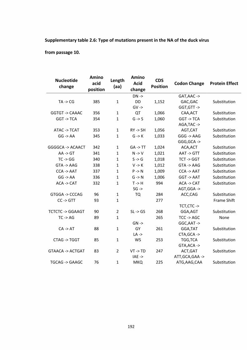
192
Supplementary table 2.6: Type of mutations present in the NA of the duck virus
from passage 10.
Nucleotide change
Amino acid
position
Length (aa)
Amino Acid
change
CDS Position
Codon Change Protein Effect
TA -> CG 385 1 DN ->
DD 1,152 GAT,AAC ->
GAC,GAC Substitution
GGTGT -> CAAAC 356 1 GV ->
QT 1,066 GGT,GTT ->
CAA,ACT Substitution
GGT -> TCA 354 1 G -> S 1,060 GGT -> TCA Substitution
ATAC -> TCAT 353 1 RY -> SH 1,056 AGA,TAC ->
AGT,CAT Substitution
GG -> AA 345 1 G -> K 1,033 GGG -> AAG Substitution
GGGGCA -> ACAACT 342 1 GA -> TT 1,024 GGG,GCA ->
ACA,ACT Substitution
AA -> GT 341 1 N -> V 1,021 AAT -> GTT Substitution
TC -> GG 340 1 S -> G 1,018 TCT -> GGT Substitution
GTA -> AAG 338 1 V -> K 1,012 GTA -> AAG Substitution
CCA -> AAT 337 1 P -> N 1,009 CCA -> AAT Substitution
GG -> AA 336 1 G -> N 1,006 GGT -> AAT Substitution
ACA -> CAT 332 1 T -> H 994 ACA -> CAT Substitution
GTGGA -> CCCAG 96 1 SG ->
TQ 284 AGT,GGA ->
ACC,CAG Substitution
CC -> GTT 93 1 277 Frame Shift
TCTCTC -> GGAAGT 90 2 SL -> GS 268 TCT,CTC -> GGA,AGT Substitution
TC -> AG 89 1 265 TCC -> AGC None
CA -> AT 88 1 GN ->
GY 261 GGC,AAT ->
GGA,TAT Substitution
CTAG -> TGGT 85 1 LA -> WS 253
CTA,GCA -> TGG,TCA Substitution
GTAACA -> ACTGAT 83 2 VT -> TD 247 GTA,ACA ->
ACT,GAT Substitution
TGCAG -> GAAGC 76 1 IAE -> MKQ 225
ATT,GCA,GAA -> ATG,AAG,CAA Substitution

193
Supplementary figure 2.1: Average coverage for each segment from the IAV
genomic sequencing.
0
1 000
2 000
3 000
4 000
5 000
6 000
7 000
murreH1N2AB319
murreH1N2AB327
murreH1N2AB341
gullH13N6GR654
gullH13N6GR658
gullH13N6GR848
duckH1N1
MW668
duckH3N2GR679
duckH3N2GR256
Ave
rage
re
ads
nu
mb
er
PB2 PB1 PA HA NA NP M NS

194

195
Supplementary figure 2.2: Relationships between segment shared variants and
sequence homology. The scatter plot shows the two values for each pair of
segments compared within the different hosts: A) gull viruses, B) murre viruses, and
C) duck viruses.

196
Supplementary documents in chapter 3
Supplementary table 3.1: List of RACE primers used.
Primer Primer sequence (5' to 3')
3’ RACE
3CM-PB2-1 CATTAACGGAGCAATCTTGCAG
3CM-PB2-2 GTGATACCATTACTCTGTCTGATCC
3HG-PB2-1 CCGGTAGGAATCTGGTCTTGC
3HG-PB2-2 ACACCATCACCCTATCAGAACC
3DD-PB2-1 ACTGCTTGTTCCACCAGCC
3DD-PB2-2 CACGGCCAGAGGTGATACC
3-CM-PB1-1 CCGGCTGATTCCTGTTTAGTG
3-PB1-2 ATATCCACTTGGCTCATTGTCC
3-HGDD-PB1-1 CCGGTTGATTCCTGTTTAATGTC
3-PA-1 CAATGCAGCCGTTCGGTT
3-CM-PA-2 ATTATCGATTCACCTCGTTCATC
3-HG-PA-2 CAGATGCTATTCACCACAGTCC
3-DD-PA-2 GATTCCACAATTATTGATTCGCC
3-CM-HA-1 GTGTCCAATAATAGTTCATCCTGCC
3-HG-HA-1 CGGGTGGAAACTAGTGTATAAGG
3-DD-HA-1 GTGTCCAGTAATAGTTCATCCTGCC
3-CMDD-HA-2 TTCAGGCTGCAGAGCTTCC

197
3-HG-HA-2 CTCCATTCAAAGAACAGTATGTTCC
3-CM-NA-1 CATGGCAACTTGAGCTGGAC
3-HG-NA-1 ATGACATGAGGTGCTGGACC
3-DD-NA-1 CAATTGTCAACCAGCTAATGCC
3-CM-NA-2 CGATGTGGAATTCTATCATGTATTG
3-HG-NA-2 CTCTCCTATTCTAATTGCATTGTCC
3-DD-NA-2 GCCTGCTAATGTTATTGGAGCC
3-CMDD-NP-1 GTGGCATCATTCAGATTGGAAT
3-HG-NP-1 GTGGCGTCATTCAAGTTAGAATG
3-HG-NP-2 CTGAGCTTGAGTTCGGTGCAC
3-DD-NP-2 GATGTTCCTCAAGGTATCTGTTCC
3-CM-NP-2 CCAGATACTTGTTTCTCCTCTCATC
3-M-1 GCTCACAAGTGGCACATACTAGAC
3-M-2 CTACGCTGCAGTCCTCGCT
3-NS-1 AGTATGTCCTGGAAGAGAAGG
3-CMDD-NS-2 CAGTTAGGTAGCGTGAAGTAGGC
3-HG-NS-2 CTCTTGACATCTCCTCTGGAGAC
5’ RACE
5-CM-PB2-1 GTAAGGACGCTATTCCAGCAGAT
5-HG-PB2-1 CAAGGCTGCTAGAGGCCAATAC
5-DD-PB2-1 CGTGAGGACACTATTCCAGCAG
5-CM-PB2-2 GGAGTGGAATCTGCGGTATTAAG

198
5-HG-PB2-2 GGATGCAGGTTCATTGACAGAAG
5-DD-PB2-2 GCATTGACTGAAGATCCAGATGA
5-CM-PB2-3 GATATGGACCAGCATTGAGCAT
5-HG-PB2-3 GCATCAACGAACTGAGCAATC
5-DD-PB2-3 GGAGAGAAGGCTAATGTGCTAATTG
5-PB1-1 CATGAGCATTGGAGTAACAG
5-PB1-2 GACACACAAATTCAGACAAG
5-PB1-3 CCACAAAGAGATAGAGTCTGT
5-CM-PA-1 GTATGTGAGGACCAATGGAACTTC
5-HG-PA-1 GAGAGCATGATTGAAGCTGAATC
5-DD-PA-1 GACCGCAATAGGCCAAGTATC
5-CM-PA-2 GGAGTAGAGGAAGGTTCCATTG
5-HG-PA-2 GGCTCCATCGGTAAAGTGTG
5-DD-PA-2 GGAAGGCTCTATTGGGAAGGTAT
5-PA-3 CTCATTGTTCAGGCACTTAG
5-CMDD-HA-1 GAATTCAACAACCTAGAGAG
5-HG-HA-1 GAGTTCAGCCAGGTAGAA
5-CMDD-HA-2 GAGTTCTATCACAAGTGTGAT
5-HG-HA-2 GAGCTCCTTCATAAATGTAAT
5-CM-HA-3 CGGAGTGAAACTAGAGTCGATGG
5-HG-HA-3 GCTGAATATGCAGAAGAATCG
5-DD-HA-3 CGGCACATATGACTATCCCAAATAT

199
5-CM-NA-1 GGAACGATGATAGCTCTAG
5-HG-NA-1 GCAAATACTTATGTTCAAAG
5-DD-NA-1 GTACTAGCTCAAGGAGTGG
5-CM-NA-2 CGGCTAATTCCAAGTCACAGG
5-HG-NA-2 GCAGAAACAGACAATCAGTCCG
5-DD-NA-2 GGATGGACAGAGACGGACG
5-CM-NA-3 GGTGGACTTCAAATAGTATCGTC
5-HG-NA-3 GGAATTAATCAGAGGCAGGCC
5-DD-NA-3 GAGCTAATCAGAGGAAGACCCAAG
5-NP-1 GTGTCAAGCTTCATCAGA
5-CM-NP-2 GAGAGAGCAACCATTATGGCA
5-HG-NP-2 GGAGGAAACACCAACCAAC
5-DD-NP-2 GGAAACACTGAAGGCAGAACTTC
5-NP-3 CTGAGATCATAAGGATGATGG
5-M-1 GTGCCACTTGTGAGCAGATTG
5-M-2 GGCTAGACAGATGGTGCA
5-M-3 GCAGCGATTCAAGTGATCCTC
5-CMDD-NS-1 GCCTTCTCTTCCAGGAC
5-HG-NS-1 GACATGTCTCCAGAGGAG
5-CMDD-NS-2 GGACTTGAATGGAATGATAACACAG
5-HG-NS-2 CTCACCATTACCTTCTCTTCCAGG
5-CMDD-NS-3 CGCTTGGAGAAGCAGTGATG

200
5-HG-NS-3 GGAACAAGAACTGGAGAGAACAAT
The abbreviations HG, CM, and DD refer to the avian viruses: herring gull, common murre, and duck
respectively.

201
Supplementary table 3.2: Number of NCRs analyzed by host.
Swine Duck Human Poultry Murre Shorebird Swan/Goose Gull Equine
H1 191 200 380 11 26 36
H9
38
33
4 3
H13
2
41
N6
10
14
NP
29 16 23
36
M 16 12 18
7
NS
16 10 12
15

202
Supplementary figure 3.1: NS 3’ NCRs from different host species and locations.
Hosts of the viruses are indicated on the left.

203
Supplementary documents in chapter 4
Supplementary table 4.1: Pairwise genetic distancea matrices comparing gene
segments of A/herring gull/Newfoundland/YH019/2010(H16N3) to segments from
other related viruses.
Virusb/Segment Location Virus numberc
PB2
1 2 3
1 A/herring_gull/Newfoundland/YH019/2010(H16N3) Canada -
2 A/black-headed_gull/Sweden/74340/2008(H16N3) Sweden 0.034 -
3 A/glaucous_gull-
herring_gull_hybrid/Iceland/1108/2011(H16N3)
Iceland 0.042 0.022 -
PB1
1 2
1 A/herring_gull/Newfoundland/YH019/2010(H16N3) Canada -
2 A/common_gull/Norway/10_1313/2009(H13N2) Norway 0.019 -
PA
1 2
1 A/herring gull/Newfoundland/YH019/2010(H16N3) Canada -
2 A/black-headed gull/Iceland/713/2010(H16N3) Iceland 0.009 -
HA
1 2 3

204
1 A/herring_gull/Newfoundland/YH019/2010(H16N3) Canada -
2 A/black-headed_gull/Iceland/713/2010(H16N3) Iceland 0.013 -
3 A/duck/Hokkaido/WZ82/2013(H16N3) Japan 0.021 0.011 -
4 A/Herring_Gull/Norway/10_2298/2007(H16) Norway 0.029 0.019 0.02
7
NP
1 2
1 A/herring_gull/Newfoundland/YH019/2010(H16N3) Canada -
2 A/black-headed_gull/Iceland/713/2010(H16N3) Iceland 0.005 -
NA
1 2
1 A/herring_gull/Newfoundland/YH019/2010(H16N3) Canada -
2 A/common_gull/Norway/10_1617/2006(H16N3) Norway 0.067 -
M
1 2
1 A/blackhead_gull/Astrakhan/65/1983(H13N6) Russia -
2 A/herring_gull/Newfoundland/YH019/2010(H16N3) Canada 0.031 -
NS
1 2
1 A/herring_gull/Newfoundland/YH019/2010(H16N3) Canada -
2 A/yellow-legged_gull/Ukraine/912306/2005(H13) Ukraine 0.02 -
a Genetic distance was calculated as 1 - % identity from nucleotide alignments.

205
b Gull sequences from North America are in green, gull sequences from Eurasia are in dark blue, and waterfowl sequences from Eurasia are in red. c Also corresponds to numbers in leftmost column.

206
Supplementary table 4.2: Pairwise genetic distancesa for comparison of gull virus
HA gene segments to related segments from different hosts.
HA
Virusesb Distancec
Gull-waterfowl
H1
A/laughing_gull/Delaware_Bay/61/2002(H1N9)
A/muscovy_duck/New_York/21211-5/2005(H1N1) 0.025
A/mallard/Maryland/13/2003(H1N1) 0.030
A/gull/Delaware/428/2009(H1N1)
A/mallard/Nova Scotia/02151/2007(H1N1) 0.033
H3
A/glaucous_gull/Alaska/44201-161/2006(H3N8)
A/northern_pintail/Alaska/44204-075/2006(H3N8) 0.000
H5
A/slaty-backed gull/Shandong/59/04(H5N1)
A/duck/Hubei/wg/2002(H5N1) 0.006
1
A/Black Headed Gull/HK/12.1/2003(H5N1)
A/duck/Hunan/1386/2003(H5N1) 0.009

207
H7
A/gull/Italy/692-2/93(H7N2)
A/duck/Heinersdorf/S495/6/86(H7N7) 0.038
A/yellow-legged_gull/Republic_of_Georgia/1/2012/mixed(H7)
A/mallard/Republic_of_Georgia/2/2010(H7N7) 0.029
A/laughing_gull/New_York/AI00-2455/2000(H7N3)
A/mallard/Ohio/322/1998(H7N3) 0.012
H13
A/glaucous-winged_gull/Southeastern_Alaska/9JR0822R0/2009(H13N6) 0.031
A/duck/Hokkaido/WZ68/2012(H13N2)
A/lesser_snow_goose/Alaska/44199-115/2006(H13N9)
A/glaucous_gull/Alaska/44199-097/2006(H13N3) 0.003
A/black_headed_gull/Mongolia/1766/2006(H13N6) 0.019
A/black-headed_gull/Sweden/74342/2008(H13N8) 0.030
A/mallard/Korea/SH38-45/2010(H13N2)
A/black_headed_gull/Mongolia/1766/2006(H13N6) 0.045
A/black-headed_gull/Sweden/74342/2008(H13N8) 0.046

208
A/herring_gull/New_Jersey/AI09-1105/2009(H13N6)
A/hooded_merganser/New_Brunswick/03750/2009(H13N6) 0.007
A/mallard-black_duck_hybrid/New_Brunswick/03736/2009(H13N6) 0.005
H16
A/gull/Denmark/68110/2002(H16N3) 0.048
A/mallard/Quebec/02916-1/2009(H16N3)
A/California gull/California/1196P/2013(H16N3)
A/duck/Hokkaido/WZ82/2013(H16N3) 0.032
A/black-headed gull/Iceland/713/2010(H16N3) 0.021
A/waterfowl/GA/96623-7/2001(H16N3)
A/herring gull/New York/AI00-532/2000(H16N3) 0.001
A/mallard/Gurjev/785/83(H16N3)
A/black-headed gull/Sweden/2/99(H16N3) 0.070
Gull-poultry
H5
A/slaty-backed_gull/Shandong/38/04(H5N1) 0.002
A/chicken/Yichang/lung-1/04(H5N1)

209
A/brown-head_gull/Thailand/vsmu-4/2008(H5N1)
A/chicken/Thailand/ICRC-V143/2007(H5N1) 0.000
A/chicken/Thailand/ICRC-213/2007(H5N1) 0.000
A/black_headed_gull/HK/12.1/2003(H5N1)
A/chicken/Hong_Kong/3123.1/2002(H5N1) 0.007
H7
A/gull/Italy/692-2/93(H7N2)
A/turkey/Israel/Ramon/79(H7N2) 0.016
A/chicken/Hebei/1/2002(H7N2) 0.031
A/chicken/Jena/1816/87(H7N7) 0.037
A/yellow-legged_gull/Republic_of_Georgia/1/2012/mixed(H7)
A/turkey/Italy/8000/2002(H7N3) 0.046
A/laughing_gull/New_York/AI00-2455/2000(H7N3)
A/chicken/New_York/14714-9/1999(H7N3) 0.021
H13
A/herring_gull/Delaware/660/1988(H13N6)
A/laughing_gull/Delaware_Bay/2838/1987(H13N2) 0.019
A/turkey/MN/1012/1991(H13N2) 0.020

210
Gull-swine
H1
A/laughing_gull/Delaware_Bay/61/2002(H1N9)
A/swine/Saskatchewan/18789/02(H1N1) 0.020
H5
A/slaty-backed_gull/Shandong/59/04(H5N1)
A/swine/Anhui/ca/2004(H5N1) 0.001
Gull-other mammals
H5
A/black_headed_gull/HK/12.1/2003(H5N1)
A/Hong_Kong/213/2003(H5N1) 0.009
H13
A/gull/Minnesota/945/1980(H13N6)
A/pilot_whale/Maine/328_HN/1984(H13N2) 0.042
Gull-gull
H13
A/gull/Astrakhan/3483/2002(H13N6)
A/glaucous-winged_gull/Southcentral_Alaska/9JR0738R0/2009(H13N6) 0.036
A/glaucous-winged_gull/Southcentral_Alaska/9JR0738R0/2009(H13N6)
A/great_black-headed_gull/Atyrau/773/2004(H13N6) 0.032

211
A/black_headed_gull/Mongolia/1766/2006(H13N6)
A/glaucous_gull/Alaska/44199-097/2006(H13N3) 0.022
A/black-headed_gull/Sweden/74342/2008(H13N8)
A/glaucous_gull/Alaska/44199-097/2006(H13N3) 0.033
A/gull/Astrakhan/3483/2002(H13N6)
A/glaucous-winged_gull/Southcentral_Alaska/9JR0738R0/2009(H13N6) 0.036
A/great_black-headed_gull/Atyrau/773/2004(H13N6)
A/glaucous-winged_gull/Southcentral_Alaska/9JR0738R0/2009(H13N6)
0.032
H16
A/glaucous gull-herring gull hybrid/Iceland/1108/2011(H16N3)
A/California gull/California/1196P/2013(H16N3) 0.023
a Genetic distance was calculated as 1 - % identity from nucleotide alignments. b Gull sequences from North America are in green, gull sequences from Eurasia are in blue, waterfowl sequences from Eurasia are in red, waterfowl sequences from North America are in orange, swine sequences are in pink, other mammalian sequences are in dark blue, and poultry sequences are in brown. c The distance values for each virus relative to the top listed virus sequence within groups is given.

212
Supplementary table 4.3: Pairwise distancesa between tern virus sequences and
viruses from various hosts.
Segment Virusesb Distancec
PB2
A/arctic_tern/Alaska/300/1975(H5N3)
A/gull/Astrakhan/227/1984(H13N6) 0.074
A/blackhead_gull/Astrakhan/458/1985(H13N6) 0.084
A/gull/Minnesota/945/1980(H13N6) 0.078
A/gull/Maryland/704/1977(H13N6) 0.075
PB1
A/arctic_tern/Alaska/300/1975(H5N3)
A/gull/Minnesota/945/1980(H13N6) 0.032
A/tern/South_Africa/1961(H5N3)
A/turkey/England/1963(H7N3) 0.047
A/turkey/England/N28/1973(H5N2) 0.051
A/tern/South_Africa/1961(H5N3)
A/seal/Massachusetts/3911/1992(H3N3) 0.070
PA A/arctic_tern/Alaska/300/1975(H5N3)
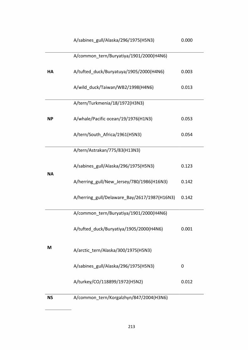
213
A/sabines_gull/Alaska/296/1975(H5N3) 0.000
HA
A/common_tern/Buryatiya/1901/2000(H4N6)
A/tufted_duck/Buryatuya/1905/2000(H4N6) 0.003
A/wild_duck/Taiwan/WB2/1998(H4N6) 0.013
NP
A/tern/Turkmenia/18/1972(H3N3)
A/whale/Pacific ocean/19/1976(H1N3) 0.053
A/tern/South_Africa/1961(H5N3) 0.054
NA
A/tern/Astrakan/775/83(H13N3)
A/sabines_gull/Alaska/296/1975(H5N3) 0.123
A/herring_gull/New_Jersey/780/1986(H16N3) 0.142
A/herring_gull/Delaware_Bay/2617/1987(H16N3) 0.142
M
A/common_tern/Buryatiya/1901/2000(H4N6)
A/tufted_duck/Buryatiya/1905/2000(H4N6) 0.001
A/arctic_tern/Alaska/300/1975(H5N3)
A/sabines_gull/Alaska/296/1975(H5N3) 0
A/turkey/CO/118899/1972(H5N2) 0.012
NS A/common_tern/Korgalzhyn/847/2004(H3N6)

214
A/mallard/Sweden/101993/2009(H2N9) 0.010
a Genetic distance was calculated as 1 - % identity from nucleotide alignments. b Tern sequences are in light blue, gull sequences from North America are in green, gull sequences from Eurasia are in blue, waterfowl sequences from Eurasia are in red, waterfowl sequences from North America are in orange, mammalian sequences are in dark blue, and poultry sequences are in brown. c The distance values for each virus relative to the top listed virus sequence within groups is given.
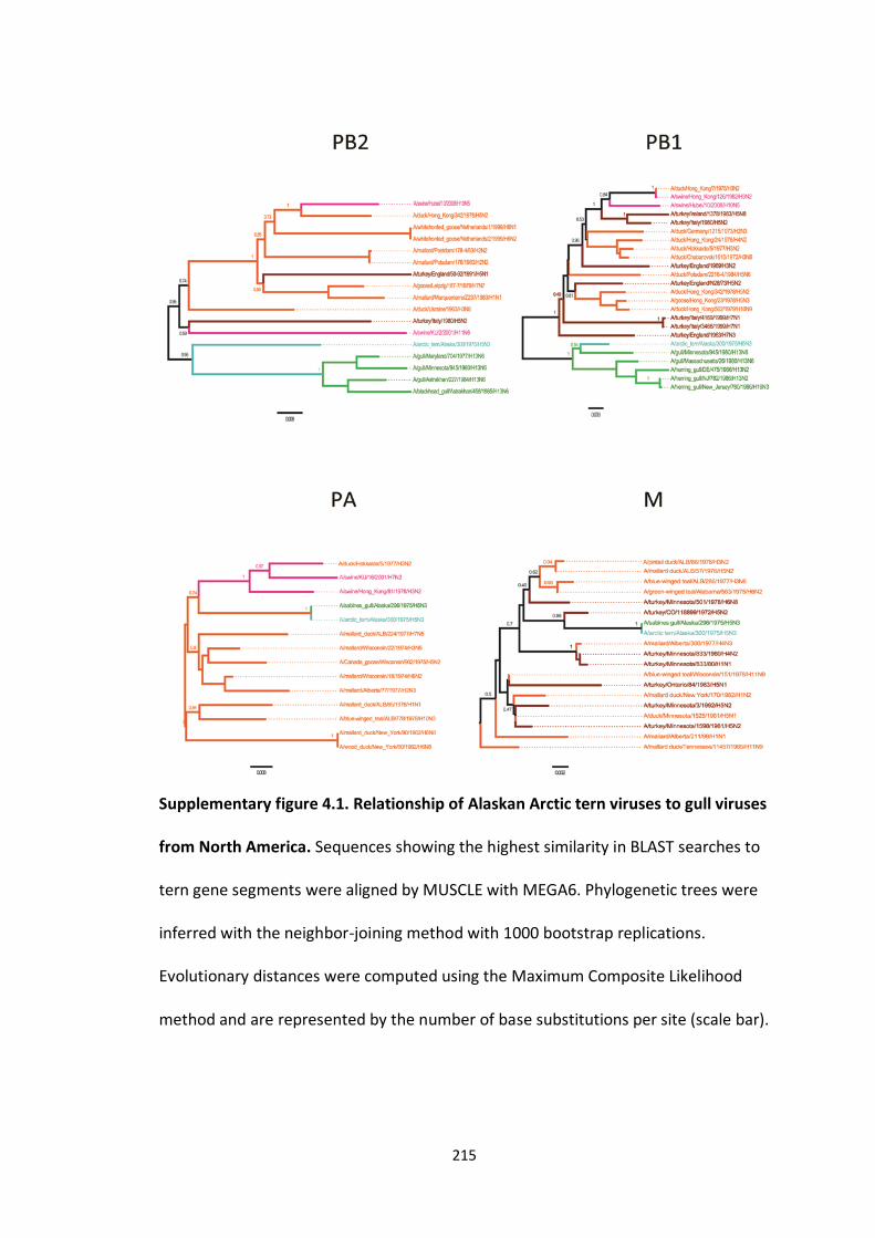
215
Supplementary figure 4.1. Relationship of Alaskan Arctic tern viruses to gull viruses
from North America. Sequences showing the highest similarity in BLAST searches to
tern gene segments were aligned by MUSCLE with MEGA6. Phylogenetic trees were
inferred with the neighbor-joining method with 1000 bootstrap replications.
Evolutionary distances were computed using the Maximum Composite Likelihood
method and are represented by the number of base substitutions per site (scale bar).

216
The branches are coloured according to the host group: green, gulls; brown, poultry;
light blue, terns; orange, waterfowl; pink, swine.

217
218
Supplementary figure 4.2. Relationships of tern viruses to waterfowl viruses.
Sequences showing the highest similarity in BLAST searches to tern gene segments
were aligned by MUSCLE with MEGA6. The phylogenetic trees were inferred with the
neighbor-joining method with 1000 bootstrap replications. Evolutionary distances
were computed using the Maximum Composite Likelihood method and are
represented by the number of base substitutions per site (scale bar). The branches
are coloured according to the host group: green, gulls; brown, poultry; light blue,
terns; orange, waterfowl; pink, swine.


![Avian Influenza 2006[1]](https://img.pdfslide.net/doc/110x75/63167d2fd16b3722ff0ce79a/avian-influenza-20061.jpg)


