Embed Size (px)
Citation preview
Genetic Factors and Orofacial Clefting
Andrew C. Lidral, DDS, PhD1,2,3,*, Lina M. Moreno, DDS, PhD1, and Steven A. Bullard, BS1
1Department of Orthodontics, University of Iowa, Iowa City, IA
2Dows Institute for Dental Research, University of Iowa, Iowa City, IA
3Craniofacial Anomalies Research Center, University of Iowa, Iowa City, IA
AbstractCleft lip with or without cleft palate is the most common facial birth defect and it is caused by acomplex interaction between genetic and environmental factors. The purpose of this review is toprovide an overview of the spectrum of the genetic causes for cleft lip and cleft palate using bothsyndromic and nonsyndromic forms of clefting as examples. Although the gene identification processfor orofacial clefting in humans is in the early stages, the pace is rapidly accelerating. Recently,several genes have been identified that have a combined role in up to 20% of all clefts. While this isa significant step forward, it is apparent that additional cleft causing genes have yet to be identified.Ongoing human genome-wide linkage studies have identified regions in the genome that likelycontain genes that when mutated cause orofacial clefting, including a major gene on chromosome 9that is positive in multiple racial groups. Currently, efforts are focused to identify which genes aremutated in these regions. In addition, parallel studies are also evaluating genes involved inenvironmental pathways. Furthermore, statistical geneticists are developing new methods tocharacterize both gene-gene and gene-environment interactions to build better models forpathogenesis of this common birth defect. The ultimate goal of these studies is to provide knowledgefor more accurate risk counseling and the development of preventive therapies.
IntroductionIn humans, orofacial clefts are common congenital anomalies with a prevalence of 1–2/1000live births. They can be separated into two different phenotypes: (1) cleft lip with or withoutcleft palate (CL/P); and (2) cleft palate only (CPO). Orofacial clefts can be further classifiedas nonsyndromic (isolated) or syndromic based upon the presence of other anomalies.Approximately 30% of CL/P and 50% of CPO patients have one of over 400 describedsyndromes.1–4 The focus of this review is primarily nonsyndromic cleft lip (CL/P) since thistrait has been studied the most in humans, while the etiology of CPO has been studied morein animal models.
*Corresponding Author, 2186 Medical Laboratories, University of Iowa, Iowa City, IA 52242, Phone: 319-335-8498, Fax: 319-335-6848,E-mail: [email protected]'s Disclaimer: This is a PDF file of an unedited manuscript that has been accepted for publication. As a service to our customerswe are providing this early version of the manuscript. The manuscript will undergo copyediting, typesetting, and review of the resultingproof before it is published in its final citable form. Please note that during the production process errors may be discovered which couldaffect the content, and all legal disclaimers that apply to the journal pertain.Statements and opinions expressed in the articles and communications herein are those of the authors and not necessarily those of theEditor or Publisher, and the Editor and Publisher disclaim any responsibility or liability for such material. Neither the Editor(s) nor thePublisher guarantee, warrant, or endorse any product or service advertised in this publication; neither do they guarantee any claim madeby the manufacturer of such product or service. Each reader must determine whether to act on the information contained in the publication,and neither the journal nor the Editor(s) shall be liable for any injury due to the publication of erroneous information.
NIH Public AccessAuthor ManuscriptSemin Orthod. Author manuscript; available in PMC 2009 June 1.
Published in final edited form as:Semin Orthod. 2008 June ; 14(2): 103–114. doi:10.1053/j.sodo.2008.02.002.
NIH
-PA Author Manuscript
NIH
-PA Author Manuscript
NIH
-PA Author Manuscript
It is generally accepted that CL/P and CPO are genetically distinct phenotypes in terms of theirinheritance patterns. CPO is less common, with a prevalence of approximately 1/1500–2000births in Caucasians, while CL/P is more common, 1–2/1000 births. The prevalence of CPOdoes not vary in different racial backgrounds, while the prevalence of CL/P varies considerably,with Asian and American Indians having the highest rate and Africans the lowest.5,6 Thereare also gender ratio differences with more males having CL/P and more females having CPO.Finally, families with one type of clefting segregating in the family do not have the other clefttype occur at a rate higher than the population prevalence.7 It will be interesting from a geneticperspective to determine the basis for the different inheritance patterns in CL/P and CPO. Forinstance, it will be important to determine whether the difference is due to locus or allelicheterogeneity, meaning that the differences are due to different genes (loci) or differentmutations (alleles) within the same gene. Given that both primary and secondary palatogenesisinvolve fusion between facial processes, it is expected that some genes may be involved inboth disorders.
Simple versus Complex Genetic TraitsIt is important to recognize that human traits (some of which are diseases or developmentalanomalies) can be caused by a variety of genetic mechanisms and phenomena, which can bereflected by the inheritance patterns within families. These mechanisms include whether thedisorder is inherited in a Mendelian pattern such as autosomal dominant, autosomal recessive,or X-linked (Figure 1–Figure 3).
The penetrance of a mutation, defined as the frequency of the disease trait in individualscarrying the disease mutation, will also affect the inheritance pattern. Expressivity, whichdescribes the severity or variation of the trait, can also vary considerably among affectedindividuals. Expressivity can affect the perceived inheritance pattern if the severity is so mildor below a certain stipulated threshold such that the person is considered normal when in factthey carry the disease mutation, and could be identified as being affected with carefulexamination or highly sensitive diagnostic techniques.
Finally, the number of genes underlying a specific trait (disease) can also vary. For example,some traits (diseases) are all caused by the same gene, meaning everyone with the trait (disease)has a mutation in the same gene. This situation is called genetic homogeneity. Interestingly,there are examples in which mutations in any one of several genes will result in the same trait(disease). This is termed locus heterogeneity. Finally, some traits (diseases) only becomeapparent when multiple genes are mutated in the same individual. Thus traits (diseases) can becaused by simple to complex mechanisms (Figure 4).8
A simple genetic trait implies that it is inherited in a Mendelian pattern, and that all individualswith the trait have a mutation in the same gene, meaning a major gene for the trait exists. Acomplex genetic trait is one that does not conform to simple Mendelian inheritance patterns.It is presumably caused by the combination of multiple mutations in different genes interactingwith environmental factors.
Inheritance patterns, penetrance, expressivity and genetic homogeneity can significantlyimpact the ability to identify causative genes. The simplest scenario is when all affectedindividuals have mutations in the same gene, the penetrance is high and the expressivity isconsistent such that one can assume that all affected individuals within a family share the sameDNA mutation and DNA markers near the mutation.
The approach in this situation involves scanning of the human genome to look for the sharedregion between affected members within a family. When this is found, it is said that linkageexists for a specific marker or region with the disease. This can be readily accomplished by
Lidral et al. Page 2
Semin Orthod. Author manuscript; available in PMC 2009 June 1.
NIH
-PA Author Manuscript
NIH
-PA Author Manuscript
NIH
-PA Author Manuscript
using approximately 400 DNA markers. The sharing is statistically evaluated under theassumption that each child has a 50:50 chance of inheriting a specific chromosome carryingthe mutated or normal copy of the gene since the genome consists of pairs of chromosomes,and only one is transferred from a given parent to an offspring.
The statistical output of these genetic tests is usually the LOD score, which is the log10 of theodds that a trait (disease) and DNA marker are linked versus the odds that they are not linked,assuming a 50:50 chance for each individual. LOD scores can be positive or negative, meaningthere is evidence for or against linkage. LOD scores greater than 3.0 are considered significantevidence for linkage, and less than −2.0 excludes linkage (for more on linkage analysis see“Investigation of Genetic Factors Affecting Complex Traits Using External Apical RootResorption as a Model” by Abass and Hartsfield in this volume).
Assuming every affected family has mutations in the same trait (disease) gene, the LOD scorescan be summed across all families. This will both increase the power to detect linkage andnarrow the region of linkage. The ability to sum LOD scores also aids studies that involvefamilies too small to independently yield significant results. Thus it is relatively easy to identifythe disease gene location region for simple traits. The next step is to identify what genes map(are located) within the linked region. This is easily accomplished by utilizing a variety ofgenetic databases and maps that are available from the successful sequencing of the humangenome. The genes can then be sequenced to identify possible causal mutations, with successbeing obtained upon finding mutations only in affected individuals.
Complex traits are much more difficult to map since not everyone has contributory mutationsin the same gene. Thus families may map to different trait (disease) genes, such that combiningthe LOD scores will result in negative results for one family canceling out the positive resultsfor another family. Furthermore, the decreased penetrance commonly observed in complextraits means some people without the trait (disease) carry mutations, thus breaking the linkagebetween a given gene or marker with the disease trait. In the early stages of gene mapping, itwas commonly believed that a researcher must know the inheritance pattern, and also havesome justification to assume trait (disease) homogeneity; otherwise the study was doomed tofail.
These beliefs, limited technology and statistical methods, and the relative ease for mappingsimple genetic traits (diseases) resulted in early success for simple traits and a delayed start forcomplex traits. However with the development of high throughput technology and improvedstatistical methods that take into account both genetic heterogeneity and decreased penetrance,successful mapping of complex traits occurred in the mid-1990’s for type 1 diabetes9,10 andmultiple sclerosis.11–14
With this as background, we will present examples of CL/P diseases that cover the spectrumof simple to complex genetic traits. Specifically, we will review the data indicatingnonsyndromic CL/P is a genetically complex trait involving genetic heterogeneity, lowpenetrance and the influence of various environmental factors.
Syndromic forms of CL/PSyndromic forms of CL/P often have simple Mendelian inheritance patterns and are thus moresuitable for conventional genetic mapping strategies.15
Van der Woude SyndromeVan der Woude syndrome (VWS) is an autosomal dominant (Figure 1) form of orofacialclefting with an estimated prevalence of 1/34,000 live births.16 Autosomal indicates that the
Lidral et al. Page 3
Semin Orthod. Author manuscript; available in PMC 2009 June 1.
NIH
-PA Author Manuscript
NIH
-PA Author Manuscript
NIH
-PA Author Manuscript
observed inheritance pattern excludes the sex chromosomes. VWS has a variety of featuresthat distinguish it from nonsyndromic CL/P, including the presence of lower lip pits (Figure5), hypodontia, and either CL/P or CPO. Furthermore, the penetrance is very high,approximately 97%. The disease gene was localized by linkage mapping to a large region onthe long arm of chromosome 1, 1q32–q41.17 Subsequent genetic studies identified severalpatients with deletions in the area that greatly narrowed the critical region to 350 kilo-bases(Kb = 1 thousand base pairs), containing over 20 known genes.18 An elegant strategy wasimplemented in which monozygotic twins, one with VWS and the other normal, weresequenced and a mutation discovered in the interferon regulatory factor 6 (IRF6) gene.19
IRF6 is a transcription factor that contains DNA binding and protein interaction domains. IRF6is expressed in the medial edge epithelia of the secondary palatal shelves. Thus it appears toregulate the expression of other genes during palatogenesis. Interestingly, VWS is an exampleof an orofacial syndrome in which CL/P and CPO can occur in the same family, suggestingthat it is likely involved in the fusion process that occurs in both primary and secondarypalatogenesis.
CL/P-Ectodermal Dysplasia SyndromeCL/P ectodermal dysplasia (CLPED1) syndrome is characterized by cleft lip, cleft palate,partial syndactyly of the fingers and toes, dental anomalies and sparse hair.20 It is a rareautosomal recessive trait (Figure 2). However there is a very high prevalence on MargaritaIsland, suggesting a founder effect in the small and relatively isolated population. The diseasegene was mapped to a 1–2 mega-base (Mb = 1 million base pairs) region on chromosome11,21 and subsequently mutations were identified in the Poliovirus Receptor-Like 1 (PVRL1)gene.22 Again, like IRF6, the PVRL1 gene name is misleading, suggesting an infectiousfunction rather than an important facial developmental gene.
PVRL1 encodes a cell-cell adhesion molecule that is expressed in the epithelia of the palatalshelves, nose and skin, as well as the dental ectoderm. Thus it appears PVRL1 is a moleculeimportant for cell-cell contact in these tissues. Interestingly, PVRL1 is used by various viruses,including the herpes simplex viruses, as a method to gain entry into cells.23
X-Linked Cleft Palate and AnkyloglossiaCleft palate occurring with ankyloglossia (CPX) has been reported segregating in an X-linkedrecessive pattern (Figure 5) in large families in Iceland and British Columbia, Canada. CPXwas the first orofacial cleft syndrome mapped, with linkage being identified to a large regionon the long arm of chromosome X.24 Recently, the causal gene was identified as TBX22,25which is expressed in the palatal shelves and tongue during development.26.27 TBX22functions by binding to specific DNA elements to regulate the expression of target genes. X-linked diseases are interesting since males have one X chromosome and females two Xchromosomes. If a male inherits a mutated TBX22 it is highly likely that he will have the diseasesince this is the only copy of the TBX22 gene.
In females, it is important to compensate the dose effect of having two X chromosomes. Thisis accomplished by X-inactivation (Lyonization) in which one X chromosome is inactivatedsuch that most genes are expressed only from the active X chromosome. This is normally arandom 50:50 process occurring early in development in each cell, with subsequent daughtercells having the same X inactivated. There is some normal range of X-inactivation if the totalnumber of cells are not split 50:50 in regard to which X is inactivated in each cell. Thus it ispossible for a female to inherit a mutated X-linked disease gene and either not have the diseaseor have a milder form of the disease depending upon the ratio and tissue distribution of X-inactivation. For example, if the X chromosome containing the disease gene is more often
Lidral et al. Page 4
Semin Orthod. Author manuscript; available in PMC 2009 June 1.
NIH
-PA Author Manuscript
NIH
-PA Author Manuscript
NIH
-PA Author Manuscript
inactivated or inactivated in the affected tissues, the female will likely not have the disease,although each son of hers has a 50:50 chance of being affected. For CPX it is hypothesizedthat X-inactivation would explain the reduced penetrance or milder phenotype in females,28though this has not been formally tested.
In summary, there has been significant success in identifying etiologic genes for syndromicforms of orofacial clefting, with approximately 15–20% cloned to date.29 Identifyingsyndromic disease genes will provide insight about the molecular processes involved in facialdevelopment and other affected tissues. Furthermore, these genes can be analyzed to determineif different mutations are associated with the more common nonsyndromic form of CL/P.
Genetics of Nonsyndromic CL/PNonsyndromic CL/P is an example of a genetically complex trait.30 The majority of affectedpatients have no positive family history and the evaluation of inheritance patterns in the familialcases has not revealed a simple Mendelian mode of inheritance (Figure 6). It is also clear thatthere is reduced penetrance. However, there is solid evidence that CL/P is a genetic trait sincethere is a 40 fold risk for CL/P amongst first degree relatives of an affected individual and thereis greater concordance in identical (monozygotic) compared to fraternal (dizygotic) twins.However, the concordance rate in monozygotic twins is only 40–60%, suggesting the influenceof environmental factors is also important.
Studies have estimated that 3–14 genes interacting multiplicatively may be involved, indicatingthat CL/P is a heterogeneous disorder,31 making it more difficult to map these genes sinceonly a portion of affected individuals will have a mutation in the same gene and currently thereis not any method to identify different genetic subsets a priori. However the impact for a givenCL/P gene is estimated to be sufficiently large enough to be mapped using a variety ofstrategies.32,33 Another limitation is that very large families with CL/P are rare, thus it isnecessary to combine the LOD scores across families when using a linkage approach, reducingpower and increasing the likelihood for missing a gene.
Human studies have used both association and linkage analyses to evaluate the role of candidategenes in the etiology of CL/P. Candidate genes have been chosen based on expression patternsduring facial development, cleft phenotype in transgenic or knockout mouse models,association with syndromic forms of clefting, previous positive findings in humans, role innutritional or xenobiotic pathways, and cytogenetic location adjacent to chromosomalanomalies associated with orofacial clreft phenotypes. Association mapping is theidentification of nonrandom correlations (associations) between alleles at two loci in apopulation. A simple way to envision this is the higher occurrence of a given allele in cases ascompared to controls. Association approaches have more study power especially in thepresence of genetic heterogeneity. Using a combination of both linkage and association canalso be successful for complex traits.34
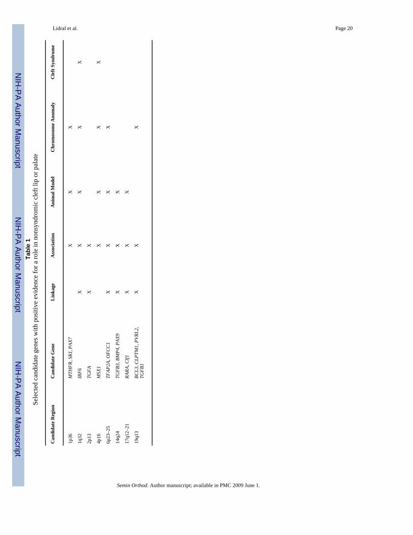
Candidate GenesInitial efforts to identify genes for nonsyndromic CL/P relied on candidate gene approaches.5,35 Genes at 1q32 (IRF6), 2p13 (TGFA), 4p16 (MSX1), 6p23–25, 14q24 (TGFB3), 17q21(RARA) and 19q13 (BCL3, TGFB1) have the most supporting data (Table 1). Below we willhighlight several of these genes.
The VWS Gene, IRF6 is associated with Nonsyndromic CL/PAs mentioned previously, mutations in IRF6 cause VWS. Since VWS has a very similarpresentation to isolated CL/P, IRF6 was evaluated as a candidate gene, and a highly significantassociation between IRF6 variants and CL/P was identified.36 Estimates suggest that genetic
Lidral et al. Page 5
Semin Orthod. Author manuscript; available in PMC 2009 June 1.
NIH
-PA Author Manuscript
NIH
-PA Author Manuscript
NIH
-PA Author Manuscript
variation in IRF6 contributes to 12% of CL/P and triples the recurrence risk in some families.These results have been replicated in additional populations,37–39 although the specificmutations have not yet been identified. This discovery constitutes one of the most excitingdiscoveries so far in the field of isolated CL/P.
MSX1MSX1 is a DNA binding transcription factor that when inactivated in mice results in cleft palateand tooth agenesis.40 This finding greatly aided the identification of a MSX1 mutation in afamily with hereditary tooth agenesis that was recruited through an orthodontic clinic.41Simultaneously, DNA variations in MSX1 were shown to be associated with CL/P.5,42–46Knowing these findings, a MSX1 mutation was found in a family with tooth agenesis, CL/Pand/or CPO.47 Recently, mutations in MSX1 have been identified in 2% of patients withnonsyndromic orofacial clefting.48,49 These findings indicate that MSX1 is involved in bothprimary and secondary palatogenesis, even though the mouse phenotype only involved thelatter. This is supported by the strong mesenchymal expression in the nasal and maxillaryprocesses during primary palatogenesis (Figure 7).
Transforming Growth Factor Beta 3 (TGFB3)Studies of TGFB3 further underscore the importance of animal studies as the observation ofcleft palate in mice missing TGFB350,51 led to the discovery of associations with CL/P inhumans.5 Furthermore, linkage studies have showed positive results for the region containingTGFB3.52 Nevertheless, only 15% of the families were linked to this region, which may inpart explain the observed inconsistencies in some previous studies. It is plausible that the nearbyBMP4 and PAX9 genes, both associated with orofacial clefting when inactivated in mice53,54 may be the cause of the positive findings, or are also involved.
19q13.1 (BCL3, CLPTM1, PVRL2, TGFB1)Several studies have found linkage or association with candidate genes on the long arm ofchromosome 19.5,55,56 Furthermore, a chromosomal anomaly involving this region wasfound in a family with CL/P.57 Candidate genes in this area include BCL3, PVRL2,CLPTM1 and TGFB1. Of these, PVRL2 is of interest since it is similar to PVRL1 that causesCLPED1, and carriers for a PVRL1 mutation are thought to have increased risk fornonsyndromic CL/P.58 Hence it is possible that PVRL2 has an analogous role.
Syndromic Orofacial Clefts Provide Important CluesIn addition to the IRF6 and PVRL1 examples of syndromic genes playing a role innonsyndromic clefting, efforts are underway to determine whether variants in other cleftsyndrome genes have similar roles. Mutations and deletions in the FGFR1 gene account for10% of Kallman syndrome patients, which have orofacial clefting, dental anomalies,hypogonadism and anosmia59 Suggestive association and linkage to CL/P has been found formarkers within the FGFR1 gene60 Also, mutations in the CPX gene, TBX22, have beenidentified in 4% of patients with CPO or ankyloglossia.61 These findings highlight theimportance of studying rare forms of diseases since in addition to identifying genes andpathways involved in a disease process, variants in the same gene may be associated with morecommon forms of the disease.62
Scanning the Genome for Additional CL/P GenesAs mentioned earlier, the application of linkage to complex traits is complicated by geneticheterogeneity and low penetrance. One approach around this is to collect families with multipleaffected individuals to look for sharing of genetic markers between only these related affectedpeople. This circumvents the low penetrance issue of not knowing if an unaffected person is a
Lidral et al. Page 6
Semin Orthod. Author manuscript; available in PMC 2009 June 1.
NIH
-PA Author Manuscript
NIH
-PA Author Manuscript
NIH
-PA Author Manuscript
disease gene carrier or not. Yet at the same time this approach has limited power, necessitatingthe study of hundreds of families. Previously, technology limited this approach to theevaluation of candidate genes. However, with the emergence of high-throughput genotypingtechnologies and powerful statistical approaches, this approach has been expanded to scan theentire genome to identify additional disease genes.
The first CL/P scan was published in 200063 and subsequently 5 additional scans of varyingsize have been published. In general the results have been modest with the exception of a LODscore of 3.0 at 17p13.1 in a scan of two large Syrian families64 The most consistent loci are2p13 (TGFA), 2q35–q37, 3p21–p24, 4q32–q33, 6p23–p25, 9q22–q33, 14q12–q31 and 18q11–q12. These results reflect genetic heterogeneity both within and between populations, limitedstudy power and a likely high false positive rate for loci with low levels of significance.
A meta-analysis of these 6 published and 7 ongoing genome scans revealed significant resultsfor 11 regions at 1q32, 2q32–q35, 3p25, 6q23–q25, 8p21, 8q23, 12p11, 14q21–q24, 17q21,18q21, and 20q13.52 Also, linkage analysis was performed allowing for genetic heterogeneity,and the summed results from 7 populations revealed significant heterogeneity LOD scores forchromosomal regions 1p12–p13, 6p23, 6q23–q25, 9q22–q33, 14q21–q24 and 15q15. Of these,the 9q22–q33 region was the most striking with a heterogeneity LOD score equal to 6.6, whichis the most significant result ever reported for CL/P. This is a new discovery and the regionlikely contains a major gene for CL/P.
Gene-Environment InteractionsEpidemiologic studies have revealed an increased risk for CL/P with alcohol65 andsmoking66 exposure during pregnancy. Furthermore, some studies suggest periconceptionalfolate or multivitamin supplementation has a protective effect against CL/P.67 However, notall mothers who drink or smoke have children with CL/P, nor do all mothers takingmultivitamins have normal children. Thus it is likely that certain genes that interact with theseenvironmental factors and that genetic variation within these genes affect the risk for CL/P.Researchers have tested this hypothesis by looking at both candidate genes for CL/P and genesinvolved in the environmental pathways.68
Given that neural tube defects can be prevented by folate supplementation and similar evidenceexists for CL/P, some investigators have evaluated genes in the folate pathway with somesuccess. However, it is not clear whether one should look at the DNA of the children, mothersor both. Clearly, environmental agents interact with maternal gene products, but it is not alwaysclear if the same is true for fetal gene products, although it is likely in some situations. It isplausible that a fetus may have a low risk for CL/P due to its genes, but that this risk increasesdue to maternal environmental exposures and her genetic susceptibility to these exposures.
This uncertainty has impacted the results for folate pathway genes.67 For example functionalvariations in the methylenetetrahydrofolate reductase (MTHFR) and reduced folate carrier(RFC1) genes revealed no association with CL/P in a South American population.69 However,a method looking at the infants genotype and maternal environmental exposures revealedsignificant gene-environment interactions between CP infants with certain variations inMTHFR and maternal folic acid consumption70 and these results were also found to be truefor CL/P.71 Alternatively, an increased risk has been observed for maternal MTHFR variants.72,73
Genetic variation has been identified in a variety of genes involved in the biotransformationof agents found in tobacco smoke including common deletions of the Glutathione S-transferasetheta 1-1 (GSTT1) and Glutathione S-transferase Mu 1 (GSTM1) genes. Mothers carrying theGSTT1-null allele and who smoked had a higher although not significant risk of having a child
Lidral et al. Page 7
Semin Orthod. Author manuscript; available in PMC 2009 June 1.
NIH
-PA Author Manuscript
NIH
-PA Author Manuscript
NIH
-PA Author Manuscript
with CL/P.74 A 6 fold risk increase was found for fetuses lacking both GSTT1 and GSTM1and whose mothers smoked.75 Furthermore, variations in NAT1, another gene involved indetoxification of cigarette smoke, resulted in a two or four fold increased risk for CL/P amonginfants homozygous for this polymorphism if their mothers did not use multivitamins orsmoked respectively.76,77 These studies highlight the importance of identifying suchinteractions so that individuals with disease susceptibility alleles can modify their risk byceasing detrimental exposures and improving their nutritional status.
SummaryIn general, the gene identification process for CL/P is still in the early stages, especiallycompared to other common diseases. However, the candidate gene approaches have identifiedvariations that are associated with up to 20% of patients with CL/P. Furthermore, the genomewide linkage scans have identified the location of several genes, including the previouslyunknown locus on chromosome 9. Current efforts are ongoing to narrow these regions andidentify disease causing mutations. Overall, the published findings support the hypothesis thatmultiple genes are involved in the etiology of CL/P. Future studies will determine how thesegenes interact with each other and the environment to develop models for improved geneticcounseling and public health policies.
AcknowledgementsOur work has been greatly aided by discussions with Mauricio Arcos-Burgos, Sandy Daack-Hirsch, Mike Dixon,David FitzPatrick, Brion Maher, Mary Marazita, Brian Schutte, Alex Vieira and George Wehby. Also we would liketo thank families that over the years have participated in our research studies.
Dr. Moreno is supported by a Fogarty International Maternal and Child Health Research and Training Fellowship(1D43 TW-05503) and Dr. Lidral is supported by NIH Grants RO1DE14677, KO2DE015291 and P50DE016215 withadditional funding from both the University of Iowa Craniofacial Anomalies Research Center, brilliantly directed byJeff Murray, and the College of Dentistry. While this work was not directly supported by the American Associationof Orthodontists Foundation, Dr. Lidral’s career has been greatly aided by three AAOF Faculty Development Awards.
References1. Cohen MM. Syndromes with cleft lip and cleft palate. Cleft Palate J 1978;15:306–328. [PubMed:
281275]2. Shprintzen RJ, Siegel VL, Amato J, et al. Anomalies associated with cleft lip, cleft palate, or both.
American Journal of Medical Genetics 1985;20:585–595. [PubMed: 3993684]3. Gorlin, RJ. Syndromes of the head and neck. New York: Oxford University Press; 1990.4. Gabriel SB, Schaffner SF, Nguyen H, et al. The structure of haplotype blocks in the human genome.
Science 2002;296:2225–2229. [PubMed: 12029063]5. Marazita ML, Mooney MP. Current concepts in the embryology and genetics of cleft lip and cleft
palate. Clin Plast Surg 2004;31:125–140. [PubMed: 15145658]6. Vanderas AP. Incidence of cleft lip, cleft palate, and cleft lip and palate among races: a review. Cleft
Palate J 1987;24:216–225. [PubMed: 3308178]7. Fraser, FC. The genetics of cleft lip and palate: yet another look. In: Pratt, Christiansen, editor. Current
trends in prenatal craniofacial development. 1980. p. 357-366.8. Beaudet AL. 1998 ASHG presidential address. Making genomic medicine a reality. Am J Hum Genet
1999;64:1–13. [PubMed: 9915936]9. Davies JL, Kawaguchi Y, Bennett ST, et al. A genome-wide search for human type 1 diabetes
susceptibility genes. Nature 1994;371:130–136. [PubMed: 8072542]10. Hashimoto L, Habita C, Beressi JP, et al. Genetic mapping of a susceptibility locus for insulin-
dependent diabetes mellitus on chromosome 11q. Nature 1994;371:161–164. [PubMed: 8072544]11. Sawcer S, Jones HB, Feakes R, et al. A genome screen in multiple sclerosis reveals susceptibility loci
on chromosome 6p21 and 17q22. Nat Genet 1996;13:464–468. [PubMed: 8696343]
Lidral et al. Page 8
Semin Orthod. Author manuscript; available in PMC 2009 June 1.
NIH
-PA Author Manuscript
NIH
-PA Author Manuscript
NIH
-PA Author Manuscript
12. Haines JL, Ter-Minassian M, Bazyk A, et al. A complete genomic screen for multiple sclerosisunderscores a role for the major histocompatability complex. Nat Genet 1996;13:469–471. [PubMed:8696344]
13. Ebers GC, Kukay K, Bulman DE, et al. A full genome search in multiple sclerosis. Nat Genet1996;13:472–476. [PubMed: 8696345]
14. Kuokkanen S, Gschwend M, Rioux JD, et al. Genomewide scan of multiple sclerosis in Finnishmultiplex families. Am J Hum Genet 1997;61:1379–1387. [PubMed: 9399895]
15. Murray JC. Face facts: genes, environment, and clefts. Am J Hum Genet 1995;57:227–232. [PubMed:7668246]
16. Rintala AE, Ranta R. Lower lip sinuses: I. Epidemiology, microforms and transverse sulci. Br J PlastSurg 1981;34:26–30. [PubMed: 7459520]
17. Murray JC, Nishimura DY, Buetow KH, et al. Linkage of an autosomal dominant clefting syndrome(Van der Woude) to loci on chromosome 1q. Am J Hum Genet 1990;46:486–491. [PubMed:2309700]
18. Schutte BC, Bjork BC, Coppage KB, et al. A preliminary gene map for the Van der Woude syndromecritical region derived from 900 kb of genomic sequence at 1q32–q41. Genome Res 2000;10:81–94.[PubMed: 10645953]
19. Kondo S, Schutte BC, Richardson RJ, et al. Mutations in IRF6 cause Van der Woude and poplitealpterygium syndromes. Nat Genet 2002;32:285–289. [PubMed: 12219090]
20. Zlotogora J. Syndactyly, ectodermal dysplasia, and cleft lip/palate. J Med Genet 1994;31:957–959.[PubMed: 7891379]
21. Suzuki K, Bustos T, Spritz RA. Linkage disequilibrium mapping of the gene for Margarita Islandectodermal dysplasia (ED4) to 11q23. Am J Hum Genet 1998;63:1102–1107. [PubMed: 9758630]
22. Suzuki K, Hu D, Bustos T, et al. Mutations of PVRL1, encoding a cell-cell adhesion molecule/herpesvirus receptor, in cleft lip/palate-ectodermal dysplasia. Nat Genet 2000;25:427–430. [PubMed:10932188]
23. Subramanian RP, Dunn JE, Geraghty RJ. The nectin-1[alpha] transmembrane domain, but not thecytoplasmic tail, influences cell fusion induced by HSV-1 glycoproteins. Virology 2005;339:176–191. [PubMed: 16005040]
24. Moore GE, Ivens A, Chambers J, et al. Linkage of an X-chromosome cleft palate gene. Nature1987;326:91–92. [PubMed: 2881212]
25. Braybrook C, Doudney K, Marcano AC, et al. The T-box transcription factor gene TBX22 is mutatedin X-linked cleft palate and ankyloglossia. Nat Genet 2001;29:179–183. [PubMed: 11559848]
26. Braybrook C, Lisgo S, Doudney K, et al. Craniofacial expression of human and murine TBX22correlates with the cleft palate and ankyloglossia phenotype observed in CPX patients. Hum MolGenet 2002;11:2793–2804. [PubMed: 12374769]
27. Bush JO, Lan Y, Maltby KM, et al. Isolation and developmental expression analysis of Tbx22, themouse homolog of the human X-linked cleft palate gene. Dev Dyn 2002;225:322–326. [PubMed:12412015]
28. Stanier P, Forbes SA, Arnason A, et al. The localization of a gene causing X-linked cleft palate andankyloglossia (CPX) in an Icelandic kindred is between DXS326 and DXYS1X. Genomics1993;17:549–555. [PubMed: 8244369]
29. Wilkie AO, Morriss-Kay GM. Genetics of craniofacial development and malformation. Nat RevGenet 2001;2:458–468. [PubMed: 11389462]
30. Wyszynski, DF. Cleft Lip and Palate: From Origin to Treatment. Oxford: Oxford University Press;2002.
31. Schliekelman P, Slatkin M. Multiplex relative risk and estimation of the number of loci underlyingan inherited disease. Am J Hum Genet 2002;71:1369–1385. [PubMed: 12454800]
32. Mitchell LE, Christensen K. Analysis of the recurrence patterns for nonsyndromic cleft lip with orwithout cleft palate in the families of 3,073 Danish probands. Am J Med Genet 1996;61:371–376.[PubMed: 8834050]
33. Farrall M, Buetow KH, Murray JC. Resolving an apparent paradox concerning the role of TGFA inCL/P. Am J Hum Genet 1993;52:434–436. [PubMed: 8094269]
Lidral et al. Page 9
Semin Orthod. Author manuscript; available in PMC 2009 June 1.
NIH
-PA Author Manuscript
NIH
-PA Author Manuscript
NIH
-PA Author Manuscript
34. Horikawa Y, Oda N, Cox NJ, et al. Genetic variation in the gene encoding calpain-10 is associatedwith type 2 diabetes mellitus. Nat Genet 2000;26:163–175. [PubMed: 11017071]
35. Carinci F, Pezzetti F, Scapoli L, et al. Recent developments in orofacial cleft genetics. J CraniofacSurg 2003;14:130–143. [PubMed: 12621282]
36. Zucchero TM, Cooper ME, Maher BS, et al. Interferon regulatory factor 6 (IRF6) gene variants andthe risk of isolated cleft lip or palate. N Engl J Med 2004;351:769–780. [PubMed: 15317890]
37. Houdayer C, Bonaiti-Pellie C, Erguy C, et al. Possible relationship between the van der Woudesyndrome (VWS) locus and nonsyndromic cleft lip with or without cleft palate (NSCL/P). Am J MedGenet 2001;104:86–92. [PubMed: 11746036]
38. Scapoli L, Palmieri A, Martinelli M, et al. Strong Evidence of Linkage Disequilibrium betweenPolymorphisms at the IRF6 Locus and Nonsyndromic Cleft Lip With or Without Cleft Palate, in anItalian Population. Am J Hum Genet 2005;76:180–183. [PubMed: 15558496]
39. Srichomthong C, Siriwan P, Shotelersuk V. Significant association between IRF6 820G->A and non-syndromic cleft lip with or without cleft palate in the Thai population. J Med Genet 2005;42:e46.[PubMed: 15994871]
40. Satokata I, Maas R. Msx1 deficient mice exhibit cleft palate and abnormalities of craniofacial andtooth development. Nat Genet 1994;6:348–356. [PubMed: 7914451]
41. Vastardis H, Karimbux N, Guthua SW, et al. A human MSX1 homeodomain missense mutationcauses selective tooth agenesis. Nat Genet 1996;13:417–421. [PubMed: 8696335]
42. Marazita ML, Field LL, Tuncbilek G, et al. Genome-scan for loci involved in cleft lip with or withoutcleft palate in consanguineous families from Turkey. Am J Med Genet 2004;126A:111–122.
43. Moreno LM, Arcos-Burgos M, Marazita ML, et al. Genetic analysis of candidate loci in non-syndromic cleft lip families from Antioquia-Colombia and Ohio. Am J Med Genet 2004;125A:135–144.
44. Schultz RE, Cooper ME, Daack-Hirsch S, et al. Targeted scan of fifteen regions for nonsyndromiccleft lip and palate in Filipino families. Am J Med Genet 2004;125A:17–22.
45. Vieira AR, Orioli IM, Castilla EE, et al. MSX1 and TGFB3 Contribute to Clefting in South America.J Dent Res 2003;82:289–292. [PubMed: 12651933]
46. Suazo J, Santos JL, Carreno H, et al. Linkage Disequilibrium between MSX1 and Non-syndromicCleft Lip/Palate in the Chilean Population. J Dent Res 2004;83:782–785. [PubMed: 15381719]
47. van den Boogaard MJ, Dorland M, Beemer FA, et al. MSX1 mutation is associated with orofacialclefting and tooth agenesis in humans [published erratum appears in Nat Genet 2000 May;25:125].Nat Genet 2000;24:342–343. [PubMed: 10742093]
48. Jezewski PA, Vieira AR, Nishimura C, et al. Complete sequencing shows a role for MSX1 in non-syndromic cleft lip and palate. J Med Genet 2003;40:399–407. [PubMed: 12807959]
49. Suzuki Y, Jezewski PA, Machida J, et al. In a Vietnamese population, MSX1 variants contribute tocleft lip and palate. Genet Med 2004;6:117–125. [PubMed: 15354328]
50. Proetzel G, Pawlowski SA, Wiles MV, et al. Transforming growth factor-B3 is required for secondarypalate fusion. Nat Genet 1995;11:409–414. [PubMed: 7493021]
51. Kaartinen V, Voncken JW, Shuler C, et al. Abnormal lung development and cleft palate in micelacking TGF-B3 indicates defects of epithelial-mesenchymal interaction. Nature Genetics1995;11:415–421. [PubMed: 7493022]
52. Marazita ML, Murray JC, Lidral AC, et al. Meta-Analysis of 13 Genome Scans Reveals MultipleCleft Lip/Palate Genes with Novel Loci on 9q21 and 2q32–35. Am J Hum Genet 2004;75:161–173.[PubMed: 15185170]
53. Liu W, Sun X, Braut A, et al. Distinct functions for Bmp signaling in lip and palate fusion in mice.Development 2005:dev.01676.
54. Peters H, Neubuser A, Kratochwil K, et al. Pax9-deficient mice lack pharyngeal pouch derivativesand teeth and exhibit craniofacial and limb abnormalities. Genes Dev 1998;12:2735–2747. [PubMed:9732271]
55. Blanco R, Suazo J, Santos JL, et al. Association between 10 microsatellite markers and nonsyndromiccleft lip palate in the Chilean population. Cleft Palate Craniofac J 2004;41:163–167. [PubMed:14989688]
Lidral et al. Page 10
Semin Orthod. Author manuscript; available in PMC 2009 June 1.
NIH
-PA Author Manuscript
NIH
-PA Author Manuscript
NIH
-PA Author Manuscript
56. Fujita H, Nagata M, Ono K, et al. Linkage analysis between BCL3 and nearby genes on 19q13.2 andnon-syndromic cleft lip with or without cleft palate in multigenerational Japanese families. OralDiseases 2004;10:353–359. [PubMed: 15533211]
57. Yoshiura, K-i; Machida, J.; Daack-Hirsch, S., et al. Characterization of a Novel Gene Disrupted bya Balanced Chromosomal Translocation t(2;19)(q11.2;q13.3) in a Family with Cleft Lip and Palate*1.Genomics 1998;54:231–240. [PubMed: 9828125]
58. Sozen MA, Suzuki K, Tolarova MM, et al. Mutation of PVRL1 is associated with sporadic, non-syndromic cleft lip/palate in northern Venezuela. Nat Genet 2001;29:141–142. [PubMed: 11559849]
59. Sato N, Katsumata N, Kagami M, et al. Clinical Assessment and Mutation Analysis of KallmannSyndrome 1 (KAL1) and Fibroblast Growth Factor Receptor 1 (FGFR1, or KAL2) in Five Familiesand 18 Sporadic Patients. J Clin Endocrinol Metab 2004;89:1079–1088. [PubMed: 15001591]
60. Schultz, RE. Genetics Program. Iowa City: University of Iowa; 2004. Identification of genetic lociinvolved in nonsyndromic cleft lip with or without cleft palate.
61. Marcano ACB, Doudney K, Braybrook C, et al. TBX22 mutations are a common cause of cleft palate.J Med Genet 2004;000:1–8.
62. Stanier P, Moore GE. Genetics of cleft lip and palate: syndromic genes contribute to the incidenceof nonsyndromic clefts. Hum. Mol. Genet 2004:ddh052.
63. Prescott NJ, Lees MM, Winter RM, et al. Identification of susceptibility loci for nonsyndromic cleftlip with or without cleft palate in a two stage genome scan of affected sib-pairs. Hum Genet2000;106:345–350. [PubMed: 10798365]
64. Wyszynski DF, Albacha-Hejazi H, Aldirani M, et al. A genome-wide scan for loci predisposing tonon-syndromic cleft lip with or without cleft palate in two large Syrian families. Am J Med Genet2003;123A:140–147.
65. Jones KL, Smith DW, Ullelaand CN, et al. Pattern of malformation in offspring of chronic alcoholicmothers. Lancet 1973;9:1267–1271. [PubMed: 4126070]
66. Wyszynski DF, Duffy DL, Beaty TH. Maternal cigarette smoking and oral clefts: a meta-analysis.Cleft Palate Craniofac J 1997;34:206–210. [PubMed: 9167070]
67. Prescott NJ, Malcolm S. Folate and the face: evaluating the evidence for the influence of folate geneson craniofacial development. Cleft Palate Craniofac J 2002;39:327–331. [PubMed: 12019010]
68. Murray J. Gene/environment causes of cleft lip and/or palate. Clin Genet 2002;61:248–256. [PubMed:12030886]
69. Vieira AR, Murray JC, Trembath D, et al. Studies of reduced folate carrier 1 (RFC1) A80G and 5,10-methylenetetrahydrofolate reductase (MTHFR) C677T polymorphisms with neural tube andorofacial cleft defects. Am J Med Genet A 2005;135:220–223. [PubMed: 15880745]
70. Jugessur A, Wilcox AJ, Lie RT, et al. Exploring the Effects of Methylenetetrahydrofolate ReductaseGene Variants C677T and A1298C on the Risk of Orofacial Clefts in 261 Norwegian Case-ParentTriads. Am J Epidemiol 2003;157:1083–1091. [PubMed: 12796044]
71. van Rooij IALM, Vermeij-Keers C, Kluijtmans LAJ, et al. Does the Interaction between MaternalFolate Intake and the Methylenetetrahydrofolate Reductase Polymorphisms Affect the Risk of CleftLip with or without Cleft Palate? Am J Epidemiol 2003;157:583–591. [PubMed: 12672677]
72. Shotelersuk V, Ittiwut C, Siriwan P, et al. Maternal 677CT/1298AC genotype of the MTHFR geneas a risk factor for cleft lip. J Med Genet 2003;40:e64. [PubMed: 12746418]
73. Pezzetti F, Martinelli M, Scapoli L, et al. Maternal MTHFR variant forms increase the risk in offspringof isolated nonsyndromic cleft lip with or without cleft palate. Hum Mutat 2004;24:104–105.[PubMed: 15221800]
74. van Rooij IALM, Ocke MC, Straatman H, et al. Periconceptional folate intake by supplement andfood reduces the risk of nonsyndromic cleft lip with or without cleft palate. Preventive Medicine2004;39:689–694. [PubMed: 15351534]
75. Lammer EJ, Shaw GM, Iovannisci DM, et al. Maternal smoking, genetic variation of glutathione s-transferases, and risk for orofacial clefts. Epidemiology 2005;16:698–701. [PubMed: 16135950]
76. Lammer EJ, Shaw GM, Iovannisci DM, et al. Periconceptional multivitamin intake during earlypregnancy, genetic variation of acetyl-N-transferase 1 (NAT1), and risk for orofacial clefts. BirthDefects Res A Clin Mol Teratol 2004;70:846–852. [PubMed: 15523664]
Lidral et al. Page 11
Semin Orthod. Author manuscript; available in PMC 2009 June 1.
NIH
-PA Author Manuscript
NIH
-PA Author Manuscript
NIH
-PA Author Manuscript
77. Lammer EJ, Shaw GM, Iovannisci DM, et al. Maternal smoking and the risk of orofacial clefts:Susceptibility with NAT1 and NAT2 polymorphisms. Epidemiology 2004;15:150–156. [PubMed:15127906]
GlossaryAllele, A genetic variation for a given marker or locuscM, Centimorgan. 1cM is equal to 1% recombination and approximately 1 million base pairs.Expressivity, The severity of a disease in individuals carrying the disease mutation. Fordiseases that involve multiple features, the expressivity can describe both the number ofaffected features as well as the severity for each feature.Gene, Typically, this describes the DNA sequence encoding a protein. But this is also used todescribe the DNA sequence that is used to make the message RNA (mRNA). The term mayalso be expanded to describe the entire gene, meaning the coding sequence, the mRNAsequence and all the regulatory DNA elements that control the expression of the mRNA andprotein.Genome, The entire genetic material for a given organism. For humans this consists of 22 pairsof chromosomes and two sex chromosomes either XX or XY.Genotyping, A laboratory technique used to determine which alleles exist for a marker in aperson.Haplotype, A combination of alleles for markers as they occur on a chromosomeHeterozygosity, The presence of two different alleles for a given markerHomozygosity, The presence of the same alleles for a given markerLocus, A given region of the human genome,;specifically, the location of a gene or particularDNA sequence on a chromosome (plural loci)LOD Score, The statistic used to measure genetic linkageMarker, A region of the human genome that contains genetic variation within it. The variationis used as a marker to follow inheritance of the genetic material within the locus.Penetrance, The frequency that the disease phenotype is expressed when the disease mutationis present.
Lidral et al. Page 12
Semin Orthod. Author manuscript; available in PMC 2009 June 1.
NIH
-PA Author Manuscript
NIH
-PA Author Manuscript
NIH
-PA Author Manuscript
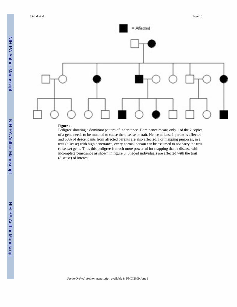
Figure 1.Pedigree showing a dominant pattern of inheritance. Dominance means only 1 of the 2 copiesof a gene needs to be mutated to cause the disease or trait. Hence at least 1 parent is affectedand 50% of descendants from affected parents are also affected. For mapping purposes, in atrait (disease) with high penetrance, every normal person can be assumed to not carry the trait(disease) gene. Thus this pedigree is much more powerful for mapping than a disease withincomplete penetrance as shown in figure 5. Shaded individuals are affected with the trait(disease) of interest.
Lidral et al. Page 13
Semin Orthod. Author manuscript; available in PMC 2009 June 1.
NIH
-PA Author Manuscript
NIH
-PA Author Manuscript
NIH
-PA Author Manuscript
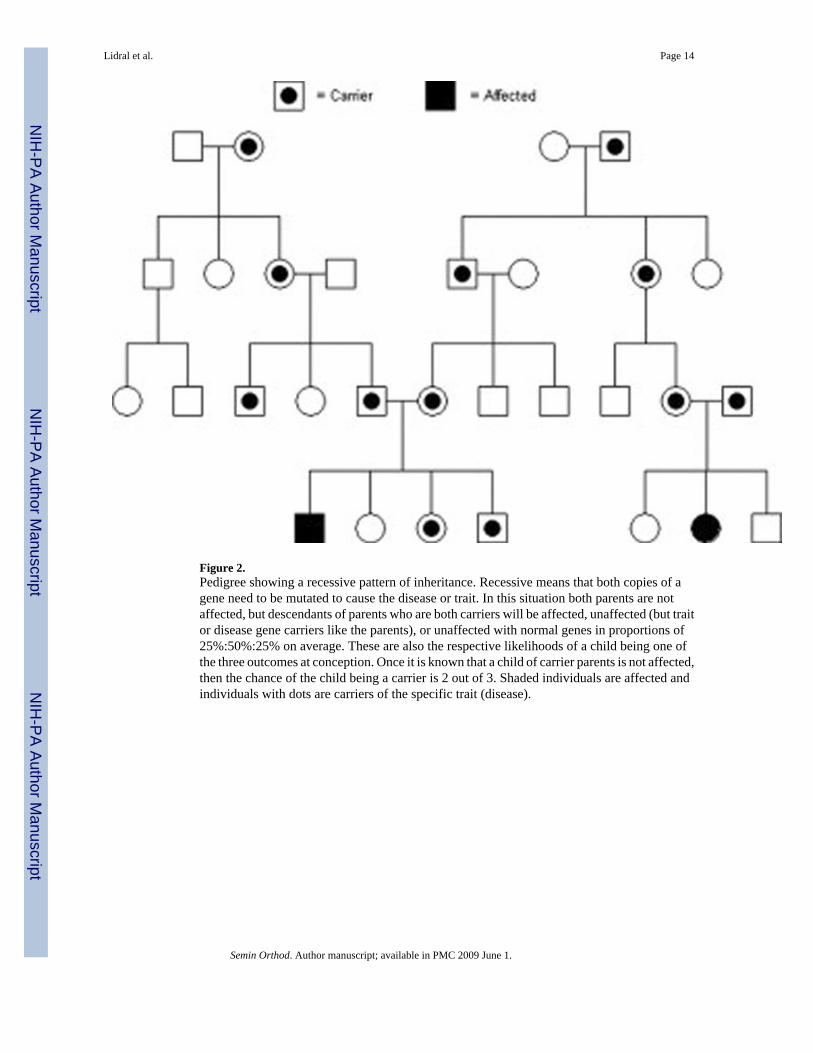
Figure 2.Pedigree showing a recessive pattern of inheritance. Recessive means that both copies of agene need to be mutated to cause the disease or trait. In this situation both parents are notaffected, but descendants of parents who are both carriers will be affected, unaffected (but traitor disease gene carriers like the parents), or unaffected with normal genes in proportions of25%:50%:25% on average. These are also the respective likelihoods of a child being one ofthe three outcomes at conception. Once it is known that a child of carrier parents is not affected,then the chance of the child being a carrier is 2 out of 3. Shaded individuals are affected andindividuals with dots are carriers of the specific trait (disease).
Lidral et al. Page 14
Semin Orthod. Author manuscript; available in PMC 2009 June 1.
NIH
-PA Author Manuscript
NIH
-PA Author Manuscript
NIH
-PA Author Manuscript
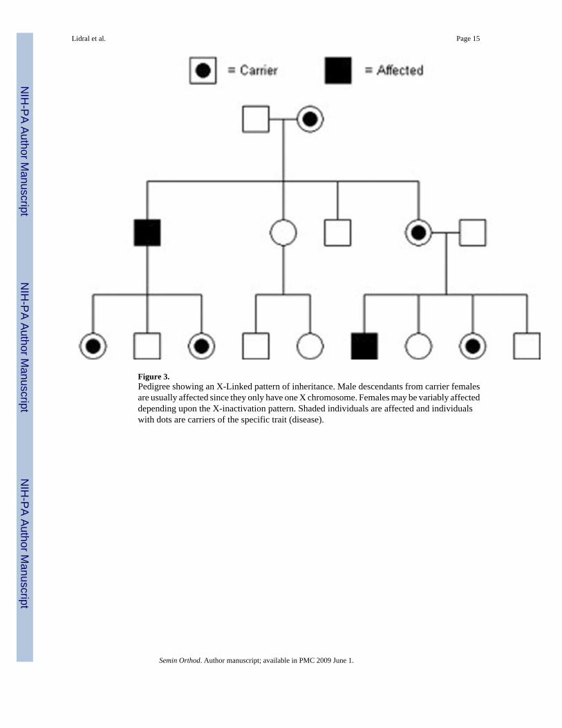
Figure 3.Pedigree showing an X-Linked pattern of inheritance. Male descendants from carrier femalesare usually affected since they only have one X chromosome. Females may be variably affecteddepending upon the X-inactivation pattern. Shaded individuals are affected and individualswith dots are carriers of the specific trait (disease).
Lidral et al. Page 15
Semin Orthod. Author manuscript; available in PMC 2009 June 1.
NIH
-PA Author Manuscript
NIH
-PA Author Manuscript
NIH
-PA Author Manuscript
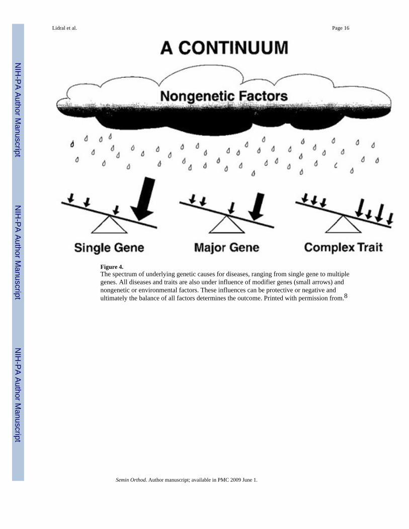
Figure 4.The spectrum of underlying genetic causes for diseases, ranging from single gene to multiplegenes. All diseases and traits are also under influence of modifier genes (small arrows) andnongenetic or environmental factors. These influences can be protective or negative andultimately the balance of all factors determines the outcome. Printed with permission from.8
Lidral et al. Page 16
Semin Orthod. Author manuscript; available in PMC 2009 June 1.
NIH
-PA Author Manuscript
NIH
-PA Author Manuscript
NIH
-PA Author Manuscript
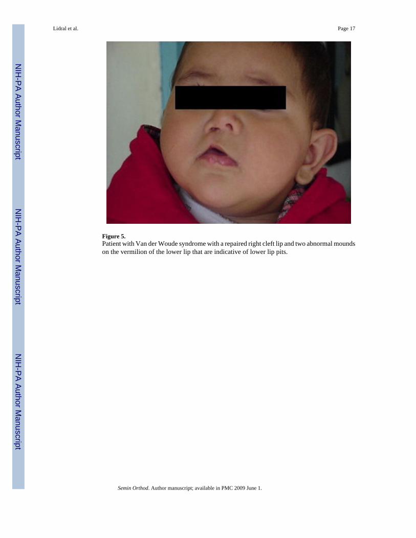
Figure 5.Patient with Van der Woude syndrome with a repaired right cleft lip and two abnormal moundson the vermilion of the lower lip that are indicative of lower lip pits.
Lidral et al. Page 17
Semin Orthod. Author manuscript; available in PMC 2009 June 1.
NIH
-PA Author Manuscript
NIH
-PA Author Manuscript
NIH
-PA Author Manuscript
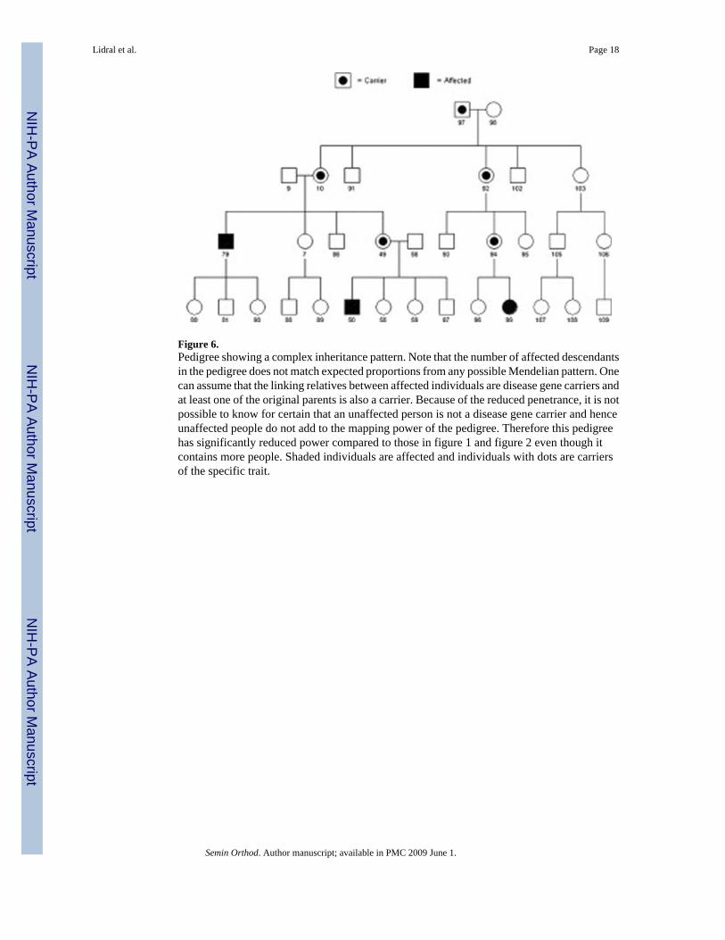
Figure 6.Pedigree showing a complex inheritance pattern. Note that the number of affected descendantsin the pedigree does not match expected proportions from any possible Mendelian pattern. Onecan assume that the linking relatives between affected individuals are disease gene carriers andat least one of the original parents is also a carrier. Because of the reduced penetrance, it is notpossible to know for certain that an unaffected person is not a disease gene carrier and henceunaffected people do not add to the mapping power of the pedigree. Therefore this pedigreehas significantly reduced power compared to those in figure 1 and figure 2 even though itcontains more people. Shaded individuals are affected and individuals with dots are carriersof the specific trait.
Lidral et al. Page 18
Semin Orthod. Author manuscript; available in PMC 2009 June 1.
NIH
-PA Author Manuscript
NIH
-PA Author Manuscript
NIH
-PA Author Manuscript
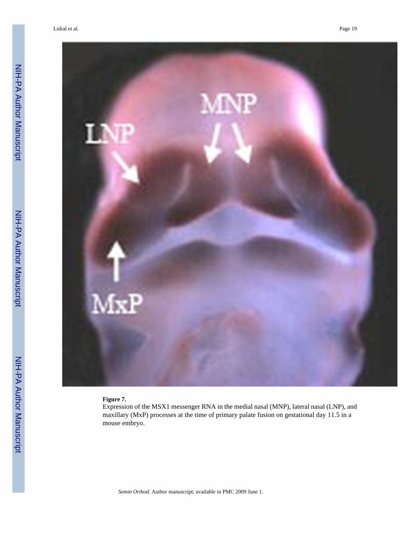
Figure 7.Expression of the MSX1 messenger RNA in the medial nasal (MNP), lateral nasal (LNP), andmaxillary (MxP) processes at the time of primary palate fusion on gestational day 11.5 in amouse embryo.
Lidral et al. Page 19
Semin Orthod. Author manuscript; available in PMC 2009 June 1.
NIH
-PA Author Manuscript
NIH
-PA Author Manuscript
NIH
-PA Author Manuscript
NIH
-PA Author Manuscript
NIH
-PA Author Manuscript
NIH
-PA Author Manuscript
Lidral et al. Page 20Ta
ble
1Se
lect
ed c
andi
date
gen
es w
ith p
ositi
ve e
vide
nce
for a
role
in n
onsy
ndro
mic
cle
ft lip
or p
alat
e
Can
dida
te R
egio
nC
andi
date
Gen
eL
inka
geA
ssoc
iatio
nA
nim
al M
odel
Chr
omos
ome
Ano
mal
yC
left
Synd
rom
e
1p36
MTH
FR, S
KI,
PAX7
XX
X
1q32
IRF6
XX
XX
X
2p13
TGFA
XX
4p16
MSX
1X
XX
X
6p23
–25
TFAP
2A, O
FCC
1X
XX
X
14q2
4TG
FB3,
BM
P4, P
AX9
XX
X
17q1
2–21
RARA
, Clf1
XX
X
19q1
3BC
L3, C
LPTM
1, P
VRL2
,TG
FB1
XX
X
Semin Orthod. Author manuscript; available in PMC 2009 June 1.


