Embed Size (px)
Citation preview

MOLECULAR PAINProchazkova et al. Molecular Pain 2013, 9:66http://www.molecularpain.com/content/9/1/66
RESEARCH Open Access
Activation of cyclin-dependent kinase 5 mediatesorofacial mechanical hyperalgesiaMichaela Prochazkova1, Anita Terse1, Niranjana D Amin2, Bradford Hall1, Elias Utreras1,3, Harish C Pant2
and Ashok B Kulkarni1*
Abstract
Background: Cyclin-dependent kinase 5 (Cdk5) is a unique member of the serine/threonine kinase family. Thiskinase plays an important role in neuronal development, and deregulation of its activity leads to neurodegenerativedisorders. Cdk5 also serves an important function in the regulation of nociceptive signaling. Our previous studiesrevealed that the expression of Cdk5 and its activator, p35, is upregulated in nociceptive neurons during peripheralinflammation. The aim of the present study was to characterize the involvement of Cdk5 in orofacial pain. Sincemechanical hyperalgesia is the distinctive sign of many orofacial pain conditions, we adapted an existing orofacialstimulation test to assess the behavioral responses to mechanical stimulation in the trigeminal region of thetransgenic mice with either reduced or increased Cdk5 activity.
Results: Mice overexpressing or lacking p35, an activator of Cdk5, showed altered phenotype in response tonoxious mechanical stimulation in the trigeminal area. Mice with increased Cdk5 activity displayed aversivebehavior to mechanical stimulation as indicated by a significant decrease in reward licking events and licking time.The number of reward licking/facial contact events was significantly decreased in these mice as the mechanicalintensity increased. By contrast, mice deficient in Cdk5 activity displayed mechanical hypoalgesia.
Conclusions: Collectively, our findings demonstrate for the first time the important role of Cdk5 in orofacialmechanical nociception. Modulation of Cdk5 activity in primary sensory neurons makes it an attractive potentialtarget for the development of novel analgesics that could be used to treat multiple orofacial pain conditions.
Keywords: Cdk5, p35, Trigeminal ganglia, Orofacial pain, Mouse model
BackgroundOrofacial pain affects millions of people worldwide. It ischaracterized by throbbing, sharp or burning pain in thehead, neck, face, mouth, gums or teeth. Epidemiologicalstudies indicate that orofacial pain occurs in approxi-mately 10% of the adult population [1], and women aremore often affected than men by a ratio of 2:1 [2]. Oro-facial pain episodes are usually very debilitating forthe patient. However, relatively few studies are focusedon characterizing orofacial pain, particularly due to thelimited number of animal models available to studynociception in the trigeminal region. Most of thesemodels have been adapted from those used for studying
* Correspondence: [email protected] Genomics Section, Laboratory of Cell and DevelopmentalBiology, National Institute of Dental and Craniofacial Research, NationalInstitutes of Health, Bethesda, MD 20892, USAFull list of author information is available at the end of the article
© 2013 Prochazkova et al.; licensee BioMed CeCreative Commons Attribution License (http:/distribution, and reproduction in any medium
peripheral pain and are primarily based on the inductionof inflammation by the administration of nociceptiveagents, such as complete Freund’s adjuvant [3,4], carra-geenan [5,6], and formalin [7-10]. Other models arebased on the direct damage to a nerve (cutting, ligating,or crushing) [11-14]. These models suffer from certainlimitations, such as variation in subjective observation,inability to escape from the noxious stimulus, andinduction of the stress in a test animal. The recentlyreported operant behavioral assay using a reward-conflict paradigm wherein a test animal can decidebetween receiving a reward or escaping an aversivestimulus present new perspectives on measuring pain inthe orofacial region [15-18].There is accumulating evidence that protein kinases are
involved in mediating several types of pain. Cdk5 is aserine/threonine kinase widely distributed in differentmammalian tissues, but its kinase activity is observed
ntral Ltd. This is an open access article distributed under the terms of the/creativecommons.org/licenses/by/2.0), which permits unrestricted use,, provided the original work is properly cited.

Prochazkova et al. Molecular Pain 2013, 9:66 Page 2 of 12http://www.molecularpain.com/content/9/1/66
primarily in neuronal cells, due to the selective expressionof its activators, p35 and p39. Cdk5 plays important rolesin many vital processes, including brain development andfunction, neuronal migration, neurotransmitter release,cell adhesion and survival, drug addiction, learning, mem-ory [19-23], and also in many non-neuronal functions[24-26]. Cdk5 knockout mice are embryonic lethal withnumerous lesions in the central nervous system [27].Efforts to delineate molecular roles of Cdk5 in vivo led tothe generation of mice overexpressing [28] or lacking [29]p35, an activator of Cdk5. Recently, we and others discov-ered that Cdk5 activity regulates peripheral pain signaling,and that it is required for the basal responses to noxiousheat [30,31]. The p35 knockout mice (with residual Cdk5activity) showed delayed responses to painful thermalstimulation (hypoalgesia), whereas mice overexpressingp35 (with significantly increased Cdk5 activity) were moresensitive to painful thermal stimulation showing hyper-algesia. Moreover, we have identified that the expressionof p35, as well as Cdk5 kinase activity, is present in thedorsal root ganglia and trigeminal ganglia neurons, andboth are significantly increased upon the induction ofperipheral inflammation [32]. Furthermore, nociceptor-specific Cdk5 conditional knockout mice developedhypoalgesia associated with reduced phosphorylation ofthe TRPV1 channel [30].The goal of the current study was to evaluate the
role of Cdk5 in orofacial mechanosensation and tocharacterize the behavioral changes of mice lacking oroverexpressing p35 using adapted orofacial stimulationtest (Additional file 1).
ResultsCdk5 activity in transgenic p35 (Tgp35) and p35 knockout(p35−/−) miceWe initially examined the expression and activity ofCdk5/p35 in the trigeminal ganglia, brainstem, andbrain of mice that overexpress or lack p35. Analysisof the Tgp35 mice revealed a significant increase inp35 mRNA (Figure 1A) as well as in p35 protein levels(Figure 1B). There was a three-fold increase in Cdk5 activ-ity (Figure 1C) in the trigeminal ganglia of Tgp35 micecompared with the wild-type (WT) mice (p < 0.001). TheTgp35 mice also showed a significant increase in p35mRNA and protein levels as well as in Cdk5 activity inbrainstem and in brain (Figure 1A-C). The analysis of thep35−/− mice showed almost undetectable p35 mRNA andprotein levels (Figure 2A and B) and significantly de-creased Cdk5 activity (Figure 2C) in tissue homogenatesfrom the trigeminal ganglia, brainstem, and brain, as com-pared to controls (p < 0.001). The p35 expression levelsand Cdk5 activity correlated with the mouse genotype,thus confirming that the p35 level is the limiting factor forthe Cdk5 activity [28].
Normal motor coordination and locomotion in Tgp35 andp35−/− miceHaving established the p35 expression levels and theCdk5 activity in Tgp35 and p35−/− mice, we determinedwhether this difference in expression and activity ofCdk5/p35 could affect the motor coordination and loco-motion of these genetically modified animals. We didnot observe any motor deficit using the acceleration testwith rotarod when the rotation was set to acceleratefrom 4 to 40 rpm during the defined period of time. Theaverage latency to drop from the cylinder was 269 sec-onds in Tgp35 and 274 seconds in wild-type FVBN con-trol mice, respectively (Additional file 2A). Similarly, nosignificant difference in the fall latency was observed inp35−/− mice (average latency 253 seconds in p35−/−mice and 256 seconds in the wild-type C57 controls)(Additional file 2A). There were no significant differ-ences in the fall latency during the testing using the con-stant rotation speed. The p35−/−, Tgp35, and theirrespective, wild-type controls showed no motor deficitsas evident from the mean time spent on the rotarod(Additional file 2B).
Normal anxiety level and exploratory behavior in Tgp35and p35−/− miceBefore performing the orofacial operant assay on thesemice, we also assessed anxiety, exploratory activity, andstereotypical behavioral using the open-field test. Neitherhorizontal (Additional file 2C) or vertical (Additional file2D) activity was affected in the p35−/− or Tgp35 mice,in comparison to the control mice. Since the middle of anon-familiar arena is anxiogenic for rodents, anxiety wasstudied by analyzing the time spent and total distancetravelled in the middle of the cage. There were no sig-nificant differences in the time spent in the center ofthe cage as well as the center distance travelled bythe Tgp35 and their littermate controls (Additionalfile 3A), and the p35−/− and wild-type C57 mice con-trols (Additional file 4A). There were also no significantchanges in the stereotypical behavioral (Additional file3B and Additional file 4B) or time spent at the differentcorners of the activity cage (Additional file 3C andAdditional file 4C), indicating that the difference in thep35 levels did not cause any change in anxiety or ex-ploratory behavior of the mice.
Normal baseline behavior in orofacial operant assayIt was previously reported that the reward conflict para-digm (using parameters such as reward licking events,facial contact events, and facial contact duration) couldserve as a characteristic marker of pain in the orofacialarea [15,16]. In the mouse orofacial operant assay thatwe report here, the number of attempts the test mousemade to acquire the reward and the duration that it

TG Brainstem Brain
Brainstem Brain
A
BWT Tgp35kDa
35
51
TG
Brainstem BrainTG
p35
α-tubulin
WT Tgp350
50
100
150 ***
WT Tgp35 WT Tgp350
50
100
150
200 *** ***
WT Tgp35WT Tgp35
CWT
P32-Histone H1
WT Tgp35 WT Tgp35 WT Tgp350
50
100
150
200
250 *** ** ***
Tgp35 WT Tgp35 WT Tgp35
WT Tgp350.0
0.5
1.0
1.5
0.0
0.5
1.0
1.5***
WT Tgp35
***
WT Tgp350.0
0.5
1.0
1.5
2.0 *p
35 m
RN
A le
vels
(re
lati
ve t
o H
PR
T)
p35
mR
NA
leve
ls (
rela
tive
to
HP
RT
)
p35
mR
NA
leve
ls (
rela
tive
to
HP
RT
)
Mea
n d
ensi
ty
0
50
100
150
Mea
n d
ensi
ty
Mea
n d
ensi
ty
Mea
n d
ensi
ty
0
50
100
150
200
250
Mea
n d
ensi
ty
0
50
100
150
200
250
Mea
n d
ensi
ty
Figure 1 (See legend on next page.)
Prochazkova et al. Molecular Pain 2013, 9:66 Page 3 of 12http://www.molecularpain.com/content/9/1/66

(See figure on previous page.)Figure 1 Analysis of the p35 expression profile in transgenic p35 (Tgp35) mice. The p35 expression levels and Cdk5 activity in thetrigeminal ganglia, brainstem, and brain of the transgenic p35 mice: (A) q-PCR analysis revealed significantly enhanced levels of p35 mRNAin the trigeminal ganglia, brainstem, and the brain of the Tgp35 mice. Each data set was normalized to the expression seen in control wild-typeanimals. The results obtained from four different animals are expressed as mean ± SEM and analyzed by an unpaired t-test (***p < 0.001). (B)Representative Western blots showing p35 protein levels from Tgp35 tissue lysates together with corresponding densitometric analysis. Datawere normalized to the levels of p35 in wild-type controls, and are presented as mean ± SEM (unpaired t-test, ***p < 0.001). (C) Cdk5 activity inthe trigeminal ganglia, brainstem, and the brain of the Tgp35 mice. The data are presented as mean ± SEM and analyzed by an unpairedt-test (***p < 0.001).
Prochazkova et al. Molecular Pain 2013, 9:66 Page 4 of 12http://www.molecularpain.com/content/9/1/66
spent acquiring the reward were the basic outcomes forthis behavioral testing. Naïve animals were initiallytrained to access the reward through the drinking win-dow with an innocuous module. During the baselinemeasurements, the Tgp35 and WT FVBN control micedid not show any aversive behavior, and there was nodifference in the number of beam breaks and time theanimals spent with the licking recorded (Figure 3A).After the completion of five different training sessions,we observed the effects of the mechanical stimuli onorofacial outcome measures.
Mechanical hyperalgesia in Tgp35 miceIn the presence of mechanical stimulators, the Tgp35mice showed aversive behavior to mechanical stimuli asindicated by a decrease in the number of attempts toaccess the reward and the contact time compared tothe WT FVBN mice. The Tgp35 mice exhibited signifi-cant mechanical hyperalgesia when subjected to orofa-cial stimulation using plates with either 6 + 6 wires(Figure 3B), 9 + 9 wires (Figure 3C), or 13 + 13 wires(Figure 3D). The Tgp35 mice made significantly fewer at-tempts to acquire the reward and spent much less timelicking the reward compared to the wild-type controls(p < 0.001). The mechanical hyperalgesia caused by theplates with the highest number of wires produced asignificantly lower reward intake as well as reduction inthe licking time, compared to the plates with the lowernumbers of the wires. We determined that the specificbehavioral changes between the Tgp35 and the wild-typemice in the licking episodes were caused by the induc-tion of nociception in the trigeminal area (Figure 3A-D).
Mechanical hypoalgesia in p35 knockout miceSimilar to the Tgp35 mice, there were no changes inthe baseline reward licking paradigm in the p35−/−mice compared with the wild-type C57 control mice(Figure 4A). However, we found that the p35−/− micedisplayed a significant mechanical hypoalgesia as com-pared to the wild-type mice when tested with the platecontaining 6 + 6 wires (Figure 4B; p < 0.001). An add-itional noxious stimulus caused a significant decrease inthe number of attempts to get the reward and timespent licking reward by the wild-type control mice
compared to the p35−/− mice (Figure 4C; p < 0.001).The most obvious difference was noticed using the platewith the highest number of the wires. The wild-typemice spent only 1.7 seconds licking, whereas the p35−/−mice licked for more than 13 seconds, indicating muchlower pain sensation in the p35−/− mice (Figure 4D;p < 0.001). The number of attempts and the licking timedecreased significantly in the case of the wild-type miceover the entire test period. However, there were nochanges in the licking patterns of the p35−/− mice afterinducing the mild pain, and more obvious changes wereobserved inducing more painful conditions. In compar-ing the wild-type and the p35−/− mice, there were alsoclear changes in the licking pattern episodes caused bydifferent nociceptive stimulation (Figure 4A-D).
DiscussionThe present study shows that Cdk5 has an importantrole in orofacial pain signaling, and that this kinase isassociated with mechanical nociception in the mousevibrissal pad. We utilized two sets of mice with signifi-cantly altered Cdk5 activity to confirm its associationwith orofacial pain. Both p35 knockout (with residualCdk5 activity) and transgenic p35 (with significantly in-creased Cdk5 activity) mice depicted an altered responsetowards the mechanical stimulation. When tested withmechanical stimuli, the mice lacking the p35 geneshowed hypoalgesia, whereas the mice overexpressingp35 hyperalgesia. Thus, these results clearly establish acorrelation between Cdk5 activity and mechanicalnociception.Since the discovery of Cdk5, numerous studies have
revealed its multifunctional roles in important physio-logical processes, such as brain development andfunction, neuronal migration, synaptic plasticity, mem-ory, learning, and neurodegenerative disease processes[19-23,26,33]. Our previous studies demonstrated thatp35, as well as Cdk5, are expressed in the dorsal rootand the trigeminal ganglia, and that the expression andactivity of Cdk5/p35 is increased during the inflamma-tion. We and others have also reported that Cdk5 isrequired for the basal responses to noxious heat [30,31].The p35 knockout mice showed delayed responsesto the painful thermal stimulation whereas the mice

A
B
C
TG Brainstem Brain
WT p35-/-
35
51
p35
α-tubulin
kDa
TG Brainstem Brain
TG Brainstem Brain
WT p35-/- WT p35-/- WT p35-/-0
50
100
150 *** ***
0
50
100
150
200 ***
WT p35-/-WT p35-/-
WT p35-/- WT p35-/-
P32-Histone H1
WT p35-/-
WT p35-/- WT p35-/- WT p35-/-0
50
100
150
200
250 *** *** ***
WT p35-/-0
10
20
30***
WT p35-/-0
20
40
60
80***
WT p35-/-0
50
100
150
200***
p35
mR
NA
leve
ls (
rela
tive
to
HP
RT
)
p35
mR
NA
leve
ls (
rela
tive
to
HP
RT
)
p35
mR
NA
leve
ls (
rela
tive
to
HP
RT
)
Mea
n d
ensi
ty
0
50
100
150
Mea
n d
ensi
ty
Mea
n d
ensi
ty
Mea
n d
ensi
ty
0
50
100
150
200
250
Mea
n d
ensi
ty
0
50
100
150
200
250
Mea
n d
ensi
ty
Figure 2 Analysis of the p35 expression profile in p35 knockout mice. The p35 expression levels and Cdk5 activity in the trigeminal ganglia,brainstem, and brain of the p35 knockout mice: (A) q-PCR analysis revealed significantly decreased levels of p35 mRNA in the trigeminal ganglia,brainstem, and brain of the p35 knockout mice. Each data set was normalized to the expression seen in control wild-type animals. Results ob-tained from four different animals are expressed as mean ± SEM and analyzed by an unpaired t-test (***p < 0.001). (B) Representative Westernblots showing p35 protein levels from p35 knockout mice together with corresponding, densitometric analysis. Data were normalized to thelevels of p35 in wild-type controls, and are presented as mean ± SEM (unpaired t-test, ***< 0.001). (C) Cdk5 activity in the trigeminal ganglia, brain-stem, and brain of the p35 knockout mice. Data are presented as mean ± SEM and analyzed by an unpaired t-test (**p < 0.01, ***p < 0.001).
Prochazkova et al. Molecular Pain 2013, 9:66 Page 5 of 12http://www.molecularpain.com/content/9/1/66
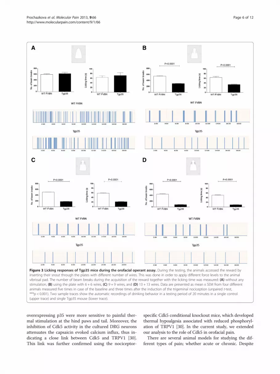
Figure 3 Licking responses of Tgp35 mice during the orofacial operant assay. During the testing, the animals accessed the reward byinserting their snout through the plates with different number of wires. This was done in order to apply different force levels to the animalvibrissal pad. The number of beam breaks during the acquisition of the reward together with the licking time was measured: (A) without anystimulation, (B) using the plate with 6 + 6 wires, (C) 9 + 9 wires, and (D) 13 + 13 wires. Data are presented as mean ± SEM from four differentanimals measured five times in case of the baseline and three times after the induction of the trigeminal nociception (unpaired t-test,***p < 0.001). Two sample traces show the automatic recordings of drinking behavior in a testing period of 20 minutes in a single control(upper trace) and single Tgp35 mouse (lower trace).
Prochazkova et al. Molecular Pain 2013, 9:66 Page 6 of 12http://www.molecularpain.com/content/9/1/66
overexpressing p35 were more sensitive to painful ther-mal stimulation at the hind paws and tail. Moreover, theinhibition of Cdk5 activity in the cultured DRG neuronsattenuates the capsaicin evoked calcium influx, thus in-dicating a close link between Cdk5 and TRPV1 [30].This link was further confirmed using the nociceptor-
specific Cdk5 conditional knockout mice, which developedthermal hypoalgesia associated with reduced phosphoryl-ation of TRPV1 [30]. In the current study, we extendedour analysis to the role of Cdk5 in orofacial pain.There are several animal models for studying the dif-
ferent types of pain; whether acute or chronic. Despite

Figure 4 Licking responses of the p35 knockout mice during the orofacial operant assay. During the testing, the animals accessed thereward by inserting their snout through the plates with the different number of wires, in order to apply different force levels to the animalvibrissal pad. The number of beam breaks during the acquisition of the reward together with the licking time was measured: (A) without anystimulation, (B) using the plate with 6 + 6 wires, (C) 9 + 9 wires, and (D) 13 + 13. The data are expressed as mean ± SEM from four differentanimals, measured five times in case of the baseline, and three times after induction of the trigeminal nociception (unpaired t-test, ***p < 0.001).Two sample traces show the automatic recordings of drinking behavior in a testing period of 20 minutes in a single control (upper trace) andsingle p35 knockout mouse (lower trace).
Prochazkova et al. Molecular Pain 2013, 9:66 Page 7 of 12http://www.molecularpain.com/content/9/1/66
this, there is still a paucity of animal models to studyorofacial pain, especially in mice. The majority of the be-havioral pain tests in the orofacial area are based onpain-related spontaneous behavior. All these tests inducethe pain by an injection of various chemicals (formalin,
carrageenan, or capsaicin) into the upper lip or thevibrissal pad, and observe licking or grooming behavior.Recent studies use a mouse grimace scale for the meas-urement of short term nociception [34] or a devicethat quantifies a gnawing function in the mouse [35].

Prochazkova et al. Molecular Pain 2013, 9:66 Page 8 of 12http://www.molecularpain.com/content/9/1/66
However, all of the current protocols for studying orofa-cial pain have many limitations, including variation insubjective observations, inability to escape from a nox-ious stimulus, and the induction of stress in the testanimal. All of these can cause a large variation in themeasured results.The operant behavioral assay developed and intro-
duced by Neubert in 2005 [16] shows that use of areward-aversion paradigm offers more benefits. Thismodel is based on the reward-conflict paradigm, wherethe test animal can decide between receiving a reward,or it can escape from the aversive stimulus by which itcan control and modify its own behavior. Therefore, ascompared to the other orofacial behavioral tests the useof this operant assay reduces the stress in the testing an-imals, there is a possibility to provide the multiple mea-surements using the same animal, and most importantly,it is free from investigator bias when it comes to evaluat-ing the results that are recorded automatically. To thebest of our knowledge, there is no reported study usingorofacial mechanical stimulation test in mice, and we be-lieve our present study will fill this void.Although there are many common features in pain
transduction and processing in the trigeminal and spinalsystems, there are key differences in the anatomical andfunctional features of the primary afferent neurons ofthe trigeminal ganglia that distinguish them from neu-rons of the spinal dorsal root ganglia. Recent studieshave shown that not only anatomical, but also electro-physiological and pharmacological differences [36-38] ofthe trigeminal afferents innervating unique target tissuessuch as meninges, cornea, teeth, oral/nasal mucosa, andthe temporomandibular joint. These differences are con-sistent with our observations. It has been reported that theintrathecal administration of roscovitine, a Cdk5 inhibitor,inhibited Cdk5 activity and attenuated a formalin-inducednociceptive response in rats [39]. However, we did not ob-serve any changes in trigeminal p35 mRNA and proteinlevels, nor in the Cdk5 activity after the vibrissal formalininjection (data not shown), thus supporting the theoryabout the differential regulation of nociception at the per-iphery and in the vibrissal pad.We do not know the precise molecular mechanism by
which Cdk5 activity can affect the orofacial nociception.There are several possibilities. First, the activation of theTRPV1 channel by Cdk5-mediated phosphorylationcould participate in this mechanism. The TRPV1 recep-tor, consistent with its role as a pain regulator, isexpressed in the peripheral and central nervous systemsinvolved in pain detection, transmission, and regulation.Phosphorylation and dephosphorylation reactions regu-late TRPV1 receptor activity, which is crucial in promot-ing inflammatory pain [40,41]. There is clear evidencethat the TRPV1 channel activation at the periphery is
involved in the development of inflammatory thermalhyperalgesia and heat sensitivity [42]. We have also pre-viously reported that Cdk5 modulates thermal, nocicep-tive signaling through the phosphorylation of TRPV1 atthreonine-407 [30]. Another recent study points out thatCdk5 can control TRPV1 membrane trafficking, andthus regulate the heat sensitivity of the nociceptors [43].Furthermore, the systemic or intrathecal administrationof TRPV1 antagonists is effective in reducing both ther-mal hyperalgesia, as well as mechanical allodynia associ-ated with chronic or neuropathic pain [44-46], whichindicates that TRPV1 could play an important role in in-tegrating multiple pain-producing stimuli. More recentstudies have uncovered the involvement of TRPV1 inthe central mechanical nociception together in connec-tion with the other TRP channel – TRPA1 [47]. Otherstudies speculate that the central mechanical hyperalge-sia could be induced by the functional interaction be-tween P2X3 [48] or NMDA receptor [49] and TRPV1.These studies provide the evidence that TRPV1 channelsare important not only for the peripheral pain sensation,but they can also play an important role in the centralmechanical nociception.An interesting possibility is that Cdk5 can mediate
orofacial mechanical hyperalgesia through the regulationof the neurotransmitter release, thus indicating that thiskinase could be an important presynaptic control par-ameter. Deregulation of its activity could affect nocicep-tion via the presynaptic mechanism with the subsequentinitiation of the pain sensation [50-52]. Another possibil-ity is that Cdk5 could mediate the orofacial mechanicalhyperalgesia through the activation of other potentialmechanotransducers. It is well known that the upregula-tion of Cdk5 activity can lead to phosphorylation of deltaopioid receptor [53,54], NMDA receptor [55,56], P2X3receptor [57], and voltage gated calcium channels [58].Additionally, there are other potential candidates likeTRPA1 [59-61] or TREK channels [62,63] that containthe Cdk5 phosphorylation consensus sequence and maybe involved in the Cdk5-mediated activation andmechanotransduction in the orofacial area. To under-stand the precise mechanism through which Cdk5 regu-lates orofacial mechanosensitisation will require furtherstudies; including molecular, electrophysiological, andbehavioral methods to map the functional role of Cdk5in this type of the nociception.
ConclusionsWe have adapted orofacial stimulation test for mice thatcould be used for orofacial pain studies, and using thistest we have identified that Cdk5 activity has an import-ant role in orofacial mechanical nociception. Moreover,our studies also demonstrate that genetically engineeredmice with the altered Cdk5/p35 levels will prove to be

Prochazkova et al. Molecular Pain 2013, 9:66 Page 9 of 12http://www.molecularpain.com/content/9/1/66
valuable models to identify and characterize the inhibi-tors of Cdk5/p35 as novel analgesics to treat orofacialpain.
MethodsAnimalsThe p35−/− mice and the age-matched, wild-type con-trols were maintained in C57BL6/129SVJ background.Tgp35 mice and the wild-type littermate controls weremaintained in FVBN background. All of the animalswere housed and bred in standard cages, and they weremaintained in climate and light controlled rooms withfree access to food and water in accordance with theU.S. National Institutes of Health Guide for Care andUse of Laboratory Animals. All of the experimentsadhered to the guidelines of the IASP Committee forResearch and Ethical Issue [64].
Rotarod testThe p35−/− and Tgp35 mice were evaluated for motorabilities, coordination, and balance by performance onthe rotarod (Model 7650, Ugo Basile, Italy). This instru-ment consists of a platform under a rotating cylinderthat can be set either to accelerate from 4 to 40 rpm inthe defined period of time or used at a constant speed.The automated rotarod system has a timer linked to theplatform panel onto which the mouse falls (approxi-mately 15 cm below the rotating cylinder). During thetesting, the animals have to continuously walk forwardto keep from falling off the rotating cylinder. After themice became familiarized with this procedure, they wereplaced on the rotating cylinder, and the time until theanimal fell down was measured in three different testsperformed on three consecutive days.
Open field testAnxiety, exploratory activity, as well as spontaneousmotor activity were evaluated using the VersaMaxAnimal activity monitoring system (AccuScan Instru-ments Inc., Columbus, OH, USA). The testing instru-ment consists of a clear, plexiglass, rectangular cage(42 × 42 × 30 cm), with the transparent side wall. Twosets of sixteen photocells are aligned from the front toback of the cage and from side to side for recordinghorizontal activity. Vertical activity is assessed by anadditional sixteen photocells located above the horizon-tal cells. The mice were placed individually in the centerof the open field apparatus, and their behavior was mon-itored for ten minutes. Horizontal or vertical activity(rearing measured by counting the number of beam in-terruptions), time spent in the center area of the openfield, and stereotypic counts were automatically recordedusing the VersaMax software system.
Mouse orofacial stimulation testUsing a small plastic reducer, we have modified theOrofacial Stimulation Test (31300, Ugo Basile, Italy),which was previously used only for the measurement ofhypersensitivity to thermal or mechanical stimulation ofthe trigeminal area in rats, and adapted it for assessingbehavioral responses to mechanical stimulation in mice(Additional file 1C).An orofacial stimulation test consists of several parts -
the plastic cage (26.5 cm wide, 20 cm deep, and 48 cmlong) with the interface wall containing a drinking win-dow (1.5 cm wide × 2 cm high) that allows a mouse anaccess to a reward; an infrared photo beam built on theexterior aspect of the window linked to the ORO soft-ware that automatically quantifies feeding behavior bymeasuring the number of attempts the animals made toacquire the reward, and the total duration of feedingtime (Additional file 1A). The equipment is supple-mented with metal inserts containing a different numberand configuration of the Nitinol wires (0,155 mm) uti-lized to induce varying degrees of trigeminal, mechanicalnociception (Additional file 1B). Each of these metal in-serts can be mounted onto the opposite aspect of thedrinking window with six screws.During the orofacial operant assay, the test mouse in-
serts its snout through the drinking window in the inter-face wall to access a nozzle of the bottle containing reward(30% sucrose) [65]. Simultaneously, while accessing thenozzle, the mouse breaks an analog output from the infra-red detectors (Banner Engineering, Minneapolis, MN,USA) placed directly behind the drinking window, andthus its attempts to get the reward can be recorded andanalyzed with the ORO software (Additional file 1D).The mice were deprived of food and water for at least
a period of eight hours prior to the testing sessions,which was done to increase the incentive for reward ac-quisition. After the completion of tests, access for foodand water was restored. The mice were trained andtested in the same cage and at the same time each day,and they had a day of rest between the testing.Initially, the animals were trained to access the reward
through the blank plate (without the wires). For mech-anical testing, access to the reward was impeded byinserting the plate with the different number of the wiresproviding mechanical contact with the vibrissal pad re-gion of the tested animal (Additional file 1D). We usedthree different plates with 6 + 6, 9 + 9, or 13 + 13 wires ateach side in order to apply a different level of mechan-ical force to the animal vibrissal pad, so as to induce dif-ferent degrees of pain. After completing five, different,twenty-minute training sessions, the animals wereretested three times using each plate with a differentnumber of wires, in increasing order, starting with thelowest to the highest number of wires. The outcome

Prochazkova et al. Molecular Pain 2013, 9:66 Page 10 of 12http://www.molecularpain.com/content/9/1/66
measures that were collected during the testing con-sisted of the total duration of time the test mouse spentacquiring the reward over a twenty minute period oftime, with or without (baseline measurements) mechan-ical stimulators and the number of attempts the testmouse made to access the reward. Both were determinedautomatically by an interruption of an infrared beamwhen the animal placed its snout through the opening inthe interface wall.
Quantitative real-time PCR (q-PCR)The trigeminal ganglia, brainstem, and the brain weredissected out from p35−/−, Tgp35, and wild-type controlmice and these tissues were immediately frozen at−80°C. The total RNA was extracted using the RNeasyMini Kit (Qiagen, Valencia, CA, and USA), according tothe manufacturer’s instructions. The purity of the RNAwas assessed by the ratio of absorbance at 260 nm and280 nm. The RNA from each sample was reverse tran-scribed using a High Capacity cDNA Reverse Transcrip-tion Kit (Applied Biosystems, Foster City, CA, USA).The q-PCR reactions were conducted using cDNA,
specific primers (Assays on Demand Gene ExpressionProducts), and TaqMan Universal PCR Master Mix(Applied Biosystems, Foster City, CA, USA), and theywere run in duplicates using the Real-time PCR System7500 (Applied Biosystems, Foster City, CA, USA). Thep35 mRNA levels were normalized to the levels of HPRTusing the comparative cycle threshold (ct) method.
AntibodiesAnti-p35 (C19), Anti-Cdk5 (C8) and secondary horse-radish peroxidase-conjugated anti-mouse and anti-rabbitantibodies were obtained from Santa Cruz Biotechnology(Santa Cruz Biotechnology, Santa Cruz, CA, USA). Theanti α-tubulin antibody was purchased from SigmaAldrich (Sigma-Aldrich, St. Louis, MO, USA).
Western blottingThe tissue homogenates were lysed in tissue protein ex-traction reagent (Thermo Scientific, Rockford, IL, USA)containing a cocktail of protease (Complete Mini, Roche,Indianapolis, IN, USA) and phosphatase (PhosSTOP,Roche, Indianapolis, IN, USA) inhibitors to avoid deg-radation of the proteins. After thirty minutes of incuba-tion on ice, the samples were spun down at 14000 rpmat 4°C for 30 min. The supernatant was assayed fortotal protein concentration using the Bradford ProteinAssay (Bio-Rad, Hercules, CA, USA). The proteins weredenatured by boiling them with NuPAGE LDS samplebuffer and NuPAGE sample reducing agent (Invitrogen,Carlsbad, CA, USA) for 10 min. Each sample was sepa-rated by 4-12% SDS PAGE gels (Invitrogen, Carlsbad,CA, USA) and transferred to 0.45 μm nitrocellulose
membrane (Invitrogen, Carlsbad, CA, USA). The blotswere blocked for 1 h in phosphate buffered saline con-taining 5% nonfat dry milk and 0.05% Tween20 (Sigma-Aldrich, St. Louis, MO, USA), and then they were blot-ted with primary antibodies overnight at 4°C. The mem-branes were then probed with horseradish peroxidase-conjugated anti-mouse or anti-rabbit IgG at roomtemperature for one hour, and they were finally developedby SuperSignal West Pico or Dura ChemiluminescentSubstrate (Thermo Scientific, Rockford, IL, USA). The im-munoblots were analyzed by densitometry using ImageJanalysis system software.
Immuno-precipitation and Cdk5 activity assayImmuno-precipitation and Cdk5 kinase activity wereperformed as described previously [66]. Briefly, the pro-tein G (+) A-agarose beads were washed three timeswith tris-buffered saline (TBS) and incubated with Cdk5antibody (1–2 ug/500 ug of protein lysate, Santa CruzBiotechnology, Santa Cruz, CA, USA) for 1 h at roomtemperature with gentle mixing. The beads were centri-fuged and washed three times with TBS and then sus-pended in TBS. The protein lysates from the trigeminalganglia, brainstem, and brain were incubated with anti-body conjugated beads for 2 h and 30 min at 4°C on arotating wheel. The beads were subsequently centrifugedand washed two times with TBS, one time with 1Xkinase buffer and suspended in kinase buffer (50 mMTris/HCl pH 7.4, containing 1 mM EGTA, and 5 mMMgCl2). The immunoprecipitated beads were used as anenzyme source for the kinase activity. For the kinaseassay, a total volume of 50 μL of kinase assay mixturewas used, containing 50 μM Tris/HCl (pH 7.4) withEGTA, 1 mM dithiothreitol, 5 mM MgCI2, 10 μg of his-tone H1, and 10 μL of Cdk5 immunoprecipitates. Thephosphorylation reaction was initiated by the addition of0.1 mM [γ-32P] ATP and incubated at 30°C for one hour.The reaction was stopped by the addition of theLaemmli sample buffer. The reaction mixture washeated for five minutes at 90°C and electrophoresed on a4-20% SDS-PAGE gel stained with Coomassie blue, andthen dried and exposed overnight for the detection of32P-labeled Histone H1 by autoradiography. The filmswere scanned, and the bands were quantified using Ima-geJ software.
Statistical analysisAll data are expressed as a mean ± SEM. The statisticalevaluation was done with GraphPad Prism software,version 5.0 (GraphPad, San Diego, CA, USA). Statisticaldifferences between the experiments were assessedby unpaired t-test. The significance level was set atp < 0.05.

Prochazkova et al. Molecular Pain 2013, 9:66 Page 11 of 12http://www.molecularpain.com/content/9/1/66
Additional files
Additional file 1: Mouse orofacial stimulation test system used forthe measurement of orofacial mechanical nociception. (A) Orofacialstimulation test device. (B) The mechanical inserts with different numberof wires used for the induction of trigeminal pain. (C) The plastic reducerused for the modification of the existing system to be applicable for thecharacterization of mouse orofacial pain. (D) An example showing themouse during the reward licking while its vibrissal region is in directcontact with the wires.
Additional file 2: Effect of different p35 genotype on locomotorand exploratory activity. (A) The mean performance time determinedas time spent on the rotating cylinder during the acceleration. (B) Thelatency to fall from the rotating cylinder by the constant speed. (C) Theunaffected horizontal and (D) vertical activity as revealed by the openfield test. The data analyzed by the unpaired t-test are expressed asmean ± SEM and represent the mean from four different animals.
Additional file 3: The effect of upregulated p35 and Cdk5 activityon mouse behavior in an open-field test. (A) The center distancetravelled and time spent in the center of the activity cage, (B) stereotypyand the time Tgp35 mice spent with the stereotypic behavior. (C) Thetime Tgp35 mice spent in the different parts of the activity cage duringten minutes of measurement. These values represent the mean ± SEMfrom four animals.
Additional file 4: The effect of downregulated p35 and Cdk5activity on mouse behavior in an open-field test. (A) The centerdistance travelled and time spent in the center of the activity cage, (B)stereotypy and the time p35 knockout mice spent with the stereotypicbehavior. (C) The time p35-/- mice spent in the different parts of theactivity cage during 10 min of measurement. These values represent themean ± SEM from four animals.
AbbreviationsCdk5: Cyclin-dependent kinase 5; TRPV1: Transient receptor potentialvanilloid 1; TG: Trigeminal ganglia; Tgp35: Transgenic p35 mice; p35-/-: p35knockout mice; q-PCR: Quantitative real-time PCR; WT: Wild-type.
Competing interestThe authors declare that they have no conflict of interest.
Authors’ contributionsMP and ABK designed the experiments. MP, AT, EU, BH, and NDA performedresearch; MP, AT, EU, BH, NDA, HCP and ABK analyzed data, and MP and ABKwrote the manuscript. All authors read and approved the final manuscript.
AcknowledgementsThis work was supported by the Divisions of Intramural Research, NationalInstitute of Dental and Craniofacial Research and the National Institute ofNeurological Disorders and Stroke, National Institutes of Health. We wouldlike to thank Drs. Michael Iadarola, Jason Keller, Santosh Mishra, and VarshaShukla for their helpful discussions during the course of these studies, andMr. Larry Jones for his expert editorial assistance.
Author details1Functional Genomics Section, Laboratory of Cell and DevelopmentalBiology, National Institute of Dental and Craniofacial Research, NationalInstitutes of Health, Bethesda, MD 20892, USA. 2Laboratory ofNeurochemistry, National Institute of Neurological Disorders and Stroke,National Institutes of Health, Bethesda, MD 20892, USA. 3Laboratory ofCellular and Neuronal Dynamics, Faculty of Science, University of Chile,Santiago, Chile.
Received: 13 August 2013 Accepted: 17 December 2013Published: 21 December 2013
References1. Madland G, Newton-John T, Feinmann C: Chronic idiopathic orofacial pain:
I: what is the evidence base? Br Dent J 2001, 191:22–24.2. Kohlmann T: Epidemiology of orofacial pain. Schmerz 2002, 16:339–345.
3. Romero-Reyes M, Akerman S, Nguyen E, Vijjeswarapu A, Hom B, Dong HW,Charles AC: Spontaneous behavioral responses in the orofacial region: amodel of trigeminal pain in mouse. Headache 2013, 53:137–151.
4. Yasuda M, Shinoda M, Kiyomoto M, Honda K, Suzuki A, Tamagawa T, Kaji K,Kimoto S, Iwata K: P2X3 receptor mediates ectopic mechanical allodyniawith inflamed lower lip in mice. Neurosci Lett 2012, 528:67–72.
5. Poh KW, Lutfun N, Manikandan J, Ong WY, Yeo JF: Global gene expressionanalysis in the mouse brainstem after hyperalgesia induced by facialcarrageenan injection–evidence for a form of neurovascular coupling?Pain 2009, 142:133–141.
6. Poh KW, Yeo JF, Stohler CS, Ong WY: Comprehensive gene expressionprofiling in the prefrontal cortex links immune activation and neutrophilinfiltration to antinociception. J Neurosci 2012, 32:35–45.
7. Bornhof M, Ihmsen H, Schwilden H, Yeomans DC, Tzabazis A: The orofacialformalin test in mice revisited–effects of formalin concentration, age,morphine and analysis method. J Pain 2011, 12:633–639.
8. Luccarini P, Childeric A, Gaydier AM, Voisin D, Dallel R: The orofacialformalin test in the mouse: a behavioral model for studying physiologyand modulation of trigeminal nociception. J Pain 2006, 7:908–914.
9. Miranda HF, Noriega V, Sierralta F, Prieto JC: Interaction betweendexibuprofen and dexketoprofen in the orofacial formalin test in mice.Pharmacol Biochem Behav 2011, 97:423–427.
10. Miranda HF, Noriega V, Zepeda RJ, Sierralta F, Prieto JC: Systemic synergismbetween codeine and morphine in three pain models in mice. PharmacolRep 2013, 65:80–88.
11. Cha M, Kohan KJ, Zuo X, Ling JX, Gu JG: Assessment of chronic trigeminalneuropathic pain by the orofacial operant test in rats. Behav Brain Res2012, 234:82–90.
12. Krzyzanowska A, Pittolo S, Cabrerizo M, Sanchez-Lopez J, Krishnasamy S,Venero C, Avendano C: Assessing nociceptive sensitivity in mouse modelsof inflammatory and neuropathic trigeminal pain. J Neurosci Methods2011, 201:46–54.
13. Rossi HL, Jenkins AC, Kaufman J, Bhattacharyya I, Caudle RM, Neubert JK:Characterization of bilateral trigeminal constriction injury using anoperant facial pain assay. Neuroscience 2012, 224:294–306.
14. Teodoro FC, Tronco Junior MF, Zampronio AR, Martini AC, Rae GA,Chichorro JG: Peripheral substance P and neurokinin-1 receptors have arole in inflammatory and neuropathic orofacial pain models.Neuropeptides 2013, 47:199–206.
15. Neubert JK, King C, Malphurs W, Wong F, Weaver JP, Jenkins AC, Rossi HL,Caudle RM: Characterization of mouse orofacial pain and the effects oflesioning TRPV1-expressing neurons on operant behavior. Mol Pain2008, 4:43.
16. Neubert JK, Widmer CG, Malphurs W, Rossi HL, Vierck CJ Jr, Caudle RM: Useof a novel thermal operant behavioral assay for characterization oforofacial pain sensitivity. Pain 2005, 116:386–395.
17. Nolan TA, Hester J, Bokrand-Donatelli Y, Caudle RM, Neubert K: Adaptationof novel operant orofacial testing system to characterize both mechan-ical and thermal pain. Behav Brain Res 2011, 217:477–480.
18. Rossi HL, Vierck CJ Jr, Caudle RM, Neubert JK: Characterization of coldsensitivity and thermal preference using an operant orofacial assay.Mol Pain 2006, 2:37.
19. Dhariwala FA, Rajadhyaksha MS: An unusual member of the Cdk family:Cdk5. Cell Mol Neurobiol 2008, 28:351–369.
20. Dhavan R, Tsai LH: A decade of CDK5. Nat Rev Mol Cell Biol 2001,2:749–759.
21. Lalioti V, Pulido D, Sandoval IV: Cdk5, the multifunctional surveyor.Cell Cycle 2010, 9:284–311.
22. Liebl J, Furst R, Vollmar AM, Zahler S: Twice switched at birth: cell cycle-independent roles of the “neuron-specific” cyclin-dependent kinase 5(Cdk5) in non-neuronal cells. Cell Signal 2011, 23:1698–1707.
23. Su SC, Tsai LH: Cyclin-dependent kinases in brain development anddisease. Annu Rev Cell Dev Biol 2011, 27:465–491.
24. Arif A: Extraneuronal activities and regulatory mechanisms of theatypical cyclin-dependent kinase Cdk5. Biochem Pharmacol 2012,84:985–993.
25. Contreras-Vallejos E, Utreras E, Gonzalez-Billault C: Going out of the brain:non-nervous system physiological and pathological functions of Cdk5.Cell Signal 2012, 24:44–52.
26. Rosales JL, Lee KY: Extraneuronal roles of cyclin-dependent kinase 5.Bioessays 2006, 28:1023–1034.

Prochazkova et al. Molecular Pain 2013, 9:66 Page 12 of 12http://www.molecularpain.com/content/9/1/66
27. Ohshima T, Ward JM, Huh CG, Longenecker G, Veeranna, Pant HC, Brady RO,Martin LJ, Kulkarni AB: Targeted disruption of the cyclin-dependent kinase5 gene results in abnormal corticogenesis, neuronal pathology andperinatal death. Proc Natl Acad Sci USA 1996, 93:11173–11178.
28. Takahashi S, Ohshima T, Cho A, Sreenath T, Iadarola MJ, Pant HC, Kim Y,Nairn AC, Brady RO, Greengard P, Kulkarni AB: Increased activity of cyclin-dependent kinase 5 leads to attenuation of cocaine-mediated dopaminesignaling. Proc Natl Acad Sci USA 2005, 102:1737–1742.
29. Chae T, Kwon YT, Bronson R, Dikkes P, Li E, Tsai LH: Mice lacking p35, aneuronal specific activator of Cdk5, display cortical lamination defects,seizures, and adult lethality. Neuron 1997, 18:29–42.
30. Pareek TK, Keller J, Kesavapany S, Agarwal N, Kuner R, Pant HC, Iadarola MJ,Brady RO, Kulkarni AB: Cyclin-dependent kinase 5 modulates nociceptivesignaling through direct phosphorylation of transient receptor potentialvanilloid 1. Proc Natl Acad Sci USA 2007, 104:660–665.
31. Yang YR, He Y, Zhang Y, Li Y, Li Y, Han Y, Zhu H, Wang Y: Activation ofcyclin-dependent kinase 5 (Cdk5) in primary sensory and dorsal hornneurons by peripheral inflammation contributes to heat hyperalgesia.Pain 2007, 127:109–120.
32. Pareek TK, Keller J, Kesavapany S, Pant HC, Iadarola MJ, Brady RO, KulkarniAB: Cyclin-dependent kinase 5 activity regulates pain signaling. Proc NatlAcad Sci USA 2006, 103:791–796.
33. Maccioni RB, Otth C, Concha II, Munoz JP: The protein kinase Cdk5.Structural aspects, roles in neurogenesis and involvement in Alzheimer’spathology. Eur J Biochem 2001, 268:1518–1527.
34. Langford DJ, Bailey AL, Chanda ML, Clarke SE, Drummond TE, Echols S, GlickS, Ingrao J, Klassen-Ross T, Lacroix-Fralish ML, et al: Coding of facial expres-sions of pain in the laboratory mouse. Nat Methods 2010, 7:447–449.
35. Dolan JC, Lam DK, Achdjian SH, Schmidt BL: The dolognawmeter: a novelinstrument and assay to quantify nociception in rodent models oforofacial pain. J Neurosci Methods 2010, 187:207–215.
36. Ambalavanar R, Moritani M, Dessem D: Trigeminal P2X3 receptorexpression differs from dorsal root ganglion and is modulated by deeptissue inflammation. Pain 2005, 117:280–291.
37. Harriott AM, Gold MS: Serotonin type 1D receptors (5HTR) aredifferentially distributed in nerve fibres innervating craniofacial tissues.Cephalalgia 2008, 28:933–944.
38. Harriott AM, Gold MS: Electrophysiological properties of dural afferents inthe absence and presence of inflammatory mediators. J Neurophysiol2009, 101:3126–3134.
39. Wang CH, Chou WY, Hung KS, Jawan B, Lu CN, Liu JK, Hung YP, Lee TH:Intrathecal administration of roscovitine inhibits Cdk5 activity andattenuates formalin-induced nociceptive response in rats. Acta PharmacolSin 2005, 26:46–50.
40. Caterina MJ, Schumacher MA, Tominaga M, Rosen TA, Levine JD, Julius D:The capsaicin receptor: a heat-activated ion channel in the painpathway. Nature 1997, 389:816–824.
41. Tominaga M, Caterina MJ, Malmberg AB, Rosen TA, Gilbert H, Skinner K,Raumann BE, Basbaum AI, Julius D: The cloned capsaicin receptorintegrates multiple pain-producing stimuli. Neuron 1998, 21:531–543.
42. Caterina MJ, Leffler A, Malmberg AB, Martin WJ, Trafton J, Petersen-Zeitz KR,Koltzenburg M, Basbaum AI, Julius D: Impaired nociception and pain sen-sation in mice lacking the capsaicin receptor. Science 2000, 288:306–313.
43. Xing BM, Yang YR, Du JX, Chen HJ, Qi C, Huang ZH, Zhang Y, Wang Y:Cyclin-dependent kinase 5 controls TRPV1 membrane trafficking andthe heat sensitivity of nociceptors through KIF13B. J Neurosci 2012,32:14709–14721.
44. Cui M, Honore P, Zhong C, Gauvin D, Mikusa J, Hernandez G, Chandran P,Gomtsyan A, Brown B, Bayburt EK, et al: TRPV1 receptors in the CNS play akey role in broad-spectrum analgesia of TRPV1 antagonists. J Neurosci2006, 26:9385–9393.
45. Watabiki T, Kiso T, Tsukamoto M, Aoki T, Matsuoka N: Intrathecaladministration of AS1928370, a transient receptor potential vanilloid 1antagonist, attenuates mechanical allodynia in a mouse model ofneuropathic pain. Biol Pharm Bull 2011, 34:1105–1108.
46. Yu L, Yang F, Luo H, Liu FY, Han JS, Xing GG, Wan Y: The role of TRPV1 indifferent subtypes of dorsal root ganglion neurons in rat chronicinflammatory nociception induced by complete Freund’s adjuvant.Mol Pain 2008, 4:61.
47. Ro JY, Lee JS, Zhang Y: Activation of TRPV1 and TRPA1 leads to musclenociception and mechanical hyperalgesia. Pain 2009, 144:270–277.
48. Saloman JL, Chung MK, Ro JY: P2X (3) and TRPV1 functionally interact andmediate sensitization of trigeminal sensory neurons. Neuroscience 2012,232:226–238.
49. Lee J, Saloman JL, Weiland G, Auh QS, Chung MK, Ro JY: Functionalinteractions between NMDA receptors and TRPV1 in trigeminal sensoryneurons mediate mechanical hyperalgesia in the rat masseter muscle.Pain 2012, 153:1514–1524.
50. Kim SH, Ryan TA: CDK5 serves as a major control point inneurotransmitter release. Neuron 2010, 67:797–809.
51. Kim SH, Ryan TA: Balance of calcineurin aalpha and CDK5 activities setsrelease probability at nerve terminals. J Neurosci 2013, 33:8937–8950.
52. Zhang HH, Zhang XQ, Wang WY, Xue QS, Lu H, Huang JL, Gui T, Yu BW:Increased synaptophysin is involved in inflammation-induced heathyperalgesia mediated by cyclin-dependent kinase 5 in rats. PLoS One2012, 7:e46666.
53. Chen HJ, Xie WY, Hu F, Zhang Y, Wang J, Wang Y: Disruption of delta-opioid receptor phosphorylation at threonine 161 attenuates morphinetolerance in rats with CFA-induced inflammatory hypersensitivity.Neurosci Bull 2012, 28:182–192.
54. Xie WY, He Y, Yang YR, Li YF, Kang K, Xing BM, Wang Y: Disruption ofCdk5-associated phosphorylation of residue threonine-161 of the delta-opioid receptor: impaired receptor function and attenuated morphineantinociceptive tolerance. J Neurosci 2009, 29:3551–3564.
55. Zhang R, Liu Y, Zhang J, Zheng Y, Gu X, Ma Z: Intrathecal administrationof roscovitine attenuates cancer pain and inhibits the expression ofNMDA receptor 2B subunit mRNA. Pharmacol Biochem Behav 2012,102:139–145.
56. Zhang S, Edelmann L, Liu J, Crandall JE, Morabito MA: Cdk5 regulates thephosphorylation of tyrosine 1472 NR2B and the surface expression ofNMDA receptors. J Neurosci 2008, 28:415–424.
57. Nair A, Simonetti M, Fabbretti E, Nistri A: The Cdk5 kinase downregulatesATP-gated ionotropic P2X3 receptor function via serine phosphorylation.Cell Mol Neurobiol 2010, 30:505–509.
58. Tomizawa K, Ohta J, Matsushita M, Moriwaki A, Li ST, Takei K, Matsui H:Cdk5/p35 regulates neurotransmitter release through phosphorylationand downregulation of P/Q-type voltage-dependent calcium channelactivity. J Neurosci 2002, 22:2590–2597.
59. Brierley SM, Castro J, Harrington AM, Hughes PA, Page AJ, Rychkov GY,Blackshaw LA: TRPA1 contributes to specific mechanically activatedcurrents and sensory neuron mechanical hypersensitivity. J Physiol 2011,589:3575–3593.
60. Brierley SM, Hughes PA, Page AJ, Kwan KY, Martin CM, O’Donnell TA,Cooper NJ, Harrington AM, Adam B, Liebregts T, et al: The ion channelTRPA1 is required for normal mechanosensation and is modulated byalgesic stimuli. Gastroenterology 2009, 137:2084–2095. e2083.
61. Kwan KY, Glazer JM, Corey DP, Rice FL, Stucky CL: TRPA1 modulatesmechanotransduction in cutaneous sensory neurons. J Neurosci 2009,29:4808–4819.
62. Alloui A, Zimmermann K, Mamet J, Duprat F, Noel J, Chemin J, Guy N,Blondeau N, Voilley N, Rubat-Coudert C, et al: TREK-1, a K + channelinvolved in polymodal pain perception. EMBO J 2006, 25:2368–2376.
63. Dedman A, Sharif-Naeini R, Folgering JH, Duprat F, Patel A, Honore E: Themechano-gated K (2P) channel TREK-1. Eur Biophys J 2009, 38:293–303.
64. Zimmermann M: Ethical guidelines for investigations of experimentalpain in conscious animals. Pain 1983, 16:109–110.
65. Nolan TA, Caudle RM, Neubert JK: Effect of caloric and non-caloric sweetreward solutions on thermal facial operant conditioning. Behav Brain Res2011, 216:723–725.
66. Veeranna A, Shetty KT, Amin N, Grant P, Albers RW, Pant HC: Inhibitionof neuronal cyclin-dependent kinase-5 by staurosporine and purineanalogs is independent of activation by Munc-18. Neurochem Res 1996,21:629–636.
doi:10.1186/1744-8069-9-66Cite this article as: Prochazkova et al.: Activation of cyclin-dependentkinase 5 mediates orofacial mechanical hyperalgesia. Molecular Pain2013 9:66.