Embed Size (px)
Citation preview

Genomic Characterization of Campylobacter jejuni StrainM1Carsten Friis1*, Trudy M. Wassenaar1,2, Muhammad A. Javed3, Lars Snipen1,4, Karin Lagesen1,5,6, Peter
F. Hallin1, Diane G. Newell7,8, Monique Toszeghy7, Anne Ridley7, Georgina Manning3, David W. Ussery1
1 Department of Systems Biology, The Technical University of Denmark, Lyngby, Denmark, 2 Molecular Microbiology and Genomics Consultants, Zotzenheim, Germany,
3 School of Science and Technology, Nottingham Trent University, Nottingham, United Kingdom, 4 Department of Chemistry, Biotechnology and Food Sciences,
Norwegian University of Life Sciences, As, Norway, 5 Institute of Medical Microbiology, Oslo University Hospital, Oslo, Norway, 6 Department of Informatics, University of
Oslo, Oslo, Norway, 7 Veterinary Laboratories Agencies, Addlestone, United Kingdom, 8 Food-borne Zoonoses Consultancy, Andover, United Kingdom
Abstract
Campylobacter jejuni strain M1 (laboratory designation 99/308) is a rarely documented case of direct transmission of C. jejunifrom chicken to a person, resulting in enteritis. We have sequenced the genome of C. jejuni strain M1, and compared this to12 other C. jejuni sequenced genomes currently publicly available. Compared to these, M1 is closest to strain 81116. Basedon the 13 genome sequences, we have identified the C. jejuni pan-genome, as well as the core genome, the auxiliary genes,and genes unique between strains M1 and 81116. The pan-genome contains 2,427 gene families, whilst the core genomecomprised 1,295 gene families, or about two-thirds of the gene content of the average of the sequenced C. jejuni genomes.Various comparison and visualization tools were applied to the 13 C. jejuni genome sequences, including a species pan- andcore genome plot, a BLAST Matrix and a BLAST Atlas. Trees based on 16S rRNA sequences and on the total gene families ineach genome are presented. The findings are discussed in the background of the proven virulence potential of M1.
Citation: Friis C, Wassenaar TM, Javed MA, Snipen L, Lagesen K, et al. (2010) Genomic Characterization of Campylobacter jejuni Strain M1. PLoS ONE 5(8): e12253.doi:10.1371/journal.pone.0012253
Editor: Iddo Friedberg, Miami University, United States of America
Received April 20, 2010; Accepted July 22, 2010; Published August 26, 2010
Copyright: � 2010 Friis et al. This is an open-access article distributed under the terms of the Creative Commons Attribution License, which permits unrestricteduse, distribution, and reproduction in any medium, provided the original author and source are credited.
Funding: Danish Center for Scientific Computing (DCSC) http://www.dcsc.dk/; Danish Natural Science Research Council (FNU grant 26-06-0349) http://en.fi.dk/councils-commissions/the-danish-council-for-independent-research; Defra, Great Britain, funded the work leading to the isolation and characterization of C. jejuni99/308 http://www.defra.gov.uk/. Dr. Wassenaar’s salary was co-financed between The Technical University of Denmark and by Molecular Microbiology andGenomics Consultants MMGC (http://www.mmgc.eu/). The funders had no role in study design, data collection and analysis, decision to publish, or preparation ofthe manuscript.
Competing Interests: The authors have no conflicts or competing interests, but to clarify the involvment of the companies of Professor Newell andDr. Wassenaar they here present statements from both parties declaring their interests. These statements can be provided with signatures if requested. Until 2008Dr Diane G Newell was employed by the Veterinary Laboratories Agency (VLA). Following her retirement from the VLA she became the sole owner of FoodborneZoonoses Consultancy, which provides advice to UK and EC governmental bodies. In her capacity as an employee of the VLA she initiated, participated in,supervised and managed the research associated with the isolation and characterization of strain M1 and the delivery of the DNA for the genomic analysis.Subsequently, Dr Newell, of Foodborne Zoonoses Consultancy, was contracted by the VLA to provide the time and expertise to collaborate in the interpretationof the genomic data and publication of the results of this research. There are no conflicts of, or competing, interests associated with this employment. All PLoSONE policies regarding the sharing of results or data will be complied with. Since 2000, Dr. Trudy M. Wassenaar is founder and owner of Molecular Microbiologyand Genomics Consultants (MMGC), which provides advice to academic, governmental and commercial research groups working on bacterial molecular biologyor genomics. Through this company, Dr. Wassenaar provided insights and expertise in the analysis and interpretation of the M1 genome. The time invested in thiswork was paid in part by The Technical University of Denmark and in part by MMGC but no other financial input was rendered. There is no commercial interest ofMMGC to any of the findings disclosed in the manuscript. There are no conflicts of, or competing, interests associated with MMGC. All PLoS ONE policiesregarding the sharing of results or data will be complied with.
* E-mail: [email protected]
Introduction
Campylobacter jejuni is the most common cause of known bacterial
enteritis in Europe and in the US, and the second-most cause
(following Salmonella infections) in many other countries [1,2].
Campylobacteriosis is often food-transmitted, and frequently
attributed to the consumption and handling of poultry meat [3].
Chickens are commonly asymptomatically colonized with C. jejuni,
and, depending on the season and country, 20% to 100% of flocks
can be found positive at slaughter [4]. Contamination of carcasses
occurs during processing, so that up to 80% of retail poultry meat
is positive for Campylobacter in countries like the UK [5,6].
There is considerable circumstantial evidence from epidemio-
logical studies, like case control investigations, that poultry is a
major attributable source of human infection with C. jejuni.
Molecular epidemiological investigations also provide supporting
evidence in that the Campylobacter populations in the human and
chicken hosts substantially overlap [7,8,9]. However, there is a
wide diversity of C. jejuni strains in poultry flocks. These strains
vary in both ability to survive the environmental stresses during
processing and in putative virulence properties, such as invasive-
ness and toxin expression [10,11]. Given this diversity it seems
likely that many of these strains may be non-pathogenic i.e. not
cause disease in humans. Either they lack virulence factors, do not
colonize humans, or are not persistent during slaughter, processing
and storage. Unfortunately, the virulence pathway or pathways by
which C. jejuni causes disease are still enigmatic, not least because
of the lack of a reliable animal model of disease [12].
The first complete genome sequence of a C. jejuni strain was
published in 2001 for C. jejuni strain 11168 [13]. This laboratory-
adapted strain was originally isolated from a case of campylo-
bacteriosis in 1977. This strain displays a number of aberrant
PLoS ONE | www.plosone.org 1 August 2010 | Volume 5 | Issue 8 | e12253

phenotypic characteristics, such as poor motility, an atypical
straight body morphology, and poor colonization in chickens. In
contrast a minimally-subcultured variant of the original clinical
isolate displayed normal characteristics [14]. The virulence
potential in humans of the sequenced variant is unknown, but
other strains with impaired motility have been shown to have
reduced virulence in a human volunteer study [15]. Genome
sequences of other C. jejuni strains have since become publicly
available, but for none of these sequenced organisms direct
evidence exists of virulence in humans, or of transmissability from
chicken to humans. For example, the sequenced C. jejuni strain
81116 [16] was originally isolated from a human case during a
outbreak of campylobacteriosis in a school [17]. This outbreak was
hypothesized to be due to the fecal contamination of drinking
water by wild birds, but this was unconfirmed. Strain CG8486 is a
recent clinical isolate, but has no stated epidemiological association
with poultry [18]. In contrast, strain RM1221 was isolated from
chicken [19], but its virulence potential in humans remains
untested.
Following a research visit to a poultry abattoir, one surveillance
team member developed campylobacteriosis. The strains isolated
from the patient and from the poultry flock sampled at the abattoir
were identical by serotyping, fla typing, pulsed-field gel electro-
phoresis, amplified fragment polymorphism and multilocus
sequence typing (MLST), thus providing evidence of the direct
transmission of pathogenic Campylobacter from a poultry source to a
human.
The complete genome sequence of this strain, laboratory
number 99/308, but designated here by its common name of
M1, was determined in anticipation that its gene content would
contain both colonization factors for chicken and virulence factors
for human disease. With rapid advances in sequencing technol-
ogies and the subsequent explosion in the amount of available
sequence data, it is now possible to describe microbial genomes
not only as individual entities, but also to analyze their collective
pan- and core genomes [20]. A core genome was first defined by
Lan and Reeves (2000) [21] as comprising those genes present in
almost all individuals of a species. The pan-genome was
introduced by Tettelin and coworkers [22] as comprising any
gene observed in the species. For defining the core and pan-
genome, genes are considered members of a gene family when
they share significant homology, using specified criteria. In this
work we describe the comparison of the complete genome of
C. jejuni M1 with other complete C. jejuni genome sequences.
Results and Discussion
Sampling of C. jejuni M1 and confirmation of chicken-to-human transmission
C. jejuni M1 (laboratory designation 99/308) was isolated from
the diarrheic stools of one research team member 9 days after
visiting a poultry processing plant. C. jejuni isolate M1 was found to
be identical by all typing methods to isolates from samples from
the processing plant derived from one flock (flock 1) but not from a
second subsequent flock (flock 2) processed over the surveillance
period. Figure 1 shows the AFLP patterns obtained. The strain
persisted in the abattoir environment sufficiently to subsequently
contaminate the crates and carcasses of the second flock processed
(flock 2), indicating strong environmental survival properties.
Genome sequence and characterization of C. jejuni M1The human isolate C. jejuni M1, and epidemiologically related
poultry strains are motile, S-shaped organisms with MLST
Sequence Type (ST) 137, fla-type 2,5 [23], and serotype HS21.
Additional characterization showed the strain to be without
detectable plasmids. Further the M1 strain displays a low
invasiveness to INT407 and CaCO2 cell lines [24] and to express
low CDT activity as measured by in vitro assays (data not shown).
The genome sequence of strain M1 obtained from paired end
reads, consisted of 18 contigs that were oriented using in-house perl
scripts, and then assembled into a complete genome using directed
PCR. To ensure the genome sequence corresponded with the human
isolate, an in silico MLST was performed indicating Sequence Type
137 in accordance with the results obtained in vitro. An in silico flaA
RFLP was also performed as a further confirmation (data not shown).
Gene finding predicted 1,624 protein encoding genes. Predic-
tion of gene function, performed as described in the Methods,
provided inferred functionality for 1,229 of these protein-coding
genes. Non-translated genes coding rRNA or tRNA were also
predicted. In addition to the expected three rRNA operons, strain
M1 possesses 44 tRNA genes.
Genes relating to invasiveness and colonization ofchickens
A number of Campylobacter genes have been previously described as
being related to chicken colonization and/or mediating adherence and
invasion of human cells in vitro, on the basis of site-directed mutagenesis
causing substantial loss of colonization or invasion potential. Most of
these genes were present in strain M1. For example, cadF, which
Figure 1. AFLP on strains isolated from abattoir. Isolates were from flock 1 (designated F1) and flock 2 (F2) and a selection of caecal isolatesand crate swabs is shown. Two AFLP banding patterns were recognized of which the lower one is identical to that of the human isolate M1.doi:10.1371/journal.pone.0012253.g001
The Genome of C. jejuni M1
PLoS ONE | www.plosone.org 2 August 2010 | Volume 5 | Issue 8 | e12253

encodes the Campylobacter adhesion to fibronectin protein and is
important in chicken colonization [25,26,27], jlpA [28] and peb1A,
which encodes a 27-kDa putative adhesin [29] were identified as
CJM1_1423, CJM1_0958 and CJM1_0885, respectively. In addition
porA, which encodes the C. jejuni 43-kDa major outer membrane
protein (MOMP), which is a porin and a potential adhesin [30], was
identified as CJM1_1240. A putative adhesin Cj1279c, recently
designated as fibronectin-like protein A coded by flpA, which has been
shown as being required for efficient cell adherence and chicken
colonization [31], was identified as CJM1_1260. Strain M1 also
contains ciaB, a Campylobacter invasion antigen (CJM1_0879), the
presence of which is reported to be associated with severity of
campylobacteriosis in piglets [32]. Another putative Campylobacter
invasion-associated gene [33], tentatively named ciaC but otherwise
known as Cj1242, is identical to CJM1_1224.
Not all C. jejuni genes previously described as related to invasiveness
were found in the genome of strain M1. Completely absent is capA, an
autotransporter protein reported to be associated with both
adherence to human epithelial cells and the colonization of chickens
[34]. However, capA is known to be absent in many C. jejuni isolates
[31]. The relationship between the absence of this gene and the
phenotype of strain M1 is at yet unknown. A Genome Atlas of the
strain M1 chromosome is shown in Figure 2. This Genome Atlas
illustrates various features of the genome in eight circles [35,36].
Based on the change in GC skew and on the presence of the dnaA
gene, the first nucleotide of the sequence was positioned at the top of
the circle as the likely origin of DNA replication. The fifth circle
illustrates the annotated genes and reveals a slight preference for
genes being located on the leading strand compared with the lagging
strand. Around 1,050–1,100 kb there is a strong signal in the stacking
energy lane, which suggests this region will readily melt, correspond-
ing with the high local AT region mentioned previously. The
functional annotation for this region inferred from Pfam indicates
that genes in this area are involved in glycosylation and are
responsible for biosynthesis of the lipooligosaccharide (LOS)
structures of C. jejuni. The presence of readily melting DNA correlates
with a higher AT content compared to the genome average and
suggests that this region may be less thermodynamically stable and
likely subject to an increased mutation rate [37]. The three rRNA
operon loci are located in regions characterized by direct global
repeats where the DNA is highly flexible, as shown from the green
signal in the position preference lane, with an absence of protein
coding genes. The flexible DNA in this region facilitates the binding
of the RNA polymerase and a high expression level for these genes.
Figure 2. Genome Atlas of C. jejuni M1. The Atlas shows various nucleotide and structural properties as well as the genes of the M1 genome inrelation to chromosomal position. The intrinsic curvature, stacking energy and position preference (a measure of helix flexibility, where green colordenotes flexible sequences) are calculated from simple weight matrix models previously published [35], while global direct and inverted repeats arederived from BLASTN alignments [65]. The second inner-most circle displays the GC-skew, visible as the bias of G’s towards the replication leadingstrand. Genes discussed in the text are marked on the atlas. A zoomable version is available online in the supplementary section.doi:10.1371/journal.pone.0012253.g002
The Genome of C. jejuni M1
PLoS ONE | www.plosone.org 3 August 2010 | Volume 5 | Issue 8 | e12253

Core and Pan-genome AnalysisCampylobacters have a relatively small genome, which has
implication for the sizes of the pan- and core genomes. With only
roughly 25–33% as many genes as E. coli strains, such small
genomes would be expected to contain more essential (and hence
‘core’) genes and relatively fewer dispensable or auxiliary genes
[38].
An estimate was made of the pan- and core genomes of C. jejuni
based on the 13 complete or nearly complete genomes (including
M1) of C. jejuni publically available at the time of writing. The core
genome of the genus Campylobacter could be estimated tentatively
using 8 additional genomes of other Campylobacter species, although
more data would allow for higher accuracy. The core genomes for
each of the non-jejuni species could not be assessed, however, as
only single genome sequences for each species were available.
The number of conserved gene families in the core and pan-
genomes of the genus Campylobacter, identified by consecutive
addition of species, starting with the C. jejuni genome containing
the most genes (C. jejuni RM1221) and then subsequently adding
the remaining C. jejuni genomes one by one, is shown (Figure 3).
The resulting numbers of newly added gene families were plotted
as bars, whereas the lines connect the sum of all recognized gene
families (the pan-genome) and the combined conserved gene
families (the core genome).
With the addition of each genome, the size of the pan-genome
increases, whilst the core genome decreases at a slower rate. In
total 2,427 gene families were identified in the pan-genome whilst
the core genome comprised 1,295 gene families. Note that in
Figure 3, the genome of strain M1 reduced the estimated core
genome of C. jejuni by 6 gene families, but contributed 16 new gene
families to the pan-genome. Based on the 13 C. jejuni genomes
available, and using a method previously described [39] for
extrapolating the boundaries of core and pan-genomes, we
estimate the total species core genome to stabilize no lower than
608 gene families. A similar Chao-lower-bound estimate of the
pan-genome size is 3,047 gene families which represents the
minimum number of gene families one would expect to see with
infinite data available [40]. These numbers were inferred using a
strict regimen requiring core genes to be present in all 13 genomes
and should be considered the extreme lower boundaries for the
core and pan-genomes. Two recent studies reported an estimate
number of 647 or 847 for the core genome of the complete genus
Campylobacter using different approaches [40,41]. A similar estimate
for the genus using the method applied here would not be justified
with the amount of data available.
The pan-genome of all 13 C. jejuni genomes analyzed is roughly
twice that of their core genomes, and only about 1.5 times the
average size of these 13 C. jejuni genomes. Consequently only about
one third of any of these genomes will comprise auxiliary genes (i.e.
genes not belonging to the conserved core genome) and these must
then account for strain-to-strain phenotypic variation that is due to
gene content (as opposed to variations due to gene expression or
the minor sequence variations possible while remaining inside the
same gene family). Such genes may be functionally dispensable
Figure 3. Pan-genome plots of Campylobacter. The columns give the number of new gene families introduced with the addition of each newgenome to the consideration. The curves shows the evolution of the pan genome as an accumulated sum of gene families (blue line) and the coregenome (red line), as described in the Methods. The lower-bound estimates of the sizes of the C. jejuni core and pan-genomes are indicated by thedotted lines.doi:10.1371/journal.pone.0012253.g003
The Genome of C. jejuni M1
PLoS ONE | www.plosone.org 4 August 2010 | Volume 5 | Issue 8 | e12253
and redundant to some degree. However, it is not known if all such
genes would be non-essential as a given ‘essential’ functionality
could theoretically be provided by different auxiliary genes in
alternative genetic backgrounds.
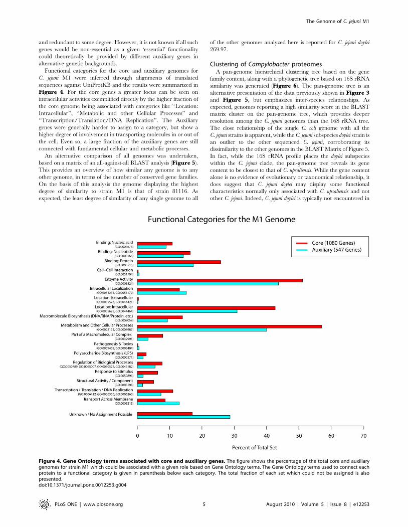
Functional categories for the core and auxiliary genomes for
C. jejuni M1 were inferred through alignments of translated
sequences against UniProtKB and the results were summarized in
Figure 4. For the core genes a greater focus can be seen on
intracellular activities exemplified directly by the higher fraction of
the core genome being associated with categories like ‘‘Location:
Intracellular’’, ‘‘Metabolic and other Cellular Processes’’ and
‘‘Transcription/Translation/DNA Replication’’. The Auxiliary
genes were generally harder to assign to a category, but show a
higher degree of involvement in transporting molecules in or out of
the cell. Even so, a large fraction of the auxiliary genes are still
connected with fundamental cellular and metabolic processes.
An alternative comparison of all genomes was undertaken,
based on a matrix of an all-against-all BLAST analysis (Figure 5).
This provides an overview of how similar any genome is to any
other genome, in terms of the number of conserved gene families.
On the basis of this analysis the genome displaying the highest
degree of similarity to strain M1 is that of strain 81116. As
expected, the least degree of similarity of any single genome to all
of the other genomes analyzed here is reported for C. jejuni doylei
269.97.
Clustering of Campylobacter proteomesA pan-genome hierarchical clustering tree based on the gene
family content, along with a phylogenetic tree based on 16S rRNA
similarity was generated (Figure 6). The pan-genome tree is an
alternative presentation of the data previously shown in Figure 3and Figure 5, but emphasizes inter-species relationships. As
expected, genomes reporting a high similarity score in the BLAST
matrix cluster on the pan-genome tree, which provides deeper
resolution among the C. jejuni genomes than the 16S rRNA tree.
The close relationship of the single C. coli genome with all the
C. jejuni strains is apparent, while the C. jejuni subspecies doylei strain is
an outlier to the other sequenced C. jejuni, corroborating its
dissimilarity to the other genomes in the BLAST Matrix of Figure 5.
In fact, while the 16S rRNA profile places the doylei subspecies
within the C. jejuni clade, the pan-genome tree reveals its gene
content to be closest to that of C. upsaliensis. While the gene content
alone is no evidence of evolutionary or taxonomical relationship, it
does suggest that C. jejuni doylei may display some functional
characteristics normally only associated with C. upsaliensis and not
other C. jejuni. Indeed, C. jejuni doylei is typically not encountered in
Figure 4. Gene Ontology terms associated with core and auxiliary genes. The figure shows the percentage of the total core and auxiliarygenomes for strain M1 which could be associated with a given role based on Gene Ontology terms. The Gene Ontology terms used to connect eachprotein to a functional category is given in parenthesis below each category. The total fraction of each set which could not be assigned is alsopresented.doi:10.1371/journal.pone.0012253.g004
The Genome of C. jejuni M1
PLoS ONE | www.plosone.org 5 August 2010 | Volume 5 | Issue 8 | e12253

Figure 5. Blast Matrix of C. jejuni. The figure presents the absolute number of gene families preserved between any two species alongwith the total number of families between them. The relative percentage between these numbers is also given and is used as a basis for thecolor intensity. The orange squares deserve special mention since these are based on alignments of the organism against itself, and thusshow the internal homology within each organism’s proteome. The highest homology was found between genomes M1 and 81116,highlighted by a box.doi:10.1371/journal.pone.0012253.g005
Figure 6. 16S rRNA and Pan-genomic tree of the Campylobacter genus. The left dendrogram displays the distance between the genomesbased on the sequence of the 16S rRNA gene. The right dendrogram is based on presence or absence of the gene families in the pan-genome. Therelative manhattan distance indicates the proportion of the pan-genome where genomes differ in present/absent status. Bootstrap values are givenas percentages to indicate the stability of the branching.doi:10.1371/journal.pone.0012253.g006
The Genome of C. jejuni M1
PLoS ONE | www.plosone.org 6 August 2010 | Volume 5 | Issue 8 | e12253

food or animals, but only in humans, where it appears more apt at
causing bacteremia [42], suggesting a different ecology.
The high degree of whole-genome similarity between strains M1
and 81116 is confirmed by the relative Manhattan distance being
less than 0.05, i.e. these strains differ in less than 5% of the pan-
genome (ignoring all ORFans, see Methods).
Comparison of 81116 and M1C. jejuni strain 81116 is the closest related sequenced strain to
M1. It is known to be serotype HS6, ST-283 (which differs in 3 of
7 alleles with strain M1). Although strain 81116 has the same
AFLP profile as strain M1 (data not shown), its DNA is indigestible
by SmaI so it is untypable by PFGE using this enzyme [43]. Strain
81116 is one of a group of strains that were isolated over time and
distance with identical genotypes [43] and consequently share
phenotypic characteristics. A closer comparison of M1 with strain
81116 was undertaken.
The frequency of genes sharing decreasing percentages of
identity between these two genomes was determined (Table 1).
These data illustrate the differences that can occur, on a complete
genome scale, between isolates that have an identical AFLP
genotype and flaA type. The divergence in gene content between
these strains is considerable, even at the protein level. Such
observations would obviously be overlooked when isolates were
only characterized by genotyping, and this could lead to the
erroneous interpretation that isolates with identical genotypes are
completely identical.
In the comparison of the genomes of strains 81116 and M1, a
number of genes were recognized as being unique to either one or
the other strain (Table 1). The comparison was performed first
with a tBLASTX alignment of the genes of C. jejuni M1 against the
full genome of 81116 and then vice-versa. Similarity was relaxed to
require 50% identity over only 20% of the length of the query
sequence (i.e. ,10% identity over the entire sequence), so that only
genes that are truly missing (as opposed to divergent genes) in
either of the two strains were detected. There were 36 genes
unique to strain 81116 and 19 unique to strain M1. One of the
genes present in strain 81116, but not in strain M1, was C8J_0133,
which has similarity to a DNA methylase in Lactococcus lactis subsp.
cremoris (P34877). The absence of this gene might explain the
resistance to SmaI digestion observed strain 81116 but not in strain
M1.
Blast Atlas of C. jejuni strain M1A final visualization tool of genome comparison applied to the
C. jejuni genomes was the Blast Atlas [44] (Figure 7) (a
‘‘zoomable’’ version of this is available online [45]). This analysis
used the Genome Atlas of M1 as a reference point, around which
all BLAST hits detected in the other C. jejuni genomes were
plotted, followed by the other available genomes of the
Campylobacter genus. The atlas illustrates how well conserved most
regions of strain M1 are within the other C. jejuni species, and even
to a large extent within the Campylobacter genus, with the exception
of a few loci. As this analysis is based on BLASTP, RNA genes
show up as ‘gaps’ although they are in fact conserved in all
genomes.
Despite the degree of conservation, several highly variable
regions are also visible, such as the gene cluster located in the
region from 1,335 kb to 1,360 kb. This region encodes several
genes which are hypothetical, but also many capsular poly-
saccharide biosynthesis genes. The LOS locus of C. jejuni also
displays high degrees of variability between strains, as previously
reported [13].
At the position from 52,774 to 51,104 bp the gene CJM1_0038
is located. This gene encodes a gamma-glutamyltranspeptidase,
which has been shown to enhance the ability of C. jejuni to
colonize the avian intestine [46,47]. This gene is far from
ubiquitous in C. jejuni but is found (other than in M1) in strains
81116, HB93-13, 81-176 and C. jejuni subsp doylei 269.97.
Although all these strains were isolated from cases of campylo-
bacteriosis [16,48], other clinical strains, such as NCTC 11168,
do not possess this gene.
Table 1. Genes identified in a comparison between strains 81116 and M1 as unique relative to the comparator.
Number of Proteins
BLASTP %-identity M1 proteome (1624 proteins) vs. 81116 proteome 81116 proteome (1623 proteins) vs. M1 proteome
100% 1166 1194
$90% 392 322
$80% 23 19
$70% 7 13
$60% 6 8
$50% 2 11
$40% 0 4
$30% 4 5
$20% 5 11
,20% 19 36
Proteins with ,20% identity in the other genome
CJM1_0032, CJM1_0040, CJM1_0041, CJM1_0055, CJM1_0056,CJM1_0057, CJM1_0138, CJM1_0278, CJM1_0414, CJM1_0728,CJM1_0745, CJM1_1118, CJM1_1119, CJM1_1120, CJM1_1372,CJM1_1373, CJM1_1375, CJM1_1381, CJM1_1382
C8J_0035, C8J_0036, C8J_0063, C8J_0132, C8J_0133, C8J_0154,C8J_0265, C8J_0266, C8J_0267, C8J_0272, C8J_0273, C8J_0274,C8J_0275, C8J_0321, C8J_0529, C8J_0651, C8J_0776, C8J_0852,C8J_0918, C8J_1080, C8J_1081, C8J_1083, C8J_1329, C8J_1330,C8J_0131, C8J_1332, C8J_1334, C8J_1335, C8J_1337, C8J_1338,C8J_1340, C8J_1341, C8J_1342, C8J_1456, C8J_1458, C8J_1459
*Gene names given in bold refer to the sequences having matches to verified proteins in UniProtKB/Swiss-Prot, while italics indicate a match to the NCBI nr database.doi:10.1371/journal.pone.0012253.t001
The Genome of C. jejuni M1
PLoS ONE | www.plosone.org 7 August 2010 | Volume 5 | Issue 8 | e12253

Another region of interest, from 1,455 kb to 1,465 kb, encodes
two Type I restriction modification genes hsdR and hsdS
(CJM1_1490 and CJM1_1493) and the associated DNA methy-
lase, hsdM, (CJM1_1494). This restriction/modification (R/M)
region has been shown to be involved in a hsd type I restriction–
modification system of C. jejuni but is not present in C. jejuni strain
NCTC 11168 [49]. The region appears conserved in only a few C.
jejuni strains in addition to M1, namely in strains 81116, CG8421,
and GC8486, as well as in C. coli RM2228. While hsdR and hsdM
were found conserved in strain HB93-13 the hsdS gene was not.
The presence of this R/M region would suggest implications for
the methylation pattern and susceptibility of these genomes
towards the HsdR restriction enzyme.
Finally, the R/M region in strain M1 also contains two slightly
overlapping genes, rloA and rloB (CJM1_1491 and CJM1_1492)
with no inferred function, but which are also conserved in strains
81116 and CG8421. A subsequent examination of these genomes
revealed that the location of the rlo genes inside the R/M island is
conserved in strains CG8486 and 81116 (data not shown).
In conclusion, strain M1, a rarely reported C. jejuni isolate from
colonized poultry with direct evidence of its ability to infect and
cause diarrhea in a human, provides a unique opportunity to
investigate the genomic basis of colonization in poultry and
pathogenicity in humans. The complete elucidation of the genome
sequence of strain M1 not only provides the scientific community
with a valuable strain to use in in vitro and in vivo models of
colonization and virulence, but also allows in silico approaches such
as microarray analysis to be applied.
Materials and Methods
Ethics StatementThe study was approved by the VLA Ethical Review
Committee on the 25th of March 2010. The patient has provided
written informed consent for the collection of samples and
subsequent analysis.
Isolation and characterization of C. jejuni strain M1A research team of four individuals visited a poultry processing
plant to investigate the sites and diversity of Campylobacter
contamination. Three days after this visit, one member of the
research team became ill with watery diarrhoea, fever and severe
Figure 7. Blast Atlas of C. jejuni M1. The proteome of the M1 strain was aligned against the proteomes of 20 other Campylobacter genomes usingBLASTP and the results are displayed as colored circles with increasing color intensity signifying increased similarity. Only BLAST results of proteins areshown. The three rRNA islands are marked as well as the lipooligosaccharide (LOS) and extracellular polysaccharide biosynthesis (PBS) clusters.doi:10.1371/journal.pone.0012253.g007
The Genome of C. jejuni M1
PLoS ONE | www.plosone.org 8 August 2010 | Volume 5 | Issue 8 | e12253
abdominal cramps. The diarrhoea terminated after about six days.
Six days after the onset of symptoms a faecal sample was cultured
as described below and C. jejuni was isolated. Four separate
colonies from the faecal sample were recovered and stored.
The team sampled two flocks (flocks 1 and 2) at the abattoir.
The flocks were reared on different farms, and sequentially
processed through the processing plant. Caeca were removed from
the poultry carcasses during evisceration, and the contents
sampled aseptically. Swab samples were also collected from
various sites in the abattoir and from up to five poultry carcasses
[50] as they progressed through the processing plant. The methods
of chicken sampling and culture have been previously described
[51]. Briefly all samples were directly plated onto blood agar
containing selective antibiotics [52] with actidione (100 ug/ml)
and cefoperazone (30 mg/ml), incubated microaerobically at
37uC for 2 days, with or without pre-enrichment in Exeter
medium [53]. Identification was based on microaerobic growth at
42uC, hippurate and indoxylacetate hydrolysis and catalase and
oxidase activities. A single colony from each sample was stored in
glycerol broth (10% v/v glycerol in 1% w/v proteose peptone) at
280uC for subsequent typing.
All isolates were fla-typed according to the technique of Ayling
et al. [23] using the restriction enzymes DdeI and HinfI.
Representatives of the human and chicken isolates were also
serotyped [54] and molecular typed by pulsed-field gel electro-
phoresis (PFGE) using the enzymes SmaI and KpnI [55], amplified
fragment length polymorphism (AFLP) [56] and multilocus
sequence typing (MLST) [57].
Phenotypic characterization of strain M1 (Laboratory designa-
tion 99/308) was performed to determine in vitro invasiveness of
INT407 cells and CaCO2 cells and to detect the expression of
cytolethal distending toxin (CDT) as previously described [58,59].
Genome sequencing and annotation of strain M1The genome of strain M1 was sequenced by Agencourt Bioscience,
Beverly MA, USA using a 454 GS FLEX instrument and assembled
using the ARACHNE system of Batzoglou et al [60]. This resulted in
18 contigs ranging in size from 108 bp to 950,427 bp, altogether
totaling 1,611,563 bp. Gaps were closed by direct sequencing of PCR
amplifications using primers flanking the gapped regions. The gene
annotation (as described below) for strain M1 was visualized on a
Genome Atlas as previously published [35,36]. A zoomable version of
the Atlas is available online [45]. The complete annotated sequence
has been submitted to NCBI/GenBank under the accession number
CP001900 (Genome Project ID 38041).
Identification of protein coding genes and inference ofgene function
Gene finding was carried out using the EasyGene v1.2b gene
finder using the pre-calculated model for C. jejuni (CJ02) [61,62],
which resulted in 1624 inferred protein coding genes. The quality
of the gene finding is difficult to gauge, but was found to be
acceptable in an assessment performed according to Skovgaard et
al. [63] (See supplementary section [45]). Even so, in their second
publication, the authors of EasyGene note that the gene finder has
a small tendency to disfavor non-ATG start codons, particularly if
an in-frame ATG is nearby [61]. Thus, to verify the correct
prediction of CDS start codons, all inferred genes were aligned
against the NCBI non-redundant database (nr database) using
BLASTX [64,65], which identified 233 entries as having no
perfect match in the database. For each of these 233 entries, the
highest scoring sequence in the nr database was extracted and
aligned back against the entire strain M1 genome using
TBLASTN. If this resulted in a perfect hit to the sequence of
the originally predicted gene in strain M1 covering the entire
sequence in the nr database without any in-frame stop codons, the
strain M1 annotations were updated. In this case a new
transcription start position was assigned to the M1 gene matching
that of the sequence from the nr database, provided that the new
start codon was a valid. As a result of this, 85 start codons were
changed, many of which resulted in non-standard, but valid start
codons of GTG or TTG. A table of all affected genes is available
in the supplemental section [45].
Functional information was inferred through prediction of gene
function. This entailed the comparison of translated sequences to
the validated part of UniProt (Swiss-prot) [66], and to the
annotated genes of the previously sequenced Campylobacter
genomes in GenBank (Table 2) [67]. For BLASTX alignment a
similarity criterion was used of $50% identity of the amino acids
over $50% of the length of the longest sequence. Hits to Uniprot
were preferred over those to the NCBI database, however, the
curated Swiss-prot part of Uniprot was preferred over all others. In
total, similarity to Swiss-prot provided inference of annotation for
527 protein coding genes, while a further 1060 could be
characterized from the rest of Uniprot (UniProt/TrEMBL) and
8 from NCBI nr. Of these 1595 genes, 495 were annotated as
‘‘putative’’, ‘‘uncharacterized’’ or ‘‘hypothetical’’.
Supplemental functional annotation was inferred through the
alignment of translated sequences against active sites in Pfam using
the HMM-based tool provided by Pfam [68]. Default settings were
used and the built-in Pfam TC trusted threshold cutoffs were relied
upon. In case of overlapping results, only the best match was used.
Identification of non-protein encoding RNAsRibosomal genes coding rRNA were predicted using RNAm-
mer [69], while tRNA genes were predicted using tRNAscan [70].
Both programs were used at the default settings.
Identification of gene conservation and definition ofgene families
The Campylobacter genomes used for comparative studies, as
listed in Table 2, were obtained from NCBI [67].
The set of gene families for this collection of Campylobacter
genomes were found by BLASTing all proteins in each genome
against all proteins in the query genome. A hit was considered
significant if the alignment covered at least 50% of both sequences,
and contained at least 50% identities [71]. This included aligning
proteins within each proteome, and while self-hits were ignored,
hits to other homologous genes within the same genome were
recorded. A pair of genes having significant hits was considered as
belonging to the same gene family. Two gene families were
merged into one if any one gene in a given family would meet the
similarity criterion in an alignment against any member of another
gene family [71,72].
BLAST matrixThe results of the gene conservation analysis were visualized as
a triangle-shaped matrix providing the amount of conserved gene
families between any two proteomes, both as an absolute number
and as the fraction of the total number of gene families shared. In
addition to the comparisons across proteomes, the matrix also
shows the results from aligning proteins within each proteomes
against each other.
16S rRNA tree for the Campylobacter genusRNAmmer was used to find the sequences for the 16S rRNA
phylogenetic tree. The sequences had to be between 1400 and
The Genome of C. jejuni M1
PLoS ONE | www.plosone.org 9 August 2010 | Volume 5 | Issue 8 | e12253

1700 nucleotides and have an RNAmmer score above 1700 to be
chosen. In case several sequences from one organism satisfied
these criteria, one sequence was arbitrarily chosen. Alignment was
done using PRANK [73,74], and the program MEGA4 was used
to construct a phylogenetic tree [75]. Within MEGA4, the tree was
created using the Neighbor-Joining method with the uniform rate
Jukes–Cantor distance measure and the complete-delete option. A
thousand re-samplings were done to find the bootstrap values.
Pan-genome tree for the Campylobacter genusThe pan-genome tree is another method for visualization of the
differences between proteomes in a pan-genome context. It is a
hierarchical clustering of genomes based on a weighted Manhat-
tan distance computed from a pan-matrix. A pan-matrix is a
matrix of 1s and 0s where each row corresponds to a gene family,
as described above, and each column to a genome. Cell (i,j) in the
matrix is 1 if gene family i is present in genome j, or 0 if it is absent.
The distance between two genomes is the proportion of gene
families where their present/absent status differs. Shorter distances
represent genomes with many gene families in common, and
larger distances reflect genomes with fewer gene families in
common. Genes only occurring in a single genome were given a
weight of ‘‘0’’ – that is, they did not provide any difference in the
computed distance in the matrix. These ‘ORFans’ are frequently
annotated as ‘hypothetical protein’ and may in cases be falsely
predicted genes. By discarding these genes the tree obtained is
more robust against gene-prediction errors, at the cost of possibly
losing some information. Bootstrap values (percentages) were
computed for each inner node by re-sampling the rows of the pan-
matrix.
Core- and pan-genome analysisConservation data across various genomes were also
visualized by an accumulative plot showing the changes to
the number of gene families in the core and pan-genomes as
more sequential genomes were added to the dataset [76]. The
number of novel gene families for each added genome is also
depicted. Any novel gene family encountered is automatically
added to the pan-genome unless it is found to be conserved in
the previously considered genomes. Because the quality of
genome annotation is not consistent, and because Table 2includes genomes which have not been completely sequenced,
we have relaxed the definition of the core genome as follows;
we added any gene family of the nth analyzed genome to the
core genome if by analyzing n+1 genomes this gene family was
present in at least n genomes. This approach provided
tolerance while preserving intuitiveness in plotting. Otherwise,
the reduction in the core genome caused by, for example, the
addition of C. concisus 13826 would be drawn for the genome to
its right (C. fetus). For the same reason the core genome curve
does not extend to the rightmost genome since no n+1 genome
exists.
GO categories were inferred for the core and auxiliary genes in
strain M1 through the alignment of translated sequences against
Uniprot meeting the similarity criterion of at least 50% identity
covering at least 50% of both sequences. GO terms were inferred
from all alignments meeting the criterion to minimize problems
with incomplete annotations in the database. For each go term
assigned to a given protein, all parent terms were considered as
well. GO assignments were then grouped together into functional
category based on similarity and overlap.
Table 2. Campylobacter Genomes included in this study.
Species Name Project ID Contigs Accession Reference
C. jejuni M1 38041 1 CP001900 This Study
C. jejuni 260.94 16229 10* AANK01000000 -
C. jejuni 81116 17953 1 CP000814.1 [16]
C. jejuni 81-176A 16135 3 CP000538.1, CP000549.1, CP000550.1 [48]
C. jejuni 81-176B 17341 1 NZ_AASL00000000 [46]
C. jejuni 84–25 16367 5* AANT02000000 -
C. jejuni CF93-6 16265 14* AANJ01000001 [77]
C. jejuni CG8421 21037 20* ABGQ01000000 [78]
C. jejuni CG8486 17055 19* AASY01000000 [18]
C. jejuni doylei 269.97 17163 1 CP000768.1 -
C. jejuni HB93-13 16267 35* AANQ0100000 -
C. jejuni NCTC 11168 8 1 AL111168.1 [13]
C. jejuni RM1221 303 1 CP000025.1 [19]
C. coli RM2228 12516 38* AAFL01000000 [19]
C. concisus 13826 17159 3 CP000792.1, CP000793.1, CP000794.1 -
C. curvus 525.92 17161 1 CP000767.1 -
C. fetus 82–40 16293 1 CP000487.1 -
C. hominis ATCC BAA-381 20083 2 CP000775.1, CP000776.1 -
C. lari RM2100 12517 2 CP000932.1, CP000933.1 [79]
C. rectus RM3267 31017 89* ACFU01000000 -
C. upsaliensis RM3195 12518 20* AAFJ01000000 [19]
If the genome project listed any plasmids, these were included in the number of contigs and their accession numbers are included.*For genomes represented by multiple contigs the published sequence may still be incomplete.doi:10.1371/journal.pone.0012253.t002
The Genome of C. jejuni M1
PLoS ONE | www.plosone.org 10 August 2010 | Volume 5 | Issue 8 | e12253

BLAST atlasA Blast Atlas was produced using a previously described
approach [44]. The atlas displays results of the BLASTP
comparisons of the products of the predicted genes of C. jejuni
M1 with the annotated proteomes of the other campylobacters
from Table 2. A zoomable version is available online [45].
Author Contributions
Conceived and designed the experiments: CF TMW DGN DWU.
Performed the experiments: CF MAJ MT AR GM. Analyzed the data:
CF TMW LS KL PFH. Contributed reagents/materials/analysis tools:
MT AR GM. Wrote the paper: CF TMW DGN DWU. The primary
author of the paper and the primary person responsible for driving the
computational work from start to finish: CF. The primary behind all
computational aspects of the work, including the genome annotation of
M1: CF. Has been instrumental in maintaining the link between the
computational work and the experimental work: TMW. Lent her
experience to all aspects of the project and has contributed significantly
at all stages writing and proof reading of the manuscript: TMW. Helped
with the analysis of genome sequence of M1, designed and synthesized
primers to fill the gaps between contigs of the M1 genome: MAJ. Did the
amplicons generation and sequencing followed by the analysis: MAJ.
Helped with the preparation of the manuscript: MAJ. Did the computa-
tions related to the pan-genome tree and the estimates of the population
core- and pan-genome sizes, and contributed to the corresponding parts of
the manuscript: LS. Developed a phylogenetic tree for these genomes
based on predicted 16S rRNA sequences: KL. Contributed text to the
paper and participated in the final proof reading of the article: KL. Offered
assistance in adopting his previously established atlas methods into this
project: PFH. Contributed particularly to the supplementary section by
setting up the interactive figures included there: PFH. Participated in the
proof reading of the manuscript: PFH. Managed the research team
throughout the period of the strains isolation, identification and
characterization providing expertise and leadership to recognize the
importance of this strain for future research: DGN. Instigated the idea of
sequencing the strain and collaborated with DWU and TMW to get this
done: DGN. Contributed significantly to the drafting of the manuscript by
providing expert knowledge, intellectual content and experience: DGN.
Was solely responsible for acquiring the M1 - she is the person who was
infected: MT. Recovered the strains from herself and the appropriate
chickens: MT. Purified, stored and initially characterized the strains: MT.
Was responsible for the molecular characterization of the human and
chickens strains including the molecular typing - demonstrating that they
were identical: AR GM. Prepared, quality controlled and provided the
strain DNA for sequencing: AR. Contributed to the drafting and approval
of the final manuscript: MT AR. Was involved in the research carried out
on M1 since its first isolation at the VLA; was specifically responsible for
the molecular typing of this strain by MLST as well as overseeing the
invasion and toxin studies: GM. The closing of the gaps was funded by
research money that was awarded to her at NTU and she oversaw that
work: GM. Contributed to previous drafts of the paper and the final proof
reading of the article: GM. Conceptualized much of the work, especially
the computational aspects: DWU. Managed the bioinformatic research
team and has provided considerable feedback and ideas: DWU.
Contributed text and assisted in proof reading of the manuscript: DWU.
References
1. Nauta M, Hill A, Rosenquist H, Brynestad S, Fetsch A, et al. (2009) A
comparison of risk assessments on Campylobacter in broiler meat. Int J Food
Microbiol 129: 107–123.
2. Wagenaar JA, Mevius DJ, Havelaar AH (2006) Campylobacter in primary animal
production and control strategies to reduce the burden of human campylo-
bacteriosis. Rev Sci Tech 25: 581–594.
3. Wingstrand A, Neimann J, Engberg J, Nielsen EM, Gerner-Smidt P, et al. (2006)
Fresh chicken as main risk factor for campylobacteriosis, Denmark. Emerg Infect
Dis 12: 280–285.
4. Jacobs-Reitsma W (2000) Campylobacter in the food supply. In: Nachamkin I,
Blaser MJ, eds. Campylobacter. 2nd ed. Washington, D.C: American Society for
Microbhioology. pp 467–481.
5. Little CL, Richardson JF, Owen RJ, de Pinna E, Threlfall EJ (2008) Prevalence,
characterisation and antimicrobial resistance of Campylobacter and Salmonella in
raw poultrymeat in the UK, 2003-2005. Int J Environ Health Res 18: 403–414.
6. Jorgensen F, Bailey R, Williams S, Henderson P, Wareing DR, et al. (2002)
Prevalence and numbers of Salmonella and Campylobacter spp. on raw, whole
chickens in relation to sampling methods. Int J Food Microbiol 76: 151–164.
7. Wassenaar TM, Fernandez-Astorga A, Alonso R, Marteinsson VT,
Magnusson SH, et al. (2009) Comparison of Campylobacter fla-SVR genotypes
isolated from humans and poultry in three European regions. Lett Appl
Microbiol.
8. Sheppard SK, Dallas JF, MacRae M, McCarthy ND, Sproston EL, et al. (2009)
Campylobacter genotypes from food animals, environmental sources and clinical
disease in Scotland 2005/6. Int J Food Microbiol 134: 96–103.
9. Miller WG, Englen MD, Kathariou S, Wesley IV, Wang G, et al. (2006)
Identification of host-associated alleles by multilocus sequence typing of
Campylobacter coli strains from food animals. Microbiology 152: 245–255.
10. Wassenaar TM (1997) Toxin production by Campylobacter spp. Clin Microbiol
Rev 10: 466–476.
11. Carrillo CD, Taboada E, Nash JH, Lanthier P, Kelly J, et al. (2004) Genome-
wide expression analyses of Campylobacter jejuni NCTC11168 reveals
coordinate regulation of motility and virulence by flhA. J Biol Chem 279:
20327–20338.
12. Newell DG (2001) Animal models of Campylobacter jejuni colonization and disease
and the lessons to be learned from similar Helicobacter pylori models. Symp Ser Soc
Appl Microbiol. pp 57S–67S.
13. Parkhill J, Wren BW, Mungall K, Ketley JM, Churcher C, et al. (2000) The
genome sequence of the food-borne pathogen Campylobacter jejuni reveals
hypervariable sequences. Nature 403: 665–668.
14. Gaynor EC, Cawthraw S, Manning G, MacKichan JK, Falkow S, et al. (2004)
The genome-sequenced variant of Campylobacter jejuni NCTC 11168 and the
original clonal clinical isolate differ markedly in colonization, gene expression,
and virulence-associated phenotypes. J Bacteriol 186: 503–517.
15. Black RE, Levine MM, Clements ML, Hughes TP, Blaser MJ (1988)
Experimental Campylobacter jejuni infection in humans. J Infect Dis 157: 472–479.
16. Pearson BM, Gaskin DJ, Segers RP, Wells JM, Nuijten PJ, et al. (2007) Thecomplete genome sequence of Campylobacter jejuni strain 81116 (NCTC11828).
J Bacteriol 189: 8402–8403.
17. Palmer SR, Gully PR, White JM, Pearson AD, Suckling WG, et al. (1983)Water-borne outbreak of campylobacter gastroenteritis. Lancet 1: 287–290.
18. Poly F, Read T, Tribble DR, Baqar S, Lorenzo M, et al. (2007) Genomesequence of a clinical isolate of Campylobacter jejuni from Thailand. Infect Immun
75: 3425–3433.
19. Fouts DE, Mongodin EF, Mandrell RE, Miller WG, Rasko DA, et al. (2005)Major structural differences and novel potential virulence mechanisms from the
genomes of multiple campylobacter species. PLoS Biol 3: e15.
20. Medini D, Serruto D, Parkhill J, Relman DA, Donati C, et al. (2008)Microbiology in the post-genomic era. Nat Rev Microbiol 6: 419–430.
21. Lan R, Reeves PR (2000) Intraspecies variation in bacterial genomes: the need
for a species genome concept. Trends Microbiol 8: 396–401.
22. Medini D, Donati C, Tettelin H, Masignani V, Rappuoli R (2005) The
microbial pan-genome. Curr Opin Genet Dev 15: 589–594.
23. Ayling RD, Woodward MJ, Evans S, Newell DG (1996) Restriction fragmentlength polymorphism of polymerase chain reaction products applied to the
differentiation of poultry campylobacters for epidemiological investigations. ResVet Sci 60: 168–172.
24. Cell lines were obtained from the European Cell Culture Collection, Health
Protection Agency. Porton Down. Salisbury, UK.
25. Konkel ME, Garvis SG, Tipton SL, Anderson DE, Jr., Cieplak W, Jr. (1997)
Identification and molecular cloning of a gene encoding a fibronectin-bindingprotein (CadF) from Campylobacter jejuni. Mol Microbiol 24: 953–963.
26. Monteville MR, Yoon JE, Konkel ME (2003) Maximal adherence and invasion
of INT 407 cells by Campylobacter jejuni requires the CadF outer-membraneprotein and microfilament reorganization. Microbiology 149: 153–165.
27. Ziprin RL, Young CR, Stanker LH, Hume ME, Konkel ME (1999) The absence
of cecal colonization of chicks by a mutant of Campylobacter jejuni not expressingbacterial fibronectin-binding protein. Avian Dis 43: 586–589.
28. Jin S, Joe A, Lynett J, Hani EK, Sherman P, et al. (2001) JlpA, a novel surface-exposed lipoprotein specific to Campylobacter jejuni, mediates adherence to host
epithelial cells. Mol Microbiol 39: 1225–1236.
29. Kervella M, Pages JM, Pei Z, Grollier G, Blaser MJ, et al. (1993) Isolation andcharacterization of two Campylobacter glycine-extracted proteins that bind to
HeLa cell membranes. Infect Immun 61: 3440–3448.
30. Moser I, Schroeder W, Salnikow J (1997) Campylobacter jejuni major outermembrane protein and a 59-kDa protein are involved in binding to fibronectin
and INT 407 cell membranes. FEMS Microbiol Lett 157: 233–238.
31. Flanagan RC, Neal-McKinney JM, Dhillon AS, Miller WG, Konkel ME (2009)Examination of Campylobacter jejuni putative adhesins leads to the identification of
a new protein, designated FlpA, required for chicken colonization. Infect Immun77: 2399–2407.
32. Raphael BH MM, Klena JD, Joens LA, Konkel M (2005) Interactions of
Campylobacter jejuni with non-professional phagocytic cells. In: Ketley JM KM, ed.
The Genome of C. jejuni M1
PLoS ONE | www.plosone.org 11 August 2010 | Volume 5 | Issue 8 | e12253

Campylobacter: Molecular and Cellular Biology. Norfolk NR: Horizon
Bioscience. pp 397–413.
33. Christensen JE, Pacheco SA, Konkel ME (2009) Identification of a Campylobacter
jejuni secreted protein required for maximal invasion of host cells. Mol Microbiol.
34. Ashgar SS, Oldfield NJ, Wooldridge KG, Jones MA, Irving GJ, et al. (2007)
CapA, an autotransporter protein of Campylobacter jejuni, mediates associationwith human epithelial cells and colonization of the chicken gut. J Bacteriol 189:
1856–1865.
35. Jensen LJ, Friis C, Ussery DW (1999) Three views of microbial genomes. Res
Microbiol 150: 773–777.
36. Pedersen AG, Jensen LJ, Brunak S, Staerfeldt HH, Ussery DW (2000) A DNA
structural atlas for Escherichia coli. J Mol Biol 299: 907–930.
37. Sinden RR, Hashem VI, Rosche WA (1999) DNA-directed mutations. Leadingand lagging strand specificity. Ann N Y Acad Sci 870: 173–189.
38. Raskin DM, Seshadri R, Pukatzki SU, Mekalanos JJ (2006) Bacterial genomicsand pathogen evolution. Cell 124: 703–714.
39. Chao A (1987) Estimating the population size for capture-recapture data withunequal catchability. Biometrics 43: 783–791.
40. Snipen L, Almoy T, Ussery DW (2009) Microbial comparative pan-genomicsusing binomial mixture models. BMC Genomics 10: 385.
41. Lefebure T, Stanhope MJ (2009) Pervasive, genome-wide positive selectionleading to functional divergence in the bacterial genus Campylobacter. Genome
Res 19: 1224–1232.
42. Parker CT, Miller WG, Horn ST, Lastovica AJ (2007) Common genomic
features of Campylobacter jejuni subsp. doylei strains distinguish them from C.jejuni subsp. jejuni. BMC Microbiol 7: 50.
43. Manning G, Duim B, Wassenaar T, Wagenaar JA, Ridley A, et al. (2001)Evidence for a genetically stable strain of Campylobacter jejuni. Appl Environ
Microbiol 67: 1185–1189.
44. Hallin PF, Binnewies TT, Ussery DW (2008) The genome BLASTatlas-a
GeneWiz extension for visualization of whole-genome homology. Mol Biosyst 4:
363–371.
45. Friis C, Wassenaar T, Javed MA, Snipen L, Lagersen K, et al. (2009)
Supplementary Section for ‘‘Genomic Characterization of Campylobacter jejunistrain M1’’. Available from: http://www.cbs.dtu.dk/services/GenomeAtlas/
Campy_M1/.
46. Hofreuter D, Novik V, Galan JE (2008) Metabolic diversity in Campylobacter jejuni
enhances specific tissue colonization. Cell Host Microbe 4: 425–433.
47. Barnes IH, Bagnall MC, Browning DD, Thompson SA, Manning G, et al.
(2007) Gamma-glutamyl transpeptidase has a role in the persistent colonizationof the avian gut by Campylobacter jejuni. Microb Pathog 43: 198–207.
48. Russell RG, Blaser MJ, Sarmiento JI, Fox J (1989) Experimental Campylobacter
jejuni infection in Macaca nemestrina. Infect Immun 57: 1438–1444.
49. Miller WG, Pearson BM, Wells JM, Parker CT, Kapitonov VV, et al. (2005)Diversity within the Campylobacter jejuni type I restriction-modification loci.
Microbiology 151: 337–351.
50. Simonsen B (1971) Methods for determining the microbial counts of ready-to-
cook chicken. World’s Poultry Science: 27–368.
51. Newell DG, Shreeve JE, Toszeghy M, Domingue G, Bull S, et al. (2001)
Changes in the carriage of Campylobacter strains by poultry carcasses during
processing in abattoirs. Appl Environ Microbiol 67: 2636–2640.
52. Skirrow MB (1977) Campylobacter enteritis: a ‘‘new’’ disease. Br Med J 2: 9–11.
53. Humphrey T, Mason M, Martin K (1995) The isolation of Campylobacter jejunifrom contaminated surfaces and its survival in diluents. Int J Food Microbiol 26:
295–303.
54. Frost JA, Oza AN, Thwaites RT, Rowe B (1998) Serotyping scheme for
Campylobacter jejuni and Campylobacter coli based on direct agglutination ofheat-stable antigens. J Clin Microbiol 36: 335–339.
55. Gibson J, Lorenz E, Owen RJ (1997) Lineages within Campylobacter jejunidefined by numerical analysis of pulsed-field gel electrophoretic DNA profiles.
J Med Microbiol 46: 157–163.
56. Duim B, Wassenaar TM, Rigter A, Wagenaar J (1999) High-resolution
genotyping of Campylobacter strains isolated from poultry and humans with
amplified fragment length polymorphism fingerprinting. Appl Environ Micro-
biol 65: 2369–2375.57. Dingle KE, Colles FM, Falush D, Maiden MC (2005) Sequence typing and
comparison of population biology of Campylobacter coli and Campylobacter
jejuni. J Clin Microbiol 43: 340–347.58. Fearnley C, Manning G, Bagnall M, Javed MA, Wassenaar TM, et al. (2008)
Identification of hyperinvasive Campylobacter jejuni strains isolated frompoultry and human clinical sources. J Med Microbiol 57: 570–580.
59. Abuoun M, Manning G, Cawthraw SA, Ridley A, Ahmed IH, et al. (2005)
Cytolethal distending toxin (CDT)-negative Campylobacter jejuni strains andanti-CDT neutralizing antibodies are induced during human infection but not
during colonization in chickens. Infect Immun 73: 3053–3062.60. Batzoglou S, Jaffe DB, Stanley K, Butler J, Gnerre S, et al. (2002) ARACHNE: a
whole-genome shotgun assembler. Genome Res 12: 177–189.61. Nielsen P, Krogh A (2005) Large-scale prokaryotic gene prediction and
comparison to genome annotation. Bioinformatics 21: 4322–4329.
62. Larsen TS, Krogh A (2003) EasyGene—a prokaryotic gene finder that ranksORFs by statistical significance. BMC Bioinformatics 4: 21.
63. Skovgaard M, Jensen LJ, Brunak S, Ussery D, Krogh A (2001) On the totalnumber of genes and their length distribution in complete microbial genomes.
Trends Genet 17: 425–428.
64. Pruitt KD, Tatusova T, Klimke W, Maglott DR (2009) NCBI ReferenceSequences: current status, policy and new initiatives. Nucleic Acids Res 37:
D32–36.65. Altschul SF, Madden TL, Schaffer AA, Zhang J, Zhang Z, et al. (1997) Gapped
BLAST and PSI-BLAST: a new generation of protein database searchprograms. Nucleic Acids Res 25: 3389–3402.
66. Bairoch A, Apweiler R, Wu CH, Barker WC, Boeckmann B, et al. (2005) The
Universal Protein Resource (UniProt). Nucleic Acids Res 33: D154–159.67. Benson DA, Karsch-Mizrachi I, Lipman DJ, Ostell J, Wheeler DL (2008)
GenBank. Nucleic Acids Res 36: D25–30.68. Finn RD, Tate J, Mistry J, Coggill PC, Sammut SJ, et al. (2008) The Pfam
protein families database. Nucleic Acids Res 36: D281–288.
69. Lagesen K, Hallin P, Rodland EA, Staerfeldt HH, Rognes T, et al. (2007)RNAmmer: consistent and rapid annotation of ribosomal RNA genes. Nucleic
Acids Res 35: 3100–3108.70. Schattner P, Brooks AN, Lowe TM (2005) The tRNAscan-SE, snoscan and
snoGPS web servers for the detection of tRNAs and snoRNAs. Nucleic AcidsRes 33: W686–689.
71. Binnewies TT, Hallin PF, Staerfeldt HH, Ussery DW (2005) Genome Update:
proteome comparisons. Microbiology 151: 1–4.72. Ussery DW, Wassenaar TM, Borini S (2009) Computing for Comparative
Microbial Genomics: Bioinformatics for Microbiologists: Springer.73. Loytynoja A, Goldman N (2005) An algorithm for progressive multiple
alignment of sequences with insertions. Proc Natl Acad Sci U S A 102:
10557–10562.74. Loytynoja A, Goldman N (2008) Phylogeny-aware gap placement prevents
errors in sequence alignment and evolutionary analysis. Science 320:1632–1635.
75. Tamura K, Dudley J, Nei M, Kumar S (2007) MEGA4: Molecular EvolutionaryGenetics Analysis (MEGA) software version 4.0. Mol Biol Evol 24: 1596–1599.
76. Tettelin H, Masignani V, Cieslewicz MJ, Donati C, Medini D, et al. (2005)
Genome analysis of multiple pathogenic isolates of Streptococcus agalactiae:implications for the microbial ‘‘pan-genome’’. Proc Natl Acad Sci U S A 102:
13950–13955.77. Koga M, Gilbert M, Li J, Koike S, Takahashi M, et al. (2005) Antecedent
infections in Fisher syndrome: a common pathogenesis of molecular mimicry.
Neurology 64: 1605–1611.78. Poly F, Read TD, Chen YH, Monteiro MA, Serichantalergs O, et al. (2008)
Characterization of two Campylobacter jejuni strains for use in volunteerexperimental-infection studies. Infect Immun 76: 5655–5667.
79. Miller WG, Wang G, Binnewies TT, Parker CT (2008) The complete genome
sequence and analysis of the human pathogen Campylobacter lari. FoodbornePathog Dis 5: 371–386.
The Genome of C. jejuni M1
PLoS ONE | www.plosone.org 12 August 2010 | Volume 5 | Issue 8 | e12253