Embed Size (px)
Citation preview
i
SURVIVAL OF Campylobacter jejuni IN THE
ENVIRONMENT
Thesis submitted in accordance with the requirements of the University of Liverpool for the
degree of Doctor in Philosophy
By
KASEM HAMED MUSTAFA
Department of Medical Microbiology
Institute of Infection and Global Health
University of Liverpool
March 2016
i
ABSTRACT
Campylobacter jejuni is an emerging food borne pathogen and a successful human
pathogen, with the infection mostly transmitted to humans through consumption of
contaminated under-cooked poultry meat. However, the environment can also play a
role in transmission either directly or indirectly to humans. The microorganism has
reservoirs in water and various animals. Its survival outside the host is generally
thought to be poor, but the organism survives well in poultry meat. Previous studies
have suggested that the ability to survive may vary between different strain types of
C. jejuni.
A number of survival experiments were conducted, based on the ability of different C.
jejuni strains to retain culturability in sterile distilled water. These experiments
demonstrated that survival varies between different strains of C. jejuni and that the
retention of culturability was much better at low temperatures (4 °C) than higher
temperatures (25 °C). Survival was also better in non-autoclaved natural water. One
strain, C. jejuni M1, lost culturability more quickly at both temperatures than the
others tested. However, cells remained viable in these samples, suggesting that the
bacteria had entered into a viable but non-culturable (VBNC) state under stress
conditions. These variations may contribute to the transmission of C. jejuni from the
environment to humans or farm animals.
Using end-point and Q-PCR assays, a set of stress response genes, including genes
implicated previously in the formation of VBNC cells, were targeted for different C.
jejuni strains during survival in sterile distilled water. Differences in gene expression
between different strains of C. jejuni were identified, including in key genes (luxS,
htrA, ppk1), suggesting that these genes might have contributed to strain M1
ii
switching to a VBNC state in response to starvation (sterile distilled water). This is the
first report suggesting a role for the C. jejuni luxS (a gene likely to be involved in
quorum sensing) in the formation of VBNC state and survival in water.
Co-existence with other microorganisms, such as Pseudomonas spp., is one of the
suggested survival strategies of C. jejuni in the environment. In a small study using
environmental PCR assays, it was demonstrated that when C. jejuni is present in the
farm environment, Pseudomonas spp. are also always present. In preliminary in vitro
experiments, we demonstrated that some fluorescent Pseudomonas spp. could
secret proteinaceous products that enhance the growth of Campylobacter. In natural
environments, it is likely that interactions with other species, such as Pseudomonas,
play an important role in C. jejuni survival and subsequent transmission to humans or
animals.
iii
DECLARATION
This thesis is a presentation of my own research work. The material presented here
has not been presented and is not being presented, either wholly or in part for any
other degree or qualification. Some of the technical procedures were carried out in
collaboration with other people and reference has been made to specific data from
other colleagues where appropriate.
The research work was carried out under the guidance of Professor Craig Winstanley
at the Institute of Infection and Global Health, Department of Clinical infection,
Microbiology and Immunology, University of Liverpool.
Kasem Mustafa

iv
ACKNOWLEDGEMENTS
Firstly, I would like to express my thankfulness to my supervisor, Professor Craig
Winstanley, for all the continuous support and guidance throughout my PhD research
and especially during writing of this thesis. I would like to thank my secondary
supervisor Professor Nicola Williams for supervising me during my work at Leahurst
campus. I would also like to thank Dr. Christina Bronowski for her technical support
and suggestions for different parts of this project in particular, with respect to
RNAseq.
I would also like to thank Professor Tom Humphrey, Professor Paul Wigley, Dr. Trevor
Jones, Dr. Amy Wedley and other members of my committee at Leahurst for the
assistance they provided at Leahurst. Thanks also to Dr. Jo Fothergill for the guidance
and technical supports she provided at all levels of the study. I must also
acknowledge Dr. Chloe James, who has left for the University of Salford, for her
assistance and willingness to help during the first year of this research.
Finally I would like to thank my wife, Rizgar, for her support and encouragement
during the period of my study.
v
CONTENTS
ABSTRACT i
DECLARATION iii
ACKNOLEDGMENT iv
CONTENTS v
LIST OF FIGURES xi
LIST OF TABLES xvi
ABBREVIATIONS xviii
CHAPTER 1: GENERAL INTRODUCTION 1
1.1 The genus Campylobacter 1
1.2 The history of Campylobacter 6
1.3 Diseases associated with Campylobacter infection 8
1.3.1 Campylobacteriosis in Humans 8
1.3.2 Campylobacter in poultry 11
1.3.3 Campylobacter in other animals 12
1.4 Pathogenesis 14
1.5 Incidence and Epidemiology 17
1.5.1 Seasonality 18
1.5.2 Guillain-Barre syndrome (GBS) 19
1.5.3 Miller Fisher syndrome (MFS) 19
1.5.4 Reactive arthritis (ReA) 19
1.5.5 Inflammatory bowel disease (IBD) 20
1.6 Treatment of campylobacteriosis 21
1.7 Strain typing methods 21
1.7.1 Serotyping 23
1.7.2 Macro-restriction PFGE 24
1.7.3 Flagellin typing 24
vi
1.7.4 MLST 25
1.7.4.1 Common C. jejuni clonal complexes 30
1.8 Campylobacter spp. in the environment 31
1.9 Aims of this study 34
CHAPTER 2: MATERIALS AND METHODS 35
2.1 Bacterial strains used in this study 35
2.1.1 Campylobacter strains 35
2.1.2 Pseudomonas strains 37
2.2 Growth conditions 38
2.3 Experiments to test survival of C. jejuni in water 38
2.3.1 Preparation of cell suspensions for testing 38 survival in sterile water
2.3.2 Preparation of sterile water sample 39
2.3.3 Inoculation of C. jejuni strains in sterile 39
distilled water
2.3.4 Bacterial survival in sterile distilled water: 40
determination of culturable cells counts
2.4 Survival of C. jejuni strains in natural water (Troughs) 40
2.4.1 Preparation of the inoculum 40
2.4.2 Collection of trough water sample 41
2.4.3 Inoculation of C. jejuni strains 42
in natural (trough) water
2.4.4 Bacterial survival in natural water 42
2.5 Assay for viable but non culturable (VBNC) cells 43
2.5.1 Counting of VBNC cells of C. jejuni strains 43
2.6 Genomic DNA preparation 44
2.6.1 Wizard Genomic DNA Purification Kit (Promega) 44
vii
2.6.2 Chelex Genomic DNA extraction method 45
2.6.3 Preparation of bacterial DNA from boiled suspension 45
2.7 PCR protocol for amplification of bacterial genomic DNA 45
2.7.1 Standard PCR protocol for the identification 47
of C. jejuni strains
2.7.2 PCR protocol for identification of Campylobacter 47
isolates from environmental water samples
2.7.3 PCR protocol for identification of Pseudomonas 48
Isolates from environmental water samples
2.7.4 Temperature gradient PCR 49
2.7.5 PCR amplification of cDNA 49
2.8 Agarose gel electrophoresis 50
2.9 Gene expression analysis using Real Time Quantitative- 51
PCR (Q-PCR) and RNA-Sequencing (RNAseq)
2.9.1 Oligonucleotide Primers 51
2.9.2 RNA extraction from water survival 52
experiments and cDNA synthesis
2.9.3 Q-PCR 54
2.9.4 RNAseq 55
2.9.4.1 RNA extraction from water survival experiment 55
2.9.4.2 Library construction and sequencing 56
2.9.4.3 RNAseq data analysis 57
2.10 Interaction between supernatants of cultures 58
of Pseudomonas spp. and C. jejuni strains.
2.10.1 Bacterial strains and growth conditions 58
2.10.2 Preparation of supernatants 58
of Pseudomonas spp.
2.10.3 Interaction assay 59
2.10.4 Treatment of supernatants by boiling 59
or proteinase K
viii
2.11 Experiments to asses co-existence of C. jejuni and 60
Pseudomonas spp. in environmental samples
2.11.1 Collection of environmental water samples 60
2.11.2 Enrichment of collected water samples 60
2.11.3 Extraction of bacterial DNA from environmental 61
water samples
CHAPTER 3: SURVIVAL OF C. jejuni STRAINS IN STERILE 62
DISTILLED WATER AND NATURAL WATER
3.1 Introduction 62
3.1.1 Survival of Campylobacter spp. in water 62
3.1.2 VBNC state 65
3.1.3 Aims of this chapter 68
3.2 Results 69
3.2.1 Survival in sterile distilled water and classification 69
of C. jejuni strains into groups
3.2.2 Survival of selected strains in sterile distilled water 76
using three biological replicates
3.2.3 Survival of selected strains in natural water using 79
three biological replicates
3.2.4 Direct viable cell counts 83
3.3 Discussion 87
3.4 Conclusions 94
CHAPTER 4: GENE EXPRESSION DURING SURVIVAL 95 IN STERILE DISTILLED WATER
4.1 Introduction 95
4.1.2 Aims of this chapter 100
4.2 Results 101
4.2.1 Temperature gradient PCR to determine optimal 101
annealing temperatures for end-point PCR assays
ix
4.2.2 RNA extraction and conversion to cDNA 103
4.2.3 PCR amplification of cDNA 103
4.2.4 Development of Q-PCR assays 114
4.2.5 Gene expression analysis using Q-PCR 119
4.2.6 RNAseq analysis of strain M1 gene expression 126
4.3 Discussion 133
4.4 Conclusions 141
CHAPTER 5: CO-EXISTENCE OF C. jejuni AND Pseudomonas spp. IN THE 142
ENVIRONMENT, AND INTERACTION BETWEEN THEM
5.1 Introduction 142
5.1.1 The genus Pseudomonas 144
5.1.2 Bacterial secretome 145
5.1.3 Pseudomonas secretome 146
5.1.4 Bacterial QS systems 148
5.1.5 Pseudomonas QS systems 149
5.1.6 Cross talk between bacterial species 151
via secreted products
5.1.7 Aims of this chapter 152
5.2 Results 153
5.2.1 Laboratory interaction assay between 153
C. jejuni and Pseudomonas spp.
5.2.2 The effect of pre-treated supernatant on growth 154
5.2.3 Environmental PCR assays to assess co-existence of 159
C. jejuni and Pseudomonas spp. in environmental samples
5.3 Discussion 162
5.4 Conclusions 167

x
CHAPTER 6: GENERAL DISCUSSION 168
APPENDIX 1 (MEDIA) 180
APPENDIX 2 (BUFFERS) 181
REFERENCES 183
xi
LIST OF FIGURES
Chapter 1
1.1 C. jejuni observed microscopically 2
1.2 Image of unipolar and bipolar flagella and corkscrew shape of C. jejuni 2
under electron microscope
1.3 Phylogenetic tree of the family Campylobacteraceae and close relatives, 7
based on similarity of 16S rRNA
1.4 Clinical conditions associated with Campylobacter species in humans 10
1.5 Number of laboratory confirmed cases of campylobacteriosis 20
and salmonellosis in the UK between 2000-2012
1.6 Chromosomal locations of MLST loci 31
1.7 Transmission routes of C. jejuni 33
Chapter 2
2.1 Sampling area of natural water at Leahurst 41
2.2 DNA size markers used in this study 51
Chapter 3
3.1A Rapid screen for culturability of C. jejuni cells (27 strains) during 70
incubation periods in sterile distilled water
3.1B Rapid screen for culturability of C. jejuni cells (27 strains) during 71
incubation periods in sterile distilled water
3.1C CFU/mL reduction of C. jejuni cells (27 strains) during incubation 72
xii
periods in sterile distilled water at 25 °C
3.1D CFU/mL reduction of C. jejuni cells (27 strains) during incubation 73
Periods in sterile distilled water at 4 °C
3.2A and 3.2B Culturability of C. jejuni cells (M1, 1336, 414) during inoculation 78
periods in sterile distilled water at 4 °C and 25 °C
3.2C CFU/mL reduction of C. jejuni cells during incubation periods 79
in sterile distilled water at 25 °C (T1) and 4 °C (T3)
3.3A Culturability of C. jejuni cells (M1) during incubation periods 80
in autoclaved and non-autoclaved natural water
3.3B and 3.3C Culturability of C. jejuni cells (1336 and 414) during 81
incubation periods in autoclaved and non-autoclaved natural water
3.4A CFU/mL reductions of C. jejuni (M1, 1336 and 414) cells during 82
incubation periods in autoclaved (AC) and non-autoclaved (NAC)
natural water at 25 °C (T1)
3.4B CFU/mL reductions of C. jejuni (M1, 1336 and 414) cells during 83
incubation periods in autoclaved (AC) and non-autoclaved (NAC)
natural water at 4 °C (T3)
3.5 Total viable cells of C. jejuni strain M1 at three time points visualised 85
with fluorescence microscopy
3.6 Percentage of viable and dead cells determined by LIVE/DEAD staining 86
using fluorescence microscopy
xiii
Chapter 4
4.1 (A sub set of stress response genes in C. jejuni) 99
4.2 PCR amplification using primer pairs at annealing temperature of 55 °C 102
4.3 PCR amplification of segments of 16S rRNA gene during survival test 105
of C. jejuni strains
4.4 PCR amplification of segments of rpoA gene during survival test 106
of C. jejuni strains
4.5 PCR amplification of segments of luxS gene during survival test 107
of C. jejuni strains
4.6 PCR amplification of segments of spoT gene during survival test 108
of C. jejuni strains
4.7 PCR amplification of segments of ppk1 gene during survival test 109
of C. jejuni strains
4.8 PCR amplification of segments of amiA gene during survival test 110
of C. jejuni strains
4.9 PCR amplification of segments of htrB gene during survival test 111
of C. jejuni strains
4.10 PCR amplification of segments of htrA gene during survival test 112
of C. jejuni strains
4.11 Alignments of the genomes of C. jejuni strains 414, 1336, 11168 115
and M1 in Mauve The primer binding sites for htrA primers
4.12 Alignments of the genomes of C. jejuni strains 414, 1336, 11168 116
xiv
and M1 in Mauve. The primer binding sites for amiA primers
4.13 Alignments of the genomes of C. jejuni strains 414, 1336, 11168 117
and M1 in Mauve. The primer binding sites for ppk1 primers
4.14 Alignments of the genomes of C. jejuni strains 414, 1336, 11168 118
and M1 in Mauve. The primer binding sites for spoT primers
4.15 Standard curves for Q-PCR for the 16S rRNA 121
4.16 Standard curves for Q-PCR for the rpoA 121
4.17 Standard curves for Q-PCR for the luxS 122
4.18 Standard curves for Q-PCR for the spot 122
4.19 Standard curves for Q-PCR for the ppk1 123
4.20 Standard curves for Q-PCR for the amiA 123
4.21 Standard curves for Q-PCR for the htrB 124
4.22 Standard curve for Q-PCR for the htrA 124
4.23A and B Relative expressions data analysis 125
4.24 RNAseq data of selected stress response genes in strain M1 128
compared with control (MHB) data
Chapter 5
5.1 Growth of C. jejuni (ST-42) cultures under microaerobic conditions at 37 °C 155
in the presence of supernatants of 24 strains of Pseudomonas spp.
5.2 Growth of C. jejuni (ST-42) cultures under microaerobic conditions at 30 °C 156
in the presence of supernatants of 6 strains of Pseudomonas spp.
xv
5.3 Growth of C. jejuni (ST-42) cultures under microaerobic conditions at 25 °C 156
in the presence of supernatants of 6 strains of Pseudomonas spp.
5.4 Effects of supernatants (treated by boiling or proteinase K) of 157
P. syringae pv. glycinea 49a/90 on growth of C. jejuni (ST-42) culture
5.5 PCR amplification results for detection C. jejuni and Pseudomonas spp. 160
5.6 Amplification results of both the environmental and plate culture PCR 161
for detection of C. jejuni and Pseudomonas spp.
xvi
LIST OF TABLES
Chapter 1
1.1 Currently recognized species and subspecies within the 3
genus Campylobacter
1.2 Genotyping methods that have been used for bacterial strain typing 22
1.3 Studies on Campylobacter spp. MLST clonal complexes and sequence 28
types in different countries.
1.4 Seven housekeeping gene chosen for C. jejuni MLST scheme 31
with their protein products.
Chapter 2
2.1 C. jejuni strains used in this study 36
2.2 Pseudomonas spp. used in this study 37
2.3 Oligonucleotide Primers used in this study 45
Chapter 3
3.1 Classification of C. jejuni strains on the basis of maintenance of CFU/mL 74
3.2 Distribution of isolates from different clonal complex groups amongst 75
survival groups.
3.3 Distribution of isolates from different source groups amongst survival 75
groups
3.4 % Culturable cells of C. jejuni (M1, 1336, 414) during incubation periods 82
in sterile distilled water, sterile natural water and natural water at 4 °C
and 25°C
xvii
Chapter 4
4.1 Selected stress response and other genes in C. jejuni 99
4.2A RNA quantity and quality measurements of C. jejuni strains 104
(M1, 1336, 414) at three time points
4.2B cDNA quantity and quality measurements of C. jejuni strains 104
(M1, 1336, 414) at three time points
4.3 End point PCR assays for stress response gene expression 113
4.4 Summary of Q-PCR assays for stress response genes used in this study 120
4.5 Statistical analyses [p-value, using One-Way ANOVA] of normalised data 129
for htrA, luxS and ppk1 in C. jejuni strains M1, 1336 and 414
4.6 RNAseq data for strain M1 during survival in sterile distilled water at 130
different time points
Chapter 5
5.1 secretion systems and scretomes in Pseudomonas aeruginosa PAO1 strain 147
5.2 QS systems in the different Pseudomonas species. 150
5.3 Statistical analyses [p-value, using One-Way ANOVA] for 72 h of Co-culture 158
of C. jejuni (ST-42) culture and supernatants of P. spp. microaerobically at 37 °C
5.4 Amplification results of the environmental PCR assays (plate culture PCR 162
assay and broth culture PCR assay) for detection C. jejuni and P. spp.
Chapter 6
6.1 Key regulators of the stress defence system found in E. coli are absent in 176
C. jejuni
xviii
ABBREVIATIONS
AFLP Amplified fragment length polymorphism
AI Autoinducer
AP-PCR Arbitrarily primed polymerase chain reaction amplification
BP Base pair
°C Celsius
CBA Columbia Blood Agar
CCs Clonal complexes
CDT Cytolethal distending toxin
CFU Colony forming unit
Cia Campylobacter invasion antigens
CLOs Campylobacter-like organisms
CN Cetrimide and Sodium nalidixate
Cq Quantification cycle values
CSF Cerebrospinal fluid
DCs Dendritic cells
DGGE Denaturing gradient gel electrophoresis
DNase Deoxyribonuclease
ED Entner-Doudoroff
fla Flagellin
FSA Food Standards Agency
GBS Guillain-Barré syndrome
H Hour
HRM High –resolution melting
IBD Inflammatory bowel diseases
xix
IBS Irritable bowel syndrome
IgA Immunoglobulin A
IID Infectious intestinal disease
IL Interleukin
ITS Internal transcribed spacer
Kb Kilobase
LB Luria Bertani
LOSs Lipooligosaccharides
mCCDA Charcoal Cefoperazine Deoxycholate Agar
MFS Miller Fisher syndrome
MHB Muller Hinton Broth
MK Menaquinone
MLST Multilocus sequence typing
MLVA Multiple-locus variable number tandem repeat analysis
MST Multispacer typing
N Number of samples
PCR Polymerase chain reaction
PFGE Pulsed-Field gel electrophoresis
Q-PCR Quantitative polymerase chain reaction
QS Quorum sensing
REP-PCR Repetitive sequencing-based PCR
RFLP Restriction fragment length polymorphism
ROS Reactive oxygen species
rRNA Ribosomal RNA
SDW Sterile distilled water
STs Sequence types

xx
TBE Tris borate EDTA
TLRs Toll-like receptors
VBNC Viable but non culturable
V/V Volume/Volume
1
C h a p t e r 1
GENERAL INTRODUCTION
1.1 The Genus Campylobacter
The term Campylobacter is derived from the Greek word meaning curved rod.
Campylobacter species are small Gram-negative rods, 0.2 to 0. 5 µm wide and 0.5 to 8
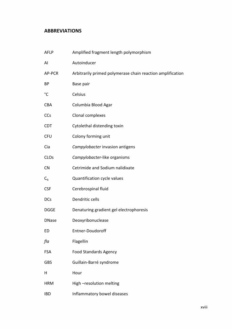
µm long characterised by S-shaped cells and microaerophilic in nature (Figure 1.1)
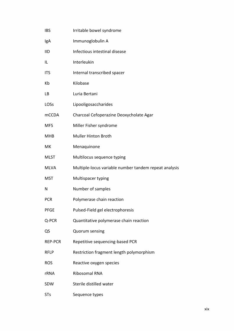
(Halablab et al., 2008; Penner, 1988). They are generally flagellated with either bipolar
flagella or a single polar flagellum enabling the organism to be motile with a
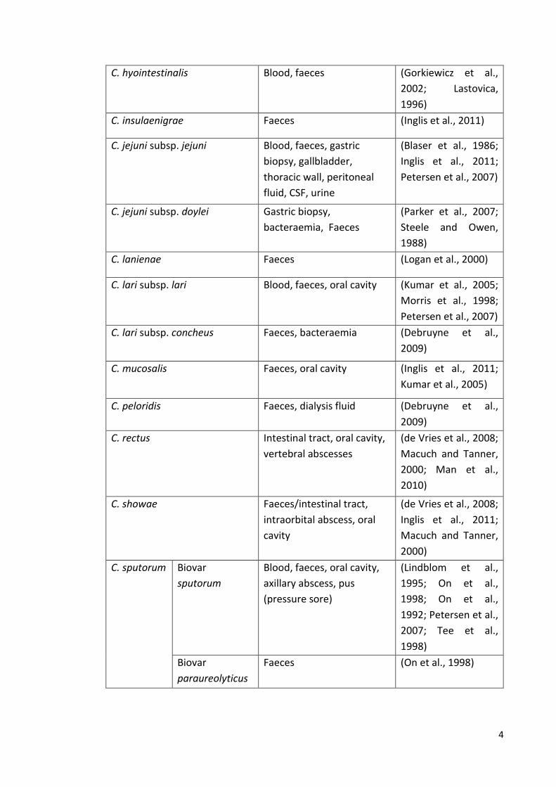
characteristic corkscrew movement (Figure 1.2) (Smibert, 1978). Table 1.1 summarises
currently recognized species and subspecies within the genus Campylobacter. The
thermophilic species C. jejuni and C. coli, which grow best at 42 °C, are the most
significant species in terms of food safety and are the major cause of Campylobacter
infections in developed countries (Gillespie et al., 2002; Ketley, 1995; Man, 2011;
Penner, 1988). As well as being microaerophilic, members of the genus Campylobacter
have other fastidious growth requirements. They are unable to survive atmospheric
oxygen levels and grow optimally in atmospheres containing 5% (v/v) oxygen and their
optimal growth is at 42 °C, but they do not grow at temperatures below 30 °C. They
are catalase positive, and previous studies suggested Campylobacter species are
unable to ferment carbohydrates as a carbon source due to the lack of 6-
phosphofructokinase, but they use amino acids or tricarboxylic acid cycle
intermediates to obtain their energy (Adzitey and Nurul, 2011; Velayudhan and Kelly,
2002). However, one very recent study has revealed that some strains of C. coli (and C.
jejuni subsp. doylei) are glycolytic and can utilise glucose via the pentose phosphate
and Entner-Doudoroff (ED) pathway which is encoded by a genomic island. Hence, it
2
has been suggested that these glycolytic capabilities of some C. coli and C. jejuni subsp.
doylei strains have been acquired via horizontal gene transfer (Vorwerk et al., 2015).
Because of the fastidious growth requirements, these pathogens are unable to
multiply during food processing or storage (Nachamkin et al., 2008; Park, 2002).
Campylobacter infection is usually associated with a low infective dose of 500
organisms (Griffiths and Park, 1990). The bacterium can be found in the intestinal
tract, reproductive organs, and oral cavity of humans and animals (Nachamkin et al.,
2008).
Figure 1.1 C. jejuni observed microscopically, recovered from a stool culture obtained from a one year old patient suffering from Campylobacter infection. Taken from http://www.microbelibrary.org.
Figure 1.2 Image of unipolar and bipolar flagella and corkscrew shape of C. jejuni under electron microscope. Taken from http://www.microbelibrary.org.
3
Table 1.1 Currently recognized species and subspecies within the genus Campylobacter and isolation sites in humans and animals.
Campylobacter spp. Isolation site in humans Reference
C. coli Blood, faeces/intestinal
tract, gallbladder,
retroperitoneal abscess,
cerebrospinal fluid (CSF)
(Blaser et al., 1986;
Inglis et al., 2011;
Petersen et al., 2007)
C. concisus Blood, faeces/intestinal
tract, oral cavity, duodenal
biopsy, brain biopsy
(de Vries et al., 2008;
Istivan et al., 2008;
Macuch and Tanner,
2000; Vandamme et
al., 1989; Zhang et
al., 2009)
C. curvus Faeces, oral cavity, alveolar
abscess.
(Inglis et al., 2011;
Macuch and Tanner,
2000; Petersen et al.,
2007)
C. fetus subsp. fetus Blood, faeces, gastric
aspirate, CSF, vagina, liver,
lungs, skin and spleen of an
aborted foetus.
(Dronda et al., 1998;
Ichiyama et al., 1998;
Petersen et al., 2007;
Sauerwein et al.,
1993; Simor et al.,
1986)
C. fetus subsp. venerealis Blood (Petersen et al.,
2007)
C. fetus subsp. testudinum Blood, Faeces, pleural fluid,
hematoma, bile
(Fitzgerald et al.,
2014; Patrick et al.,
2013)
C. gracilis Faeces, oral cavity, brain
abscess
(de Vries et al., 2008;
Inglis et al., 2011;
Macuch and Tanner,
2000; Man et al.,
2010)
C. hominis Blood, faeces, intestinal
tract
(Inglis et al., 2011;
Linscott et al., 2005;
Zhang et al., 2009)
C. helveticus Faeces (Inglis et al., 2011)
4
C. hyointestinalis Blood, faeces (Gorkiewicz et al.,
2002; Lastovica,
1996)
C. insulaenigrae Faeces (Inglis et al., 2011)
C. jejuni subsp. jejuni Blood, faeces, gastric
biopsy, gallbladder,
thoracic wall, peritoneal
fluid, CSF, urine
(Blaser et al., 1986;
Inglis et al., 2011;
Petersen et al., 2007)
C. jejuni subsp. doylei Gastric biopsy,
bacteraemia, Faeces
(Parker et al., 2007;
Steele and Owen,
1988)
C. lanienae Faeces (Logan et al., 2000)
C. lari subsp. lari Blood, faeces, oral cavity (Kumar et al., 2005;
Morris et al., 1998;
Petersen et al., 2007)
C. lari subsp. concheus Faeces, bacteraemia (Debruyne et al.,
2009)
C. mucosalis Faeces, oral cavity (Inglis et al., 2011;
Kumar et al., 2005)
C. peloridis Faeces, dialysis fluid (Debruyne et al.,
2009)
C. rectus Intestinal tract, oral cavity,
vertebral abscesses
(de Vries et al., 2008;
Macuch and Tanner,
2000; Man et al.,
2010)
C. showae Faeces/intestinal tract,
intraorbital abscess, oral
cavity
(de Vries et al., 2008;
Inglis et al., 2011;
Macuch and Tanner,
2000)
C. sputorum Biovar
sputorum
Blood, faeces, oral cavity,
axillary abscess, pus
(pressure sore)
(Lindblom et al.,
1995; On et al.,
1998; On et al.,
1992; Petersen et al.,
2007; Tee et al.,
1998)
Biovar
paraureolyticus
Faeces (On et al., 1998)
5
C. upsaliensis Blood, faeces, breast
abscesses
(Gaudreau and
Lamothe, 1992;
Lindblom et al.,
1995; Patton et al.,
1989)
C. ureolyticus Faeces, intestinal tract, oral
abscess, gangrenous
lesions of lower limb,
genital infections, genital
abscess, soft tissue
infections, amniotic fluids,
urine
(Burgos-Portugal et
al., 2012; Duerden et
al., 1982; Petersen et
al., 2007)
Campylobacter spp. that have not been found in humans
Campylobacter spp. Source Isolation
site
Reference
C. avium poultry Caeca (Rossi et al., 2009)
C. canadensis Whooping
cranes
Cloaca (Inglis et al., 2007)
C. corcagiensis Captive lion Faeces (Koziel et al., 2014)
C. cuniculorum Rabbits Caeca (Zanoni et al., 2009)
C. hyointestinalis subsp.
hyointestinalis
Cattle, deer,
pigs, hamsters
Faeces,
small and
large
Intestine
(Gebhart et al., 1985;
Hill et al., 1987;
Petersen et al., 2007)
C. hyointestinalis subsp.
lawsonii
Pigs Stomach (On et al., 1995)
C. subantarcticus Wild birds in the
sub-Antarctic
area
Cloaca (Debruyne et al.,
2010a)
C. troglodytis Chimpanzee Faeces (Kaur et al., 2011)
C. volucris Black-headed
gulls
Cloaca (Debruyne et al.,
2010b)
'Campylobacter sp. Dolphin
DP' (provisional)
Dolphin Oral cavity (Goldman et al.,
2011)
'Campylobacter sp. Prairie
Dog' (provisional)
Prairie dogs Intestine,
liver
(Beisele et al., 2011)
Data adapted from (Kaakoush et al., 2015b; Man, 2011; On, 2013)
6
1.2 The history of Campylobacter
Campylobacter (previously named ‘‘Vibrio’’) was first isolated from aborted ovine
foetuses by McFaydean and Stockman in 1913. A few years later the identical Vibrio
was isolated from aborted bovine foetuses by Smith and Taylor in 1919, and from the
blood of three pregnant women by Vincent et al in 1947 (Farrell and Harris, 1992;
Skirrow, 2006; Zilbauer et al., 2008). Afterwards, the name Campylobacter was created
by Sebald and Vernon in 1963, and the organism was recognised as a human pathogen
causing bacterial gastroenteritis in man in the 1970s (Butzler, 2004; Nachamkin et al.,
2008). In April 1982 in the Microbiology Department, Royal Perth Hospital, Western
Australia, a culture of human gastric mucosa harvested a spiral bacterium with some
characteristics of the genus Campylobacter and was named Campylobacter pyloridis
(Itoh et al., 1987; Marshall et al., 1984). Afterwards, the name was changed to C. pylori
(Marshall and Goodwin, 1987). Subsequently it was shown that C. pylori did not fit
within the genus Campylobacter due to differences in their ribosomal RNA sequences,
fatty acids, ultrastructural features, antibiotic susceptibilities and absence of
methylated menaquinone 6 (MK-6) in C. pylori (Goodwin et al., 1989; Goodwin et al.,
1986). Therefore, it was suggested to create a new genus, Helicobacter, and to transfer
C. pylori associated with gastritis to this genus (Goodwin et al., 1989; Griffiths and
Park, 1990). In the 1980s the interest in Campylobacter research was renewed. As a
result, many Campylobacter-like organisms (CLOs) were isolated from different sources
including humans, animals and the environment, and new species were reported
(Benjamin et al., 1983; Fox et al., 1989; Gebhart et al., 1985; Marshall et al., 1984).
Also, during the 1980s, bacterial phylogeny studies at the level of the rRNA cistron
were established, and it was revealed that Campylobacter was heterogeneous. As a
7
consequence, bacterial classification schemes were revised (Nachamkin et al., 2008;
Romaniuk et al., 1987).
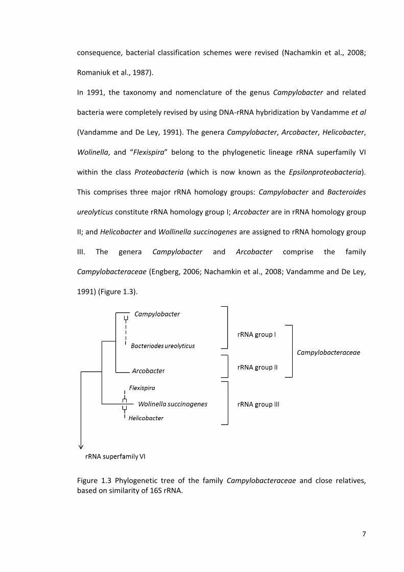
In 1991, the taxonomy and nomenclature of the genus Campylobacter and related
bacteria were completely revised by using DNA-rRNA hybridization by Vandamme et al
(Vandamme and De Ley, 1991). The genera Campylobacter, Arcobacter, Helicobacter,
Wolinella, and “Flexispira” belong to the phylogenetic lineage rRNA superfamily VI
within the class Proteobacteria (which is now known as the Epsilonproteobacteria).
This comprises three major rRNA homology groups: Campylobacter and Bacteroides
ureolyticus constitute rRNA homology group I; Arcobacter are in rRNA homology group
II; and Helicobacter and Wollinella succinogenes are assigned to rRNA homology group
III. The genera Campylobacter and Arcobacter comprise the family
Campylobacteraceae (Engberg, 2006; Nachamkin et al., 2008; Vandamme and De Ley,
1991) (Figure 1.3).
Figure 1.3 Phylogenetic tree of the family Campylobacteraceae and close relatives, based on similarity of 16S rRNA.
8
1.3 Diseases associated with Campylobacter infection
1.3.1 Campylobacteriosis in Humans
C. jejuni is the main bacterial cause of foodborne human intestinal disease worldwide,
followed by Salmonella spp., Shigella spp., and Escherichia coli O157 (Acheson and
Allos, 2001). Infection in humans is frequently associated with consumption of
undercooked poultry meat, non-chlorinated water or unpasteurised milk (Shane,
2000). Campylobacteriosis is an acute diarrhoeal infection caused by members of the
bacterial genus Campylobacter, most commonly C. jejuni. Clinical syndromes are
similar to those of other acute bacterial enteritis including frequency of diarrhoea,
blood in stools, vomiting, and abdominal pain (Coker et al., 2002; Nachamkin et al.,
2008). The symptoms of campylobacteriosis induced by C. coli are clinically similar to
that induced by C. jejuni. Usually, the incubation period for C. jejuni following ingestion
is 24-72 h but longer incubation periods are possible, especially with low infectious
doses. The peak of symptoms can last 24-48 h and may be associated with severe
abdominal pain that mimics appendicitis (Blaser, 1997). Although campylobacteriosis
rarely causes death, it may cause severe disabling sequelae including arthritis,
autoimmune disorders [Guillain-Barré syndrome (GBS) and Miller Fisher syndrome
(MFS)], and inflammatory bowel diseases (IBD), such as Crohn’s Disease (Endtz et al.,
2000; Lamhonwah et al., 2005; Wingstrand et al., 2006). It has also led to bacteraemia
and /or meningitis of a newborn by the infected mother during the delivery process or
shortly after birth, and may cause pancreatitis following C. jejuni enterocolitis
(Peterson, 1994; Smith, 2002). Moreover, it has been suggested that Campylobacter
species may play a role in driving the chronic esophageal inflammation that develops
to cancer, and may be involved in colorectal cancer (Kaakoush et al., 2015a; Kaakoush
9
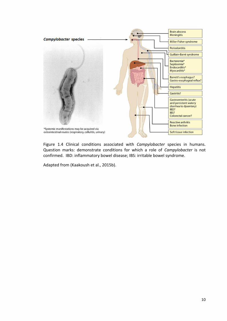
et al., 2015b). It has also been reported that Campylobacter species may cause
extragastrointestinal symptoms such as bacteraemia, lung infections, brain abscesses
and reactive arthritis (Kaakoush et al., 2015b). Figure 1.4 summarises clinical
manifestations in humans associated with Campylobacter species.
In a one year study, it was estimated that C .coli contributed to 18.6% of human
campylobacteriosis, with detection of a winter peak of human C. coli cases which may
be attributable to pork and pork products (Gillespie et al., 2002; Gürtler et al., 2005). A
comparison study of the characteristics of patients infected with C. jejuni, C. coli, and
C. fetus demonstrated that C. coli was more prevalent in slightly older patients [34.6
years (mean age) for C. coli versus 27.5 years (mean age)] for C. jejuni and in those who
had a history of a travel abroad, but less often in hot months than C. jejuni. C. fetus
infection was found more often in much older patients than infection with C. jejuni and
C. coli [68.4 years (mean age) for C. fetus versus 28.6 (mean age)] for C. jejuni and C.
coli and those who had been hospitalised with a systemic disease (Bessède et al.,
2014). Campylobacter infection in humans is likely to be a challenge in terms of global
health for some time and it is considered to be of high public health significance
(Kaakoush et al., 2015b).
10
Figure 1.4 Clinical conditions associated with Campylobacter species in humans. Question marks: demonstrate conditions for which a role of Campylobacter is not confirmed. IBD: inflammatory bowel disease; IBS: irritable bowel syndrome.
Adapted from (Kaakoush et al., 2015b).
11
1.3.2 Campylobacter in poultry
Chickens are a prominent reservoir for thermotolerant Campylobacter, which colonises
primarily the caecum and gastrointestinal tract, and chicken meat contaminated with
Campylobacter is the primary source of human campylobacteriosis (Ghareeb et al.,
2013; Hermans et al., 2011). Also, undercooked paté made with chicken liver has been
considered as an important source of human campylobacteriosis in Europe and in the
United States (Abid et al., 2013; O'Leary et al., 2009; Scott et al., 2015). Differential
gene expression at two different temperatures (42 °C and 37 °C) may enable the
organism to respond differentially to its chicken reservoir niche and the human host
(Stintzi, 2003). Moreover, it has been shown that some C. jejuni isolates grown at 42 °C
were more motile than C. jejuni grown at 37 °C, whereas some grown at 37 °C were
more invasive into human T84 cells. C. fetus subsp. fetus was less able to grow and
invade at 42 °C compared to at 37 °C (Aroori et al., 2013).
Previously, Campylobacter in poultry was considered to be a commensal organism
because of its persistent caecal colonization in large numbers [106 -108 Colony Forming
Unit (CFU)/g] with inefficient intestinal mucosal immune response to the bacterium
(Hermans et al., 2012). However, studies have shown the induction of innate immune
responses in the chicken gut and proliferation of heterophils in the caecum through
recognition of C. jejuni by activated Toll-like receptors 4 (TLR4) and TLR21 in the
chicken gut (de Zoete et al., 2010; Smith et al., 2008). However, post-mortem
examination showed no caecal lesions despite heterophilia which clearly suggests that
the immune responses did not lead to disease (Smith et al., 2008). This could be due to
the differences between the immune systems of poultry and humans (Kaiser et al.,
2005). Extra-intestinal Campylobacter spp. that are found in poultry livers may have
12
been implicated in extra-intestinal disease, such as vibrionic hepatitis of broiler
chickens which is characterised by focal lesions in the liver and that adaptive T cell
response (Jennings et al., 2011). Recently, it has been demonstrated that C. jejuni
infections in some commercial broiler chickens can lead to disease characterised by
pathological changes in the gut mucosa and diarrhoea, which in turn causes
pododermatitis in flocks (Humphrey et al., 2014).
1.3.3 Campylobacter in other animals
It has been shown that both cattle and sheep can also act as a source of
Campylobacter infections in humans. One study has estimated that cattle and sheep
contribute to 35% and 4.3% of human cases of campylobacteriosis, respectively
(Wilson et al., 2008).
The organism can be found in the digestive tract of healthy cattle (Atabay and Corry,
1998; Humphrey et al., 2007). Although cattle are considered an important source of
human campylobacteriosis, unlike chicken, red meat is not massively contaminated at
the point of sale. However, raw milk or possibly incompletely pasteurised milk can
cause sporadic infections, or may cause outbreaks (Anand et al., 2015). It has been
shown that C. jejuni is the most frequent species of Campylobacter detected in cattle
followed by C. coli and other species including C. fetus subsp. fetus, C. hyointestinalis
subsp. hyointestinalis and C. lanienae (Bae et al., 2005; Inglis and Kalischuk, 2003). It
has been demonstrated that C. fetus subsp. venerealis and C. fetus subsp. fetus (mostly
in sheep) are two important pathogens in cattle and sheep affecting the reproductive
ducts and gastrointestinal tract, respectively. The former disease is associated with
13
abortion due to early embryonic death (Campero et al., 2005; Truyers et al., 2014).
Multilocus sequence typing (MLST) analysis was used to characterise C. jejuni isolates
obtained in a 2-year longitudinal study of 15 dairy farms and 4 sheep farms in
Lancashire, UK. The most prevalent clonal complexes (CCs) in cattle were ST-61, ST-21,
ST-403 and ST-45 while in sheep the most prevalent clonal complexes were ST-42, ST-
21, ST-48 and ST-52. The clonal complex ST-45, previously shown to be predominant
during warm months in human cases, was also found to be more prevalent during
warm months in ruminant samples (Grove-White et al., 2011).
A study in New Zealand has estimated the prevalence of C. jejuni in the faeces of wild
birds and pets. The prevalence of C. jejuni in the faecal samples (n=906) was 20% in
ducks, 18% in starlings (n=835), 9% in Canadian goose (n=23), 5% in dogs (n=498) and
7% in cats (n=82), and generally this prevalence was relatively higher during summer
(Mohan, 2015). It has been shown that C. upsaliensis and C. helveticus are the most
frequent species of Campylobacter found in dogs and cats, respectively (Baker et al.,
1999; Moser et al., 2001; Parsons et al., 2010; Rossi et al., 2008). In a Danish
longitudinal study (2-year study) of the prevalence of Campylobacter spp. in young pet
dogs, of the 278 Campylobacter positive samples, 75% were positive for C. upsaliensis,
19.4% for C. jejuni, 2.1% for C. lari, 0.7% for C. coli and 2.8% only identified to
Campylobacter spp. (Hald et al., 2004a). Campylobacter spp. may cause gastroenteritis
associated with diarrhoea in dogs, but can also be found in the intestinal tracts without
showing any gastroenteritis signs (Grøndalen et al., 2008). Using MLST analysis, it was
reported that the majority of sequence types (STs) of C. jejuni isolates found in various
populations of dogs (kennels and veterinary practices) were the same as those found
in isolates from humans, including ST-45 and ST-21 (Parsons et al., 2009). Therefore,
14
Campylobacter carriage in pet dogs is considered a potential source for human
campylobacteriosis (Procter et al., 2014; Rossi et al., 2008; Tenkate and Stafford,
2001). In a parallel study carried out in Liverpool, MLST analysis of isolates recovered
from Cheshire bank voles revealed that all possess a novel, unique ST (ST-3704)
(Williams et al., 2010). ST-3704 strains have now been isolated from bank voles
inhabiting six different sites across Cheshire and Wirral. Furthermore, persistent ST-
3704 strain infections have occurred in naturally-infected, captured bank voles and
even their (captive-reared) F1 off-spring (Nicola Williams, unpublished data).
1.4 Pathogenesis
The mechanisms by which Campylobacter causes diseases are poorly understood
(Dasti et al., 2010; Young et al., 2007). Following ingestion of a low infective dose,
around 500 organisms, Campylobacter starts to adhere and colonise the intestinal
mucosa aided by the flagellum, which also secretes invasive antigens named
Campylobacter invasion antigens (Cia) (Dasti et al., 2010; Ketley, 1995; Nachamkin et
al., 2008). It has been shown that the ability of the nonmotile C. jejuni (ΔmotAB)
mutant, possessing paralysed flagella, to invade Caco-2 cells was reduced. This mutant
C. jejuni failed to colonize the caeca of chickens (Mertins et al., 2012). Campylobacter
species are able to adhere to intestinal epithelial cells by secretion of the outer
membrane protein CadF, which binds to fibronectin (Konkel et al., 1997). Unlike most
bacteria, C. jejuni does not possess many classical virulence factors, but it is the only
pathogen that carries out N-linked glycosylation of more than 30 proteins linked with
adherence, colonization and invasion (Dasti et al., 2010). The pathogenicity of
15
Campylobacter may also be influenced by other microbes. A recent study has shown
the role of the gut microbiota composition of humans in conferring resistance to
enteropathogen colonization and that the colonization can change the composition of
the human gut microbiota. For example, Campylobacter-positive abattoir workers
were found to have significantly higher abundance of Bacteroides, Escherichia species,
Phascolarctobacterium and Streptococcus in the gut microbiota than Campylobacter-
negative workers, who had high proportions of Clostridiales, unclassified
Lachnospiraceae and Anaerovorax (Dicksved et al., 2014). More recently, it has been
shown that colonisation of C. jejuni in poultry leads to a change within the composition
of the intestinal microbiota, associated with a higher water content of faecal samples,
which indicates the beginning phase of diarrhoea (Sofka et al., 2015). The most
important immunoglobulin secreted during infection with Campylobacter is IgA which
can cross the gut wall, attenuate the organism and provide short term of immunity
against the organism, while other immunoglobulins prevent bacteraemia (Wallis,
1994). Even though cell invasion may be responsible for the occurrence of clinical
symptoms of campylobacteriosis, bacterial toxins might also aggravate the
pathogenesis process (Ketley, 1995). Campylobacter produces a cytolethal distending
toxin (CDT), which might play a role in disease pathogenesis; the toxin consists of three
subunits CdtA, CdtB and CdtC (Ceelen et al., 2006; Johnson and Lior, 1988). The
subunit CdtB acts as a Deoxyribonuclease (DNase) 1-like protein which causes cell cycle
arrest, cytoplasm distension, chromatin disruption and cell death, whereas both
subunits CdtA and CdtC are essential to deliver CdtB into the host cell (Lara-Tejero and
Galán, 2000; Lara-Tejero and Galán, 2001). Hence, exposure of intestinal epithelial cells
16
to CDT triggers release of interleukin-8 (IL-8) which induces acute inflammatory
response of the intestine (Hickey et al., 1999).
It has been shown that the autotransporter proteins comprise a group of outer
membrane proteins of most Gram-negative bacteria that have unique structural
properties and are often associated with virulence functions such as adhesion (Wells et
al., 2007). For example, the autotransporter protein CapA of C. jejuni strain NCTC11168
may play an important role in adhesion to human epithelial cells and colonization in
chickens. This was shown using the capA mutant of C. jejuni strain NCTC11168
demonstrated a significant decrease in its ability to invade Caco-2 cells and failed to
colonize chicken gut (Ashgar et al., 2007).
Bacteria have evolved enzymes involved in the membrane disruption processes of host
cell, in which phospholipids and proteins are the major chemical components, by
hydrolysing these chemicals and haemolytic activity during invasion (Songer, 1997). For
example, studies have indicated the role for the phospholipase A in the lysis of
erythrocytes by Campylobacter species (Grant et al., 1997; Istivan et al., 2004).
Moreover, it has been demonstrated that the C. jejuni pldA, which encodes
phospholipase, mutant was shown reduced the ability in caecum colonization of
chicken (Ziprin et al., 2001). It has been shown that the Peb protein is one of the most
abundant periplasmic immunogenic proteins in C. jejuni which may play a key role in
host-cell adhesion of eukaryotic cell membranes (Kervella et al., 1993) and in the
utilization of amino acids, such as aspartate and glutamate, that are an essential
carbon sources for the pathogen (Del Rocio Leon‐Kempis et al., 2006).
17
1.5 Incidence and Epidemiology Although the epidemiology of campylobacteriosis is poorly understood, poultry is
considered the number one source of Campylobacter infections in humans. At the
beginning, a few colonized broilers at the age of >2 weeks will infect the entire flock
until the end of rearing (Hermans et al., 2012). The Food Standards Agency (FSA) has
reported that the presence of Campylobacter in fresh chicken at retail in the UK is 65%,
and its presence in chilled chicken is higher than in frozen chicken (at 48% and 14%,
respectively). Additionally, a Scottish study estimated that poultry meat can contribute
to up to 60-80% of human campylobacteriosis cases (FSA, 2009; Goddard et al., 2014).
In recent years in many European countries, campylobacteriosis has been the most
frequently reported zoonosis, followed by other infectious diseases such as
salmonellosis (Eurosurveillance Editorial, 2012). In the United States in 2009,
campylobacteriosis was the second most frequent cause of laboratory-confirmed
cases of bacterial enteritis after salmonellosis, and the incidence was highest among
the <4 years age group (Centers for Disease and Prevention, 2010). In population-
based studies of infectious intestinal disease (IID) in the UK over 15 years, changes in
incidence and etiology of pathogens associated with IID were examined in two studies
(IID1 and IID2) covering different time periods (1993-1996 and 2008-2009,
respectively). Campylobacter spp. was the most frequently identified bacterial cause of
infectious intestinal disease in the IID1 and IID2 studies (Tam et al., 2012). The
prevalence of salmonellosis cases reduced between IID1 and IID2, which reflects the
success of Salmonella control strategy in both broilers and laying chickens in the UK
(Tam et al., 2012). In developed countries, the incidence of campylobacteriosis is
highest among infancy and age groups 15-44 years, with most infections occurring via
18
handling and consumption of chicken meat, whereas in developing countries the
highest incidence of the disease is restricted to young children, and most infections are
acquired by exposure to poorly treated water and farm animals (Butzler, 2004). It was
shown that Campylobacter infections had risen to 1 million cases in England and Wales
between 1989 and 2011, with the highest increase among age groups >50 years.
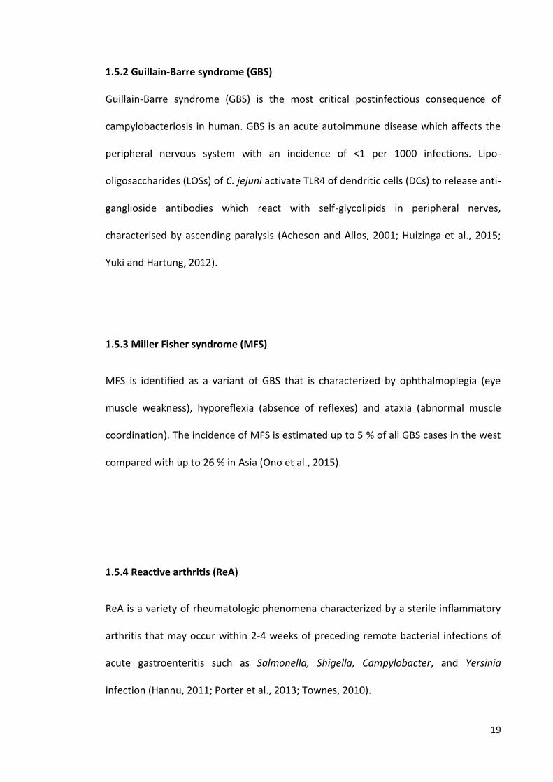
(Nichols et al., 2012). Figure 1.5 shows the number of laboratory confirmed cases of
Campylobacter infection in humans in the UK between 2000-2012. In general,
salmonellosis continues to fall in the UK. Thus, the key aim of the FSA strategic plan
2010-2015 was to reduce campylobacteriosis in humans through decreasing
Campylobacter levels in chicken (https://www.gov.uk /government /publications
/zoonoses-report-uk-2012).
1.5.1 Seasonality
Cases of campylobacteriosis show distinct seasonality. In a long term study in England
and Wales between 1989 and 2011, it was shown that constant seasonal increase in
campylobacteriosis cases was observed between early May and early June across
years, ages and regions, and was greater in children in rural areas. Seasonality could be
due to a number of causes. It has been suggested that one cause may be attributed to
flies depositing the organism onto food (Nichols, 2005; Nichols et al., 2012;
Wingstrand et al., 2006).
19
1.5.2 Guillain-Barre syndrome (GBS)
Guillain-Barre syndrome (GBS) is the most critical postinfectious consequence of
campylobacteriosis in human. GBS is an acute autoimmune disease which affects the
peripheral nervous system with an incidence of <1 per 1000 infections. Lipo-
oligosaccharides (LOSs) of C. jejuni activate TLR4 of dendritic cells (DCs) to release anti-
ganglioside antibodies which react with self-glycolipids in peripheral nerves,
characterised by ascending paralysis (Acheson and Allos, 2001; Huizinga et al., 2015;
Yuki and Hartung, 2012).
1.5.3 Miller Fisher syndrome (MFS)
MFS is identified as a variant of GBS that is characterized by ophthalmoplegia (eye
muscle weakness), hyporeflexia (absence of reflexes) and ataxia (abnormal muscle
coordination). The incidence of MFS is estimated up to 5 % of all GBS cases in the west
compared with up to 26 % in Asia (Ono et al., 2015).
1.5.4 Reactive arthritis (ReA)
ReA is a variety of rheumatologic phenomena characterized by a sterile inflammatory
arthritis that may occur within 2-4 weeks of preceding remote bacterial infections of
acute gastroenteritis such as Salmonella, Shigella, Campylobacter, and Yersinia
infection (Hannu, 2011; Porter et al., 2013; Townes, 2010).
20
1.5.5 Inflammatory bowel disease (IBD)
IBD is considered as an emergent worldwide disease characterized by chronic
inflammation of the gastrointestinal tract. The disease has two clinical subtypes, (1)
Crohn’s disease (CD) that can affect any part of the intestine, and (2) ulcerative colitis
(UC) which is restricted to the colon and rectum. Although the exact causative agent of
IBD is not known, it has been suggested that mucosa-associated bacteria, such as
Campylobacter species, may play a key role in the development of IBD. The mechanism
behind this could be immunopathogenetic, such as autoantibody generation (Castaño-
Rodríguez et al., 2015; Kaakoush et al., 2014; Lamhonwah et al., 2005).
Figure 1.5 Number of laboratory confirmed cases of infectious intestinal diseases (campylobacteriosis and salmonellosis) in the UK between 2000-2012. Data from Food Standard Agency https://www.food.gov.uk/science/microbiology/fds/58736.
0
10,000
20,000
30,000
40,000
50,000
60,000
70,000
80,000
Cas
es
Year
Campylobacterspp.
Salmonallaspp.
21
1.6 Treatment of campylobacteriosis
Human Campylobacter gastroenteritis is commonly self-limiting, and antibacterial
treatment is not generally recommended in the United Kingdom, but ciprofloxacin
(fluoroquinolone) and a macrolide are the drugs of choice in severe or prolonged
cases. There has been a steady increase in the proportion of clinical ciprofloxacin-
resistant Campylobacter isolates in the UK from 3% in 1991 to 37.5% in 2008 (Cody et
al., 2010). Furthermore, some studies have shown a link between certain C. jejuni
genotypes and resistance to ciprofloxacin, and correlation between chicken
consumption and acquisition of resistance to ciprofloxacin (Habib et al., 2009; Kinana
et al., 2006). Recently, it has been reported that a horizontally transferrable gene
erm(B), encoding rRNA methylase, can confer macrolide resistance in C. coli (Wang et
al., 2014).
1.7 Strain typing methods
Bacterial strain typing is the establishment of the relatedness of a group of bacterial
isolates. It is a method for source tracing of bacterial strains that cause infections and
for studying the epidemiology of diseases (Li et al., 2009; Tenover et al., 1995;
Wassenaar and Newell, 2000). In general, the diversity within a bacterial species is
caused by a combination of genetic events which include: mutation, horizontal gene
transfer, gene loss, gene duplication and recombination (Fraser-Liggett, 2005).
Typing methods can be based on either phenotyping or genotyping. Phenotyping
methods include morphology of colonies on culture media, serology, growth
characteristics, biochemical tests, pathogenicity and antibiotic susceptibility. Bacterial
22
phenotypes are often not sufficiently variable to distinguish closely related strains.
Because of their greater resolution, genotyping methods or DNA fingerprinting, based
on the genetic structure of bacterial strains, have largely superseded phenotypic
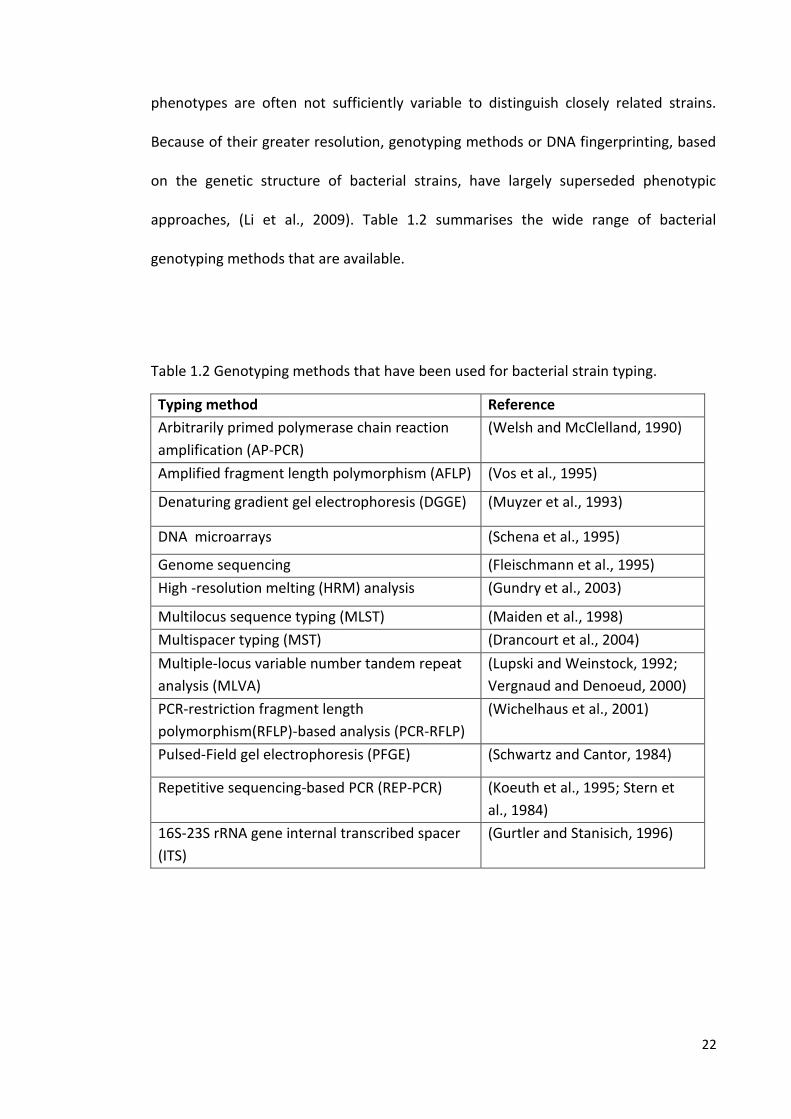
approaches, (Li et al., 2009). Table 1.2 summarises the wide range of bacterial
genotyping methods that are available.
Table 1.2 Genotyping methods that have been used for bacterial strain typing.
Typing method Reference
Arbitrarily primed polymerase chain reaction
amplification (AP-PCR)
(Welsh and McClelland, 1990)
Amplified fragment length polymorphism (AFLP) (Vos et al., 1995)
Denaturing gradient gel electrophoresis (DGGE) (Muyzer et al., 1993)
DNA microarrays (Schena et al., 1995)
Genome sequencing (Fleischmann et al., 1995)
High -resolution melting (HRM) analysis (Gundry et al., 2003)
Multilocus sequence typing (MLST) (Maiden et al., 1998)
Multispacer typing (MST) (Drancourt et al., 2004)
Multiple-locus variable number tandem repeat
analysis (MLVA)
(Lupski and Weinstock, 1992;
Vergnaud and Denoeud, 2000)
PCR-restriction fragment length
polymorphism(RFLP)-based analysis (PCR-RFLP)
(Wichelhaus et al., 2001)
Pulsed-Field gel electrophoresis (PFGE) (Schwartz and Cantor, 1984)
Repetitive sequencing-based PCR (REP-PCR) (Koeuth et al., 1995; Stern et
al., 1984)
16S-23S rRNA gene internal transcribed spacer
(ITS)
(Gurtler and Stanisich, 1996)
23
1.7.1 Serotyping: is the most commonly used phenotypic method to characterise
Campylobacter strains despite the lack of discriminatory power. This method is based
on the differences of surface structures (antigens) of bacteria which can be detected
by antibodies and antisera. Using this approach strains can be differentiated by their
different surface structures (Wiedmann, 2002).
Penner and Hennessy (1980) showed that bacteria, later classified as C. fetus subsp.
jejuni could be serotyped on the basis of their thermostable antigens (penner
scheme).Briefly, Bacterial antigens, C. fetus subsp. jejuni strains, were extracted from
cell suspensions by heating at 100 °C in saline or by exposure to EDTA. The extracted
thermostable antigens were diluted (1:10 in phosphate buffer saline) and incubated at
37 °C for 1 h with an equal amount of PBS + 1% sheep erythrocytes. The sensitized
erythrocytes (adherence of antigens to the surface of erythrocytes) were washed and
resuspended in phosphate buffer saline, and titrated against diluted rabbit antisera,
for agglutination of sensitized erythrocytes with the presence of antibodies specific for
lipopolysaccharides (LPSs) in antisera (Penner and Hennessy, 1980). The variability of
the somatic “O” antigen of outer membrane of the Campylobacter LPS is believed to
contribute to the antigenic basis of the Penner serotyping system (Shi et al., 2002). The
scheme was used to identify the distinct antigenic specificities between C. jejuni and C.
coli (Penner et al., 1983). The scheme has long been used as laboratory based
epidemiologic method by researchers worldwide to study the transmission of
Campylobacter infection from food, animal, and water to humans (Woodward and
Rodgers, 2002). With the scheme, the strains are differentiated on the basis of heat-
stable antigens presence on the bacterial surface (Moran and Penner, 1999).
24
1.7.2 Macro-restriction PFGE: Is a useful typing technique for many bacteria that can
resolve large DNA molecules (20-200 kb). The organisms are embedded in agarose,
lysed and the chromosomal DNA is digested with restrictions enzymes that cleave the
bacterial DNA infrequently. The DNA fragments in the agarose (blocks) are loaded onto
agarose gels for electrophoresis. The DNA fragments are resolved into discrete bands
by electrophoresis in the gel where the direction of the electric field is changed or
pulsed to allow resolution of very large DNA fragments (Tenover et al., 1995;
Wassenaar and Newell, 2000). With Campylobacter, the technique was originally used
for C. jejuni, and was later applied to C. coli, C. hyointestinalis, C.fetus and C.
upsaliensis (Bourke et al., 1996; Fujita et al., 1995; Salama et al., 1992; Yan et al.,
1991).
1.7.3 Flagellin typing (fla typing): The proteins of Campylobacter flagella are encoded
by two highly homologous genes; a major flagellin gene (flaA) and a minor flagellin
gene (flaB) (Guerry, 2007). Because both flaA and flaB genes have variable central
regions and highly conserved flanking regions, they can be analysed using RFLP analysis
of amplified PCR products. In fla typing, the flaA gene is amplified by PCR followed by
digestion of the amplified DNA with a specific restriction enzyme to produce PCR
product fragments which are separated according to their lengths by agarose gel
electrophoresis (Eberle and Kiess, 2012; Fitzgerald et al., 2001). Although the
technique has high discriminatory power, it is not the favoured method used in
epidemiology studies (Eberle and Kiess, 2012). A database for flaA genotypes isolates is
25
available for scientists and publically accessible to share information on flaA gene
typing (http://pubmlst.org/campylobacter/). However, in recent years MLST has
superseded this approach and has become the gold standard for Campylobacter
typing.
1.7.4 MLST
MLST was established in 1998. It is a molecular biological method for the
characterization of bacterial isolates using the DNA sequences of internal fragments of
housekeeping genes (commonly seven) located at various parts of the chromosome.
MLST data are freely accessible over the World-Wide Web (www.mlst.net) and can be
implemented for a wide range of bacterial species. Due to its ability to determine
variation which is building up gradually within a population, MLST can be used to trace
lineages in bacterial populations, enabling detailed studies of the epidemiology,
evolution and pathogenicity of bacteria (Chan et al., 2001; Maiden, 2006; Maiden et
al., 1998).
In the Campylobacter MLST system, the sequences of seven housekeeping genes (allele
fragments), which are present in all isolates and are stabilized for conservation of
metabolic function, are determined after amplification of fragments of 400-550 bp per
gene from genomic DNA. For each individual gene, new allele fragment-sequences are
assigned an allele number, with the first reported sequence being designated “1”.
These numbers are obtained for each of the seven loci and are stored electronically in
the ‘‘profile database’’ using web-sites such as mlst.net. These allele numbers are
joined into sequence types (STs), which are also assigned an arbitrary number. STs are
26
grouped into ‘‘clonal complexes’’ (which can differ in allele number for up to two of
the seven loci) and stored in the profiles database (Maiden, 2006; Nachamkin et al.,
2008). This makes MLST a highly portable genotyping method (i.e., easily comparable
data between laboratories). In addition, its use can decrease the risk of transporting
live bacteria because killed bacterial suspensions, genomic DNA, or clinical material
can be used to carry out nucleotide sequence determination from PCR products
(Dingle et al., 2001). Table 1.3 summarises a number of studies where MLST has been
applied to Campylobacter in different countries. Using MLST data, it has been found
that Campylobacter is genetically highly diverse, with a weak clonal population
structure, and has strong intra- and interspecies lateral genetic exchange (Dingle et al.,
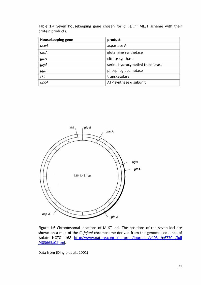
2001). Table 1.4 shows the seven housekeeping loci that were chosen for the C. jejuni
MLST scheme with their protein products. These genes were chosen from multiple
chromosomal locations encoding proteins responsible for intermediary metabolism
and were suitable for primer design. The minimum distance between loci was 70 kb
(Figure 1.6), which suggests that the coinheritance of any of the loci in a recombination
event was unlikely. The MLST scheme was initially developed using 194 C. jejuni
isolates from different sources including humans, animals and the environment. There
were 155 STs identified which were assigned to 62 clonal complexes. The data
indicated that there was horizontal gene exchange, including import of alleles from
other Campylobacter species including C. coli (Dingle et al., 2001). Using this previously
described MLST system for C. jejuni, a MLST scheme was established for C. coli (Dingle
et al., 2005). There was an identity of approximately 86.5% between the two species,
C. jejuni and C. coli, at the nucleotide sequence level within the MLST loci, with genetic
exchange of the housekeeping genes only observed at a very low rate. In contrast, it
27
was shown that the flaA gene repeatedly exchanged between the two species (Dingle
et al., 2005). Using MLST data, it has been indicated that Campylobacter isolates from
bovine, ovine, poultry, pets, pigs and the environment are considered potential
reservoirs of human campylobacteriosis. Associations between source and certain
clonal complex have been demonstrated, suggesting niche adaptation of isolates
(Hepworth et al., 2011; Manning et al., 2003; Williams et al., 2010). Figures 1.6
summarise the most common clonal complexes and sequence types, and their
prevalence.

28
Table 1.3 Studies on Campylobacter spp. MLST clonal complexes and sequence types in different countries. The table shows only the most common and more predominant MLST CCs than other CCs.
Country Year Source Number of isolates
Most common and more predominant CC
Reference
C. jejuni C. coli
Denmark 2002-2003
Humans
122 Gastro enteritis (96) Reactive arthritis (18) GBS (8)
- ST-21 (4% reactive arthritis) ST-45 (4% reactive arthritis) ST-22 (4% GBS)
(Nielsen et al., 2010)
Finland 2012 Humans
95 - ST-45 ST-283 ST-677
(Kovanen et al., 2014)
Finland 1998-2007
Humans (blood)
73 - ST-677 ST-45 ST-21
(Feodoroff et al., 2013)
Italy 2009 Humans 11 7
ST-257 ST-206 ST-443 ST-21 ST-353 ST-828/C.coli
(Piccirillo et al., 2014)
Chicken 23 16
ST-21 ST-446 ST-443 ST-828/C.coli ST-1150/C.coli
Scotland
2005- 2006
Pigs poultry Sheep Cattle gulls
443 (C. jejuni +C.coli)
ST-828 /C. coli (% of total dataset) 5-10 2.5-5 2.5-5 0.62-1.25 0.00-0.62
(Ogden et al., 2009)
29
Pigeons gulls
ST-179 (% of total dataset) 5-10 0.00-0.62
Gulls Ducks pigeons
ST-1275 (% of total dataset) 5-10 062-1.25 0.00-0.62
Cattle Sheep Poultry birds
ST-61 (% of total dataset) 2.5-5% 1.25-2.5 1.25-2.5 0.00-0.62
Cattle Sheep Bird Ducks Poultry Pigeons
ST-21 (% of total dataset) 2.5-5% 1.25-2.5 0.62-1.25 0.62-1.25 0.62-1.25 0.00-0.62
Ducks Birds Gees Pigeons poultry Cattle Sheep gulls
ST-45 (% of total dataset) 2.5-5.00 1.25-2.5 1.25-2.5 1.25-2.5 1.25-2.5 0.00-0.62 0.00-0.62 0.00-0.62
Cattle Sheep Gulls
ST-42 (% of total dataset) 1.25-2.5 0.62-1.25 0.62-1.25
Cattle Sheep birds
ST-48 (% of total dataset) 0.62-1.25 0.62-1.25 0.00-0.62
30
South Korea
2012 Duck
46 9
ST-21 ST-45 ST-828 /C.coli
(Wei et al., 2014)
UK
2006-2008
Cattle
849 - ST-61 ST-21 ST-403 ST-45
(Grove-White et al., 2011)
Sheep 154 ST-42 ST-2114 ST-4814 ST-52
UK 2005-2008
Dogs 33 - ST-45 (11) Rescue dog (6) Hunt dog ( 4) house hold dog (1) ST-21 (4) Hunt dog ST-508 (4) Boarding dog (2) hunt dog (1) vet visiting dog (1) ST-403 (3) Hunt dog (2) household dog (1)
(Parsons et al., 2009)
UK
2004-2006
Broilers flocks
226 8.5 %
ST-45 ST-21 ST-574 ST-443 ST-828(C.coli)
(Jorgensen et al., 2011)
UK 2003- 2004
Humans
326 30
ST-21 ST-45 ST-257 C. coli
(Sopwith et al., 2006)
USA
2011-2012
Turkey
19 80
ST-353 ST-828 ST-828 /C.coli
(Kashoma et al., 2014)
31
Table 1.4 Seven housekeeping gene chosen for C. jejuni MLST scheme with their protein products.
Housekeeping gene product
aspA aspartase A
glnA glutamine synthetase
gltA citrate synthase
glyA serine hydroxymethyl transferase
pgm phosphoglucomutase
tkt transketolase
uncA ATP synthase α subunit
Figure 1.6 Chromosomal locations of MLST loci. The positions of the seven loci are shown on a map of the C. jejuni chromosome derived from the genome sequence of isolate NCTC11168 http://www.nature.com /nature /journal /v403 /n6770 /full /403665a0.html. Data from (Dingle et al., 2001)
30
1.7.4.1 Common C. jejuni clonal complexes
Clonal complex ST-21 is the most common of the clonal complexes, comprising
approximately 22% of all of the yielded isolates (Figures 1.6A and 1.6B-last accessed
July 2015), and has a total of 667 different STs in it (Figure 1.6C-last accessed July
2015). It has been detected in multiple sources, including food-producing animals and
humans, and is therefore considered to be a niche generalist (Dingle et al., 2001;
http://pubmlst.org/campylobacter).
Contrary to the isolates of clonal complex ST-21, distributed among a wide range of
hosts, it has been shown that certain clonal complexes are associated with particular
farm animals. For example, one study, most of isolates were from the United
Kingdom and the rest was from Northern Europe, reported that isolates from clonal
complex ST-45 were the most prevalent in poultry, isolates from both clonal
complexes ST-48 and ST-61 were the most prevalent in cattle, isolates from clonal
complex ST-42 were overrepresented in sheep, and isolates from clonal complex ST-
403 were predominant in pigs (Manning et al., 2003). Despite being poor survivors
outside the body of their hosts, some C. jejuni have adapted to survive in
environmental niches (Sopwith et al., 2008). For example, in one UK study using MLST
analysis, the C. jejuni community isolated from faeces of livestock and wild animals,
environmental water and soil samples in dairy cattle farmland in the UK was
analysed. The clonal complex ST-45 isolates were overrepresented in wildlife faeces
and environmental water, whereas the clonal complex ST-61 isolates were
overrepresented in cattle faeces (French et al., 2005). A longitudinal study of C. jejuni
in dairy cattle farms using MLST demonstrated predominance of three clonal
complexes ST-61 (24.2%), ST-21 (23.6%) and ST-42 (20.5%) among the cattle isolates.
31
This indicates that there is an association between cattle and some genotypes of C.
jejuni (Kwan et al., 2008b). A cross-sectional study of molecular epidemiology of C.
jejuni in a dairy farm demonstrated that C. jejuni clonal complexes ST-21, ST-45, and
ST-61, which have been commonly associated with human Campylobacter
gastroenteritis disease, comprised the majority of genotypes isolated, again
demonstrating potential host-associations (Kwan et al., 2008a). It has been suggested
that the contaminated faeces of wild birds may contribute to the distribution of one
of the most common genotypes (ST-45) in river water (Carter et al., 2009). It has been
shown that livestock including poultry, cattle and sheep are principle sources of
human campylobacteriosis. Using MLST studies in England and Scotland
supplemented with statistical genetic approaches, it was revealed that 97% (1195)
cases of sporadic campylobacteriosis in England could be attributed to the meat of
farm animals and poultry, and that 76% cases of Campylobacter infections in humans
in Scotland may be due to consumption of contaminated poultry meat. This indicates
that applying strict biosecurity on farms could greatly reduce campylobacteriosis in
humans (Sheppard et al., 2009; Wilson et al., 2008).
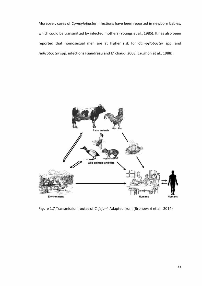
1.8 Campylobacter spp. in the environment
Campylobacter species are continually shed from all reservoirs into the environment
(soil and water), which in turns plays a key role in transmission, either directly to
humans or indirectly by livestock (Figure 1.7). Using MLST analyses, it has become
clear that isolates of some clonal complexes, such as ST-45, are more frequently
found in environmental water (surface water) and are associated with the late spring
32
incidence of human campylobacteriosis in north-western England, which indicates a
link between the prevalence of this clonal complex in the environment and human
campylobacteriosis (Bronowski et al., 2014; Ogden et al., 2009; Sopwith et al., 2008).
Transmission of the organism from these reservoirs to humans is via different routes
(Figure 1.7). In general, the transmission is associated with the consumption of
contaminated chicken meat during carcass processing at slaughter, and cross
contamination in the food preparation environment (Humphrey et al., 2007). It has
also been shown that the organism can be transmitted to humans by eating
contaminated raw vegetables (Carvalho et al., 2013). More recently, it has been
suggested that Campylobacter survival is enhanced in water when there is interaction
(gene product involvement) with the free-living protozoa Acanthamoeba (Vieira et al.,
2015). It has also been shown that flies can contribute to Campylobacter transmission
to humans and to chicken flocks during the summer (Hald et al., 2004b; Nichols,
2005). The possibility of Campylobacter contamination in the environment has been
demonstrated in specific climatic conditions, which may play a role in Campylobacter
outbreaks. For example, in June 2007 in British Columbia/Canada, one of the largest
campylobacteriosis outbreaks was reported in wet muddy conditions. Among 537
bike racers (included in the study), 225 racers (42%) suffered diarrhoeal illness due to
mud ingestion (Stuart et al., 2010). Moreover, it has been suggested that
transmission can occur person-to-person (Domingues et al., 2012), despite the
relatively lower rate of documentation compared to the total number of
campylobacteriosis cases (Musher and Musher, 2004). For example, it has been
suggested that the outbreaks of Campylobacter infections in families could be
transmitted by faecal-oral route, via infected faeces of infants (Blaser et al., 1981).
33
Moreover, cases of Campylobacter infections have been reported in newborn babies,
which could be transmitted by infected mothers (Youngs et al., 1985). It has also been
reported that homosexual men are at higher risk for Campylobacter spp. and
Helicobacter spp. infections (Gaudreau and Michaud, 2003; Laughon et al., 1988).
Figure 1.7 Transmission routes of C. jejuni. Adapted from (Bronowski et al., 2014)
34
1.9 Aims of this study
The survival of C. jejuni strains in the environment is likely to play a key role in the
transmission of this pathogen, either directly or indirectly, to humans. The overall
objective of this study was to increase our understanding of Campylobacter survival in
natural environments, variations between Campylobacter genotypes, the genes that
are important in survival, and interactions between Campylobacter and other species
that might enhance survival. The specific aims were:
• To compare the survival of a diverse panel of C. jejuni strains from various
sources and representing different MLSTs in sterilized water (and natural
water) at selected time points [time 0, day 1 (at 25 °C) and day 3 (at 4 °C)] by
measuring the ability to form colony forming units.
• To investigate the viable but non culturable (VBNC) state in C. jejuni strains
(M1, 1336 and 414) in sterile distilled water (starvation conditions) at selected
time points by detecting and counting viable cells of C. jejuni in sterile distilled
water.
• To study gene expression variations of previously reported stress response
genes between different C. jejuni strains (M1, 1336 and 414) during survival in
water at selected time points by using both end-point PCR and real-time
quantitative PCR (Q-PCR) assays for selected stress response genes.
• To investigate the effects of supernatants from Pseudomonas spp. cultures on
the growth of Campylobacter in vitro and determine the co-existence of
Campylobacter spp. with fluorescent Pseudomonas spp. in natural water by
using environmental PCR assays.
35
C h a p t e r 2
MATERIALS AND METHODS
2.1 Bacterial strains used in this study
2.1.1 Campylobacter strains
The Campylobacter jejuni strains (n=27) used in this study were obtained from the
University of Liverpool, Leahurst culture collection assembled and subjected to MLST
during previous projects (French et al., 2005; Wilson et al., 2008; Wilson et al., 2009)
(Table 2.1). The strains were chosen arbitrarily but to represent various sources and
different clonal complexes. They comprised seven isolates from human sources, five
from water sources, two from soil, six from cattle, four from chicken, one from a wild
bird and one from bank-vole, as well C. jejuni isolate (ST-42). The strain panel
included the widely studied strain M1 (Friis et al., 2010) and representatives of
different clonal complexes (Table 2.1). All strains were stored at -80 °C on
cryopreservation beads (Technical Service Consultants Ltd) according to
manufacturer’s instructions. When required, a bead was taken from the partially
thawed stock to inoculate a blood agar plate.
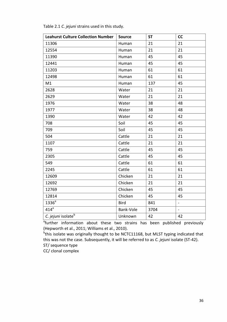
36
Table 2.1 C. jejuni strains used in this study.
Leahurst Culture Collection Number Source ST CC
11306 Human 21 21
12554 Human 21 21
11390 Human 45 45
12441 Human 45 45
11203 Human 61 61
12498 Human 61 61
M1 Human 137 45
2628 Water 21 21
2629 Water 21 21
1976 Water 38 48
1977 Water 38 48
1390 Water 42 42
708 Soil 45 45
709 Soil 45 45
504 Cattle 21 21
1107 Cattle 21 21
759 Cattle 45 45
2305 Cattle 45 45
549 Cattle 61 61
2245 Cattle 61 61
12609 Chicken 21 21
12692 Chicken 21 21
12769 Chicken 45 45
12814 Chicken 45 45
1336a Bird 841 -
414a Bank-Vole 3704 -
C. jejuni isolateb Unknown 42 42 afurther information about these two strains has been published previously (Hepworth et al., 2011; Williams et al., 2010). bthis isolate was originally thought to be NCTC11168, but MLST typing indicated that this was not the case. Subsequently, it will be referred to as C. jejuni isolate (ST-42). ST/ sequence type CC/ clonal complex
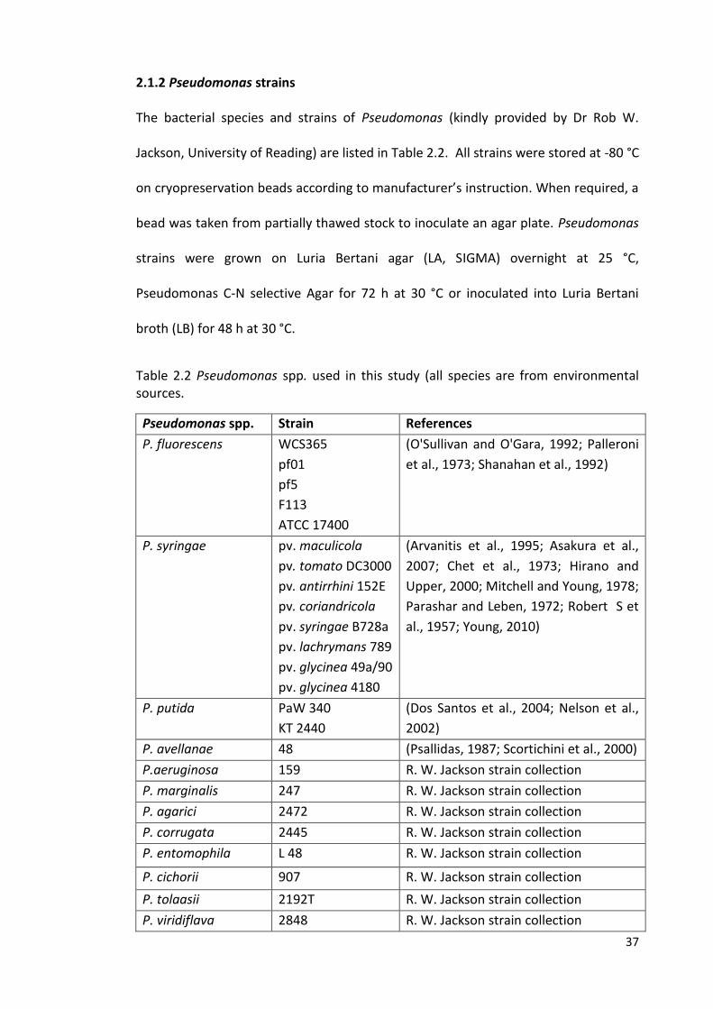
37
2.1.2 Pseudomonas strains
The bacterial species and strains of Pseudomonas (kindly provided by Dr Rob W.
Jackson, University of Reading) are listed in Table 2.2. All strains were stored at -80 °C
on cryopreservation beads according to manufacturer’s instruction. When required, a
bead was taken from partially thawed stock to inoculate an agar plate. Pseudomonas
strains were grown on Luria Bertani agar (LA, SIGMA) overnight at 25 °C,
Pseudomonas C-N selective Agar for 72 h at 30 °C or inoculated into Luria Bertani
broth (LB) for 48 h at 30 °C.
Table 2.2 Pseudomonas spp. used in this study (all species are from environmental sources.
Pseudomonas spp. Strain References
P. fluorescens WCS365
pf01
pf5
F113
ATCC 17400
(O'Sullivan and O'Gara, 1992; Palleroni
et al., 1973; Shanahan et al., 1992)
P. syringae pv. maculicola
pv. tomato DC3000
pv. antirrhini 152E
pv. coriandricola
pv. syringae B728a
pv. lachrymans 789
pv. glycinea 49a/90
pv. glycinea 4180
(Arvanitis et al., 1995; Asakura et al.,
2007; Chet et al., 1973; Hirano and
Upper, 2000; Mitchell and Young, 1978;
Parashar and Leben, 1972; Robert S et
al., 1957; Young, 2010)
P. putida PaW 340
KT 2440
(Dos Santos et al., 2004; Nelson et al.,
2002)
P. avellanae 48 (Psallidas, 1987; Scortichini et al., 2000)
P.aeruginosa 159 R. W. Jackson strain collection
P. marginalis 247 R. W. Jackson strain collection
P. agarici 2472 R. W. Jackson strain collection
P. corrugata 2445 R. W. Jackson strain collection
P. entomophila L 48 R. W. Jackson strain collection
P. cichorii 907 R. W. Jackson strain collection
P. tolaasii 2192T R. W. Jackson strain collection
P. viridiflava 2848 R. W. Jackson strain collection
38
2.2 Growth conditions
The media and solutions used were sterilised by autoclaving at 121 °C for 15 min. C.
jejuni strains (Table 2.1) were grown on Columbia Blood Agar Base (CBA, Oxoid) with
5% (v/v) defibrinated horse blood (Oxoid) at 37 °C for 48 h under microaerobic
conditions (85% [v/v] N2, 5% [v/v] O2, and 10% [v/v] CO2) by using anaerobic jars
equipped with a Campygen gas pack (Oxoid) or in a Whitley VA500 Workstation
incubator (Don Whitley Scientific Ltd). Details of the media used in this study are
shown in Appendix 1 and details of solutions and buffers prepared in this study are
shown in Appendix 2.
2.3 Experiments to test survival of C. jejuni in water
2.3.1 Preparation of cell suspensions for testing survival in sterile water
For survival experiments, bacteria were sub-cultured on CBA containing 5% (v/v)
defibrinated horse blood for 24 h at 37 °C under microaerobic conditions. A loop-ful
(STAR LAB) of 5 µL was taken, and suspended in 5 mL of Muller-Hinton Broth (MHB,
Oxoid) supplemented with Campylobacter growth supplement ( LAB M) in a 30 mL
Universal tube (Ramboldi Ltd). Suspended bacterial samples in 30 mL Universal tubes
were adjusted to a final optical density at 600 nm (OD600) of 0.05 (3.8×10⁷-3.5×10⁸
CFU/mL) (Spectronic Biomate 5).
39
2.3.2 Preparation of sterile water sample
Filtered-tap water (PUR1TE SELECT) was collected, and autoclaved at 121 °C for 15
min. Aliquots of 99 mL (pH6.5) of autoclaved water sample were transferred into a
250 mL sterile borosilicate glass bottles with screw caps (Schott, Duran, Germany) in
triplicate.
2.3.3 Inoculation of C. jejuni strains in sterile distilled water
250 mL sterile Borosilicate glass bottles (Schott) containing 99 mL sterile distilled
water were inoculated with 1 mL from bacterial suspensions to a final concentration
of 8×10⁵-3.7×10⁶ cells/mL. The inoculated samples were kept in the dark at 4 °C (for
up to 10 days) or 25 °C (for up to 3 days). An un-inoculated sterile distilled water
sample for each temperature was used as a control for the presence of
contamination. All experiments were conducted using three independent technical
replicates. For a subset of samples, three biological replicates were carried out at
days 0 and 1 (for 25 °C), and 0 and 3 (for 4 °C) (strains M1, 1336 and 414). The error
bars represented standard deviation calculated from three independent replicates.
Previous studies have shown that mean survival times in water were much longer at
4°C and 10°C than at 22°C and 37°C (Buswell et al., 1998). Two represent both of
these scenarios, in this study the temperatures 4°C and 25°C were selected for
convenience (easy availability of incubators at those temperatures) and to fit in with
previous studies (Blaser et al., 1980; González and Hänninen, 2012; González et al.,
2009).
40
2.3.4 Bacterial survival in sterile distilled water: determination of culturable cells
counts
At days 0, 1, 2, and 3 (for 25 °C), and 0, 1, 2, 3, and 10 (for 4 °C), a 100 µL sample was
taken and 10-fold dilutions were made in MHB supplemented with Campylobacter
growth supplement. A 10 µL spot assay of appropriate dilutions was carried out on
CBA plates in triplicate. The plates were incubated for 48 h at 37 °C under
microaerobic conditions, and the survival was then determined by enumerating the
CFU/ mL.
2.4. Survival of C. jejuni strains in natural water (Troughs)
2.4.1 Preparation of the inoculum
C. jejuni strains M1, 1336 and 414 (Table 2.1) were grown microaerobically on CBA for
48 h at 42 °C. The strains were sub-cultured microaerobically on CBA for 24 h at 42 °C.
A loop-ful of 5 µL was harvested and inoculated into a 7 mL sterile bijou tube
(Appleton Woods) containing 5 mL of MHB supplemented with Campylobacter
growth supplement. The suspension was adjusted to a final optical density at 600 nm
(OD600) of 0.05 (3.2x10⁷-2.9x10⁸ CFU/mL).
41
2.4.2 Collection of trough water sample
Trough water samples were collected in sterile borosilicate glass bottles (1 litre,
Schott) from field 6 at Leahurst (Figure 2.1). Samples were transported to the
laboratory at ambient temperature within 1 h of sampling and half of the water
sample was autoclaved at 121 °C for 15 min. Aliquots of 99 mL (pH7.5) of autoclaved
and non-autoclaved water samples were transferred into a 250 mL sterile borosilicate
glass bottles with screw caps in triplicate.
Figure 2.1 Sampling area of natural water at Leahurst
42
2.4.3 Inoculation of C. jejuni strains in natural (trough) water
250 mL sterile Borosilicate glass bottles containing 99 mL of autoclaved or non-
autoclaved trough water were inoculated with 1 mL from bacterial suspensions so
that the final concentration was 7.3×10⁵-3×10⁶ cells/mL. The inoculated samples were
kept in the dark at 4 °C (for 3 days) and 25 °C (for 1 day). An un-inoculated water
sample for each temperature was used as a control for the presence of
contamination. The experiments were conducted in three independent replicates and
the error bars represented standard deviation calculated from three independent
replicates.
2.4.4 Bacterial survival in natural water
At days 0 and 1 (for 25 °C), and 0 and 3 (for 4 °C), a 100 µL sample was taken and 10-
fold dilutions were made in MHB supplemented with Campylobacter growth
supplement. A 10 µL spot assay of appropriate dilutions was carried out in triplicate
on modified Charcoal Cefoperazine Deoxycholate Agar Base (mCCDA) supplemented
with Tetracycline (30 µg/mL SIGMA). The plates were incubated for 48 h at 42 °C
under microaerobic conditions, and survival was determined by enumerating of the
CFU/mL.
43
2.5 Assay for viable but non culturable (VBNC) cells
2.5.1 Counting of VBNC cells of C. jejuni strains
At days 0, 1 (for 25 °C), and 0 and 3 (for 4 °C), inoculated samples in Borosilicate glass
bottles for C. jejuni strains M1, 414 and 1336 were concentrated by centrifugation at
3893×g for 20 min (3-16PK, SIGMA) in a 50 mL Falcon tube (Corning, Appleton
Woods). Supernatant was removed but 1 mL kept at the bottom of each Falcon tube
and subsequently transferred into a 1.5 mL Eppendorf tube. Cells were pelleted by
centrifugation at 5000×g for 20 min. The pellet was re-suspended in 1 mL of sterile
distilled water. Using the LIVE/DEAD BacLight, Invitrogen kit, 3 µL of the mixture
(SYTO 9 green-fluorescent and propidium iodide red-fluorescent) were added for
each 1 mL of bacterial suspension and this mixture was incubated for 15 min at room
temperature. 5 µL of the cell suspension was then placed on a clean microscope slide,
covered with a 22×22 mm cover slip, sealed with nail polish and investigated with
Fluorescence microscopy (Nikon ECLIPSE 80i). Enumeration of VBNC cells was carried
out by using two nucleic acid stains, SYTO 9 dye that penetrates live cells (intact
membranes) causing the cells to stain fluorescent green, and propidium iodide dye
that penetrates only dead cells (damaged membranes) causing the cells to stain
fluorescent red (LIVE/DEAD BacLight, Invitrogen kit) .
For each sample, three fields were enumerated at an average of 90-180 cells in each
field. The percentage of viable cells was calculated as follows: % viable cells= [viable
cell count (green cells)/total cell count (green cells +red cells)] ×100. The experiment
was conducted in three independent replicates.
44
2.6 Genomic DNA preparation
2.6.1 Wizard Genomic DNA Purification Kit (Promega)
Genomic DNA was extracted from C. jejuni strains M1, 1336 and 414 by using the
Wizard Genomic DNA Purification Kit (Promega), according to the manufacturer’s
instructions. Briefly, colonies of bacterial culture on CBA were mixed with 600 µL
Nuclei Lysis Solution in a 1.5 mL Eppendorf tube (Appleton Woods). This suspension
was incubated at 80 °C for 5 min and then cooled to room temperature. 3 µL of RNase
(0.012 mg) solution was added and the mixture incubated for 30 min at 37 °C and
then cooled to room temperature. 200 µL of protein precipitation solution was added
and mixed well, after which the mixture was incubated on ice for 5 min and then
centrifuged at maximum speed for 3 min. The supernatant was transferred to a clean
1.5 mL Eppendorf tube containing 600 µL of room temperature isopropanol, mixed
gently and centrifuged for 2 min at maximum speed. The supernatant was discarded,
600 µL of room temperature 70% (v/v) ethanol was added, and the centrifugation
was repeated at maximum speed for 2 min. The ethanol was aspirated and the DNA
pellet was air-dried for 15 min and rehydrated in 100 µL of rehydration solution for 1
h at 65 °C. DNA concentrations were determined by using a NanoDrop-1000
spectrophotometer (Labtech) and the extracted DNA was stored at -20 °C.
45
2.6.2 Chelex Genomic DNA extraction method
Bacterial templates were prepared by re-suspending single colonies in 300 µL 20%
(w/v) Chelex (BIORAD) in a 1.5 mL Eppendorf tube, heating to 95 °C for 10 min and
centrifugation at 10, 000×g for 2 min. 50 µL of the supernatant was added into a fresh
1.5 mL Eppendorf tube containing 450 µL of sterile distilled water (SDW) and was
used directly in PCR assays. DNA extracts were stored at -20 °C.
2.6.3 Preparation of bacterial DNA from boiled suspension
Bacterial templates of C. jejuni strains were prepared by suspending single colonies
from CBA plates in 50 µL of SDW. The suspension was then mixed by vortexing, boiled
for 5 min and was then used directly in PCR assays.
2.7 PCR protocol for amplification of bacterial genomic DNA
All oligonucleotide primers were obtained from SIGMA. All primers used in this study
are listed in Table 2.3.
Table 2.3 Oligonucleotide Primers used in this study.
Name Sequence (5’-3’) Amplicon size (bp)
Target gene Temp* (°C)
Reference
C412F GGATGACACTTT TCG GAGC
857 16S rRNA 58 (Katzav et al., 2008; Linton et al., 1996) CampR2 GGCTTCATGCTC
TCGAGTT
q-rpoA-F GCTTTAGATGCTTTCTTTAC
119 rpoA 55 (Phongsisay et al., 2007)
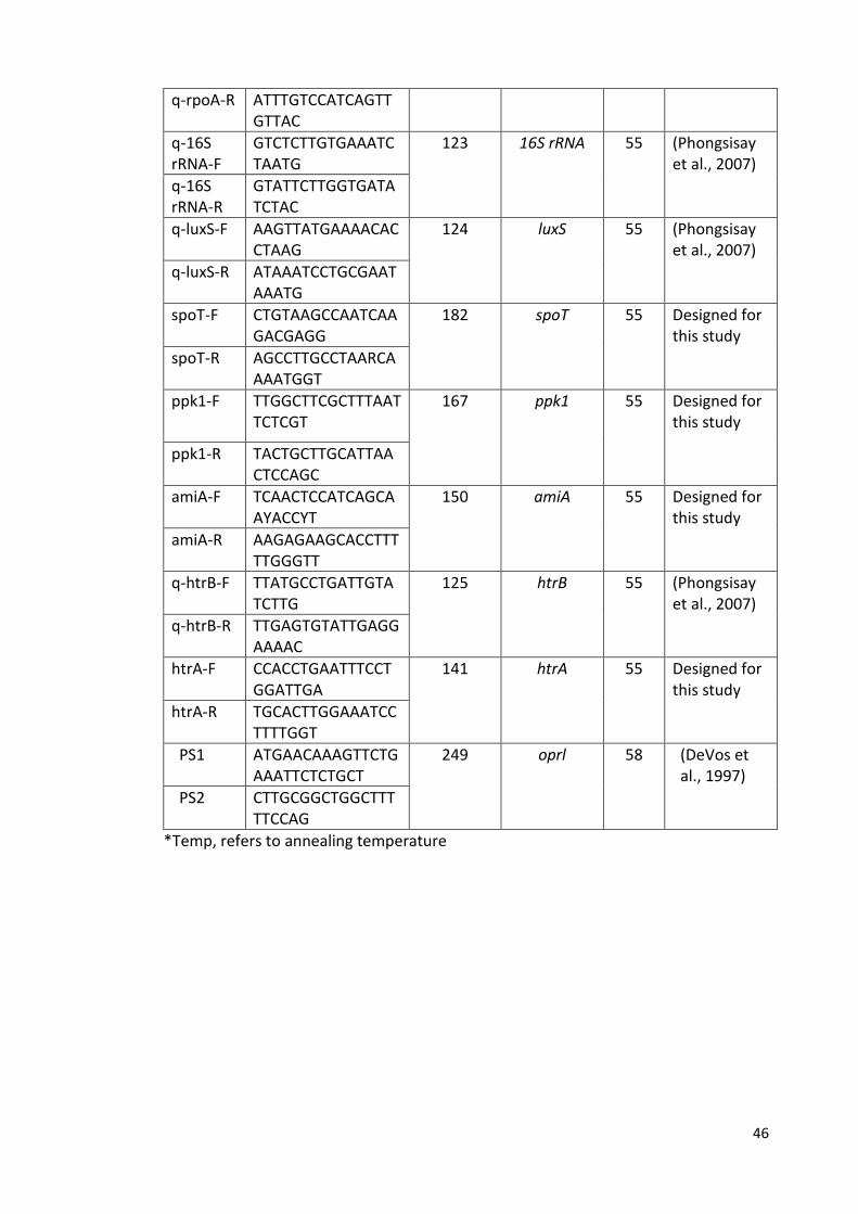
46
q-rpoA-R ATTTGTCCATCAGTTGTTAC
q-16S rRNA-F
GTCTCTTGTGAAATCTAATG
123 16S rRNA 55 (Phongsisay et al., 2007)
q-16S rRNA-R
GTATTCTTGGTGATATCTAC
q-luxS-F AAGTTATGAAAACACCTAAG
124 luxS 55 (Phongsisay et al., 2007)
q-luxS-R ATAAATCCTGCGAATAAATG
spoT-F CTGTAAGCCAATCAAGACGAGG
182 spoT 55
Designed for this study
spoT-R
AGCCTTGCCTAARCAAAATGGT
ppk1-F TTGGCTTCGCTTTAATTCTCGT
167 ppk1 55 Designed for this study
ppk1-R TACTGCTTGCATTAACTCCAGC
amiA-F TCAACTCCATCAGCAAYACCYT
150 amiA 55 Designed for this study
amiA-R AAGAGAAGCACCTTTTTGGGTT
q-htrB-F TTATGCCTGATTGTATCTTG
125 htrB 55 (Phongsisay et al., 2007)
q-htrB-R TTGAGTGTATTGAGGAAAAC
htrA-F CCACCTGAATTTCCTGGATTGA
141 htrA 55 Designed for this study
htrA-R TGCACTTGGAAATCCTTTTGGT
PS1 ATGAACAAAGTTCTGAAATTCTCTGCT
249 oprl 58 (DeVos et al., 1997)
PS2 CTTGCGGCTGGCTTTTTCCAG
*Temp, refers to annealing temperature
47
2.7.1 Standard PCR protocol for the identification of C. jejuni strains
Modified Linton primers (Katzav et al., 2008; Linton et al., 1996) targeting the 16S
rRNA gene were used to confirm that bacteria were C. jejuni. PCR amplification was
performed in 25 µL volumes. Each reaction contained 5 µL of the template from
boiled suspension (section 2.6.3), 1.25 U of GoTaq polymerase (Promega), 5 mL of 5 x
Taq buffer, 2.5 µM MgCl2, 200 µM of nucleotides (dATP, dCTP, dGTP, dTTP), 12 pmol
of each of the primers and 10.25 µL of distilled water. The PCR reaction was
performed in a Thermal cycler (Labnet) using the following conditions:
One cycle of 95 °C for 5 min, followed by 30 cycles of 95 °C for 30 s, 58 °C for 30s and
72 °C for 1 min. The final elongation step lasted 10 min at 72 °C. The PCR product was
visualised on a 1% (w/v) agarose gel (Bioline) which contained 0.2 µg/ mL of ethidium
bromide. A DNA molecular weight marker [1kb plus ladder (Figure 2.4)], positive
control (C. jejuni) and no template control (dH2O) were included in each gel. 1 µL of
6x loading buffer (Thermo Scientific) was mixed with 5 µL of PCR product prior to
loading. Amplicons were visualised under UV light (SYNGENE) and the presence of a
product of 857 bp was considered positive.
2.7.2 PCR protocol for identification of Campylobacter isolates from environmental
water samples
PCR amplification was performed in 25 µL volumes containing 2 µL of the template
extracted by Chelex method, 22.5 µL of PCR master mix (1.1 x ReddyMix PCR master
mixes with 1.5 mM MgCl2, Thermo Scientific) and 12 pmol (0.25 µL) of each of the
primers (Modified Linton primers, Table 2.3).
48
The PCR reaction was performed in a Thermal cycler (Applied Biosystems) using the
following conditions: one cycle of 94 °C for 5 min, followed by 30 cycles of 95 °C for 1
min, 58 °C for 1 min and 72 °C for 1 min. The final stage carried out for 10 min at
72°C. The PCR products were visualised on a 1% (w/v) agarose gel (Alpha laboratories)
which contained 6 µL peqgreen (peQlab). A DNA molecular mass marker (GeneRuler
100 bp ladder), a positive control (C. jejuni) and no template control (dH2O) were
included in each PCR run. Amplicons were visualised under UV light (Trasilluminator)
and the presence of a single product of (857 bp) was considered as positive for C.
jejuni.
2.7.3 PCR protocol for identification of Pseudomonas isolates from environmental
water samples
PCR amplification was performed in 25 µL volumes containing 2 µL of the template
extracted by the Chelex method, 22.2 µL of PCR master mix (1.1x ReddyMix PCR
master mix with 1.5 mM MgCl2, Thermo Scientific) and 4 pmol (0.4 µL) of each of the
Pseudomonas-specific-primers (PS1 and PS2, Table 2.3). The PCR reaction was
performed in a Thermal cycler using the following conditions:
One cycle of 94 °C for 5 min, followed by 30 cycles of 95 °C for 1 min, 58 °C for 1 min
and 72 °C for 1 min. The final elongation step was carried out for 10 min at 72 °C. The
PCR products were visualised on a 1% (w/v) agarose gel which contained 6 µL
peqgreen (peQlab). A DNA molecular mass marker (GeneRuler 100 bp ladder), a
positive control (P. aeruginosa) and a no template control (dH2O) were included in
49
each PCR run. Amplicons were visualised under UV light and the presence of a single
amplified product of (249 bp) was considered as positive for Pseudomonas spp.
2.7.4 Temperature gradient PCR
Gradient PCR (Mastercycler® thermal cycler) was used with a temperature gradient of
45.3 to 64.7 °C. The amplification was performed in a reaction volume of 25 µL. Each
reaction contained 1 µL of DNA template (C. jejuni ST-42 strain) extracted by the
Promega Wizard method (section 2.6.1), 1.25 U of GoTaq® DNA polymerase
(Promega), 5 µL of 5 × GoTaq buffer, 2.5 µM MgCl2, 200 µM of nucleotides (dATP,
dCTP, dGTP, dTTP), 7.5 pmol of each of the oligonucleotide primers and 14.25 µL of
distilled water.
The following PCR conditions were set: one cycle of 95 °C for 5 min, followed by 30
cycles of 95 °C for 1 min, annealing temperature 45.3 to 64.7 °C for 1 min, and 72 °C
for 2 min. The final elongation step was carried out for 10 min. The PCR product was
visualised on a 1% (w/v) agarose gel which contained 0.2 µg/mL of ethidium bromide.
A DNA molecular weight marker (1 Kb plus ladder) was included in each gel. The
amplicons were visualised under UV light (SYNGENE).
2.7.5 PCR amplification of cDNA
PCR amplification was performed at a reaction volume of 25 µL. Each reaction
contained 5 µL of the cDNA template, 1.25 U of GoTaq®DNA polymerase (Promega),
5 µL of 5 × GoTaq buffer, 2.5 µM MgCl2, 200 µM of nucleotides (dATP, dCTP, dGTP,
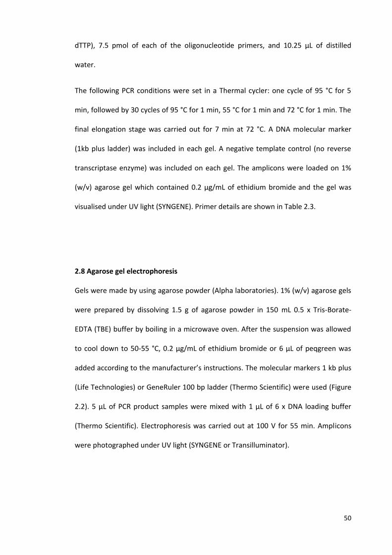
50
dTTP), 7.5 pmol of each of the oligonucleotide primers, and 10.25 µL of distilled
water.
The following PCR conditions were set in a Thermal cycler: one cycle of 95 °C for 5
min, followed by 30 cycles of 95 °C for 1 min, 55 °C for 1 min and 72 °C for 1 min. The
final elongation stage was carried out for 7 min at 72 °C. A DNA molecular marker
(1kb plus ladder) was included in each gel. A negative template control (no reverse
transcriptase enzyme) was included on each gel. The amplicons were loaded on 1%
(w/v) agarose gel which contained 0.2 µg/mL of ethidium bromide and the gel was
visualised under UV light (SYNGENE). Primer details are shown in Table 2.3.
2.8 Agarose gel electrophoresis
Gels were made by using agarose powder (Alpha laboratories). 1% (w/v) agarose gels
were prepared by dissolving 1.5 g of agarose powder in 150 mL 0.5 x Tris-Borate-
EDTA (TBE) buffer by boiling in a microwave oven. After the suspension was allowed
to cool down to 50-55 °C, 0.2 µg/mL of ethidium bromide or 6 µL of peqgreen was
added according to the manufacturer’s instructions. The molecular markers 1 kb plus
(Life Technologies) or GeneRuler 100 bp ladder (Thermo Scientific) were used (Figure
2.2). 5 µL of PCR product samples were mixed with 1 µL of 6 x DNA loading buffer
(Thermo Scientific). Electrophoresis was carried out at 100 V for 55 min. Amplicons
were photographed under UV light (SYNGENE or Transilluminator).
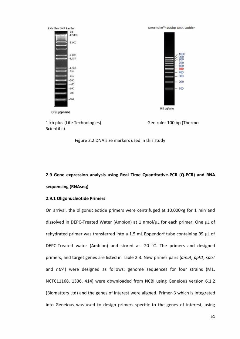
51
1 kb plus (Life Technologies) Gen ruler 100 bp (Thermo Scientific)
Figure 2.2 DNA size markers used in this study
2.9 Gene expression analysis using Real Time Quantitative-PCR (Q-PCR) and RNA
sequencing (RNAseq)
2.9.1 Oligonucleotide Primers
On arrival, the oligonucleotide primers were centrifuged at 10,000×g for 1 min and
dissolved in DEPC-Treated Water (Ambion) at 1 nmol/µL for each primer. One µL of
rehydrated primer was transferred into a 1.5 mL Eppendorf tube containing 99 µL of
DEPC-Treated water (Ambion) and stored at -20 °C. The primers and designed
primers, and target genes are listed in Table 2.3. New primer pairs (amiA, ppk1, spoT
and htrA) were designed as follows: genome sequences for four strains (M1,
NCTC11168, 1336, 414) were downloaded from NCBI using Geneious version 6.1.2
(Biomatters Ltd) and the genes of interest were aligned. Primer-3 which is integrated
into Geneious was used to design primers specific to the genes of interest, using
52
default settings. Primers were designed with Q-PCR in mind, aiming for amplicon sizes
of around 180 bp.
Artemis Comparison Tool (ACT) (https://www.sanger.ac.uk/resources/software/act)
was used to identify the differences between the genomes of C. jejuni strains used in
this study (M1, 1336, 414) and the strain NCTC11168.
The Primers were designed by Dr. Christina Bronowski, University of Liverpool.
2.9.2 RNA extraction from water survival experiments and cDNA synthesis
100 mL of inoculated water samples containing C. jejuni strains M1, 414 and 1336
(section 2.3.3) at days 0, 1 (for 25 °C) and 3 (for 4 °C), were concentrated by
centrifugation at 3893×g (3-16 pk, SIGMA) for 15 min at 4 °C in Falcon tubes (Corning,
Appleton Woods). Supernatants were removed but, for each sample, 1 mL was
retained at the bottom of each Falcon tube and subsequently transferred into a 1.5
mL Eppendorf tube. Cells were pelleted by centrifugation (SIGMA) at 5000×g for 20
min. Total RNA of the C. jejuni strains was isolated by using the RiboPure-Bacteria Kit
(Ambion) according to the manufacturer’s instructions. Briefly, cell pellets were re-
suspended in 350 µL RNAwiz and the suspension was vortexed vigorously for 15 s.
Suspensions were then transferred to a tube containing 250 µL Zirconia Beads and
exposed to beating by vortex for 10 min at room temperature. Zirconia Beads were
pelleted by centrifugation at 16,000×g for 5 min at 4 °C and the bacterial lysate was
transferred to a 1.5 mL Eppendorf tube. A 0.2 volume (v/v) of chloroform (typically 40
µL) was added, mixed well and incubated for 10 min at room temperature. The lysate
53
was separated into aqueous and organic phases by centrifugation at 16,000×g for 5
min at 4 °C, and the aqueous phase (top layer) was transferred to a 1.5 mL Eppendorf
tube. 0.5 volumes (v/v) of 100% ethanol (500-750 µL) were added to the aqueous
phase of each sample. Samples were passed through filter cartridges in 2 mL
collection tubes for each sample. Samples were centrifuged at 16,000×g for 1 min at
room temperature, after which the flow-through was discarded and the filter
cartridge left in the tube.
The filter was washed with 700 µL wash solution 1, centrifuged at 16,000×g for 1 min
at room temperature and the flow-through was discarded. The filter was washed
twice with 500 µL wash solution 2/3, centrifuged at 16,000×g for 1 min at room
temperature and the flow-through was discarded. The filter cartridge for each sample
was centrifuged at 16,000×g for 1 min at room temperature to remove excess wash
solution from the filter and then transferred to a fresh 2 mL collection tube. RNA was
eluted with 50 µL preheated (100 °C) elution solution and centrifuged at 16,000×g for
1 min at room temperature. Maximum total RNA was yielded by repeating the elution
step with a further 50 µL preheated elution solution into the same tube. Yielded RNA
was purified from contaminated genomic DNA by adding 1/9th (11 µL) volume (v/v) of
10 × DNase buffer, 4 µL (8 U) of DNase 1 (Ambion) and incubated for 30 min at 37 °C.
A volume of DNase inactivation reagent equal to 20% (v/v) (20 µL) of the volume of
RNA treated was added to each sample. Samples were mixed well, then left at room
temperature for 2 min, before centrifugation for 1 min at maximum speed, after
which the RNA solution was transferred to a new RNAase-free tube.
First-strand cDNA synthesis was carried out by using the SuperScriptVILO Kit
(Invitrogen). The reaction for each sample was carried out in a volume of 20 µL
54
containing 14 µL (1.98-7.03 ng/µL) total RNA, 4 µL 5 × reaction mix and 2 µL 10 ×
superscript enzyme mix. The reactions were carried out at 25 °C for 10 min, 42 °C for
60 min and 85 °C for 5 min. Both extracted RNA and synthesised cDNA were
quantified by using a NanoDrop-1000 spectrophotometer (Thermo Scientific).
2.9.3 Q-PCR
Q-PCR analysis using Two Standard Curve Method was carried out according to the
instructions in the operator manual of the Rotor-GeneTM 6000 (Corbett Life Science).
Briefly, the method depended on performing a standard curve for each gene. The
expression of the gene of interest was then normalised with the internal control gene
(for example, 16S rRNA). Normalisation is the calculation of the ratios of the mRNA
concentrations of the genes of interest to the concentrations of the internal control
genes (Bustin et al., 2009). The standard curves were obtained by analysis using a 10-
fold serial dilution of genomic DNA of C. jejuni (ST-42). The quantification cycle values
(Cq) were plotted against log10 concentration of the genomic DNA (standard curve of a
10-fold serial dilution of genomic DNA). The Cq values determined by software
analysis of the unknown samples were compared to the standard curve to determine
the concentration of each unknown sample. The efficiency and the coefficient (R2) of
the standard curves were calculated automatically by the software for the Rotor-gene
Q cycler.
Q-PCR was performed in a 72 well Rotor-Gene Q cycler (Qiagen), using a SYBR green
PCR Kit (Qiagen). The PCR reaction was performed in a total volume of 12.5 µL in 0.1
mL PCR tubes with cap strips (Qiagen) containing 0.5 µL of the cDNA template and 12
55
µL of a PCR master mix (6.25 µL SYBR green, 12.5 pmol (1.25 µL) each of the primers
and 3.25 µL dH2O).
The following PCR conditions were set: an initial denaturation step of 95°C for 5 min
followed by 40 cycles of 95 °C for 10 s, 55 °C for 15 s and 72 °C for 20 s. On each run, a
no template control (dH2O) was included. The amplification was carried out with
three replicates of each dilution. Each specific amplicon was confirmed by the
presence of a single melting peak. Cq values and data analysis were conducted using
Rotor-Gene cycler software.
2.9.4 RNAseq
RNAseq or transcriptome sequencing is a newly developed technology to study
transcriptome profiling that uses next-generation sequencing techniques, providing
important information about internal conditions of cells. For example, RNAseq can be
used to quantify and determine gene expression level changes under various
conditions (Wang et al., 2009).
2.9.4.1 RNA extraction from water survival experiments
100 mL of inoculated water samples were prepared for C. jejuni M1 in triplicate as
described previously at days 0, 1 (25 °C) and 3 (4 °C). Cells were concentrated by
centrifugation at 3893×g (3-16pk, SIGMA) for 20 min at the corresponding
temperature in 50 mL Falcon tubes (Corning, Appleton Woods). Supernatants were
removed but, for each sample, 1 mL was retained at the bottom of each Falcon tube
56
and subsequently combined per sample and transferred into a 1.5 mL Eppendorf
tube. Cells were pelleted by centrifugation at 5000 ×g for 10 min. A negative control
was included using only sterile distilled water. Additionally a positive control of C.
jejuni M1 grown in MHB was prepared. Cells were grown to approximately 1x10⁷
CFU/mL (OD600 of 0.05) in a 25 cm2 cell culture flask with gas exchange lid (Corning) at
37 oC microaerophilic conditions for 18 h (exponential phase) . Cells were collected by
centrifugation at 3000xg in a microcentrifuge (SIGMA). Once collected, cells were
immediately re-suspended in TRIzol solution (3 times TRIzol volume to 1 volume of
cells) (Ambion) and stored at -80 °C until further processing.
TRIzol samples were allowed to reach room temperature and cells were disrupted
using vigorous vortexing. The samples were then incubated at room temperature for
5 min. RNA was extracted using the Direct-zol RNA MiniPrep Kit (Zymo Research),
following the manufacturer’s instructions.
These experiments were carried out by Dr. Christina Bronowski, University of
Liverpool.
2.9.4.2 Library construction and sequencing
rRNA was depleted from the total RNA sample using Ribozero (Epicentre), following
the manufacturer’s instructions. Libraries were prepared using the strand-specific
ScriptSeq kit (Epicentre), following the manufacture’s instruction. Sequencing was
performed on the Illumina HiSeq 2500 using v4 chemistry (Illumina). This work was
carried out at the Centre for Genomic Research, University of Liverpool.
57
2.9.4.3 RNAseq data analysis
The raw Fastq data files were trimmed for the presence of Illumina adapter
sequences using Cutadapt (v1.2.1.) (Martin, 2011), using the -O3 option. The reads
were further trimmed using Sickle (v1.200) (https://github.com/najoshi/sickle) with a
minimum window quality score of 20. Reads shorter than 10 bp after trimming were
removed. Sense and antisense overlaps between the annotation and mapped reads
were counted using the HTSEQ package (Anders et al., 2015). Read counts were then
normalised and Differential Expression calculated using EdgeR implemented in R
(version 3.1.2 (2014-10-31), using Loess-style weighting to estimate the trended
dispersion values.
For pairwise Differential Expression analysis between samples, the data was re-
mapped to the C. jejuni M1 genome (Friis et al., 2010) using Bowtie2, and parsed
using the BitSeq (Bayesian Inference of Transcripts from Sequencing data) pipeline
(Glaus et al., 2012). BitSeq takes into account biological replicates and technical noise,
and thereby calculates a posterior distribution of differential expression between
samples.
Statistical Significance of BitSeq results was visualized in Artemis (Carver et al., 2008).
Regions of Difference between the genomes of M1 (Friis et al., 2010), 1336, 414
(Hepworth et al., 2011) and NCTC11168 (Gundogdu et al., 2007; Parkhill et al., 2000)
were derived through pairwise genome comparisons in ACT (Carver et al., 2005).
Putative/Hypothetical genes were selected in Artemis and a putative function was
derived from BLASTX searches
58
(http://blast.ncbi.nlm.nih.gov/blast/Blast.cgi?PROGRAM=blastx&PAGE_TYPE=BlastSe
arch&LINK_LOC=blasthome).
This work was carried out by Dr. Christina Bronowski, University of Liverpool.
2.10 Interaction between supernatants of cultures of Pseudomonas spp. and C.
jejuni strains.
2.10.1 Bacterial strains and growth conditions
The bacterial species and strains of Pseudomonas, and Campylobacter used in this
study are listed in Tables 2.1 and 2.2. C. jejuni isolate (ST-42) was streaked out from
frozen stock (-80 °C) on CBA for 48 h at 37 °C. Cells were harvested by sub-culturing
colonies microaerobically on CBA plate at 37 °C for 24 h. Aliquots of 5 mL of MHB
supplemented with Campylobacter growth supplement were inoculated with a 5 µL
loop -ful of C. jejuni isolate (ST-42) in 30 mL Universal tube and adjusted to an OD600
of 0.05 (Spectronic BioMate 5).
Bacterial cells of Pseudomonas spp. were harvested by streaking out from frozen
stock (-80 °C) on Luria Agar (LA) and incubated aerobically at 25 °C for 24 h (Table
2.2).
2.10.2 Preparation of supernatants of Pseudomonas spp.
Aliquots of 5 mL of (LB) broth were inoculated with a 5 µL loop of Pseudomonas spp.
in 30 mL Universal tube (Ramboldi Ltd) for each strain of Pseudomonas spp. (24
strains) and incubated aerobically at 25 °C while shaking at 180 rpm (Stuart) for 24 h.
59
Each culture was adjusted to OD600 of 0.5 (Spectronic BioMate 5) using LB Broth. The
cultures were filtered using a 0.2 µm filter (Minisart) and the supernatant was stored
at 4 °C.
2.10.3 Interaction assay
Aliquots of 180 µL of Campylobacter culture (OD600 of 0.05) were mixed with 20 µL of
supernatant of Pseudomonas spp. (24 strains) in 72 wells of a sterile 96 well plate
(Costar) in triplicate (for each strain). The following controls were also carried out: 6
replicates of C. jejuni culture mixed with LB (180 µL+20 µL respectively) and 6
replicates of C. jejuni only (200 µL). The plate was incubated microaerobically at 25 °C,
30 °C or 37 °C for 72 h. At 24 h intervals the OD600 was measured inside a Microplate
reader (FLUOstar Omega). Each experiment was conducted in three independent
replicates.
2.10.4 Treatment of supernatants by boiling or proteinase K
Aliquots of 180 µL of Campylobacter culture (OD600 of 0.05) were inoculated into 60
wells of a sterile 96 well plate. The following were added: aliquots of 20 µL of
supernatant of P. syringae pv. glycinea 49a/90 in 12 replicates, aliquots of 20 µL of
boiled supernatant of P. syringae pv. glycinea 49a/90 (boiled at 95 °C for 5 min) in 12
replicates, aliquots of 20 µL of supernatant of P. syringae pv. glycinea 49a/90 treated
with proteinase K (Applied Biosystems) in 12 replicates, aliquots of 20 µL of LB
treated with proteinase k in 12 replicates and aliquots of 20 µL of LB in 12 replicates
as a control. The plate was incubated microaerobically at 37°C for 72 h. At 24 h
60
intervals the OD600 was measured inside Microplate reader (FLUOstar Omega).
Proteinase K was added at 1 µL (0.02 mg)/200 µL sample and incubated at 37 °C for
30 min. After incubation, protease inhibiter cocktail (MERCK MILLIPORE) was added
at 0.5 µL/200 µL sample. The experiment was conducted in triplicate.
2.11 Experiments to asses co-existence of C. jejuni and Pseudomonas spp. in
environmental samples
2.11.1 Collection of environmental water samples
Environmental water samples were collected from troughs in Leahurst fields 1, 3, 6, 8
and 12 (n=5), ditches in fields 1, 3 and cuckoo lane (n=3), ponds in field 9 (n=1) and
permanent puddles in fields 6 and 7 (n=2) (Figure 2.1). Samples (30 mL for each one)
were collected from Leahurst fields in sterile disposable syringes (50 mL, TERUMO)
and transferred to the laboratory at ambient temperature within 1 h of sampling. All
samples were tested within 1 h of sampling.
2.11.2 Enrichment of collected water samples
To isolate C. jejuni strains, aliquots of 10 mL of water sample were mixed with 10 mL
of Campylobacter Enrichment Broth (Double Exeter Broth, appendix 2) containing 5%
(v/v) lysed horse blood (Southern group laboratories) in 30 mL Universal tube.
Enrichment cultures were incubated for 48 h at 42 °C under microaerobic conditions.
After incubation, aliquots of 50-100 µL of enrichment culture broth were spread with
a sterile loop on to a sterile filter paper 0.45 µm pore size (Sartorius), which had been
previously placed on mCCDA plate. Plates were left on the bench for 30 min and were
61
then incubated micoaerobically for 24-48 h at 42 °C after aseptically removing the
filter papers. Using 5 µL sterile plastic loops, each mCCDA plate positive for grown
colonies was sub-cultured on two CBA plates with the same inoculum and incubated
for 48 h at both 30 °C (aerobically) and 42 °C (microaerobically). CBA Plates positive
for grown colonies at both 30 °C (aerobically) and 42 °C (microaerobically) were
suspected Arcobacter, whereas colonies present when there was growth on CBA
plates only at 42 °C (microaerobically) were considered as likely to be Campylobacter
isolates. The suspensions of these colonies were stored at -80 °C on beads
(Microbank).
To isolate Pseudomonas spp., aliquots of 10 mL of water samples were mixed with 10
mL of LB broth (appendix 2) in 30 mL Universal tubes. Enrichment cultures were
incubated aerobically for 48 h at 30°C. After incubation, aliquots of 50-100 µL of
enrichment culture broth were plated out on Pseudomonas C-N Selective Agar
(Oxoid) containing Cetrimide and Sodium nalidixate (CN). Plates were incubated
aerobically at 30 °C for 48-72 h.
2.11.3 Extraction of bacterial DNA from environmental water samples
DNA was extracted directly from enrichment broth cultures (Double Exeter Broth
culture) after incubation microaerobically for 48 h at 42 °C or from LB broth cultures
after aerobic incubation for 48-72 h at 30 °C (section 2.11.2) by using the Chelex
method (section 2.6.2). DNA samples were stored at 4 °C or at -20 °C for long term
storage.
62
C h a p t e r 3
SURVIVAL OF C. jejuni STRAINS IN STERILE DISTILLED WATER AND NATURAL WATER
3.1 Introduction
3.1.1 Survival of Campylobacter spp. in water
Although campylobacteriosis is primarily considered to be a zoonotic infection, with
the infection mostly transmitted to humans through consumption of contaminated
food, especially under-cooked poultry meat, it is clear that the environment can also
play a role in transmission either directly or indirectly to humans. For example,
Campylobacter infections can be acquired directly via un-chlorinated drinking water
(Olson et al., 2008). In addition, the environment potentially plays an important role
in transmission of Campylobacter to livestock (Stanley and Jones, 2003).
Little is known about the survival mechanisms of Campylobacter outside the host
body compared to the survival of other enteric pathogens such as Salmonella spp.
and Escherichia coli. The genome sequence of C. jejuni NCTC 11168 and other strains
suggested that the capability of the organism for regulating gene expression in
response to stresses associated with environmental survival may be limited compared
to other bacteria (Murphy et al., 2006).
However, it is clear that Campylobacter, despite its requirement for microaerophilic
conditions in the laboratory, and its apparently limited ability to respond to stress,
can survive in the environment. Water-borne outbreaks of Campylobacter have been
reported worldwide due to contamination of untreated water with Campylobacter
spp. (Clark et al., 2003). For instance, Campylobacter outbreaks have been linked with
faecal contamination of drinking water in the Scandinavian countries of Finland
(Miettinen et al., 2001), Norway (Kapperud et al., 1992) and Sweden (Martin et al.,
63
2006). C. jejuni was the causative agent of seven reported waterborne epidemics in
Finland between 1998 and 2001, with most outbreaks happening in July, August,
September or October (Hänninen and Kärenlampi, 2004). More recently, whole
genome sequencing of C. jejuni was used to analyse isolates from a waterborne
outbreak in Finland (Revez et al., 2014). Furthermore, outbreaks of contaminated
drinking water have been reported in England and Wales (Said et al., 2003; Smith et
al., 2006), Scotland (Jones and Roworth, 1996) and the United States (Taylor et al.,
2013). Tap water consumption has also caused outbreaks of campylobacteriosis in
Wales due to contamination with surface water of pasture land (Richardson et al.,
2007).
Isolation of indistinguishable strains of C. jejuni from groundwater and a dairy farm
suggested that the source of contamination of groundwater could be from the dairy
farm within the drainage or shed area (Jones, 2001). Indeed contaminated
groundwater is considered to be a potential source of transmission of Campylobacter
to poultry flocks and broiler chickens through the drinking water system (Pearson et
al., 1993; Van de Giessen et al., 1996). It has also been suggested that these
organisms can survive in natural water in a VBNC state after environmental water
contamination by animal hosts (Guillou et al., 2008). Moreover, drinking bottled
water has been a public health concern due to the ability of Campylobacter to survive
in water for several months during storage before consumption (Evans et al., 2003;
Gillespie et al., 2002). Thus, water is considered as one of the important transmission
routes of campylobacteriosis in humans, either directly or indirectly, following
transmission to livestock.
64
Previous studies have revealed that survival of Campylobacter in natural water
samples or ground water is highly temperature and strain origin-dependent, as well
as varying according to the salinity and pH value of the water. Low temperatures
(around 4 °C) enhanced the survival of Campylobacter, whereas at increased
temperatures (20-25 °C) viability declined rapidly (Cools et al., 2003; Thomas et al.,
2002). Previous studies have determined that survival times for C. jejuni in water can
vary from a few days at ambient temperatures up to 4 months at 4 °C (Buswell et al.,
1998; Rollins and Colwell, 1986). It has been postulated that strain variations
contribute to these differences in survival times. Various factors could contribute to
Campylobacter survival within surface aquifers, including low oxidative stress, lack of
oxygen in deeper zones, protection from UV light and lower temperature (Jones,
2001).
In the farming environment, water types potentially play an important role in the
epidemiology of Campylobacter spp., with C. jejuni the most frequently species found
in trough and running water, whereas C. coli is the most common species found in
standing water (Kemp et al., 2005), highlighting the variations within the genus.
However, previous comparisons have not addressed variations based on strain
genotyping. Yet previous epidemiological studies have suggested that some strain
types are more commonly found in the environment. Using MLST it has been shown
that C. jejuni ST-61, associated with cattle faecal isolates, is only rarely isolated from
the environment, whereas the ST-45 complex is not only commonly found amongst
livestock and wildlife faecal isolates, but also amongst water isolates (Colles et al.,
2003; French et al., 2005). In addition, there are particular MLSTs found exclusively
amongst wildlife or environmental sources (Hepworth et al., 2011). There are
65
significant gaps in our knowledge concerning the survival of different Campylobacter
genotypes in the environment. In order to address the food safety and public health
concerns on gastroenteritis in the community, a better understanding of the
occurrence and behaviour of Campylobacter in contaminated water sources, and the
mechanisms underlying variations, is needed (Van Dyke et al., 2010).
3.1.2 VBNC state
Although the concept of the VBNC state remains controversial, it has a long history
and can be a useful measure of physiological change by bacteria in response to stress.
It is a state in which bacterial cells remain viable according to various measures of
metabolic activity but are unable to grow on the routine culture media, on which the
bacteria would normally be able to grow and form colonies (Rollins and Colwell,
1986). It has been demonstrated that bacteria in this state can remain capable of
causing infections or colonising a host and can be returned to a state of culturability
(Chaisowwong et al., 2012).
Rollins and Colwell were the first to describe the phenomenon of the VBNC state in C.
jejuni in 1986. In water microcosms, Campylobacter forms VBNC coccoid-shape cells
with an intact cell membrane when exposed to stresses such as low nutrient
conditions (Rollins and Colwell, 1986). Further studies have since described the
influence of a harsh environment on survival of Campylobacter. Combinations of low
temperature and starvation, and pH stress, are factors that can drive C. jejuni to enter
into a VBNC state (Baffone et al., 2006; Chaveerach et al., 2003). Furthermore, high
temperature and oxidative stress can also drive Campylobacter cells to form a VBNC
66
state (Klančnik et al., 2006). It has also been demonstrated that bactericidal processes
for bacteria such as pasteurization of milk (Gunasekera et al., 2002) and chlorination
of waste water (Oliver, 2005) may result in formation of the VBNC state.
Bacterial cells in a VBNC state generally show dwarfing or coccoid shape, and during
this stage significant metabolic changes take place, such as decreasing nutrient
transport, respiration rate and the levels of DNA, RNA and protein synthesis (Oliver,
2000; Porter et al., 1995). Starvation and cold shock proteins are synthesized during
this period (McGovern and Oliver, 1995; Morton and Oliver, 1994). It has also been
shown that due to the low metabolic activity of flagellated coccoid cells in the VBNC
state, these cells are non-motile even with the presence of flagella (Moore, 2001).
Campylobacter species in the VBNC state have been detected in a farm’s water supply
by immunofluorescence microscopy; chickens became colonised with the organism
after drinking this contaminated water (Pearson et al., 1993). The cells in VBNC state
may play an important role in the pathogenicity of C. jejuni; for example, the
expression of the virulence gene cadF, encoding an outer-membrane protein of C.
jejuni involved in adhesion to intestinal fibronectin was still detected at high levels up
to the third week of entering a VBNC state (Patrone et al., 2013). Moreover, it has
been shown that the coccoid cells of C. jejuni are capable of adhering to chicken skin
under aerobic conditions (Jang et al., 2007), and are also capable of invading Caco-2
human epithelial cells in vitro (Chaisowwong et al., 2012). Therefore the bacteria in
this state are believed to be a potential concern for both food safety and public
health (Chaisowwong et al., 2012).
It has been shown that detection rates of Campylobacter based on culture methods
within a watershed were much lower than prevalence rates based on the use of Q-
67
PCR assays. This difference in detection levels was attributed to the Campylobacter
entering the VBNC state (Van Dyke et al., 2010). Furthermore, despite the high
number of waterborne outbreaks of Campylobacter, it is often difficult to detect
Campylobacter in drinking water samples using culture. This may be due to using
poor methods to recover the cells or because the cells in the environment are often
presence in the form of a VBNC state (Percival et al., 2013). Although the mechanisms
underlying a switch to the VBNC state are not well understood, some genes have
been implicated in the process in Campylobacter. A mutant C. jejuni (Δppk1) showed
decreased ability to enter VBNC state due to lack in poly-P synthesis Poly-P, a source
of energy and an essential molecule for survival during starvation, is synthesized by
mediating the key enzyme polyphosphate kinase 1 (ppk1) (Gangaiah et al., 2009;
Kassem and Rajashekara, 2011). Inorganic polyphosphate (poly-P) is a linear polymer
of phosphate residues linked by phosphoanhydride bonds which provide a high
energy to the cell (Kornberg, 1999).
Reverse transcription-PCR (RT-PCR) methods have been employed to monitor
continued expression of genes of cells in the VBNC state (del Mar Lleò et al., 2000).
Furthermore, Detection of bacterial mRNAs in VBNC cells has been suggested as an
indicator for cell viability considering the short half-life of mRNA inside the cell (del
Mar Lleò et al., 2000; Sheridan et al., 1998)
68
3.1.3 Aims of this chapter
The aims of this chapter are:
1. To compare the survival of a diverse panel of C. jejuni strains from various sources
and representing different MLSTs in sterile distilled water at two different
temperatures (4 °C and 25 °C) by measuring the ability to form colony forming units.
2. To investigate the VBNC state in C. jejuni strains under starvation conditions at two
different temperatures (4 °C and 25 °C) by detecting and counting viable cells of C.
jejuni in sterile distilled water.
3. To compare the survival of C. jejuni strains in autoclaved and non-autoclaved
natural water at two different temperatures (4 °C and 25 °C) by measuring the ability
to form colony forming units.
69
3.2 Results
3.2.1 Survival in sterile distilled water and classification of C. jejuni strains into
groups
27 strains of C. jejuni (Table 2.1), chosen to represent a diverse range of sources and
MLST clonal complexes, were screened for survival based on the retention of
culturability in sterile distilled water at two different temperatures (4 °C and 25 °C) in
the dark. This rapid screen (Figure 3.1A, 3.1B 3.1C and 3.1D), based on a single
biological replicate, was used to classify the strains into four groups (>75%, 50 -75%,
10-50% and <10%) on the basis of maintenance of CFU/mL on CBA plates at 4 °C (for
day 3) and at 25 °C (for day 1). Maintenance of culturability at greater than 75% of
the inoculum CFU/mL was considered as a high maintenance of CFU/mL, 10-75%
CFU/mL was considered as an intermediate maintenance of CFU/mL, and less than
10% CFU/mL was considered as a low maintenance of CFU/mL (Table 3.1). The
distribution of strain types according to MLST and source is shown in tables 3.2 and
3.3.
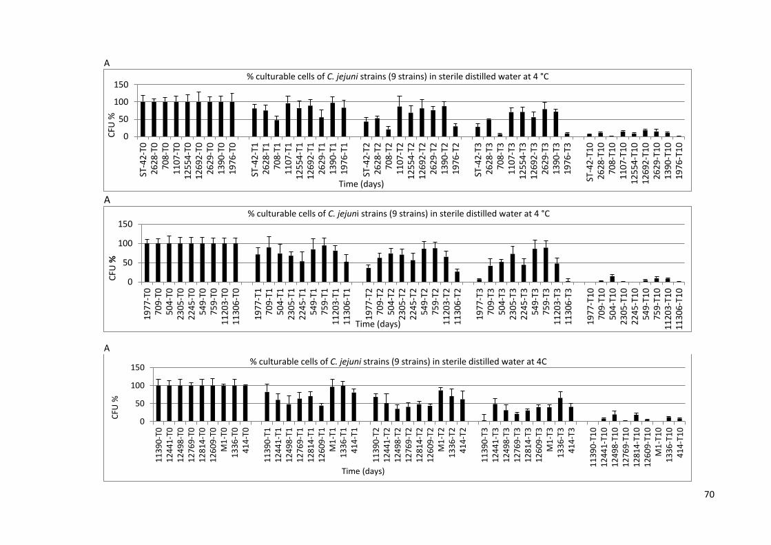
70
A
A
A
0
50
100
150
ST-4
2-T0
2628
-T0
708-
T011
07-T
01
25
54
-T0
12
69
2-T
026
29-T
013
90-T
019
76-T
0
ST-4
2-T1
2628
-T1
708-
T111
07-T
11
25
54
-T1
12
69
2-T
126
29-T
113
90-T
119
76-T
1
ST-4
2-T2
2628
-T2
708-
T211
07-T
21
25
54
-T2
12
69
2-T
226
29-T
213
90-T
219
76-T
2
ST-4
2-T3
2628
-T3
708-
T311
07-T
31
25
54
-T3
12
69
2-T
326
29-T
313
90-T
319
76-T
3
ST-4
2-T1
02
62
8-T
10
708-
T10
11
07
-T1
012
554
-T1
012
692
-T1
02
62
9-T
10
13
90
-T1
01
97
6-T
10
CFU
%
Time (days)
% culturable cells of C. jejuni strains (9 strains) in sterile distilled water at 4 °C
0
50
100
150
19
77-T
070
9-T0
504-
T023
05-T
02
245
-T0
549-
T075
9-T0
11
203
-T0
11
306
-T0
19
77-T
170
9-T1
504-
T12
305
-T1
22
45-T
154
9-T1
759-
T11
120
3-T
111
306
-T1
19
77-T
270
9-T2
504-
T22
305
-T2
22
45-T
254
9-T2
759-
T21
120
3-T
21
130
6-T
2
19
77-T
370
9-T3
504-
T32
305
-T3
22
45-T
354
9-T3
759-
T31
120
3-T
31
130
6-T
3
1977
-T10
70
9-T1
05
04-
T10
23
05-T
10
22
45-T
10
54
9-T1
07
59-
T10
11
203
-T1
01
130
6-T
10
CFU
%
Time (days)
% culturable cells of C. jejuni strains (9 strains) in sterile distilled water at 4 °C
0
50
100
150
11
39
0-T
01
24
41
-T0
12
49
8-T
01
27
69
-T0
12
81
4-T
01
26
09
-T0
M1
-T0
13
36
-T0
41
4-T
0
11
39
0-T
11
24
41
-T1
12
49
8-T
11
27
69
-T1
12
81
4-T
11
26
09
-T1
M1
-T1
13
36
-T1
41
4-T
1
11
39
0-T
21
24
41
-T2
12
49
8-T
21
27
69
-T2
12
81
4-T
21
26
09
-T2
M1
-T2
13
36
-T2
41
4-T
2
11
39
0-T
31
24
41
-T3
12
49
8-T
31
27
69
-T3
12
81
4-T
31
26
09
-T3
M1
-T3
13
36
-T3
41
4-T
3
11
39
0-T
10
12
44
1-T
10
12
49
8-T
10
12
76
9-T
10
12
81
4-T
10
12
60
9-T
10
M1
-T1
01
33
6-T
10
41
4-T
10
CFU
%
Time (days)
% culturable cells of C. jejuni strains (9 strains) in sterile distilled water at 4C
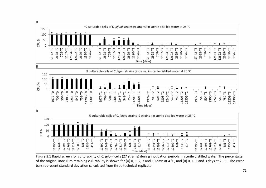
71
B
B
B
Figure 3.1 Rapid screen for culturability of C. jejuni cells (27 strains) during incubation periods in sterile distilled water. The percentage of the original inoculum retaining culurability is shown for (A) 0, 1, 2, 3 and 10 days at 4 °C, and (B) 0, 1, 2 and 3 days at 25 °C. The error bars represent standard deviation calculated from three technical replicate
0
50
100
150
ST-4
2-T0
2628
-T0
708-
T0
1107
-T0
1255
4-T
0
1269
2-T
0
2629
-T0
1390
-T0
1976
-T0
ST-4
2-T1
2628
-T1
708-
T1
1107
-T1
1255
4-T
1
1269
2-T
1
2629
-T1
1390
-T1
1976
-T1
ST-4
2-T2
2628
-T2
708-
T2
1107
-T2
1255
4-T
2
1269
2-T
2
2629
-T2
1390
-T2
1976
-T2
ST-4
2-T3
2628
-T3
708-
T3
1107
-T3
1255
4-T
3
1269
2-T
3
2629
-T3
1390
-T3
1976
-T3
CFU
%
Time (days)
% culturable cells of C. jejuni strains (9 strains) in sterile distilled water at 25 °C
0
50
100
150
19
77
-T0
709-
T0
504-
T0
23
05
-T0
22
45
-T0
549-
T0
759-
T0
1120
3-T
0
1130
6-T
0
19
77
-T1
709-
T1
504-
T1
23
05
-T1
22
45
-T1
549-
T1
759-
T1
1120
3-T
1
1130
6-T
1
19
77
-T2
709-
T2
504-
T2
23
05
-T2
22
45
-T2
549-
T2
759-
T2
1120
3-T
2
1130
6-T
2
19
77
-T3
709-
T3
504-
T3
23
05
-T3
22
45
-T3
549-
T3
759-
T3
1120
3-T
3
1130
6-T
3
CFU
%
Time (days)
% culturable cells of C. jejuni strains (9strains) in sterile distilled water at 25 °C
0
50
100
150
11
39
0-T
0
12
44
1-T
0
12
49
8-T
0
12
76
9-T
0
12
81
4-T
0
12
60
9-T
0
M1
-T0
13
36
-T0
41
4-T
0
11
39
0-T
1
12
44
1-T
1
12
49
8-T
1
12
76
9-T
1
12
81
4-T
1
12
60
9-T
1
M1
-T1
13
36
-T1
41
4-T
1
11
39
0-T
2
12
44
1-T
2
12
49
8-T
2
12
76
9-T
2
12
81
4-T
2
12
60
9-T
2
M1
-T2
13
36
-T2
41
4-T
2
11
39
0-T
3
12
44
1-T
3
12
49
8-T
3
12
76
9-T
3
12
81
4-T
3
12
60
9-T
3
M1
-T3
13
36
-T3
41
4-T
3
CFU
%
Time (days)
% culturable cells of C. jejuni strains (9 strains ) in sterile distilled water at 25 °C
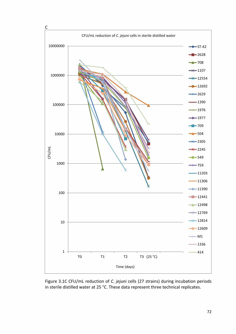
72
C
Figure 3.1C CFU/mL reduction of C. jejuni cells (27 strains) during incubation periods in sterile distilled water at 25 °C. These data represent three technical replicates.
1
10
100
1000
10000
100000
1000000
10000000
T0 T1 T2 T3 (25 °C)
CFU
/mL
Time (days)
CFU/mL reduction of C. jejuni cells in sterile distilled water
ST-42
2628
708
1107
12554
12692
2629
1390
1976
1977
709
504
2305
2245
549
759
11203
11306
11390
12441
12498
12769
12814
12609
M1
1336
414
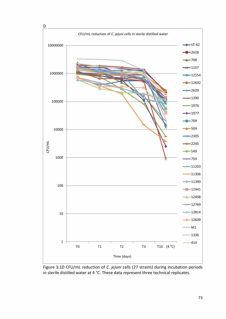
73
D
Figure 3.1D CFU/mL reduction of C. jejuni cells (27 strains) during incubation periods in sterile distilled water at 4 °C. These data represent three technical replicates.
1
10
100
1000
10000
100000
1000000
10000000
T0 T1 T2 T3 T10 (4 °C)
CFU
/mL
Time (days)
CFU/mL reduction of C. jejuni cells in sterile distilled water
ST-42
2628
708
1107
12554
12692
2629
1390
1976
1977
709
504
2305
2245
549
759
11203
11306
11390
12441
12498
12769
12814
12609
M1
1336
414
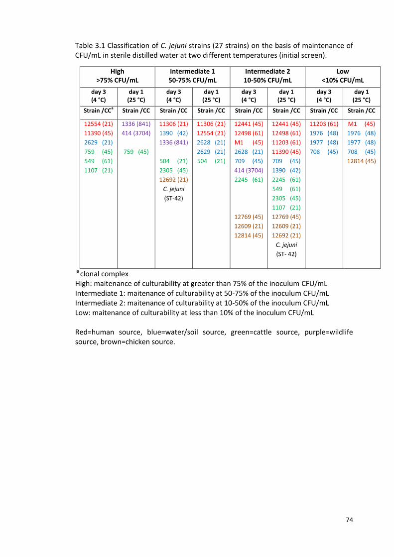
74
Table 3.1 Classification of C. jejuni strains (27 strains) on the basis of maintenance of CFU/mL in sterile distilled water at two different temperatures (initial screen).
High >75% CFU/mL
Intermediate 1 50-75% CFU/mL
Intermediate 2 10-50% CFU/mL
Low <10% CFU/mL
day 3 (4 °C)
day 1 (25 °C)
day 3 (4 °C)
day 1 (25 °C)
day 3 (4 °C)
day 1 (25 °C)
day 3 (4 °C)
day 1 (25 °C)
Strain /CCa Strain /CC Strain /CC Strain /CC Strain /CC Strain /CC Strain /CC Strain /CC
12554 (21)
11390 (45)
2629 (21)
759 (45)
549 (61)
1107 (21)
1336 (841)
414 (3704)
759 (45)
11306 (21)
1390 (42)
1336 (841)
504 (21)
2305 (45)
12692 (21)
C. jejuni
(ST-42)
11306 (21)
12554 (21)
2628 (21)
2629 (21)
504 (21)
12441 (45)
12498 (61)
M1 (45)
2628 (21)
709 (45)
414 (3704)
2245 (61)
12769 (45)
12609 (21)
12814 (45)
12441 (45)
12498 (61)
11203 (61)
11390 (45)
709 (45)
1390 (42)
2245 (61)
549 (61)
2305 (45)
1107 (21)
12769 (45)
12609 (21)
12692 (21)
C. jejuni
(ST- 42)
11203 (61)
1976 (48)
1977 (48)
708 (45)
M1 (45)
1976 (48)
1977 (48)
708 (45)
12814 (45)
a clonal complex High: maitenance of culturability at greater than 75% of the inoculum CFU/mL Intermediate 1: maitenance of culturability at 50-75% of the inoculum CFU/mL Intermediate 2: maitenance of culturability at 10-50% of the inoculum CFU/mL Low: maitenance of culturability at less than 10% of the inoculum CFU/mL Red=human source, blue=water/soil source, green=cattle source, purple=wildlife source, brown=chicken source.
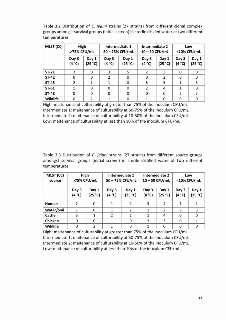
75
Table 3.2 Distribution of C. jejuni strains (27 strains) from different clonal complex groups amongst survival groups (initial screen) in sterile disilled water at two different temperatures
MLST (CC) High >75% CFU/mL
Intermediate 1 50 – 75% CFU/mL
Intermediate 2 10 – 50 CFU/mL
Low <10% CFU/mL
Day 3 (4 °C)
Day 1 (25 °C)
Day 3 (4 °C)
Day 1 (25 °C)
Day 3 (4 °C)
Day 1 (25 °C)
Day 3 (4 °C)
Day 1 (25 °C)
ST-21 3 0 3 5 2 3 0 0
ST-42 0 0 2 0 0 2 0 0
ST-45 2 1 1 0 5 5 1 3
ST-61 1 0 0 0 2 4 1 0
ST-48 0 0 0 0 0 0 2 2
Wildlife 0 2 1 0 1 0 0 0
High: maitenance of culturability at greater than 75% of the inoculum CFU/mL Intermediate 1: maitenance of culturability at 50-75% of the inoculum CFU/mL Intermediate 2: maitenance of culturability at 10-50% of the inoculum CFU/mL Low: maitenance of culturability at less than 10% of the inoculum CFU/mL
Table 3.3 Distribution of C. jejuni strains (27 strains) from different source groups amongst survival groups (initial screen) in sterile distilled water at two different temperatures
MLST (CC) source
High >75% CFU/mL
Intermediate 1 50 – 75% CFU/mL
Intermediate 2 10 – 50 CFU/mL
Low <10% CFU/mL
Day 3 (4 °C)
Day 1 (25 °C)
Day 3 (4 °C)
Day 1 (25 °C)
Day 3 (4 °C)
Day 1 (25 °C)
Day 3 (4 °C)
Day 1 (25 °C)
Human 2 0 1 2 3 4 1 1
Water/Soil 1 0 1 2 2 2 3 3
Cattle 3 1 2 1 1 4 0 0
Chicken 0 0 1 0 3 3 0 1
Wildlife 0 2 1 0 1 0 0 0
High: maitenance of culturability at greater than 75% of the inoculum CFU/mL Intermediate 1: maitenance of culturability at 50-75% of the inoculum CFU/mL Intermediate 2: maitenance of culturability at 10-50% of the inoculum CFU/mL Low: maitenance of culturability at less than 10% of the inoculum CFU/mL
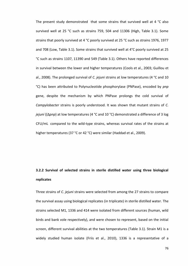
76
The present study demonstrated that some strains that survived well at 4 °C also
survived well at 25 °C such as strains 759, 504 and 11306 (High, Table 3.1). Some
strains that poorly survived at 4 °C poorly survived at 25 °C such as strains 1976, 1977
and 708 (Low, Table 3.1). Some strains that survived well at 4°C poorly survived at 25
°C such as strains 1107, 11390 and 549 (Table 3.1). Others have reported differences
in survival between the lower and higher temperatures (Cools et al., 2003; Guillou et
al., 2008). The prolonged survival of C. jejuni strains at low temperatures (4 °C and 10
°C) has been attributed to Polynucleotide phosphorylase (PNPase), encoded by pnp
gene, despite the mechanism by which PNPase prolongs the cold survival of
Campylobacter strains is poorly understood. It was shown that mutant strains of C.
jejuni ((Δpnp) at low temperatures (4 °C and 10 °C) demonstrated a difference of 3 log
CFU/mL compared to the wild-type strains, whereas survival rates of the strains at
higher temperatures (37 °C or 42 °C) were similar (Haddad et al., 2009).
3.2.2 Survival of selected strains in sterile distilled water using three biological
replicates
Three strains of C. jejuni strains were selected from among the 27 strains to compare
the survival assay using biological replicates (in triplicate) in sterile distilled water. The
strains selected M1, 1336 and 414 were isolated from different sources (human, wild
birds and bank vole respectively), and were chosen to represent, based on the initial
screen, different survival abilities at the two temperatures (Table 3.1). Strain M1 is a
widely studied human isolate (Friis et al., 2010), 1336 is a representative of a
77
previously reported water/wild-life group of strains (Hepworth et al., 2011) and 414 is
a niche specialist found associated with the bank vole (Hepworth et al., 2011;
Williams et al., 2010). All three have been genome sequenced (Friis et al., 2010;
Hepworth et al., 2011).
The results from the initial screen (based on 1 replicate) differed a little from the
results obtained when the experiments were done in triplicate. Figures 3.2A, 3.2B,
and 3.2C show the results obtained for % survival and CFU/mL reduction for the three
selected strains of C. jejuni based on culturability throughout incubation in sterile
distilled water held at 4 °C or 25 °C (100% represents the number of viable cells at
time zero). The results confirmed that the culturability of the cells varied between the
different strains of C. jejuni. At 4 °C the greatest variability between strains was
observed at day 3; whereas only approximately 17% of strain M1 cells were still
culturable after 3 days at 4 °C, for strains 1336 and 414 much higher levels (64% and
48% respectively) remained culturable. Indeed, the survival (based on culture) of
strain M1 was significantly different from survival of either of the other two strains (p
< 0.01). At 25 °C the greatest variability between strains was observed at day 1.
Whereas only approximately 1.2% of strain M1 cells remained culturable after 1 day
at 25 °C, for strains 1336 and 414 approximately 71% and 82% respectively remained
culturable. The survival of strain M1 was significantly different from survival of either
of the other two strains (P < 0.01).
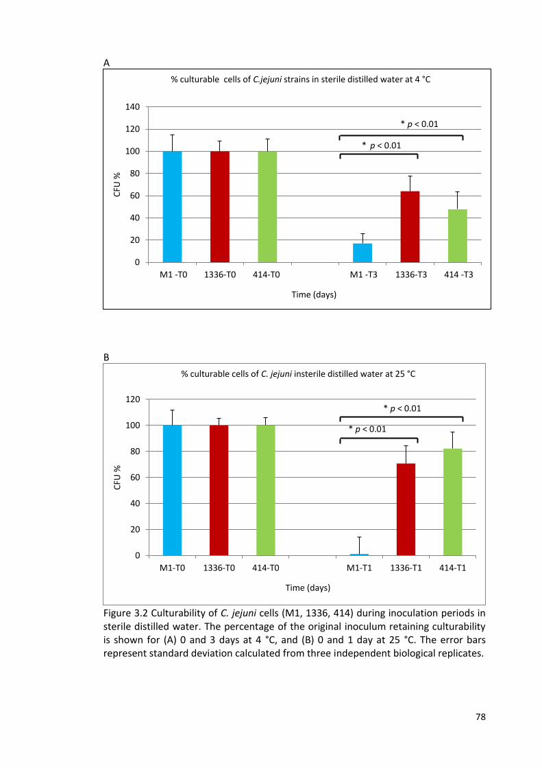
78
A
B
Figure 3.2 Culturability of C. jejuni cells (M1, 1336, 414) during inoculation periods in sterile distilled water. The percentage of the original inoculum retaining culturability is shown for (A) 0 and 3 days at 4 °C, and (B) 0 and 1 day at 25 °C. The error bars represent standard deviation calculated from three independent biological replicates.
0
20
40
60
80
100
120
140
M1 -T0 1336-T0 414-T0 M1 -T3 1336-T3 414 -T3
CFU
%
Time (days)
% culturable cells of C.jejuni strains in sterile distilled water at 4 °C
0
20
40
60
80
100
120
M1-T0 1336-T0 414-T0 M1-T1 1336-T1 414-T1
CFU
%
Time (days)
% culturable cells of C. jejuni insterile distilled water at 25 °C
* p < 0.01
* p < 0.01
* p < 0.01
* p < 0.01
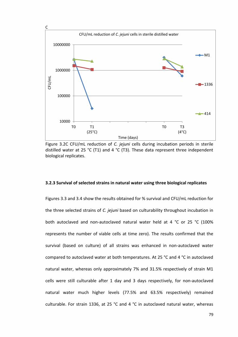
79
C
Figure 3.2C CFU/mL reduction of C. jejuni cells during incubation periods in sterile distilled water at 25 °C (T1) and 4 °C (T3). These data represent three independent biological replicates.
3.2.3 Survival of selected strains in natural water using three biological replicates
Figures 3.3 and 3.4 show the results obtained for % survival and CFU/mL reduction for
the three selected strains of C. jejuni based on culturability throughout incubation in
both autoclaved and non-autoclaved natural water held at 4 °C or 25 °C (100%
represents the number of viable cells at time zero). The results confirmed that the
survival (based on culture) of all strains was enhanced in non-autoclaved water
compared to autoclaved water at both temperatures. At 25 °C and 4 °C in autoclaved
natural water, whereas only approximately 7% and 31.5% respectively of strain M1
cells were still culturable after 1 day and 3 days respectively, for non-autoclaved
natural water much higher levels (77.5% and 63.5% respectively) remained
culturable. For strain 1336, at 25 °C and 4 °C in autoclaved natural water, whereas
10000
100000
1000000
10000000
T0 T1(25°C)
T0 T3(4°C)
CFU
/mL
Time (days)
CFU/mL reduction of C. jejuni cells in sterile distilled water
M1
1336
414
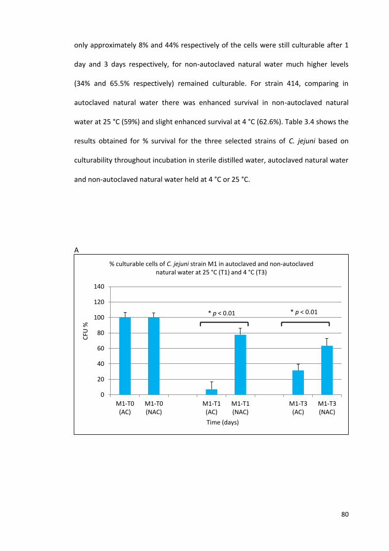
80
only approximately 8% and 44% respectively of the cells were still culturable after 1
day and 3 days respectively, for non-autoclaved natural water much higher levels
(34% and 65.5% respectively) remained culturable. For strain 414, comparing in
autoclaved natural water there was enhanced survival in non-autoclaved natural
water at 25 °C (59%) and slight enhanced survival at 4 °C (62.6%). Table 3.4 shows the
results obtained for % survival for the three selected strains of C. jejuni based on
culturability throughout incubation in sterile distilled water, autoclaved natural water
and non-autoclaved natural water held at 4 °C or 25 °C.
A
0
20
40
60
80
100
120
140
M1-T0(AC)
M1-T0(NAC)
M1-T1(AC)
M1-T1(NAC)
M1-T3(AC)
M1-T3(NAC)
CFU
%
Time (days)
% culturable cells of C. jejuni strain M1 in autoclaved and non-autoclaved natural water at 25 °C (T1) and 4 °C (T3)
* p < 0.01 * p < 0.01
81
B
C
Figure 3.3 Culturability of C. jejuni cells (M1, 1336, 414) during incubation periods in autoclaved and non-autoclaved natural water. The percentage of the original inoculum retaining culturability is shown for (A, B and C) 0 and 1 days at 25 °C, and 0 and 3 days at 4 °C. The error bars represent standard deviation calculated from three independent biological replicates. AC / autoclaved NAC/non-autoclaved
0
20
40
60
80
100
120
140
1336-T0(AC)
1336-T0(NAC)
1336-T1(AC)
1336-T1(NAC)
1336-T3(AC)
1336-T3(NAC)
CFU
%
Time (days)
% culturabe cells of C. jejuni strain 1336 in autoclaved and non-autoclaved natural water at 25°C (T1) and 4 °C (T3)
0
20
40
60
80
100
120
140
414-T0(AC)
414-T0(NAC)
414-T1(AC)
414-T1(NAC)
414-T3(AC)
414-T3(NAC)
CFU
%
Time (days)
% culturable cells of C. jejuni strain 414 in autoclaved and non-autoclaved natural water at 25 °C (T1) and 4 °C (T3)
* p < 0.01
* p < 0.01
* p = 0.15
* p = 0.05
62.6% 61.8%
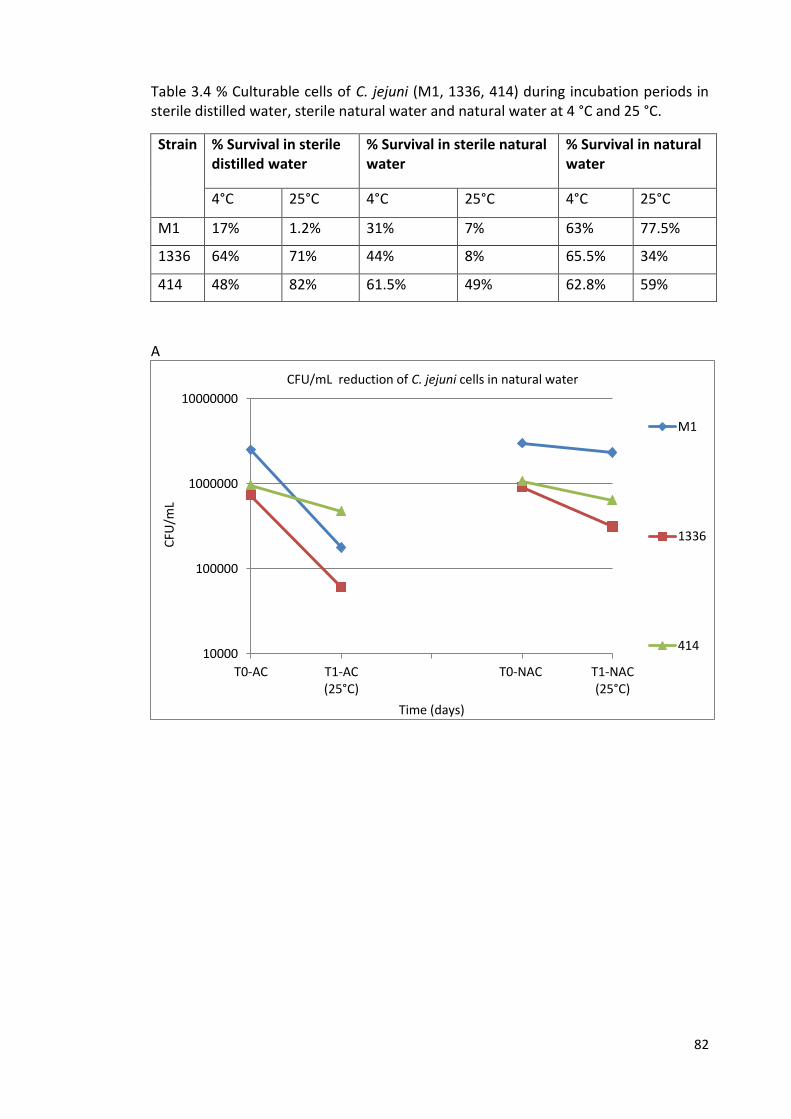
82
Table 3.4 % Culturable cells of C. jejuni (M1, 1336, 414) during incubation periods in sterile distilled water, sterile natural water and natural water at 4 °C and 25 °C.
Strain % Survival in sterile distilled water
% Survival in sterile natural water
% Survival in natural water
4°C 25°C 4°C 25°C 4°C 25°C
M1 17% 1.2% 31% 7% 63% 77.5%
1336 64% 71% 44% 8% 65.5% 34%
414 48% 82% 61.5% 49% 62.8% 59%
A
10000
100000
1000000
10000000
T0-AC T1-AC(25°C)
T0-NAC T1-NAC(25°C)
CFU
/mL
Time (days)
CFU/mL reduction of C. jejuni cells in natural water
M1
1336
414
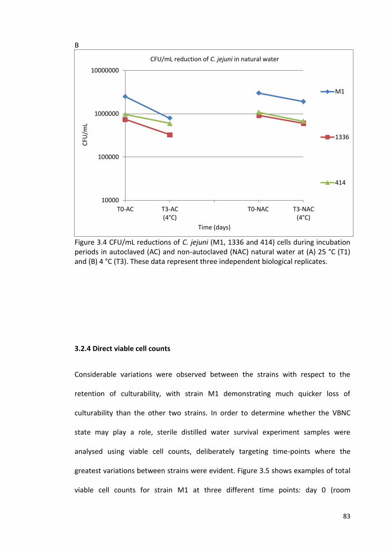
83
B
Figure 3.4 CFU/mL reductions of C. jejuni (M1, 1336 and 414) cells during incubation periods in autoclaved (AC) and non-autoclaved (NAC) natural water at (A) 25 °C (T1) and (B) 4 °C (T3). These data represent three independent biological replicates.
3.2.4 Direct viable cell counts
Considerable variations were observed between the strains with respect to the
retention of culturability, with strain M1 demonstrating much quicker loss of
culturability than the other two strains. In order to determine whether the VBNC
state may play a role, sterile distilled water survival experiment samples were
analysed using viable cell counts, deliberately targeting time-points where the
greatest variations between strains were evident. Figure 3.5 shows examples of total
viable cell counts for strain M1 at three different time points: day 0 (room
10000
100000
1000000
10000000
T0-AC T3-AC(4°C)
T0-NAC T3-NAC(4°C)
CFU
/mL
Time (days)
CFU/mL reduction of C. jejuni in natural water
M1
1336
414

84
temperature), day 1 (at 25 °C) and day 3 (at 4 °C) using fluorescence microscopy.
Whereas the prevalence of culturable cells on CBA media for these strains declined
rapidly, especially for strain M1, at both 4 °C and 25 °C (Figure 3.2C), the numbers of
viable cells (green cells) remained constant throughout the experiment at both
temperatures (p < 0.001) (Figure 3.5 and 3.6).
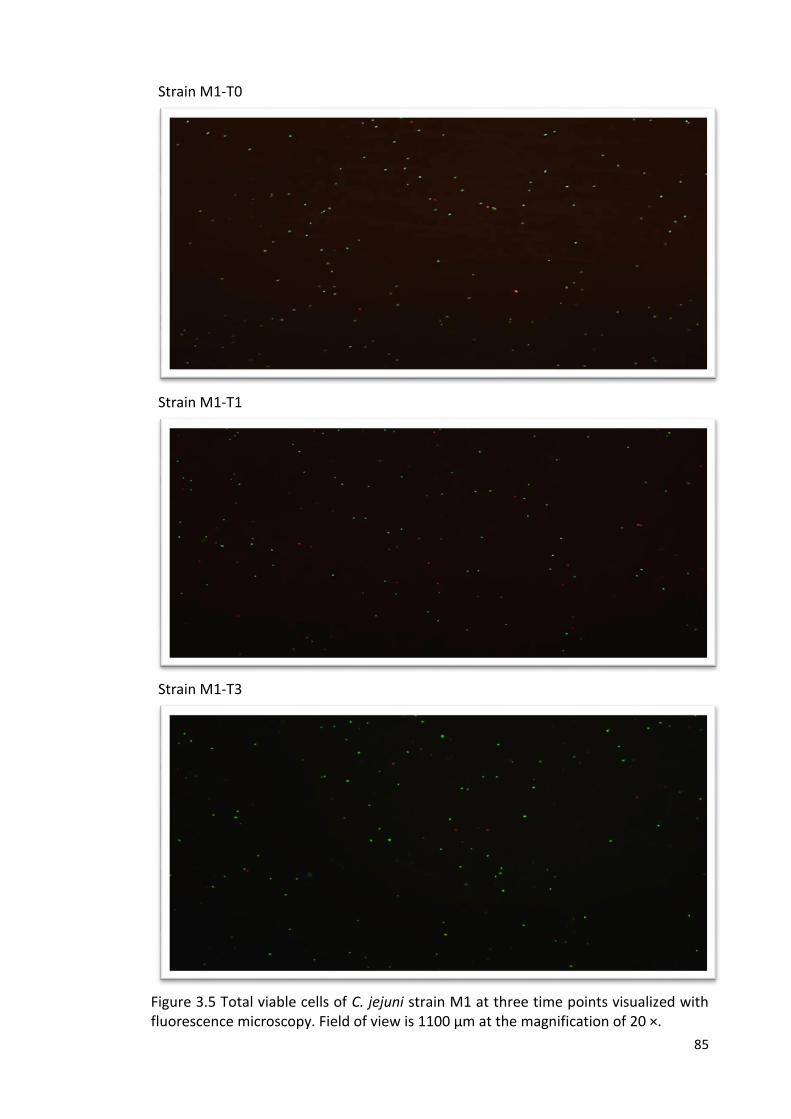
85
Strain M1-T0
Strain M1-T1
Strain M1-T3
Figure 3.5 Total viable cells of C. jejuni strain M1 at three time points visualized with fluorescence microscopy. Field of view is 1100 µm at the magnification of 20 ×.
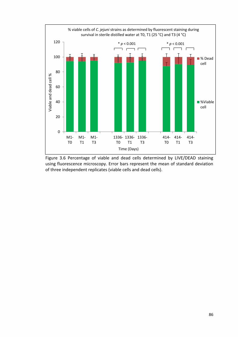
86
Figure 3.6 Percentage of viable and dead cells determined by LIVE/DEAD staining using fluorescence microscopy. Error bars represent the mean of standard deviation of three independent replicates (viable cells and dead cells).
0
20
40
60
80
100
120
M1-T0
M1-T1
M1-T3
1336-T0
1336-T1
1336-T3
414-T0
414-T1
414-T3
Via
ble
an
d d
ead
cel
l %
Time (Days)
% viable cells of C. jejuni strains as determined by fluorescent staining during survival in sterile distilled water at T0, T1 (25 °C) and T3 (4 °C)
% Deadcell
%Viablecell
* p < 0.001 * p < 0.001
87
3.3 Discussion
Although C. jejuni has been isolated from diverse animal and environmental sources,
the factors influencing the ability of C. jejuni to colonise specific hosts or survive in
the environment are poorly understood. However, there is now a persuasive body of
evidence to support the hypothesis that C. jejuni strains have adapted to exploit
specific ecological niches, suggesting that survival may vary. Several studies have
sought to determine clonal prevalence amongst isolates from diverse sources by
applying MLST (Colles et al., 2003; Dingle et al., 2005; Dingle et al., 2001; French et al.,
2005; Kärenlampi et al., 2007; Manning et al., 2003; McCarthy et al., 2007; Sails et al.,
2003). Whilst some MLST clonal complexes, such as the ST-21 complex, are
widespread, others have a more restricted provenance (Colles et al., 2003; French et
al., 2005). For example, members of the ST-45 complex (which are commonly
associated with human infection) are encountered significantly more frequently in
wildlife and water than other common clonal complexes and may be associated with
environmental exposure, particularly in rural areas (Sopwith et al., 2006). The
introduction of full allelic profiling provided further evidence for an association
between specific C. jejuni genotypes and particular host species, and has given insight
into the genetic mechanisms that permit adaptation to specific niches (McCarthy et
al., 2007).
In previous work carried out at the University of Liverpool, MLST was applied to a
large number of C. jejuni isolates obtained from a wide range of animal and
environmental sources sampled in a structured way during a narrow spatiotemporal
window (French et al., 2005; Kwan et al., 2008a). In these studies most wildlife and
water isolates possessed unusual or novel STs, unassigned to existing clonal
88
complexes and never found amongst isolates from humans or livestock (Hepworth et
al., 2011).
Gene flow analysis has revealed that although most isolates from wildlife (albeit
represented mainly by wild birds) and environmental sources represent a genetically
indistinguishable population, there is only very limited genetic exchange between this
C. jejuni population and strains associated with ruminant livestock (Kwan et al.,
2008a). These findings are consistent with the existence of restricted transmission
pathways for different C. jejuni ‘‘ecotypes’’ that have adapted to exploit the specific
niches they occupy. Those isolates solely associated with wildlife and water sources
have been referred to as the water-wildlife group and include strain 1336 (Hepworth
et al., 2011). DNA microarray technology (comparative phylogenomics) was employed
to differentiate C. jejuni into ‘livestock’ and ‘nonlivestock’ clades (Champion et al.,
2005; Stabler et al., 2013) and subsequently expanded to multiplex PCR assays
(mPCR) enabling predictive analysis to investigate if human campylobacteriosis cases
derived from domesticated (livestock) or water and wildlife sources (Stabler et al.,
2013). The specific association between ST-3704 and Cheshire bank voles appears to
be an extreme example of the well-recognised host-association for some C. jejuni
STs/CCs. Strain 414 is a representative of this group.
In this study, a selection of isolates mostly taken from the previous large surveys
carried out at the University of Liverpool was screened for variations in the ability to
survive in water. The isolates were chosen to include widespread MLST clonal
complexes (such as ST-21), including clonal complexes more often isolated from the
environment (ST-45), clonal complexes found amongst human isolates but with
evidence of host restriction (such as the cattle associated ST-61), and representatives
89
of water/wild-life specialists (1336 and the bank vole specialist 414). The isolates
were also chosen to represent a variety of sources (human, cattle, poultry,
environment, and wild-life). In the initial screen, there was no clear association
between the ability to retain culturability in the water (at either 4 °C or 25 °C) and
either MLST clonal complex or source, though there was a tendency for the ST-45
isolates to be amongst the poor or lower-intermediate survivors, based on culture.
However, the numbers representing each group were insufficient to draw many
conclusions from this comparison, which was also only based on one biological
replicate per strain. The initial screen did however enable the assignment of strains
into various categories based on their ability to retain culturability, enabling the
selection of three isolates for closer study, M1, 1336 and 414, representing different
survival abilities.
The results of this study demonstrated that the survival time (based on culture) in
sterile distilled water varies significantly between different strains of C. jejuni. Strain
M1, a representative of the ST-45 clonal complex (Friis et al., 2010), underwent the
most rapid decline in culturability of the three strains at both temperatures tested. It
has been reported previously that Campylobacter population decline occurs more
rapidly at higher temperatures (such as >20 °C) (Cools et al., 2003; Guillou et al.,
2008; Thomas et al., 1999) and this was the case for each of the three strains
analysed in detail.
Experiments were carried out in both autoclaved and non-autoclaved natural water
to compare the same three strains. In general, the results obtained in autoclaved
natural water resembled those obtained in sterile distilled water. In particular,
differences between the strains were observed, with strain 414 better able to retain
90
culturability than the other two strains. However, it was notable, especially for strain
M1, that culturability was better retained in non-autoclaved natural water, suggesting
that some component of the natural water system that is sensitive to autoclaving can
help strain M1 to retain culturability. This study was not designed to identify the
active component, but possible candidates are other microorganisms or temperature-
sensitive secreted products from other microorganisms. This observation does
however support the idea that bacteria may behave very differently in complex
ecological systems than they do in simplified laboratory systems.
It has been shown that survival times of Campylobacter in water microcosms,
representing environmental water, were temperature-dependant and origin-
dependant. They are generally able to survive much better at low temperatures (4
and 10 °C), untreated natural water and are able to survive for long time in well water
(Buswell et al., 1998; Cools et al., 2003; González and Hänninen, 2012; Korhonen and
Martikalnon, 1991). These studies are compatible with the concept of strain
discrimination on the basis of survival in water. Some strains of C. jejuni appear to be
adapted to survive well in environmental water and able to cause a risk of human
campylobacteriosis through the consumption of this contaminated water.
Strain M1 is ST-137, a member of the ST-45 clonal complex, and was isolated from a
human infection associated with transmission from chicken (Friis et al., 2010). It has
also been used in experiments to highlight strain to strain variations in the infection
ecology of C. jejuni in chickens (Chaloner et al., 2014). Given that the ST-45 clonal
complex is more often associated with isolation from the environment (French et al.,
2005), it is perhaps surprising that strain M1, the ST-45 isolate used in this study, was
apparently the least able to survive (least culturable). However, it should also be
91
noted that considerable genomic divergence can occur within clonal complexes
(Revez et al., 2014), and that adaptation to a new host can lead to alterations in
phenotype (Hanel et al., 2009; Leblanc-Maridor et al., 2011). Campylobacter genomes
contain numerous contingency loci that can facilitate such adaptation (Jerome et al.,
2011; Thomas et al., 2014). In addition, other adaptations can also occur (Lefébure
and Stanhope, 2009). Indeed, phenotypic instability due to high frequency phase
variations makes Campylobacter spp. notoriously difficult to work with (Bayliss et al.,
2012). Hence, the lower ability of strain M1, a human isolate that had also passed
through chicken, could have been due to adaptive changes. Strain 1336 and 414 had
not been exposed to the same host environments. However, more rapid loss of
culturability amongst members of the ST-45 complex was also found in the initial
screen on a larger selection of isolates from different sources, where the ST-45
isolates were generally poor or lower intermediate survivors (Table.3.1 and 3.2).
The result of the LIVE/DEAD staining (fluorescent microscopy)(Fakruddin et al., 2013)
experiments indicated that the viability of the C. jejuni cells remained constant,
suggesting that the loss of culturability may not indicate poor survival, but could be
due to the ability of strain M1 to enter more quickly into a VBNC state in nutrient-
poor environment such as sterile distilled water. Previous studies (Federighi et al.,
1998; Guillou et al., 2008; Uyttendaele et al., 2001) demonstrated this phenomenon
in microcosm water. Even though the time-points at which strain variations based on
culture were most evident were selected [4 °C (for day 3) and at 25 °C (for day 1)],
variations in the percentage of viable cells were minor (Figure 3.6). Hence, it is
feasible that some strains (such as strain M1) might actually enhance their survival in
the environment by being able to enter the VBNC state more rapidly than others.
92
Further work is needed to fully characterise the extent of variations between
different strains of C. jejuni, and to better determine whether there are clear
associations between particular genotypes (such as clonal complexes) and the rate of
loss of culturabilty, and whether this correlates with observations about the relative
prevalence in environmental samples. However, it should also be noted that most
environmental surveys rely on culture to characterise the Campylobacter populations
present. This does not take into account the issue of whether VBNC cells are present.
However, the ST-45 complex is well represented amongst isolates from the
environment, suggesting that loss of culturability is not an issue that leads to the ST-
45 complex being undetectable in natural samples. Clearly, the situation is more
complex in natural environments. However, the work presented here does suggest
that there are variations between strains and that such variations could play a role in
the better survival (or better culturability) of some strains in natural environmental
samples.
Although the nature of the VBNC state remains controversial, the present study is in
agreement with findings suggesting that Campylobacter can enter a VBNC state under
stress conditions such as in nutrient-poor water (Baffone et al., 2006; Chaveerach et
al., 2003; Rollins and Colwell, 1986; Tholozan et al., 1999). It also supports previous
observations that the capability of C. jejuni to enter the VBNC state varies between
different strains (Cools et al., 2003). It has been shown previously that resuscitation
from a VBNC state to cause infections can also vary (Beumer et al., 1992; Mederma et
al., 1992) and that the physiology of the VBNC cells can vary according to temperature
(Hazeleger et al., 1995). Other studies contradict the idea of resuscitation to cause
infection and conclude that it is doubtful that the VBNC state plays an important role
93
in Campylobacter epidemiology (Hald et al., 2001; Ziprin et al., 2003; Ziprin and
Harvey, 2004). However, given that there are evident variations between strains, and
that the VBNC state itself may vary depending upon the conditions used to induce it,
there may be no clear single answer to the question of how important the VBNC state
is.
The survival experiments of Campylobacter in water were conducted in the dark
because it has been reported previously that survival time could be affected when
bacteria are exposed to the light due to the lethal effect of Ultraviolet (UV) radiation
(Obiri‐Danso et al., 2001). We used a non-selective medium (CBA) for recovery and
counting CFUs of C. jejuni strains in sterile distilled water because the recovery of
stressed bacterial cells, due to starvation, on selective media is lower (Cools et al.,
2003; Uyttendaele et al., 2001). Furthermore, our results confirm the studies showing
that refrigerated temperatures support the survival of C. jejuni in drinking water
including tap water, and bottled water (Cools et al., 2003; Evans et al., 2003; González
et al., 2009; Guillou et al., 2008).
This study provides clear evidence that there are variations between strains in terms
of the ability to retain culturability in water, and the rate at which C. jejuni can enter a
VBNC state. However, it will be necessary to screen far larger numbers of isolates
from different sources and representing different clonal complex, to identify any clear
links between these variations and the epidemiology of Campylobacter infections.
94
3.4 Conclusions
Survival in water, based on culturability, varies between different strains of C. jejuni,
with potential implications for strain transmission. However, LIVE/DEAD staining
suggests that cells remain viable in samples where culturability has dropped to very
low levels. The retention of culturability is prolonged at low temperatures as well as in
untreated natural water. These variations may contribute to the fact that some strains
of C. jejuni are better adapted to survive in aquatic environments, contributing to the
transmission of Campylobacter species from the environment, directly or indirectly to
humans, constituting public health threat. Our observations are consistent with the
notion that Campylobacter can enter into a VBNC state in response to starvation
(nutrient-poor environment) such as water, but that this ability varies between
strains, and may be reduced in more complex natural environments.
95
C h a p t e r 4
GENE EXPRESSION DURING SURVIVAL IN STERILE DISTILLED WATER
4.1 Introduction
In chapter 3, clear variations between strains with respect to the ability to retain
culturability were demonstrated. In order to survive outside the host body, C. jejuni
must confront and survive conditions where they are under stress. Despite the lack of
several common stress response genes that exist in other bacteria such as Escherichia
coli, C. jejuni are able to overcome harsh environmental conditions during
transmission (Gaynor et al., 2005; Kassem and Rajashekara, 2011). Results that have
been obtained from PCR amplification and DNA sequencing in previous studies
indicate that the C. jejuni genome contains a luxS gene which encodes autoinducer-2
(AI-2), known as a quorum sensing (QS) signal molecule (Cloak et al., 2002).
Additionally, this gene may play an important role in response to oxidative stress,
ability in colonisation, expression of CDT, expression of flagellar genes and regulation
of motility (Plummer, 2012). Studies have shown that C. jejuni is able to use the
stringent response to control various stress survival and virulence characteristics such
as survival in stationary phase, survival under high oxygen rate/low carbon dioxide
rate conditions and resisting rifampicin. This involves the key regulator gene spoT
(Gaynor et al., 2005). C. jejuni needs to adapt to starvation and survive in nutrient-
poor environments such as water. It was shown that a mutant C. jejuni (Δppk1) was
deficient in poly-P (energy and phosphate store) accumulation, associated with a
decreased ability to form VBNC cells under stress. In addition, this gene might
contribute to each of natural transmission, antibiotic and bile acid resistance and
regulation of stress starvation via synthesis of poly-P (Gangaiah et al., 2009). Although
96
there have been few studies focusing on the coccoid form of Campylobacter, it has
been suggested that the C. jejuni amiA gene is involved in transforming the cells into
coccoid form (Ikeda and Karlyshev, 2012; Nachamkin et al., 2008). It has also been
indicated by using Q-PCR that expression of the C. jejuni htrB gene may be critical for
survival and stress responses such as heat, acid, osmotic and oxidative stresses
(Phongsisay et al., 2007). A mutant C. jejuni (ΔhtrA) showed defective growth at 44 °C
and other characteristics, suggesting that the htrA gene is essential for C. jejuni to
resist heat, protect periplasmic proteins from misfolding, survive at high oxygen
tensions and to invade epithelial cells in human gut (Andersen et al., 2005; Baek et al.,
2011a; Brøndsted et al., 2005). It is essential for C. jejuni during transmission to
counter atmospheric oxygen tensions and the toxicity of reactive oxygen species
(ROS). These toxic products of the metabolism of oxygen can cause oxidative damage
to the nucleic acid of the organism. It has been shown that the expression of the
catalase (katA) gene but not the superoxide dismutase (sodB) gene, the main
antioxidant defence of most organisms, of C. jejuni increased after exposure to
oxidative stress (Garénaux et al., 2008), despite the important role for sodB gene of C.
jejuni in intracellular survival (Pesci et al., 1994). Additionally, it has been suggested
that catalase in C. jejuni might play an important role in intracellular survival (Day et
al., 2000). Moreover, an ankyrin-containing protein (Cj1386) has recently been
identified in C. jejuni, encoded by a gene based directly downstream of the katA gene,
and is thought to be involved in the same detoxification pathway as catalase (Flint et
al., 2012). Amongst three classified types of superoxide dismutases (SODs) based on
their metal cofactors, all possessed by Escherichia coli, C. jejuni only possess one type
(SodB) (Bakshi et al., 2006; Purdy et al., 1999).
97
Unlike E. coli, in which alkyl hydroxide reductase (Ahp) consists of the catalytic site
(AhpC) and flavoprotein (AhpF) subunit, C. jejuni only possess the catalytic site (AhpC)
of Ahp. This protein may play a role in oxidative stress resistance in C. jejuni (Atack
and Kelly, 2009; Baillon et al., 1999; Poole et al., 2000). Furthermore, C. jejuni is the
first Gram-negative organism to be described in which the expression of katA and
ahpC genes are regulated by ‘‘peroxide stress regulator’’ (PerR) but not oxidative
stress regulator (OxyR) (Van Vliet et al., 1999). It has been shown that in C. jejuni,
methionine sulphoxide reductases (MsrA and MsrB) appear to be encoded by two
genes (Cj0637c and Cj1112c) and may play a role in repairing the oxidized methionine
residues (MetO) in proteins (Atack and Kelly, 2008; Moskovitz, 2005). C. jejuni is able
to avoid the damaging effect of iron-mediated oxidative stress on the cells by
acquisition of the iron-binding proteins ferritin and Dps, which contribute to iron
assimilation and protection from ROS by sequestering intracellular free iron (Ishikawa
et al., 2003; Wai et al., 1996). For C. jejuni to colonise its host in the human gut (37 °C)
and the chicken gut (42 °C), the organism may up-regulate the expression of various
genes at these two temperatures. For instance, it has been shown that the
temperature change can alter expression of up to 336 genes in C. jejuni (Stintzi,
2003).
In general, C. jejuni as a respiratory microorganism is unable to utilize sugars as a
carbon source due to the lack of the glycolytic enzyme phosphofructokinase (Parkhill
et al., 2000; Sellars et al., 2002; Velayudhan and Kelly, 2002). Therefore, C. jejuni
depend on the availability of amino acids such as serine, glutamine, aspartate and
proline for its growth (Guccione et al., 2008; Hofreuter et al., 2008). The
immunogenic protein (Cj0917) in C. jejuni, is homologous to carbon starvation protein

98
A (CstA) in E. coli, and is thought to play a role in in vivo peptide uptake. It is the main
up-regulated gene during starvation (Rasmussen et al., 2013).
For the purpose of this study, a sub-set of stress response genes (Table 4.1 and Figure
4.1), highlighted in the literature as important for survival in stressful conditions, was
selected for analysis using Q-PCR assays during survival of C. jejuni strains in sterile
distilled water at two different temperatures .
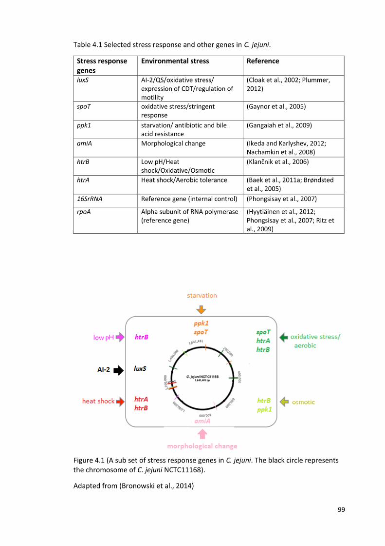
99
Table 4.1 Selected stress response and other genes in C. jejuni.
Stress response genes
Environmental stress Reference
luxS AI-2/QS/oxidative stress/ expression of CDT/regulation of motility
(Cloak et al., 2002; Plummer, 2012)
spoT oxidative stress/stringent response
(Gaynor et al., 2005)
ppk1 starvation/ antibiotic and bile acid resistance
(Gangaiah et al., 2009)
amiA Morphological change (Ikeda and Karlyshev, 2012; Nachamkin et al., 2008)
htrB Low pH/Heat shock/Oxidative/Osmotic
(Klančnik et al., 2006)
htrA Heat shock/Aerobic tolerance (Baek et al., 2011a; Brøndsted et al., 2005)
16SrRNA Reference gene (internal control) (Phongsisay et al., 2007)
rpoA Alpha subunit of RNA polymerase (reference gene)
(Hyytiäinen et al., 2012; Phongsisay et al., 2007; Ritz et al., 2009)
Figure 4.1 (A sub set of stress response genes in C. jejuni. The black circle represents the chromosome of C. jejuni NCTC11168).
Adapted from (Bronowski et al., 2014)
100
4.1.2 Aims of this chapter
The overall aim of this chapter was to identify genes that might play an important
role in the differences observed between strains in the ability to retain culturability
during survival in water, as described in the previous chapter. This will be achieved
by:
1. At selected time-points, in both 4 °C and 25 °C water survival experiments,
determining differences in gene expression of a panel of previously reported stress
response genes between the different C. jejuni strains (M1, 1336 and 414) using end-
point PCR assays.
2. Developing Q-PCR assays for selected response genes.
3. At selected time-points, in both 4 °C and 25 °C water survival experiments, using Q-
PCR assays to determine variations in gene expression of stress response genes
between the C. jejuni strains
101
4.2 Results
4.2.1 Temperature gradient PCR to determine optimal annealing temperatures for
end-point PCR assays
We used Q-PCR to look for variations in the expression of 8 selected stress response
genes (Table 4.1) highlighted in the literature as important for survival in low nutrient
or stressful conditions, or the formation of biofilm/VBNC forms. For targeting these
genes, four oligonucleotide primer pairs were designed (section 2.9.1) for the
purpose of this study and the other four primer pairs (published previously) were
purchased from SIGMA (Table 2.3).
Temperature gradient PCR was performed to determine the optimal annealing
temperatures of the oligonucleotide primers used in this study. For each primer pair
there were a range of temperatures at which single amplicons of the correct size
were obtained. The temperature (55 °C) was selected as the annealing temperature
for all of the primer pairs.
To test the ability of the primers to amplify from each of the strains used in this study
using the annealing temperature 55 °C, the primer pairs were next tested on C. jejuni
strains M1, 1336, 414. The result showed that the amplification primers were
effective at annealing temperature 55 °C. Furthermore, non-specific PCR products
were not detected from the no template control. Interestingly, this experiment
revealed the absence of luxS gene from the genome of C. jejuni strain 1336 (Figure
4.2).
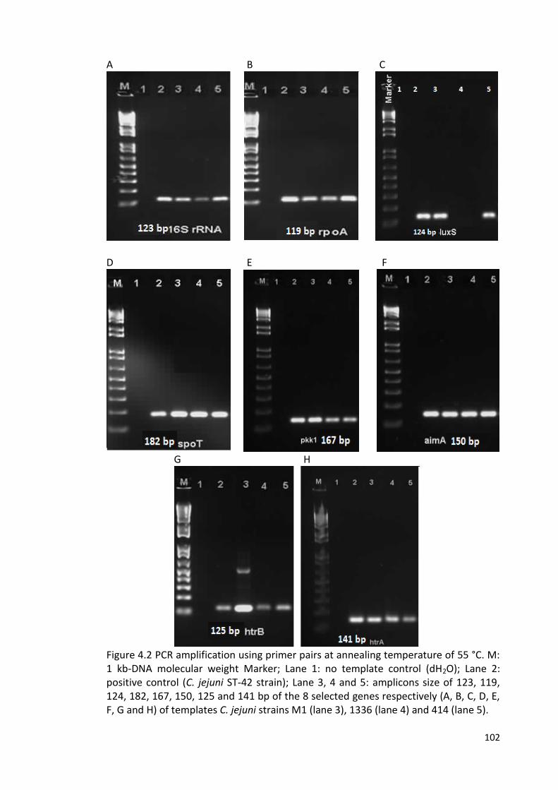
102
A B C
D E F
G H
Figure 4.2 PCR amplification using primer pairs at annealing temperature of 55 °C. M: 1 kb-DNA molecular weight Marker; Lane 1: no template control (dH2O); Lane 2: positive control (C. jejuni ST-42 strain); Lane 3, 4 and 5: amplicons size of 123, 119, 124, 182, 167, 150, 125 and 141 bp of the 8 selected genes respectively (A, B, C, D, E, F, G and H) of templates C. jejuni strains M1 (lane 3), 1336 (lane 4) and 414 (lane 5).
103
4.2.2 RNA extraction and conversion to cDNA
At T0 (day 0 at room temperature) and for the days where we identified greatest
variation between strains (M1, 1336, 414) with respect to maintenance of
culturability, T1 (day 1 at 25 °C) and T3 (day 3 at 4 °C), RNA was extracted from the
water survival experiment and the amounts and the purity of extracted RNA and
cDNA of C. jejuni strains were determined by using a NanoDrop-1000
spectrophotometer (Thermo Scientific). The absorbance was measured at 260 nm
(A260) and the purity was determined by measuring A260/A280 ratio (Table 4.2A and
Table 4.2B).
4.2.3 PCR amplification of cDNA
In order to determine whether expression of various stress genes was detectable at
the chosen time points in the water survival experiments, a series of end-point PCR
assays were carried out on the cDNA samples.
Figures 4.3-4.10 show the results of end point PCR amplification of cDNA targeting
specific stress response genes and control genes during survival of C. jejuni strains
M1, 1336 and 414 in sterile distilled water at days 0, 1 (for 25 °C) and 3 (for 4 °C).
Expression of gene-specific cDNA levels of the stress response genes was evaluated
and is summarised in Table 4.3.
The rpoA gene was chosen as the reference (control) gene for this study because it
had been reported to be “the most suitable internal control to study stress response
in C. jejuni by RT-qPCR” (Ritz et al., 2009). However, because detection of expression
of the rpoA gene was not consistent, 16S rRNA was chosen as the reference (control)
gene for subsequent Q-PCR assays rather than rpoA.
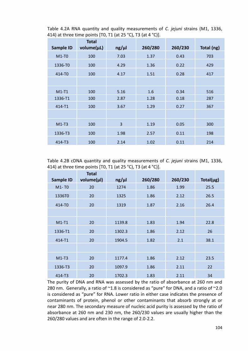
104
Table 4.2A RNA quantity and quality measurements of C. jejuni strains (M1, 1336, 414) at three time points [T0, T1 (at 25 °C), T3 (at 4 °C)].
Sample ID Total
volume(µL) ng/µl 260/280 260/230
Total (ng)
M1-T0 100 7.03 1.37 0.43 703
1336-T0 100 4.29 1.36 0.22 429
414-T0 100 4.17 1.51 0.28 417
M1-T1 100 5.16 1.6 0.34 516
1336-T1 100 2.87 1.28 0.18 287
414-T1 100 3.67 1.29 0.27 367
M1-T3 100 3 1.19 0.05 300
1336-T3 100 1.98 2.57 0.11 198
414-T3 100 2.14 1.02 0.11 214
Table 4.2B cDNA quantity and quality measurements of C. jejuni strains (M1, 1336, 414) at three time points [T0, T1 (at 25 °C), T3 (at 4 °C)].
Sample ID Total
volume(µl) ng/µl 260/280 260/230
Total(µg)
M1- T0 20 1274 1.86 1.99 25.5
1336T0 20 1325 1.86 2.12 26.5
414-T0 20 1319 1.87 2.16 26.4
M1-T1 20 1139.8 1.83 1.94 22.8
1336-T1 20 1302.3 1.86 2.12 26
414-T1 20 1904.5 1.82 2.1 38.1
M1-T3 20 1177.4 1.86 2.12 23.5
1336-T3 20 1097.9 1.86 2.11 22
414-T3 20 1702.3 1.83 2.11 34
The purity of DNA and RNA was assessed by the ratio of absorbance at 260 nm and 280 nm. Generally, a ratio of ~1.8 is considered as “pure” for DNA, and a ratio of ~2.0 is considered as “pure” for RNA. Lower ratio in either case indicates the presence of contaminants of protein, phenol or other contaminants that absorb strongly at or near 280 nm. The secondary measure of nucleic acid purity is assessed by the ratio of absorbance at 260 nm and 230 nm, the 260/230 values are usually higher than the 260/280 values and are often in the range of 2.0-2.2.
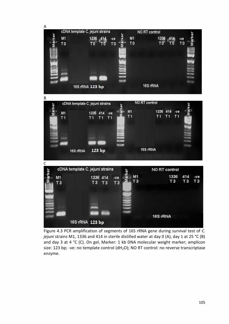
105
A
B
C
Figure 4.3 PCR amplification of segments of 16S rRNA gene during survival test of C. jejuni strains M1, 1336 and 414 in sterile distilled water at day 0 (A), day 1 at 25 °C (B) and day 3 at 4 °C (C). On gel, Marker: 1 kb DNA molecular weight marker; amplicon size: 123 bp; -ve: no template control (dH2O); NO RT control: no reverse transcriptase enzyme.
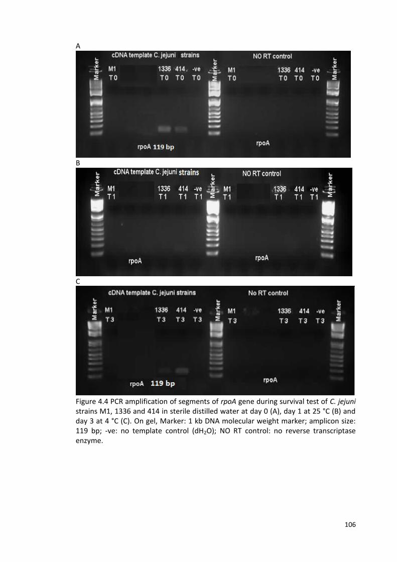
106
A
B
C
Figure 4.4 PCR amplification of segments of rpoA gene during survival test of C. jejuni strains M1, 1336 and 414 in sterile distilled water at day 0 (A), day 1 at 25 °C (B) and day 3 at 4 °C (C). On gel, Marker: 1 kb DNA molecular weight marker; amplicon size: 119 bp; -ve: no template control (dH2O); NO RT control: no reverse transcriptase enzyme.
107
A
B
C
Figure 4.5 PCR amplification of segments of luxS gene during survival test of C. jejuni strains M1, 1336 and 414 in sterile distilled water at day 0 (A), day 1 at 25 °C (B) and day 3 at 4 °C (C). On gel, Marker: 1 kb DNA molecular weight marker; amplicon size: 124 bp; -ve: no template control (dH2O); NO RT control: no reverse transcriptase enzyme.
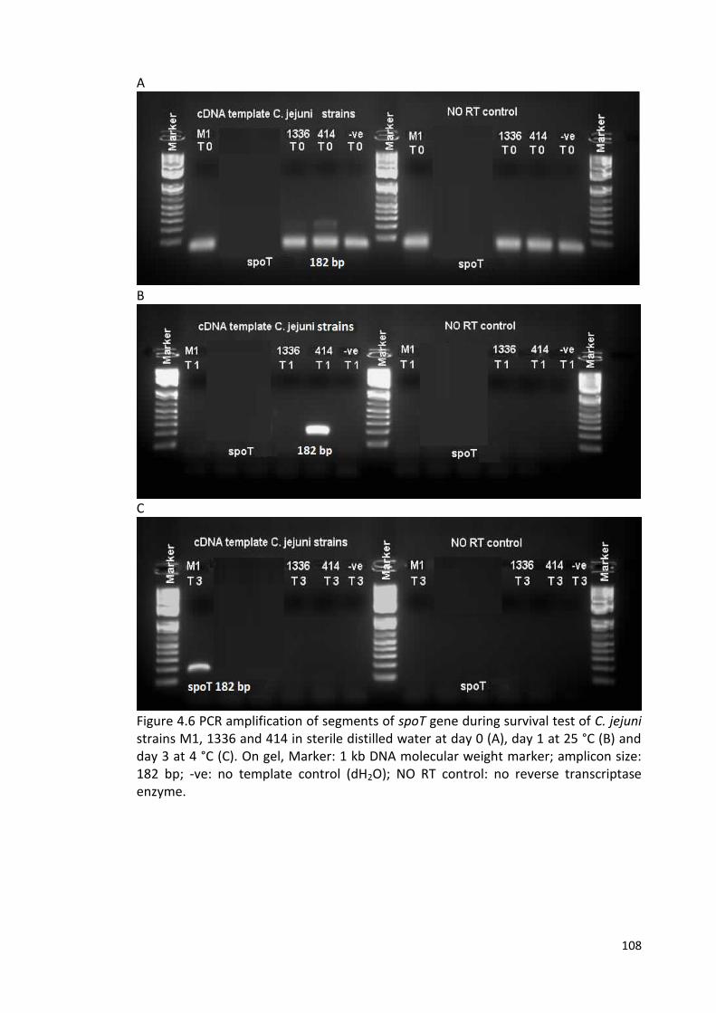
108
A
B
C
Figure 4.6 PCR amplification of segments of spoT gene during survival test of C. jejuni strains M1, 1336 and 414 in sterile distilled water at day 0 (A), day 1 at 25 °C (B) and day 3 at 4 °C (C). On gel, Marker: 1 kb DNA molecular weight marker; amplicon size: 182 bp; -ve: no template control (dH2O); NO RT control: no reverse transcriptase enzyme.
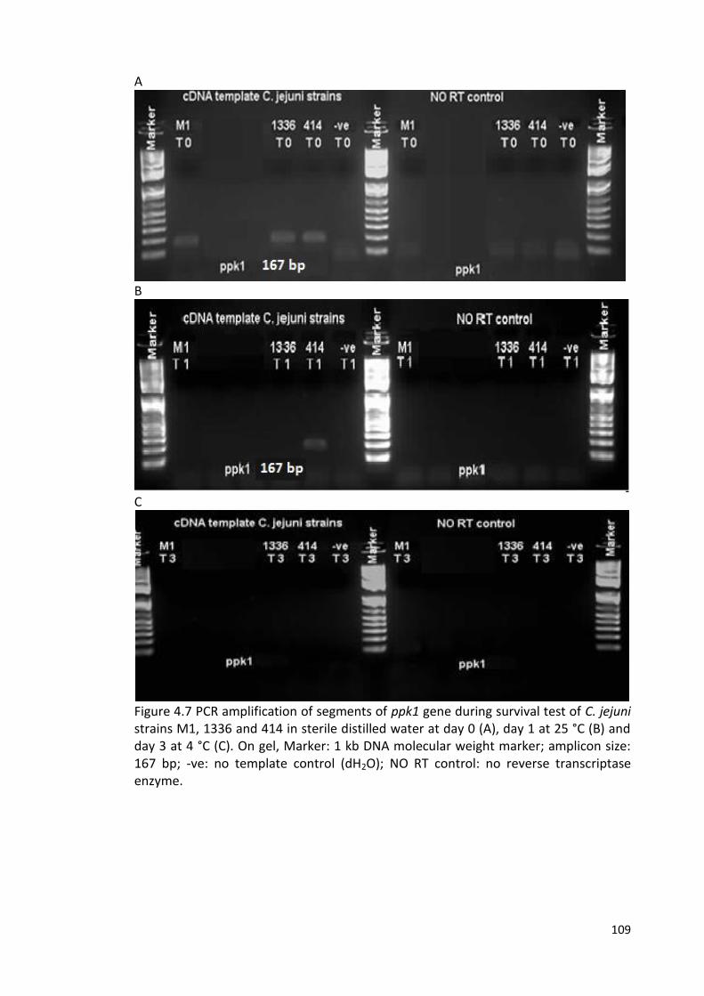
109
A
B
C
Figure 4.7 PCR amplification of segments of ppk1 gene during survival test of C. jejuni strains M1, 1336 and 414 in sterile distilled water at day 0 (A), day 1 at 25 °C (B) and day 3 at 4 °C (C). On gel, Marker: 1 kb DNA molecular weight marker; amplicon size: 167 bp; -ve: no template control (dH2O); NO RT control: no reverse transcriptase enzyme.
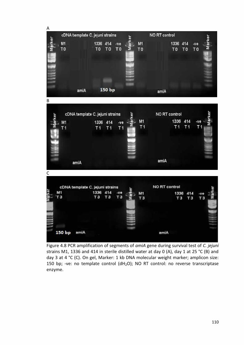
110
A
B
C
Figure 4.8 PCR amplification of segments of amiA gene during survival test of C. jejuni strains M1, 1336 and 414 in sterile distilled water at day 0 (A), day 1 at 25 °C (B) and day 3 at 4 °C (C). On gel, Marker: 1 kb DNA molecular weight marker; amplicon size: 150 bp; -ve: no template control (dH2O); NO RT control: no reverse transcriptase enzyme.
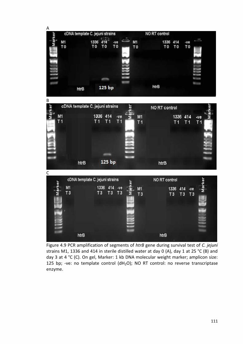
111
A
B
C
Figure 4.9 PCR amplification of segments of htrB gene during survival test of C. jejuni strains M1, 1336 and 414 in sterile distilled water at day 0 (A), day 1 at 25 °C (B) and day 3 at 4 °C (C). On gel, Marker: 1 kb DNA molecular weight marker; amplicon size: 125 bp; -ve: no template control (dH2O); NO RT control: no reverse transcriptase enzyme.
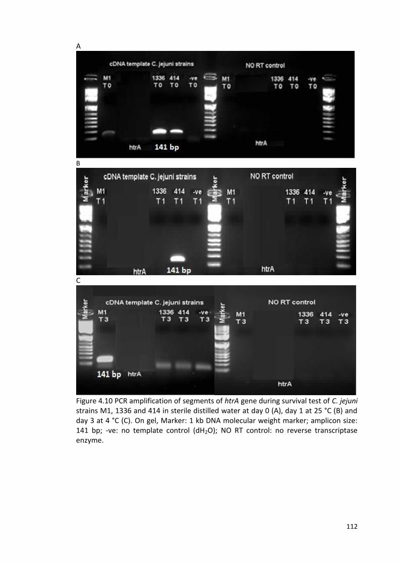
112
A
B
C
Figure 4.10 PCR amplification of segments of htrA gene during survival test of C. jejuni strains M1, 1336 and 414 in sterile distilled water at day 0 (A), day 1 at 25 °C (B) and day 3 at 4 °C (C). On gel, Marker: 1 kb DNA molecular weight marker; amplicon size: 141 bp; -ve: no template control (dH2O); NO RT control: no reverse transcriptase enzyme.
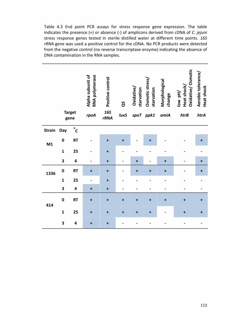
113
Table 4.3 End point PCR assays for stress response gene expression. The table indicates the presence (+) or absence (-) of amplicons derived from cDNA of C. jejuni stress response genes tested in sterile distilled water at different time points. 16S rRNA gene was used a positive control for the cDNA. No PCR products were detected from the negative control (no reverse transcriptase enzyme) indicating the absence of DNA contamination in the RNA samples.
Alp
ha
sub
un
it o
f R
NA
po
lym
eras
e
Po
siti
ve c
on
tro
l
QS
Oxi
dat
ive/
st
arva
tio
n
Osm
oti
c st
ress
/ st
arva
tio
n
Mo
rph
olo
gica
l
chan
ge
low
pH
/ H
eat
sho
ck/
Oxi
dat
ive/
Osm
oti
c
Aer
ob
ic t
ole
ran
ce/
Hea
t sh
ock
Target gene rpoA 16S
rRNA luxS spoT ppk1 amiA htrB htrA
Strain Day o
C
M1
0 RT - + + - + - - +
1 25 - + - - - - - -
3 4 - + - + - + - +
1336
0 RT + + - + + + - +
1 25 - + - - - - - -
3 4 + + - - - - - -
414
0 RT + + + + + + + +
1 25 + + + + + - + +
3 4 + + - - - - - -
114
4.2.4 Development of Q-PCR assays
Primer pairs targeting four genes (amiA, ppk1, spoT and htrA) were designed for the
purpose of this study as follows: genome sequences for four strains (M1, NCTC11168,
1336, 414) were downloaded from NCBI using Geneious version 6.1.2 (Biomathes Ltd)
and the genes of interest were aligned (Figures 4.11-4.14). Primer-3, which is
integrated into Geneious, was used to design primers specific to the genes of interest,
using default settings. Primers were designed with Q-PCR in mind, aiming for
amplicon sizes at around 180 bp. Initially, temperature gradient PCR was performed
to determine optimal annealing temperature of the primers used in this study (Table
2.3). The temperature (55 °C) was selected as optimal annealing temperature for all
of the primer pairs. All of the primer pairs were next tested on DNA from C. jejuni
strains M1, 1336, 414 for amplification at annealing temperature 55 °C. Standard
curves were obtained from a 10-fold dilution series of genomic DNA of C. jejuni (ST-
42). The Cq values were plotted against log10 concentration of the genomic DNA. The
Cq values determined by software analysis of the unknown samples were compared
to the standard curve to determine the concentration of each unknown sample. The
efficiency and the coefficient (R2) of the standard curves were calculated
automatically by the software for the Rotor-gene Q cycler.
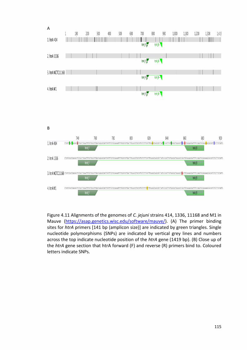
115
A
B
Figure 4.11 Alignments of the genomes of C. jejuni strains 414, 1336, 11168 and M1 in Mauve (https://asap.genetics.wisc.edu/software/mauve/). (A) The primer binding sites for htrA primers [141 bp (amplicon size)] are indicated by green triangles. Single nucleotide polymorphisms (SNPs) are indicated by vertical grey lines and numbers across the top indicate nucleotide position of the htrA gene (1419 bp). (B) Close up of the htrA gene section that htrA forward (F) and reverse (R) primers bind to. Coloured letters indicate SNPs.
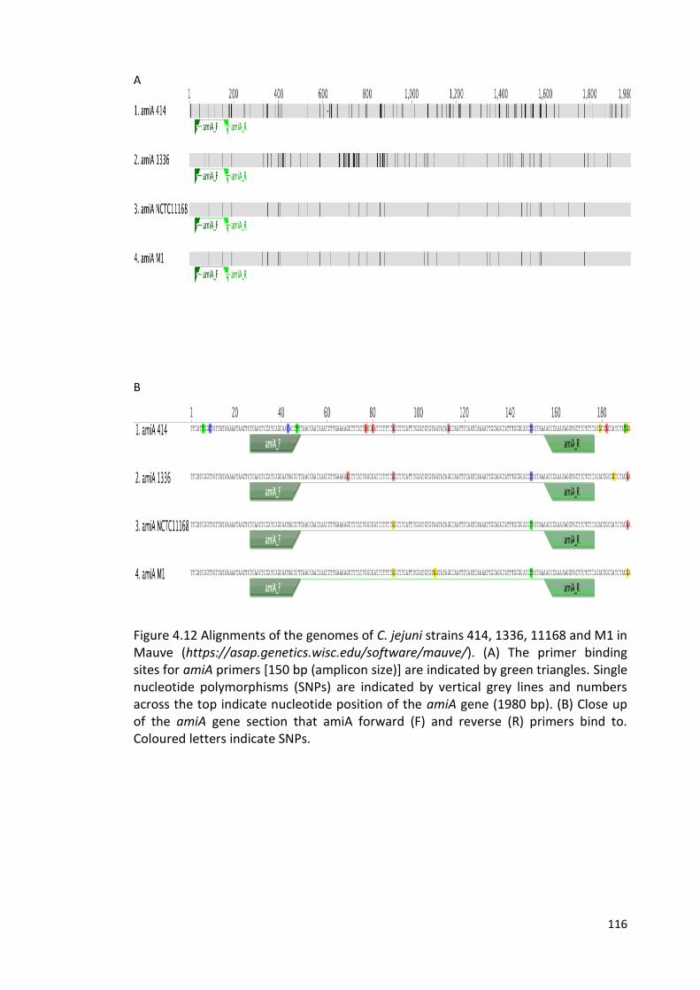
116
A
B
Figure 4.12 Alignments of the genomes of C. jejuni strains 414, 1336, 11168 and M1 in Mauve (https://asap.genetics.wisc.edu/software/mauve/). (A) The primer binding sites for amiA primers [150 bp (amplicon size)] are indicated by green triangles. Single nucleotide polymorphisms (SNPs) are indicated by vertical grey lines and numbers across the top indicate nucleotide position of the amiA gene (1980 bp). (B) Close up of the amiA gene section that amiA forward (F) and reverse (R) primers bind to. Coloured letters indicate SNPs.
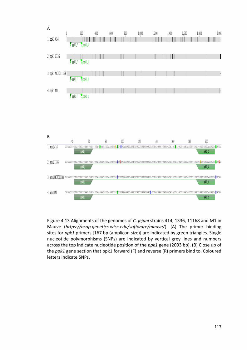
117
A
B
Figure 4.13 Alignments of the genomes of C. jejuni strains 414, 1336, 11168 and M1 in Mauve (https://asap.genetics.wisc.edu/software/mauve/). (A) The primer binding sites for ppk1 primers [167 bp (amplicon size)] are indicated by green triangles. Single nucleotide polymorphisms (SNPs) are indicated by vertical grey lines and numbers across the top indicate nucleotide position of the ppk1 gene (2093 bp). (B) Close up of the ppk1 gene section that ppk1 forward (F) and reverse (R) primers bind to. Coloured letters indicate SNPs.
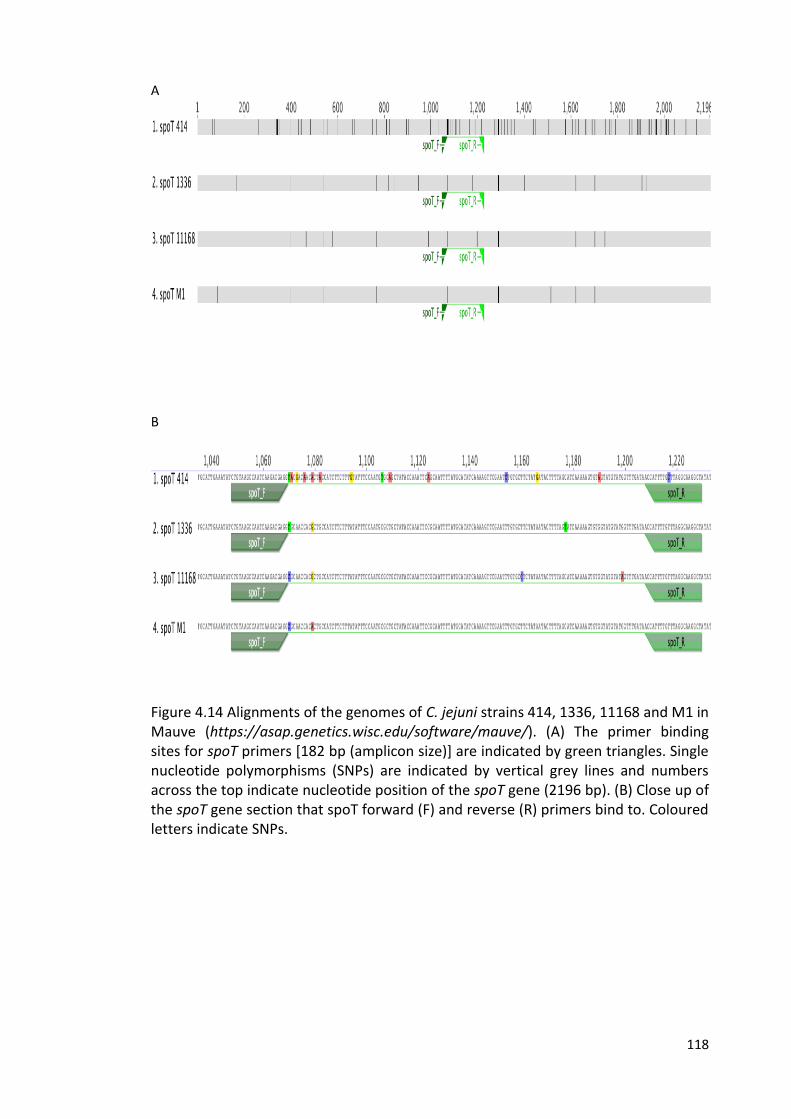
118
A
B
Figure 4.14 Alignments of the genomes of C. jejuni strains 414, 1336, 11168 and M1 in Mauve (https://asap.genetics.wisc.edu/software/mauve/). (A) The primer binding sites for spoT primers [182 bp (amplicon size)] are indicated by green triangles. Single nucleotide polymorphisms (SNPs) are indicated by vertical grey lines and numbers across the top indicate nucleotide position of the spoT gene (2196 bp). (B) Close up of the spoT gene section that spoT forward (F) and reverse (R) primers bind to. Coloured letters indicate SNPs.
119
4.2.5 Gene expression analysis using Q-PCR
Given the limitations of end-point PCR with respect to quantification, further
investigations were carried out using Q-PCR assays.
For the days where we identified greatest variation between C. jejuni strains with
respect to maintenance of culturability (Chapter 3), we used 2-step Q-PCR to look for
variations in the expression of our chosen panel of stress response genes highlighted
in the literature as important for survival in low nutrient environment or stressful
conditions, or the formation of biofilm/VBNC forms (Table 4.1). The assay included
two steps of (I) reverse transcription reaction and (II) Q-PCR method. The method was
used to quantify the relative concentration of each stress response genes in C. jejuni
strains, expressed during survival in sterile distilled water at two different
temperatures 4 °C (day 3) and 25 °C (day 1). Amplification efficiency, R2 value and
detection limit were determined (Table 4.4 and Figures 4.15-4.22). The 16S rRNA
gene was used as an internal control to normalise the expression of gene of interest
and the results obtained are shown in Figures 4.25. Statistical analyses of normalised
data of the Q-PCR in Table 4.5.
From the normalised data, notable findings were that (i) luxS expression was up-
regulated in strain M1 compared to strain 414 at both the day 1 (25 °C) and day 3 (4
°C) time-points, and compared to M1 at time zero; (ii) the htrA gene was up-regulated
in strain 414 compared to either of the other strains at both the day 1 (25 °C) and day
3 (4 °C) time-points and at time zero; (iii) the ppk1 gene was up-regulated in strain M1
compared to either of the other strains at day 1 (25 °C) and compared to time zero;
(iv) only very low levels of gene expression were detectable in strain 1336.
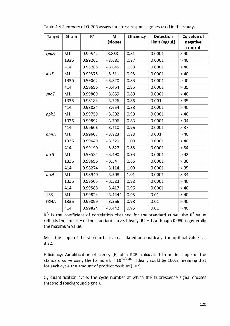
120
Table 4.4 Summary of Q-PCR assays for stress response genes used in this study.
Target Strain R2 M (slope)
Efficiency Detection limit (ng/µL)
Cq value of negative control
rpoA M1 0.99542 -3.863 0.81 0.0001 > 40
1336 0.99262 - 3.680 0.87 0.0001 > 40
414 0.98288 - 3.645 0.88 0.0001 > 40
luxS M1 0.99375 - 3.511 0.93 0.0001 > 40
1336 0.99062 - 3.820 0.83 0.0001 > 40
414 0.99696 - 3.454 0.95 0.0001 > 35
spoT M1 0.99809 - 3.659 0.88 0.0001 > 40
1336 0.98184 - 3.726 0.86 0.001 > 35
414 0.98834 - 3.654 0.88 0.0001 > 40
ppk1 M1 0.99759 - 3.582 0.90 0.0001 > 40
1336 0.99892 - 3.796 0.83 0.0001 > 34
414 0.99606 - 3.410 0.96 0.0001 > 37
amiA M1 0.99607 - 3.823 0.83 0.001 > 40
1336 0.99649 - 3.329 1.00 0.0001 > 40
414 0.99190 - 3.827 0.83 0.0001 > 34
htrB M1 0.99524 - 3.490 0.93 0.0001 > 32
1336 0.99696 - 3.54 0.85 0.0001 > 36
414 0.98274 - 3.114 1.09 0.0001 > 35
htrA M1 0.98940 - 3.308 1.01 0.0001 > 34
1336 0.99505 - 3.523 0.92 0.0001 > 40
414 0.99588 - 3.417 0.96 0.0001 > 40
16S
rRNA
M1 0.99824 - 3.4442 0.95 0.01 > 40
1336 0.99899 - 3.366 0.98 0.01 > 40
414 0.99824 - 3.442 0.95 0.01 > 40
R2: is the coefficient of correlation obtained for the standard curve, the R2 value reflects the linearity of the standard curve. Ideally, R2 = 1, although 0.980 is generally the maximum value. M: is the slope of the standard curve calculated automaticaly, the optimal value is -3.32. Efficiency: Amplification efficiency (E) of a PCR, calculated from the slope of the standard curve using the formula E = 10–1/slope. Ideally sould be 100%, meaning that for each cycle the amount of product doubles (E=2). Cq=quantification cycle: the cycle number at which the fluorescence signal crosses threshold (background signal).
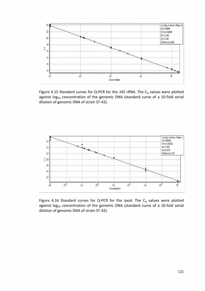
121
Figure 4.15 Standard curves for Q-PCR for the 16S rRNA. The Cq values were plotted against log10 concentration of the genomic DNA (standard curve of a 10-fold serial dilution of genomic DNA of strain ST-42).
Figure 4.16 Standard curves for Q-PCR for the rpoA. The Cq values were plotted against log10 concentration of the genomic DNA (standard curve of a 10-fold serial dilution of genomic DNA of strain ST-42).

122
Figure 4.17 Standard curves for Q-PCR for the luxS. The Cq values were plotted against log10 concentration of the genomic DNA (standard curve of a 10-fold serial dilution of genomic DNA of strain ST-42).
Figure 4.18 Standard curves for Q-PCR for the spoT. The Cq values were plotted against log10 concentration of the genomic DNA (standard curve of a 10-fold serial dilution of genomic DNA of strain ST-42).
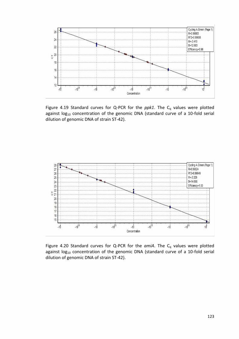
123
Figure 4.19 Standard curves for Q-PCR for the ppk1. The Cq values were plotted against log10 concentration of the genomic DNA (standard curve of a 10-fold serial dilution of genomic DNA of strain ST-42).
Figure 4.20 Standard curves for Q-PCR for the amiA. The Cq values were plotted against log10 concentration of the genomic DNA (standard curve of a 10-fold serial dilution of genomic DNA of strain ST-42).
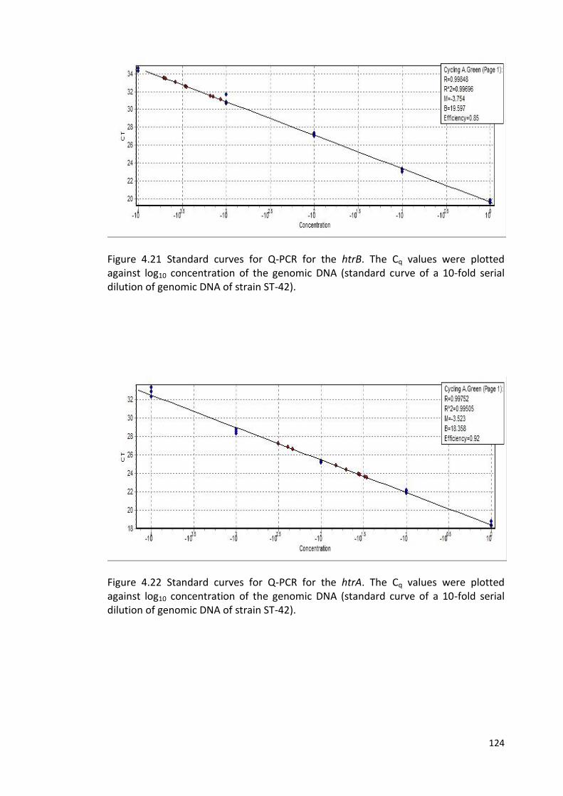
124
Figure 4.21 Standard curves for Q-PCR for the htrB. The Cq values were plotted against log10 concentration of the genomic DNA (standard curve of a 10-fold serial dilution of genomic DNA of strain ST-42).
Figure 4.22 Standard curves for Q-PCR for the htrA. The Cq values were plotted against log10 concentration of the genomic DNA (standard curve of a 10-fold serial dilution of genomic DNA of strain ST-42).
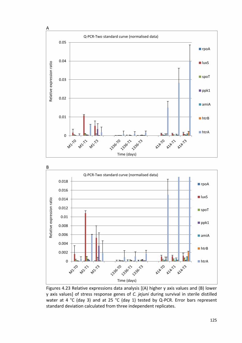
125
A
B
Figures 4.23 Relative expressions data analysis [(A) higher y axis values and (B) lower y axis values] of stress response genes of C. jejuni during survival in sterile distilled water at 4 °C (day 3) and at 25 °C (day 1) tested by Q-PCR. Error bars represent standard deviation calculated from three independent replicates.
0
0.01
0.02
0.03
0.04
0.05
Rel
ativ
e ex
pre
ssio
n r
atio
Time (days)
Q-PCR-Two standard curve (normalised data)
rpoA
luxS
spoT
ppk1
amiA
htrB
htrA
0
0.002
0.004
0.006
0.008
0.01
0.012
0.014
0.016
0.018
Rel
ativ
e ex
pre
ssio
n r
atio
Time (days)
Q-PCR-Two standard curve (normalised data)
rpoA
luxS
spoT
ppk1
amiA
htrB
htrA
126
4.2.6 RNAseq analysis of strain M1 gene expression
The RNAseq method was applied to study the expression of genes in strain M1 under
a number of different conditions. These included growth in MHB at 37 °C to OD600 of
0.05. Henceforth, I will refer to this as the control sample. The other conditions used
were the same as used for the Q-PCR assays in this study, namely strain M1 in sterile
distilled water at time zero (T0), at day 1 (T1) at 25 °C, and at day 3 (T3) at 4 °C. The
RNAseq data were generated by Dr. Christina Bronowski, University of Liverpool.
Using these data, analysis was carried out to look at variations in the expression of
genes which from previous work were potentially associated with survival in water.
Figure 4.24 shows the RNAseq data obtained for the genes used in the Q-PCR analysis
in this study, comparing each of these three time points in water survival experiments
with gene expression in the control (grown in MHB). Table 4.6 provides a summary of
these data and identifies statistically significant variations. In addition, data for a far
wider selection of genes implicated in stress responses, survival, biofilm formation or
the VBNC form are included in Table 4.6.
Figure 4.24 and Table 4.6 indicate that the luxS gene is up-regulated compared to the
control at all three time-points. “In agreement with the Q-PCR findings, luxS was up-
regulated in strain M1 at day 1 (T1) at 25 °C, and at day 3 (T3) at 4 °C compared to
time zero”. Other genes significantly up-regulated (p < 0.05) during survival in water
compared to growth in broth include katA (all three time-points), fliA (T0 and T3
only), sodB (all three time-points), ahpC (all three time-points), clpB (all three time-
points), hrcA (T0 and T3 only), grpE (all three time-points), groS (all three time-
points), groL (all three time-points), and yjiY (all three time-points). The peb1c gene
was also up-regulated at all three time-points (p range 0.005 – 0.055).
127
It has been demonstrated that CosR (Campylobacter oxidative stress regulator;
Cj0355c) may play a role in regulation of many oxidative stress genes especially sodB
by binding to the promoter regions of sodB and ahpC and regulates their
transcriptions (Hwang et al., 2011).
Amongst genes significantly down-regulated during survival in water (p < 0.05) were
rpoA, spoT, ppk1, amiA (T1 and T3 only), htrB (T1 and T3 only), nrfA, racS (T1 and T3
only), dcuA, clpP, cheA, fdxA (T1 and T3 only), sdhA, sdhB, sdhC, hslU, lon (T1 and T3
only), clpA (T1 and T3 only) (Table 4.6).
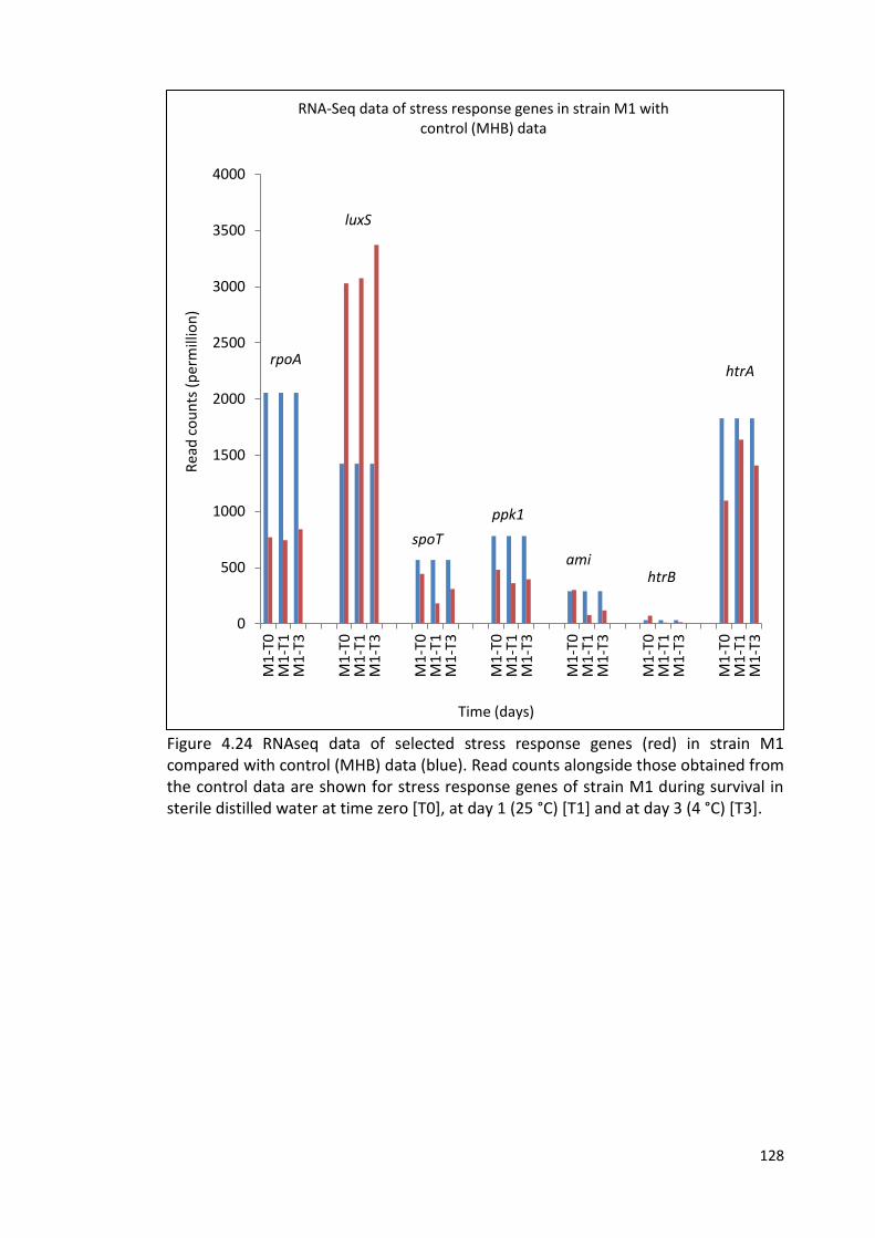
128
Figure 4.24 RNAseq data of selected stress response genes (red) in strain M1 compared with control (MHB) data (blue). Read counts alongside those obtained from the control data are shown for stress response genes of strain M1 during survival in sterile distilled water at time zero [T0], at day 1 (25 °C) [T1] and at day 3 (4 °C) [T3].
0
500
1000
1500
2000
2500
3000
3500
4000
M1
-T0
M1
-T1
M1
-T3
M1
-T0
M1
-T1
M1
-T3
M1
-T0
M1
-T1
M1
-T3
M1
-T0
M1
-T1
M1
-T3
M1
-T0
M1
-T1
M1
-T3
M1
-T0
M1
-T1
M1
-T3
M1
-T0
M1
-T1
M1
-T3
Rea
d c
ou
nts
(p
erm
illio
n)
Time (days)
RNA-Seq data of stress response genes in strain M1 with control (MHB) data
luxS
spoT
ppk1
amihtrB
htrA rpoA
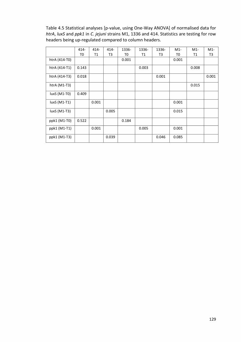
129
Table 4.5 Statistical analyses [p-value, using One-Way ANOVA] of normalised data for htrA, luxS and ppk1 in C. jejuni strains M1, 1336 and 414. Statistics are testing for row headers being up-regulated compared to column headers.
414- T0
414- T1
414- T3
1336- T0
1336- T1
1336- T3
M1- T0
M1- T1
M1- T3
htrA (414-T0) 0.001 0.001
htrA (414-T1) 0.143 0.003 0.008
htrA (414-T3) 0.018 0.001 0.001
htrA (M1-T3) 0.015
luxS (M1-T0) 0.409
luxS (M1-T1) 0.001 0.001
luxS (M1-T3) 0.005 0.015
ppk1 (M1-T0) 0.522 0.184
ppk1 (M1-T1) 0.001 0.005 0.001
ppk1 (M1-T3) 0.039 0.046 0.085
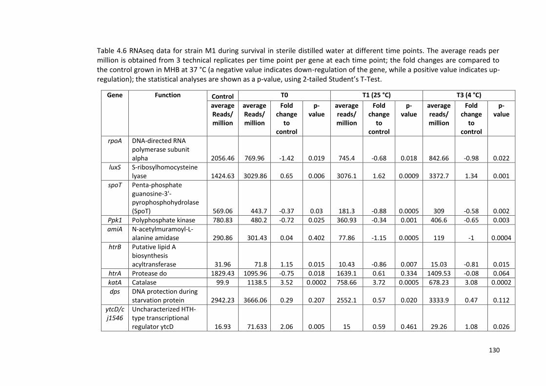
130
Table 4.6 RNAseq data for strain M1 during survival in sterile distilled water at different time points. The average reads per million is obtained from 3 technical replicates per time point per gene at each time point; the fold changes are compared to the control grown in MHB at 37 °C (a negative value indicates down-regulation of the gene, while a positive value indicates up-regulation); the statistical analyses are shown as a p-value, using 2-tailed Student’s T-Test.
Gene Function Control T0 T1 (25 °C) T3 (4 °C)
average Reads/ million
average Reads/million
Fold change
to control
p-value
average reads/ million
Fold change
to control
p-value
average reads/ million
Fold change
to control
p-value
rpoA DNA-directed RNA polymerase subunit alpha 2056.46 769.96 -1.42 0.019 745.4 -0.68 0.018 842.66 -0.98 0.022
luxS S-ribosylhomocysteine lyase 1424.63 3029.86 0.65 0.006 3076.1 1.62 0.0009 3372.7 1.34 0.001
spoT Penta-phosphate guanosine-3'-pyrophosphohydrolase (SpoT) 569.06 443.7 -0.37 0.03 181.3 -0.88 0.0005 309 -0.58 0.002
Ppk1 Polyphosphate kinase 780.83 480.2 -0.72 0.025 360.93 -0.34 0.001 406.6 -0.65 0.003
amiA N-acetylmuramoyl-L-alanine amidase 290.86 301.43 0.04 0.402 77.86 -1.15 0.0005 119 -1 0.0004
htrB Putative lipid A biosynthesis acyltransferase 31.96 71.8 1.15 0.015 10.43 -0.86 0.007 15.03 -0.81 0.015
htrA Protease do 1829.43 1095.96 -0.75 0.018 1639.1 0.61 0.334 1409.53 -0.08 0.064
katA Catalase 99.9 1138.5 3.52 0.0002 758.66 3.72 0.0005 678.23 3.08 0.0002
dps DNA protection during starvation protein 2942.23 3666.06 0.29 0.207 2552.1 0.57 0.020 3333.9 0.47 0.112
ytcD/cj1546
Uncharacterized HTH-type transcriptional regulator ytcD 16.93 71.633 2.06 0.005 15 0.59 0.461 29.26 1.08 0.026
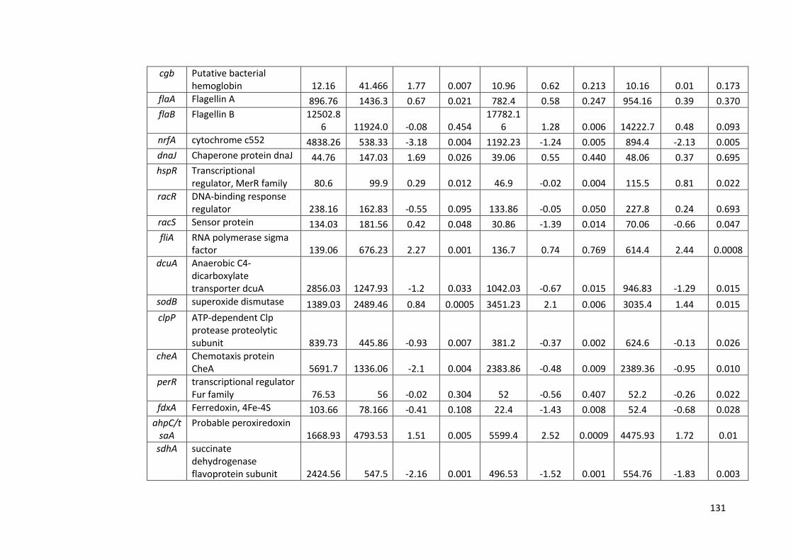
131
cgb Putative bacterial hemoglobin 12.16 41.466 1.77 0.007 10.96 0.62 0.213 10.16 0.01 0.173
flaA Flagellin A 896.76 1436.3 0.67 0.021 782.4 0.58 0.247 954.16 0.39 0.370
flaB Flagellin B 12502.86 11924.0 -0.08 0.454
17782.16 1.28 0.006 14222.7 0.48 0.093
nrfA cytochrome c552 4838.26 538.33 -3.18 0.004 1192.23 -1.24 0.005 894.4 -2.13 0.005
dnaJ Chaperone protein dnaJ 44.76 147.03 1.69 0.026 39.06 0.55 0.440 48.06 0.37 0.695
hspR Transcriptional regulator, MerR family 80.6 99.9 0.29 0.012 46.9 -0.02 0.004 115.5 0.81 0.022
racR DNA-binding response regulator 238.16 162.83 -0.55 0.095 133.86 -0.05 0.050 227.8 0.24 0.693
racS Sensor protein 134.03 181.56 0.42 0.048 30.86 -1.39 0.014 70.06 -0.66 0.047
fliA RNA polymerase sigma factor 139.06 676.23 2.27 0.001 136.7 0.74 0.769 614.4 2.44 0.0008
dcuA Anaerobic C4-dicarboxylate transporter dcuA 2856.03 1247.93 -1.2 0.033 1042.03 -0.67 0.015 946.83 -1.29 0.015
sodB superoxide dismutase 1389.03 2489.46 0.84 0.0005 3451.23 2.1 0.006 3035.4 1.44 0.015
clpP ATP-dependent Clp protease proteolytic subunit 839.73 445.86 -0.93 0.007 381.2 -0.37 0.002 624.6 -0.13 0.026
cheA Chemotaxis protein CheA 5691.7 1336.06 -2.1 0.004 2383.86 -0.48 0.009 2389.36 -0.95 0.010
perR transcriptional regulator Fur family 76.53 56 -0.02 0.304 52 -0.56 0.407 52.2 -0.26 0.022
fdxA Ferredoxin, 4Fe-4S 103.66 78.166 -0.41 0.108 22.4 -1.43 0.008 52.4 -0.68 0.028
ahpC/tsaA
Probable peroxiredoxin 1668.93 4793.53 1.51 0.005 5599.4 2.52 0.0009 4475.93 1.72 0.01
sdhA succinate dehydrogenase flavoprotein subunit 2424.56 547.5 -2.16 0.001 496.53 -1.52 0.001 554.76 -1.83 0.003
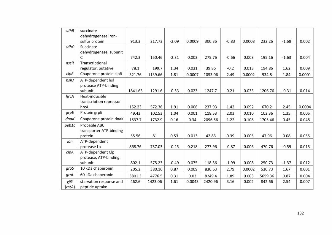
132
sdhB succinate dehydrogenase iron-sulfur protein 913.3 217.73 -2.09 0.0009 300.36 -0.83 0.0008 232.26 -1.68 0.002
sdhC Succinate dehydrogenase, subunit C 742.3 150.46 -2.31 0.002 275.76 -0.66 0.003 195.16 -1.63 0.004
nssR Transcriptional regulator, putative 78.1 199.7 1.34 0.031 39.86 -0.2 0.013 194.86 1.62 0.009
clpB Chaperone protein clpB 321.76 1139.66 1.81 0.0007 1053.06 2.49 0.0002 934.8 1.84 0.0001
hslU ATP-dependent hsl protease ATP-binding subunit 1841.63 1291.6 -0.53 0.023 1247.7 0.21 0.033 1206.76 -0.31 0.014
hrcA Heat-inducible transcription repressor hrcA 152.23 572.36 1.91 0.006 237.93 1.42 0.092 670.2 2.45 0.0004
grpE Protein grpE 49.43 102.53 1.04 0.001 118.53 2.03 0.010 102.36 1.35 0.005
dnaK Chaperone protein dnaK 1537.7 1732.9 0.16 0.34 2096.56 1.22 0.108 1705.46 0.45 0.048
peb1c Probable ABC transporter ATP-binding protein 55.56 81 0.53 0.013 42.83 0.39 0.005 47.96 0.08 0.055
lon ATP-dependent protease La 868.76 737.03 -0.25 0.218 277.96 -0.87 0.006 470.76 -0.59 0.013
clpA ATP-dependent Clp protease, ATP-binding subunit 802.1 575.23 -0.49 0.075 118.36 -1.99 0.008 250.73 -1.37 0.012
groS 10 kDa chaperonin 205.2 380.16 0.87 0.009 830.63 2.79 0.0002 530.73 1.67 0.001
groL 60 kDa chaperonin 3801.3 4776.5 0.31 0.03 8249.4 1.89 0.003 5659.36 0.87 0.004
yjiY (cstA)
starvation response and peptide uptake
462.6
1423.06
1.61
0.0043
2420.96
3.16 0.002
842.66
2.54
0.007
133
4.3 Discussion
In Chapter 3, evidence was presented suggesting that strains of C. jejuni vary in their
ability to retain culturability in water, with some strains, such as M1, rapidly losing
colony forming ability without a corresponding loss of viability, whereas others such
as 1336 and 414, retain far higher levels of culturability. We hypothesized that these
variations between different strains of C. jejuni during survival in water might be
attributed to differences in expression in genes reported previously as being
important in the response to harsh environments, including genes implicated in the
formation of VBNC cells. In order to test this hypothesis, and with the aim of
elucidating the mechanisms underlying these variations, a set of stress response
genes (Table 4.1) was targeted in end-point and Q-PCR assays at specific time points
during survival in water where variations were observed (Chapter 3).
Initial experiments using end-point PCR amplification targeting the cDNA template of
selected genes indicated considerable variation in expression between strains, genes
and time points (Table 4.3). Although it is hard to draw firm conclusions from these
data, expression of the targeted genes (luxS, spoT, ppk1, amiA, htrB, htrA) was
undetectable at the day 3 (4 °C) time point for strains 1336 and 414, whereas
expression of some genes (spoT, amiA, htrA) was detectable for strain M1. This did
correspond with a difference between strains that retained culturability better (1336
and 414) compared to a strain that did not (M1). However, at the day 1 (25 °C) time
point, the biggest variation was seen with the lack of expression of genes in strains
M1 and 1336, in contrast to strain 414. In terms of retaining culturability, strains 1336
and 414 were similar, whereas strain M1 was associated with rapid loss. Hence, there
is no clear pattern to the data. Perhaps the most surprising result was the inability to
134
detect expression of rpoA in all of the samples. This gene was included along with 16S
rRNA as a potential control/reference gene because of a previous study suggesting
that it was the best choice as an internal control to study stress response in C. jejuni
using (Q-PCR) (Ritz et al., 2009). In this previous study, Ritz et al. (2009) set out to
identify stable housekeeping genes by studying gene expression variations in
different growth phases and under different stress conditions for six target genes:
gyrA, ilvC, rpoA, slyD, thiC, and rrs (16S rRNA). The variations tested were exponential
phase, stationary phase, cold shock, oxidative stress and a combination of cold shock
and oxidative stress. The rpoA gene, encoding the alpha subunit of RNA polymerase,
was recommended because it had the least variation in gene expression when all of
these conditions were compared. It is worth noting, however, that the authors also
saw very little variation with respect to the 16S rRNA gene expression. They chose to
reject this gene in favour of rpoA because of the fact that inevitably levels of 16S
rRNA transcripts are much higher than for other genes. In this study, the 16S rRNA
gene expression could be detected in all samples, whereas rpoA expression could not
be detected in some. Hence, the decision was made to use 16S rRNA as the control
gene for Q-PCR assays. It is not clear why the rpoA expression levels are low,
especially in strain M1, but this observation was confirmed by subsequent inclusion in
the Q-PCR assays, where expression relative to 16S rRNA was low to undetectable.
The RNAseq data confirmed that the expression of this gene was variable under the
conditions used in this study.
It is worth noting that others have since suggested that rpoA gene expression is not
constitutive and is highly sensitive to environmental change. For example, it varies
during cell processing procedures prior to RNA isolation (Hyytiäinen et al., 2012). The
135
authors in this 2012 study also selected 16S rRNA in their study as an internal control
for Q-PCR, suggesting that rpoA is not an appropriate choice, and others have also the
16S rRNA in similar studies (Bui et al., 2012).
Real-time fluorescence-based Q-PCR is considered as a model for nucleic acid
quantification due to its specificity, sensitivity and speed (Bustin et al., 2009). Q-PCR
analysis demonstrated that there were differences in gene expression levels during
survival in sterile distilled water between the different strains of C. jejuni.
When the Q-PCR assays were applied to study of gene expression variations between
strains at key time points in the water survival experiments, a number of differences
were observed. In strain 414, which retains culturability at the key time points, levels
of expression of htrA were significantly higher (Table 4.5) when compared to the
other two strains at all-time points. When normalised data were used (expression
relative to 16S rRNA), expression was also significantly higher (Table 4.5) at the day 3
(4 °C) time point than at time zero for strain 414. It should be noted that during the
preparation of the inoculum for the water survival experiments, there is inevitably a
period of exposure of the C. jejuni cells to the stresses associated with being removed
from nutrient-rich to nutrient-poor conditions. Hence, it seems likely that some of the
stress responses have already occurred at the time zero point in these experiments.
This perhaps explains why there were not more differences between the Day 1 or Day
3 sample times and the Day zero sample time.
The htrA gene product, HtrA, is required for both heat and oxygen tolerance
(Brøndsted et al., 2005). HtrA displays both chaperone and protease properties, both
implicated in the ability to tolerate stress, though the chaperone activity was
identified as more important for resistance to oxidative stress (Baek et al., 2011b).
136
HtrA is a periplasmic serine protease that can contribute to host cell binding (Baek et
al., 2011a), and has been implicated in acute ulcerative enterocolitis and driving
immune responses during infection of mice (Heimesaat et al., 2014). In the context of
this study, it seems likely that a potential role in the response to oxidative stress is
more relevant than properties as a potential virulence factor. HtrA is important for
stress tolerance and survival of Gram-negative bacteria generally (Laskowska et al.,
1996; Li et al., 1996). However, it has been reported previously that htrA is down-
regulated in C. jejuni in response to low nutrient and especially oxidative stress (Bui et
al., 2012). This appears to contradict the findings in our study but probably reflects
the considerable variations between strains. In the study of Bui et al. (2012), they
used strain NCTC11168. They also used very specific oxidative stress conditions
(exposure to 10 mM hydrogen peroxide for 15 min) which differ considerably from
those used in this study. It is possible that the enhanced levels of expression of htrA in
strain 414 contribute to its ability to retain culturability better than strain M1, where
levels are significantly lower. However, the fact that levels were also lower in strain
1336, which can retain culturability, would indicate that if htrA does play a role, it
may be strain-specific, and not a mechanism that can be universally applied to C.
jejuni.
Of the three strains compared, strain M1 was clearly different in terms of its inability
to retain culturability under the experimental conditions used. The Q-PCR assays
strongly suggest that gene expression is occurring in the day 3 (4 °C) and day 1 (25 °C)
samples despite the low colony forming unit counts for M1, which supports the
conclusion from staining experiments presented in Chapter 3 that M1 is entering a
VBNC state. Clearly, the strain with lowest detectable expression levels for the genes
137
selected is strain 1336 (Figure 4.23) rather than M1. Hence, it was particularly
interesting to ascertain whether there were gene expression changes specific to
strain M1. The most notable of these related to the luxS gene, which was significantly
up-regulated (relative to 16S rRNA) in M1 compared to strain 414 in both the day 3 (4
°C) and day 1 (25 °C) samples, and which was significantly up-regulated in these
samples in comparison to the day zero sample (Table 4.5). RNAseq analysis confirmed
that luxS is expressed at high levels during survival of strain M1 in water. These
observations are consistent with a role for luxS during the conversion of M1 to the
VBNC state. luxS is responsible for encoding an autoinducer (AI-2 or LuxS) in the C.
jejuni QS system (Cloak et al., 2002). The QS system has been implicated previously in
a variety of important physiological pathways in response to cell density. These
include motility/flagellar gene expression, oxidative stress, ability to colonise animals,
expression of CDT and virulence in an animal model (Plummer, 2012). However,
different groups have reported varying observations (Elvers and Park, 2002; He et al.,
2008; Quiñones et al., 2009). As with the studies of most genes in C. jejuni, or indeed
in other common pathogens, conclusions have been drawn based on the analysis of
one or two strains, with the assumption that there is general applicability to the
whole species. In one study, luxS mutants constructed in two different strains,
NCTC11168 and 81176, were compared and differing phenotypes depending on the
strain background were reported, suggesting that this assumption may be flawed
(Adler et al., 2014).
It has been suggested that Campylobacter spp. generally possess luxS gene, but C. lari
is an exception to this (Tazumi et al., 2011). Indeed, it has become clear that not only
is luxS not carried by all Campylobacter species (Gölz et al., 2012), but that not all C.
138
jejuni carry luxS gene (Hepworth et al., 2011) and we found this to be the case for
strain 1336.
However, although the luxS gene has been reported as absent from some water/wild-
life-associated strains (Hepworth et al., 2011), it is generally present in isolates from
human sources. In a study analysing the genomes of 442 C. jejuni UK human isolates
from two large surveys of pathogens associated with intestinal infectious disease [the
IID1 and IID2 studies; (Tam et al., 2012)], luxS was present in all but six strains, five of
which shared the same ST (ST-433). In all there were six ST-433 isolates amongst the
IID1 and IID2 isolates, with one carrying the luxS gene (unpublished data, C.
Winstanley, University of Liverpool). Therefore this gene potentially helps C. jejuni to
survive in the environment, and is carried by the vast majority of strains that cause
infections in humans.
Strain 1336, isolated from a wild bird, was previously reported as a representative of
a diverse group of C. jejuni water/wild-life isolates that do not tend to be found
amongst human isolates or isolates from farm animals and are hence considered to
be uncharacteristic of human food-chain associated isolates (Hepworth et al., 2011).
Strain 414 is a representative of ST-3704, associated with bank voles. The genomes of
both strains 1336 and 414 lack regions that are normally carried by C. jejuni
associated with the human food chain. However, despite lacking a number of genetic
regions associated with the ability to colonise chickens, and lacking the luxS gene,
strain 1336, unlike strain 414, was able to colonise chickens (Hepworth et al., 2011).
Clearly further work is needed in order to resolve the role of the QS system in the
variations seen in strain M1 compared to strain 414 with respect to loss of
culturability and a switch to the VBNC state. The results of this study are consistent
139
with the idea that there is expression of luxS in VBNC populations of C. jejuni.
However, it should be noted that there may be a small proportion of vegetative cells
in which luxS could, in theory, be highly up-regulated. This study also emphasises the
likely importance of variations between strains.
There were also some significant differences in the expression of ppk1 between
strain M1 and the other two strains (Figure 4.23). At both time-points day 3 (4 °C) and
day 1 (25 °C), levels of expression of ppk1 (relative to 16S rRNA) were significantly
higher (Table 4.5) in strain M1 than in either strain 1336 or strain 414. For day 1 (25
°C), the increase in expression of ppk1 in M1 reached significance compared to the
day zero value. At the day 3 (4 °C) time-point, expression was also higher compared
to day zero, but the change did not reach statistical significance (p =0.085). ppk1
encodes polyphosphate kinase-1, a key enzyme in the synthesis of polyphosphate,
which is an essential molecule for survival, mediating stress responses and host
interactions in a number of bacterial pathogens (Kornberg, 1999). It has been
demonstrated that a ppk1 mutant of C. jejuni not only had defective polyphosphate
accumulation, but also had decreased ability to form the VBNC state under acid stress
conditions (Gangaiah et al., 2009). The authors were able to complement the
mutation and restore previous phenotypes. Given that strain M1 is seen to enter the
VBNC state at the chosen time-points in the experiments presented in this chapter,
the enhanced expression of ppk1 is consistent with the role of polyphosphate
accumulation in the formation of the VBNC state suggested by this previous study
(Candon et al., 2007; Gangaiah et al., 2009).
The low values for gene expression achieved across the board for strain 1336 suggest
that genes other than the ones targeted here are important in the survival of this
140
strain in water, and further emphasize the importance of variations between strains.
However, it should be noted that using end-point PCR assays, expression of most
genes was detectable at the zero time-point. The lack of expression of most of the
selected genes beyond the time zero point, was also observed for strain 1336 in the
end-point PCR assays.
However, overall, there was poor agreement between the end-point PCR assays and
the Q-PCR assays. The reasons behind this are unclear. For the end-point PCR assays,
only one replicate based on single RNA extractions for each assay was used. The later
observations based on Q-PCR assays suggest that this was inadequate to ensure
detection of gene expression. In particular, for strain 414, gene expression was not
detected by end-point PCR for any of the target genes in the Day 3 (4 °C) sample,
whereas Q-PCR clearly identified expression of some genes, especially htrA. Similarly,
for strain M1 the end-point PCR proved to be inadequate at detecting expression of
several genes in the Day 3 (4 °C) that were subsequently detected using Q-PCR.
The RNAseq data, obtained for strain M1 only, highlighted a number of genes up-
regulated in water compared to growth in laboratory media. Most notably, luxS was
again identified, along with genes implicated in oxidative stress (katA, sodB, ahpC).
Although the design of the RNAseq and Q-PCR experiments are not the same, the
RNAseq data are consistent with the idea that luxS plays an important role during
survival in water. Given that expression in strain M1 was higher than for other strains,
and that under the chosen conditions the M1 strain enters a VBNC state, these
observations implicate luxS in a role during this process. Further analysis of the global
transcriptome of strain M1 during survival in water is ongoing in a parallel project,
but it is clear that genes are being actively expressed, confirming the fact that
141
although strain M1 is losing culturability, it remains VBNC. Previous studies have
shown using RNAseq that gene expression can vary considerably between strains of
Campylobacter (Dugar et al., 2013). The key to understanding important phenotypic
variations, such as different survival behaviour in environmental samples, is likely to
lie within the detail of the strain-specific transcriptome profiles. In this study, some of
these variations have been identified, but further work is needed to fully characterise
the mechanisms underlying the differences between strains.
4.4 Conclusions
Gene expression differences implicated in variations in the survival behaviour in
sterile distilled water of different strains of C. jejuni were identified. In particular,
variations in the expression of key genes (luxS, ppk1, htrA) were implicated in the
conversion of strain M1 to a VBNC state. This is the first report implicating the C.
jejuni QS system (luxS) in a role in survival in water.
142
C h a p t e r 5
CO-EXISTENCE OF C. jejuni AND Pseudomonas spp. IN THE ENVIRONMENT, AND
INTERACTIONS BETWEEN THEM
5.1 Introduction
C. jejuni is a successful human pathogen and has reservoirs in water and various
animals but its survival outside the host is poor (Altekruse et al., 1999; Bronowski et
al., 2014; Young et al., 2007). Despite this, C. jejuni can often be isolated from the
environment, and apparently survives well on chicken meat, suggesting that it does
have strategies enabling it to survive. Interaction of C. jejuni with other
microorganisms is one of the suggested survival strategies of this microorganism in
the environment (Hilbert et al., 2010) and during the contamination of food. Various
bacterial species are found on chicken meat and some of these bacteria are known as
spoilage bacteria. For example, Pseudomonas species such as P. fluorescens, P. frangi,
P. chlororaphis and P. putida are found on chicken meat in levels of up to 103 to 104
CFU/g (Ghafir et al., 2008; Hilbert et al., 2010; Mead et al., 1993; Russell et al., 1995).
It has also been shown that the numbers of Campylobacter on chicken meat surface
are higher than that of internal contamination (Luber and Bartelt, 2007).
Pseudomonas species are also commonly found in a wide range of natural
environmental niches (Silby et al., 2011). It has been proposed that C. jejuni may
interact with aerobic Pseudomonas spp. found on food or in other environments in
order to counteract the toxic effects of atmospheric oxygen (Hilbert et al., 2010).
Little is understood about the mechanism by which Pseudomonas spp. enhance the
aerotolerance of C. jejuni. However, the concept of bacteria benefitting from co-
existence with other species is not a new one. In vitro experiments have shown that
143
an anaerobic microorganism such as Porphyromonas gingivalis, which causes
periodontitis in humans, is able to grow in the presence of oxygen when co-cultured
with Fusobacterium nucleatum. This is possibly due to metabolizing of both oxygen
and hydrogen peroxide molecules and the creation of a capnophilic environment by
F. nucleatum, thus supporting growth of P. gingivalis (Diaz et al., 2002; Kuramitsu et
al., 2007). In certain situations some individual organisms are found to be antibiotic
sensitive but are turned resistant within mixed infections. For example studies in vitro
have demonstrated the protection of a penicillin-sensitive strain of F. neccrophorum
from penicillin therapy by penicillin-resistant strains of Bacteroides fragilis,
pigmented Prevotella and Porphyromonas spp., and Prevotella oralis (Hackman and
Wilkins, 1976). In animal model experiments it has been demonstrated that
combinations of aerobic and anaerobic bacteria promote levels of disease that could
not be produced by individual organism. For example, mono and mixed intra-
abdominal infections with B. fragilis and E.coli in a rat fibrin clot model demonstrated
that only with mixed infection there was an increase of B. fragilis numbers about 6 h
after initial decrease, and only mixed infections resulted in abscesses lesions. It was
suggested that the presence of E.coli enabled B. fragilis to survive by changing the pH,
excreting nutrients or using up the oxygen (Verweij et al., 1991). Thus, it is possible
that aerobic organisms such as Pseudomonas spp. could play key role in the ability of
Campylobacter species to survive in the environment.
144
5.1.1 The genus Pseudomonas
The genus Pseudomonas was first reported by the Botanist Migula in 1894. It consists
of a group of bacteria with large genomes that have considerable metabolic and
physiological versatility. Their adaptability allows them to colonise diverse aquatic
and terrestrial habitats as well as causing various infections (of humans, animals and
plants) and exhibiting biochemical properties that can be exploited for
biotechnological applications (Peix et al., 2009; Silby et al., 2011). Over the years, the
taxonomy of Pseudomonas has been revised. The advent of molecular methods for
studying phylogeny revealed that organisms previously categorised as Pseudomonas
were actually phylogentically diverse and the various species (including newly
described genera) were separated on the basis of rRNA homology into different
groups (O'Sullivan and O'Gara, 1992; Palleroni et al., 1973; Palleroni and Moore,
2004; Ramos, 2011). Pseudomonas sensu stricto lie in the Gamma sub-division of
Proteobacteria, in what is known as rRNA homology group I. More recently, whole
genome sequencing has enabled much closer analysis of the relationships between
Pseudomonas species (Silby et al., 2011).
Phenotypically, Pseudomonas species are classified into fluorescent species and non-
fluorescent species. P. stutzeri is the most intensely studied species of the non-
fluorescent species (Palleroni, 2008). The fluorescent Pseudomonads are composed
of a distinct group of bacteria that have the ability to produce a yellow–green
pigment. They are Gram-negative, polar flagellated motile rods, chemoheterotrophic
and are grouped in rRNA homology group I. Many members of species of this group
are psychrotrophic, involved in food spoilage. The most common fluorescent species
in this group include P. fluorescens, P. putida, P. aeruginosa, P. chlororaphis, P.
145
syringae and P. cichorii (Arnaut‐Rollier et al., 1999a; Franzetti and Scarpellini, 2007). It
has also been shown that these spoilage bacteria have been isolated from both fresh
and refrigerated poultry skin (Arnaut-Rollier et al., 1999b). Previous studies confirmed
that pigmented and non-pigmented strains of Pseudomonas were the most
frequently bacteria found amongst the microorganisms on poultry skin (Barnes and
Thornley, 1966; Kraft, 1992; Russell et al., 1995).
5.1.2 Bacterial secretome
Bacterial Pathogens have evolved a number of complex secretion systems to
encounter hostile environments or to increase bacterial pathogenicity while infecting
their hosts. The majority of these systems depend on a large number of proteins
which administer virulence factors either into the environment or into the host cell
(Bleves et al., 2010). There are two major pathways in bacteria to secrete proteins
across the cytoplasmic membrane. The general secretion (Sec) pathway by which
most of unfolded proteins are secreted, and twin-arginine translocation (Tat)
pathway by which folded proteins are secreted (Bleves et al., 2010; Natale et al.,
2008) Natale, paol 2008).
The secretion systems are classified as type I secretion system (T1SS) up to type VI
secretion system (T6SS) (Bleves et al., 2010; Sankarasubramanian et al., 2016). The
T1SS is comprised of 3 proteins, a pore-forming outer membrane protein (OMP), a
membrane fusion protein (MFP), and an inner membrane ATP-binding cassette (ABC)
protein, the latter two proteins are cytoplasmic membrane proteins. Various sizes of
proteins are secreted via this widespread pathway (Delepelaire, 2004). The T2SS is
involved in the transport of cellulases, pectinases, phospholipases, lipases and toxins
146
from the periplasim to the outer membrane. Furthermore, these proteins can
increase the virulence of human, animal and plant bacterial pathogens (Cianciotto,
2005; Sandkvist, 2001). The T3SS is homologous to the bacterila flagellum, involved in
direct injection of proteins into the host cells (Bergeron et al., 2016).
The T4SS is related to conjugation system, which transports DNA or proteins from
donor to target cells (Bhatty et al., 2013). The T4SS plays an important role in
bacterial pathogenesis of several bacterial pathogens (Zechner et al., 2012). The T5SS
is the simplest protein secretion system in which the proteins are secreted either via
the autotransporter system (type Va), two partner secretion pathway (Vb), or type Vc
system (Henderson et al., 2004). The T6SS is an important virulence factor for several
important bacterial pathogens such as Vibrio cholerae, which can translocate proteins
directly into the host cells. Protein expression is controlled at both transcriptional and
post-transcriptional levels (Bingle et al., 2008).
5.1.3 Pseudomonas secretome
The opportunistic human pathogen Pseudomonas aeruginosa, which is implicated in
infection of the respiratory tract of cystic fibrosis (CF) cases, possesses five (excluding
the T4SS) of the six secretion systems (Table 5.1).
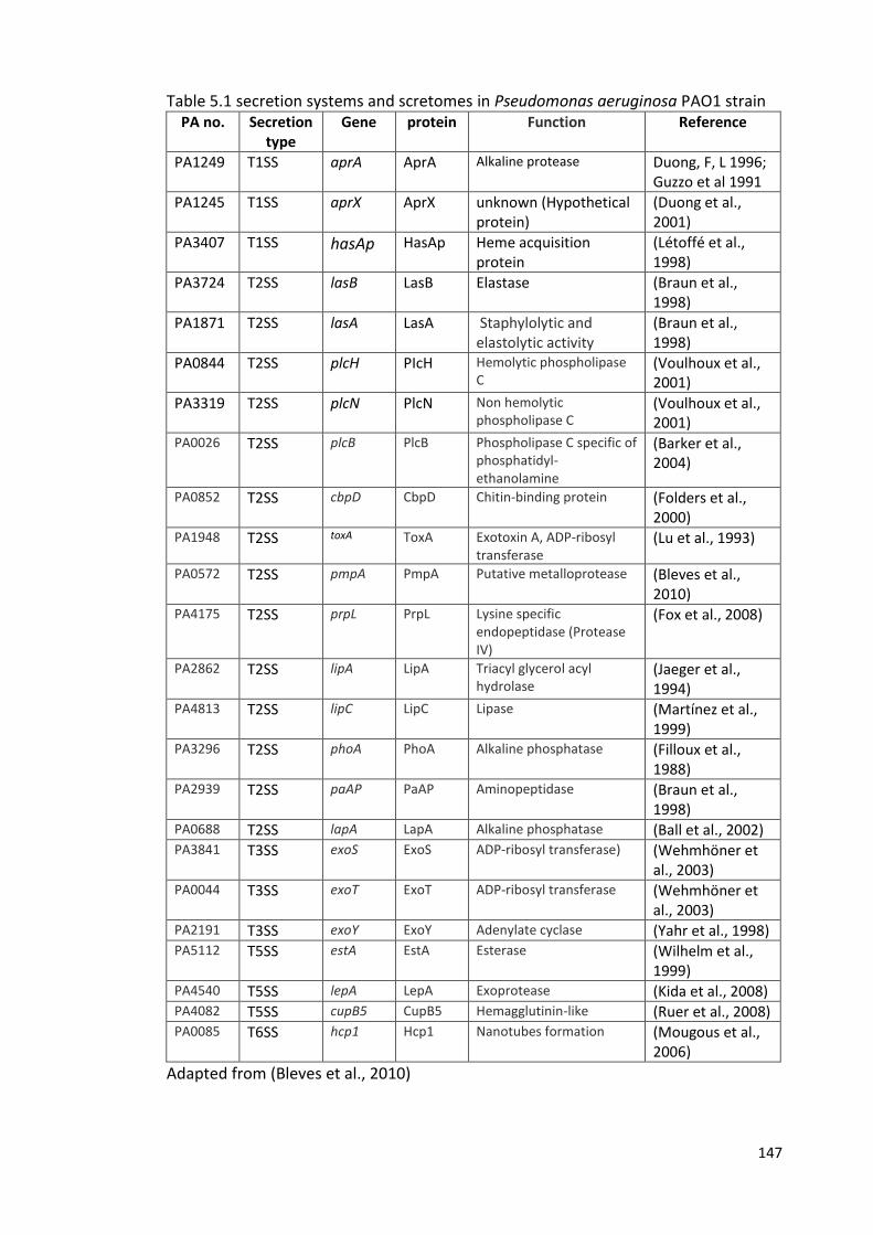
147
Table 5.1 secretion systems and scretomes in Pseudomonas aeruginosa PAO1 strain PA no. Secretion
type Gene protein Function Reference
PA1249 T1SS aprA AprA Alkaline protease Duong, F, L 1996; Guzzo et al 1991
PA1245 T1SS aprX AprX unknown (Hypothetical protein)
(Duong et al., 2001)
PA3407 T1SS hasAp HasAp Heme acquisition protein
(Létoffé et al., 1998)
PA3724 T2SS lasB LasB Elastase (Braun et al., 1998)
PA1871 T2SS lasA LasA Staphylolytic and elastolytic activity
(Braun et al., 1998)
PA0844 T2SS plcH PIcH Hemolytic phospholipase C
(Voulhoux et al., 2001)
PA3319 T2SS plcN PlcN Non hemolytic phospholipase C
(Voulhoux et al., 2001)
PA0026 T2SS plcB PlcB Phospholipase C specific of phosphatidyl-ethanolamine
(Barker et al., 2004)
PA0852 T2SS cbpD CbpD Chitin-binding protein (Folders et al., 2000)
PA1948 T2SS toxA ToxA Exotoxin A, ADP-ribosyl transferase
(Lu et al., 1993)
PA0572 T2SS pmpA PmpA Putative metalloprotease (Bleves et al., 2010)
PA4175 T2SS prpL PrpL Lysine specific endopeptidase (Protease IV)
(Fox et al., 2008)
PA2862 T2SS lipA LipA Triacyl glycerol acyl hydrolase
(Jaeger et al., 1994)
PA4813 T2SS lipC LipC Lipase (Martínez et al., 1999)
PA3296 T2SS phoA PhoA Alkaline phosphatase (Filloux et al., 1988)
PA2939 T2SS paAP PaAP Aminopeptidase (Braun et al., 1998)
PA0688 T2SS lapA LapA Alkaline phosphatase (Ball et al., 2002) PA3841 T3SS exoS ExoS ADP-ribosyl transferase) (Wehmhöner et
al., 2003) PA0044 T3SS exoT ExoT ADP-ribosyl transferase (Wehmhöner et
al., 2003) PA2191 T3SS exoY ExoY Adenylate cyclase (Yahr et al., 1998) PA5112 T5SS estA EstA Esterase (Wilhelm et al.,
1999) PA4540 T5SS lepA LepA Exoprotease (Kida et al., 2008) PA4082 T5SS cupB5 CupB5 Hemagglutinin-like (Ruer et al., 2008) PA0085 T6SS hcp1 Hcp1 Nanotubes formation (Mougous et al.,
2006)
Adapted from (Bleves et al., 2010)
148
5.1.4 Bacterial QS systems
Chemical signal molecules are used in bacteria to communicate between cells. In QS,
hormone-like molecules, which are termed autoinducers, are produced and released
by bacteria, and detected by other cells in the population. When the concentration of
the autoinducer reaches a certain threshold, because there are sufficient members of
the community producing the signal molecule, then the bacteria respond en masse to
change their gene expression and alter their behaviour as a bacterial population
(Waters and Bassler, 2005). Hence, this is considered to be a density-dependent
control system. The bacterium Vibrio fischeri, in which the phenomenon of QS was
first described, is considered the paradigm for QS in most Gram-negative bacteria
(Nealson and Hastings, 1979).
In V. fischeri, the luminescence genes are arranged in a single operon, luxCDABE,
which exists as part of luxICDABE operon. The fatty acid reductase complex, the
product of luxCDE, is involved in synthesis of fatty aldehyde substrate which is
catalysed by the luciferase LuxAB subunits leading to light production (Eberhard et al.,
1981; Meighen, 1991; Miller and Bassler, 2001). The expression of LuxICDABE is
controlled by the proteins LuxI and LuxR. LuxI, the product of the luxI gene,
synthesises the Vibrio autoinducer 3OC6-homoserine lactone, which freely diffuses
into the environment. When the autoinducer molecule reaches the threshold
concentration, the signal is transduced via the LuxR protein which acts as a regulator
to activate transcription of the lux genes encoding luminescence (Eberhard et al.,
1981; Engebrecht et al., 1983; Engebrecht and Silverman, 1984; Kaplan and
Greenberg, 1985; Stevens et al., 1994). QS systems have since been reported in a
wide range of Gram negative and Gram positive bacteria (Sifri, 2008; Williams, 2007).
149
5.1.5 Pseudomonas QS systems
The opportunistic human pathogen Pseudomonas aeruginosa is among a number of
Gram-negative bacteria in which homologous QS systems have been discovered
(Pesci and Iglewski, 1997). The pathogenicity of P. aeruginosa is increased through QS
control of the expression of many virulence genes. P. aeruginosa possesses two QS
systems las and rhl. The las system consists of the transcriptional activator protein
LasR, a homolog of a LuxR, and LasI, a homolog of a LuxI, which controls the synthesis
of the Pseudomonas autoinducer [PAI-1; (N-(3-oxododecanoyl)-L-homoserine
lactone]. When the extracellular autoinducer (PAI-1) reaches a critical threshold
concentration, it leads to activation of LasR, a regulatory protein (Venturi, 2006).
Likewise, the rhI system comprises the transcriptional activator protein RhlR and RhlI,
which controls production of a second Pseudomonas autoinducer [PAI-2; (N-butanoyl-
L-homoserine lactone]. P. aeruginosa also contains a third signalling system (the
quinolone signal system) which interacts with both the las and rhl systems (Venturi,
2006). Together these QS systems regulate the expression of numerous genes,
including those controlling the secretion of virulence factors such as elastase, alkaline
protease, pyocyanin, and hydrogen cyanide, and genes involved in biofilm formation
(McGrath et al., 2004; Venturi, 2006). Similar QS systems have also been described in
a number of other species of Pseudomonas, including P. syringae, P. fluorescens, P.
putida, P. aureofaciens and P. chlororaphils (Table 5.2) (Venturi, 2006).
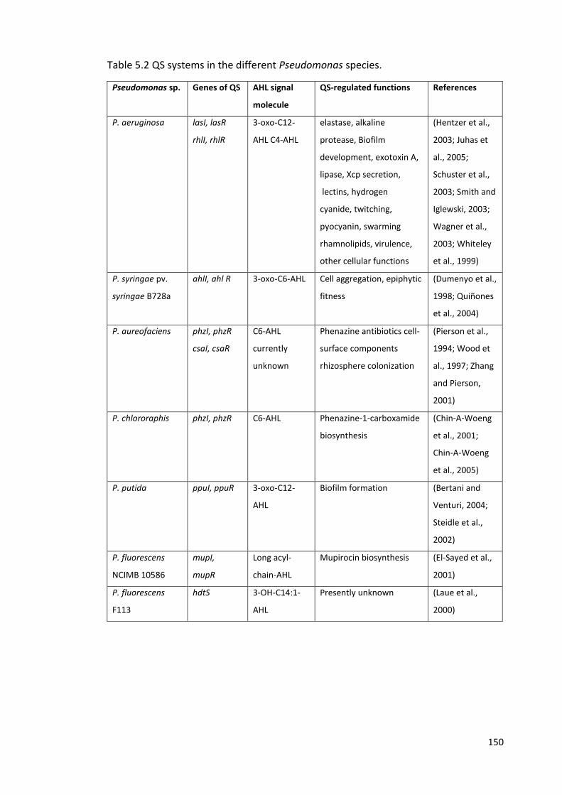
150
Table 5.2 QS systems in the different Pseudomonas species.
Pseudomonas sp. Genes of QS AHL signal
molecule
QS-regulated functions References
P. aeruginosa lasI, lasR
rhlI, rhlR
3-oxo-C12-
AHL C4-AHL
elastase, alkaline
protease, Biofilm
development, exotoxin A,
lipase, Xcp secretion,
lectins, hydrogen
cyanide, twitching,
pyocyanin, swarming
rhamnolipids, virulence,
other cellular functions
(Hentzer et al.,
2003; Juhas et
al., 2005;
Schuster et al.,
2003; Smith and
Iglewski, 2003;
Wagner et al.,
2003; Whiteley
et al., 1999)
P. syringae pv.
syringae B728a
ahlI, ahl R 3-oxo-C6-AHL Cell aggregation, epiphytic
fitness
(Dumenyo et al.,
1998; Quiñones
et al., 2004)
P. aureofaciens phzI, phzR
csaI, csaR
C6-AHL
currently
unknown
Phenazine antibiotics cell-
surface components
rhizosphere colonization
(Pierson et al.,
1994; Wood et
al., 1997; Zhang
and Pierson,
2001)
P. chlororaphis phzI, phzR C6-AHL Phenazine-1-carboxamide
biosynthesis
(Chin-A-Woeng
et al., 2001;
Chin-A-Woeng
et al., 2005)
P. putida ppuI, ppuR 3-oxo-C12-
AHL
Biofilm formation (Bertani and
Venturi, 2004;
Steidle et al.,
2002)
P. fluorescens
NCIMB 10586
mupI,
mupR
Long acyl-
chain-AHL
Mupirocin biosynthesis (El-Sayed et al.,
2001)
P. fluorescens
F113
hdtS 3-OH-C14:1-
AHL
Presently unknown (Laue et al.,
2000)
151
5.1.6 Cross talk between bacterial species via secreted products
There has been considerable interest in the possibility that bacteria in mixed species
communities may communicate via signalling systems such as QS (Williams, 2007). A
number of interactions between P. aeruginosa and other bacterial species, both
positive and negative, have been described (Tashiro et al., 2013). It has been
demonstrated previously that there can be cross-talk between the QS systems of P.
aeruginosa and other bacterial species. For example, it has been shown both in mixed
biofilms cultures and in a murine chronic infection model, that Burkholderia
cenocepacia can recognise and respond to P. aeruginosa autoinducer molecules,
though this communication is not reciprocated (Riedel et al., 2001). It has also been
shown that signal molecules produced by oropharyngeal flora can lead to up-
regulation of P. aeruginosa genes (Duan et al., 2003). There has also been reported
cross-talk between P. aeruginosa and the Gram-positive, Staphylococcus aureus,
whereby QS signal molecules from P. aeruginosa cause inhibition of the S. aureus
virulence-related agr QS system (Chan et al., 2004). In Campylobacter, the luxS gene
of Campylobacter is responsible for the production of S-ribosylhomocysteinase (LuxS)
which cleaves S-ribosylhomocysteine (SRH), yielding unstable (S)-4, 5-dihydroxy-2,3-
pentanedione (DPD) and leading to a final product of AI-2, the suggested signal for QS
(De Keersmaecker et al., 2005; Rajan et al., 2005; Winzer et al., 2002). The receptor
(LuxR equivalent) is not known but it has been demonstrated that a luxS mutant of C.
jejuni (NCTC11168) had decreased significantly in CFU after six days of incubation in
chicken meat juice at 5°C, comparing with wild type in which the CFU remained
unchanged for 27 days (Ligowska et al., 2011). This suggests that LuxS, a QS molecule
in Campylobacter, plays a role in adaptation to cold temperature and meat juice, and
152
is important for survival in the environment. In the previous chapter, evidence was
presented suggesting that luxS might also play an important role in the survival of C.
jejuni in water.
5.1.7 Aims of this chapter
Given the evidence that Pseudomonas spp. could play a key role in the ability of
Campylobacter species to survival in the environment, and previous studies showing
that Pseudomonas can interact with other species, preliminary experiments were
carried out:
1. To determine the effects of supernatants (secreted products) from Pseudomonas
spp. cultures on the growth of Campylobacter in vitro.
2. To use environmental PCR assays to establish the extent to which co-existence of
Campylobacter spp. and fluorescent Pseudomonas spp. occurs in natural water.
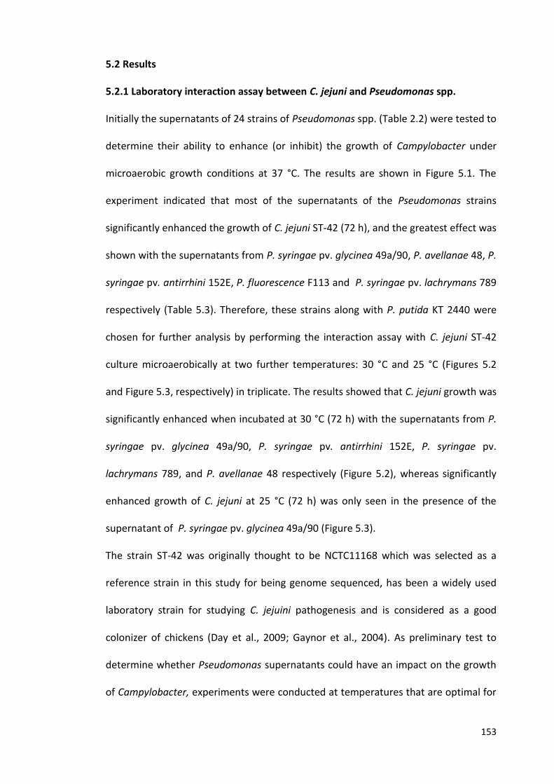
153
5.2 Results
5.2.1 Laboratory interaction assay between C. jejuni and Pseudomonas spp.
Initially the supernatants of 24 strains of Pseudomonas spp. (Table 2.2) were tested to
determine their ability to enhance (or inhibit) the growth of Campylobacter under
microaerobic growth conditions at 37 °C. The results are shown in Figure 5.1. The
experiment indicated that most of the supernatants of the Pseudomonas strains
significantly enhanced the growth of C. jejuni ST-42 (72 h), and the greatest effect was
shown with the supernatants from P. syringae pv. glycinea 49a/90, P. avellanae 48, P.
syringae pv. antirrhini 152E, P. fluorescence F113 and P. syringae pv. lachrymans 789
respectively (Table 5.3). Therefore, these strains along with P. putida KT 2440 were
chosen for further analysis by performing the interaction assay with C. jejuni ST-42
culture microaerobically at two further temperatures: 30 °C and 25 °C (Figures 5.2
and Figure 5.3, respectively) in triplicate. The results showed that C. jejuni growth was
significantly enhanced when incubated at 30 °C (72 h) with the supernatants from P.
syringae pv. glycinea 49a/90, P. syringae pv. antirrhini 152E, P. syringae pv.
lachrymans 789, and P. avellanae 48 respectively (Figure 5.2), whereas significantly
enhanced growth of C. jejuni at 25 °C (72 h) was only seen in the presence of the
supernatant of P. syringae pv. glycinea 49a/90 (Figure 5.3).
The strain ST-42 was originally thought to be NCTC11168 which was selected as a
reference strain in this study for being genome sequenced, has been a widely used
laboratory strain for studying C. jejuini pathogenesis and is considered as a good
colonizer of chickens (Day et al., 2009; Gaynor et al., 2004). As preliminary test to
determine whether Pseudomonas supernatants could have an impact on the growth
of Campylobacter, experiments were conducted at temperatures that are optimal for
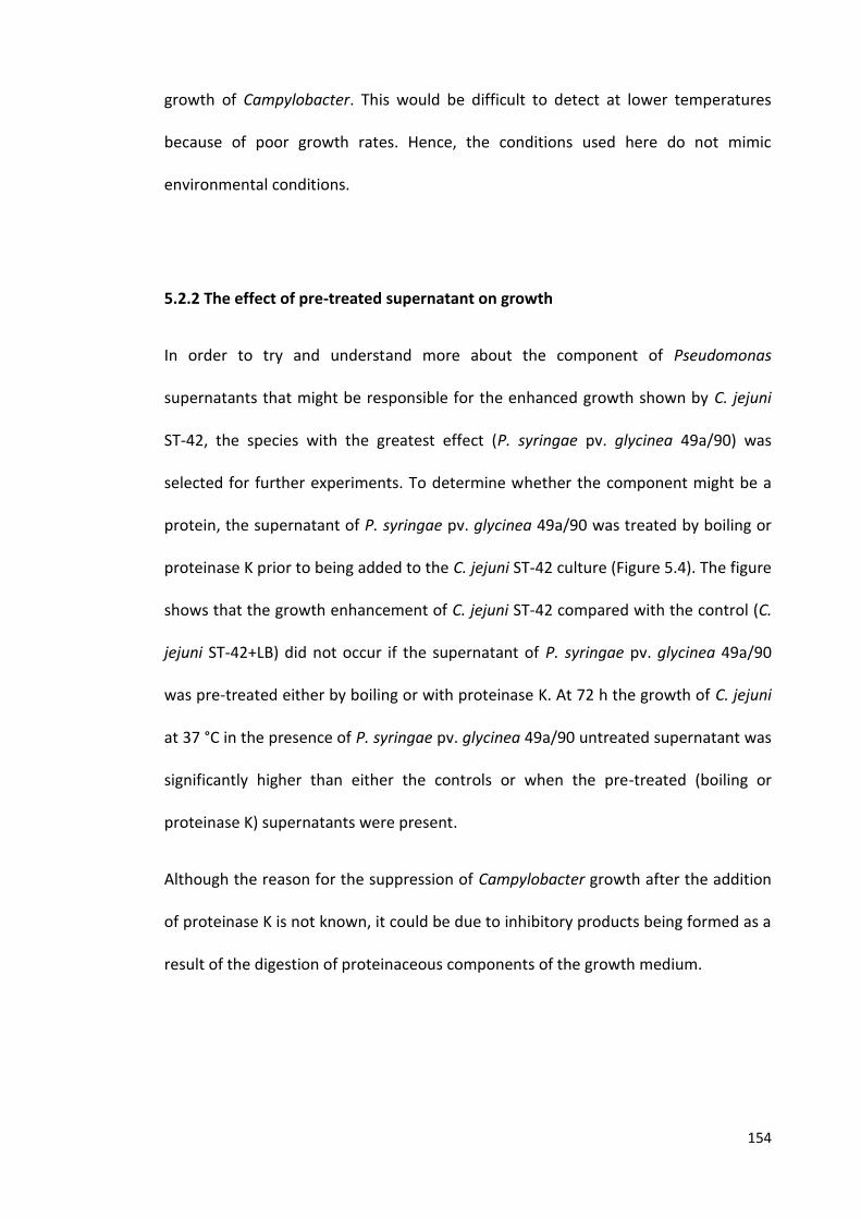
154
growth of Campylobacter. This would be difficult to detect at lower temperatures
because of poor growth rates. Hence, the conditions used here do not mimic
environmental conditions.
5.2.2 The effect of pre-treated supernatant on growth
In order to try and understand more about the component of Pseudomonas
supernatants that might be responsible for the enhanced growth shown by C. jejuni
ST-42, the species with the greatest effect (P. syringae pv. glycinea 49a/90) was
selected for further experiments. To determine whether the component might be a
protein, the supernatant of P. syringae pv. glycinea 49a/90 was treated by boiling or
proteinase K prior to being added to the C. jejuni ST-42 culture (Figure 5.4). The figure
shows that the growth enhancement of C. jejuni ST-42 compared with the control (C.
jejuni ST-42+LB) did not occur if the supernatant of P. syringae pv. glycinea 49a/90
was pre-treated either by boiling or with proteinase K. At 72 h the growth of C. jejuni
at 37 °C in the presence of P. syringae pv. glycinea 49a/90 untreated supernatant was
significantly higher than either the controls or when the pre-treated (boiling or
proteinase K) supernatants were present.
Although the reason for the suppression of Campylobacter growth after the addition
of proteinase K is not known, it could be due to inhibitory products being formed as a
result of the digestion of proteinaceous components of the growth medium.
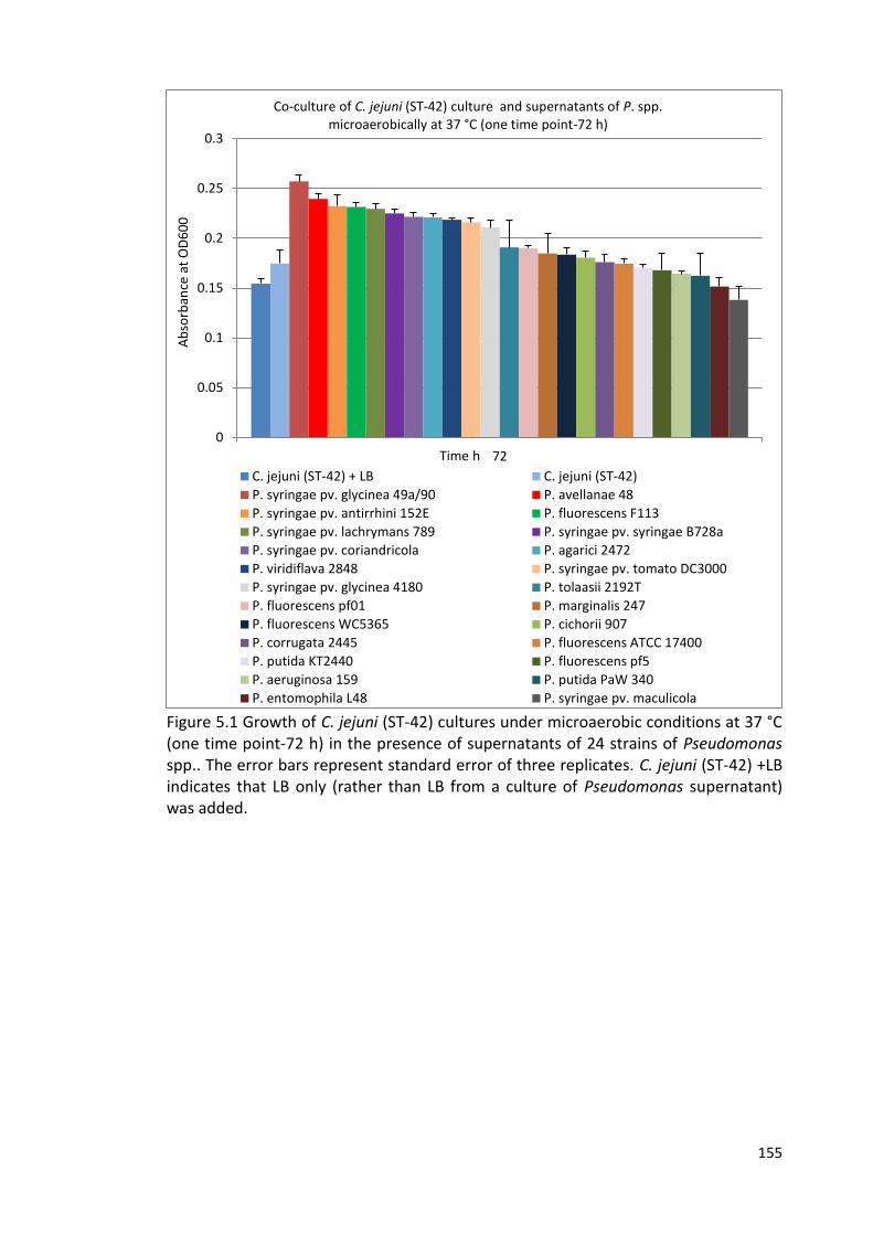
155
Figure 5.1 Growth of C. jejuni (ST-42) cultures under microaerobic conditions at 37 °C (one time point-72 h) in the presence of supernatants of 24 strains of Pseudomonas spp.. The error bars represent standard error of three replicates. C. jejuni (ST-42) +LB indicates that LB only (rather than LB from a culture of Pseudomonas supernatant) was added.
0
0.05
0.1
0.15
0.2
0.25
0.3
72
Ab
sorb
ance
at
OD
60
0
Time h
Co-culture of C. jejuni (ST-42) culture and supernatants of P. spp. microaerobically at 37 °C (one time point-72 h)
C. jejuni (ST-42) + LB C. jejuni (ST-42)
P. syringae pv. glycinea 49a/90 P. avellanae 48
P. syringae pv. antirrhini 152E P. fluorescens F113
P. syringae pv. lachrymans 789 P. syringae pv. syringae B728a
P. syringae pv. coriandricola P. agarici 2472
P. viridiflava 2848 P. syringae pv. tomato DC3000
P. syringae pv. glycinea 4180 P. tolaasii 2192T
P. fluorescens pf01 P. marginalis 247
P. fluorescens WC5365 P. cichorii 907
P. corrugata 2445 P. fluorescens ATCC 17400
P. putida KT2440 P. fluorescens pf5
P. aeruginosa 159 P. putida PaW 340
P. entomophila L48 P. syringae pv. maculicola
156
*p < 0.01 compared to C. jejuni (ST-42) +LB and C. jejuni (ST-42) Figure 5.2 Growth of C. jejuni (ST-42) cultures under microaerobic conditions at 30 °C in the presence of supernatants of 6 strains of Pseudomonas spp.. The error bars represent standard error of three replicates. C. jejuni (ST-42) +LB indicates that LB only (rather than LB from a culture of Pseudomonas supernatant) was added.
*p < 0.01 compared to C. jejuni (ST-42) Figure 5.3 Growth of C. jejuni (ST-42) cultures under microaerobic conditions at 25 °C in the presence of supernatants of 6 strains of Pseudomonas spp.. The error bars represent standard error of three replicates. C. jejuni (ST-42) +LB indicates that LB only (rather than LB from a culture of Pseudomonas supernatant) was added.
0
0.02
0.04
0.06
0.08
0.1
0.12
0.14
0.16
0 24 48 72
Ab
sorb
ance
at
60
0n
m
Time h
Co-culture of C. jejuni (ST-42) culture and supernatants of P. spp. microaerobically at 30 °C
C. jejuni (ST-42)+ LB C. jejuni (ST-42)
P. syringae pv. glycinea 49a/90 P. syringae pv.antirrhini 152E
P. syringae pv. lachrymans 789 P. avellanae 48
P. fluorescens F113 P. putida KT2440
* *
* *
0
0.02
0.04
0.06
0.08
0.1
0 24 48 72
Ab
sorb
ancc
e at
OD
60
0
Time h
Co-culture of C. jejuni (ST-42) culture and supernatants of P. spp. microarobically at 25 °C
C. jejuni (ST-42)+ LB C. jejuni (ST-42)
P. syringae pv. glycinea 49a/90 P. syringae pv. antirrhini 152E
P. fluoresscensF113 P. avellanae 48
P. putida KT2440 P. syringae pv. lachrymans 789
*
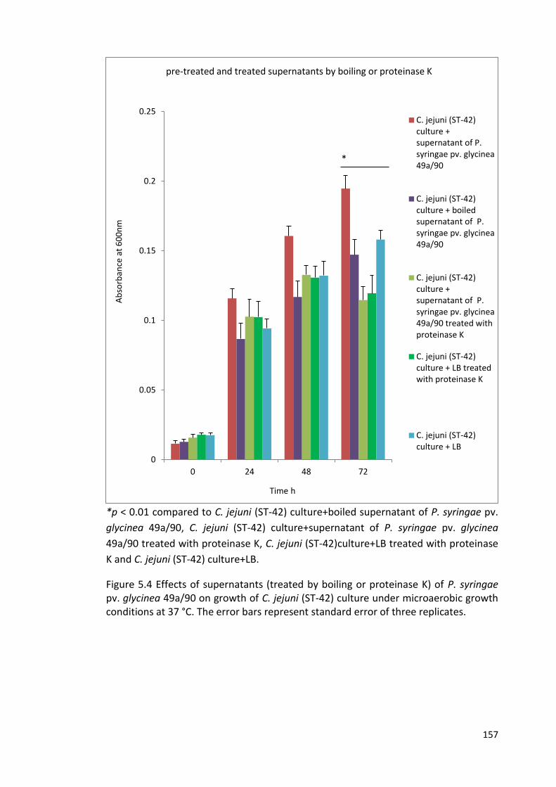
157
*p < 0.01 compared to C. jejuni (ST-42) culture+boiled supernatant of P. syringae pv.
glycinea 49a/90, C. jejuni (ST-42) culture+supernatant of P. syringae pv. glycinea
49a/90 treated with proteinase K, C. jejuni (ST-42)culture+LB treated with proteinase
K and C. jejuni (ST-42) culture+LB.
Figure 5.4 Effects of supernatants (treated by boiling or proteinase K) of P. syringae pv. glycinea 49a/90 on growth of C. jejuni (ST-42) culture under microaerobic growth conditions at 37 °C. The error bars represent standard error of three replicates.
0
0.05
0.1
0.15
0.2
0.25
0 24 48 72
Ab
sorb
ance
at
60
0n
m
Time h
pre-treated and treated supernatants by boiling or proteinase K
C. jejuni (ST-42)culture +supernatant of P.syringae pv. glycinea49a/90
C. jejuni (ST-42)culture + boiledsupernatant of P.syringae pv. glycinea49a/90
C. jejuni (ST-42)culture +supernatant of P.syringae pv. glycinea49a/90 treated withproteinase K
C. jejuni (ST-42)culture + LB treatedwith proteinase K
C. jejuni (ST-42)culture + LB
*
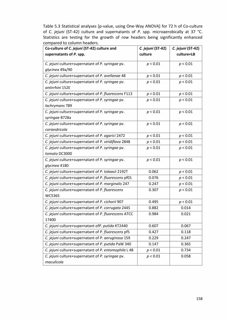
158
Table 5.3 Statistical analyses [p-value, using One-Way ANOVA] for 72 h of Co-culture of C. jejuni (ST-42) culture and supernatants of P. spp. microaerobically at 37 °C. Statistics are testing for the growth of row headers being significantly enhanced compared to column headers.
Co-culture of C. jejuni (ST-42) culture and
supernatants of P. spp.
C. jejuni (ST-42)
culture
C. jejuni (ST-42)
culture+LB
C. jejuni culture+supernatant of P. syringae pv.
glycinea 49a/90
p < 0.01 p < 0.01
C. jejuni culture+supernatant of P. avellanae 48 p < 0.01 p < 0.01
C. jejuni culture+supernatant of P. syringae pv.
antirrhini 152E
p < 0.01 p < 0.01
C. jejuni culture+supernatant of P. fluorescens F113 p < 0.01 p < 0.01
C. jejuni culture+supernatant of P. syringae pv.
lachrymans 789
p < 0.01 p < 0.01
C. jejuni culture+supernatant of P. syringae pv.
syringae B728a
p < 0.01 p < 0.01
C. jejuni culture+supernatant of P. syringae pv.
coriandricola
p < 0.01 p < 0.01
C. jejuni culture+supernatant of P. agarici 2472 p < 0.01 p < 0.01
C. jejuni culture+supernatant of P. viridiflava 2848 p < 0.01 p < 0.01
C. jejuni culture+supernatant of P. syringae pv.
tomato DC3000
p < 0.01 p < 0.01
C. jejuni culture+supernatant of P. syringae pv.
glycinea 4180
p < 0.01 p < 0.01
C. jejuni culture+supernatant of P. tolaasii 2192T 0.062 p < 0.01
C. jejuni culture+supernatant of P. fluorescens pf01 0.076 p < 0.01
C. jejuni culture+supernatant of P. marginalis 247 0.247 p < 0.01
C. jejuni culture+supernatant of P. fluorescens
WC5365
0.307 p < 0.01
C. jejuni culture+supernatant of P. cichorii 907 0.495 p < 0.01
C. jejuni culture+supernatant of P. corrugata 2445 0.882 0.014
C. jejuni culture+supernatant of P. fluorescens ATCC
17400
0.984 0.021
C. jejuni culture+supernatant ofP. putida KT2440 0.607 0.067
C. jejuni culture+supernatant of P. fluorescens pf5 0.427 0.118
C. jejuni culture+supernatant of P. aeruginosa 159 0.229 0.247
C. jejuni culture+supernatant of P. putida PaW 340 0.147 0.365
C. jejuni culture+supernatant of P. entomophila L 48 p < 0.01 0.734
C. jejuni culture+supernatant of P. syringae pv.
maculicola
p < 0.01 0.058
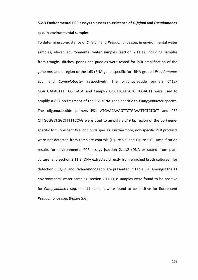
159
5.2.3 Environmental PCR assays to assess co-existence of C. jejuni and Pseudomonas
spp. in environmental samples.
To determine co-existence of C. jejuni and Pseudomonas spp. in environmental water
samples, eleven environmental water samples (section 2.11.1), including samples
from troughs, ditches, ponds and puddles were tested for PCR amplification of the
gene oprI and a region of the 16S rRNA gene, specific for rRNA group I Pseudomonas
spp. and Campylobacter respectively. The oligonucleotide primers C412F
GGATGACACTTT TCG GAGC and CampR2 GGCTTCATGCTC TCGAGTT were used to
amplify a 857 bp fragment of the 16S rRNA gene-specific to Campylobacter species.
The oligonucleotide primers PS1 ATGAACAAAGTTCTGAAATTCTCTGCT and PS2
CTTGCGGCTGGCTTTTTCCAG were used to amplify a 249 bp region of the oprI gene-
specific to fluorescent Pseudomonas species. Furthermore, non-specific PCR products
were not detected from template controls (Figure 5.5 and Figure 5.6). Amplification
results for environmental PCR assays [section 2.11.2 (DNA extracted from plate
culture) and section 2.11.3 (DNA extracted directly from enriched broth cultures)] for
detection C. jejuni and Pseudomonas spp. are presented in Table 5.4. Amongst the 11
environmental water samples (section 2.11.1), 8 samples were found to be positive
for Campylobacter spp. and 11 samples were found to be positive for fluorescent
Pseudomonas spp. (Figure 5.6).
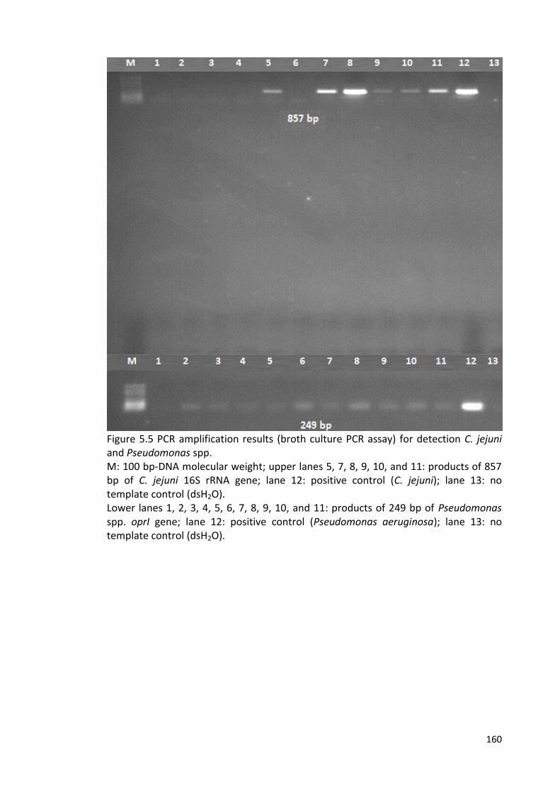
160
Figure 5.5 PCR amplification results (broth culture PCR assay) for detection C. jejuni and Pseudomonas spp. M: 100 bp-DNA molecular weight; upper lanes 5, 7, 8, 9, 10, and 11: products of 857 bp of C. jejuni 16S rRNA gene; lane 12: positive control (C. jejuni); lane 13: no template control (dsH2O). Lower lanes 1, 2, 3, 4, 5, 6, 7, 8, 9, 10, and 11: products of 249 bp of Pseudomonas spp. oprI gene; lane 12: positive control (Pseudomonas aeruginosa); lane 13: no template control (dsH2O).
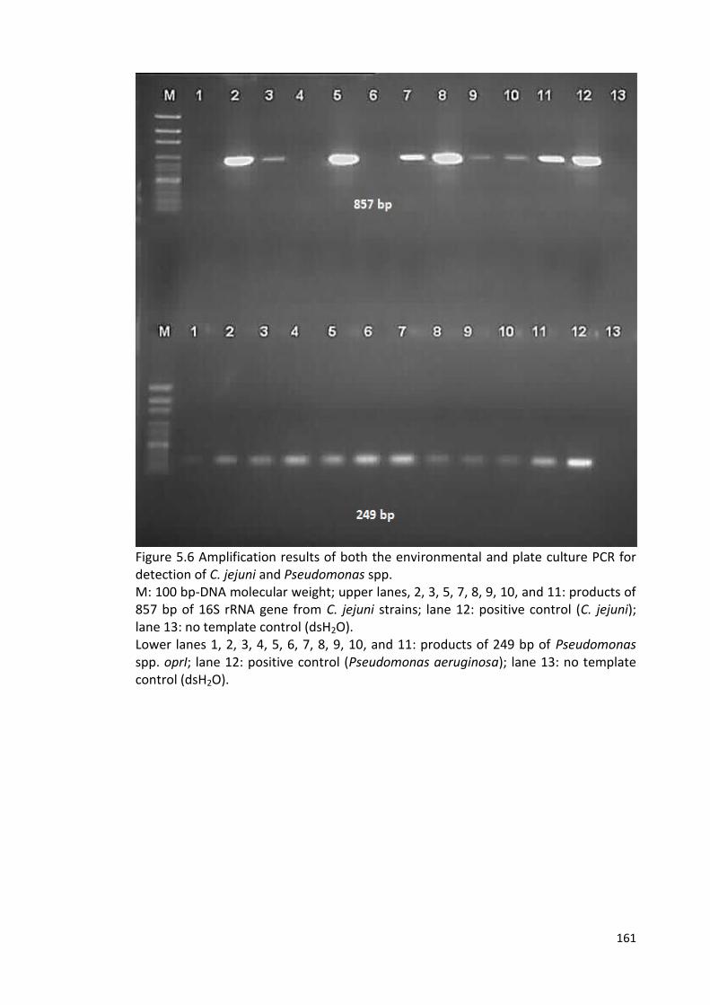
161
Figure 5.6 Amplification results of both the environmental and plate culture PCR for detection of C. jejuni and Pseudomonas spp. M: 100 bp-DNA molecular weight; upper lanes, 2, 3, 5, 7, 8, 9, 10, and 11: products of 857 bp of 16S rRNA gene from C. jejuni strains; lane 12: positive control (C. jejuni); lane 13: no template control (dsH2O). Lower lanes 1, 2, 3, 4, 5, 6, 7, 8, 9, 10, and 11: products of 249 bp of Pseudomonas spp. oprI; lane 12: positive control (Pseudomonas aeruginosa); lane 13: no template control (dsH2O).
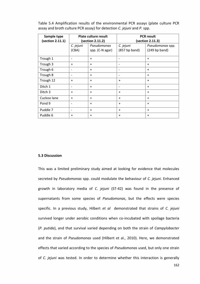
162
Table 5.4 Amplification results of the environmental PCR assays (plate culture PCR assay and broth culture PCR assay) for detection C. jejuni and P. spp.
Sample type (section 2.11.1)
Plate culture result (section 2.11.2)
PCR result (section 2.11.3)
C. jejuni (CBA)
Pseudomonas spp. (C-N agar)
C. jejuni (857 bp band)
Pseudomonas spp. (249 bp band)
Trough 1 - + - +
Trough 3 + + - +
Trough 6 - + - +
Trough 8 - + - +
Trough 12 + + + +
Ditch 1 - + - +
Ditch 3 + + + +
Cuckoo lane + + + +
Pond 9 - + + +
Puddle 7 - + + +
Puddle 6 + + + +
5.3 Discussion
This was a limited preliminary study aimed at looking for evidence that molecules
secreted by Pseudomonas spp. could modulate the behaviour of C. jejuni. Enhanced
growth in laboratory media of C. jejuni (ST-42) was found in the presence of
supernatants from some species of Pseudomonas, but the effects were species
specific. In a previous study, Hilbert et al demonstrated that strains of C. jejuni
survived longer under aerobic conditions when co-incubated with spoilage bacteria
(P. putida), and that survival varied depending on both the strain of Campylobacter
and the strain of Pseudomonas used (Hilbert et al., 2010). Here, we demonstrated
effects that varied according to the species of Pseudomonas used, but only one strain
of C. jejuni was tested. In order to determine whether this interaction is generally
163
applicable, it would be necessary to test a wider range of C. jejuni strains,
representing different sources and clonal complexes. Given the work presented in
previous chapters, where variations between strains of C. jejuni with respect to
survival in water were evident, it is likely that different strains would also exhibit
different responses with respect to this interaction with Pseudomonas.
The greatest effect was seen with the supernatant of P. syringae pv. glycinea 49a/90
isolated from field grown plants of soybean cv. Maple Arrow at Dornburg near Jena,
Germany in 1990 (Ullrich et al., 1993). This organism causes bacterial blight of
soybean and can produce a phytotoxin called coronatine. Effects were also seen with
other pathovars of P. syringae, such as lachrymans, avellanae and antirrhini, but were
not so clear with some of the more common environmental species of Pseudomonas
that are not as closely associated with plants, such as P. putida or P. fluorescens.
Hence, the significance of the effect on Campylobacter is not clear. It would be
important to test a wider range of Pseudomonas isolates, including some found in
environmental samples where Campylobacter are also present. Here, we
demonstrated that, in the environmental samples tested, wherever Campylobacter
was identified, Pseudomonas species were also present, consistent with the idea that
co-existence with Pseudomonas may act as a survival strategy for Campylobacter
outside a host body (Hilbert et al., 2010).
The work presented in this chapter suggests that the active component secreted by P.
syringae pv. glycinea 49a/90 is proteinaceous, given that it is sensitive both to boiling
and to proteinase K treatment. Little is known about the secretome of this organism,
beyond the non-protein phytotoxin coronatine. However, various P. syringae
pathovars do carry the Hrp type III secretion system implicated in plant pathogenicity
164
(O'Brien et al., 2011). It has been shown that secretion of virulence factors in several
plant pathogenic bacteria is thermoregulated. For example, Coronatin toxin in P.
syringae pathovars acts as a virulence factor, most likely by enhancing ethylene
biothensis in plant (Kenyon and Turner, 1992). The yield of coronatine was increased
after a 7-day incubation of P. syringae pv. glycinea broth cultures at 18°C, and the
toxin yields were reduced to minimal at higher temperatures (30 °C) which could be
due to reduced synthesis of the enzymes involved in the synthesis of the toxin
(Palmer and Bender, 1993). Similarly, it has been demonstrated that secretion
systems play an important role in the survival of P. aeruginosa, human opportunistic
pathogen, at lower temperatures (below 37 °C) by decreasing the channel size of the
major outer membrane porin (OprF) at suboptimal temperatures (Jaouen et al.,
2004). Moreover, this thermoadaptation is possibly specific for proteins secreted by
the T1SS (APrP), T2SS (PrpL), or T6SS (HcpB) pathways and thus facilitate nosocomial
infection at room temperature (25 °C) (Termine and Michel, 2010)
Indeed the P. avellanae secretome has been characterised (O’Brien et al., 2012).
However, induction of this system is tightly controlled and it should not be active
under the conditions used here to grow the strains. Although P. syringae do have QS
systems (Table 5.2), the signal molecule is not a protein. Therefore, it seems unlikely
that the interaction is due to cross talk between the Pseudomonas QS system and the
Campylobacter LuxS QS system. To resolve the nature of the active protein
component, it would be necessary to purify protein from the Pseudomonas
supernatants, repeating the assay on extracts to confirm that the active component is
still present. This was beyond the scope of the work here, but would be an interesting
avenue to follow. If the active protein was identified, its effect on other
165
Campylobacter phenotypes could be characterised, including potential effects on
survival in model systems more reflective of the real environment in which the two
organisms co-exist.
For detection of Pseudomonas bacteria in the environmental samples,
oligonucleotide primers were used to amplify a 249 bp of the oprI gene-specific to
fluorescent Pseudomonas spp. The oprI gene encodes lipoprotein I (OprI) which is one
of the abundant proteins present in the outer member of fluorescent Pseudomonas
spp., has a low molecular-weight (8 kDa) and may only be expressed in rRNA group I
Pseudomonas. It can be used in phylogeny of the genus Pseudomonas (De Vos et al.,
1998; De Vos et al., 1993; Duchêne et al., 1989). However, further studies would be
needed in order to characterise the species of Pseudomonas present, either by using
more specific PCR assays, or by sequencing oprI amplicons and screening against
sequence data from known species in the database.
In this study, Pseudomonas spp. were detected in all environmental water samples,
suggesting, as has been reported, that they are ubiquitous in the environment
(Witholt et al., 1992), whereas Campylobacter spp. were detectable only in some
samples. Amongst the 11 environmental water samples, 8 samples were found to be
positive for Campylobacter spp. and all 11 samples were found to be positive for
fluorescent Pseudomonas spp. (Table 5.4 and Figure 5.6). Indeed, several culture-
negative samples [Table 5.4 and Figure 5.6; Lane 9 (puddle 9) and lane 10 (puddle 7)]
were positive for the PCR test. This may be because of the presence of dead cells in
the samples, or because Campylobacter were present, but not culturable on the
media used. As was demonstrated earlier in this thesis, different C. jejuni strains
exhibited variations with respect to the retention of culturability in environmental
166
conditions. In addition, one culture-positive sample [Table 5.4 and Figure 5.6; Lane 2
(trough 3)] for Campylobacter was not positive by the PCR test [Table 5.4 and Figure
5.5; Lane 2 (trough 3)]. In this case, the culture-positive status was based on the
presence of a single colony on the mCCDA plate, whereas generally multiple colonies
were found. This anomalous result could have been due to contamination on the
plate.
Previous studies have shown that contaminated poultry meat has been considered as
the main source of campylobacteriosis in humans (Nauta et al., 2007). Campylobacter
as a microaerophilic microorganism has to survive atmospheric oxygen tension on the
poultry meat. Co-existence with Pseudomonas spp. is thought to be one of the
strategies that is followed by C. jejuni to survive atmospheric oxygen tension on
chicken meat (Hilbert et al., 2010). It would be interesting to carry out more
comprehensive studies, not only on a wider range of environmental samples, but in
the poultry meat environment, to better characterise the co-existence of
Pseudomonas and Campylobacter, and to select more relevant model strains for
study of the interactions between them.
167
5.4 Conclusions
In a limited preliminary study, it was demonstrated that Campylobacter spp. co-exist
with Pseudomonas species in the environment (natural water), supporting the notion
that interactions between Campylobacter with and other microorganisms may
enhance its survival in the atmospheric oxygen tensions of the environment. Some
Pseudomonas strains secrete products that can enhance the growth of
Campylobacter under laboratory conditions, and the activity was sensitive to boiling
and proteinase K treatment, suggesting that the active component may be a protein
(or proteins). Further work is needed to characterise the nature of this interaction,
the variations that might occur when different strains of C. jejuni or fluorescent
Pseudomonas spp. are tested, and the relevance to interactions that might occur in
natural environments.
168
C h a p t e r 6
GENERAL DISCUSSION
Campylobacter jejuni remains an important and increasing cause of food related
illness worldwide, imposing a considerable burden on global economics and posing
public health concerns. In the context of the UK in particular, the pathogen remains a
key priority, especially in relation to contaminated poultry products as a source of
infection. Compared to other enteric bacteria such as Salmonella spp. and E. coli, less
is known about the metabolism, pathogenicity and environmental survival
mechanisms of Campylobacter (Duffy et al., 2008; Murphy et al., 2006; Young et al.,
2007). Yet, there are far more cases of infectious intestinal disease associated with
Campylobacter than with the other better known bacterial agents of disease.
Various studies have reported the survival characteristics of the pathogen E.coli in
water. It has been reported that the organism may survive for two months in lakes,
troughs, rivers and faecal contaminated puddles. The bacterium can survive for long
times at cold temperatures (<10 °C), and can enter a VBNC state when exposed to
stresses such as starvation (Avery et al., 2008; Wang and Doyle, 1998). It has also
been suggested that water is one of the main reservoirs of spreading Salmonella spp.
in the environment, where the microorganism can survive long enough to be
transmitted (Gorski et al., 2011). Hence, the potential role for the environment in the
transmission of zoonotic pathogens is well established.
Although C. jejuni is a fastidious bacterium that requires microaerobic conditions for
its growth, the organism is widespread in the environment, processed meats and
animal reservoirs. It is likely that the environment plays an important role in
transmission of Campylobacter infections to humans, either directly through
169
contaminated water, or indirectly by facilitating contamination of livestock, with
subsequent transmission to humans (Clark et al., 2003; Murphy et al., 2006; Olson et
al., 2008; Park, 2002; Stanley and Jones, 2003). Compared to other bacteria, the
relatively small genome of C. jejuni lacks many of the genes that are essential for
regulating gene expression in response to adverse environmental conditions (Table
6.1), and this has further restricted our knowledge of the organisms, because many of
the widespread mechanisms considered important in other foodborne bacterial
pathogens, simply do not apply to Campylobacter. However, it is clear that
Campylobacter can survive in the environment and can cause bacterial food borne
diseases in humans (Murphy et al., 2006). It has been considered that untreated
water is one of the important routes of transmission of the pathogen to humans, and
can contaminate drinking water from which water-borne outbreaks may emerge
(Clark et al., 2003). It has also been suggested that contaminated surface water of
pastures can be a source of contamination of both tap and ground water which may
lead to waterborne outbreaks in humans and transmission of Campylobacter to
poultry flocks via drinking water systems (Jones, 2001; Pearson et al., 1993;
Richardson et al., 2007; Van de Giessen et al., 1996). It has been hypothesized that
Campylobacter follow diverse mechanisms to survive in the environment, such as
formation of biofilm, entering a VBNC state or coexistence with other microorganisms
(Hilbert et al., 2010). However, our understanding of these processes remains at a
relatively low level.
C. jejuni has been isolated from diverse animal and environmental sources where it
has adapted to specific ecological niches. Using MLST analysis, it has been shown that
whilst some MLST clonal complexes (CCs), such as ST-21 complex, are broadly
170
distributed, some others such as members of ST-45 complex, which are frequently
isolated from human campylobacteriosis, are more predominant in wildlife and
environmental water than other common clonal complexes (Colles et al., 2003; Dingle
et al., 2005; Dingle et al., 2001; French et al., 2005; Kärenlampi et al., 2007; Manning
et al., 2003; McCarthy et al., 2007; Sails et al., 2003; Sopwith et al., 2006).
In this study, a diverse panel of C. jejuni strains (from the University of Liverpool) from
various sources including human, cattle, poultry, environment, and wildlife, was
screened for variations in the ability to survive in sterile distilled water based on
culture at 4 °C and 25 °C. The isolates represented different MLST groups, including
the broadly distributed MLST clonal complexes (ST-21 and ST-45), clonal complexes
found amongst human isolates which are adapted to specific host (cattle associated
ST-61), and representatives of water/wild-life isolates (1336) and the bank vole
isolates (414). The initial screen which was only based on one biological replicate
assigned the strains into various categories based on retaining culturability by
enumerating CFU/mL. Representatives of different survival abilities, M1, 1336 and
414 were selected for closer study. This study demonstrated that the survival time in
sterile distilled water, based on the ability to retain culturabilty, varies significantly
between various strains of C. jejuni. Strain M1 is a member of ST-45 clonal complex,
which is widespread and is highly prevalent in the environment (Friis et al., 2010). Of
the three strains tested, strain M1 declined most rapidly in culturability at both
temperatures (4 °C and 25 °C) tested. Each of the three strains underwent rapid
decline at 25 °C which is in agreement with other findings (Colles et al., 2003; Guillou
et al., 2008). The study also demonstrated that C. jejuni strains were able to survive
much better at low temperature (4 °C) than warm temperature (25 °C), which is in

171
agreement with other findings (Buswell et al., 1998; Cools et al., 2003; González and
Hänninen, 2012; Korhonen and Martikalnon, 1991).
Experiments were carried out for comparing the same three strains in both
autoclaved and non-autoclaved natural water. The results demonstrated that the
culturability was better retained in non-autoclaved natural water, which may be due
to some component of the natural water system that is degraded by autoclaving, but
emphasizes the risks of drawing conclusions from less natural systems.
The result of the LIVE/DEAD staining experiments showed that the rapid loss of
culturability might not indicate poor survival, but could be due to the capability of
strain M1 to enter more quickly into a VBNC state. This result is in agreement with
findings of other studies that suggest Campylobacter can enter a VBNC state under
stress conditions such as starvation (nutrient-poor water), and this capability varies
between different strains (Baffone et al., 2006; Chaveerach et al., 2003; Cools et al.,
2003; Rollins and Colwell, 1986; Tholozan et al., 1999).
One key conclusion from these studies is that it is dangerous to assume that the
properties exhibited by one strain of a bacterial species apply to all members of that
species. For example, it has been shown that the ability of different strains of C. jejuni
varies in formation of the VBNC state, and that some strains of C. jejuni are more
frequently isolated from environmental sources (Cools et al., 2003; Hald et al., 2001;
Ziprin et al., 2003; Ziprin and Harvey, 2004). Moreover, the variation has also been
noted in the capability of these cells to resuscitate and to cause infections (Beumer et
al., 1992; Mederma et al., 1992). In addition, the behaviour variation between strains
of C. jejuni has been noted in biofilm formation. For example, most C. jejuni strains
possess the luxS gene, which encodes AI-2, implicated in formation of biofilms, but
172
some strains of C. jejuni (such as strain 1336) lack the luxS gene (Buswell et al., 1998;
Hepworth et al., 2011; Plummer, 2012). Hence, any potential role for luxS may be
strain-specific, rather than applicable to the whole species.
This emphasizes the need to ensure that reported phenotypes or mechanisms
described in one strain of C. jejuni are in fact shared more generally, before assuming
that a property is applicable to all strains of C. jejuni. Even the same strain, for
example the widely studied NCTC11168, can be subject to considerable variations,
leading to phenotypic changes (Gaynor et al., 2004; Revez et al., 2012; Thomas et al.,
2014). C. jejuni are especially adept at adaptive change because of the existence of
contingency loci (Jerome et al., 2011), which can cause significant phase variations
and hence phenotypic changes. Indeed, C. jejuni is notoriously difficult to work with
because it often hard to get consistent replicates. This genomic flexibility and the
capacity for rapid adaptive changes, emphasize the need for caution when assuming
that observations based on a single strain are widely applicable.
This study postulated that variations in the survival behaviour in sterile distilled water
between different strains of C. jejuni could de due to differences in expression of
stress response genes in the response to adverse environment, including genes
involved in the induction of VBNC state. For elucidating the mechanisms underlying
the variations between different strains of C. jejuni in the ability to retain culturability
in water, end-point PCR and Q-PCR assays were applied to target a set of stress
response genes, reported previously, at specific time points during survival in sterile
distilled water. Q-PCR analysis indicated that there were differences in gene
expression levels during survival in sterile distilled water between the different strains
of C. jejuni. Interestingly, although there were low colony forming unit counts for
173
strain M1 recovered from water, the luxS gene was significantly up-regulated in this
strain compared to strain 414 in both the day 3 (4 °C) and day 1 (25 °C) samples.
RNAseq analysis has confirmed the high levels of luxS expression during survival of
strain M1 in sterile distilled water. These observations are consistent with a role for
luxS during the conversion of M1 to the VBNC state, and this study reveals for the first
time implicating the C. jejuni QS system (luxS) in a role in survival in water. Given that
QS is considered to be a cell density dependent phenomenon, this is an interesting
finding. The bacteria are actually diluted during the process of introducing them into
the water, yet the QS-related gene is up-regulated. As yet the partner regulatory
gene associated with the putative signal molecule that LuxS is responsible for
synthesising has not been identified. Various roles for LuxS have been proposed, in
oxidative stress, flagellar gene expression, ability to colonise animals, CDT expression
and virulence in an animal model (Plummer, 2012). However, our observations
suggest that LuxS may not be part of a conventional QS system, or may have other
characteristics. A role for QS in survival has been reported for Vibrio cholerae in
relation to biofilms exposed to seawater (Joelsson et al., 2006). More recently, it has
been reported that QS signal molecules can revive V. cholerae from a dormant state
in water (i.e., recovery from VBNC state) http://www.ncbi.nlm.nih.gov
/pubmed/23716683. QS has also been implicated in biofilm formation by
Burkholderia, Vibrio and Listeria pathogens (Belval et al., 2006; Engebrecht et al.,
1983; Riedel et al., 2001). An interesting interaction between LuxS, flagella and
autoagglutination in C. jejuni has been reported (Jeon et al., 2003). This implied that
the LuxS molecule may play a role in the regulation of motility and agglutination or
the formation of surface structures in C. jejuni. Our study, coupled to parallel RNAseq
174
data indicating the up-regulation of flagellar genes (C. Bronowski and C. Winstanley,
unpublished) suggests that, LuxS plays a role in the survival of strain M1 in water, and
that this may be due to interactions with flagella and autoagglutination activity.
However, further work is needed to resolve the precise mechanisms underlying our
observations.
Co-existence of C. jejuni and Pseudomonas species is one of the survival strategies of
C. jejuni outside the host body and during food contamination (Hilbert et al., 2010).
Pseudomonas species such as P. fluorescens, P. franji, P. chlororaphis and P.putida can
be found on poultry meat, and are known spoilage bacteria that are generally
distributed in a wide range of environmental niches (Ghafir et al., 2008; Hilbert et al.,
2010; Mead et al., 1993; Russell et al., 1995; Silby et al., 2011). In order to overcome
the atmospheric oxygen stress and to enhance the aerotolerance of C. jejuni, this
organism interacts with aerobic Pseudomonas species found in natural environmental
niches or on food (Hilbert et al., 2010).
In a limited preliminary study of in vitro interaction of C. jejuni (ST-42) with
supernatants of Pseudomonas spp., we demonstrated that molecules secreted from
some species of Pseudomonas could enhance the growth of C. jejuni, and the effects
were species-dependant. Although only one strain of C. jejuni was investigated, the
survival varied according to the species of Pseudomonas used. In a previous study of
interaction between C. jejuni strains with species of Pseudomonas under aerobic
conditions, Hilbert et al. reported the survival variations were dependant on the
strain of Campylobacter and the strain of Pseudomonas used (Hilbert et al., 2010). In
this thesis the growth of C. jejuni ST-42 was significantly enhanced by the effect of
most of the supernatants of the Pseudomonas strains, and the greatest effect was

175
evident with the supernatant of P. syringae pv. glycinea 49a/90. Environmental PCR
assays demonstrated co-existence of Campylobacter spp. and fluorescent
Pseudomonas spp., which occurs in natural water, and showed that wherever
Campylobacter was found, Pseudomonas species were also present. These results are
compatible with the hypothesis that co-existence with Pseudomonas may act as a
survival strategy for Campylobacter in the environment (Hilbert et al., 2010). This
experiment demonstrated that the molecules secreted from P. syringae pv. glycinea
49a/90 and involved in the interaction with C. jejuni were sensitive both to boiling
and to proteinase K treatment, suggesting that the active component in the
secretome was proteinaceous. However, although little is known about the
secretome of this bacterium, it is unlikely that interaction is due to cross talk between
the Pseudomonas QS system and the Campylobacter LuxS QS system, given that the
signal molecule is not a protein.
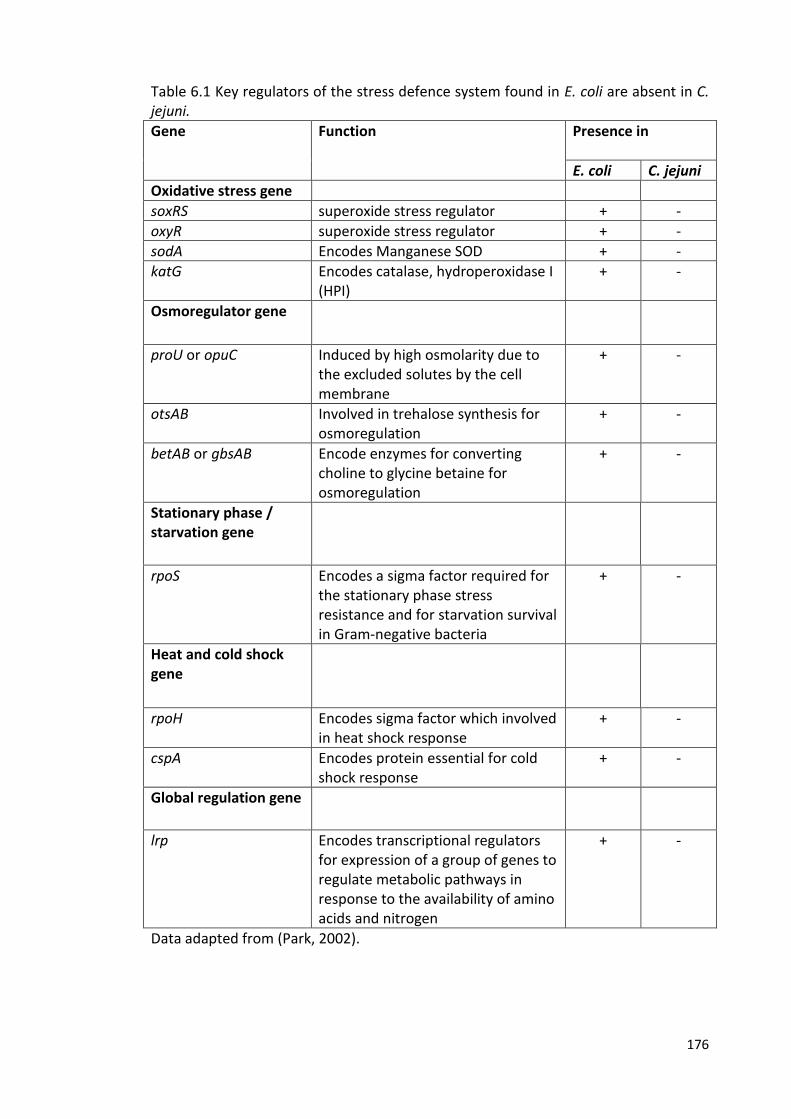
176
Table 6.1 Key regulators of the stress defence system found in E. coli are absent in C. jejuni.
Gene Function Presence in
E. coli C. jejuni
Oxidative stress gene
soxRS superoxide stress regulator + -
oxyR superoxide stress regulator + -
sodA Encodes Manganese SOD + -
katG Encodes catalase, hydroperoxidase I (HPI)
+ -
Osmoregulator gene
proU or opuC Induced by high osmolarity due to the excluded solutes by the cell membrane
+ -
otsAB Involved in trehalose synthesis for osmoregulation
+ -
betAB or gbsAB Encode enzymes for converting choline to glycine betaine for osmoregulation
+ -
Stationary phase / starvation gene
rpoS Encodes a sigma factor required for the stationary phase stress resistance and for starvation survival in Gram-negative bacteria
+ -
Heat and cold shock gene
rpoH Encodes sigma factor which involved in heat shock response
+ -
cspA Encodes protein essential for cold shock response
+ -
Global regulation gene
lrp Encodes transcriptional regulators for expression of a group of genes to regulate metabolic pathways in response to the availability of amino acids and nitrogen
+ -
Data adapted from (Park, 2002).
177
Future work
The present study suggests that survival in water varies significantly between
different strains of C. jejuni. However, more work is needed in this area (survival of C.
jejuni strains in sterile distilled water and natural water) in order to fully characterise
the extent of survival variations between different strains of C. jejuni, and to better
establish whether there are clear associations between particular clonal complexes
and the rate of loss of culturabilty, and whether this correlates with observations
about the relative prevalence in environmental samples. Also, it is important to
screen further numbers of isolates from different sources and representing different
clonal complex, to identify any clear relationships between these survival variations in
the environment and the epidemiology of Campylobacter infections in humans.
This study provides clear evidence for expression of luxS in VBNC populations of C.
jejuni (strain M1). However, further work is necessary in the area of gene expression
during survival in sterile distilled water, in order to indicate the role of other stress
response genes that may play a role in oxidative stress in C. jejuni such as katA, sodB
and msrA/B, and to determine the precise role of the QS system in the variations
observed in strain M1 compared to strain 414 with regard to loss of culturability,
survival strategies and the conversion to the VBNC state. Here, some of these
variations have been established, but further work is needed to fully identify the
mechanisms underlying the differences between strains.
Some of this work is already being carried out. Indeed a comprehensive set of RNAseq
data for the M1 survival experiments is currently being analysed in order to better
178
understand the mechanisms involved and to design follow up experiments to test the
hypotheses generated
This study provides evidence for laboratory-based interaction between C. jejuni (ST-
42) and Pseudomonas species in which the effects were species specific. Therefore, in
order to determine general applicability and to characterise the nature of this
interaction, future work is necessary to repeat the experiment of laboratory
interaction assay between C. jejuni and Pseudomonas spp. under aerobic conditions
at 4 °C and 20 °C, to test different strains of C. jejuni representing different sources
and clonal complexes such as M1 (ST-45), 11168 (ST-21), 1336 (wild bird isolate) and
strain 1107 (cattle isolate). Strains from poultry meat surfaces could also be identified
rapidly using hippurate hydrolysis, according to the methods of Hwang and Ederer
(Hilbert et al., 2010; Hwang and Ederer, 1975) and further characterized by PCR
analysis of the hippuricase gene hip (Linton et al., 1997). The identified isolates, which
are more relevant model strains, can be selected for the interaction assay with the
Pseudomonas spp. that are found on chicken meat surfaces. The Pseudomonas spp.
can be identified by plating the diluted chicken exudate on Pseudomonas selective
agar and incubated under aerobic conditions at 25°C for 48h. Additionally, the
colonies are identified by PCR amplification of the gene oprI specific for fluorescent
Pseudomonas spp.
In order to identify the nature of the molecules (proteins) secreted from fluorescent
Pseudomonas spp. that influence C. jejuni behaviour, further work is needed to
isolate the relevant protein from the Pseudomonas supernatants, for example by
repeating the assay on size-fractionated extracts in order to characterise the active
protein component. It would also be important to test the potential effects on
179
survival in model systems more reflective of the real environment in which the two
organisms co-exist. For example, co-cultures could be studied in biofilm systems.
However, to characterise the species of Pseudomonas present in this study, further
studies are needed including more specific PCR assays, or by sequencing oprI
amplicons and screening against sequence data from known species in the database.
In conclusion, it is evident that the environment plays an important role in the
transmission of Campylobacter, but that there are important strain variations that are
poorly understood. We have just started to identify some of the genes that might be
important in the various mechanisms underlying Campylobacter survival strategies
but much more work needs to be done in order to resolve the underlying processes
and potentially exploit our knowledge to design better interventions.
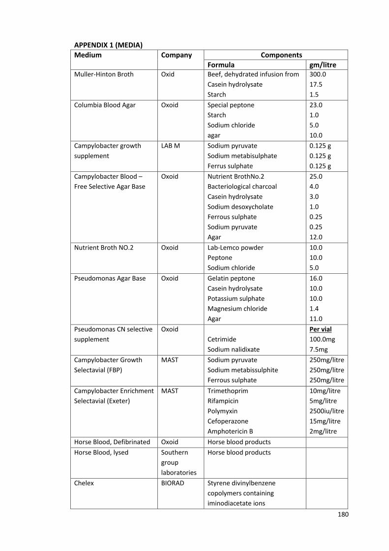
180
APPENDIX 1 (MEDIA)
Medium
Company
Components
Formula gm/litre Muller-Hinton Broth
Oxid
Beef, dehydrated infusion from
Casein hydrolysate
Starch
300.0
17.5
1.5
Columbia Blood Agar
Oxoid
Special peptone
Starch
Sodium chloride
agar
23.0
1.0
5.0
10.0
Campylobacter growth
supplement
LAB M
Sodium pyruvate
Sodium metabisulphate
Ferrus sulphate
0.125 g
0.125 g
0.125 g
Campylobacter Blood –
Free Selective Agar Base
Oxoid
Nutrient BrothNo.2
Bacteriological charcoal
Casein hydrolysate
Sodium desoxycholate
Ferrous sulphate
Sodium pyruvate
Agar
25.0
4.0
3.0
1.0
0.25
0.25
12.0
Nutrient Broth NO.2
Oxoid
Lab-Lemco powder
Peptone
Sodium chloride
10.0
10.0
5.0
Pseudomonas Agar Base
Oxoid
Gelatin peptone
Casein hydrolysate
Potassium sulphate
Magnesium chloride
Agar
16.0
10.0
10.0
1.4
11.0
Pseudomonas CN selective
supplement
Oxoid
Cetrimide
Sodium nalidixate
Per vial
100.0mg
7.5mg
Campylobacter Growth
Selectavial (FBP)
MAST
Sodium pyruvate
Sodium metabissulphite
Ferrous sulphate
250mg/litre
250mg/litre
250mg/litre
Campylobacter Enrichment
Selectavial (Exeter)
MAST
Trimethoprim
Rifampicin
Polymyxin
Cefoperazone
Amphotericin B
10mg/litre
5mg/litre
2500iu/litre
15mg/litre
2mg/litre
Horse Blood, Defibrinated Oxoid Horse blood products
Horse Blood, lysed
Southern
group
laboratories
Horse blood products
Chelex BIORAD Styrene divinylbenzene
copolymers containing
iminodiacetate ions

181
APPENDIX 2 (BUFFERS)
A. Solutions used during agarose gel electrophoresis
5x TBE buffer
54 g Tris base, 465 g EDTA and 27.5 g orthoboric acid (BDH bioscience) were mixed
with sterile distilled water and the volume was made up to 1000 mL. The buffer was
sterilised by autoclaving at 121°C for 15 min and was then diluted 1 in 10. 0.5 × TBE
was used to make agarose gel.
B. Preparation of Campylobacter growth supplement:
Using a sterile pipette, contents of the vial were dissolved with 5 mL of deionized
water and added into 500 mL of prepared medium.
C. preparation of 2% Chelex:
16 gm of Chelex100Resin was transferred into a sterile100 mL borosilicate glass bottle
containing 8 mL of Tris HCL and mixed. The suspension was filled up to 80 mL with
dH2O.
D. preparation of Tetracycline solution: 300 µg of Tetracycline powder was added into 20 mL Universal tube containing 10 mL
of SDW and dissolved. 1 mL of this stock was added into 1 litre of mCCDA.
E. Preparation of (LB) broth:
LB broth was prepared (per 1 litre) by mixing 5 g of Yeast extract (SIGMA) with 15 g of
tryptone water (SIGMA). LB agar was prepared (per 1 litre) by mixing 12 g of
Bacteriological agar (SIGMA) with LB broth.
182
F. preparation of pseudomonas C-N selective supplement:
CN agar was prepared (per 500 mL) by adding 5 mL of glycerol into 500 mL of
Pseudomonas Agar Base (Oxoid) and autoclaved at 121°C for 15 min. Both CN
supplement (1 vial per 500 mL- Oxoid) and a 2 mL o the mixture (SDW and Ethanol)
were added.
G. Preparation of Campylobacter Enrichment Broth (Exeter Broth)
Preparation Single Exeter Broth:
27.5 g of Nutrient Broth NO2 was dissolved in 1100 mL of distilled water and
autoclaved at 121°C for 15 min, aseptically the followings were added after cooling to
50°C:
1. 11 mL of Lysed Horse Blood
2. One vial of Campylobacter Growth Selectavial (dissolved in 5 mL SDW) and one vial
of Campylobacter Enrichment Selectavial (dissolved in 5 mL SDW).
Preparation Double Exeter Broth:
27.5 g of Nutrient Broth NO2 was dissolved in 550 mL of distilled water and
autoclaved at 121°C for 15 min, aseptically the followings were added after cooling to
50°C:
1. 11 mL of Lysed Horse Blood
2. One vial of Campylobacter Growth Selectavial (dissolved in 5 mL SDW) and one vial
of Campylobacter Enrichment Selectavial (dissolved in 5 mL SDW).
183
REFERENCES Abid, M., Wimalarathna, H., Mills, J., Saldana, L., Pang, W., Richardson, J.F., Maiden, M.C.J., and McCarthy, N.D. (2013). Duck liver–associated outbreak of Campylobacteriosis among humans, United Kingdom, 2011. Emerg Infectious Disease 19, 1310.
Acheson, D., and Allos, B.M. (2001). Campylobacter jejuni infections: update on emerging issues and trends. Clinical Infectious Diseases 32, 1201-1206.
Adler, L., Alter, T., Sharbati, S., and Gölz, G. (2014). Phenotypes of Campylobacter jejuni luxS mutants are depending on strain background, kind of mutation and experimental conditions. PloS one 9, e104399.
Adzitey, F., and Nurul, H. (2011). Campylobacter in poultry: Incidences and possible control measures. Research Journal of Microbiology 6, 182-192.
Altekruse, S.F., Stern, N.J., Fields, P.I., and Swerdlow, D.L. (1999). Campylobacter jejuni-an emerging foodborne pathogen. Emerg Infectious Disease 5, 28.
Anand, F., Sooria, B., Caroline, W., Helen, W., Maiden, M.C., and McCarthy, N.D. (2015). Partial failure of milk pasteurisation as a risk for the transmission of Campylobacter from cattle to humans. Clinical Infectious Diseases, civ431.
Anders, S., Pyl, P.T., and Huber, W. (2015). HTSeq--a Python framework to work with high-throughput sequencing data. Bioinformatics 31, 166-169.
Andersen, M.T., Brøndsted, L., Pearson, B.M., Mulholland, F., Parker, M., Pin, C., Wells, J.M., and Ingmer, H. (2005). Diverse roles for HspR in Campylobacter jejuni revealed by the proteome, transcriptome and phenotypic characterization of an hspR mutant. Microbiology 151, 905-915.
Arnaut-Rollier, I., De Zutter, L., and Van Hoof, J. (1999a). Identities of the Pseudomonas spp. in flora from chilled chicken. International Journal of Food Microbiology 48, 87-96.
Arnaut‐Rollier, I., Vauterin, L., De Vos, P., Massart, D.L., Devriese, L.A., De Zutter, L., and Van Hoof, J. (1999b). A numerical taxonomic study of the Pseudomonas flora isolated from poultry meat. Journal of Applied Microbiology 87, 15-28.
Aroori, S.V., Cogan, T.A., and Humphrey, T.J. (2013). The effect of growth temperature on the pathogenicity of Campylobacter. Current Microbiology 67, 333-340.
Arvanitis, N., Vargas, C., Tegos, G., Perysinakis, A., Nieto, J.J., Ventosa, A., and Drainas, C. (1995). Development of a gene reporter system in moderately halophilic bacteria by employing the ice nucleation gene of Pseudomonas syringae. Applied and Environmental Microbiology 61, 3821-3825.
Asakura, H., Yamasaki, M., Yamamoto, S., and Igimi, S. (2007). Deletion of peb4 gene impairs cell adhesion and biofilm formation in Campylobacter jejuni. FEMS Microbiology Letters 275, 278-285.
184
Ashgar, S.S.A., Oldfield, N.J., Wooldridge, K.G., Jones, M.A., Irving, G.J., Turner, D.P.J., and Ala'Aldeen, D.A.A. (2007). CapA, an autotransporter protein of Campylobacter jejuni, mediates association with human epithelial cells and colonization of the chicken gut. Journal of Bacteriology 189, 1856-1865.
Atabay, H.I., and Corry, J.E.L. (1998). The isolation and prevalence of Campylobacters from dairy cattle using a variety of methods. Journal of Applied Microbiology 84, 733-740.
Atack, J.M., and Kelly, D.J. (2008). Contribution of the stereospecific methionine sulphoxide reductases MsrA and MsrB to oxidative and nitrosative stress resistance in the food-borne pathogen Campylobacter jejuni. Microbiology 154, 2219-2230.
Atack, J.M., and Kelly, D.J. (2009). Oxidative stress in Campylobacter jejuni: responses, resistance and regulation. Future Microbiology 4, 677-690.
Avery, L.M., Williams, A.P., Killham, K., and Jones, D.L. (2008). Survival of Escherichia coli O157: H7 in waters from lakes, rivers, puddles and animal-drinking troughs. Science of the Total Environment 389, 378-385.
Bae, W., Kaya, K.N., Hancock, D.D., Call, D.R., Park, Y.H., and Besser, T.E. (2005). Prevalence and antimicrobial resistance of thermophilic Campylobacter spp. from cattle farms in Washington State. Applied and Environmental Microbiology 71, 169-174.
Baek, K.T., Vegge, C.S., and Brondsted, L. (2011a). HtrA chaperone activity contributes to host cell binding in Campylobacter jejuni. Gut Pathogenes 3, 13.
Baek, K.T., Vegge, C.S., Skorko-Glonek, J., and Brondsted, L. (2011b). Different contributions of HtrA protease and chaperone activities to Campylobacter jejuni stress tolerance and physiology. Applied and Environmental Microbiology 77, 57-66.
Baffone, W., Casaroli, A., Citterio, B., Pierfelici, L., Campana, R., Vittoria, E., Guaglianone, E., and Donelli, G. (2006). Campylobacter jejuni loss of culturability in aqueous microcosms and ability to resuscitate in a mouse model. International Journal of Food Microbiology 107, 83-91.
Baillon, M.-L.A., van Vliet, A.H.M., Ketley, J.M., Constantinidou, C., and Penn, C.W. (1999). An iron-regulated alkyl hydroperoxide reductase (AhpC) confers aerotolerance and oxidative stress resistance to the microaerophilic pathogen Campylobacter jejuni. Journal of Bacteriology 181, 4798-4804.
Baker, J., Barton, M.D., and Lanser, J. (1999). Campylobacter species in cats and dogs in South Australia. Australian Veterinary Journal 77, 662-666.
Bakshi, C.S., Malik, M., Regan, K., Melendez, J.A., Metzger, D.W., Pavlov, V.M., and Sellati, T.J. (2006). Superoxide dismutase B gene (sodB)-deficient mutants of Francisella tularensis demonstrate hypersensitivity to oxidative stress and attenuated virulence. Journal of Bacteriology 188, 6443-6448.
Ball, G., Durand, E., Lazdunski, A., and Filloux, A. (2002). A novel type II secretion system in Pseudomonas aeruginosa. Molecular Microbiology 43, 475-485.
185
Barker, A.P., Vasil, A.I., Filloux, A., Ball, G., Wilderman, P.J., and Vasil, M.L. (2004). A novel extracellular phospholipase C of Pseudomonas aeruginosa is required for phospholipid chemotaxis. Molecular Microbiology 53, 1089-1098.
Barnes, E.M., and Thornley, M.J. (1966). The spoilage flora of eviscerated chickens stored at different temperatures. International Journal of Food Science & Technology 1, 113-119.
Bayliss, C.D., Bidmos, F.A., Anjum, A., Manchev, V.T., Richards, R.L., Grossier, J.P., Wooldridge, K.G., Ketley, J.M., Barrow, P.A., Jones, M.A., et al. (2012). Phase variable genes of Campylobacter jejuni exhibit high mutation rates and specific mutational patterns but mutability is not the major determinant of population structure during host colonization. Nucleic Acids Research 40, 5876-5889.
Beisele, M., Shen, Z., Parry, N., Mobley, M., Taylor, N.S., Buckley, E., Abedin, M.Z., Dewhirst, F.E., and Fox, J.G. (2011). Helicobacter marmotae and novel Helicobacter and Campylobacter species isolated from the livers and intestines of prairie dogs. Journal of Medical Microbiology 60, 1366-1374.
Belval, S.C., Gal, L., Margiewes, S., Garmyn, D., Piveteau, P., and Guzzo, J. (2006). Assessment of the roles of LuxS, S-ribosyl homocysteine, and autoinducer 2 in cell attachment during biofilm formation by Listeria monocytogenes EGD-e. Applied and Environmental Microbiology 72, 2644-2650.
Benjamin, J., Leaper, S., Owen, R.J., and Skirrow, M.B. (1983). Description of Campylobacter laridis, a new species comprising the nalidixic acid resistant thermophilic Campylobacter (NARTC) group. Current Microbiology 8, 231-238.
Bergeron, J.R.C., Fernández, L., Wasney, G.A., Vuckovic, M., Reffuveille, F., Hancock, R.E.W., and Strynadka, N.C.J. (2016). The structure of a type 3 secretion system (T3SS) ruler protein suggests a molecular mechanism for needle length sensing. Journal of Biological Chemistry 291, 1676-1691.
Bertani, I., and Venturi, V. (2004). Regulation of the N-acyl homoserine lactone-dependent quorum-sensing system in rhizosphere Pseudomonas putida WCS358 and cross-talk with the stationary-phase RpoS sigma factor and the global regulator GacA. Applied and Environmental Microbiology 70, 5493-5502.
Bessède, E., Lehours, P., Labadi, L., Bakiri, S., and Mégraud, F. (2014). Comparison of characteristics of patients infected by Campylobacter jejuni, Campylobacter coli, and Campylobacter fetus. Journal of Clinical Microbiology 52, 328-330.
Beumer, R.R., De Vries, J., and Rombouts, F.M. (1992). Campylobacter jejuni non-culturable coccoid cells. International Journal of Food Microbiology 15, 153-163.
Bhatty, M., Gomez, J.A.L., and Christie, P.J. (2013). The expanding bacterial type IV secretion lexicon. Research in Microbiology 164, 620-639.
Bingle, L.E.H., Bailey, C.M., and Pallen, M.J. (2008). Type VI secretion: a beginner's guide. Current opinion in Microbiology 11, 3-8.
186
Blaser, M.J. (1997). Epidemiologic and clinical features of Campylobacter jejuni infections. Journal of Infectious Diseases 176, S103-S105.
Blaser, M.J., Hardesty, H.L., Powers, B., and Wang, W.L. (1980). Survival of Campylobacter fetus subsp. jejuni in biological milieus. Journal of Clinical Microbiology 11, 309-313.
Blaser, M.J., Perez, G.P., Smith, P.F., Patton, C., Tenover, F.C., Lastovica, A.J., and Wang, W.-I.L. (1986). Extraintestinal Campylobacter jejuni and Campylobacter coli infections: host factors and strain characteristics. Journal of Infectious Diseases 153, 552-559.
Blaser, M.J., Waldman, R.J., Barrett, T., and Erlandson, A.L. (1981). Outbreaks of Campylobacter enteritis in two extended families: evidence for person-to-person transmission. The Journal of Pediatrics 98, 254-257.
Bleves, S., Viarre, V., Salacha, R., Michel, G.P.F., Filloux, A., and Voulhoux, R. (2010). Protein secretion systems in Pseudomonas aeruginosa: A wealth of pathogenic weapons. International Journal of Medical Microbiology 300, 534-543.
Bourke, B., Sherman, P.M., Woodward, D., Lior, H., and Chan, V.L. (1996). Pulsed-field gel electrophoresis indicates genotypic heterogeneity among Campylobacter upsaliensis strains. FEMS Microbiology Letters 143, 57-61.
Braun, P., de Groot, A., Bitter, W., and Tommassen, J. (1998). Secretion of elastinolytic enzymes and their propeptides by Pseudomonas aeruginosa. Journal of Bacteriology 180, 3467-3469.
Brøndsted, L., Andersen, M.T., Parker, M., Jørgensen, K., and Ingmer, H. (2005). The HtrA protease of Campylobacter jejuni is required for heat and oxygen tolerance and for optimal interaction with human epithelial cells. Applied and Environmental Microbiology 71, 3205-3212.
Bronowski, C., James, C.E., and Winstanley, C. (2014). Role of environmental survival in transmission of Campylobacter jejuni. FEMS Microbiology Letters.
Bui, X.T., Qvortrup, K., Wolff, A., Bang, D.D., and Creuzenet, C. (2012). Effect of environmental stress factors on the uptake and survival of Campylobacter jejuni in Acanthamoeba castellanii. BMC Microbiology 12, 232.
Burgos-Portugal, J.A., Kaakoush, N.O., Raftery, M.J., and Mitchell, H.M. (2012). Pathogenic potential of Campylobacter ureolyticus. Infection and Immunity 80, 883-890.
Bustin, S.A., Benes, V., Garson, J.A., Hellemans, J., Huggett, J., Kubista, M., Mueller, R., Nolan, T., Pfaffl, M.W., and Shipley, G.L. (2009). The MIQE guidelines: minimum information for publication of quantitative real-time PCR experiments. Clinical Chemistry 55, 611-622.
Buswell, C.M., Herlihy, Y.M., Lawrence, L.M., McGuiggan, J.T., Marsh, P.D., Keevil, C.W., and Leach, S.A. (1998). Extended survival and persistence of Campylobacter
187
spp. in water and aquatic biofilms and their detection by immunofluorescent-antibody and -rRNA staining. Applied and Environmental Microbiology 64, 733-741.
Butzler, J.P. (2004). Campylobacter, from obscurity to celebrity. Clinical Microbiology and Infection 10, 868-876.
Campero, C.M., Anderson, M.L., Walker, R.L., Blanchard, P.C., Barbano, L., Chiu, P., Martínez, A., Combessies, G., Bardon, J.C., and Cordeviola, J. (2005). Immunohistochemical identification of Campylobacter fetus in natural cases of bovine and ovine abortions. Journal of Veterinary Medicine, Series B 52, 138-141.
Candon, H.L., Allan, B.J., Fraley, C.D., and Gaynor, E.C. (2007). Polyphosphate kinase 1 is a pathogenesis determinant in Campylobacter jejuni. Journal of Bacteriology 189, 8099-8108.
Carter, P.E., McTavish, S.M., Brooks, H.J.L., Campbell, D., Collins-Emerson, J.M., Midwinter, A.C., and French, N.P. (2009). Novel clonal complexes with an unknown animal reservoir dominate Campylobacter jejuni isolates from river water in New Zealand. Applied and Environmental Microbiology 75, 6038-6046.
Carvalho, A.F.d., Silva, D.M.d., Azevedo, S.S., Piatti, R.M., Genovez, M.E., and Scarcelli, E. (2013). Detection of CDT toxin genes in Campylobacter spp. strains isolated from broiler carcasses and vegetables in São Paulo, Brazil. Brazilian Journal of Microbiology 44, 693-699.
Carver, T., Berriman, M., Tivey, A., Patel, C., Bohme, U., Barrell, B.G., Parkhill, J., and Rajandream, M.A. (2008). Artemis and ACT: viewing, annotating and comparing sequences stored in a relational database. Bioinformatics 24, 2672-2676.
Carver, T.J., Rutherford, K.M., Berriman, M., Rajandream, M.A., Barrell, B.G., and Parkhill, J. (2005). ACT: the Artemis Comparison Tool. Bioinformatics 21, 3422-3423.
Castaño-Rodríguez, N., Kaakoush, N.O., Lee, W.S., and Mitchell, H.M. (2015). Dual role of Helicobacter and Campylobacter species in IBD: a systematic review and meta-analysis. Gut, Gutjnl-2015-310545.
Ceelen, L.M., Decostere, A., Ducatelle, R., and Haesebrouck, F. (2006). Cytolethal distending toxin generates cell death by inducing a bottleneck in the cell cycle. Microbiological Research 161, 109-120.
Centers for Disease, C., and Prevention (2010). Preliminary FoodNet data on the incidence of infection with pathogens transmitted commonly through food-10 states, 2009. MMWR Morbidity and Mortality Weekly Report 59, 418.
Chaisowwong, W., Kusumoto, A., Hashimoto, M., Harada, T., Maklon, K., and Kawamoto, K. (2012). Physiological characterization of Campylobacter jejuni under cold stresses conditions: its potential for public threat. The Journal of Veterinary Medical Science/the Japanese Society of Veterinary Science 74, 43-50.
Chaloner, G., Wigley, P., Humphrey, S., Kemmett, K., Lacharme-Lora, L., Humphrey, T., and Williams, N. (2014). Dynamics of dual infection with Campylobacter jejuni strains
188
in chickens reveals distinct strain-to-strain variation in infection ecology. Applied and Environmental Microbiology 80, 6366-6372.
Champion, O.L., Gaunt, M.W., Gundogdu, O., Elmi, A., Witney, A.A., Hinds, J., Dorrell, N., and Wren, B.W. (2005). Comparative phylogenomics of the food-borne pathogen Campylobacter jejuni reveals genetic markers predictive of infection source. Proceedings of the National Academy of Sciences of the United States of America 102, 16043.
Chan, M.-S., Maiden, M.C.J., and Spratt, B.G. (2001). Database-driven multi locus sequence typing (MLST) of bacterial pathogens. Bioinformatics 17, 1077-1083.
Chan, W.C., Coyle, B.J., and Williams, P. (2004). Virulence regulation and quorum sensing in Staphylococcal infections: Competitive AgrC antagonists as quorum sensing inhibitors §. Journal of Medicinal Chemistry 47, 4633-4641.
Chaveerach, P., Ter Huurne, A., Lipman, L.J.A., and Van Knapen, F. (2003). Survival and resuscitation of ten strains of Campylobacter jejuni and Campylobacter coli under acid conditions. Applied and Environmental Microbiology 69, 711-714.
Chet, I., Zilberstein, Y., and Henis, Y. (1973). Chemotaxis of Pseudomonas lachrymans to plant extracts and to water droplets collected from the leaf surfaces of resistant and susceptible plants. Physiological Plant Pathology 3, 473-479.
Chin-A-Woeng, T.F.C., van den Broek, D., de Voer, G., van der Drift, K.M.G.M., Tuinman, S., Thomas-Oates, J.E., Lugtenberg, B.J.J., and Bloemberg, G.V. (2001). Phenazine-1-carboxamide production in the biocontrol strain Pseudomonas chlororaphis PCL1391 is regulated by multiple factors secreted into the growth medium. Molecular Plant-Microbe Interactions 14, 969-979.
Chin-A-Woeng, T.F.C., van den Broek, D., Lugtenberg, B.J.J., and Bloemberg, G.V. (2005). The Pseudomonas chlororaphis PCL1391 sigma regulator psrA represses the production of the antifungal metabolite phenazine-1-carboxamide. Molecular Plant-Microbe Interactions 18, 244-253.
Cianciotto, N.P. (2005). Type II secretion: a protein secretion system for all seasons. Trends in Microbiology 13, 581-588.
Clark, C.G., Price, L., Ahmed, R., Woodward, D.L., Melito, P.L., Rodgers, F.G., Jamieson, F., Ciebin, B., Li, A., and Ellis, A. (2003). Characterization of waterborne outbreak–associated Campylobacter jejuni, Walkerton, Ontario. Emerging Infectious Diseases 9, 1232.
Cloak, O.M., Solow, B.T., Briggs, C.E., Chen, C.-Y., and Fratamico, P.M. (2002). Quorum sensing and production of autoinducer-2 in Campylobacter spp., Escherichia coli O157: H7, and Salmonella enterica serovar Typhimurium in foods. Applied and Environmental Microbiology 68, 4666-4671.
Cody, A.J., Clarke, L., Bowler, I.C.J.W., and Dingle, K.E. (2010). Ciprofloxacin-resistant campylobacteriosis in the UK. The Lancet 376, 1987.
189
Coker, A.O., Isokpehi, R.D., Thomas, B.N., Amisu, K.O., and Obi, C.L. (2002). Human campylobacteriosis in developing countries. Emerging Infectious Disease 8, 237.
Colles, F.M., Jones, K., Harding, R.M., and Maiden, M.C. (2003). Genetic diversity of Campylobacter jejuni isolates from farm animals and the farm environment. Applied and Environmental Microbiology 69, 7409-7413.
Cools, I., Uyttendaele, M., Caro, C., D'Haese, E., Nelis, H.J., and Debevere, J. (2003). Survival of Campylobacter jejuni strains of different origin in drinking water. Journal of Applied Microbiollogy 94, 886-892.
Dasti, J.I., Tareen, A.M., Lugert, R., Zautner, A.E., and Groß, U. (2010). Campylobacter jejuni: a brief overview on pathogenicity-associated factors and disease-mediating mechanisms. International Journal of Medical Microbiology 300, 205-211.
Day, C.J., Tiralongo, J., Hartnell, R.D., Logue, C.-A., Wilson, J.C., von Itzstein, M., and Korolik, V. (2009). Differential carbohydrate recognition by Campylobacter jejuni strain 11168: influences of temperature and growth conditions. PloS one 4, e4927.
Day, W.A., Sajecki, J.L., Pitts, T.M., and Joens, L.A. (2000). Role of catalase in Campylobacter jejuni intracellular survival. Infection and Immunity 68, 6337-6345.
De Keersmaecker, S.C.J., Varszegi, C., van Boxel, N., Habel, L.W., Metzger, K., Daniels, R., Marchal, K., De Vos, D., and Vanderleyden, J. (2005). Chemical synthesis of (S)-4, 5-dihydroxy-2, 3-pentanedione, a bacterial signal molecule precursor, and validation of its activity in Salmonella typhimurium. Journal of Biological Chemistry 280, 19563-19568.
De Vos, D., Bouton, C., Sarniguet, A., De Vos, P., Vauterin, M., and Cornelis, P. (1998). Sequence diversity of the oprI gene, coding for major outer membrane lipoprotein I, among rRNA group I pseudomonads. Journal of Bacteriology 180, 6551-6556.
De Vos, D., Lim, A., De Vos, P., Sarniguet, A., Kersters, K., and Cornelis, P. (1993). Detection of the outer membrane lipoprotein I and its gene in fluorescent and non-fluorescent pseudomonads: implications for taxonomy and diagnosis. Journal of General Microbiology 139, 2215-2223.
de Vries, J.J.C., Arents, N.L.A., and Manson, W.L. (2008). Campylobacter species isolated from extra-oro-intestinal abscesses: a report of four cases and literature review. European Journal of Clinical Microbiology & Infectious Diseases 27, 1119-1123.
de Zoete, M.R., Keestra, A.M., Roszczenko, P., and van Putten, J.P.M. (2010). Activation of human and chicken toll-like receptors by Campylobacter spp. Infection and Immunity 78, 1229-1238.
Debruyne, L., Broman, T., Bergström, S., Olsen, B., On, S.L.W., and Vandamme, P. (2010a). Campylobacter subantarcticus sp. nov., isolated from birds in the sub-Antarctic region. International Journal of Systematic and Evolutionary Microbiology 60, 815-819.
190
Debruyne, L., Broman, T., Bergström, S., Olsen, B., On, S.L.W., and Vandamme, P. (2010b). Campylobacter volucris sp. nov., isolated from black-headed gulls (Larus ridibundus). International Journal of Systematic and Evolutionary Microbiollogy 60, 1870-1875.
Debruyne, L., On, S.L.W., De Brandt, E., and Vandamme, P. (2009). Novel Campylobacter lari-like bacteria from humans and molluscs: description of Campylobacter peloridis sp. nov., Campylobacter lari subsp. concheus subsp. nov. and Campylobacter lari subsp. lari subsp. nov. Interational Journal of Systematic and Evolutionary Microbiology 59, 1126-1132.
del Mar Lleò, M., Pierobon, S., Tafi, M.C., Signoretto, C., and Canepari, P. (2000). mRNA detection by reverse transcription-PCR for monitoring viability over time in an Enterococcus faecalis viable but nonculturable population maintained in a laboratory microcosm. Applied and Environmental Microbiology 66, 4564-4567.
Del Rocio Leon‐Kempis, M., Guccione, E., Mulholland, F., Williamson, M.P., and Kelly, D.J. (2006). The Campylobacter jejuni PEB1a adhesin is an aspartate/glutamate-binding protein of an ABC transporter essential for microaerobic growth on dicarboxylic amino acids. Molecular Microbiology 60, 1262-1275.
Delepelaire, P. (2004). Type I secretion in gram-negative bacteria. Biochimica et Biophysica Acta (BBA)-Molecular Cell Research 1694, 149-161.
DeVos, D., Lim, A., Pirnay, J.P., Struelens, M., Vandenvelde, C., Duinslaeger, L., Vanderkelen, A., and Cornelis, P. (1997). Direct detection and identification of Pseudomonas aeruginosa in clinical samples such as skin biopsy specimens and expectorations by multiplex PCR based on two outer membrane lipoprotein genes, oprI and oprL. Journal of Clinical Microbiology 35, 1295-1299.
Diaz, P.I., Zilm, P.S., and Rogers, A.H. (2002). Fusobacterium nucleatum supports the growth of Porphyromonas gingivalis in oxygenated and carbon-dioxide-depleted environments. Microbiology 148, 467-472.
Dicksved, J., Ellström, P., Engstrand, L., and Rautelin, H. (2014). Susceptibility to Campylobacter infection is associated with the species composition of the human fecal microbiota. Mbio 5, e01212-01214.
Dingle, K.E., Colles, F.M., Falush, D., and Maiden, M.C.J. (2005). Sequence typing and comparison of population biology of Campylobacter coli and Campylobacter jejuni. Journal of Clinical Microbiology 43, 340-347.
Dingle, K.E., Colles, F.M., Wareing, D.R.A., Ure, R., Fox, A.J., Bolton, F.E., Bootsma, H.J., Willems, R.J.L., Urwin, R., and Maiden, M.C.J. (2001). Multilocus sequence typing system for Campylobacter jejuni. Journal of Clinical Microbiology 39, 14-23.
Domingues, A.R., Pires, S.M., Halasa, T., and Hald, T. (2012). Source attribution of human campylobacteriosis using a meta-analysis of case-control studies of sporadic infections. Epidemiology and Infection 140, 970-981.
191
Dos Santos, V.A.P., Heim, S., Moore, E.R.B., Strätz, M., and Timmis, K.N. (2004). Insights into the genomic basis of niche specificity of Pseudomonas putida KT2440. Environmental Microbiology 6, 1264-1286.
Drancourt, M., Roux, V., Tran-Hung, L., Castex, D., Chenal-Francisque, V., Ogata, H., Fournier, P.-E., Crubézy, E., and Raoult, D. (2004). Genotyping, orientalis-like Yersinia pestis, and plague pandemics. Emerging Infectious Diseases 10, 1585-1592.
Dronda, F., García-Arata, I., Navas, E., and de Rafael, L. (1998). Meningitis in adults due to Campylobacter fetus subspecies fetus. Clinical Infectious Diseases 27, 906-907.
Duan, K., Dammel, C., Stein, J., Rabin, H., and Surette, M.G. (2003). Modulation of Pseudomonas aeruginosa gene expression by host microflora through interspecies communication. Molecular Microbiology 50, 1477-1491.
Duchêne, M., Barron, C., Schweizer, A., von Specht, B.U., and Domdey, H. (1989). Pseudomonas aeruginosa outer membrane lipoprotein I gene: molecular cloning, sequence, and expression in Escherichia coli. Journal of Bacteriology 171, 4130-4137.
Duerden, B., Bennet, K.W., and Faulkner, J. (1982). Isolation of Bacteroides ureolyticus (B corrodens) from clinical infections. Journal of Clinical Pathology 35, 309-312.
Duffy, G., Lynch, O.A., and Cagney, C. (2008). Tracking emerging zoonotic pathogens from farm to fork. Meat science 78, 34-42.
Dugar, G., Herbig, A., Förstner, K.U., Heidrich, N., Reinhardt, R., Nieselt, K., and Sharma, C.M. (2013). High-resolution transcriptome maps reveal strain-specific regulatory features of multiple Campylobacter jejuni isolates. PLoS Genetics 9, e1003495.
Dumenyo, C.K., Mukherjee, A., Chun, W., and Chatterjee, A.K. (1998). Genetic and physiological evidence for the production of N-acyl homoserine lactones by Pseudomonas syringae pv. syringae and other fluorescent plant pathogenic Pseudomonas species. European Journal of Plant Pathology 104, 569-582.
Duong, F., Bonnet, E., Géli, V., Lazdunski, A., Murgier, M., and Filloux, A. (2001). The AprX protein of Pseudomonas aeruginosa: a new substrate for the Apr type I secretion system. Gene 262, 147-153.
Eberhard, A., Burlingame, A.L., Eberhard, C., Kenyon, G.L., Nealson, K.H., and Oppenheimer, N.J. (1981). Structural identification of autoinducer of Photobacterium fischeri luciferase. Biochemistry 20, 2444-2449.
Eberle, K.N., and Kiess, A.S. (2012). Phenotypic and genotypic methods for typing Campylobacter jejuni and Campylobacter coli in poultry. Poultry Science 91, 255-264.
El-Sayed, A.K., Hothersall, J., and Thomas, C.M. (2001). Quorum-sensing-dependent regulation of biosynthesis of the polyketide antibiotic mupirocin in Pseudomonas fluorescens NCIMB 10586. Microbiology 147, 2127-2139.
Elvers, K.T., and Park, S.F. (2002). Quorum sensing in Campylobacter jejuni: detection of a luxS encoded signalling molecule. Microbiology 148, 1475-1481.
192
Endtz, H.P., Ang, C.W., van den Braak, N., Duim, B., Rigter, A., Price, L.J., Woodward, D.L., Rodgers, F.G., Johnson, W.M., and Wagenaar, J.A. (2000). Molecular characterization of Campylobacter jejuni from patients with Guillain-Barré and Miller Fisher syndromes. Journal of Clinical Microbiology 38, 2297-2301.
Engberg, J. (2006). Contributions to the epidemiology of Campylobacter infections. Danish Medical Bulletin 53, 361-389.
Engebrecht, J., Nealson, K., and Silverman, M. (1983). Bacterial bioluminescence: isolation and genetic analysis of functions from Vibrio fischeri. Cell 32, 773-781.
Engebrecht, J., and Silverman, M. (1984). Identification of genes and gene products necessary for bacterial bioluminescence. Proceedings of the National Academy of Sciences 81, 4154-4158.
Eurosurveillance Editorial, T. (2012). The European union summary report on trends and sources of zoonoses, zoonotic agents and food-borne outbreaks in 2010. Euro surveillance: Bulletin Européen sur les Maladies Transmissibles=European Communicable Disease Bulletin 17.
Evans, M.R., Ribeiro, C.D., and Salmon, R.L. (2003). Hazards of healthy living: bottled water and salad vegetables as risk factors for Campylobacter infection. Emerging Infectious Diseases 9, 1219.
Fakruddin, M., Mannan, K.S.B., and Andrews, S. (2013). Viable but nonculturable bacteria: food safety and public health perspective. ISRN Microbiology 2013.
Farrell, D.J., and Harris, M.T. (1992). A case of intrauterine fetal death associated with maternal Campylobacter coli bacteraemia. Australian and New Zealand Journal of Obstetrics and Gynaecology 32, 172-174.
Federighi, M., Tholozan, J.L., Cappelier, J.M., Tissier, J.P., and Jouve, J.L. (1998). Evidence of non-coccoid viable but non-culturable Campylobacter jejuni cells in microcosm water by direct viable count, CTC-DAPI double staining, and scanning electron microscopy. Food Microbiology 15, 539-550.
Feodoroff, B., de Haan, C.P.A., Ellström, P., Sarna, S., Hänninen, M.-L., and Rautelin, H. (2013). Clonal distribution and virulence of Campylobacter jejuni isolates in blood. Emerging Infectious Diseases 19, 1653.
Filloux, A., Bally, M., Soscia, C., Murgier, M., and Lazdunski, A. (1988). Phosphate regulation in Pseudomonas aeruginosa: cloning of the alkaline phosphatase gene and identification of phoB-and phoR-like genes. Molecular and General Genetics MGG 212, 510-513.
Fitzgerald, C., chao Tu, Z., Patrick, M., Stiles, T., Lawson, A.J., Santovenia, M., Gilbert, M.J., Van Bergen, M., Joyce, K., and Pruckler, J. (2014). Campylobacter fetus subsp. testudinum subsp. nov., isolated from humans and reptiles. International Journal of Systematic and Evolutionary Microbiology 64, 2944-2948.
Fitzgerald, C., Stanley, K., Andrew, S., and Jones, K. (2001). Use of pulsed-field gel electrophoresis and flagellin gene typing in identifying clonal groups of
193
Campylobacter jejuni and Campylobacter coli in farm and clinical environments. Applied and Environmental Microbiology 67, 1429-1436.
Fleischmann, R.D., Adams, M.D., White, O., Clayton, R.A., Kirkness, E.F., Kerlavage, A.R., Bult, C.J., Tomb, J.-F., Dougherty, B.A., and Merrick, J.M. (1995). Whole-genome random sequencing and assembly of Haemophilus influenzae Rd. Science 269, 496-512.
Flint, A., Sun, Y.-Q., and Stintzi, A. (2012). Cj1386 is an ankyrin-containing protein involved in heme trafficking to catalase in Campylobacter jejuni. Journal of Bacteriology 194, 334-345.
Folders, J., Tommassen, J., Van Loon, L.C., and Bitter, W. (2000). Identification of a chitin-binding protein secreted by Pseudomonas aeruginosa. Journal of Bacteriology 182, 1257-1263.
Fox, A., Haas, D., Reimmann, C., Heeb, S., Filloux, A., and Voulhoux, R. (2008). Emergence of secretion-defective sublines of Pseudomonas aeruginosa PAO1 resulting from spontaneous mutations in the vfr global regulatory gene. Applied and Environmental Microbiology 74, 1902-1908.
Fox, J.G., Chilvers, T., Goodwin, C.S., Taylor, N.S., Edmonds, P., Sly, L.I., and Brenner, D.J. (1989). Campylobacter mustelae, a new species resulting from the elevation of Campylobacter pylori subsp. mustelae to species status. International Journal of Systematic Bacteriology 39, 301-303.
Franzetti, L., and Scarpellini, M. (2007). Characterisation of Pseudomonas spp. isolated from foods. Annals of Microbiology 57, 39-47.
Fraser-Liggett, C.M. (2005). Insights on biology and evolution from microbial genome sequencing. Genome Research 15, 1603-1610.
French, N., Barrigas, M., Brown, P., Ribiero, P., Williams, N., Leatherbarrow, H., Birtles, R., Bolton, E., Fearnhead, P., and Fox, A. (2005). Spatial epidemiology and natural population structure of Campylobacter jejuni colonizing a farmland ecosystem. Environmental Microbiology 7, 1116-1126.
Friis, C., Wassenaar, T.M., Javed, M.A., Snipen, L., Lagesen, K., Hallin, P.F., Newell, D.G., Toszeghy, M., Ridley, A., and Manning, G. (2010). Genomic characterization of Campylobacter jejuni strain M1. PloS one 5, e12253.
FSA (2009). A UK survey of Campylobacter and Salmonella contamination of fresh chicken at retail sale. Food Survey Information Sheet 04/09 (http://www.food.gov.uk/multimedia/pdfs/fsis0409.pdf). Accessed March 2011.
Fujita, M., Fujimoto, S., Morooka, T., and Amako, K. (1995). Analysis of strains of Campylobacter fetus by pulsed-field gel electrophoresis. Journal of Clinical Microbiology 33, 1676-1678.
Gangaiah, D., Kassem, II, Liu, Z., and Rajashekara, G. (2009). Importance of polyphosphate kinase 1 for Campylobacter jejuni viable-but-nonculturable cell
194
formation, natural transformation, and antimicrobial resistance. Appl Environ Microbiol 75, 7838-7849.
Garénaux, A., Jugiau, F., Rama, F., De Jonge, R., Denis, M., Federighi, M., and Ritz, M. (2008). Survival of Campylobacter jejuni strains from different origins under oxidative stress conditions: effect of temperature. Current Microbiology 56, 293-297.
Gaudreau, C., and Lamothe, F. (1992). Campylobacter upsaliensis isolated from a breast abscess. Journal of Clinical Microbiology 30, 1354-1356.
Gaudreau, C., and Michaud, S. (2003). Cluster of erythromycin-and ciprofloxacin-resistant Campylobacter jejuni subsp. jejuni from 1999 to 2001 in men who have sex with men, Québec, Canada. Clinical Infectious Diseases 37, 131-136.
Gaynor, E.C., Cawthraw, S., Manning, G., MacKichan, J.K., Falkow, S., and Newell, D.G. (2004). The genome-sequenced variant of Campylobacter jejuni NCTC 11168 and the original clonal clinical isolate differ markedly in colonization, gene expression, and virulence-associated phenotypes. Journal of Bacteriology 186, 503-517.
Gaynor, E.C., Wells, D.H., MacKichan, J.K., and Falkow, S. (2005). The Campylobacter jejuni stringent response controls specific stress survival and virulence-associated phenotypes. Molecular Microbiology 56, 8-27.
Gebhart, C.J., Edmonds, P., Ward, G.E., Kurtz, H.J., and Brenner, D.J. (1985). " Campylobacter hyointestinalis" sp. nov.: a new species of Campylobacter found in the intestines of pigs and other animals. Journal of Clinical Microbiology 21, 715-720.
Ghafir, Y., China, B., Dierick, K., De Zutter, L., and Daube, G. (2008). Hygiene indicator microorganisms for selected pathogens on beef, pork, and poultry meats in Belgium. Journal of food Protection 71, 35-45.
Ghareeb, K., Awad, W.A., Mohnl, M., Schatzmayr, G., and Boehm, J. (2013). Control strategies for Campylobacter infection in poultry production. World's Poultry Science Journal 69, 57-76.
Gillespie, I.A., O'Brien, S.J., Frost, J.A., Adak, G.K., Horby, P., Swan, A.V., Painter, M.J., and Neal, K.R. (2002). A case-case comparison of Campylobacter coli and Campylobacter jejuni infection: a tool for generating hypotheses. Emerging Infectious Diseases 8, 937-942.
Glaus, P., Honkela, A., and Rattray, M. (2012). Identifying differentially expressed transcripts from RNA-seq data with biological variation. Bioinformatics 28, 1721-1728.
Goddard, A.D., Arnold, M.E., Allen, V.M., and Snary, E.L. (2014). Estimating the time at which commercial broiler flocks in Great Britain become infected with Campylobacter: a Bayesian approach. Epidemiology and Infection 142, 1884-1892.
Goldman, C.G., Matteo, M.J., Loureiro, J.D., Almuzara, M., Barberis, C., Vay, C., Catalano, M., Heredia, S.R., Mantero, P., and Boccio, J.R. (2011). Novel gastric Helicobacters and oral Campylobacters are present in captive and wild cetaceans. Veterinary Microbiology 152, 138-145.
195
Gölz, G., Adler, L., Huehn, S., and Alter, T. (2012). LuxS distribution and AI-2 activity of Campylobacter spp. Journal of Applied Microbiology 112, 571-578.
González, M., and Hänninen, M.L. (2012). Effect of temperature and antimicrobial resistance on survival of Campylobacter jejuni in well water: application of the Weibull model. Journal of Applied Microbiology 113, 284-293.
González, M., Skandamis, P.N., and Hänninen, M.L. (2009). A modified Weibull model for describing the survival of Campylobacter jejuni in minced chicken meat. International Journal of Food Microbiology 136, 52-58.
Goodwin, C.S., Armstrong, J.A., Chilvers, T., Peters, M., Collins, M.D., Sly, L., McConnell, W., and Harper, W.E. (1989). Transfer of Campylobacter pylori and Campylobacter mustelae to Helicobacter gen. nov. as Helicobacter pylori comb. nov. and Helicobacter mustelae comb. nov., respectively. International Journal of Systematic Bacteriology 39, 397-405.
Goodwin, C.S., Blake, P., and Blincow, E. (1986). The minimum inhibitory and bactericidal concentrations of antibiotics and anti-ulcer agents against Campylobacter pyloridis. The Journal of Antimicrobial Chemotherapy 17, 309-314.
Gorkiewicz, G., Feierl, G., Zechner, R., and Zechner, E.L. (2002). Transmission of Campylobacter hyointestinalis from a pig to a human. Journal of Clinical Microbiology 40, 2601-2605.
Gorski, L., Parker, C.T., Liang, A., Cooley, M.B., Jay-Russell, M.T., Gordus, A.G., Atwill, E.R., and Mandrell, R.E. (2011). Prevalence, distribution, and diversity of Salmonella enterica in a major produce region of California. Applied and Environmental Microbiology 77, 2734-2748.
Grant, K.A., Belandia, I.U., Dekker, N., Richardson, P.T., and Park, S.F. (1997). Molecular characterization of pldA, the structural gene for a phospholipase A from Campylobacter coli, and its contribution to cell-associated hemolysis. Infection and Immunity 65, 1172-1180.
Griffiths, P.L., and Park, R.W. (1990). Campylobacters associated with human diarrhoeal disease. The Journal of Applied Bacteriology 69, 281-301.
Grøndalen, J., Sævik, B., and Sørum, H. (2008). Companion animals as reservoir for zoonotic diseases. European Journal of Companion Animal Practice 18, 213-222.
Grove-White, D.H., Leatherbarrow, A.J.H., Cripps, P.J., Diggle, P.J., and French, N.P. (2011). Molecular epidemiology and genetic diversity of Campylobacter jejuni in ruminants. Epidemiology and Infection 139, 1661-1671.
Guccione, E., Del Rocio Leon‐Kempis, M., Pearson, B.M., Hitchin, E., Mulholland, F., Van Diemen, P.M., Stevens, M.P., and Kelly, D.J. (2008). Amino acid-dependent growth of Campylobacter jejuni: key roles for aspartase (AspA) under microaerobic and oxygen-limited conditions and identification of AspB (Cj0762), essential for growth on glutamate. Molecular Microbiology 69, 77-93.
196
Guerry, P. (2007). Campylobacter flagella: not just for motility. Trends in Microbiology 15, 456-461.
Guillou, S., Leguérinel, I., Garrec, N., Renard, M.A., Cappelier, J.M., and Federighi, M. (2008). Survival of Campylobacter jejuni in mineral bottled water according to difference in mineral content: Application of the Weibull model. Water Research 42, 2213-2219.
Gunasekera, T.S., Sørensen, A., Attfield, P.V., Sørensen, S.J., and Veal, D.A. (2002). Inducible gene expression by nonculturable bacteria in milk after pasteurization. Applied and Environmental Microbiology 68, 1988-1993.
Gundogdu, O., Bentley, S.D., Holden, M.T., Parkhill, J., Dorrell, N., and Wren, B.W. (2007). Re-annotation and re-analysis of the Campylobacter jejuni NCTC11168 genome sequence. BMC Genomics 8, 162.
Gundry, C.N., Vandersteen, J.G., Reed, G.H., Pryor, R.J., Chen, J., and Wittwer, C.T. (2003). Amplicon melting analysis with labeled primers: a closed-tube method for differentiating homozygotes and heterozygotes. Clinical Chemistry 49, 396-406.
Gürtler, M., Alter, T., Kasimir, S., and Fehlhaber, K. (2005). The importance of Campylobacter coli in human campylobacteriosis: prevalence and genetic characterization. Epidemiology and Infection 133, 1081-1087.
Gurtler, V., and Stanisich, V.A. (1996). New approaches to typing and identification of bacteria using the 16S-23S rDNA spacer region. Microbiology-Sgm 142, 3-16.
Habib, I., Miller, W.G., Uyttendaele, M., Houf, K., and De Zutter, L. (2009). Clonal population structure and antimicrobial resistance of Campylobacter jejuni in chicken meat from Belgium. Applied and Environmental Microbiology 75, 4264-4272.
Hackman, A.S., and Wilkins, T.D. (1976). Influence of penicillinase production by strains of Bacteroides melaninogenicus and Bacteroides oralis on penicillin therapy of an experimental mixed anaerobic infection in mice. Archives of Oral Biology 21, 385-389.
Haddad, N., Burns, C.M., Bolla, J.M., Prévost, H., Fédérighi, M., Drider, D., and Cappelier, J.M. (2009). Long-term survival of Campylobacter jejuni at low temperatures is dependent on polynucleotide phosphorylase activity. Applied and Environmental Microbiology 75, 7310-7318.
Halablab, M.A., Mohammed, K.A.S., and Miles, R.J. (2008). Growth and survival of Campylobacter pathogens in the presence of different metabolic inhibitors. Journal of Medical Science 8, 262-268.
Hald, B., Knudsen, K., Lind, P., and Madsen, M. (2001). Study of the infectivity of saline-stored Campylobacter jejuni for day-old chicks. Applied and Environmental Microbiology 67, 2388-2392.
Hald, B., Pedersen, K., Wainø, M., Jørgensen, J.C., and Madsen, M. (2004a). Longitudinal study of the excretion patterns of thermophilic Campylobacter spp. in young pet dogs in Denmark. Journal of Clinical Microbiology 42, 2003-2012.
197
Hald, B., Skovgård, H., Bang, D.D., Pedersen, K., Dybdahl, J., Jespersen, J.B., and Madsen, M. (2004b). Flies and Campylobacter infection of broiler flocks. Emerging Infectious Diseases 10, 1490.
Hanel, I., Borrmann, E., Muller, J., Muller, W., Pauly, B., Liebler-Tenorio, E.M., and Schulze, F. (2009). Genomic and phenotypic changes of Campylobacter jejuni strains after passage of the chicken gut. Veterinary Microbiology 136, 121-129.
Hänninen, M.L., and Kärenlampi, R. (2004). Campylobacter in waterborne epidemics in Finland. Water Science and Technology: Water Supply 4, 39-45.
Hannu, T. (2011). Reactive arthritis. Best Practice & Research Clinical Rheumatology 25, 347-357.
Hazeleger, W.C., Janse, J.D., Koenraad, P.M., Beumer, R.R., Rombouts, F.M., and Abee, T. (1995). Temperature-dependent membrane fatty acid and cell physiology changes in coccoid forms of Campylobacter jejuni. Applied and Environmental Microbiology 61, 2713-2719.
He, Y., Frye, J.G., Strobaugh Jr, T.P., and Chen, C.-Y. (2008). Analysis of AI-2/LuxS-dependent transcription in Campylobacter jejuni strain 81-176. Foodborne Pathogens and Disease 5, 399-415.
Heimesaat, M.M., Alutis, M., Grundmann, U., Fischer, A., Tegtmeyer, N., Böhm, M., Kühl, A.A., Göbel, U.B., Backert, S., and Bereswill, S. (2014). The role of serine protease HtrA in acute ulcerative enterocolitis and extra-intestinal immune responses during Campylobacter jejuni infection of gnotobiotic IL-10 deficient mice. Frontiers in Cellular and Infection Microbiology 4.
Henderson, I.R., Navarro-Garcia, F., Desvaux, M., Fernandez, R.C., and Ala'Aldeen, D. (2004). Type V protein secretion pathway: the autotransporter story. Microbiology and Molecular Biology Reviews 68, 692-744.
Hentzer, M., Wu, H., Andersen, J.B., Riedel, K., Rasmussen, T.B., Bagge, N., Kumar, N., Schembri, M.A., Song, Z., and Kristoffersen, P. (2003). Attenuation of Pseudomonas aeruginosa virulence by quorum sensing inhibitors. European Molecular Biology Organization (EMBO) Journal 22, 3803-3815.
Hepworth, P.J., Ashelford, K.E., Hinds, J., Gould, K.A., Witney, A.A., Williams, N.J., Leatherbarrow, H., French, N.P., Birtles, R.J., and Mendonca, C. (2011). Genomic variations define divergence of water/wildlife‐associated Campylobacter jejuni niche specialists from common clonal complexes. Environmental Microbiology 13, 1549-1560.
Hermans, D., Pasmans, F., Heyndrickx, M., Van Immerseel, F., Martel, A., Van Deun, K., and Haesebrouck, F. (2012a). A tolerogenic mucosal immune response leads to persistent Campylobacter jejuni colonization in the chicken gut. Critical Reviews in Microbiology 38, 17-29.
Hermans, D., Pasmans, F., Messens, W., Martel, A., Van Immerseel, F., Rasschaert, G., Heyndrickx, M., Van Deun, K., and Haesebrouck, F. (2012b). Poultry as a host for the
198
zoonotic pathogen Campylobacter jejuni. Vector-Borne and Zoonotic Diseases 12, 89-98.
Hermans, D., Van Deun, K., Messens, W., Martel, A., Van Immerseel, F., Haesebrouck, F., Rasschaert, G., Heyndrickx, M., and Pasmans, F. (2011). Campylobacter control in poultry by current intervention measures ineffective: urgent need for intensified fundamental research. Veterinary Microbiology 152, 219-228.
Hickey, T.E., Baqar, S., Bourgeois, A.L., Ewing, C.P., and Guerry, P. (1999). Campylobacter jejuni-stimulated secretion of interleukin-8 by INT407 cells. Infection and Immunity 67, 88-93.
Hilbert, F., Scherwitzel, M., Paulsen, P., and Szostak, M.P. (2010). Survival of Campylobacter jejuni under conditions of atmospheric oxygen tension with the support of Pseudomonas spp. Applied and Environmental Microbiology 76, 5911-5917.
Hill, B.D., Thomas, R.J., and Mackenzie, A.R. (1987). Campylobacter hyointestinalis-associated enteritis in Moluccan rusa deer (Cervus timorensis subsp. Moluccensis). Journal of Comparative Pathology 97, 687-694.
Hirano, S.S., and Upper, C.D. (2000). Bacteria in the leaf ecosystem with emphasis on Pseudomonas syringae-a pathogen, ice nucleus, and epiphyte. Microbiology and Molecular Biology Reviews 64, 624-653.
Hofreuter, D., Novik, V., and Galán, J.E. (2008). Metabolic Diversity in Campylobacter jejuni enhances specific tissue colonization. Cell host & Microbe 4, 425-433.
http://pubmlst.org/Campylobacter.
Huizinga, R., van den Berg, B., van Rijs, W., Tio-Gillen, A.P., Fokkink, W.J., Bakker-Jonges, L.E., Geleijns, K., Samsom, J.N., P, A.v.D., Laman, J.D., et al. (2015). Innate immunity to Campylobacter jejuni in Guillain-Barre syndrome. Annals of Neurology.
Humphrey, S., Chaloner, G., Kemmett, K., Davidson, N., Williams, N., Kipar, A., Humphrey, T., and Wigley, P. (2014). Campylobacter jejuni is not merely a commensal in commercial broiler chickens and affects bird welfare. Mbio 5, e01364-01314.
Humphrey, T., O'Brien, S., and Madsen, M. (2007). Campylobacters as zoonotic pathogens: a food production perspective. International Journal of Food Microbiology 117, 237-257.
Hwang, M.-N., and Ederer, G.M. (1975). Rapid hippurate hydrolysis method for presumptive identification of group B Streptococci. Journal of Clinical Microbiology 1, 114-115.
Hwang, S., Kim, M., Ryu, S., and Jeon, B. (2011). Regulation of oxidative stress response by CosR, an essential response regulator in Campylobacter jejuni. PloS one 6, e22300.
Hyytiäinen, H., Juntunen, P., Akrenius, N., and Hänninen, M.-L. (2012). Importance of RNA stabilization: evaluation of ansB, ggt, and rpoA transcripts in microaerophilic Campylobacter jejuni 81-176. Archives of Microbiology 194, 803-808.
199
Ichiyama, S., Hirai, S., Minami, T., Nishiyama, Y., Shimizu, S., Shimokata, K., and Ohta, M. (1998). Campylobacter fetus subspecies fetus cellulitis associated with bacteremia in debilitated hosts. Clinical Infectious Diseases 27, 252-255.
Ikeda, N., and Karlyshev, A.V. (2012). Putative mechanisms and biological role of coccoid form formation in Campylobacter jejuni. European Journal of Microbiology and Immunology 2, 41-49.
Inglis, G.D., Boras, V.F., and Houde, A. (2011). Enteric campylobacteria and RNA viruses associated with healthy and diarrheic humans in the Chinook health region of southwestern Alberta, Canada. Journal of Clinical Microbiology 49, 209-219.
Inglis, G.D., Hoar, B.M., Whiteside, D.P., and Morck, D.W. (2007). Campylobacter canadensis sp. nov., from captive whooping cranes in Canada. International Journal of Systematic and Evolutionary Microbiology 57, 2636-2644.
Inglis, G.D., and Kalischuk, L.D. (2003). Use of PCR for direct detection of Campylobacter species in bovine feces. Applied and Environmental Microbiology 69, 3435-3447.
Ishikawa, T., Mizunoe, Y., Kawabata, S.-i., Takade, A., Harada, M., Wai, S.N., and Yoshida, S.-i. (2003). The iron-binding protein Dps confers hydrogen peroxide stress resistance to Campylobacter jejuni. Journal of Bacteriology 185, 1010-1017.
Istivan, T.S., Coloe, P.J., Fry, B.N., Ward, P., and Smith, S.C. (2004). Characterization of a haemolytic phospholipase A2 activity in clinical isolates of Campylobacter concisus. Journal of Medical Microbiology 53, 483-493.
Istivan, T.S., Smith, S.C., Fry, B.N., and Coloe, P.J. (2008). Characterization of Campylobacter concisus hemolysins. FEMS Immunology & Medical Microbiology 54, 224-235.
Itoh, T., Yanagawa, Y., Shingaki, M., Takahashi, M., Kai, A., Ohashi, M., and Hamana, G. (1987). Isolation of Campylobacter pyloridis from human gastric mucosa and characterization of the isolates. Microbiology and Immunology 31, 603-614.
Jaeger, K.-E., Ransac, S., Dijkstra, B.W., Colson, C., van Heuvel, M., and Misset, O. (1994). Bacterial lipases. FEMS Microbiology Reviews 15, 29-63.
Jang, K.-I., Kim, M.-G., Ha, S.-D., Kim, K.-S., Lee, K.-H., Chung, D.-H., Kim, C.-H., and Kim, K.-Y. (2007). Morphology and adhesion of Campylobacter jejuni to chicken skin under varying conditions. Journal of Microbiology and Biotechnology 17, 202-206.
Jaouen, T., Dé, E., Chevalier, S., and Orange, N. (2004). Pore size dependence on growth temperature is a common characteristic of the major outer membrane protein OprF in psychrotrophic and mesophilic Pseudomonas species. Applied and Environmental Microbiology 70, 6665-6669.
Jennings, J.L., Sait, L.C., Perrett, C.A., Foster, C., Williams, L.K., Humphrey, T.J., and Cogan, T.A. (2011). Campylobacter jejuni is associated with, but not sufficient to cause vibrionic hepatitis in chickens. Veterinary Microbiology 149, 193-199.
200
Jeon, B., Itoh, K., Misawa, N., and Ryu, S. (2003). Effects of quorum sensing on flaA transcription and autoagglutination in Campylobacter jejuni. Microbiology and Immunology 47, 833-839.
Jerome, J.P., Bell, J.A., Plovanich-Jones, A.E., Barrick, J.E., Brown, C.T., and Mansfield, L.S. (2011). Standing genetic variation in contingency loci drives the rapid adaptation of Campylobacter jejuni to a novel host. PloS one 6, e16399-e16399
Joelsson, A., Liu, Z., and Zhu, J. (2006). Genetic and phenotypic diversity of quorum-sensing systems in clinical and environmental isolates of Vibrio cholerae. Infection and Immunity 74, 1141-1147.
Johnson, W.á., and Lior, H. (1988). A new heat-labile cytolethal distending toxin (CLDT) produced by Campylobacter spp. Microbial Pathogenesis 4, 115-126.
Jones, I.G., and Roworth, M. (1996). An outbreak of Escherichia coli 0157 and campylobacteriosis associated with contamination of a drinking water supply. Public Health 110, 277-282.
Jones, K. (2001). Campylobacters in water, sewage and the environment. Journal of Applied Microbiology 90, 68S-79S.
Jorgensen, F., Ellis-Iversen, J., Rushton, S., Bull, S.A., Harris, S.A., Bryan, S.J., Gonzalez, A., and Humphrey, T.J. (2011). Influence of season and geography on Campylobacter jejuni and C. coli subtypes in housed broiler flocks reared in Great Britain. Applied and Environmental Microbiology 77, 3741-3748.
Juhas, M., Eberl, L., and Tümmler, B. (2005). Quorum sensing: the power of cooperation in the world of Pseudomonas. Environmental Microbiology 7, 459-471.
Kaakoush, N.O., Castaño-Rodríguez, N., Man, S.M., and Mitchell, H.M. (2015a). Is Campylobacter to esophageal adenocarcinoma as Helicobacter is to gastric adenocarcinoma? Trends in Microbiology 8, 455-462.
Kaakoush, N.O., Castaño-Rodríguez, N., Mitchell, H.M., and Man, S.M. (2015b). Global epidemiology of Campylobacter infection. Clinical Microbiology Reviews 28, 687-720.
Kaakoush, N.O., Mitchell, H.M., and Man, S.M. (2014). Role of emerging Campylobacter species in inflammatory bowel diseases. Inflammatory Bowel Diseases 20, 2189-2197.
Kaiser, P., Poh, T.Y., Rothwell, L., Avery, S., Balu, S., Pathania, U.S., Hughes, S., Goodchild, M., Morrell, S., and Watson, M. (2005). A genomic analysis of chicken cytokines and chemokines. Journal of Interferon & Cytokine Research 25, 467-484.
Kaplan, H.B., and Greenberg, E.P. (1985). Diffusion of autoinducer is involved in regulation of the Vibrio fischeri luminescence system. Journal of Bacteriology 163, 1210-1214.
Kapperud, G., Skjerve, E., Bean, N.H., Ostroff, S.M., and Lassen, J. (1992). Risk factors for sporadic Campylobacter infections: results of a case-control study in southeastern Norway. Journal of Clinical Microbiology 30, 3117-3121.
201
Kärenlampi, R., Rautelin, H., Schönberg-Norio, D., Paulin, L., and Hänninen, M.-L. (2007). Longitudinal study of Finnish Campylobacter jejuni and C. coli isolates from humans, using multilocus sequence typing, including comparison with epidemiological data and isolates from poultry and cattle. Applied and Environmental Microbiology 73, 148-155.
Kashoma, I.P., Kumar, A., Sanad, Y.M., Gebreyes, W., Kazwala, R.R., Garabed, R., and Rajashekara, G. (2014). Phenotypic and genotypic diversity of thermophilic Campylobacter spp. in commercial turkey flocks: A longitudinal study. Foodborne Pathogens and Disease 11, 850-860.
Kassem, I.I., and Rajashekara, G. (2011). An ancient molecule in a recalcitrant pathogen: the contributions of poly-P to the pathogenesis and stress responses of Campylobacter jejuni. Future Microbiology 6, 1117-1120.
Katzav, M., Isohanni, P., Lund, M., Hakkinen, M., and Lyhs, U. (2008). PCR assay for the detection of Campylobacter in marinated and non-marinated poultry products. Food Microbiology 25, 908-914.
Kaur, T., Singh, J., Huffman, M.A., Petrželková, K.J., Taylor, N.S., Xu, S., Dewhirst, F.E., Paster, B.J., Debruyne, L., and Vandamme, P. (2011). Campylobacter troglodytis sp. nov., isolated from feces of human-habituated wild chimpanzees (Pan troglodytes schweinfurthii) in Tanzania. Applied and Environmental Microbiology 77, 2366-2373.
Kemp, R., Leatherbarrow, A.J., Williams, N.J., Hart, C.A., Clough, H.E., Turner, J., Wright, E.J., and French, N.P. (2005). Prevalence and genetic diversity of Campylobacter spp. in environmental water samples from a 100-square-kilometer predominantly dairy farming area. Applied and Environmental Microbiology 71, 1876-1882.
Kenyon, J.S., and Turner, J.G. (1992). The stimulation of ethylene synthesis in Nicotiana tabacum leaves by the phytotoxin coronatine. Plant Physiology 100, 219-224.
Kervella, M., Pagès, J.-M., Pei, Z., Grollier, G., Blaser, M.J., and Fauchere, J.L. (1993). Isolation and characterization of two Campylobacter glycine-extracted proteins that bind to HeLa cell membranes. Infection and Immunity 61, 3440-3448.
Ketley, J.M. (1995). Virulence of Campylobacter species: a molecular genetic approach. Journal of Medical Microbiology 42, 312-327.
Kida, Y., Higashimoto, Y., Inoue, H., Shimizu, T., and Kuwano, K. (2008). A novel secreted protease from Pseudomonas aeruginosa activates NF κB through protease activated receptors. Cellular Microbiology 10, 1491-1504.
Kinana, A.D., Cardinale, E., Tall, F., Bahsoun, I., Sire, J.-M., Garin, B., Breurec, S., Boye, C.S.-B., and Perrier-Gros-Claude, J.-D. (2006). Genetic diversity and quinolone resistance in Campylobacter jejuni isolates from poultry in Senegal. Applied and Environmental Microbiology 72, 3309-3313.
202
Klančnik, A., Botteldoorn, N., Herman, L., and Možina, S.S. (2006). Survival and stress induced expression of groEL and rpoD of Campylobacter jejuni from different growth phases. International Journal of Food Microbiology 112, 200-207.
Koeuth, T., Versalovic, J., and Lupski, J.R. (1995). Differential subsequence conservation of interspersed repetitive Streptococcus pneumoniae BOX elements in diverse bacteria. Genome Research 5, 408-418.
Konkel, M.E., Garvis, S.G., Tipton, S.L., Anderson Jr, D.E., and Cieplak Jr, W. (1997). Identification and molecular cloning of a gene encoding a fibronectin-binding protein (CadF) from Campylobacter jejuni. Molecular Microbiology 24, 953-963.
Korhonen, L.K., and Martikalnon, P.J. (1991). Survival of Escherichia coli and Campylobacter jejuni in untreated and filtered lake water. Journal of Applied Bacteriology 71, 379-382.
Kornberg, A. (1999). Inorganic polyphosphate: a molecule of many functions. In Inorganic polyphosphates (Springer), pp. 1-18.
Kovanen, S.M., Kivistö, R.I., Rossi, M., Schott, T., Kärkkäinen, U.-M., Tuuminen, T., Uksila, J., Rautelin, H., and Hänninen, M.-L. (2014). Multilocus sequence typing (MLST) and whole-genome MLST of Campylobacter jejuni isolates from human infections in three districts during a seasonal peak in Finland. Journal of Clinical Microbiology 52, 4147-4154.
Koziel, M., O’Doherty, P., Vandamme, P., Corcoran, G.D., Sleator, R.D., and Lucey, B. (2014). Campylobacter corcagiensis sp. nov., isolated from faeces of captive lion-tailed macaques (Macaca silenus). International Journal of Systematic and Evolutionary Microbiology 64, 2878-2883.
Kraft, A.A. (1992). Psychotropic bacteria in foods disease and spoilage (CRC Press).
Kumar, P.S., Griffen, A.L., Moeschberger, M.L., and Leys, E.J. (2005). Identification of candidate periodontal pathogens and beneficial species by quantitative 16S clonal analysis. Journal of Clinical Microbiology 43, 3944-3955.
Kuramitsu, H.K., He, X., Lux, R., Anderson, M.H., and Shi, W. (2007). Interspecies interactions within oral microbial communities. Microbiology and Molecular Biology Reviews 71, 653-670.
Kwan, P.S., Barrigas, M., Bolton, F.J., French, N.P., Gowland, P., Kemp, R., Leatherbarrow, H., Upton, M., and Fox, A.J. (2008a). Molecular epidemiology of Campylobacter jejuni populations in dairy cattle, wildlife, and the environment in a farmland area. Applied and Environmental Microbiology 74, 5130-5138.
Kwan, P.S.L., Birtles, A., Bolton, F.J., French, N.P., Robinson, S.E., Newbold, L.S., Upton, M., and Fox, A.J. (2008b). Longitudinal study of the molecular epidemiology of Campylobacter jejuni in cattle on dairy farms. Applied and Environmental Microbiology 74, 3626-3633.
Lamhonwah, A.-M., Ackerley, C., Onizuka, R., Tilups, A., Lamhonwah, D., Chung, C., Tao, K.S., Tellier, R., and Tein, I. (2005). Epitope shared by functional variant of
203
organic cation/carnitine transporter, OCTN1, Campylobacter jejuni and Mycobacterium paratuberculosis may underlie susceptibility to Crohn’s disease at 5q31. Biochemical and Biophysical Research Communications 337, 1165-1175.
Lara-Tejero, M., and Galán, J.E. (2000). A bacterial toxin that controls cell cycle progression as a deoxyribonuclease I-like protein. Science 290, 354-357.
Lara-Tejero, M.a., and Galán, J.E. (2001). CdtA, CdtB, and CdtC form a tripartite complex that is required for cytolethal distending toxin activity. Infection and Immunity 69, 4358-4365.
Laskowska, E., Kuczyńska‐Wiśnik, D., Skórko‐Glonek, J., and Taylor, A. (1996). Degradation by proteases Lon, Clp and HtrA, of Escherichia coli proteins aggregated in vivo by heat shock; HtrA protease action in vivo and in vitro. Molecular Microbiology 22, 555-571.
Lastovica, A.J. (1996). Campylobacter/Helicobacter bacteraemia in Cape Town, South Africa, 1977–1995. In Campylobacters, Helicobacters, and Related Organisms (Springer), pp. 475-479.
Laue, B.E., Jiang, Y., Chhabra, S.R., Jacob, S., Stewart, G.S.A.B., Hardman, A., Downie, J.A., O’Gara, F., and Williams, P. (2000). The biocontrol strain Pseudomonas fluorescens F113 produces the Rhizobium small bacteriocin, N-(3-hydroxy-7-cis-tetradecenoyl) homoserine lactone, via HdtS, a putative novel N-acylhomoserine lactone synthase. Microbiology 146, 2469-2480.
Laughon, B.E., Vernon, A.A., Druckman, D.A., Fox, R., Quinn, T.C., Polk, B.F., and Bartlett, J.G. (1988). Recovery of Campylobacter species from homosexual men. The Journal of Infectious Diseases, 464-467.
Leblanc-Maridor, M., Denis, M., Chidaine, B., Rossero, A., Seegers, H., Beaudeau, F., and Belloc, C. (2011). Genetic instability of Campylobacter coli in the digestive tract of experimentally infected pigs. Veterinary Microbiology 154, 171-179.
Lefébure, T., and Stanhope, M.J. (2009). Pervasive, genome-wide positive selection leading to functional divergence in the bacterial genus Campylobacter. Genome Research 19, 1224-1232.
Létoffé, S., Redeker, V., and Wandersman, C. (1998). Isolation and characterization of an extracellular haem binding protein from Pseudomonas aeruginosa that shares function and sequence similarities with the Serratia marcescens HasA haemophore. Molecular Microbiology 28, 1223-1234.
Li, S.-R., Dorrell, N., Everest, P.H., Dougan, G., and Wren, B.W. (1996). Construction and characterization of a Yersinia enterocolitica O: 8 high-temperature requirement (htrA) isogenic mutant. Infection and Immunity 64, 2088-2094.
Li, W., Raoult, D., and Fournier, P.-E. (2009). Bacterial strain typing in the genomic era. FEMS Microbiology Reviews 33, 892-916.
Ligowska, M., Cohn, M.T., Stabler, R.A., Wren, B.W., and Brøndsted, L. (2011). Effect of chicken meat environment on gene expression of Campylobacter jejuni and its
204
relevance to survival in food. International Journal of Food Microbiology 145, S111-S115.
Lindblom, G.-B., Sjögren, E., Hansson-Westerberg, J., and Kaijser, B. (1995). Campylobacter upsaliensis, C. sputorum sputorum and C. concisus as common causes of diarrhoea in Swedish children. Scandinavian Journal of Infectious Diseases 27, 187-188.
Linscott, A.J., Flamholtz, R.B., Shukla, D., Song, Y., Liu, C., and Finegold, S.M. (2005). Fatal septicemia due to Clostridium hathewayi and Campylobacter hominis. Anaerobe 11, 97-98.
Linton, D., Lawson, A.J., Owen, R.J., and Stanley, J. (1997). PCR detection, identification to species level, and fingerprinting of Campylobacter jejuni and Campylobacter coli direct from diarrheic samples. Journal of Clinical Microbiology 35, 2568-2572.
Linton, D., Owen, R.J., and Stanley, J. (1996). Rapid identification by PCR of the genus Campylobacter and of five Campylobacter species enteropathogenic for man and animals. Research in Microbiology 147, 707-718.
Logan, J.M., Burnens, A., Linton, D., Lawson, A.J., and Stanley, J. (2000). Campylobacter lanienae sp. nov., a new species isolated from workers in an abattoir. International Journal of Systematic and Evolutionary Microbiology 50, 865-872.
Lu, H.M., Mizushima, S., and Lory, S. (1993). A periplasmic intermediate in the extracellular secretion pathway of Pseudomonas aeruginosa exotoxin A. Journal of Bacteriology 175, 7463-7467.
Luber, P., and Bartelt, E. (2007). Enumeration of Campylobacter spp. on the surface and within chicken breast fillets. Journal of Applied Microbiology 102, 313-318.
Lupski, J.R., and Weinstock, G.M. (1992). Short, interspersed repetitive DNA sequences in prokaryotic genomes. Journal of Bacteriology 174, 4525.
Macuch, P.J., and Tanner, A.C.R. (2000). Campylobacter species in health, gingivitis, and periodontitis. Journal of Dental Research 79, 785-792.
Maiden, M.C. (2006). Multilocus sequence typing of bacteria. Annual Review of Microbiology 60, 561-588.
Maiden, M.C.J., Bygraves, J.A., Feil, E., Morelli, G., Russell, J.E., Urwin, R., Zhang, Q., Zhou, J., Zurth, K., and Caugant, D.A. (1998). Multilocus sequence typing: a portable approach to the identification of clones within populations of pathogenic microorganisms. Proceedings of the National Academy of Sciences 95, 3140-3145.
Man, S.M. (2011). The clinical importance of emerging Campylobacter species. Nature Reviews Gastroenterology and Hepatology 8, 669-685.
Man, S.M., Zhang, L., Day, A.S., Leach, S.T., Lemberg, D.A., and Mitchell, H. (2010). Campylobacter concisus and other Campylobacter species in children with newly diagnosed Crohn's disease. Inflammatory Bowel Diseases 16, 1008-1016.
205
Manning, G., Dowson, C.G., Bagnall, M.C., Ahmed, I.H., West, M., and Newell, D.G. (2003). Multilocus sequence typing for comparison of veterinary and human isolates of Campylobacter jejuni. Applied and Environmental Microbiology 69, 6370-6379.
Marshall, B.J., and Goodwin, C.S. (1987). Notes: Revised Nomenclature of Campylobacter pyloridis. International Journal of Systematic Bacteriology 37, 68-68.
Marshall, B.J., Royce, H., Annear, D.I., Goodwin, C.S., Pearman, J.W., Warren, J.R., and Armstrong, J.A. (1984). Original isolation of Campylobacter pyloridis from human gastric mucosa. Microbios Letters 25, 83-88.
Martin, M. (2011). Cutadapt removes adapter sequences from high-throughput sequencing reads. European Molecular Biology Network (EMBnet) Journal 17, 2.
Martin, S., Penttinen, P., Ljungstrom, M., Allestam, G., Andersson, Y., Giesecke, J., and Hedin, G. (2006). A case-cohort study to investigate concomitant waterborne outbreaks of Campylobacter and gastroenteritis in Suderhamn, Sweden, 2002-3. Journal of Water and Health 4, 417-424.
Martínez, A., Ostrovsky, P., and Nunn, D.N. (1999). LipC, a second lipase of Pseudomonas aeruginosa, is LipB and Xcp dependent and is transcriptionally regulated by pilus biogenesis components. Molecular Microbiology 34, 317-326.
McCarthy, N.D., Colles, F.M., Dingle, K.E., Bagnall, M.C., Manning, G., Maiden, M.C.J., and Falush, D. (2007). Host-associated genetic import in Campylobacter jejuni. Emerging Infectious Diseases 13, 267.
McGovern, V.P., and Oliver, J.D. (1995). Induction of cold-responsive proteins in Vibrio vulnificus. Journal of Bacteriology 177, 4131-4133.
McGrath, S., Wade, D.S., and Pesci, E.C. (2004). Dueling quorum sensing systems in Pseudomonas aeruginosa control the production of the Pseudomonas quinolone signal (PQS). Federation of European Microbiological Societies (FEMS) Microbiology Letters 230, 27-34.
Mead, G.C., Hudson, W.R., and Hinton, M.H. (1993). Microbiological survey of 5 poultry-processing plants in the UK. British Poultry Science 34, 497-503.
Mederma, G.J., Schets, F.M., Giessen, A.W., and Havelaar, A.H. (1992). Lack of colonization of 1 day old chicks by viable, non culturable Campylobacter jejuni. Journal of Applied Microbiology 72, 512-516.
Meighen, E.A. (1991). Molecular biology of bacterial bioluminescence. Microbiological Reviews 55, 123-142.
Mertins, S., Allan, B.J., Townsend, H.G., Köster, W., and Potter, A.A. (2012). Role of motAB in adherence and internalization in polarized Caco-2 cells and in cecal colonization of Campylobacter jejuni. Avian Diseases 57, 116-122.
Miettinen, I., Zacheus, O., Von Bonsdorff, C., and Vartiainen, T. (2001). Waterborne epidemics in Finland in 1998-1999. Water Science & Technology 43, 67-71.
206
Miller, M.B., and Bassler, B.L. (2001). Quorum sensing in bacteria. Annual Reviews in Microbiology 55, 165-199.
Mitchell, R.E., and Young, H. (1978). Identification of a chlorosis-inducing toxin of Pseudomonas glycinea as coronatine. Phytochemistry 17, 2028-2029.
Mohan, V. (2015). Faeco-prevalence of Campylobacter jejuni in urban wild birds and pets in New Zealand. BioMed Central (BMC) Research Notes 8, 1.
Moore, J.E. (2001). Bacterial dormancy in Campylobacter: abstract theory or cause for concern? International Journal of Food Science & Technology 36, 593-600.
Moran, A.P., and Penner, J.L. (1999). Serotyping of Campylobacter jejuni based on heat stable antigens: relevance, molecular basis and implications in pathogenesis. Journal of Applied Microbiology 86, 361-377.
Morris, C.N., Scully, B., and Garvey, G.J. (1998). Campylobacter lari associated with permanent infection and bacteremia. Clinical Infectious Diseases 27, 220-221.
Morton, D.S., and Oliver, J.D. (1994). Induction of carbon starvation-induced proteins in Vibrio vulnificus. Applied and Environmental Microbiology 60, 3653-3659.
Moser, I., Rieksneuwöhner, B., Lentzsch, P., Schwerk, P., and Wieler, L.H. (2001). Genomic heterogeneity and O-antigenic diversity of Campylobacter upsaliensis and Campylobacter helveticus strains isolated from dogs and cats in Germany. Journal of Clinical Microbiology 39, 2548-2557.
Moskovitz, J. (2005). Methionine sulfoxide reductases: ubiquitous enzymes involved in antioxidant defense, protein regulation, and prevention of aging-associated diseases. Biochimica et Biophysica Acta (BBA)-Proteins and Proteomics 1703, 213-219.
Mougous, J.D., Cuff, M.E., Raunser, S., Shen, A., Zhou, M., Gifford, C.A., Goodman, A.L., Joachimiak, G., Ordoñez, C.L., and Lory, S. (2006). A virulence locus of Pseudomonas aeruginosa encodes a protein secretion apparatus. Science 312, 1526-1530.
Murphy, C., Carroll, C., and Jordan, K.N. (2006). Environmental survival mechanisms of the foodborne pathogen Campylobacter jejuni. Journal of Applied Microbiology 100, 623-632.
Musher, D.M., and Musher, B.L. (2004). Contagious acute gastrointestinal infections. New England Journal of Medicine 351, 2417-2427.
Muyzer, G., De Waal, E.C., and Uitterlinden, A.G. (1993). Profiling of complex microbial populations by denaturing gradient gel electrophoresis analysis of polymerase chain reaction-amplified genes coding for 16S rRNA. Applied and Environmental Microbiology 59, 695-700.
Nachamkin, I., Szymanski, C.M., and Blaser, M.J. (2008). Campylobacter. Washington, DC: American Society for Microbiology (ASM) Press, Third Edition.
207
Natale, P., Brüser, T., and Driessen, A.J.M. (2008). Sec-and Tat-mediated protein secretion across the bacterial cytoplasmic membrane—distinct translocases and mechanisms. Biochimica et Biophysica Acta (BBA)-Biomembranes 1778, 1735-1756.
Nauta, M.J., Jacobs‐Reitsma, W.F., and Havelaar, A.H. (2007). A risk assessment model for Campylobacter in broiler meat. Risk Analysis 27, 845-861.
Nealson, K.H., and Hastings, J.W. (1979). Bacterial bioluminescence: its control and ecological significance. Microbiological Reviews 43, 496.
Nelson, K.E., Weinel, C., Paulsen, I.T., Dodson, R.J., Hilbert, H., Martins dos Santos, V.A.P., Fouts, D.E., Gill, S.R., Pop, M., and Holmes, M. (2002). Complete genome sequence and comparative analysis of the metabolically versatile Pseudomonas putida KT2440. Environmental Microbiology 4, 799-808.
Nichols, G.L. (2005). Fly transmission of Campylobacter. Emerging Infectious Diseases 11, 361-364.
Nichols, G.L., Richardson, J.F., Sheppard, S.K., Lane, C., and Sarran, C. (2012). Campylobacter epidemiology: a descriptive study reviewing 1 million cases in England and Wales between 1989 and 2011. BMJ Open 2, e001179.
Nielsen, L.N., Sheppard, S.K., McCarthy, N.D., Maiden, M.C.J., Ingmer, H., and Krogfelt, K.A. (2010). MLST clustering of Campylobacter jejuni isolates from patients with gastroenteritis, reactive arthritis and Guillain–Barré syndrome. Journal of Applied Microbiology 108, 591-599.
O'Brien, H.E., Thakur, S., and Guttman, D.S. (2011). Evolution of plant pathogenesis in Pseudomonas syringae: a genomics perspective. Annual Review of Phytopathology 49, 269-289.
O'Leary, M.C., Harding, O., Fisher, L., and Cowden, J. (2009). A continuous common-source outbreak of campylobacteriosis associated with changes to the preparation of chicken liver pâté. Epidemiology and Infection 137, 383-388.
O'Sullivan, D.J., and O'Gara, F. (1992). Traits of fluorescent Pseudomonas spp. involved in suppression of plant root pathogens. Microbiological Reviews 56, 662-676.
O’Brien, H.E., Thakur, S., Gong, Y., Fung, P., Zhang, J., Yuan, L., Wang, P.W., Yong, C., Scortichini, M., and Guttman, D.S. (2012). Extensive remodeling of the Pseudomonas syringae pv. avellanae type III secretome associated with two independent host shifts onto hazelnut. BioMed Central (BMC) Microbiology 12, 141.
Obiri‐Danso, K., Paul, N., and Jones, K. (2001). The effects of UVB and temperature on the survival of natural populations and pure cultures of Campylobacter jejuni, Camp. coli, Camp. lari and urease positive thermophilic Campylobacters (UPTC) in surface waters. Journal of Applied Microbiology 90, 256-267.
Ogden, I.D., Dallas, J.F., MacRae, M., Rotariu, O., Reay, K.W., Leitch, M., Thomson, A.P., Sheppard, S.K., Maiden, M., and Forbes, K.J. (2009). Campylobacter excreted
208
into the environment by animal sources: prevalence, concentration shed, and host association. Foodborne Pathogens and Disease 6, 1161-1170.
Oliver, J.D. (2000). Problems in detecting dormant (VBNC) cells, and the role of DNA elements in this response. Tracking Genetically-Engineered Microorganisms Landes Biosciences, Georgetown, Tex, 1-15.
Oliver, J.D. (2005). The viable but nonculturable state in bacteria. Journal of Microbiology 43 Spec No, 93-100.
Olson, C.K., Ethelberg, S., Pelt, W.v., Tauxe, R.V., Nachamkin, I., Szymanski, C.M., and Blaser, M.J. (2008). Epidemiology of Campylobacter jejuni infections in industrialized nations. Campylobacter, 163-189.
On, S.L.W. (2013). Isolation, identification and subtyping of Campylobacter: where to from here? Journal of Microbiological Methods 95, 3-7.
On, S.L.W., Atabay, H.I., Corry, J.E.L., Harrington, C.S., and Vandamme, P. (1998). Emended description of Campylobacter sputorum and revision of its infrasubspecific (biovar) divisions, including C. sputorum biovar paraureolyticus, a urease-producing variant from cattle and humans. International Journal of Systematic Bacteriology 48, 195-206.
On, S.L.W., Bloch, B., Holmes, B., Hoste, B., and Vandamme, P. (1995). Campylobacter hyointestinalis subsp. lawsonii subsp. nov., isolated from the porcine stomach, and an emended description of Campylobacter hyointestinalis. International Journal of Systematic Bacteriology 45, 767-774.
On, S.L.W., Ridgwell, F., Cryan, B., and Azadian, B.S. (1992). Isolation of Campylobacter sputorum biovar sputorum from an axillary abscess. Journal of Infection 24, 175-179.
Ono, M., Sato, H., Shirahashi, M., Tomioka, N., Maeda, J., Watanabe, K., Amagata, T., Ikeda, T., Yakubo, K., and Fukuiya, T. (2015). Clinical features of Miller-Fisher syndrome in pregnancy. Case Reports in Obstetrics and Gynecology 2015, 840680.
Palleroni, N.J. (2008). The road to the taxonomy of Pseudomonas. Pseudomonas: Genomics and Molecular Biology, 1-18.
Palleroni, N.J., Kunisawa, R., Contopoulou, R., and Doudoroff, M. (1973). Nucleic acid homologies in the genus Pseudomonas. International Journal of Systematic Bacteriology 23, 333-339.
Palleroni, N.J., and Moore, E.R.B. (2004). Taxonomy of Pseudomonads: experimental approaches. Pseudomonas (Springer), pp. 3-44.
Palmer, D.A., and Bender, C.L. (1993). Effects of environmental and nutritional factors on production of the polyketide phytotoxin coronatine by Pseudomonas syringae pv. glycinea. Applied and Environmental Microbiology 59, 1619-1626.
Parashar, R.D., and Leben, C. (1972). Detection of Pseudomonas glycinea in soybean seed lots. Phytopathology 62, 1075-1077.
209
Park, S.F. (2002). The physiology of Campylobacter species and its relevance to their role as foodborne pathogens. International Journal of Food Microbiology 74, 177-188.
Parker, C.T., Miller, W.G., Horn, S.T., and Lastovica, A.J. (2007). Common genomic features of Campylobacter jejuni subsp. doylei strains distinguish them from C. jejuni subsp. jejuni. BioMed Central (BMC) Microbiology 7, 50.
Parkhill, J., Wren, B.W., Mungall, K., Ketley, J.M., Churcher, C., Basham, D., Chillingworth, T., Davies, R.M., Feltwell, T., Holroyd, S., et al. (2000). The genome sequence of the food-borne pathogen Campylobacter jejuni reveals hypervariable sequences. Nature 403, 665-668.
Parsons, B.N., Cody, A.J., Porter, C.J., Stavisky, J.H., Smith, J.L., Williams, N.J., Leatherbarrow, A.J.H., Hart, C.A., Gaskell, R.M., and Dingle, K.E. (2009). Typing of Campylobacter jejuni isolates from dogs by use of multilocus sequence typing and pulsed-field gel electrophoresis. Journal of Clinical Microbiology 47, 3466-3471.
Parsons, B.N., Porter, C.J., Ryvar, R., Stavisky, J., Williams, N.J., Pinchbeck, G.L., Birtles, R.J., Christley, R.M., German, A.J., and Radford, A.D. (2010). Prevalence of Campylobacter spp. in a cross-sectional study of dogs attending veterinary practices in the UK and risk indicators associated with shedding. The Veterinary Journal 184, 66-70.
Patrick, M.E., Gilbert, M.J., Blaser, M.J., Tauxe, R.V., Wagenaar, J.A., and Fitzgerald, C. (2013). Human infections with new subspecies of Campylobacter fetus. Emerging Infectious Diseases 19, 1678.
Patrone, V., Campana, R., Vallorani, L., Dominici, S., Federici, S., Casadei, L., Gioacchini, A.M., Stocchi, V., and Baffone, W. (2013). CadF expression in Campylobacter jejuni strains incubated under low-temperature water microcosm conditions which induce the viable but non-culturable (VBNC) state. Antonie van Leeuwenhoek 103, 979-988.
Patton, C.á., Shaffer, N., Edmonds, P., Barrett, T.J., Lambert, M.A., Baker, C., Perlman, D.M., and Brenner, D.J. (1989). Human disease associated with" Campylobacter upsaliensis"(catalase-negative or weakly positive Campylobacter species) in the United States. Journal of Clinical Microbiology 27, 66-73.
Pearson, A.D., Greenwood, M., Healing, T.D., Rollins, D., Shahamat, M., Donaldson, J., and Colwell, R.R. (1993). Colonization of broiler chickens by waterborne Campylobacter jejuni. Applied and Environmental Microbiology 59, 987-996.
Peix, A., Ramírez-Bahena, M.-H., and Velázquez, E. (2009). Historical evolution and current status of the taxonomy of genus Pseudomonas. Infection, Genetics and Evolution 9, 1132-1147.
Penner, J.L. (1988). The genus Campylobacter: a decade of progress. Clin Microbiol Rev 1, 157-172.
Penner, J.L., and Hennessy, J.N. (1980). Passive hemagglutination technique for serotyping Campylobacter fetus subsp. jejuni on the basis of soluble heat-stable antigens. Journal of Clinical Microbiology 12, 732-737.
210
Penner, J.L., Hennessy, J.N., and Congi, R.V. (1983). Serotyping of Campylobacter jejuni and Campylobacter coli on the basis of thermostable antigens. European Journal of Clinical Microbiology 2, 378-383.
Percival, S.L., Yates, M.V., Williams, D., Chalmers, R., and Gray, N. (2014). Microbiology of waterborne diseases: microbiological aspects and risks. Academic Press 2nd Edition, 68-70
Pesci, E.C., Cottle, D.L., and Pickett, C.L. (1994). Genetic, enzymatic, and pathogenic studies of the iron superoxide dismutase of Campylobacter jejuni. Infection and Immunity 62, 2687-2694.
Pesci, E.C., and Iglewski, B.H. (1997). The chain of command in Pseudomonas quorum sensing. Trends in Microbiology 5, 132-134.
Petersen, R.F., Harrington, C.S., Kortegaard, H.E., and On, S.L.W. (2007). A PCR DGGE method for detection and identification of Campylobacter, Helicobacter, Arcobacter and related Epsilobacteria and its application to saliva samples from humans and domestic pets. Journal of Applied Microbiology 103, 2601-2615.
Peterson, M.C. (1994). Clinical aspects of Campylobacter jejuni infections in adults. Western Journal of Medicine 161, 148.
Phongsisay, V., Perera, V.N., and Fry, B.N. (2007). Expression of the htrB gene is essential for responsiveness of Salmonella typhimurium and Campylobacter jejuni to harsh environments. Microbiology 153, 254-262.
Piccirillo, A., Giacomelli, M., Salata, C., Bettanello, S., De Canale, E., and Palù, G. (2014). Multilocus sequence typing of Campylobacter jejuni and Campylobacter coli from humans and chickens in North-Eastern Italy. The New Microbiologica 37, 557-562.
Pierson, L.S., Keppenne, V.D., and Wood, D.W. (1994). Phenazine antibiotic biosynthesis in Pseudomonas aureofaciens 30-84 is regulated by PhzR in response to cell density. Journal of Bacteriology 176, 3966-3974.
Plummer, P.J. (2012). LuxS and quorum-sensing in Campylobacter. Frontiers in Cellular and Infection Microbiology 2, 22
Poole, L.B., Godzik, A., Nayeem, A., and Schmitt, J.D. (2000). AhpF can be dissected into two functional units: tandem repeats of two thioredoxin-like folds in the N-terminus mediate electron transfer from the thioredoxin reductase-like C-terminus to AhpC. Biochemistry 39, 6602-6615.
Porter, C.K., Choi, D., and Riddle, M.S. (2013). Pathogen-specific risk of reactive arthritis from bacterial causes of foodborne illness. The Journal of Rheumatology 40, 712-714.
Porter, J., Edwards, C., and Pickup, R.W. (1995). Rapid assessment of physiological status in Escherichia coli using fluorescent probes. Journal of Applied Bacteriology 79, 399-408.
211
Procter, T.D., Pearl, D.L., Finley, R.L., Leonard, E.K., Janecko, N., Reid‐Smith, R.J., Weese, J.S., Peregrine, A.S., and Sargeant, J.M. (2014). A Cross sectional study examining Campylobacter and other zoonotic enteric pathogens in dogs that frequent dog parks in three cities in south western Ontario and risk factors for shedding of Campylobacter spp. Zoonoses and Public Health 61, 208-218.
Psallidas, P.G. (1987). The problem of bacterial canker of hazelnut in Greece caused by Pseudomonas syringae pv. avellanae. European and Mediterranean Plant Protection Organization (EPPO) Bulletin 17, 257-261.
Purdy, D., Cawthraw, S., Dickinson, J.H., Newell, D.G., and Park, S.F. (1999). Generation of a superoxide dismutase (SOD)-deficient mutant of Campylobacter coli: evidence for the significance of SOD in Campylobacter survival and colonization. Applied and Environmental Microbiology 65, 2540.
Quiñones, B., Miller, W.G., Bates, A.H., and Mandrell, R.E. (2009). Autoinducer-2 production in Campylobacter jejuni contributes to chicken colonization. Applied and Environmental Microbiology 75, 281-285.
Quiñones, B., Pujol, C.J., and Lindow, S.E. (2004). Regulation of AHL production and its contribution to epiphytic fitness in Pseudomonas syringae. Molecular Plant-Microbe Interactions 17, 521-531.
Rajan, R., Zhu, J., Hu, X., Pei, D., and Bell, C.E. (2005). Crystal structure of S-ribosylhomocysteinase (LuxS) in complex with a catalytic 2-ketone intermediate. Biochemistry 44, 3745-3753.
Ramos, J.-L. (2011). Pseudomonas: volume 1 genomics, life style and molecular architecture (Springer Science & Business Media).
Rasmussen, J.J., Vegge, C.S., Frøkiær, H., Howlett, R.M., Krogfelt, K.A., Kelly, D.J., and Ingmer, H. (2013). Campylobacter jejuni carbon starvation protein A (CstA) is involved in peptide utilization, motility and agglutination, and has a role in stimulation of dendritic cells. Journal of Medical Microbiology 62, 1135-1143.
Revez, J., Llarena, A.-K., Schott, T., Kuusi, M., Hakkinen, M., Kivistö, R., Hänninen, M.-L., and Rossi, M. (2014). Genome analysis of Campylobacter jejuni strains isolated from a waterborne outbreak. BioMed Central (BMC) Genomics 15, 768.
Revez, J., Schott, T., Rossi, M., and Hänninen, M.-L. (2012). Complete genome sequence of a variant of Campylobacter jejuni NCTC 11168. Journal of Bacteriology 194, 6298-6299.
Richardson, G., Thomas, D.R., Smith, R.M., Nehaul, L., Ribeiro, C.D., Brown, A.G., and Salmon, R.L. (2007). A community outbreak of Campylobacter jejuni infection from a chlorinated public water supply. Epidemiology and Infection 135, 1151-1158.
Riedel, K., Hentzer, M., Geisenberger, O., Huber, B., Steidle, A., Wu, H., Høiby, N., Givskov, M., Molin, S., and Eberl, L. (2001). N-acylhomoserine-lactone-mediated communication between Pseudomonas aeruginosa and Burkholderia cepacia in mixed biofilms. Microbiology 147, 3249-3262.
212
Ritz, M., Garenaux, A., Berge, M., and Federighi, M. (2009). Determination of rpoA as the most suitable internal control to study stress response in C. jejuni by RT-qPCR and application to oxidative stress. Journal of Microbiological Methods 76, 196-200.
Robert S, B., MURRAY, E.G.D., and SMITH, N.R. (1957). Full text of ''Bergey's manual of determinative bacteriology''Seventh Edition.
Rollins, D.M., and Colwell, R.R. (1986). Viable but nonculturable stage of Campylobacter jejuni and its role in survival in the natural aquatic environment. Applied and Environmental Microbiology 52, 531-538.
Romaniuk, P.J., Zoltowska, B., Lane, D.J., Olsen, G.J., Pace, N.R., and Stahl, D.A. (1987). Campylobacter pylori, the spiral bacterium associated with human gastritis, is not a true Campylobacter sp. Journal of Bacteriology 169, 2137-2141.
Rossi, M., Debruyne, L., Zanoni, R.G., Manfreda, G., Revez, J., and Vandamme, P. (2009). Campylobacter avium sp. nov., a hippurate-positive species isolated from poultry. International Journal of Systematic and Evolutionary Microbiology 59, 2364-2369.
Rossi, M., Hänninen, M.L., Revez, J., Hannula, M., and Zanoni, R.G. (2008). Occurrence and species level diagnostics of Campylobacter spp., enteric Helicobacter spp. and Anaerobiospirillum spp. in healthy and diarrheic dogs and cats. Veterinary Microbiology 129, 304-314.
Ruer, S., Ball, G., Filloux, A., and de Bentzmann, S. (2008). The ‘P usher’, a novel protein transporter involved in fimbrial assembly and TpsA secretion. European Molecular Biology Organization (EMBO) Journal 27, 2669-2680.
Russell, S.M., Fletcher, D.L., and Cox, N.A. (1995). Spoilage bacteria of fresh broiler chicken carcasses. Poultry Science 74, 2041-2047.
Said, B., Wright, F., Nichols, G.L., Reacher, M., and Rutter, M. (2003). Outbreaks of infectious disease associated with private drinking water supplies in England and Wales 1970–2000. Epidemiology and Infection 130, 469-479.
Sails, A.D., Swaminathan, B., and Fields, P.I. (2003). Clonal complexes of Campylobacter jejuni identified by multilocus sequence typing correlate with strain associations identified by multilocus enzyme electrophoresis. Journal of Clinical Microbiology 41, 4058-4067.
Salama, S.á., Tabor, H., Richter, M., and Taylor, D.E. (1992). Pulsed-field gel electrophoresis for epidemiologic studies of Campylobacter hyointestinalis isolates. Journal of Clinical Microbiology 30, 1982-1984.
Sandkvist, M. (2001). Type II secretion and pathogenesis. Infection and Immunity 69, 3523-3535.
Sankarasubramanian, J., Vishnu, U.S., Dinakaran, V., Sridhar, J., Gunasekaran, P., and Rajendhran, J. (2016). Computational prediction of secretion systems and secretomes of Brucella: identification of novel type IV effectors and their interaction with the host. Molecular BioSystems 12, 178-190.
213
Sauerwein, R.W., Horrevorts, A.M., and Bisseling, J. (1993). Septic abortion associated with Campylobacter fetus subspeciesfetus infection: Case report and review of the literature. Infection 21, 331-333.
Schena, M., Shalon, D., Davis, R.W., and Brown, P.O. (1995). Quantitative monitoring of gene expression patterns with a complementary DNA microarray. Science 270, 467-470.
Schuster, M., Lostroh, C.P., Ogi, T., and Greenberg, E.P. (2003). Identification, timing, and signal specificity of Pseudomonas aeruginosa quorum-controlled genes: a transcriptome analysis. Journal of Bacteriology 185, 2066-2079.
Schwartz, D.C., and Cantor, C.R. (1984). Separation of yeast chromosome-sized DNAs by pulsed field gradient gel electrophoresis. Cell 37, 67-75.
Scortichini, M., Marchesi, U., Rossi, M.P., Angelucci, L., and Dettori, M.T. (2000). Rapid identification of Pseudomonas avellanae field isolates, causing hazelnut decline in central Italy, by repetitive PCR genomic fingerprinting. Journal of Phytopathology 148, 153-159.
Scott, M.K., Geissler, A., Poissant, T., DeBess, E., Melius, B., Eckmann, K., Salehi, E., and Cieslak, P.R. (2015). Notes from the field: campylobacteriosis outbreak associated with consuming undercooked chicken liver pate-Ohio and Oregon, december 2013-january 2014. MMWR Morbidity and Mortality Weekly Report 64, 399-399.
Sellars, M.J., Hall, S.J., and Kelly, D.J. (2002). Growth of Campylobacter jejuni supported by respiration of fumarate, nitrate, nitrite, trimethylamine-N-oxide, or dimethyl sulfoxide requires oxygen. Journal of Bacteriology 184, 4187-4196.
Shanahan, P., O'Sullivan, D.J., Simpson, P., Glennon, J.D., and O'Gara, F. (1992). Isolation of 2, 4-diacetylphloroglucinol from a fluorescent pseudomonad and investigation of physiological parameters influencing its production. Applied and Environmental Microbiology 58, 353-358.
Shane, S.M. (2000). Campylobacter infection of commercial poultry. Revue scientifique et technique (International Office of Epizootics) 19, 376-395.
Sheppard, S.K., Dallas, J.F., Strachan, N.J.C., MacRae, M., McCarthy, N.D., Wilson, D.J., Gormley, F.J., Falush, D., Ogden, I.D., and Maiden, M.C.J. (2009). Campylobacter genotyping to determine the source of human infection. Clinical Infectious Diseases 48, 1072-1078.
Sheridan, G.E.C., Masters, C.I., Shallcross, J.A., and Mackey, B.M. (1998). Detection of mRNA by reverse transcription-PCR as an indicator of viability in Escherichia coli cells. Applied and Environmental Microbiology 64, 1313-1318.
Shi, F., Chen, Y.Y., Wassenaar, T.M., Woods, W.H., Coloe, P.J., and Fry, B.N. (2002). Development and application of a new scheme for typing Campylobacter jejuni and Campylobacter coli by PCR-based restriction fragment length polymorphism analysis. Journal of Clinical Microbiology 40, 1791-1797.
214
Sifri, C.D. (2008). Quorum sensing: bacteria talk sense. Clinical Infectious Diseases 47, 1070-1076.
Silby, M.W., Winstanley, C., Godfrey, S.A.C., Levy, S.B., and Jackson, R.W. (2011). Pseudomonas genomes: diverse and adaptable. Federation of European Microbiological Societies (FEMS) Microbiology Reviews 35, 652-680.
Simor, A.E., Karmali, M.A., Jadavji, T., and Roscoe, M. (1986). Abortion and perinatal sepsis associated with Campylobacter infection. Review of Infectious Diseases 8, 397-402.
Skirrow, M.B. (2006). John McFadyean and the centenary of the first isolation of Campylobacter species. Clinical Infectious Diseases 43, 1213-1217.
Smibert, R.M. (1978). The genus Campylobacter. Annual Reviews in Microbiology 32, 673-709.
Smith, A., Reacher, M., Smerdon, W., Adak, G.K., Nichols, G., and Chalmers, R.M. (2006). Outbreaks of waterborne infectious intestinal disease in England and Wales, 1992–2003. Epidemiology and Infection 134, 1141-1149.
Smith, C.K., AbuOun, M., Cawthraw, S.A., Humphrey, T.J., Rothwell, L., Kaiser, P., Barrow, P.A., and Jones, M.A. (2008). Campylobacter colonization of the chicken induces a proinflammatory response in mucosal tissues. Federation of European Microbiological Societies (FEMS) Immunology & Medical Microbiology 54, 114-121.
Smith, J.L. (2002). Campylobacter jejuni infection during pregnancy: long-term consequences of associated bacteremia, Guillain-Barré syndrome, and reactive arthritis. Journal of Food Protection® 65, 696-708.
Smith, R.S., and Iglewski, B.H. (2003). P. aeruginosa quorum-sensing systems and virulence. Current Opinion in Microbiology 6, 56-60.
Sofka, D., Pfeifer, A., Gleiss, B., Paulsen, P., and Hilbert, F. (2015). Changes within the intestinal flora of broilers by colonisation with Campylobacter jejuni. Berliner und Münchener Tierärztliche Wochenschrift 128.
Songer, J.G. (1997). Bacterial phospholipases and their role in virulence. Trends in Microbiology 5, 156-161.
Sopwith, W., Birtles, A., Matthews, M., Fox, A., Gee, S., Painter, M., Regan, M., Syed, Q., and Bolton, E. (2006). Campylobacter jejuni multilocus sequence types in humans, northwest England, 2003–2004. Emerging Infectious Diseases 12, 1500.
Sopwith, W., Birtles, A., Matthews, M., Fox, A., Gee, S., Painter, M., Regan, M., Syed, Q., and Bolton, E. (2008). Identification of potential environmentally adapted Campylobacter jejuni strain, United Kingdom. Emerging Infectious Diseases 14, 1769.
Stabler, R.A., Larsson, J.T., Al‐Jaberi, S., Nielsen, E.M., Kay, E., Tam, C.C., Higgins, C.D., Rodrigues, L.C., Richardson, J.F., and O'Brien, S.J. (2013). Characterization of water and wildlife strains as a subgroup of Campylobacter jejuni using DNA microarrays. Environmental Microbiology 15, 2371-2383.
215
Stanley, K., and Jones, K. (2003). Cattle and sheep farms as reservoirs of Campylobacter. Journal of Applied Microbiology 94, 104-113.
Steele, T.W., and Owen, R.J. (1988). NOTES: Campylobacter jejuni subsp. doylei subsp. nov., a subspecies of nitrate-negative Campylobacters isolated from human clinical specimens. International Journal of Systematic Bacteriology 38, 316-318.
Steidle, A., Allesen-Holm, M., Riedel, K., Berg, G., Givskov, M., Molin, S., and Eberl, L. (2002). Identification and characterization of an N-acylhomoserine lactone-dependent quorum-sensing system in Pseudomonas putida strain IsoF. Applied and Environmental Microbiology 68, 6371-6382.
Stern, M.J., Ames, G.F.-L., Smith, N.H., Robinson, E.C., and Higgins, C.F. (1984). Repetitive extragenic palindromic sequences: a major component of the bacterial genome. Cell 37, 1015-1026.
Stevens, A.M., Dolan, K.M., and Greenberg, E.P. (1994). Synergistic binding of the Vibrio fischeri LuxR transcriptional activator domain and RNA polymerase to the lux promoter region. Proceedings of the National Academy of Sciences 91, 12619-12623.
Stintzi, A. (2003). Gene expression profile of Campylobacter jejuni in response to growth temperature variation. Journal of Bacteriology 185, 2009-2016.
Stuart, T.L., Sandhu, J., Stirling, R., Corder, J., Ellis, A., Misa, P., Goh, S., Wong, B., Martiquet, P., and Hoang, L. (2010). Campylobacteriosis outbreak associated with ingestion of mud during a mountain bike race. Epidemiology and Infection 138, 1695-1703.
Tam, C.C., O’Brien, S.J., Tompkins, D.S., Bolton, F.J., Berry, L., Dodds, J., Choudhury, D., Halstead, F., Iturriza-Gómara, M., and Mather, K. (2012). Changes in causes of acute gastroenteritis in the United Kingdom over 15 years: microbiologic findings from 2 prospective, population-based studies of infectious intestinal disease. Clinical Infectious Diseases, cis028.
Tashiro, Y., Yawata, Y., Toyofuku, M., Uchiyama, H., and Nomura, N. (2013). Interspecies interaction between Pseudomonas aeruginosa and other microorganisms. Microbes and Environments 28, 13-24.
Taylor, E.V., Herman, K.M., Ailes, E.C., Fitzgerald, C., Yoder, J.S., Mahon, B.E., and Tauxe, R.V. (2013). Common source outbreaks of Campylobacter infection in the USA, 1997–2008. Epidemiology and Infection 141, 987-996.
Tazumi, A., Negoro, M., Tomiyama, Y., Misawa, N., Itoh, K., Moore, J.E., Millar, B.C., and Matsuda, M. (2011). Uneven distribution of the luxS gene within the genus Campylobacter. British Journal of Biomedical Science 68, 19-22.
Tee, W., Luppino, M., and Rambaldo, S. (1998). Bacteremia due to Campylobacter sputorum Biovar sputorum. Clinical Infectious Diseases 27, 1544-1545.
Tenkate, T.D., and Stafford, R.J. (2001). Risk factors for Campylobacter infection in infants and young children: a matched case-control study. Epidemiology and Infection 127, 399-404.
216
Tenover, F.C., Arbeit, R.D., Goering, R.V., Mickelsen, P.A., Murray, B.E., Persing, D.H., and Swaminathan, B. (1995). Interpreting chromosomal DNA restriction patterns produced by pulsed-field gel electrophoresis: criteria for bacterial strain typing. Journal of Clinical Microbiology 33, 2233.
Termine, E., and Michel, G.P.F. (2010). Transcriptome and secretome analyses of the adaptive response of Pseudomonas aeruginosa to suboptimal growth temperature. International Microbiology 12, 7-12.
Tholozan, J.L., Cappelier, J.M., Tissier, J.P., Delattre, G., and Federighi, M. (1999). Physiological characterization of viable-but-nonculturable Campylobacter jejuni cells. Applied and Environmental Microbiology 65, 1110-1116.
Thomas, C., Hill, D., and Mabey, M. (2002). Culturability, injury and morphological dynamics of thermophilic Campylobacter spp. within a laboratory-based aquatic model system. Journal of Applied Microbiology 92, 433-442.
Thomas, C., Hill, D.J., and Mabey, M. (1999). Morphological changes of synchronized Campylobacter jejuni populations during growth in single phase liquid culture. Letters in Applied Microbiology 28, 194-198.
Thomas, D.K., Lone, A.G., Selinger, L.B., Taboada, E.N., Uwiera, R.R.E., Abbott, D.W., and Inglis, G.D. (2014). Comparative variation within the genome of Campylobacter jejuni NCTC 11168 in human and murine hosts. PloS one 9.
Townes, J.M. (2010). Reactive arthritis after enteric infections in the United States: the problem of definition. Clinical Infectious Diseases 50, 247-254.
Truyers, I., Luke, T., Wilson, D., and Sargison, N. (2014). Diagnosis and management of venereal campylobacteriosis in beef cattle. BioMed Central (BMC) Veterinary Research 10, 280.
Ullrich, M., Bereswill, S., Volksch, B., Fritsche, W., and Geider, K. (1993). Molecular characterization of field isolates of Pseudomonas syringae pv. glycinea differing in coronatine production. Journal of General Microbiology 139, 1927-1937.
Uyttendaele, M., Taverniers, I., and Debevere, J. (2001). Effect of stress induced by suboptimal growth factors on survival of Escherichia coli O157: H7. International Journal of Food Microbiology 66, 31-37.
Van de Giessen, A.W., Bloemberg, B.P.M., Ritmeester, W.S., and Tilburg, J. (1996). Epidemiological study on risk factors and risk reducing measures for Campylobacter infections in Dutch broiler flocks. Epidemiology and Infection 117, 245-250.
Van Dyke, M.I., Morton, V.K., McLellan, N.L., and Huck, P.M. (2010). The occurrence of Campylobacter in river water and waterfowl within a watershed in southern Ontario, Canada. Journal of Applied Microbiology 109, 1053-1066.
Van Vliet, A.H.M., Baillon, M.-L.A., Penn, C.W., and Ketley, J.M. (1999). Campylobacter jejuni contains two fur homologs: characterization of iron-responsive regulation of peroxide stress defense genes by the PerR repressor. Journal of Bacteriology 181, 6371-6376.
217
Vandamme, P., and De Ley, J. (1991). Proposal for a new family, Campylobacteraceae. International Journal of Systematic Bacteriology 41, 451-455.
Vandamme, P., Falsen, E., Pot, B., Hoste, B., Kersters, K., and De Ley, J. (1989). Identification of EF group 22 Campylobacters from gastroenteritis cases as Campylobacter concisus. Journal of Clinical Microbiology 27, 1775-1781.
Velayudhan, J., and Kelly, D.J. (2002). Analysis of gluconeogenic and anaplerotic enzymes in Campylobacter jejuni: an essential role for phosphoenolpyruvate carboxykinase. Microbiology 148, 685-694.
Venturi, V. (2006). Regulation of quorum sensing in Pseudomonas. Federation of European Microbiological Societies (FEMS) Microbiology Reviews 30, 274-291.
Vergnaud, G., and Denoeud, F. (2000). Minisatellites: mutability and genome architecture. Genome Research 10, 899-907.
Verweij, W.R., Namavar, F., Schouten, W.F., and MacLaren, D.M. (1991). Early events after intra-abdominal infection with Bacteroides fragilis and Escherichia coli. Journal of Medical Microbiology 35, 18-22.
Vieira, A., Seddon, A.M., and Karlyshev, A.V. (2015). Campylobacter-Acanthamoeba interactions. Microbiology 161, 933–947
Vorwerk, H., Huber, C., Mohr, J., Bunk, B., Bhuju, S., Wensel, O., Sproer, C., Fruth, A., Flieger, A., Schmidt-Hohagen, K., et al. (2015). A transferable plasticity region in Campylobacter coli allows isolates of an otherwise non-glycolytic food-borne pathogen to catabolize glucose. Molecular Microbiology 98, 809-830.
Vos, P., Hogers, R., Bleeker, M., Reijans, M., Van de Lee, T., Hornes, M., Friters, A., Pot, J., Paleman, J., and Kuiper, M. (1995). AFLP: a new technique for DNA fingerprinting. Nucleic Acids Research 23, 4407-4414.
Voulhoux, R., Ball, G., Ize, B., Vasil, M.L., Lazdunski, A., Wu, L.F., and Filloux, A. (2001). Involvement of the twin-arginine translocation system in protein secretion via the type II pathway. European Molecular Biology organization(EMBO) Journal 20, 6735-6741.
Wagner, V.E., Bushnell, D., Passador, L., Brooks, A.I., and Iglewski, B.H. (2003). Microarray analysis of Pseudomonas aeruginosa quorum-sensing regulons: effects of growth phase and environment. Journal of Bacteriology 185, 2080-2095.
Wai, S.N., Nakayama, K., Umene, K., Moriya, T., and Amako, K. (1996). Construction of a ferritin-deficient mutant of Campylobacter jejuni: contribution of ferritin to iron storage and protection against oxidative stress. Molecular Microbiology 20, 1127-1134.
Wallis, M.R. (1994). The pathogenesis of Campylobacter jejuni. British Journal of Biomedical Science 51, 57-64.
Wang, G., and Doyle, M.P. (1998). Survival of enterohemorrhagic Escherichia coli O157: H7 in water. Journal of Food Protection® 61, 662-667.
218
Wang, Y., Zhang, M., Deng, F., Shen, Z., Wu, C., Zhang, J., Zhang, Q., and Shen, J. (2014). Emergence of multidrug-resistant Campylobacter species isolates with a horizontally acquired rRNA methylase. Antimicrobial Agents and Chemotherapy 58, 5405-5412.
Wang, Z., Gerstein, M., and Snyder, M. (2009). RNA-Seq: a revolutionary tool for transcriptomics. Nature Reviews Genetics 10, 57-63.
Wassenaar, T.M., and Newell, D.G. (2000). Genotyping of Campylobacter spp. Applied and Environmental Microbiology 66, 1-9.
Waters, C.M., and Bassler, B.L. (2005). Quorum sensing: cell-to-cell communication in bacteria. Annual Reviews Cell Develomental Biology 21, 319-346.
Wehmhöner, D., Häussler, S., Tümmler, B., Jänsch, L., Bredenbruch, F., Wehland, J., and Steinmetz, I. (2003). Inter-and intraclonal diversity of the Pseudomonas aeruginosa proteome manifests within the secretome. J Bacteriol 185, 5807-5814.
Wei, B., Cha, S.-Y., Kang, M., Roh, J.-H., Seo, H.-S., Yoon, R.-H., and Jang, H.-K. (2014). Antimicrobial susceptibility profiles and molecular typing of Campylobacter jejuni and Campylobacter coli isolates from ducks in south Korea. Applied and Environmental Microbiology 80, 7604-7610.
Wells, T.J., Tree, J.J., Ulett, G.C., and Schembri, M.A. (2007). Autotransporter proteins: novel targets at the bacterial cell surface. Federaion of European Microbiological Societies (FEMS) Microbiology Letters 274, 163-172.
Welsh, J., and McClelland, M. (1990). Fingerprinting genomes using PCR with arbitrary primers. Nucleic Acids Research 18, 7213-7218.
Whiteley, M., Lee, K.M., and Greenberg, E.P. (1999). Identification of genes controlled by quorum sensing in Pseudomonas aeruginosa. Proceedings of the National Academy of Sciences 96, 13904-13909.
Wichelhaus, T.A., Hunfeld, K.-P., Böddinghaus, B., Kraiczy, P., Schafer, V., and Brade, V. (2001). Rapid molecular typing of methicillin-resistant Staphylococcus aureus by PCR-RFLP. Infection Control 22, 294-298.
Wiedmann, M. (2002). Molecular subtyping methods for Listeria monocytogenes. Journal of AOAC International 85, 524-531.
Wilhelm, S., Tommassen, J., and Jaeger, K.-E. (1999). A novel lipolytic enzyme located in the outer membrane of Pseudomonas aeruginosa. Journal of Bacteriology 181, 6977-6986.
Williams, N.J., Jones, T.R., Leatherbarrow, H.J., Birtles, R.J., Lahuerta-Marin, A., Bennett, M., and Winstanley, C. (2010). Isolation of a novel Campylobacter jejuni clone associated with the bank vole, Myodes glareolus. Applied and Environmental Microbiology 76, 7318-7321.
Williams, P. (2007). Quorum sensing, communication and cross-kingdom signalling in the bacterial world. Microbiology 153, 3923-3938.
219
Wilson, D.J., Gabriel, E., Leatherbarrow, A.J.H., Cheesbrough, J., Gee, S., Bolton, E., Fox, A., Fearnhead, P., Hart, C.A., and Diggle, P.J. (2008). Tracing the source of campylobacteriosis. PLoS Genetics 4, e1000203.
Wilson, D.J., Gabriel, E., Leatherbarrow, A.J.H., Cheesbrough, J., Gee, S., Bolton, E., Fox, A., Hart, C.A., Diggle, P.J., and Fearnhead, P. (2009). Rapid evolution and the importance of recombination to the gastroenteric pathogen Campylobacter jejuni. Molecular Biology and Evolution 26, 385-397.
Wingstrand, A., Neimann, J., Engberg, J., Nielsen, E.M., Gerner-Smidt, P., Wegener, H.C., and Molbak, K. (2006). Fresh chicken as main risk factor for campylobacteriosis, Denmark. Emerging Infectious Diseases 12, 280-285.
Winzer, K., Hardie, K.R., Burgess, N., Doherty, N., Kirke, D., Holden, M.T.G., Linforth, R., Cornell, K.A., Taylor, A.J., and Hill, P.J. (2002). LuxS: its role in central metabolism and the in vitro synthesis of 4-hydroxy-5-methyl-3 (2H)-furanone. Microbiology 148, 909-922.
Witholt, B., Silver, S., and Galli, E. (1992). Pseudomonas: molecular biology and biotechnology (American Society for Microbiology).
Wood, D.W., Gong, F., Daykin, M.M., Williams, P., and Pierson, L. (1997). N-acyl-homoserine lactone-mediated regulation of phenazine gene expression by Pseudomonas aureofaciens 30-84 in the wheat rhizosphere. Journal of Bacteriology 179, 7663-7670.
Woodward, D.L., and Rodgers, F.G. (2002). Identification of Campylobacter heat-stable and heat-labile antigens by combining the Penner and Lior serotyping schemes. Journal of Clinical Microbiology 40, 741-745.
Yahr, T.L., Vallis, A.J., Hancock, M.K., Barbieri, J.T., and Frank, D.W. (1998). ExoY, an adenylate cyclase secreted by the Pseudomonas aeruginosa type III system. Proceedings of the National Academy of Sciences 95, 13899-13904.
Yan, W., Chang, N., and Taylor, D.E. (1991). Pulsed-field gel electrophoresis of Campylobacter jejuni and Campylobacter coli genomic DNA and its epidemiologic application. Journal of Infectious Diseases 163, 1068-1072.
Young, J.M. (2010). Taxonomy of Pseudomonas syringae. Journal of Plant Pathology, S5-S14.
Young, K.T., Davis, L.M., and DiRita, V.J. (2007). Campylobacter jejuni: molecular biology and pathogenesis. Nature Reviews Microbiology 5, 665-679.
Youngs, E.R., Roberts, C., and Davidson, D.C. (1985). Campylobacter enteritis and bloody stools in the neonate. Archives of Disease in Childhood 60, 480-481.
Yuki, N., and Hartung, H.-P. (2012). Guillain–Barré syndrome. New England Journal of Medicine 366, 2294-2304.
Zanoni, R.G., Debruyne, L., Rossi, M., Revez, J., and Vandamme, P. (2009). Campylobacter cuniculorum sp. nov., from rabbits. International Journal of Systematic and Evolutionary Microbiology 59, 1666-1671.
220
Zechner, E.L., Lang, S., and Schildbach, J.F. (2012). Assembly and mechanisms of bacterial type IV secretion machines. Philosophical Transactions of the Royal Society of London B: Biological Sciences 367, 1073-1087.
Zhang, L., Man, S.M., Day, A.S., Leach, S.T., Lemberg, D.A., Dutt, S., Stormon, M., Otley, A., O'Loughlin, E.V., and Magoffin, A. (2009). Detection and isolation of Campylobacter species other than C. jejuni from children with Crohn's disease. Journal of Clinical Microbiology 47, 453-455.
Zhang, Z., and Pierson, L.S. (2001). A Second quorum-sensing system regulates cell surface properties but not phenazine antibiotic production in Pseudomonas aureofaciens. Applied and Environmental Microbiology 67, 4305-4315.
Zilbauer, M., Dorrell, N., Wren, B.W., and Bajaj-Elliott, M. (2008). Campylobacter jejuni-mediated disease pathogenesis: an update. Transactions of the Royal Society of Tropical Medicine and Hygiene 102, 123-129.
Ziprin, R.L., Droleskey, R.E., Hume, M.E., and Harvey, R.B. (2003). Failure of viable nonculturable Campylobacter jejuni to colonize the cecum of newly hatched leghorn chicks. Avian Diseases 47, 753-758.
Ziprin, R.L., and Harvey, R.B. (2004). Inability of cecal microflora to promote reversion of viable nonculturable Campylobacter jejuni. Avian Diseases 48, 647-650.
Ziprin, R.L., Young, C.R., Byrd, J.A., Stanker, L.H., Hume, M.E., Gray, S.A., Kim, B.J., and Konkel, M.E. (2001). Role of Campylobacter jejuni potential virulence genes in cecal colonization. Avian Diseases, 549-557.


