Embed Size (px)
Citation preview

Science of the Total Environment 441 (2012) 117–124
Contents lists available at SciVerse ScienceDirect
Science of the Total Environment
j ourna l homepage: www.e lsev ie r .com/ locate /sc i totenv
Genotoxic effects of copper oxide nanoparticles in Neuro 2A cell cultures
François Perreault a,b, Silvia Pedroso Melegari a, Cristina Henning da Costa a,Ana Letícia de Oliveira Franco Rossetto a, Radovan Popovic b, William Gerson Matias a,⁎a Laboratório de ToxicologiaAmbiental, LABTOX-Depto. de Engenharia Sanitária e Ambiental, Universidade Federal de SantaCatarina, CampusUniversitário, CEP: 88040-970, Florianópolis, SC, Brazilb Department of Chemistry, University of Quebec in Montreal, C.P. 8888, Succ. Centre-Ville, Montreal, Quebec, Canada, H3C 3P8
H I G H L I G H T S
► Particles formed large agglomerates of over 300 nm in the medium.► CuO nanoparticles are cytotoxic and genotoxic for Neuro 2A cells.► CuO NPs induce significant lipid peroxidation and DNA fragmentation at 25 mg l−1.► CuO nanoparticles increase micronucleus frequency at 12.5 mg l−1.► Genotoxic effects of CuO NPs are an important aspect of its toxicological risk.
⁎ Corresponding author. Tel.: +55 48 37217742; fax:E-mail address: [email protected] (W.G. Matias).
0048-9697/$ – see front matter © 2012 Elsevier B.V. Allhttp://dx.doi.org/10.1016/j.scitotenv.2012.09.065
a b s t r a c t
a r t i c l e i n f oArticle history:Received 30 May 2012Received in revised form 25 September 2012Accepted 25 September 2012Available online 6 November 2012
Keywords:NanotoxicologyMicronucleusDNA methylationLipid peroxidationCytotoxicity
Copper oxide nanoparticles (CuO NPs) are used for their biocide potential however they were also shown to behighly toxic to mammalian cells. Therefore, the effects of CuO NPs should be carefully investigated to determinethe most sensitive processes for CuO NP toxicity. In this study, the genotoxicity of CuO NPs was investigatedin vitro, using the mouse neuroblastoma cell line Neuro-2A. Genotoxic effects related to DNA fragmentation,DNAmethylation and chromosomal damage, as well as lipid peroxidation, were investigated and compared to cy-totoxic effects, measured by themitochondrial reduction of 3-(4,5-dimethylthiazol-2-yl)-2,5-diphenyltetrazoliumbromide into formazan. Based on mitochondrial activity, CuO NPs were found to be cytotoxic. At the highest con-centration tested (400 mg l−1), 63% of cell viability was found in Neuro-2A cells after 24 h of treatment to CuONPs. CuO NPs were also found to induce DNA fragmentation, lipid peroxidation and micronucleus formation.The micronucleus assay was the most sensitive to evaluate CuO NP genotoxicity and micronucleus frequencywas increased significantly at 12.5 mg l−1 CuO NPs after 24 h of treatment. At this concentration, no significantchange of cell viability was found using the mitochondrial activity assay. These results highlight the importantrisk of genotoxic effects of CuO NPs and show that genotoxicity assays are a sensitive approach to evaluate therisk of CuO NP toxicity.
© 2012 Elsevier B.V. All rights reserved.
1. Introduction
Nanotechnology is a newpromisingfieldwith potential applicationsin domestic, industrial and biomedical products (Peralta-Videa et al.,2011). Due to the growing number of applications, there is an increas-ing risk of human and environmental exposure to nanomaterials.Their potential toxicological impacts are still a matter of investigationand our actual knowledge on the effects of nano-sized contaminantson biological systems remains incomplete (Singh et al., 2009; Skocajet al., 2011). These effects need to be carefully assessed in order to pro-vide a scientific basis for a safe development of nanotechnologies.
Copper oxidenanoparticles (CuONPs) possess biocide properties in-teresting for applications in antimicrobial textiles, paints and plastics(Ren et al., 2009; Dastjerdi and Montazer, 2010; Delgado et al., 2011).
+55 48 37219823.
rights reserved.
However, CuO NPs were found to be highly toxic compared to othercarbon or metal oxide nanomaterials (Karlsson et al., 2008; Wang etal., 2011). Due to their small size, NPs may cross biological barriers toreach different organs and, according to their size and surface proper-ties, accumulation of metal NPs was previously observed in all thedifferent organs in vivo (for a review, see Li and Chen, 2011). MetallicNPs such as MnO2 NPs or gold NPs were found to accumulate inthe brain of rats and mice, respectively (Lasagna-Reeves et al., 2010;Oszlanczi et al., 2010). CuO NP treatment is known to induce adisruption of the blood–brain barrier in vivo in mice and rats (Sharmaet al., 2010). Moreover, under in vitro conditions, CuO NPs were alsofound to induce toxic effects in different types of neuronal cells suchas the human SH-SY5Y neuroblastoma and H4 neuroglioma cells (Li etal., 2007; Chen et al., 2008). However, several aspects of CuONP toxicityon such cellular systems remain unknown. Genotoxicity of nano-materials is of particular concern since an alteration of the geneticmaterial may favor cancer development or fertility impairment (Singh

118 F. Perreault et al. / Science of the Total Environment 441 (2012) 117–124
et al., 2009). Nanomaterials were shown to induce DNA alterations bytwo pathways: via direct association with DNA strands for NPs ofsmall (1–2 nm) diameter (Tsoli et al., 2005), or mediated by oxidativestress induced by NPs (Shukla et al., 2011). Previous studies, usinghuman lung epithelial cells A549 indicated that CuO NPs may have del-eterious effects on DNA integrity (Karlsson, 2010; Ahamed et al., 2010).However, further investigations are required to provide a better under-standing of the risk of genotoxicity of CuO NPs.
Genotoxic effects of contaminantsmay be induced by different path-ways, such as direct DNA damage (Zhivotosky and Orrenius, 2001;Karlsson, 2010) or by chromosomal damage caused by an alteration ofDNA integrity or the disruption of cell division processes (Fenech,2000). Indirect processes may also be implicated in genotoxic effectsof contaminants. For example, epigenetic changes such as methylationof cytosine (dC) into 5-methyldeoxycytosine (m5dC)may alter gene ex-pression and lead to carcinogenesis (Zukiel et al., 2004). Change in them5dC:dC ratio, in addition to favoring mutagenesis by m5dC deamina-tion to thymine, can trigger the activation of proto-oncogenes or theinactivation of tumor suppression genes (Gonzalgo and Jones, 1997).Finally, cellular stress may lead to genotoxic effects via oxidation by-products such as malondialdehyde (MDA), a mutagenic and carcino-genic by-product of lipid peroxidation (Marnett, 1999).
In this report, CuO NP genotoxic effects were evaluated in vitro andcompared with cytotoxic effects to better understand the risk of CuONP exposure. The Neuro-2A (N2A) mouse neuroblastoma cell line wasused as a model to determine the potential toxicological effect of CuONP accumulation in the brain. The change of different toxicity endpointswas compared to identify themost sensitive processes in CuONP actionat the cellular level.
2. Materials and methods
2.1. Cell culture
The N2A mouse neuroblastoma cell line was obtained from theNational Museum of Nature, Paris, France (European Collection of CellCultures cat. no. 89121404; PortonDown, UK). Cells were grown inRPMI-1640 medium supplemented with 10% fetal bovine serum,2 mM glutamine, 1 mM sodium pyruvate, 50 mg ml−1 streptomycinand 50 U ml−1 penicillin (all reagents were obtained from Sigma-Aldrich, Lyon, France). The culture wasmaintained at 37 °C in a humid-ified air:CO2 (95:5) atmosphere (Humpage et al., 2007).
2.2. Preparation of the NP suspension
CuOnanopowderwas obtained fromMTI corporation (Richmond, CA)and, according to themanufacturer, had an average size of 30–40 nmanda purity higher than 99%. Nanopowder was exposed to UV illuminationfor 30 min before use to avoid bacterial contamination. Stock CuONP sus-pension (4 mg ml−1) was prepared in sterile nanopure water and soni-cated 2×150 s at 50 W, with 10 s of vortex between each sonication.CuO NPs were stabilised using albumin in a protocol modified fromBihari et al. (2008), as this protein is already present in the culturemedia, has a high affinity to NPs and a loweffect on biochemical reactions(Bihari et al., 2008). Stabilisation of NPs by albumin is a widely usedapproach to stabilise NPs in biological medium and is used by theNANOGENOTOX joint action as a standard operation procedure (Jensenet al., 2011). After the two initial sonication steps, bovine serum albuminwas added to a final concentration of 1.5 mg ml−1 and the suspensionwas sonicated a third time for 60 s at 37.5 W. The CuO NP suspensionwas prepared just before treatment.
Raw nanopowder was visualized by scanning electronic microscopy(SEM) to determine initial particle size. CuO NPs were first dispersed innanopure water, deposed on the sample holder and left to dry. Sampleswere covered with a 5 nm gold film and desiccated for 3 days. Analysiswas realized on a JEOL JSM-6701Fmicroscope at the Laboratório Central
deMicroscopia Eletrônica (UFSC, Brazil). CuONPswere characterized inthe RPMI media used for treatments using a ZetaPlus particle sizer(Brookhaven Instruments Corporation, NY). Particle size distributionwas determined by dynamic light scattering and zeta potential of NPsby the electrophoretic mobility method. NP size and shape were alsoverified by transmission electronicmicroscopy (TEM). For TEManalysis,a drop of NP suspension (1 mg ml−1) was placed on a formvar-coatedcopper grid and, after 1 min, the media was removed with a Whatmanfilter paper. Samples were visualized using a FEI Tecnai 12 120 kV mi-croscope and pictures taken with a Gatan 792 Bioscan 1 k×1 k WideAngle Multiscan CCD camera.
2.3. NP treatments
N2A cells (105cells ml−1) were first seeded in 6, 24 or 96-wellmicroplates (according to the assay used) with 5% of CO2 at 37 °Cduring 24 h. Then, the medium was removed and cells were exposedto CuO NP concentrations from 6.25 to 400 mg l−1 diluted in RPMI1640 without any supplements to limit cell proliferation. N2A cellswere exposed during 24 h to CuO NPs, in 5% of CO2 at 37 °C in the dark.
2.4. Mitochondrial activity assay
Cell viability was determined with the mitochondrial activity assayby themeasure of 3-(4,5-dimethylthiazol-2-yl)-2,5-diphenyltetrazoliumbromide (MTT) reduction. N2A cellswere exposed to CuONPs in 96-wellplates during 24 h. At the end of the treatment, the supernatant wasdiscarded and 200 μl of MTT (0.5 mg ml−1 in RPMI-1640) was addedand cells were incubated for 2 h. Then, the MTT solution was discardedand 200 μl of dimethyl sulfoxide was added to each well to dissolvethe formazan crystals formed. Absorbance was read at 560 nm using aBioTek EL 3 800 microplate reader (BioTek Instruments, Winooski, VT).Reference wells were used to correct for any absorbance of CuO NPs at560 nm. From the MTT results, the EC10 value was calculated by fittinga 4 parameter sigmoidal equation (Hill-slope model).
Interactions between CuO NPs and the MTT assay was evaluatedby the same protocol as described without the N2A cells. For eachCuO NP concentration used, the NPs were incubated in RPMI mediasupplemented with MTT solution for 2 h at 37 °C. The absorbance at560 nm was used to monitor MTT reduction into formazan by NPs.Reference wells were used to correct for any absorbance of CuO NPsat 560 nm.
2.5. Lipid peroxidation
MDA quantification was performed according to Matias and Creppy(1998), with modifications. N2A cells were exposed to CuO NPs (12.5,25, 50 and 100 mg l−1 in RPMI-1640) in 24-well plates during 24 h.At the end of the exposition, the media was discarded; the cells werecollected in 500 μl PBS and centrifuged at 1000 g for 5 min. PBS wasdiscarded and 150 μl of SET buffer (0.1 M NaCl, 20 mM EDTA, 50 mMTris–HCl, pH 8.0) was added. The samples were vortexed and 20 μlwas taken to measure the protein content using the Bradford assay(Bradford, 1976). Then, 25 μl of 7% sodium dodecyl sulfate, 300 μl of0.1 M HCl, 40 μl of 1% phosphotungstic acid and 300 μl of 0.67%thiobarbituric acid (TBA) were added to each sample and vortexed.Samples were incubated at 90 °C for 1 h in the dark, and then cooledin ice bath for 15 min. 300 μl of n-butanol was added to each sampleand vortexed. Samples were centrifuged at 3000 g for 10 min and then-butanol phase containing the MDA-TBA adduct was separated andquantified using a HP1050 HPLC system (Hewlett Packard, Barueri,Brazil) equipped with Supelcosil LC-18 column (250×4.6 mm, 5 μm).The mobile phase consisted of methanol:water 40:60 (v/v), pH 8.4.The flow rate was kept at 0.5 ml min−1, and the injection volume was50 μl. Detectionwas done using a Programmable Fluorescence DetectorHP 1064A. The excitation and emission wavelengths were of 515 and
119F. Perreault et al. / Science of the Total Environment 441 (2012) 117–124
553 nm, respectively. MDA calibration curve was prepared with eightsamples of standard MDA concentrations varying between 7.5 and6×107nM, prepared the same way as samples to form the MDA–TBAcomplex. Quantification of MDA–TBA was done in relation to the totalprotein content.
2.6. DNA extraction and genotoxicity assay
N2A cells (105cells ml−1) were seeded in 6-wells microplates andtreated with CuO NPs (12.5, 25, 50 and 100 mg l−1 in RPMI-1640). Apositive control has been performed in parallel using cyclophospha-mide (1 mg l−1). After 24 h of treatment, the culture medium was re-moved and the cells were harvested by scraping with 1 ml of PBS.DNAwas extracted from control and treated cells using aWizard Geno-mic DNA Purification Kit (Promega) according to the manufacturers'protocol. Total DNA was quantified using the ratio of absorbance at260/280 nm (adapted from Kouadio et al., 2007).
2.7. DNA fragmentation assay in agarose gel electrophoresis
Qualitative evaluation of DNA fragmentation was done by agarosegel electrophoresis. 10 μg of purified DNA of N2A cells was carried outby electrophoresis (MSMIDI 738, Biosystems) in 1.5% agarose gel pre-pared in TBE buffer (Tris 44.5 mM, boric acid 44 mM and EDTA50 mM). DNA migration was carried out in electrophoresis bufferunder 70 V during 2 h. At the end of themigration, the gel was incubat-ed in TBE buffer containing 0.02 mg ml−1 ethidium bromide and DNAwas revealed under UV light using a transiluminator (LTB-ST LoccusBiotecnologia, SP-Brazil). DNA gel images were captured with a digitalcamera (L-PIX Loccus Biotecnologia, SP-Brazil) and analyzed with theImage J software (National Institutes of Health, Bethesda, MD) (Sladeet al., 2009).
2.8. DNA methylation
For m5dC quantification, 10 μg of purified DNA from exposed N2Acells was dissolved in 10 μl of nanopure water and incubated at100 °C for 2 min. Then, 1 μl of 250 mM potassium acetate buffer(pH 5.4), 1 μl of 10 mM zinc sulfate, and 10 μl of nuclease P1(0.5 Uml−1) was added and samples were kept at 37 °C overnight.After incubation, the samples were treated with 2 μl of 0.5 M Tris–HCl(pH 8.3), plus 2 μl of the buffer containing 0.31 Uml−1 alkaline phos-phatase. Samples were incubated at 37 °C for 2 h. DNA bases composi-tionwas analyzed on a HP1050HPLC system (Hewlett Packard, Barueri,Brazil) equipped with a HP1050 Series Variable Wavelength Detectorand a Agilent Zorbax SB-Phenyl column (250×4.6 mm2, 5 μM). Elutionwas carried outwith 6.5 mM(NH4)H2PO4 pH3.95 and4%methanol (v/v)at a flow rate of 1 ml min−1. Eluates were monitored at 254 nm. Thesampleswere diluted to 1:4 and the injection volumewas 20 μl. StandardDNA bases (dC, dT, dG, dA; 100 μg ml−1 each) and m5dC (10 μg ml−1)were used for quantification. The results in μg ml−1 were used to calcu-late the % rate of m5dC as compared to [m5dC+dC]×100.
2.9. Micronucleus assay
Cytochalasin B-blocked micronucleus assay was performed as de-scribed in Ouanes et al. (2003), with some modification. For micronu-cleus assay, N2A cells (105cells ml−1) were cultivated for 24 h in24-well plates, and then exposed to CuO NP, with RPMI 1640 mediumas negative control and cyclophosphamide (1 mg l−1) as positive con-trol. Cytochalasin B (Cyt-B, 5 μg ml−1) was subsequently added to thetreated cultures 6 h after start of treatment for a further 18 h incubationperiod. Cells were harvested after 24 h treatment, transferred to0.075 M KCl for a mild hypotonic treatment and then immediatelycentrifuged at 5000 g for 5 min. Supernatant was discarded and cellswere fixed in 250 μl methanol/acetic acid 3:1 (v:v) for 20 min. Fixed
cells were spread on glass slides and dried for 30 min. Staining wasdone with acridine orange/ethidium bromide and slides were analyzedat 400× magnification using a fluorescent microscope (Olympus BX40,Japan). One thousand binucleated cells were assessed per slide.
2.10. Data analysis and statistics
All experiments were done in three replicates. For toxicity thresh-olds (EC50 and EC10), toxicity data were fitted with a 4 parametersigmoidal equation (Hill-slope model) using the GraphPad Prism soft-ware (GraphPad Software, Inc., La Jolla, CA). Results were comparedusing an ANOVA followed by a Tukeys' test. Results were consideredsignificant for pb0.05.
3. Results
3.1. CuO NP aggregation in the media
SEM microscopy visualization of CuO NPs indicated that CuO NPswere agglomerated after being dispersed in nanopurewater. Indeed, in-dividual CuO particles had a diameter between 70 and 100 nm whilelarger agglomerates of several hundred nanometers were observed(Fig. 1A). After sonication and suspension in the culturemedia, TEMmi-crographs showed a CuO core of 102±34 nm located inside a less elec-tronically dense material that can be attributed to albumin proteins asdescribed in Bihari et al. (2008). The protein–NPs complex had a totalaverage size of 356±70 nm (Fig. 1B). These values were in agreementwith dynamic light scattering analysis,which indicated aNPs size distri-bution in liquid media mostly centered around 300 nm (99.1% of allparticles), with some agglomerate formationwithmicron-sized diame-ter (2.1 μm) (Fig. 2). Therefore, in the culture media, particles formedcomplexes larger than the nanoscale range (1–100 nm). Similarly tothe observation of Bihari et al. (2008), formation of a protein layeraround CuO NPs decreased particle surface charge from −46.05±3.54 to −6.04±1.00 mV.
3.2. MTT assay
TheMTTmitochondrial activity assaywas used to evaluate the effectof CuO NP treatment on N2A cell viability. This approach was evaluatedfor any interactions betweenNPs and theMTT assay and itwas conclud-ed that CuO NPs alone do not induce a significant reduction of MTT intoformazan, which validate this assay for evaluating their effect on N2Acells (Fig. 3, insert). After 24 h of N2A treatment to CuO NPs, low con-centrations (6.25 and 12.5 mg l−1) were found to increase mitochon-drial activity while higher concentrations decreased mitochondrialactivity (Fig. 3). At the highest concentration tested (400 mg l−1), mi-tochondrial activity was decreased to 63.1±11.7% of control sample.Based on the MTT results, EC10 value of 20.84±2.46 mg l−1 (2.3–189.2 mg l−1, 95% confidence interval) was found by fitting a 4 param-eter sigmoidal equation to the experimental data. However, this toxicitythreshold value should be considered with care since the concentrationrange tested did not cover the complete dose–response curve andtherefore results in a poor fit of the dose–response curve (R2=0.72)(Sebaugh, 2010). Similarly, EC50 valuewas not calculated since the con-centration range used in this study did not provide a toxic responselower than 63%.
3.3. Lipid peroxidation
Exposition of N2A cells to CuO NP treatments resulted in oxidativestress, measured by the formation of MDA. After 24 h of treatment,CuO effect on MDA content was significant from a CuO concentrationof 25 mg l−1 (pb0.05) or higher. CuO NP treatment at 25 mg l−1
increased the MDA content at 217% of the negative control value,while this value reached 543% for 100 mg l−1 CuO NPs (Fig. 4). In

Fig. 1. A. SEM microscopy picture of CuO NPs in water. B. TEM microscopy picture ofCuO NPs in the culture media.
Fig. 3. Change of N2A cell viability after 24 h of exposure to CuO NPs. Cell viability wasdetermined using the mitochondrial MTT assay. Inset: change in MTT reduction in-duced by CuO NPs alone. Data are presented as mean±standard deviation (n=3).
120 F. Perreault et al. / Science of the Total Environment 441 (2012) 117–124
comparison, cyclophosphamide treatment, which was used as a posi-tive control, increased the MDA content to 333% of the control value.
3.4. DNA fragmentation
Agarose gel electrophoresis of total DNA extracted from N2A cellsafter 24 h of CuO NP treatments indicated the presence of DNA frag-mentation in all treatments (Fig. 5A). CuO NP exposure, compared
Fig. 2. Size distribution of CuO NPs in the culture media. Bold line indicates the size dis-tribution of NPs. The dashed line indicates the cumulative total fraction of particles.
to the negative control (untreated sample) increased DNA fragmenta-tion up to a concentration of 50 mg l−1, while exposure of N2A cellsto 100 mg l−1 reduced DNA fragmentation. Using the image analysissoftware Image J, DNA fragmentation was quantitatively compared tothe control (Fig. 5B). At 50 mg l−1 CuO NPs, DNA fragmentation wasincreased by 30% compared to control. This effect was similar to theeffect of the positive control toxicant cyclophosphamide (1 mg l−1).DNA fragmentation was significantly different from the negative con-trol from a concentration of 25 mg l−1 CuO NPs.
3.5. DNA methylation
Methylation of dC into m5dC was evaluated by the change in them5dC/dC ratio. Negative control N2A cells had an m5dC/dC ratio of0.38%, which is close to previous findings for this cell line (Perreaultet al., 2011). However, CuO NPs treatment did not show any signifi-cant change of m5dC/dC ratio even at 100 mg l−1 (Fig. 6). Changesin the m5dC/dC ratio induced by CuO NPs were less than 1%, whichmay indicate that CuO NPs do not alter DNA methylation rate invitro. It should be noted that the positive control, cyclophosphamide,did not result in a significant change of DNA methylation either.Therefore, from such results, no specific conclusions may be donefor CuO NPs effect on DNA methylation processes in N2A cells.
Fig. 4. MDA concentrations in N2A cells after a 24 h exposure to 12.5, 25, 50 and100 mg l−1 CuO NPs. Cyclophosphamide was used as a positive control (Pos Ctr).Data are presented as mean±standard deviation (n=3).

Fig. 5. DNA fragmentation in N2A cells after a 24 h exposure to 12.5, 25, 50 and100 mg l−1 CuO NPs. Cyclophosphamide was used as a positive control (Pos Ctr).A. Agarose gel electrophoresis of Neuro-2A DNA extracts. B. Image analysis of DNAband intensity. Data are presented as mean±standard deviation (n=3).
121F. Perreault et al. / Science of the Total Environment 441 (2012) 117–124
3.6. Micronucleus formation
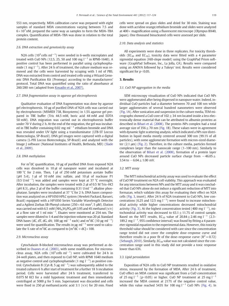
Chromosomal damage induced by CuO NPs was evaluated using thecytochalasin B-blocked micronucleus assay, in which cell division isblocked to allow the numbering of dividing cells only (binucleatedcells, Fig. 7A). In the untreated cells, micronucleus frequency was lessthan two per 1000 binucleated cells (Fig. 8). CuO NP treatments in-creased significantly the frequency of micronucleus formation at thelowest concentration tested (12.5 mg l−1, pb0.05) and reached ashigh as 15 micronuclei per 1000 binucleated cells for 100 mg l−1 CuONPs (Fig. 8). CuO NPs treatment induced micronuclei of both type I(micronucleus smaller than 1/4 the size of the nucleus, Fig. 7B) andtype II (micronucleus between 1/4 and 1/2 the size of the nucleus,Fig. 7C and D). Type I micronuclei are considered as an indicator of aclastogenic effect and contain small chromosome fragments, whileType II micronuclei are considered as indicating aneugenic effects andcontain whole chromosomes (Tinwell and Ashby, 1991; Hashimoto etal., 2010). Type I micronuclei were more frequent in all CuO NPs treat-ment, with a frequency between 70 and 80%, and did not increasewith increasing concentration of CuO NPs. This stability in the Type I:Type II ratiomay be an indicator of clastogenic effects however it shouldbe mentioned that total number of micronucleus counted was not suf-ficient to distinguishwith certainty between aneugenic and clastogenic
Fig. 6. Change in the ratio m5dC/(dC+m5dC) in DNA extracts of N2A cells after a 24 hexposure to 12.5, 25, 50 and 100 mg l−1 CuO NPs. Cyclophosphamide was used as apositive control (Pos Ctr). Data are presented as mean±standard deviation (n=3).
effect of CuO NPs using the micronucleus size-classification approach(Hashimoto et al., 2010).
4. Discussion
CuO NPs were previously identified as highly toxic with effects oncell viability, cellular oxidative balance and DNA integrity (Karlsson,2010; Ahamed et al., 2010; Wang et al., 2011). The genotoxic effect ofCuO NPs is considered to be mainly attributed to the nanoparticulateform and not to the presence of soluble copper ions (Karlsson, 2010;Wang et al., 2012). However, several aspects of the genotoxicity ofCuO NPs still remained to be investigated. In this study, we furtherinvestigated the genotoxic effects of CuO NPs using the N2A mouseneuroblastoma cell lines.
When N2A cells were exposed to CuONPs, a decrease of cell viabilitywas observed after 24 h of treatments. However, at the highest concen-tration tested (400 mg l−1) cell viability was still 63.1% of the controlvalue, indicating that N2A cells are not very sensitive to CuO NP effectscompared to other in vitro cell cultures. For the human cell lines THP-Iand A549, the concentrations inducing a 50% decrease of cell viabilityfor a 24 h treatment to 45 nm CuO NPs were between 3.89 and31.07 mg l−1 (Lanone et al., 2009). Ahamed et al. (2010) indicated a48% decrease of cell viability, using theMTT assay, in A549 cells exposedto 50 mg l−1 CuONPs of 65 nm. The differences in the toxic response ofN2A cells compared to THP-I and A549 cells may be associatedwith cel-lular differences (membrane composition, number of mitochondria)due to their origin, phenotype or cellular function (Watanabe et al.,2002; Souid-Mensi et al., 2008). The lower sensitivity of N2A cells mayalso be caused by the different agglomeration states of CuO NPs in theculture media. The N2A culture medium has a high ionic strength andcontains several types of protein, amino acids or glucose moleculesthat can be adsorbed on the NP surface (Amirnasr et al., 2011;Monopoli et al., 2011; Joshi et al., 2012). Change of NP surface propertiesinduced by the interaction with the culture medium may influence thephysicochemical properties and therefore the toxicity of nanomaterials(Mukherjee et al., 2010). In the culture medium of A549 cells, Ahamedet al. (2010) found a particle hydrodynamic diameter of 65.59 nm forCuO NPs having an initial particle diameter of 50–60 nm, while CuONPs having an initial particle diameter of 70–100 nm were found tohave a hydrodynamic diameter of 307 nm in the N2A culture medium.These complexesmay be hypothesized to be due to the binding of albu-min, which was used for particles stabilisation (Bihari et al., 2008), toform a protein corona at the CuO NP surface. The presence of such pro-tein corona may have a significant impact on its biological effect(Monopoli et al., 2011). In addition, agglomeration of CuO NPs inthe media increase the particle size above the nanometer range(1–100 nm), thus reducing the surface specific effects that are associat-ed with the nanometer range. Under those conditions, solubilisation ofthe particles may have a more important effect compared to previousfindings (Karlsson, 2010; Wang et al., 2012). Solubilisation of CuO NPsinto Cu2+ was found to be the most important process for A549 cellsexposed to CuO NPs (Hanagata et al., 2011). Therefore, for N2A cellsexposed to an agglomerated form of CuO NPs, the contribution of thesoluble Cu2+ fraction can be hypothesized to be more important.
Toxic effect of CuONPs at the cellular levelwas previously character-ized by the generation of oxidative stress and DNA damage (Karlsson,2010; Ahamed et al., 2010; Wang et al., 2012). However, such aspectsof CuO NP toxicity are still to be investigated in other cell lines besideA549, which is a heterogeneous cell line with multiple phenotypespresenting different levels of toxicological sensitivity (Watanabe et al.,2002). The genotoxicity of CuO NPs was therefore evaluated in N2Acells using different assays to characterize the DNA alterations inducedby CuONPs. Our results indicate that CuONPs can induce both oxidativestress and DNA fragmentation in N2A cells at a concentration of25 mg l−1. At this concentration, cell viability was 85% of the controlvalue. Oxidative stress is often considered as the primary mechanisms

Fig. 7. Examples of control (A) and micronucleated cells (B, C, D) for N2A cells.
122 F. Perreault et al. / Science of the Total Environment 441 (2012) 117–124
of NP toxicity (Nel et al., 2006; Shukla et al., 2011). Treatment of A549cells to CuO NPs resulted in MDA formation, activation of antioxidantenzymes and increased expression of the DNA repair proteins p53 andRad51 (Ahamed et al., 2010). In this report, the authors hypothesizedthat genotoxicity of CuO NPs may be mediated via oxidative stress andlipid peroxidation. In our study, DNA fragmentation andMDA formationwere found significantly at the same concentration (25 mg l−1). How-ever, DNA fragmentation can also be induced by several cellular pro-cesses, such as necrosis and apoptosis, therefore a complementaryapproach to evaluated the genotoxic effect of CuO NPs was used.
Micronucleus formation may be caused by an alteration of DNAintegrity which results in small chromosome fragments (castogeniceffect) or by an alteration ofmitotic processeswhich results in an abnor-mal number of chromosomes (aneugenic effect) (Kirsch-Volders et al.,2011). The formation of micronuclei in N2A cells exposed to CuO NPsmay therefore be an indicator of structural alteration of DNA.Our resultsindicate a predominance of Type Imicronuclei, which are an indicator ofa clastogenic effect. These results are in agreement with the DNA frag-mentation assay and confirm the genotoxic effect of CuO NPs in N2A
Fig. 8. Micronucleus count in N2A cells after a 24 h exposure to 12.5, 25, 50 and100 mg l−1 CuO NPs. Cyclophosphamide was used as a positive control (Pos Ctr).Data are presented as mean±standard deviation (n=3).
cells. The increase of micronucleus formation induced by CuO NPs wasfound to be low compared to other clastogenic compounds such asmethyl methanesulfonate, N-methyl–N′-nitro–N–nitroso-guanidine andmitomycin C, which increased micronucleus frequency up to more than300 micronuclei per 1000 binucleated cells (Matsushima et al., 1999).The induction of micronucleus formation by CuO NPs in N2A cells wasalso lower than for TiO2 NPs in human lymphocytes (Kang et al., 2008)but higher than for C60 NPs, SiO2 NPs and Ag NPs in hamster, mouseand human cells, respectively (Shinohara et al., 2009; Park et al., 2011;Li et al., 2012). Therefore, CuO NPs may be considered as a genotoxicnanomaterial compared to other NPs.
Themicronucleus assaywas themost sensitive approach to evaluatethe toxic effects of CuO NPs in N2A cells. Indeed, at 12.5 mg l−1 CuONPs, the micronucleus frequency was already 5 times the negative con-trol value while cell viability was not significantly changed compared tocontrol value (p=0.99). Our results indicate that, similarly to TiO2 andSiO2 NPs, micronucleus formation induced by CuO NPs in N2A cellsoccurs at lower concentration than cytotoxicity, oxidative stress orDNA fragmentation (Rahman et al., 2002; Wang et al., 2007). Recently,it was shown that the genotoxic effect of CuO NPs, in conditions whereCuO NPs were less agglomerated and soluble copper effect was low, isdue to the formation of reactive oxygen species (Wang et al., 2012).Under our conditions CuO NPs formed large (over 300 nm) agglomer-ates in the culture medium and genotoxic effects were observed atlower concentrations than lipid peroxidation induced by oxidativestress. This may indicate a different mode of action for small comparedto larger CuO NP agglomerates and this effect may be due to amore im-portant contribution of the soluble copper fraction under our condi-tions. The contribution of particle solubilisation, both in the media andinside the cell (Trojan Horse effect), will have to be determined inorder to provide a better understanding of the genotoxic potentials ofCuO NPs at the cellular level.
5. Conclusions
In this study, the toxic effects of CuO NPs were investigated in N2Acells to provide a better understanding of the toxicological risks ofCuO NPs in future nanotechnology developments. N2A cells werefound to be less sensitive to CuO NP effects than other in vitro culturedcells. This lower sensitivitymay be due to the agglomeration of CuONPs

123F. Perreault et al. / Science of the Total Environment 441 (2012) 117–124
in the culture media, which resulted in an average particle size over300 nm. Agglomeration of CuONPs reduces surface-specific effects spe-cific to nano-scale materials and increases the contribution of particlesolubilisation in the toxic response induce to N2A cells. AgglomeratedCuO NPs were found to induce both cytotoxic and genotoxic effects inN2A cells. Significant genotoxic effects were observed at a concentra-tion of 12.5 mg l−1, where no decrease of cell viability was found. Con-sidering that the NP fraction reaching the brain in vivo only accounts forless than 0.02% of total administrated dose, the risk of neurotoxicity ofCuO NPs may be less important than for other organs, such as theliver, that accumulate an higher amount of NPs (Lasagna-Reeves et al.,2010; Xie et al., 2011). However, considering the importance ofgenotoxic effect in the development of cancer, the risk of CuO NPsshould be investigated in order to ensure a safe development ofCuO-based nanomaterials. Further researchwill aimon the contributionof particle solubilisation in CuO NP toxicity and the clarification of thenature of the genotoxic effect induced by CuO NPs.
Acknowledgments
This work was supported by research grants awarded to W.G.Matias by the Conselho Nacional de Desenvolvimento Científico eTecnológico (Brazil), and to R. Popovic by the Natural Sciences andEngineering Research Council (Canada). F. Perreault was supportedby a mobility fellowship from the Ministère de l'Éducation, des Loisirset du Sport du Québec for his work in the LABTOX.
References
Ahamed M, Siddiqui MA, Akhtar MJ, Ahmad I, Pant AB, Alhadla HA. Genotoxic potentialof copper oxide nanoparticles in human lung epithelial cells. Biochem Biophys ResCommun 2010;396:578–83.
Amirnasr A, Emtiazi G, Abasi S, Yaaghoobi M. Adsorption of hemoglobin, fatty acid andglucose to iron nanoparticles as amean for drug delivery. J Biochem Tech 2011;3:280–3.
Bihari P, Vippola M, Schultes S, Praetner M, Khandoga AG, Reichel CA, et al. Optimizeddispersion of nanoparticles for biological in vitro and in vivo studies. Part FibreToxicol 2008;5:1-14.
Bradford MM. A rapid and sensitive method for the quantification of microgram quanti-ties of protein utilizing the principle of protein dye. Anal Biochem 1976;72:248–54.
Chen J, Zhu J, Cho H-H, Cui K, Li F, Zhou X, et al. Differential cytotoxicity of metal oxidenanoparticles. J Exp Nanosci 2008;3:321–8.
Dastjerdi R, Montazer M. A review on the application of inorganic nanostructuredmaterials in the modification of textiles: focus on antimicrobial properties. ColloidsSurf B Biointerfaces 2010;79:5-18.
Delgado K, Quijada R, Palma R, Palza H. Polypropylene with embedded copper metal orcopper oxide nanoparticles as a novel plastic antimicrobial agent. Lett ApplMicrobiol 2011;53:50–4.
Fenech M. The in vitro micronucleus technique. Mutat Res Fundam Mol Mech Mugag2000;455:81–95.
Gonzalgo ML, Jones PA. Mutagenic and epigenetic effects of DNA methylation. MutatRes 1997;386:107–18.
Hanagata N, Zhuang F, Connolly S, Li J, Ogawa N, Xu M. Molecular responses of humanlung epithelial cells to the toxicity of copper oxide nanoparticles inferred fromwhole genome expression analysis. ACS Nano 2011;27:9326–38.
Hashimoto K, Nakajima Y, Matsumura S, Chatani F. An in vitro micronucleus assay withsize-classified micronucleus counting to discriminate aneugens from clastogens.Toxicol In Vitro 2010;24:208–16.
Humpage AR, Ledreux A, Fanok S, Bernard C, Briand JF, Eaglesham G, et al. Applicationof the neuroblastoma assay for paralytic shellfish poisons to neurotoxic freshwatercyanobacteria: interlaboratory calibration and comparison with other methods ofanalysis. Environ Toxicol Chem 2007;26:1512–9.
Jensen KA, Kembouche Y, Christiansen E, Jacobsen NR, Wallin H, Guiot C, et al. Finalprotocol for producing suitable manufactured nanomaterial exposure media.NANOGENOTOX report n°3; 2011. July :34 pp.
Joshi S, Ghosh I, Pokhrel S, Madler L, Nau WM. Interactions of amino acids and polypep-tides with metal oxide nanoparticles probed by fluorescent indicator adsorption anddisplacement. ACS Nano 2012;6:5668–79.
Kang SJ, Kim BM, Lee YJ, Chung H. Titanium dioxide nanoparticles trigger p53-mediateddamage response in peripheral blood lymphocytes. Environ Mol Mutagen 2008;49:399–405.
Karlsson HL. The comet assay in nanotoxicology research. Anal Bioanal Chem 2010;298:651–66.
Karlsson HL, Cronholm P, Gustafsson J, Moller L. Copper oxide nanoparticles are highlytoxic: a comparison between metal oxide nanoparticles and carbon nanotubes.Chem Res Toxicol 2008;21:1726–32.
Kirsch-Volders M, Plas G, Elhajouji A, Lukamowicz M, Gonzalez L, Loock KV, et al. Thein vitro MN assay in 2011: origin and fate, biological significance, protocols, highthroughputmethodologies and toxicological relevance. Arch Toxicol 2011;85:873–99.
Kouadio JH, Danob SD, Moukha S, Mobio TA, Creppy EE. Effects of combinations of Fusar-ium mycotoxins on the inhibition of macromolecular synthesis, malondialdehydelevels, DNA methylation and fragmentation, and viability in Caco-2 cells. Toxicon2007;49:306–17.
Lanone S, Rogerieux F, Geys J, Dupont A, Maillot-Marechal E, Boczkowski J, et al. Com-parative toxicity of 24 manufactured nanoparticles in human alveolar epithelialand macrophage cell lines. Part Fibre Toxicol 2009;6:14.
Lasagna-Reeves C, Gonzalez-Romero D, Barria MA, Olmedo I, Clos A, SadagopaRamanujam VM, et al. Bioaccumulation and toxicity of gold nanoparticles afterrepeated administration in mice. Biochem Biophys Res Commun 2010;393:649–55.
Li F, Zhou X, Zhu J, Ma J, Huang X, Wong STC. High content image analysis forhuman H4 neuroglioma cells exposed to CuO nanoparticles. BMC Biotechnol2007;7:66.
Li Y-F, Chen C. Fate and toxicity of metallic and metal-containing nanoparticles for bio-medical applications. Small 2011;7:2965–80.
Li Y, Chen DH, Yan J, Chen Y, Mittelstaedt RA, Zhang Y, et al. Genotoxicity of silvernanoparticles evaluated using the Ames test and in vitro micronucleus assay.Mutat Res 2012;745:4-10.
Marnett LJ. Lipid peroxidation—DNA damage by malondialdehyde. Mutat Res FundamMol Mech Mugag 1999;424:83–95.
Matias WG, Creppy EE. 5-Methyldeoxycytosine as a biological marker of DNA damageinduced by okadaic acid in Vero cells. Environ Toxicol Water 1998;13:83–8.
Matsushima T, HayashiM, Matsuoka A, Ishidate JrM, Miura KF, Shimizu H, et al. Validationstudy of the in vitro micronucleus test in a Chinese hamster lung cell line (CHL/IU).Mutagenesis 1999;14:569–80.
Monopoli MP, Walczyk D, Campbell A, Elia G, Lynch I, Bombelli FB, et al. Physical–chemical aspects of protein corona: relevance to in vitro and in vivo biologicalimpacts of nanoparticles. J Am Chem Soc 2011;133:2525–34.
Mukherjee SP, Davoren M, Byrne HJ. In vitro mammalian cytotoxicological study ofPAMAM dendrimers—towards quantitative structure activity relationships. ToxicolIn Vitro 2010;24:1169–77.
Nel A, Xia T, Madler L, Lin N. Toxic potential of materials at the nano-level. Science2006;311:622–7.
Oszlanczi G, Vezer T, Sarkozi L, Horvath E, Konya Z, Papp A. Functional neurotoxicity ofMn-containing nanoparticles in rats. Ecotoxicol Environ Saf 2010;73:2004–9.
Ouanes Z, Abid S, Ayed I, Anane R, Mobio T, Creppy EE, et al. Induction of micronucleiby Zearalenone in Vero monkey kidney cells and in bone marrow cells of mice:protective effect of vitamin E. Mutat Res 2003;538:63–70.
Park MVDZ, Verharen HW, Zwart E, Hernandez LG, Benthem JV, Elsaesser A, et al.Genotoxicity evaluation of amorphous silica nanoparticles of different sizes usingthe micronucleus and the plasmid lacZ gene mutation assay. Nanotoxicology2011;5:168–81.
Peralta-Videa JR, Zhao L, Lopez-Moreno ML, de la Rosa G, Hong J, Gardea-Torresdey JL.Nanomaterials and the environment: a review for the biennium 2008–2010. J HazardMater 2011;186:1-15.
Perreault F, Matias MS, Melegari SP, Carvalho Pinto CRS, Creppy EE, Popovic R, et al.Investigation of animal and algal bioassays for reliable saxitoxin ecotoxicity andcytotoxicity risk evaluation. Ecotoxicol Environ Saf 2011;74:1021–6.
Rahman Q, Lohani M, Dopp E, Pemsel H, Jonas L, Weiss DG, et al. Evidence that ultrafinetitanium dioxide induces micronuclei and apoptosis in Syrian hamster embryofibroblasts. Environ Health Perspect 2002;110:797–800.
Ren G, Hu D, Cheng EWC, Vargas-Reus MA, Reip P, Allaker RP. Characterisation of copperoxide nanoparticles for antimicrobial applications. Int J Antimicrob Agents 2009;33:587–90.
Sebaugh JL. Guidelines for accurate EC50/IC50 estimation. Pharm Stat 2010;10:128–34.Sharma HS, Hussain S, Schlager J, Ali SF, Sharma A. Influence of nanoparticles on
blood-brain barrier permeability and brain edema formation in rats. Acta NeurochirSuppl 2010;106:359–64.
Shinohara N, Matsumoto K, Endoh S, Maru J, Nakanishi J. In vitro and invivo genotoxicity tests on fullerene C60 nanoparticles. Toxicol Lett 2009;191:289–96.
Shukla RK, Sharma V, Pandey AK, Singh S, Sultana S, Dhawan A. ROS-mediatedgenotoxicity induced by titanium dioxide nanoparticles in human epidermalcells. Toxicol In Vitro 2011;25:231–41.
Singh N, Manshian B, Jenkins GJS, Griffiths SM, Williams PM, Maffeis TGG, et al.NanoGenotoxicology: the DNA damaging potential of engineered nanomaterials.Biomaterials 2009;30:3891–914.
Skocaj M, Filipic M, Petkovic J, Novak S. Titanium dioxide in our everyday life; is it safe?Radiol Oncol 2011;45:227–47.
Slade D, Lindner AB, Paul G, RadmanM. Recombination and replication in DNA repair ofheavily irradiated Deinococcus radiodurans. Cell 2009;136:1044–55.
Souid-Mensi G, Moukha S, Mobio TA, Maaroufi K, Creppy EE. The cytotoxicityand genotoxicity of okadaic acid are cell-line dependent. Toxicon 2008;51:1338–44.
Tinwell H, Ashby J. Micronucleus morphology as a means to distinguish aneugens andclastogens in the mouse bone marrow micronucleus assay. Mutagenesis 1991;6:193–8.
Tsoli M, Kuhn H, Brandau W, Esche H, Schmid G. Cellular uptake and toxicity of Au55
clusters. Small 2005;1:841–4.Wang JJ, Sanderson BJS, Wang H. Cytotoxicity and genotoxicity of ultrafine crystalline
SiO2 particulate in cultured human lymphoblastoid cells. Environ Mol Mutagen2007;48:151–7.

124 F. Perreault et al. / Science of the Total Environment 441 (2012) 117–124
Wang Y, AkerWG, HwangH-M, Yedjou CG, Yu H, Tchounwou PB. A study of themechanismof in vitro cytotoxicity of metal oxide nanoparticles using catfish primary hepatocytesand human HepG2 cells. Sci Total Environ 2011;409:4753–62.
Wang Z, Li N, Zhao J, White JC, Qu O, Xing B. CuO nanoparticle interaction with humanepithelial cells: cellular uptake, location, export, and genotoxicity. Chem Res Toxicol2012;25:1512–21.
Watanabe N, Dickinson DA, Krzywanski DM, Iles KE, Zhang H, Venglarik CJ, et al. A549subclones demonstrate heterogeneity in toxicological sensitivity and antioxidantprofile. Am J Physiol Lung Cell Mol Physiol 2002;283:L726–36.
Xie G, Wang C, Sun J, Zhong G. Tissue distribution and excretion of intravenouslyadministered titanium dioxide nanoparticles. Toxicol Lett 2011;205:55–61.
Zhivotosky B, Orrenius S. Assessment of apoptosis and necrosis by DNA fragmentationand morphological criteria. Curr Protoc Cell Biol 2001;18. Unit 18.3.
Zukiel R, Nowak S, Barciszewska A-M, Gawronska I, Keith G, Barciszewska MZ. A simpleepigenetic method for the diagnosis and classification of brain tumors. Mol CancerRes 2004;2:196–202.