Embed Size (px)
Citation preview
Geometric and Electronic Structures of the NiI and Methyl-NiIII Intermediates ofMethyl-Coenzyme M Reductase†
Ritimukta Sarangi,*,‡ Mishtu Dey,§ and Stephen W. Ragsdale*,§
Stanford Synchrotron Radiation Lightsource, SLAC National Accelerator Laboratory, Menlo Park, California 94025, andDepartment of Biological Chemistry, UniVersity of Michigan, Ann Arbor, Michigan 48109-0606
ReceiVed January 23, 2009; ReVised Manuscript ReceiVed February 25, 2009
ABSTRACT: Methyl-coenzyme M reductase (MCR) catalyzes the terminal step in the formation of biologicalmethane from methyl-coenzyme M (Me-SCoM) and coenzyme B (CoBSH). The active site in MCRcontains a Ni-F430 cofactor, which can exist in different oxidation states. The catalytic mechanism ofmethane formation has remained elusive despite intense spectroscopic and theoretical investigations. Onthe basis of spectroscopic and crystallographic data, the first step of the mechanism is proposed to involvea nucleophilic attack of the NiI active state (MCRred1) on Me-SCoM to form a NiIII-methyl intermediate,while computational studies indicate that the first step involves the attack of NiI on the sulfur of Me-SCoM, forming a CH3
• radical and a NiII-thiolate species. In this study, a combination of Ni K-edgeX-ray absorption spectroscopic (XAS) studies and density functional theory (DFT) calculations have beenperformed on the NiI (MCRred1), NiII (MCRred1-silent), and NiIII-methyl (MCRMe) states of MCR to elucidatethe geometric and electronic structures of the different redox states. Ni K-edge EXAFS data are used toreveal a five-coordinate active site with an open upper axial coordination site in MCRred1. Ni K-pre-edgeand EXAFS data and time-dependent DFT calculations unambiguously demonstrate the presence of along Ni-C bond (∼2.04 Å) in the NiIII-methyl state of MCR. The formation and stability of this speciessupport mechanism I, and the Ni-C bond length suggests a homolytic cleavage of the NiIII-methyl bondin the subsequent catalytic step. The XAS data provide insight into the role of the unique F430 cofactor intuning the stability of the different redox states of MCR.
Methyl-coenzyme M reductase (MCR)1 from methano-genic archaea (1) catalyzes the terminal step in biologicalmethane synthesis. Using coenzyme B (CoBSH) as the two-electron donor, MCR reduces methyl-coenzyme M (methyl-SCoM) to form methane and the heterodisulfide product,CoBS-SCoM (2, 3). MCR contains an essential redox activenickel tetrapyrrolic cofactor called coenzyme F430 at its activesite (4, 5), which is active in the reduced NiI state (MCRred1).All of the biologically generated methane, amounting to 1billion tons per annum globally, is formed by MCR.Furthermore, recent evidence indicates that anaerobic meth-ane oxidation is also catalyzed by MCR and occurs by areversal of the methane synthesis reaction (6, 7). The centralrole of MCR in the synthesis of this important fuel, which
is also a potent greenhouse gas, makes it important tounderstand the catalytic mechanism of methane formation.
Two limiting mechanisms for MCR-catalyzed methaneformation have been proposed (Figure 1). In mechanism I,based on the NiII crystal structure and model chemistry (8-11),NiI performs an SN2 attack on the methyl group of methyl-SCoM to form a NiIII-methyl intermediate that undergoesone-electron reduction to form a NiII-methyl species, whichthen undergoes protonation to form methane, and a CoMS•
radical. Condensation of the CoMS• radical with CoBSHgenerates a CoBSSCoM• radical anion that reduces NiII tothe active NiI state. Mechanism II, which is based on densityfunctional theory calculations (12, 13) and considers NiIII
not to be a feasible intermediate, proposes that NiI-MCRred1
† This work has been supported by NIH Grant 1P20RR17675 toS.W.R. We are grateful for funding from a Department of Energy grant(DE-FG02-08ER15931) to S.W.R.
* To whom correspondence should be addressed. R.S.: e-mail,[email protected]; phone, (650) 926-4621; fax, (650) 926-4100.S.W.R.: e-mail, [email protected]; phone, (734) 615-4621; fax,(734)763-4581.
‡ SLAC National Accelerator Laboratory.§ University of Michigan.1 Abbreviations: CoBSH, coenzyme B; DFT, density functional
theory; ENDOR, electron nuclear double resonance; EPR, electronparamagnetic resonance; EXAFS, extended X-ray absorption finestructure; MCR, methyl-coenzyme M reductase; methyl-SCoM, methyl-coenzyme M; PDB, Protein Data Bank; TD-DFT, time-dependentdensity functional theory; XAS, X-ray absorption spectroscopy.
FIGURE 1: Schematic diagram showing the proposed structures uponconversion of the active MCRred1 to MCRred1-silent and MCRMe. TheNiIII-PS state is analogous to the NiIII-methyl state and is formedby the reaction of MCRred1 with bromopropane sulfonate.
Biochemistry 2009, 48, 3146–31563146
10.1021/bi900087w CCC: $40.75 2009 American Chemical SocietyPublished on Web 02/25/2009
reacts with the sulfur of Me-SCoM to generate a methylradical and a NiII-thiolate complex. The methyl radical isproposed to abstract a hydrogen atom from CoBSH togenerate a CoBS• radical that reacts with bound CoM togenerate NiII and the same CoBS-SCoM• radical anionproposed in mechanism 1. Finally, reduction of NiII andgeneration of the CoBS-SCoM product occur as in mech-anism I. The major distinction between the two mechanismslies in the intermediate generated in the first step of catalysis:NiIII-methyl or methyl radical and NiII-SCoM. However,so far, none of the proposed intermediates have been trappedduring reactions with the natural substrates; presumably,these intermediates form and decay too fast to be observedby stopped-flow and rapid freeze quench EPR methods.
Much of our understanding of the structure and mechanismof MCR catalysis is based on crystal structures of variousinactive NiII states (MCRsilent, MCRox1-silent, and MCRred1-silent)(8, 10, 14), since the crystal structures of the active states ofMCR have not been reported. In all states, the central Niatom is coordinated by four corphin ring nitrogens and alower axial glutamine oxygen atom. The upper axial ligandsin MCRox1-silent and MCRsilent are the thiol group of coenzymeM and the sulfonate oxygen of the heterodisulfide product,respectively (8, 10, 14). In MCRred1-silent, two structures havebeen proposed: a five-coordinate site lacking an upper axialligand and a six-coordinate site with the thiol group ofcoenzyme M as the upper axial ligand (10). EXAFS studieshave also been performed on various NiII forms of MCR,which are consistent with crystal structures; however, high-kEXAFS studies have not been reported (8, 15).
In this study, NiI and NiIII-methyl species, which are thestarting state and a putative intermediate state in the MCR-catalyzed reaction, respectively, have been trapped andcharacterized. Although a NiIII-methyl intermediate has notyet been identified during the reaction with the naturalsubstrate Me-SCoM, its formation has been demonstratedin the reaction of MCRred1 with methyl bromide (16) andmethyl iodide (17). Analogous NiIII-alkyl species are formedby the reaction of MCRred1 with corresponding alkylhalides (18-20). Furthermore, this species has been shownto react with HSCoM (and other thiolates) to generate theNiI-MCRred1 state and methyl-SCoM (or other alkyl thio-ethers). The similarity between the rate constants for methaneformation from the MCRMe species with HSCoM andCoBSH (1.1 s-1) and the steady-state kcat for methaneformation from natural substrates (4.5 s-1 at 25 °C) isconsistent with the catalytic intermediacy of the methyl-Nispecies (17). Thus, there is significant evidence supportingthe catalytic relevance of the MCRMe intermediate. Althoughthe crystal structure of active MCRred1 is not known, muchof our understanding about the active site structure inMCRred1 comes from Ni K-edge EXAFS studies. Twostructures have been proposed: a five-coordinate site withan open upper axial ligand (8) and a six-coordinate site withan oxygen atom as the upper axial ligand (15). However, inboth studies, the k range for the reported EXAFS data was2-12 Å-1, limiting the resolution ((0.16 Å) and the abilityto identify and distinguish the equatorial and axial ligands.
The goal of this study was to determine high-resolutionstructures of the active NiI state with accurate metricalparameters. Although direct structural information about theNiIII-methyl state is not available, EPR, ENDOR, and
HYSCORE spectroscopic studies have determined the elec-tronic structure of the active site Ni center (16, 17). Thesestudies describe MCRMe to be formally NiIII with a methyl-Nibond formed by the oxidative addition reaction of methyliodide with the NiI-MCRred1 complex. The presence of alarge 13C hyperfine coupling indicates that the methyl groupis coordinated to the paramagentic NiIII center by a covalentbond, with a Ni-C bond distance of approximately 1.9-2.0Å (16, 17), although the actual bond distance cannot beprecisely determined by analysis of these hyperfine couplings.Similarly, EPR studies on the adduct between propane-sulfonate and MCR demonstrated the presence of a Ni(III)-Cbond with an approximate bond distance of 2 Å (21).However, the active site geometry of the MCRMe state hasnot been determined by any structure determination technique.
In the work described here, Ni K-pre-edge and edge X-rayabsorption spectroscopy and EXAFS investigations havebeen combined with time-dependent density functional theory(DFT) calculations to determine the local geometric structureat the active sites in MCRred1-silent, MCRred1, and MCRMe.The ∼0.1 Å resolution (k ) 17 Å-1) EXAFS data presentedhere provide a precise description of the active site geometryin the active MCRred1 state with the ability to differentiatebetween the axial Ni-O(Gln) bond distance and theNi-N(F430) distances. This ability to discriminate the axialand equatorial bond distances was not present in previouslyreported EXAFS data, which extended up to k ) 12Å-1 (8, 15). The results presented in this study also providethe first experimentally determined atomic-level descriptionof the Ni center in the proposed intermediate methyl-Nistate with precise Ni-first neighbor bond distance measure-ments. The trends in the Ni K-pre-edge and edge energypositions are used to determine the changes in bonding andligand field. Together, the XAS and EXAFS studies are usedto provide insight into the electronic structures of the activesites and how they relate to the mechanism of formation ofmethane by MCR.
EXPERIMENTAL PROCEDURES
Sample Preparation. (i) Material and Organisms. Metha-nothermobacter marburgensis was obtained from the OregonCollection of Methanogens catalogue as OCM82. All buffers,mediUM ingredients, and other reagents were acquired fromSigma-Aldrich and, unless otherwise stated, were of thehighest purity available. Solutions were prepared usingnanopure deionized water. N2 (99.98%), H2/CO2 (80%/20%),and ultra-high-purity (UHP) H2 (99.999%) were obtainedfrom Cryogenic Gases (Grand Rapids, MI). Ti(III) citratesolutions were prepared from a stock solution of 200 mMTi(III) citrate, which was synthesized by adding sodiumcitrate to Ti(III) trichloride (30 wt % solution in 2 Nhydrochloric acid) under anaerobic conditions and adjustingthe pH to 7.0 with sodium bicarbonate. The concentrationof Ti(III) citrate was determined routinely by titrating againsta methyl viologen solution.
(ii) M. marburgensis Growth, HarVest, and MCRred1
Purification. MCRred1 was isolated from M. marburgensiscultured on H2/CO2 (80%/20%) at 65 °C in a 14 L fermentor.Culture media were prepared as previously described (18)with a slight modification of the sulfur and reducing source.Instead of H2S used previously, 50 mM sodium sulfide was
Ni K-Edge X-ray Absorption Spectroscopy on MCRred1 and MCRMe Biochemistry, Vol. 48, No. 14, 2009 3147
added at a flow rate of 1 mL/min during the entire growthperiod. MCRred1 was generated in vivo and purified asdescribed previously (18). This purification procedure rou-tinely generates ∼70% MCRred1 as determined by UV-visibleand EPR spectroscopy.
(iii) Preparation of MCRred1 and MCRMe Samples. MCRred1
was prepared in 50 mM Tris-HCl (pH 7.6) containing 30%glycerol. The MCRMe samples were prepared in the anaerobicchamber by incubating MCRred1 with excess methyl iodidein 50 mM Tris-HCl (pH 7.6). The reaction mixture was splitinto three aliquots. One sample was transferred to a cuvetteto monitor the conversion of the NiI-MCRred1 complex toNiIII/NiII by UV-visible spectroscopy; a second aliquot wasfrozen in liquid nitrogen in an EPR tube to measure theconcentration of MCRMe species, and a third sample wasloaded in 1 mm lucite cells with 37 µm Kapton windowsfor X-ray absorption studies, frozen in liquid nitrogen, andmaintained under liquid N2 conditions until data werecollected. An important point to note is that the reaction ofMCRred1 to MCRMe always goes to at least 100% conversion,if not more. This observation has been made repeatedly, andin these studies, we observed a 3% increase in the MCRMe
concentration compared to the starting MCRred1 concentrationthat was used.
X-ray Absorption Spectroscopy. Ni K-edge X-ray absorp-tion spectra of MCRred1-silent, MCRred1, and MCRMe weremeasured at the Stanford Synchrotron Radiation Laboratoryon 16-pole, 2.0 T, wiggler beamline 9-3. A liquid N2-cooledSi(220) double-crystal monochromator was used for energyselection. A Rh-coated harmonic rejection mirror and acylindrical Rh-coated bent focusing mirror were used. AnOxford Instruments CF1208 continuous-flow liquid Hecryostat was used to maintain the sample temperature at ∼10K throughout the course of data measurement. Data weremeasured up to k ) 18 Å-1 in fluorescence mode using aCanberra Ge-30 element array detector. Internal energycalibration was accomplished by simultaneous measurementof the absorption of a Ni foil placed between two ionizationchambers situated downstream of the sample. The firstinflection point of the Ni foil was assigned to 8331.6 eV.All samples were closely monitored for photoreduction.However, the Ni active site in all three states of the proteinwas resistant to photoreduction. Spectra presented here area 10-scan, 24-scan, and 28-scan average for MCRred1-silent,MCRred1, and MCRMe, respectively. The energy-calibrateddata were averaged and processed by fitting a second-orderpolynomial to the pre-edge region, which was subtractedfrom the entire spectrum as background using Pyspline (22).A three-region spline function of order 2,3 and 3 was usedto model the background atomic absorption and subtractedfrom the spectrum. Normalization was accomplished bydividing the entire spectrum by a polynomial of order 1,which was fit to the postedge region. The experimentalthreshold energy was chosen to be 8340 eV (8, 15). Theintensities and energies of the pre-edge transitions werequantitated by performing least-squares refinement usingEDG-FIT (23). The pre-edge features were modeled byusing 1:1 Gaussian/Lorentzian Pseudo-Voigt line shapes tosimulate the convolution of instrument and core-hole lifetimebroadening. Additional Pseudo-Voigt line shapes were alsorequired to mimic the rising-edge transition and shouldersin the edge region. The data were fit over two different
energy ranges: 8125-8135 and 8127-8140 eV. The least-squares error and a comparison of the second derivatives ofthe data and fit were used to estimate the goodness of thefit. Standard deviations in energy position and intensity wereused to quantitate the errors in these parameters. TheoreticalEXAFS phase and amplitude parameters were calculatedusing FEFF (Macintosh version 8.4) (24-26) and thepublished crystal structure of MCRred1-silent (PDB entry1MRO) (27) as the initial starting model. Data were fit usingEXAFSPAK (23). The metrical parameters obtained byfitting the data indicated significant differences in the localstructures of MCRred1, MCRred1-silent, and MCRMe around thecentral absorbing Ni atom. On the basis of these preliminaryfits, a new set of theoretical EXAFS signals, �(k), werecalculated, and the data for MCRred1 and MCRMe were refitusing the new theoretical parameters generated from theirindividual refined models. The structural parameters variedduring the fitting process were restricted to the bond distance(R) and the bond variance (σ2), which is related to theDebye-Waller factor, resulting from a combination of staticand dynamic disorder (due to thermal motion) between theabsorber and scatterer pair. The nonstructural parameter, ∆E0,was also allowed to vary but was restricted to a commonvalue for every component in a given fit. Coordinationnumbers were systematically varied in the course of a fitbut were fixed within a given fit. In the case of MCRred1 andMCRMe, which were estimated to have 36 and 33% of theMCRred1-silent decay product using UV-vis spectroscopy,respectively, partial coordination numbers were also exploredduring the course of the fit. It should be noted that theEXAFS fits to the data were performed between k ) 2 and17 Å-1. Since, the Fourier transform intensity, R′, issignificant between 1.5 and 4 Å, the number of independentparameters is 26 (using the formula 2δkδR′/π + 2) (28).The maximum number of independent parameters used inthe fits is 13, which is lower than the number of maximumallowed independent parameters.
DFT Calculations. Gradient-corrected (GGA), spin-unrestricted density functional theory calculations wereperformed using the Gaussian03 (29) package on a 32-CPULinux cluster. Geometry optimizations were performed ineach case. The B3LYP (30-32) hybrid functional and thefollowing basis sets were employed: triple-� 6-311+G*(33-35) on Ni, 6-311G* (33-35) on S, and 6-31G* (36-38)on O, C, H, and N. The input structures were based on thepublished crystal structure and the EXAFS best-fit resultspresented herein. The transaxial glutamine ligand was fixedat the EXAFS distance for MCRred1 DFT calculations. Time-dependent DFT calculations were performed with theelectronic structure program ORCA (39, 40) to calculate theenergies and intensities of the Ni 1s f 3d pre-edgetransitions. Single-point ground-state calculations were per-formed using the geometry-optimized coordinates obtainedfrom the Gaussian03 package. The BP86 functional and thefollowing basis sets were employed: CP(PPP) (41, 42) onNi (core properties basis set as implemented in ORCA) andTZVP (43, 44) on N, C, H, O, and S. Tight convergencecriteria was imposed on all calculations. The calculatedenergies and intensities were convoluted with a Gaussianfunction with half-widths of 1.4 eV (45) to account for core-hole and instrument broadening. Calculations were performedin a dielectric continuum using the conductor-like screening
3148 Biochemistry, Vol. 48, No. 14, 2009 Sarangi et al.
model (COSMO). A shift of 223.0 eV was applied to thecalculated pre-edge energy positions. This is usually the casewith core-level TD-DFT calculation since DFT does notcalculate core potentials accurately, resulting in the corelevels being too high relative to the valence levels.
RESULTS
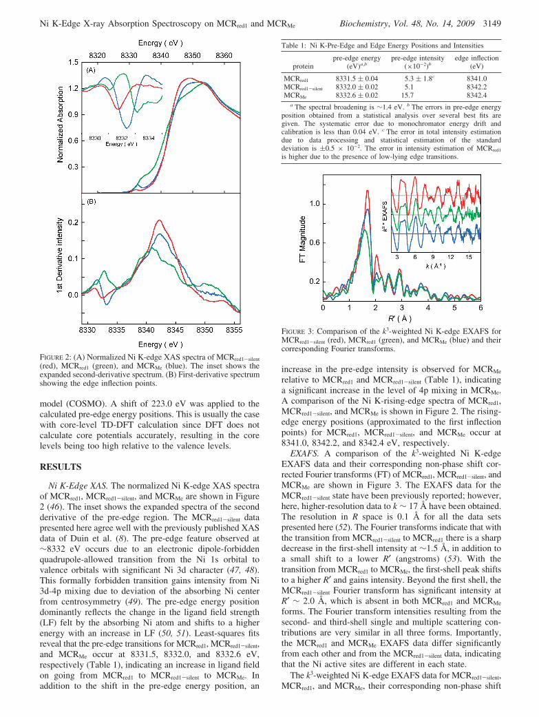
Ni K-Edge XAS. The normalized Ni K-edge XAS spectraof MCRred1, MCRred1-silent, and MCRMe are shown in Figure2 (46). The inset shows the expanded spectra of the secondderivative of the pre-edge region. The MCRred1-silent datapresented here agree well with the previously published XASdata of Duin et al. (8). The pre-edge feature observed at∼8332 eV occurs due to an electronic dipole-forbiddenquadrupole-allowed transition from the Ni 1s orbital tovalence orbitals with significant Ni 3d character (47, 48).This formally forbidden transition gains intensity from Ni3d-4p mixing due to deviation of the absorbing Ni centerfrom centrosymmetry (49). The pre-edge energy positiondominantly reflects the change in the ligand field strength(LF) felt by the absorbing Ni atom and shifts to a higherenergy with an increase in LF (50, 51). Least-squares fitsreveal that the pre-edge transitions for MCRred1, MCRred1-silent,and MCRMe occur at 8331.5, 8332.0, and 8332.6 eV,respectively (Table 1), indicating an increase in ligand fieldon going from MCRred1 to MCRred1-silent to MCRMe. Inaddition to the shift in the pre-edge energy position, an
increase in the pre-edge intensity is observed for MCRMe
relative to MCRred1 and MCRred1-silent (Table 1), indicatinga significant increase in the level of 4p mixing in MCRMe.A comparison of the Ni K-rising-edge spectra of MCRred1,MCRred1-silent, and MCRMe is shown in Figure 2. The rising-edge energy positions (approximated to the first inflectionpoints) for MCRred1, MCRred1-silent, and MCRMe occur at8341.0, 8342.2, and 8342.4 eV, respectively.
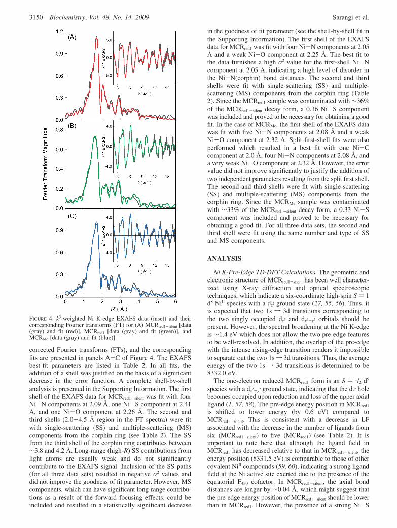
EXAFS. A comparison of the k3-weighted Ni K-edgeEXAFS data and their corresponding non-phase shift cor-rected Fourier transforms (FT) of MCRred1, MCRred1-silent, andMCRMe are shown in Figure 3. The EXAFS data for theMCRred1-silent state have been previously reported; however,here, higher-resolution data to k ∼ 17 Å have been obtained.The resolution in R space is 0.1 Å for all the data setspresented here (52). The Fourier transforms indicate that withthe transition from MCRred1-silent to MCRred1 there is a sharpdecrease in the first-shell intensity at ∼1.5 Å, in addition toa small shift to a lower R′ (angstroms) (53). With thetransition from MCRred1 to MCRMe, the first-shell peak shiftsto a higher R′ and gains intensity. Beyond the first shell, theMCRred1-silent Fourier transform has significant intensity atR′ ∼ 2.0 Å, which is absent in both MCRred1 and MCRMe
forms. The Fourier transform intensities resulting from thesecond- and third-shell single and multiple scattering con-tributions are very similar in all three forms. Importantly,the MCRred1 and MCRMe EXAFS data differ significantlyfrom each other and from the MCRred1-silent data, indicatingthat the Ni active sites are different in each state.
The k3-weighted Ni K-edge EXAFS data for MCRred1-silent,MCRred1, and MCRMe, their corresponding non-phase shift
FIGURE 2: (A) Normalized Ni K-edge XAS spectra of MCRred1-silent
(red), MCRred1 (green), and MCRMe (blue). The inset shows theexpanded second-derivative spectrum. (B) First-derivative spectrumshowing the edge inflection points.
Table 1: Ni K-Pre-Edge and Edge Energy Positions and Intensities
proteinpre-edge energy
(eV)a,bpre-edge intensity
(×10-2)bedge inflection
(eV)
MCRred1 8331.5 ( 0.04 5.3 ( 1.8c 8341.0MCRred1-silent 8332.0 ( 0.02 5.1 8342.2MCRMe 8332.6 ( 0.02 15.7 8342.4
a The spectral broadening is ∼1.4 eV. b The errors in pre-edge energyposition obtained from a statistical analysis over several best fits aregiven. The systematic error due to monochromator energy drift andcalibration is less than 0.04 eV. c The error in total intensity estimationdue to data processing and statistical estimation of the standarddeviation is (0.5 × 10⁻². The error in intensity estimation of MCRred1
is higher due to the presence of low-lying edge transitions.
FIGURE 3: Comparison of the k3-weighted Ni K-edge EXAFS forMCRred1-silent (red), MCRred1 (green), and MCRMe (blue) and theircorresponding Fourier transforms.
Ni K-Edge X-ray Absorption Spectroscopy on MCRred1 and MCRMe Biochemistry, Vol. 48, No. 14, 2009 3149
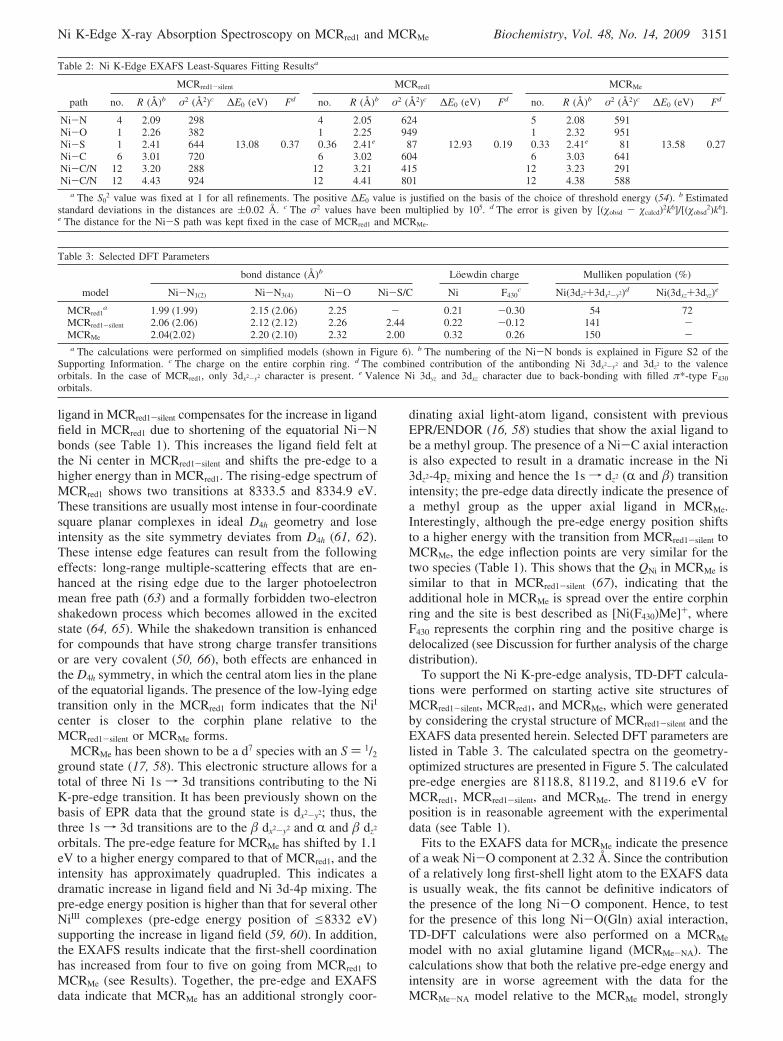
corrected Fourier transforms (FTs), and the correspondingfits are presented in panels A-C of Figure 4. The EXAFSbest-fit parameters are listed in Table 2. In all fits, theaddition of a shell was justified on the basis of a significantdecrease in the error function. A complete shell-by-shellanalysis is presented in the Supporting Information. The firstshell of the EXAFS data for MCRred1-silent was fit with fourNi-N components at 2.09 Å, one Ni-S component at 2.41Å, and one Ni-O component at 2.26 Å. The second andthird shells (2.0-4.5 Å region in the FT spectra) were fitwith single-scattering (SS) and multiple-scattering (MS)components from the corphin ring (see Table 2). The SSfrom the third shell of the corphin ring contributes between∼3.8 and 4.2 Å. Long-range (high-R) SS contributions fromlight atoms are usually weak and do not significantlycontribute to the EXAFS signal. Inclusion of the SS paths(for all three data sets) resulted in negative σ2 values anddid not improve the goodness of fit parameter. However, MScomponents, which can have significant long-range contribu-tions as a result of the forward focusing effects, could beincluded and resulted in a statistically significant decrease
in the goodness of fit parameter (see the shell-by-shell fit inthe Supporting Information). The first shell of the EXAFSdata for MCRred1 was fit with four Ni-N components at 2.05Å and a weak Ni-O component at 2.25 Å. The best fit tothe data furnishes a high σ2 value for the first-shell Ni-Ncomponent at 2.05 Å, indicating a high level of disorder inthe Ni-N(corphin) bond distances. The second and thirdshells were fit with single-scattering (SS) and multiple-scattering (MS) components from the corphin ring (Table2). Since the MCRred1 sample was contaminated with ∼36%of the MCRred1-silent decay form, a 0.36 Ni-S componentwas included and proved to be necessary for obtaining a goodfit. In the case of MCRMe, the first shell of the EXAFS datawas fit with five Ni-N components at 2.08 Å and a weakNi-O component at 2.32 Å. Split first-shell fits were alsoperformed which resulted in a best fit with one Ni-Ccomponent at 2.0 Å, four Ni-N components at 2.08 Å, anda very weak Ni-O component at 2.32 Å. However, the errorvalue did not improve significantly to justify the addition oftwo independent parameters resulting from the split first shell.The second and third shells were fit with single-scattering(SS) and multiple-scattering (MS) components from thecorphin ring. Since the MCRMe sample was contaminatedwith ∼33% of the MCRred1-silent decay form, a 0.33 Ni-Scomponent was included and proved to be necessary forobtaining a good fit. For all three data sets, the second andthird shell were fit using the same number and type of SSand MS components.
ANALYSIS
Ni K-Pre-Edge TD-DFT Calculations. The geometric andelectronic structure of MCRred1-silent has been well character-ized using X-ray diffraction and optical spectroscopictechniques, which indicate a six-coordinate high-spin S ) 1d8 NiII species with a dz2 ground state (27, 55, 56). Thus, itis expected that two 1s f 3d transitions corresponding tothe two singly occupied dz2 and dx2-y2 orbitals should bepresent. However, the spectral broadening at the Ni K-edgeis ∼1.4 eV which does not allow the two pre-edge featuresto be well-resolved. In addition, the overlap of the pre-edgewith the intense rising-edge transition renders it impossibleto separate out the two 1sf 3d transitions. Thus, the averageenergy of the two 1s f 3d transitions is determined to be8332.0 eV.
The one-electron reduced MCRred1 form is an S ) 1/2 d9
species with a dx2-y2 ground state, indicating that the dz2 holebecomes occupied upon reduction and loss of the upper axialligand (1, 57, 58). The pre-edge energy position in MCRred1
is shifted to lower energy (by 0.6 eV) compared toMCRred1-silent. This is consistent with a decrease in LFassociated with the decrease in the number of ligands fromsix (MCRred1-silent) to five (MCRred1) (see Table 2). It isimportant to note here that although the ligand field inMCRred1 has decreased relative to that in MCRred1-silent, theenergy position (8331.5 eV) is comparable to those of othercovalent NiII compounds (59, 60), indicating a strong ligandfield at the Ni active site exerted due to the presence of theequatorial F430 cofactor. In MCRred1-silent, the axial bonddistances are longer by ∼0.04 Å, which might suggest thatthe pre-edge energy position of MCRred1-silent should be lowerthan in MCRred1. However, the presence of a strong Ni-S
FIGURE 4: k3-weighted Ni K-edge EXAFS data (inset) and theircorresponding Fourier transforms (FT) for (A) MCRred1-silent [data(gray) and fit (red)], MCRred1 [data (gray) and fit (green)], andMCRMe [data (gray) and fit (blue)].
3150 Biochemistry, Vol. 48, No. 14, 2009 Sarangi et al.
ligand in MCRred1-silent compensates for the increase in ligandfield in MCRred1 due to shortening of the equatorial Ni-Nbonds (see Table 1). This increases the ligand field felt atthe Ni center in MCRred1-silent and shifts the pre-edge to ahigher energy than in MCRred1. The rising-edge spectrum ofMCRred1 shows two transitions at 8333.5 and 8334.9 eV.These transitions are usually most intense in four-coordinatesquare planar complexes in ideal D4h geometry and loseintensity as the site symmetry deviates from D4h (61, 62).These intense edge features can result from the followingeffects: long-range multiple-scattering effects that are en-hanced at the rising edge due to the larger photoelectronmean free path (63) and a formally forbidden two-electronshakedown process which becomes allowed in the excitedstate (64, 65). While the shakedown transition is enhancedfor compounds that have strong charge transfer transitionsor are very covalent (50, 66), both effects are enhanced inthe D4h symmetry, in which the central atom lies in the planeof the equatorial ligands. The presence of the low-lying edgetransition only in the MCRred1 form indicates that the NiI
center is closer to the corphin plane relative to theMCRred1-silent or MCRMe forms.
MCRMe has been shown to be a d7 species with an S ) 1/2
ground state (17, 58). This electronic structure allows for atotal of three Ni 1s f 3d transitions contributing to the NiK-pre-edge transition. It has been previously shown on thebasis of EPR data that the ground state is dx2-y2; thus, thethree 1s f 3d transitions are to the � dx2-y2 and R and � dz2
orbitals. The pre-edge feature for MCRMe has shifted by 1.1eV to a higher energy compared to that of MCRred1, and theintensity has approximately quadrupled. This indicates adramatic increase in ligand field and Ni 3d-4p mixing. Thepre-edge energy position is higher than that for several otherNiIII complexes (pre-edge energy position of e8332 eV)supporting the increase in ligand field (59, 60). In addition,the EXAFS results indicate that the first-shell coordinationhas increased from four to five on going from MCRred1 toMCRMe (see Results). Together, the pre-edge and EXAFSdata indicate that MCRMe has an additional strongly coor-
dinating axial light-atom ligand, consistent with previousEPR/ENDOR (16, 58) studies that show the axial ligand tobe a methyl group. The presence of a Ni-C axial interactionis also expected to result in a dramatic increase in the Ni3dz2-4pz mixing and hence the 1s f dz2 (R and �) transitionintensity; the pre-edge data directly indicate the presence ofa methyl group as the upper axial ligand in MCRMe.Interestingly, although the pre-edge energy position shiftsto a higher energy with the transition from MCRred1-silent toMCRMe, the edge inflection points are very similar for thetwo species (Table 1). This shows that the QNi in MCRMe issimilar to that in MCRred1-silent (67), indicating that theadditional hole in MCRMe is spread over the entire corphinring and the site is best described as [Ni(F430)Me]+, whereF430 represents the corphin ring and the positive charge isdelocalized (see Discussion for further analysis of the chargedistribution).
To support the Ni K-pre-edge analysis, TD-DFT calcula-tions were performed on starting active site structures ofMCRred1-silent, MCRred1, and MCRMe, which were generatedby considering the crystal structure of MCRred1-silent and theEXAFS data presented herein. Selected DFT parameters arelisted in Table 3. The calculated spectra on the geometry-optimized structures are presented in Figure 5. The calculatedpre-edge energies are 8118.8, 8119.2, and 8119.6 eV forMCRred1, MCRred1-silent, and MCRMe. The trend in energyposition is in reasonable agreement with the experimentaldata (see Table 1).
Fits to the EXAFS data for MCRMe indicate the presenceof a weak Ni-O component at 2.32 Å. Since the contributionof a relatively long first-shell light atom to the EXAFS datais usually weak, the fits cannot be definitive indicators ofthe presence of the long Ni-O component. Hence, to testfor the presence of this long Ni-O(Gln) axial interaction,TD-DFT calculations were also performed on a MCRMe
model with no axial glutamine ligand (MCRMe-NA). Thecalculations show that both the relative pre-edge energy andintensity are in worse agreement with the data for theMCRMe-NA model relative to the MCRMe model, strongly
Table 2: Ni K-Edge EXAFS Least-Squares Fitting Resultsa
MCRred1-silent MCRred1 MCRMe
path no. R (Å)b σ2 (Å2)c ∆E0 (eV) Fd no. R (Å)b σ2 (Å2)c ∆E0 (eV) Fd no. R (Å)b σ2 (Å2)c ∆E0 (eV) Fd
Ni-N 4 2.09 298 4 2.05 624 5 2.08 591Ni-O 1 2.26 382 1 2.25 949 1 2.32 951Ni-S 1 2.41 644 13.08 0.37 0.36 2.41e 87 12.93 0.19 0.33 2.41e 81 13.58 0.27Ni-C 6 3.01 720 6 3.02 604 6 3.03 641Ni-C/N 12 3.20 288 12 3.21 415 12 3.23 291Ni-C/N 12 4.43 924 12 4.41 801 12 4.38 588
a The S02 value was fixed at 1 for all refinements. The positive ∆E0 value is justified on the basis of the choice of threshold energy (54). b Estimated
standard deviations in the distances are (0.02 Å. c The σ2 values have been multiplied by 105. d The error is given by [(�obsd - �calcd)2k6]/[(�obsd2)k6].
e The distance for the Ni-S path was kept fixed in the case of MCRred1 and MCRMe.
Table 3: Selected DFT Parameters
bond distance (Å)b Loewdin charge Mulliken population (%)
model Ni-N1(2) Ni-N3(4) Ni-O Ni-S/C Ni F430c Ni(3dz2+3dx2-y2)d Ni(3dxz+3dyz)e
MCRred1a 1.99 (1.99) 2.15 (2.06) 2.25 - 0.21 -0.30 54 72
MCRred1-silent 2.06 (2.06) 2.12 (2.12) 2.26 2.44 0.22 -0.12 141 -MCRMe 2.04(2.02) 2.20 (2.10) 2.32 2.00 0.32 0.26 150 -a The calculations were performed on simplified models (shown in Figure 6). b The numbering of the Ni-N bonds is explained in Figure S2 of the
Supporting Information. c The charge on the entire corphin ring. d The combined contribution of the antibonding Ni 3dx2-y2 and 3dz2 to the valenceorbitals. In the case of MCRred1, only 3dx2-y2 character is present. e Valence Ni 3dyz and 3dxz character due to back-bonding with filled π*-type F430
orbitals.
Ni K-Edge X-ray Absorption Spectroscopy on MCRred1 and MCRMe Biochemistry, Vol. 48, No. 14, 2009 3151
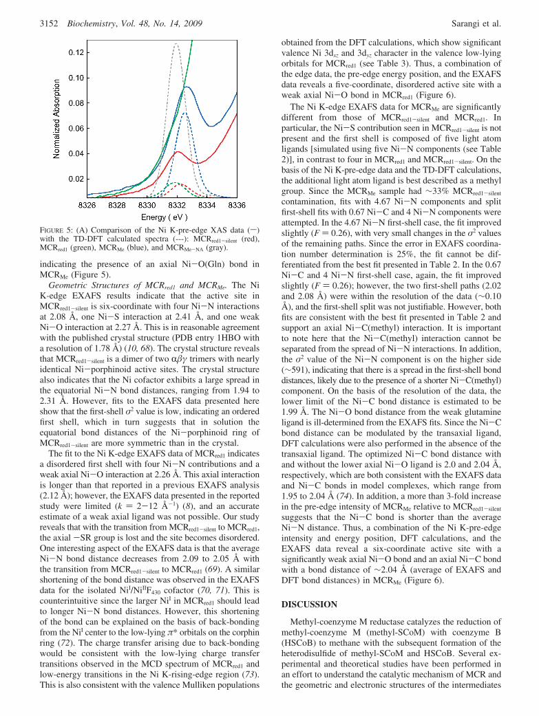
indicating the presence of an axial Ni-O(Gln) bond inMCRMe (Figure 5).
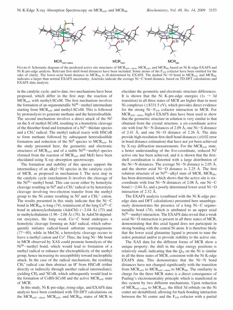
Geometric Structures of MCRred1 and MCRMe. The NiK-edge EXAFS results indicate that the active site inMCRred1-silent is six-coordinate with four Ni-N interactionsat 2.08 Å, one Ni-S interaction at 2.41 Å, and one weakNi-O interaction at 2.27 Å. This is in reasonable agreementwith the published crystal structure (PDB entry 1HBO witha resolution of 1.78 Å) (10, 68). The crystal structure revealsthat MCRred1-silent is a dimer of two R�γ trimers with nearlyidentical Ni-porphinoid active sites. The crystal structurealso indicates that the Ni cofactor exhibits a large spread inthe equatorial Ni-N bond distances, ranging from 1.94 to2.31 Å. However, fits to the EXAFS data presented hereshow that the first-shell σ2 value is low, indicating an orderedfirst shell, which in turn suggests that in solution theequatorial bond distances of the Ni-porphinoid ring ofMCRred1-silent are more symmetric than in the crystal.
The fit to the Ni K-edge EXAFS data of MCRred1 indicatesa disordered first shell with four Ni-N contributions and aweak axial Ni-O interaction at 2.26 Å. This axial interactionis longer than that reported in a previous EXAFS analysis(2.12 Å); however, the EXAFS data presented in the reportedstudy were limited (k ) 2-12 Å-1) (8), and an accurateestimate of a weak axial ligand was not possible. Our studyreveals that with the transition from MCRred1-silent to MCRred1,the axial -SR group is lost and the site becomes disordered.One interesting aspect of the EXAFS data is that the averageNi-N bond distance decreases from 2.09 to 2.05 Å withthe transition from MCRred1-silent to MCRred1 (69). A similarshortening of the bond distance was observed in the EXAFSdata for the isolated NiI/NiIIF430 cofactor (70, 71). This iscounterintuitive since the larger NiI in MCRred1 should leadto longer Ni-N bond distances. However, this shorteningof the bond can be explained on the basis of back-bondingfrom the NiI center to the low-lying π* orbitals on the corphinring (72). The charge transfer arising due to back-bondingwould be consistent with the low-lying charge transfertransitions observed in the MCD spectrum of MCRred1 andlow-energy transitions in the Ni K-rising-edge region (73).This is also consistent with the valence Mulliken populations
obtained from the DFT calculations, which show significantvalence Ni 3dxz and 3dyz character in the valence low-lyingorbitals for MCRred1 (see Table 3). Thus, a combination ofthe edge data, the pre-edge energy position, and the EXAFSdata reveals a five-coordinate, disordered active site with aweak axial Ni-O bond in MCRred1 (Figure 6).
The Ni K-edge EXAFS data for MCRMe are significantlydifferent from those of MCRred1-silent and MCRred1. Inparticular, the Ni-S contribution seen in MCRred1-silent is notpresent and the first shell is composed of five light atomligands [simulated using five Ni-N components (see Table2)], in contrast to four in MCRred1 and MCRred1-silent. On thebasis of the Ni K-pre-edge data and the TD-DFT calculations,the additional light atom ligand is best described as a methylgroup. Since the MCRMe sample had ∼33% MCRred1-silent
contamination, fits with 4.67 Ni-N components and splitfirst-shell fits with 0.67 Ni-C and 4 Ni-N components wereattempted. In the 4.67 Ni-N first-shell case, the fit improvedslightly (F ) 0.26), with very small changes in the σ2 valuesof the remaining paths. Since the error in EXAFS coordina-tion number determination is 25%, the fit cannot be dif-ferentiated from the best fit presented in Table 2. In the 0.67Ni-C and 4 Ni-N first-shell case, again, the fit improvedslightly (F ) 0.26); however, the two first-shell paths (2.02and 2.08 Å) were within the resolution of the data (∼0.10Å), and the first-shell split was not justifiable. However, bothfits are consistent with the best fit presented in Table 2 andsupport an axial Ni-C(methyl) interaction. It is importantto note here that the Ni-C(methyl) interaction cannot beseparated from the spread of Ni-N interactions. In addition,the σ2 value of the Ni-N component is on the higher side(∼591), indicating that there is a spread in the first-shell bonddistances, likely due to the presence of a shorter Ni-C(methyl)component. On the basis of the resolution of the data, thelower limit of the Ni-C bond distance is estimated to be1.99 Å. The Ni-O bond distance from the weak glutamineligand is ill-determined from the EXAFS fits. Since the Ni-Cbond distance can be modulated by the transaxial ligand,DFT calculations were also performed in the absence of thetransaxial ligand. The optimized Ni-C bond distance withand without the lower axial Ni-O ligand is 2.0 and 2.04 Å,respectively, which are both consistent with the EXAFS dataand Ni-C bonds in model complexes, which range from1.95 to 2.04 Å (74). In addition, a more than 3-fold increasein the pre-edge intensity of MCRMe relative to MCRred1-silent
suggests that the Ni-C bond is shorter than the averageNi-N distance. Thus, a combination of the Ni K-pre-edgeintensity and energy position, DFT calculations, and theEXAFS data reveal a six-coordinate active site with asignificantly weak axial Ni-O bond and an axial Ni-C bondwith a bond distance of ∼2.04 Å (average of EXAFS andDFT bond distances) in MCRMe (Figure 6).
DISCUSSION
Methyl-coenzyme M reductase catalyzes the reduction ofmethyl-coenzyme M (methyl-SCoM) with coenzyme B(HSCoB) to methane with the subsequent formation of theheterodisulfide of methyl-SCoM and HSCoB. Several ex-perimental and theoretical studies have been performed inan effort to understand the catalytic mechanism of MCR andthe geometric and electronic structures of the intermediates
FIGURE 5: (A) Comparison of the Ni K-pre-edge XAS data (s)with the TD-DFT calculated spectra (---): MCRred1-silent (red),MCRred1 (green), MCRMe (blue), and MCRMe-NA (gray).
3152 Biochemistry, Vol. 48, No. 14, 2009 Sarangi et al.
in the catalytic cycle, and to date, two mechanisms have beenproposed, which differ in the first step: the reaction ofMCRred1 with methyl-SCoM. The first mechanism involvesthe formation of an organometallic NiIII-methyl intermediatestarting from MCRred1 and methyl-SCoM. This is followedby protonolysis to generate methane and the heterodisulfide.The second mechanism involves a direct attack of the NiI
on the S of methyl-SCoM, resulting in a homolytic cleavageof the thioether bond and formation of a NiII-thiolate speciesand a CH3
• radical. The methyl radical reacts with HSCoBto form methane followed by subsequent heterodisulfideformation and reduction of the NiII species to MCRred1. Inthe study presented here, the geometric and electronicstructures of MCRred1 and of a stable NiIII-methyl species(formed from the reaction of MCRred1 and MeI) have beenelucidated using X-ray absorption spectroscopy.
The formation and stability of this species support theintermediacy of an alkyl-Ni species in the catalytic cycleof MCR, as proposed in mechanism I. The next step inthe catalytic cycle (mechanism I) involves the cleavage ofthe NiIII-methyl bond. This can occur either by homolyticcleavage resulting in NiII and a CH3
• radical or by heterolyticcleavage involving two-electron transfer from the methylgroup to the Ni center resulting in NiI and a CH3
+ cation.The results presented in this study indicate that the Ni-Cbond in MCRMe is long (74), reminiscent of the long CoIII-Cbond in adenosylcobalamin (AdoCbl) (∼2.04 Å) (75) andin methylcobalamin (1.96-2.08 Å) (76). In AdoCbl-depend-ent enzymes, the long weak Co-C bond undergoes ahomolytic cleavage forming an Ado• radical, which subse-quently initiates radical-based substrate rearrangements(77-80), while in MeCbl, a heterolytic cleavage occurs toleave a methyl cation and CoI. Thus, the long Ni-Me bondin MCR observed by XAS could promote homolysis of theNiIII-methyl bond, which would lead to formation of amethyl radical or enhance the electrophilicity of the methylgroup, hence increasing its susceptibility toward nucleophilicattack. In the case of the radical mechanism, the resultingCH3
• radical can then abstract an H• from HSCoB (eitherdirectly or indirectly through another radical intermediate),yielding CH4 and •SCoB, which subsequently would lead tothe formation of CoBS-SCoM and the active MCRred1 stateof MCR.
In this study, Ni K-pre-edge, rising-edge, and EXAFS dataanalysis have been combined with TD-DFT calculations onthe MCRred1-silent, MCRred1, and MCRMe states of MCR to
elucidate the geometric and electronic structure differences.It is shown that the Ni K-pre-edge energies (1s f 3dtransition) in all three states of MCR are higher than in mostNi complexes (e8331.5 eV), which provides direct evidencefor the strong Ni-F430 cofactor interaction in MCR. ForMCRred1-silent, high-k EXAFS data have been used to showthat the geometric structure in solution is very similar to thatobtained from the crystal structure, a six-coordinate activesite with four Ni-N distances of 2.09 Å, one Ni-S distanceof 2.41 Å, and one Ni-O distance of 2.26 Å. The dataprovide high-resolution first-shell bond distances ((0.02 errorin bond distance estimation) that have not yet been achievedby X-ray diffraction measurements. For the MCRred1 state,a better understanding of the five-coordinate, reduced NiI
active site has been achieved, and it is shown that the first-shell coordination is distorted with a large distribution ofthe Ni-N distances. The average Ni-N distance is 2.05 Å,and the shorter axial Ni-O distance is 2.25 Å. The firstsolution structure of an NiIII-alkyl state of MCR, MCRMe,has been determined, which shows that the active site is six-coordinate with four Ni-N distances of 2.08 Å, one Ni-Cbond (∼2.04 Å), and a poorly determined lower axial Ni-Ointeraction of 2.32 Å.
The EXAFS analysis (combined with the Ni K-edge pre-edge data and DFT calculations) presented here unambigu-ously demonstrates the presence of a long Ni-C organo-metallic bond (74), which is attributed to an upper axialNiIII-methyl interaction. The EXAFS data reveal that a weakaxial Ni-O interaction is present in all three states of MCR,demonstrating that this axial ligand does not participate instrong bonding with the central Ni atom. It is therefore likelythat the lower axial glutamine ligand is present to tune theredox potential and/or to provide stability to the active site.
The XAS data for the different forms of MCR show aunique property: the shift in the edge energy positions isrelatively small, indicating that the QNi on the Ni is similarin all the three states of MCR, consistent with the Ni K-edgeEXAFS data. This demonstrates that the Ni-N bonddistances have not changed significantly with the transitionfrom MCRred1 to MCRred1-silent to MCRMe. The similarity incharge for the three MCR states is a direct consequence ofPauling’s electroneutrality principle which is manifested inthis system by two different mechanisms. Upon reductionof MCRred1-silent to MCRred1, the filled 3d orbitals on the Nicenter are destabilized, allowing for back-bonding interactionbetween the Ni center and the F430 cofactor with a partial
FIGURE 6: Schematic diagram of the predicted active site structures of MCRred1-silent, MCRred1, and MCRMe based on Ni K-edge EXAFS andNi K-pre-edge analysis. Relevant first-shell bond distances have been included. Some atoms of the F430 cofactor have been omitted for thesake of clarity. The lower-axial bond distance in MCRMe is ill-determined by EXAFS. The dashed Ni-O bond in MCRred1 and MCRMe
indicates a larger than normal EXAFS uncertainty. Asterisks indicate the average Ni-C bond distance, based on TD-DFT calculations andEXAFS data analysis.
Ni K-Edge X-ray Absorption Spectroscopy on MCRred1 and MCRMe Biochemistry, Vol. 48, No. 14, 2009 3153
flow of charge from the Ni to the F430 orbitals, and hence,the charge on the central Ni atom remains closer to that inMCRred1-silent (closer to NiII than NiI). This is consistent withthe similar Loewdin charges on the Ni center in MCRred1
and MCRred1-silent obtained from DFT calculations (Table 3).Calculations also show a dramatic increase in the Ni 3dyz
and 3dxz hole character due to back-bonding interaction withthe filled π* orbitals on the F430 ring. This increases the totalvalence Ni character, leading to similar charges on the Nicenter in MCRred1 and MCRred1-silent. With the transition fromMCRred1-silent to MCRMe, the additional 3d hole created inMCRMe (NiIII in d7 configuration) undergoes covalent delo-calization over the entire active site, resulting in a speciesthat is best described as [Ni(F430)Me]+. Thus, in this casealso, the charge on the central Ni atom remains closer tothat in MCRred1-silent. Since the total charge on theMCRred1-silent and MCRMe models chosen for the DFTcalculations are different, a direct comparison of the indi-vidual fragment charges will be inaccurate; however, thecombined valence Ni 3d character in MCRred1 (150%) andMCRred1-silent (141%) are similar, with only a small increasefor MCRMe. This, combined with the small edge shift withthe transition from MCRred1-silent to MCRMe, indicates similarcharges in the two states. This noninnocent role of the F430
cofactor in tuning its bonding with the Ni center in differentoxidation states is expected to play a direct role in modulatingthe geometric and electronic structures of the active site andtherefore plays an important role in the catalytic pathway.For example, it might be expected that the NiIII site in MCRMe
would be very unstable due to a high redox potential andmight spontaneously reduce to form a NiII-methyl species.The stability of the MCRMe can be attributed to thenoninnocent participation of the F430 cofactor in bonding,which is consistent with the stability observed in reportedbiochemical studies performed with this state of MCR. Inthe case of MCRred1, the low charge on a formally NiI specieswould be expected to increase the pKa of the coordinatinganionic nitrogen on the F430 ring and destabilize the Ni-Nbond toward dissociation and protonation. Here again, theparticipation of the F430 ring in noninnocent bonding resultsin an increase in QNi and promotes the stability of theMCRred1 species. Thus, the Ni K-edge XAS data indicatethat the F430 cofactor plays a critical role in stabilizing thedifferent forms of MCR and tuning the reactivity of theprotein.
ACKNOWLEDGMENT
SSRL operations are funded by the Department of Energy,Office of Basic Energy Sciences. The SSRL StructuralMolecular Biology program is supported by the NationalInstitutes of Health, National Center for Research Resources,Biomedical Technology Program, and the Department ofEnergy, Office of Biological and Environmental Research.
SUPPORTING INFORMATION AVAILABLE
Figures and table showing the FEFF fits to the Ni K-edgeEXAFS data and their corresponding Fourier transforms ofthe MCRred1-silent subtracted MCRred1 and MCRMe forms andthe metrical parameters, respectively, and a shell-by-shellanalysis of the EXAFS data for MCRred1, MCRred1-silent, and
MCRMe. This material is available free of charge via theInternet at http://pubs.acs.org.
REFERENCES
1. Thauer, R. K. (1998) Biochemistry of methanogenesis: A tributeto Marjory Stephenson. Microbiology 144, 2377–2406.
2. DiMarco, A. A., Bobik, T. A., and Wolfe, R. S. (1990) Unusualcoenzymes of methanogenesis. Annu. ReV. Biochem. 59, 355–394.
3. Ellermann, J., Hedderich, R., Bocher, R., and Thauer, R. K. (1988)The final step in methane formation: Investigations with highlypurified methyl-coenzyme M reductase (component C) fromMethanobacterium thermoautotrophicum (strain Marburg). Eur.J. Biochem. 172, 669–678.
4. Ellefson, W. L., Wolfe, R. S., and Whitman, W. B. (1982) Nickel-containing factor F430: Chromophore of the methyl reductase ofMethanobacterium thermoautotrophicum. Proc. Natl. Acad. Sci.U.S.A. 79, 3707–3710.
5. Farber, G., Keller, W., Kratky, C., Jaun, B., Pfaltz, A., Spinner,C., Kobelt, A., and Eschenmoser, A. (1991) Coenzyme F430 frommethanogenic bacteria: Complete assignment of configuration basedon X-ray analysis of 12,13-Diepi-F430 pentamethyl ester and onNMR spectroscopy. HelV. Chim. Acta 74, 697–716.
6. Shima, S., and Thauer, R. K. (2005) Methyl-coenzyme M reductaseand the anaerobic oxidation of methane in methanotrophic archaea.Curr. Opin. Microbiol. 8, 643–648.
7. Thauer, R. K., and Shima, S. (2008) Methane as fuel for anaerobicmicroorganisms. Ann. N.Y. Acad. Sci. 1125, 158–170.
8. Duin, E. C., Cosper, N. J., Mahlert, F., Thauer, R. K., and Scott,R. A. (2003) Coordination and geometry of the nickel atom inactive methyl-coenzyme M reductase from Methanothermobactermarburgensis as detected by X-ray absorption spectroscopy. J. Biol.Inorg. Chem. 8, 141–148.
9. Signor, L., Knuppe, C., Hug, R., Schweizer, B., Pfaltz, A., andJaun, B. (2000) Methane formation by reaction of a methylthioether with a photo-excited nickel thiolate: A process mimickingmethanogenesis in archaea. Chem.sEur. J. 6, 3508–3516.
10. Grabarse, W., Mahlert, F., Duin, E. C., Goubeaud, M., Shima, S.,Thauer, R. K., Lamzin, V., and Ermler, U. (2001) On themechanism of biological methane formation: Structural evidencefor conformational changes in methyl-coenzyme M reductase uponsubstrate binding. J. Mol. Biol. 309, 315–330.
11. Duin, E. C., and McKee, M. L. (2008) A new mechanism formethane production from methyl-coenzyme M reductase as derivedfrom density functional calculations. J. Phys. Chem. B 112, 2466–2482.
12. Pelmenschikov, V., and Siegbahn, P. E. (2003) Catalysis by methyl-coenzyme M reductase: A theoretical study for heterodisulfideproduct formation. J. Biol. Inorg. Chem. 8, 653–662.
13. Pelmenschikov, V., Blomberg, M. R., Siegbahn, P. E., and Crabtree,R. H. (2002) A mechanism from quantum chemical studies formethane formation in methanogenesis. J. Am. Chem. Soc. 124,4039–4049.
14. Grabarse, W. G., Mahlert, F., Shima, S., Thauer, R. K., and Ermler,U. (2000) Comparison of three methyl-coenzyme M reductasesfrom phylogenetically distant organisms: Unusual amino acidmodification, conservation and adaptation. J. Mol. Biol. 303, 329–344.
15. Tang, Q., Carrington, P. E., Horng, Y. C., Maroney, M. J.,Ragsdale, S. W., and Bocian, D. F. (2002) X-ray absorption andresonance Raman studies of methyl-coenzyme M reductase indicat-ing that ligand exchange and macrocycle reduction accompanyreductive activation. J. Am. Chem. Soc. 124, 13242–13256.
16. Yang, N., Reiher, M., Wang, M., Harmer, J., and Duin, E. (2007)Formation of a nickel-methyl species in methyl-coenzyme Mreductase, an enzyme catalyzing methane formation. J. Am. Chem.Soc. 129, 11028–11029.
17. Dey, M., Telser, J., Kunz, R. C., Lees, N. S., Ragsdale, S. W., andHoffman, B. M. (2007) Biochemical and spectroscopic studies ofthe electronic structure and reactivity of a methyl-Ni species formedon methyl-coenzyme M reductase. J. Am. Chem. Soc. 129, 11030–11032.
18. Kunz, R. C., Horng, Y. C., and Ragsdale, S. W. (2006) Spectro-scopic and kinetic studies of the reaction of bromopropanesulfonatewith methyl-coenzyme M reductase. J. Biol. Chem. 281, 34663–34676.
19. Kunz, R. C., Dey, M., and Ragsdale, S. W. (2008) Characterizationof the thioether product formed from the thiolytic cleavage of the
3154 Biochemistry, Vol. 48, No. 14, 2009 Sarangi et al.
alkyl-nickel bond in methyl-coenzyme M reductase. Biochemistry47, 2661–2667.
20. Dey, M., Kunz, R. C., Lyons, D. M., and Ragsdale, S. W. (2007)Characterization of alkyl-nickel adducts generated by reaction ofmethyl-coenzyme M reductase with brominated acids. Biochemistry46, 11969–11978.
21. Hinderberger, D., Piskorski, R. P., Goenrich, M., Thauer, R. K.,Schweiger, A., Harmer, J., and Jaun, B. (2006) A nickel-alkyl bondin an inactivated state of the enzyme catalyzing methane formation.Angew. Chem., Int. Ed. 45, 3602–3607.
22. Tenderholt, A. Pyspline and QMForge.23. George, G. EXAFSPAK and EDG-FIT.24. Rehr, J., and Albers, R. (2000) Theoretical approaches to X-ray
absorption fine structure. ReV. Mod. Phys. 72, 621–654.25. Mustre de Leon, J., Rehr, J., Zabinsky, S., and Albers, R. (1991)
Ab initio curved wave X-ray absoprtion fine structure. Phys. ReV.B: Condens. Matter Mater. Phys. 44, 4146–4156.
26. Rehr, J., Mustre de Leon, J., Zabinsky, S., and Albers, R. (1991)Theoretical X-ray absorption fine structure standards. J. Am. Chem.Soc. 113, 5135–5140.
27. Ermler, U., Grabarse, W., Shima, S., Goubeaud, M., and Thauer,R. K. (1997) Crystal structure of methyl-coenzyme M reductase:The key enzyme of biological methane formation. Science 278,1457–1462.
28. Stern, E. A. (1993) Number of relevant independent points in X-rayabsorption fine structure spectra. Phys. ReV. B: Condens. MatterMater. Phys. 48, 9825–9827.
29. Pople, J. (2004)Gaussian 03, revision C.02.30. Lee, C., Yang, W., and Parr, R. (1988) Development of the Colle-
Salvetti correlation-energy formula into a functional of the electrondensity. Phys. ReV. B: Condens. Matter Mater. Phys. 37, 785–789.
31. Mihelich, B., Savin, A., Stoll, H., and Preuss, H. (1989) Resultsobtained with the correlation energy density functionals of Beckeand Lee, Yang and Parr. Chem. Phys. Lett. 157, 200–206.
32. Becke, A. (1993) Density functional theromochemistry: 3. The roleof exact exchange. J. Chem. Phys. 98, 5648–5652.
33. Krishnan, R., Binkley, J., Seeger, R., and Pople, J. (1980) Selfconsistent molecular-orbital methods. 20. Basis set for correlatedwave functions. J. Chem. Phys. 72, 650–654.
34. McGrath, M., and Radom, L. (1991) Extension of Gaussian-1 (G1)theory to bromine-containing molecules. J. Chem. Phys. 94, 511–516.
35. Curtiss, L., McGrath, M., Blaudeau, J., Davis, N., BIinning, R.,and Radom, L. (1995) Extension of Gaussian-2 theory to moleculescontaining 3rd row atoms Ga-Kr. J. Chem. Phys. 103, 6104–6113.
36. Rassolov, V. A., Pople, J. A., Ratner, M. A., and Windus, T. L.(1998) 6-31G* basis set for atoms K through Zn. J. Chem. Phys.109, 1223–1229.
37. Hariharan, P., and Pople, J. (1973) The influence of polarizationfunctions on molecular orbital hydrogenation energies. Theor.Chim. Acta 28, 213–222.
38. Francl, M., Pietro, W., Hehre, W., Binkley, J., Gordon, M., DeFrees,D., and Pople, J. (1982) Self consistent molecular-orbital methods.23. A polarization type basis set for 2nd-row elements. J. Chem.Phys. 77, 3654–3665.
39. Neese, F. (2004)ORCA: An ab initio, DFT and semiempiricalElectronic Structure Package.
40. Neese, F., and Olbrich, G. (2002) Efficient use of the resolutionof the identity approximation in time-dependent density functionalcalculations with hybrid density functionals. Chem. Phys. Lett. 362,170–178.
41. Neese, F. (2002) Prediction and interpretation of the Fe-57 isomershift in M. Chim. Acta 337, 181–192.
42. Sinnecker, S., Slep, L., Bill, E., and Neese, F. (2005) Performanceof nonrelativistic and quasi-relativistic hybrid DFT for the predic-tion of electric and magnetic hyperfine parameters in Fe-57 Mspectra. Inorg. Chem. 44, 2245–2254.
43. Schaefer, A., Horn, H., and Ahlrichs, R. (1992) Fully optimizedcontracted Gaussian basis sets for atoms Li to Kr. J. Chem. Phys.97, 2571–2577.
44. Schaefer, A., Huber, C., and Ahlrichs, R. (1994) Fully optimizedcontracted Gaussian basis sets of triple-� valence quality for atomsLi to Kr. J. Chem. Phys. 100, 5829–5835.
45. Krause, M., and Oliver, J. (1979) Natural widths of the atomicK-levels and L-levels, K-alpha X-ray lines and several KLL Augerlines. J. Phys. Chem. Ref. Data 8, 329–338.
46. UV-vis absorption measurements on the MCRred1 and MCRMe
samples showed 36 and 33% MCRred1-silent contamination, respec-
tively. The MCRred1-silent spectrum has been quantitatively sub-tracted from the MCRred1 and MCRMe spectra, and the data havebeen renormalized.
47. Shulman, R. G., Yafet, Y., Eisenberger, P., and Blumberg, W. E.(1976) Observation and interpretation of X-ray absorption edgesin iron compounds and proteins. Proc. Natl. Acad. Sci. U.S.A. 73,1384–1388.
48. Penner-Hahn, J. E., Scott, R. A., Hodgson, K. O., Doniach, S.,Desjardins, S. R., and Solomon, E. I. (1982) Observation of anelectric quadrupole transition in the X-ray absorption spectrum ofa Cu(II) complex. Chem. Phys. Lett. 88, 595–598.
49. Westre, T. E., Kennepohl, P., DeWitt, J. G., Hedman, B., Hodgson,K. O., and Solomon, E. I. (1997) A multiplet analysis of Fe K-edge1s f 3d pre-edge features of iron complexes. J. Am. Chem. Soc.119, 6297–6314.
50. Sarangi, R., Aboelella, N., Fujisawa, K., Tolman, W. B., Hedman,B., Hodgson, K. O., and Solomon, E. I. (2006) X-ray absorptionedge spectroscopy and computational studies on LCu O2 species:Superoxide-CuII versus peroxide-CuIII bonding. J. Am. Chem. Soc.128, 8286–8296.
51. Sarangi, R., Frank, P., Hodgson, K. O., and Hedman, B. (2008)When identical functional groups are not identical: A DFT studyof the effects of molecular environment on sulfur K-edge X-rayabsorption spectra. Inorg. Chim. Acta 361, 956–964.
52. Teo, B. (1986) EXAFS: Basic Principles and Data Analysis,Springer-Verlag, Berlin.
53. R′ denotes the non-phase shift corrected bond distance.54. The traditional E0 value for the Ni K-edge is 8340 eV. However,
as seen in Figure 2, 8340 eV is well below the white line and istoo low to be the threshold energy in the data presented here. Thisis adjusted in the fits with a ∆E0 value of ∼13 eV, which changesthe E0 value in the fits to ∼8353 eV. This value is above the whiteline and hence is a theoretically justifiable choice.
55. Cheesman, M. R., Ankel-Fuchs, D., Thauer, R. K., and Thompson,A. J. (1989) The magnetic properties of the nickel cofactor F430in the enzyme methyl-coenzyme M reductase of Methanobacteriumthermoautotrophicum. Biochem. J. 260, 613–616.
56. Hamilton, C. L., Scott, R. A., and Johnson, M. K. (1989) Themagnetic and electronic properties of Methanobacterium ther-moautotrophicum (strain delta H) methyl coenzyme M reductaseand its nickel tetrapyrrole cofactor F430. A low temperaturemagnetic circular dichroism study. J. Biol. Chem. 264, 11605–11613.
57. Telser, J., Davydov, R., Horng, Y. C., Ragsdale, S. W., andHoffman, B. M. (2001) Cryoreduction of methyl-coenzyme Mreductase: EPR characterization of forms, MCRox1 and MCRred1.J. Am. Chem. Soc. 123, 5853–5860.
58. Craft, J. L., Horng, Y.-C., Ragsdale, S. W., and Brunold, T. C.(2004) Nickel oxidation states of F430 cofactor in methyl-coenzymeM reductase. J. Am. Chem. Soc. 126, 4068–4069.
59. Colpas, G. J., Maroney, M. J., Bagyinka, C., Kumar, M., Willis,W. S., Suib, S. L., Baidya, N., and Mascharak, P. K. (1991) X-rayspectroscopies studies of nickel complexes, with application to thestructure of nickle sites in hydrogenases. Inorg. Chem. 30, 920–928.
60. Szilagyi, R. K., Lim, B. S., Glaser, T., Holm, R. H., Hedman, B.,Hodgson, K. O., and Solomon, E. I. (2003) Description of theground state wave functions of Ni dithiolenes using sulfur K-edgeX-ray absorption spectroscopy. J. Am. Chem. Soc. 125, 9158–9169.
61. Furenlid, L. R., Renner, M. W., Smith, K. M., and Fajer, J. (1990)Structural consequences of nickel versus macrocycle reductionsin F430 models: EXAFS studies of a Ni(I) anion and Ni(II)·π · anion radicals. J. Am. Chem. Soc. 112, 1634–1635.
62. Dey, M., Kunz, R. C., Van Heuvelen, K. M., Craft, J. L., Horng,Y. C., Tang, Q., Bocian, D. F., George, S. J., Brunold, T. C., andRagsdale, S. W. (2006) Spectroscopic and computational studiesof reduction of the metal versus the tetrapyrrole ring of coenzymeF430 from methyl-coenzyme M reductase. Biochemistry 45, 11915–11933.
63. Frank, P., Benfatto, M., Hedman, B., and Hodgson, K. O. (2008)Solution [Cu(amm)]2 + is a strongly solvated square pyramid: Afull account of the copper K-edge XAS spectrum within single-electron theory. Inorg. Chem. 47, 4126–4139.
64. Bair, R. A., and Goddard, W. A. I. (1980) Ab initio studies of theX-ray absorption edge in copper complexes. I. Atomic Cu2+ andCu(II)Cl2. Phys. ReV. B: Condens. Matter Mater. Phys. 22, 2767–2776.
65. Kosugi, N., Yokoyama, T., Asakura, K., and Kuroda, H. (1984)Polarized Cu K-edge XANES of square planar CuCl4
2+ ion.
Ni K-Edge X-ray Absorption Spectroscopy on MCRred1 and MCRMe Biochemistry, Vol. 48, No. 14, 2009 3155
Experimental and theoretical evidence for shake-down phenomena.Chem. Phys. 91, 249–256.
66. Sarangi, R., DeBeer George, S., Rudd, D. J., Szilagyi, R. K., Ribas,X., Rovira, C., Almeida, M., Hodgson, K. O., Hedman, B., andSolomon, E. I. (2007) Sulfur K-edge X-ray absorption spectroscopyas a probe of ligand-metal bond covalency: Metal vs ligandoxidation in copper and nickel dithiolene complexes. J. Am. Chem.Soc. 129, 2316–2326.
67. Craft, J. L., Horng, Y.-C., Ragsdale, S. W., and Brunold, T. C.(2004) Spectroscopic and computational characterization of thenickel-containing F430 cofactor of methyl-coenzyme M reductase.J. Biol. Inorg. Chem. 9, 77–89.
68. The reported average standard deviation in bond distances in 1HBO(Cruickshank’s DPI) is approximately (0.14 Å (although thestandard deviation in the Ni-N bond distances is expected to bebetter), while that for the Ni-N bond distances obtained from theEXAFS data is (0.02 Å.
69. Although the resolution of k ) 2-17 Å EXAFS data is ∼0.1 Å,the standard deviation of the first shell obtained from the EXAFSdata presented here is (0.02 Å.
70. Furenlid, L. R., Renner, M. W., and Fajer, J. (1990) EXAFSstudies of nickel(II) and nickel(I) factor 430M. Conformationalflexibility of the F430 skeleton. J. Am. Chem. Soc. 112, 8987–8989.
71. Shiemke, A. K., Shelnutt, J. A., and Scott, R. A. (1989) Coordina-tion chemistry of F430: Axial ligation equilibrium between square-planar and bis-aquo species in aqueous solution. J. Biol. Chem.264, 11236–11245.
72. Dey, M., Kunz, R. C., Van Heuvelen, K. M., Craft, J. L., Horng,Y.-C., Tang, Q., Bocian, D. F., George, S. J., Brunold, T. C., andRagsdale, S. W. (2006) Spectroscopic and computational studiesof reduction of the metal versus the tetrapyrrole ring of coenzymeF430 from methyl-coenzyme M reductase. Biochemistry 45, 11915–11933.
73. Duin, E. C., Signor, L., Piskorski, R., Mahlert, F., Clay, M. D.,Goenrich, M., Thauer, R. K., Jaun, B., and Johnson, M. K. (2004)Spectroscopic investigation of the nickel-containing porphinoidcofactor F430. Comparison of the free cofactor in the (+)1, (+)2and (+)3 oxidation states with the cofactor bound to methyl-coenzyme M reductase in the silent, red and ox forms. J. Biol.Inorg. Chem. 9, 563–576.
74. On the basis of ∼160 structures submitted to the CambridgeStructure Database (CSD), the average Ni-C(alkyl) bond distancein NiI- and NiII-containing complexes is ∼1.98 Å.
75. Ouyang, L., Rulis, P., Ching, W. Y., Nardin, G., and Randaccio,L. (2004) Accurate redetermination of the X-ray structure andelectronic bonding in adenosylcobalamin. Inorg. Chem. 43, 1235–1241.
76. Drennan, C. L., Huang, S., Drummond, J. T., Matthews, R. G.,and Ludwig, M. L. (1994) How a protein binds B12: A 3.0 ÅX-ray structure of B12-binding domains of methionine synthase.Science 266, 1669–1674.
77. Marsh, E. N., and Drennan, C. L. (2001) Adenosylcobalamin-dependent isomerases: New insights into structure and mechanism.Curr. Opin. Chem. Biol. 5, 499–505.
78. Banerjee, R. (2001) Radical peregrinations catalyzed by coenzymeB12-dependent enzymes. Biochemistry 40, 6191–6198.
79. Ludwig, M. L., and Matthews, R. G. (1997) Structure-basedperspectives on B12-dependent enzymes. Annu. ReV. Biochem. 66,269–313.
80. Warncke, K., Schmidt, J. C., and Ke, S. C. (1999) Identificationof a rearranged-substrate, product radical intermediate and thecontribution of a product radical trap in vitamin B-12 coenzyme-dependent ethanolamine deaminase catalysis. J. Am. Chem. Soc.121, 10522–10528.
BI900087W
3156 Biochemistry, Vol. 48, No. 14, 2009 Sarangi et al.


![Synthesis, Characterization, and Biological Activity of N'(Z)-(3-Methyl-5-oxo-1-phenyl-1,5-dihydro-4H-pyrazol-4ylidene)(phenyl)methyl]benzohydrazide and Its Co(II), Ni(II), and Cu(II)](https://img.pdfslide.net/doc/110x75/631f479fdbf756400702ac0a/synthesis-characterization-and-biological-activity-of-nz-3-methyl-5-oxo-1-phenyl-15-dihydro-4h-pyrazol-4ylidenephenylmethylbenzohydrazide.jpg)


![Methyl 5-bromo-2-[methyl(methylsulfonyl)amino]benzoate](https://img.pdfslide.net/doc/110x75/632436d203238a9ff60acbf8/methyl-5-bromo-2-methylmethylsulfonylaminobenzoate.jpg)