Embed Size (px)
Citation preview
Growth, telomere dynamics and successful and unsuccessful humanaging
Abraham Aviv a,*, Daniel Levy b, Marc Mangel c
a Room F-464, Hypertension Research Center, The Cardiovascular Research Institute, New Jersey Medical School, University of Medicine and Dentistry
of New Jersey, 185 South Orange Ave., Newark, NJ 07103-2714, USAb NHLBI’s and Framingham Heart Study, Framingham, MA, USA
c Department of Applied Mathematics and Statistics, Jack Baskin School of Engineering, University of California, Santa Cruz, CA, USA
Received 28 February 2003; received in revised form 23 April 2003; accepted 15 May 2003
Mechanisms of Ageing and Development 124 (2003) 829�/837
www.elsevier.com/locate/mechagedev
Abstract
This paper links mass trajectories with telomere dynamics to construct theoretical models of successful and unsuccessful aging in
human beings. It couples parameters of telomere length in somatic cells, as expressed by the terminal restriction fragment (TRF), at
birth and the rate of telomere attrition thereafter with nonlinear models of somatic growth to predict the probability of surviving
disease free, based on the assumption that telomere length in replicating somatic cells is a surrogate indicator of aging determinants
in humans. The models capture aspects of individual variation in successful and unsuccessful aging and the long-term consequences
of rapid growth early in life.
# 2003 Elsevier Ireland Ltd. All rights reserved.
Keywords: Telomeres; Aging; Growth; Somatic cells; Replication
1. Introduction
In modern humans, aging portends an escalating risk
of developing age-related disorders, including cardio-
vascular disease, diabetes mellitus, cancer, dementia,
and a host of degenerative diseases which ultimately
lead to death. Accordingly, aging has been frequently
defined in terms of the increased probability of death,
i.e. the risk of dying as a function of age (Partridge and
Mangel, 1999). The endpoint of aging is, no doubt,
death, but defining this complex biological process by its
ultimate outcome leaves a considerable void with respect
to a central question in gerontology. That question*/
how to explain and predict disorders of aging in the
context of the biological meaning of aging*/has con-
siderable clinical relevance. Such disorders arise from
progressive dysfunctions in metabolism, particularly
impaired tissue repair capacity, leading to degeneration
of tissues and organs*/a phenomenon expressed by the
cumulative compromise that ultimately results in death.There is, however, considerable variation in the rate
of progression of aging in humans. Perhaps one way to
explore the underlying reasons for this variation is to
distinguish unsuccessful from successful aging. In un-
successful aging, age-related disorders appear to proceed
faster or start earlier than in successful aging. Conse-
quently, the clinical manifestations of these disorders are
more severe and expressed at a younger (chronological)
age, causing premature disability and death. Unsuccess-
ful aging may hence be viewed as a process of early onset
aging or accelerated aging and it could occur in isolation
in one organ system, progress independently in a
number of organ systems, or proceed uniformly in the
body as a whole.
Given the enormous complexity of organisms, a
simple biological model of aging is obviously inadequate
for all purposes. Nevertheless, such a model may guide
the design of both empirical and experimental work,
facilitating the distinction between unsuccessful and
successful aging. In this paper, we combine clinical
observations and theoretical biology to construct a
* Corresponding author. Tel.: �/1-973-972-5280; fax: �/1-973-972-
5576.
E-mail address: [email protected] (A. Aviv).
0047-6374/03/$ - see front matter # 2003 Elsevier Ireland Ltd. All rights reserved.
doi:10.1016/S0047-6374(03)00143-X
mathematical model that links recent advances in
modeling growth (West et al., 2001) with insight gained
into telomere biology (Blackburn, 2000; Klapper et al.,
2001; de Lange, 2002) and its potential link with thebiology of human aging (Aviv, 2002). The model, we
suggest, points to new ways of looking at and searching
for the biological mechanisms of aging.
1.1. Telomeres, somatic cell replication and replicative
senescence in vitro
Human telomeres consist of many kilobases of
TTAGGG tandem repeats and telomeric binding pro-teins, which together cap the ends of chromosomes and
protect them from degradation and end-to-end fusion
(Blackburn, 2000; Klapper et al., 2001; Chan and
Blackburn, 2002; de Lange, 2002; Kim et al., 2002).
Because of the end replication problem (Watson, 1972;
Olovnikov, 1973), telomeres undergo erosion with each
cycle of replication of cultured somatic cells, until the
telomere length (probably telomere length in thosechromosomes with the shortest telomeres) becomes
sufficiently shortened. At this stage, a poorly under-
stood signal triggers cessation of replication, i.e. repli-
cative senescence (Wright and Shay, 2001). Thus,
telomere length is not only a record of replicative
history but also an index of replicative potential of
cultured somatic cells from humans. Telomere attrition
may also affect the silencing of gene expression in thegene-rich sub-telomeric regions, thereby modifying cell
biology before the onset of replicative senescence (Baur
et al., 2001). Recent data suggest that protecting the
chromosomes from end-fusion events may not be strictly
related to telomere length but to the configuration of
telomeric DNA in relation to telomeric binding proteins
(Blackburn, 2000; Chan and Blackburn, 2002; de Lange,
2002; Karlseder et al., 2002). Critically shortenedtelomeres evidently trigger replicative senescence be-
cause they lose the protective shield of their telomere
binding proteins.
Telomere attrition in cultured somatic cells can be
influenced by at least six processes: (a) the initial
telomere length in the primary (progenitor) cells; (b)
the rate of replication; (c) the length of the G-rich 3?single stranded telomere overhangs (SSOs) (Wright etal., 1997; Huffman et al., 2000), which determines how
much DNA is lost per division; (d) the regulation of
telomerase, (Bodnar et al., 1998; Morales et al., 1999;
Shay et al., 2001) which is expressed in many stem cells
and which slows but does not prevent telomere short-
ening; (e) the pattern of cell division (the fraction of self-
renewal vs. generation of transient amplifying cells); and
(f) the contribution of other factors such as oxidativedamage which can contribute to telomere shortening in
a fashion not strictly related to the number of divisions
(von Zglinicki et al., 2000; Serra et al., 2003). In this
manuscript we will explore the potential influence of the
first three factors. In addition, a number of non-
telomerase telomere binding proteins, including TRF2
(in telomerase-negative cells), and TRF1, TIN2 andhRAP1 (in telomerase-positive cells) are involved in
stabilizing telomere structure and regulating telomere
erosion (van Steensel and de Lange, 1997; de Lange,
2002; Kim et al., 2002). Overall, these experimental
observations are in line with the idea that telomeres are
a mitotic clock in cultured somatic cells from humans
and that telomere length reflects, in part, the replicative
history of these cells (Harley et al., 1992).
1.2. Telomere dynamics in vivo
The following features mark human telomere biology
in vivo. During intra-uterine life, telomere length is
stable and synchronized (i.e. it is approximately the
same) in different organs and tissues, evidently because
of robust telomerase activity during different phases of
intra-uterine growth (Wright et al., 1996; Youngren etal., 1998) telomere length is inversely related to the
donor age in tissues that undergo proliferation (Allsopp
et al., 1992; Slagboom et al., 1994; Kitada et al., 1995;
Jeanclos et al., 1998, 2000; Okuda et al., 2000; Aviv et
al., 2001; Benetos et al., 2001); telomere length is highly
heritable (Slagboom et al., 1994; Jeanclos et al., 2000),
longer in women than men (Jeanclos et al., 2000;
Benetos et al., 2001) and is highly variable amonghumans; this variability is observed at birth and there-
after (Slagboom et al., 1994; Okuda et al., 2002).
Although cells from highly proliferative somatic tissues
may express some telomerase activity, this activity is
insufficient to prevent age-dependent telomere attrition.
During extra-uterine life, much of the age-dependent
telomere attrition probably reflects cell replication in the
soma. Two lines of evidence further support this idea:post-mitotic, poorly proliferative tissues such as skeletal
muscle show little or no telomere erosion with age
(Decary et al., 1997). Despite considerable proliferation,
male germlines, which also express high telomerase
activity, show no age-dependent telomere erosion (All-
sopp et al., 1992; Wright et al., 1996).
1.3. Telomeres, chronological age and biological age
Telomere dynamics may serve as an index of biolo-
gical age if: (a) the cumulative number of replications of
somatic cells; (b) the rate of somatic cell replication; (c)
other factors such as the cumulative oxidative stress; or
(d) any combination of these variables register growth
and aging of humans. Biological age is different from
chronological age*/a fixed quantity that is based on thecalendar*/in that biological age reflects inter-individual
variation in rates and expressions of growth and aging.
If telomere dynamics simply tracked chronological age,
A. Aviv et al. / Mechanisms of Ageing and Development 124 (2003) 829�/837830
telomeres would provide little information beyond that
of chronological age about the biology of human aging.
Deciphering the reasons for variation in telomere
dynamics among humans may thus provide a betterunderstanding of successful and unsuccessful aging.
White blood cells (WBCs) are proliferative cells and
are readily available from humans. These cells have been
used to explore whether telomere dynamics can provide
information, additional to chronological age, about
susceptibility to disorders of aging.
The primary focus has been on the relationship
between telomere length in WBCs and indices ofcardiovascular aging. One of these indices is pulse
pressure, which is a reliable indicator of the biological
aging of central arteries such as the aorta. Elevated pulse
pressure entails a heightened risk for cardiovascular
diseases, particularly, atherosclerotic coronary heart
disease (Franklin et al., 1999, 2001; Lakatta and Levy,
2003). Telomere length, as expressed in WBCs, is
inversely correlated with pulse pressure, so that evenafter age adjustment, individuals with a shorter telomere
length are likely to show a higher pulse pressure than
their peers (Jeanclos et al., 2000; Benetos et al., 2001).
This may explain why individuals with shorter telomere
length than their age-matched peers may have an
increased risk for atherosclerosis (Samani et al., 2001).
Short, age adjusted telomere lengths in WBCs or subsets
of WBCs have been also observed in individualsdiagnosed with different forms of dementia (von Zgli-
nicki et al., 2000; Panossian et al., 2003). It follows that
a relatively short telomere length in WBCs may denote
unsuccessful aging and an increased risk for premature
death from age-related disorders (Cawthon et al., 2003).
Given that telomere dynamics may provide clues
regarding the biology of aging, a model linking telomere
dynamics with successful versus unsuccessful humanaging is in order.
1.4. A modeling framework and its predictions
The extreme boundaries of the continuum that
comprises lifespan, i.e. growth and aging, are clearly
defined by conception (the beginning) and death (the
end). Other than these boundaries, there are no clear-cut
demarcations between phases of the continuum. Ma-turation is evidently a phase bridging development with
aging. In general, however, there are no distinct set-
points that can be used to clearly and precisely mark
transitions from growth to maturation and from ma-
turation to aging (i.e. maturation and aging are not
knife-edge processes but are continuous). Consequently,
it is impossible to define accurately, in temporal terms,
phases within the continuum. Instead, one might viewgrowth and aging as dynamic, but partially overlapping
processes, from both the temporal and functional
perspectives.
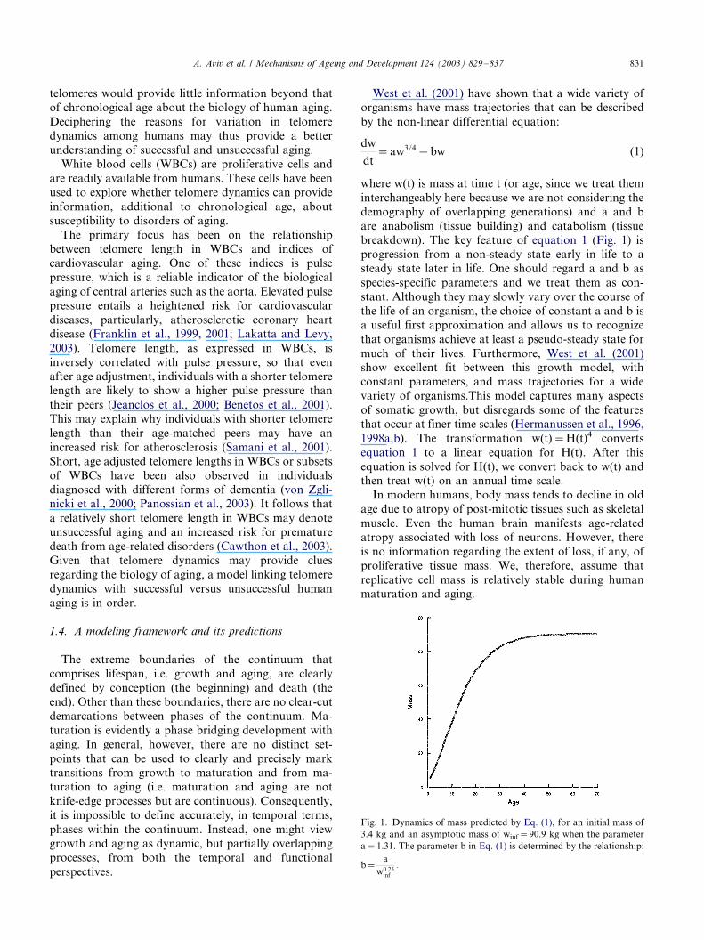
West et al. (2001) have shown that a wide variety of
organisms have mass trajectories that can be described
by the non-linear differential equation:
dw
dt�aw3=4�bw (1)
where w(t) is mass at time t (or age, since we treat them
interchangeably here because we are not considering the
demography of overlapping generations) and a and b
are anabolism (tissue building) and catabolism (tissue
breakdown). The key feature of equation 1 (Fig. 1) isprogression from a non-steady state early in life to a
steady state later in life. One should regard a and b as
species-specific parameters and we treat them as con-
stant. Although they may slowly vary over the course of
the life of an organism, the choice of constant a and b is
a useful first approximation and allows us to recognize
that organisms achieve at least a pseudo-steady state for
much of their lives. Furthermore, West et al. (2001)show excellent fit between this growth model, with
constant parameters, and mass trajectories for a wide
variety of organisms.This model captures many aspects
of somatic growth, but disregards some of the features
that occur at finer time scales (Hermanussen et al., 1996,
1998a,b). The transformation w(t)�/H(t)4 converts
equation 1 to a linear equation for H(t). After this
equation is solved for H(t), we convert back to w(t) andthen treat w(t) on an annual time scale.
In modern humans, body mass tends to decline in old
age due to atropy of post-mitotic tissues such as skeletal
muscle. Even the human brain manifests age-related
atropy associated with loss of neurons. However, there
is no information regarding the extent of loss, if any, of
proliferative tissue mass. We, therefore, assume that
replicative cell mass is relatively stable during humanmaturation and aging.
Fig. 1. Dynamics of mass predicted by Eq. (1), for an initial mass of
3.4 kg and an asymptotic mass of winf�/90.9 kg when the parameter
a�/1.31. The parameter b in Eq. (1) is determined by the relationship:
b�a
w0:25inf
:/
A. Aviv et al. / Mechanisms of Ageing and Development 124 (2003) 829�/837 831
In order to link mass trajectories with telomere
dynamics, we incorporate into the model the concept
that telomere attrition reflects the replicative history of
somatic cells. West et al. (2001) show that the para-meters a and b can be interpreted as:
a�B0mc
Ec
and b�B0
Ec
(2)
where B0 is a taxon dependent constant; mc, the mass of
a single cell and its associated extracellular matrix; Ec,
the energy required to make a single cell and Bc is the
metabolic rate of a single cell. At any age, w(t)�/mcNc,
where w is the mass and Nc(t) is the number of cells
comprising the organism. Consequently, if one assumes
that in replicating tissues of the soma hypertrophy
(growth related to an increase in cell mass) is smallrelative to hyperplasia (growth related to an increase in
cell number), changes in mass of these tissues are
reflected linearly in changes in the number of cells. In
addition, sustaining tissue growth through hypertrophy
still requires an increase in vascular supply and perhaps
in nutrient absorptive capacity via the gastrointestinal
tract. One, hence, anticipates that cells in these and
related tissues would have an increase proliferative rateto accommodate growth and turnover. Similarly, at
steady state (i.e. near asymptotic size), maintenance
(housekeeping) of the soma requires the turnover of a
fraction of cells.
To describe telomere dynamics in vivo, let L(t) in
equation 3 denote the average telomeric length (as
expressed by the mean length of the terminal restriction
fragments) at age t, and assume that telomere dynamicsis given by the discrete time (annual) dynamics:
L(t�1)�L(t)�s
�Fg
w(t � 1) � w(t)
w(t)�Fmw(t)
�(3)
where s is an annualized measure of telomere repeats
lost per cell division (TRLPD), and Fg and Fm,
respectively, relate to TRLPD as a result of cellular
proliferation that promotes growth and turnover asso-
ciated with housekeeping. We consider Fg and Fm to be
species-specific constants, much as is B0 in the growth
model.
Recent studies found that the rate of TRLPD isproportional to the length of the SSOs (Wright et al.,
1997; Huffman et al., 2000). It is possible, therefore, that
in vivo variation in age-dependent telomere erosion may
largely relate to the length of the SSOs. For this reason,
we incorporated into our model the length of the SSOs,
based on available data. For purposes of computation,
the length of the SSO is assumed to range from 0.075 to
0.175 kb, with a mean of 0.125 kb and a coefficient ofvariation of 20%. The rate of loss of telomeres at
asymptotic size is assumed to be 0.03 kb/year (Allsopp
et al., 1992; Slagboom et al., 1994; Kitada et al., 1995;
Jeanclos et al., 1998, 2000; Okuda et al., 2000; Aviv et
al., 2001; Benetos et al., 2001; Okuda et al., 2002) when
SSO length�/0.125 kb. However, as expressed in WBCs,
early in life the rate of loss of telomeres is much higher(Frenck et al., 1998; Rufer et al., 1999; Zeichner et al.,
1999) and the early life loss rate allows us to determine
Fg. We note in this regard that the etiology of rapid
telomere attrition in early life is unclear. It has been
attributed to an increased cellular turnover, but in
theory it can relate to a host of factors among them a
longer SSO length. In our model, we assumed that the
SSO length is constant throughout life.We have thus specified the dynamics of proliferative
somatic growth and of telomeric attrition. We now
assume that telomeric length is a biomarker for the
probability of disease, regardless of whether in itself
telomere dynamics is a determinant in the biology of
aging or a surrogate indicator of other aging determi-
nants. The relationship between the annual probability
of remaining disease free and telomere length L(t) at thestart of a year is currently unknown (indeed, one role of
theory is to point out what needs to be measured).
Consequently, we modeled it as exp(�/0.8(L(t)�/3)), for
the following reasons. First, the exponential distribution
is the simplest of all survival distributions; hence it is a
parsimonious choice. Second, with the exponential
model, the probability of remaining disease free depends
only upon current telomere length, and not the dy-namics of age-dependent telomere attrition, thus making
it a practical choice for empirical studies. The para-
meters 0.8 and 3 were chosen by coupling the model for
the probability of disease development with the mass
and telomere length dynamics for an individual with
mean initial telomere length and mean loss of telomere
repeats per cell division to give a probability of about
50% of reaching age 70 disease free.We applied this model to a population of 500
individuals, assuming that all individuals have the
same growth trajectory, but differed in initial telomere
length (which we assumed was normally distributed with
mean 11 kb (33) and standard deviation 1.2 kb). We
were thus able to generate a survival trajectory for the
population, while at the same time following the
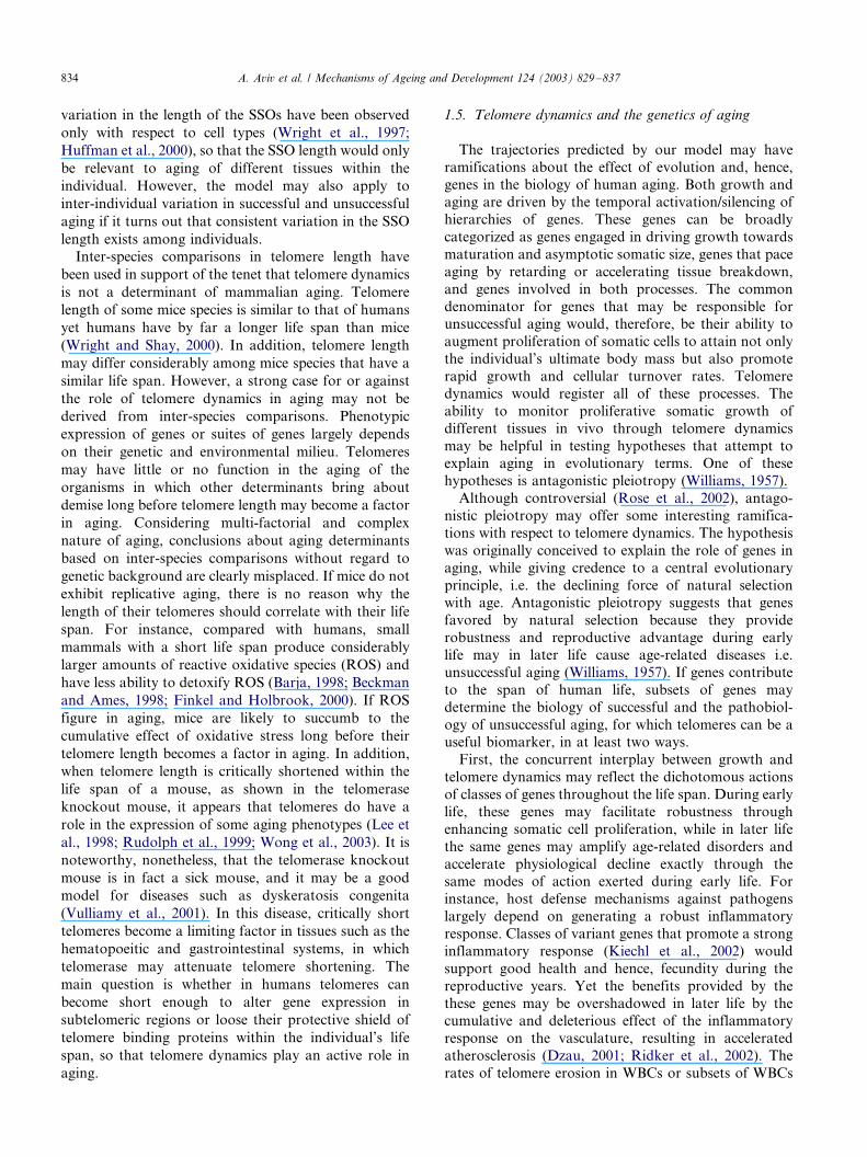
histories of individuals.To illustrate the latter point, we randomly picked five
individuals from the sample of 500. In Table 1, we
present the TRLPD(SS0), initial telomere length and the
probability that each individual survives to age 45 free
of age-related disease. Note that both initial telomere
length and TRLPD determine this probability. We can
then pose the question: given that an individual has
reached age 45 without clinical findings of cardiovas-cular disease, diabetes, cancer, etc., what is the predicted
probability of surviving to future ages free of age-related
diseases (Fig. 2). The results in this figure are analogous
to the emperical ones, in which individuals were
A. Aviv et al. / Mechanisms of Ageing and Development 124 (2003) 829�/837832
separated into groups that had longer or shorter
telomeres (Cawthon et al., 2003). This model allows us
to be more precise about such relationships.
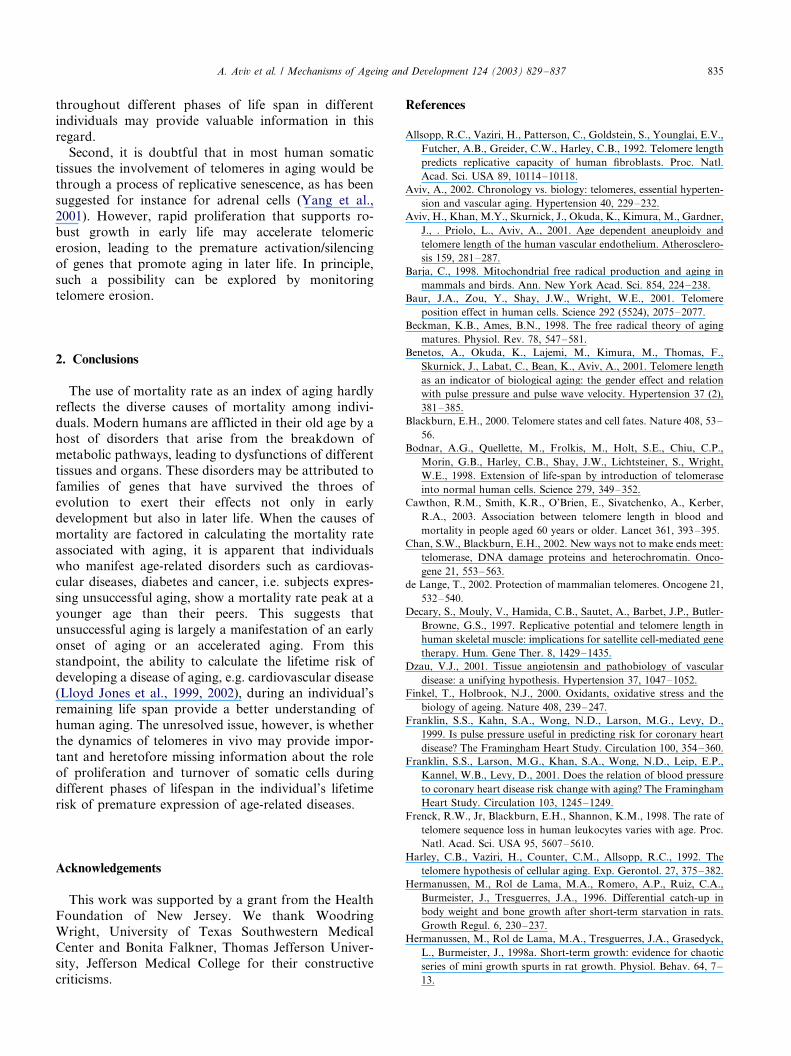
We also conducted a computer experiment to simulate
accelerated growth during early development. To do
this, we held birth mass constant, varied asymptotic size,
and assumed that all individuals had the same initial
telomere length and loss of telomere repeats per cell
division (since telomere length appears unchanged in
utero (Youngren et al., 1998)). In Fig. 3, we show the
relationship between growth rate to age 11 (defined as
[w(11)�/w(0)]/w(0)]) and the probability of reaching age
65 disease free.Figs. 2 and 3 suggest a variety of scenarios that may
define unsuccessful and successful aging from the
perspective of replicating somatic cells, organs, tissues,
or the organism as a whole. One obvious scenario is that
unsuccessful aging may be the result of deviations from
the growth trajectory that lead to accelerated growth
and that the ultimate body size is a determinant in
successful versus unsuccessful aging. It is, hence, note-
worthy that a body of epidemiological data suggests that
height inversely correlated with longevity (Samaras et
al., 2003). Furthermore, the results suggest additional
scenarios linking telomere biology to aging. They
include: an increase in proliferative, non-malignant
growth of specific somatic tissues, an increase in cellular
turnover associated with housekeeping, an increase in
tissue breakdown that generates a cellular response
resulting in enhanced cell replication, and different
combinations of these possibilities. Of relevence to
people in industrialized societies, particularly the USA,
is an increase in body mass due to obesity. Gain in
adiposity (fat mass) during adult life is attained primar-
ily through hypertrophy of fat cells. However, as
indicated earlier regarding hypertrophic growth, the
increase in fat mass may be accompanied by consider-
able angiogenesis to accommodate the metabolic needs
of the growing fat tissue (Samad et al., 1998; Li et al.,
2002; Rupnick et al., 2002). This entails proliferative
growth of vascular cells and hence telomeric erosion.
The vascular endothelium of newly formed fat mass may
hence exhibit more aged features, which might contri-
bute, for instance, to insulin resistance, associated with
obesity.If telomere length itself is not only a biomarker but
also a determinant in aging, we predict that short
telomeres length at birth and a greater TRLPD will be
ultimately linked with unsuccessful aging. If TRLPD is
correlated with the SSO length, the SSO length may be a
factor in unsuccessful aging. To date, data about
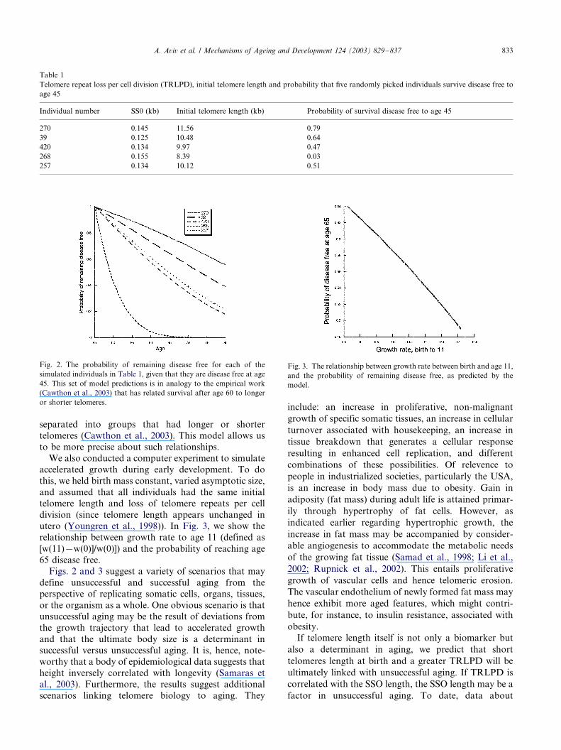
Table 1
Telomere repeat loss per cell division (TRLPD), initial telomere length and probability that five randomly picked individuals survive disease free to
age 45
Individual number SS0 (kb) Initial telomere length (kb) Probability of survival disease free to age 45
270 0.145 11.56 0.79
39 0.125 10.48 0.64
420 0.134 9.97 0.47
268 0.155 8.39 0.03
257 0.134 10.12 0.51
Fig. 2. The probability of remaining disease free for each of the
simulated individuals in Table 1, given that they are disease free at age
45. This set of model predictions is in analogy to the empirical work
(Cawthon et al., 2003) that has related survival after age 60 to longer
or shorter telomeres.
Fig. 3. The relationship between growth rate between birth and age 11,
and the probability of remaining disease free, as predicted by the
model.
A. Aviv et al. / Mechanisms of Ageing and Development 124 (2003) 829�/837 833
variation in the length of the SSOs have been observed
only with respect to cell types (Wright et al., 1997;
Huffman et al., 2000), so that the SSO length would only
be relevant to aging of different tissues within the
individual. However, the model may also apply to
inter-individual variation in successful and unsuccessful
aging if it turns out that consistent variation in the SSO
length exists among individuals.
Inter-species comparisons in telomere length have
been used in support of the tenet that telomere dynamics
is not a determinant of mammalian aging. Telomere
length of some mice species is similar to that of humans
yet humans have by far a longer life span than mice
(Wright and Shay, 2000). In addition, telomere length
may differ considerably among mice species that have a
similar life span. However, a strong case for or against
the role of telomere dynamics in aging may not be
derived from inter-species comparisons. Phenotypic
expression of genes or suites of genes largely depends
on their genetic and environmental milieu. Telomeres
may have little or no function in the aging of the
organisms in which other determinants bring about
demise long before telomere length may become a factor
in aging. Considering multi-factorial and complex
nature of aging, conclusions about aging determinants
based on inter-species comparisons without regard to
genetic background are clearly misplaced. If mice do not
exhibit replicative aging, there is no reason why the
length of their telomeres should correlate with their life
span. For instance, compared with humans, small
mammals with a short life span produce considerably
larger amounts of reactive oxidative species (ROS) and
have less ability to detoxify ROS (Barja, 1998; Beckman
and Ames, 1998; Finkel and Holbrook, 2000). If ROS
figure in aging, mice are likely to succumb to the
cumulative effect of oxidative stress long before their
telomere length becomes a factor in aging. In addition,
when telomere length is critically shortened within the
life span of a mouse, as shown in the telomerase
knockout mouse, it appears that telomeres do have a
role in the expression of some aging phenotypes (Lee et
al., 1998; Rudolph et al., 1999; Wong et al., 2003). It is
noteworthy, nonetheless, that the telomerase knockout
mouse is in fact a sick mouse, and it may be a good
model for diseases such as dyskeratosis congenita
(Vulliamy et al., 2001). In this disease, critically short
telomeres become a limiting factor in tissues such as the
hematopoeitic and gastrointestinal systems, in which
telomerase may attenuate telomere shortening. The
main question is whether in humans telomeres can
become short enough to alter gene expression in
subtelomeric regions or loose their protective shield of
telomere binding proteins within the individual’s life
span, so that telomere dynamics play an active role in
aging.
1.5. Telomere dynamics and the genetics of aging
The trajectories predicted by our model may have
ramifications about the effect of evolution and, hence,genes in the biology of human aging. Both growth and
aging are driven by the temporal activation/silencing of
hierarchies of genes. These genes can be broadly
categorized as genes engaged in driving growth towards
maturation and asymptotic somatic size, genes that pace
aging by retarding or accelerating tissue breakdown,
and genes involved in both processes. The common
denominator for genes that may be responsible forunsuccessful aging would, therefore, be their ability to
augment proliferation of somatic cells to attain not only
the individual’s ultimate body mass but also promote
rapid growth and cellular turnover rates. Telomere
dynamics would register all of these processes. The
ability to monitor proliferative somatic growth of
different tissues in vivo through telomere dynamics
may be helpful in testing hypotheses that attempt toexplain aging in evolutionary terms. One of these
hypotheses is antagonistic pleiotropy (Williams, 1957).
Although controversial (Rose et al., 2002), antago-
nistic pleiotropy may offer some interesting ramifica-
tions with respect to telomere dynamics. The hypothesis
was originally conceived to explain the role of genes in
aging, while giving credence to a central evolutionary
principle, i.e. the declining force of natural selectionwith age. Antagonistic pleiotropy suggests that genes
favored by natural selection because they provide
robustness and reproductive advantage during early
life may in later life cause age-related diseases i.e.
unsuccessful aging (Williams, 1957). If genes contribute
to the span of human life, subsets of genes may
determine the biology of successful and the pathobiol-
ogy of unsuccessful aging, for which telomeres can be auseful biomarker, in at least two ways.
First, the concurrent interplay between growth and
telomere dynamics may reflect the dichotomous actions
of classes of genes throughout the life span. During early
life, these genes may facilitate robustness through
enhancing somatic cell proliferation, while in later life
the same genes may amplify age-related disorders and
accelerate physiological decline exactly through thesame modes of action exerted during early life. For
instance, host defense mechanisms against pathogens
largely depend on generating a robust inflammatory
response. Classes of variant genes that promote a strong
inflammatory response (Kiechl et al., 2002) would
support good health and hence, fecundity during the
reproductive years. Yet the benefits provided by the
these genes may be overshadowed in later life by thecumulative and deleterious effect of the inflammatory
response on the vasculature, resulting in accelerated
atherosclerosis (Dzau, 2001; Ridker et al., 2002). The
rates of telomere erosion in WBCs or subsets of WBCs
A. Aviv et al. / Mechanisms of Ageing and Development 124 (2003) 829�/837834
throughout different phases of life span in different
individuals may provide valuable information in this
regard.
Second, it is doubtful that in most human somatictissues the involvement of telomeres in aging would be
through a process of replicative senescence, as has been
suggested for instance for adrenal cells (Yang et al.,
2001). However, rapid proliferation that supports ro-
bust growth in early life may accelerate telomeric
erosion, leading to the premature activation/silencing
of genes that promote aging in later life. In principle,
such a possibility can be explored by monitoringtelomere erosion.
2. Conclusions
The use of mortality rate as an index of aging hardly
reflects the diverse causes of mortality among indivi-
duals. Modern humans are afflicted in their old age by ahost of disorders that arise from the breakdown of
metabolic pathways, leading to dysfunctions of different
tissues and organs. These disorders may be attributed to
families of genes that have survived the throes of
evolution to exert their effects not only in early
development but also in later life. When the causes of
mortality are factored in calculating the mortality rate
associated with aging, it is apparent that individualswho manifest age-related disorders such as cardiovas-
cular diseases, diabetes and cancer, i.e. subjects expres-
sing unsuccessful aging, show a mortality rate peak at a
younger age than their peers. This suggests that
unsuccessful aging is largely a manifestation of an early
onset of aging or an accelerated aging. From this
standpoint, the ability to calculate the lifetime risk of
developing a disease of aging, e.g. cardiovascular disease(Lloyd Jones et al., 1999, 2002), during an individual’s
remaining life span provide a better understanding of
human aging. The unresolved issue, however, is whether
the dynamics of telomeres in vivo may provide impor-
tant and heretofore missing information about the role
of proliferation and turnover of somatic cells during
different phases of lifespan in the individual’s lifetime
risk of premature expression of age-related diseases.
Acknowledgements
This work was supported by a grant from the Health
Foundation of New Jersey. We thank Woodring
Wright, University of Texas Southwestern MedicalCenter and Bonita Falkner, Thomas Jefferson Univer-
sity, Jefferson Medical College for their constructive
criticisms.
References
Allsopp, R.C., Vaziri, H., Patterson, C., Goldstein, S., Younglai, E.V.,
Futcher, A.B., Greider, C.W., Harley, C.B., 1992. Telomere length
predicts replicative capacity of human fibroblasts. Proc. Natl.
Acad. Sci. USA 89, 10114�/10118.
Aviv, A., 2002. Chronology vs. biology: telomeres, essential hyperten-
sion and vascular aging. Hypertension 40, 229�/232.
Aviv, H., Khan, M.Y., Skurnick, J., Okuda, K., Kimura, M., Gardner,
J., . Priolo, L., Aviv, A., 2001. Age dependent aneuploidy and
telomere length of the human vascular endothelium. Atherosclero-
sis 159, 281�/287.
Barja, C., 1998. Mitochondrial free radical production and aging in
mammals and birds. Ann. New York Acad. Sci. 854, 224�/238.
Baur, J.A., Zou, Y., Shay, J.W., Wright, W.E., 2001. Telomere
position effect in human cells. Science 292 (5524), 2075�/2077.
Beckman, K.B., Ames, B.N., 1998. The free radical theory of aging
matures. Physiol. Rev. 78, 547�/581.
Benetos, A., Okuda, K., Lajemi, M., Kimura, M., Thomas, F.,
Skurnick, J., Labat, C., Bean, K., Aviv, A., 2001. Telomere length
as an indicator of biological aging: the gender effect and relation
with pulse pressure and pulse wave velocity. Hypertension 37 (2),
381�/385.
Blackburn, E.H., 2000. Telomere states and cell fates. Nature 408, 53�/
56.
Bodnar, A.G., Quellette, M., Frolkis, M., Holt, S.E., Chiu, C.P.,
Morin, G.B., Harley, C.B., Shay, J.W., Lichtsteiner, S., Wright,
W.E., 1998. Extension of life-span by introduction of telomerase
into normal human cells. Science 279, 349�/352.
Cawthon, R.M., Smith, K.R., O’Brien, E., Sivatchenko, A., Kerber,
R.A., 2003. Association between telomere length in blood and
mortality in people aged 60 years or older. Lancet 361, 393�/395.
Chan, S.W., Blackburn, E.H., 2002. New ways not to make ends meet:
telomerase, DNA damage proteins and heterochromatin. Onco-
gene 21, 553�/563.
de Lange, T., 2002. Protection of mammalian telomeres. Oncogene 21,
532�/540.
Decary, S., Mouly, V., Hamida, C.B., Sautet, A., Barbet, J.P., Butler-
Browne, G.S., 1997. Replicative potential and telomere length in
human skeletal muscle: implications for satellite cell-mediated gene
therapy. Hum. Gene Ther. 8, 1429�/1435.
Dzau, V.J., 2001. Tissue angiotensin and pathobiology of vascular
disease: a unifying hypothesis. Hypertension 37, 1047�/1052.
Finkel, T., Holbrook, N.J., 2000. Oxidants, oxidative stress and the
biology of ageing. Nature 408, 239�/247.
Franklin, S.S., Kahn, S.A., Wong, N.D., Larson, M.G., Levy, D.,
1999. Is pulse pressure useful in predicting risk for coronary heart
disease? The Framingham Heart Study. Circulation 100, 354�/360.
Franklin, S.S., Larson, M.G., Khan, S.A., Wong, N.D., Leip, E.P.,
Kannel, W.B., Levy, D., 2001. Does the relation of blood pressure
to coronary heart disease risk change with aging? The Framingham
Heart Study. Circulation 103, 1245�/1249.
Frenck, R.W., Jr, Blackburn, E.H., Shannon, K.M., 1998. The rate of
telomere sequence loss in human leukocytes varies with age. Proc.
Natl. Acad. Sci. USA 95, 5607�/5610.
Harley, C.B., Vaziri, H., Counter, C.M., Allsopp, R.C., 1992. The
telomere hypothesis of cellular aging. Exp. Gerontol. 27, 375�/382.
Hermanussen, M., Rol de Lama, M.A., Romero, A.P., Ruiz, C.A.,
Burmeister, J., Tresguerres, J.A., 1996. Differential catch-up in
body weight and bone growth after short-term starvation in rats.
Growth Regul. 6, 230�/237.
Hermanussen, M., Rol de Lama, M.A., Tresguerres, J.A., Grasedyck,
L., Burmeister, J., 1998a. Short-term growth: evidence for chaotic
series of mini growth spurts in rat growth. Physiol. Behav. 64, 7�/
13.
A. Aviv et al. / Mechanisms of Ageing and Development 124 (2003) 829�/837 835
Hermanussen, M., Thiel, C., von Buren, E., Rol de Lama, M.A., Perez
Romero, A., Ariznaverreta, Ruiz, C., Burmeister, J., Tresguerres,
J.A., 1998b. Micro and macro perspectives in auxology: findings
and considerations upon the variability of short term and
individual growth and the stability of population derived para-
meters. Ann. Hum. Biol. 25, 359�/385.
Huffman, K.E., Levene, S.D., Tesmer, V.M., Shay, J.W., Wright,
W.E., 2000. Telomere shortening is proportional to the size of the
C-rich telomeric 3?-overhang. J. Biol. Chem. 275, 19719�/19722.
Jeanclos, E., Krolewski, A., Skurnick, J., Kimura, M., Aviv, H.,
Warram, J.H., Aviv, A., 1998. Shortened telomere length in white
blood cells of patients with IDDM. Diabetes 47, 482�/486.
Jeanclos, E., Schork, N.J., Kyvik, K.O., Kimura, M., Skurnick, J.H.,
Aviv, A., 2000. Telomere length inversely correlates with pulse
pressure and is highly familial. Hypertension 36, 195�/200.
Karlseder, J., Smogorzewska, A., De Lange, T., 2002. Senescence
induced by altered telomere state, not telomere loss. Science 295,
2446�/2449.
Kiechl, S., Lorenz, E., Reindl, M., Weidermann, C.J., Oberhollenzer,
F., Bonora, E., Willeit, J., Schwartz, D.A., 2002. Toll-like receptor
4 polymorphisms and atherogenesis. New Engl. J. Med. 347, 185�/
192.
Kim, S.H., Kaminker, P., Campisi, J., 2002. Telomeres, ageing and
cancer: in search of a happy ending. Oncogene 21, 503�/511.
Kitada, T., Seki, S., Kawakita, N., Kuroki, T., Monna, T., 1995.
Telomere shortening in chronic liver diseases. Biochem. Biophys.
Res. Commun. 211, 33�/39.
Klapper, W., Parwaresch, R., Krupp, G., 2001. Telomere biology in
human aging and aging syndromes. Mech. Aging Dev. 122, 695�/
712.
Lakatta, E.G., Levy, D., 2003. Arterial and cardiac aging: major
shareholders in cardiovascular diseases enterprises. Part I: Aging
arteries: a ‘‘set up’’ for vascular disease. Circulation 107, 139�/146.
Lee, H.W., Blasco, M.A., Gottlieb, G.J., Homer, J.W., 2nd, Greider,
C.W., DePinho, R.A., 1998. Essential role of mouse telomerase in
highly proliferative organs. Nature 392, 569�/574.
Li, J., Yu, X., Pan, W., Unger, R.H., 2002. Gene expression profile of
rat adipose tissue at the onset of high-fat-diet-obesity. Am. J.
Physiol. Endocrinol. Metab. 282, E1334�/E1341.
Lloyd Jones, D.M., Larson, M.G., Beiser, A., Levy, D., 1999. Lifetime
risk of developing coronary heart disease. Lancet 353, 89�/92.
Lloyd Jones, D.M., Larson, M.G., Leip, E.P., Beiser, A., D’Agostino,
R.B., Kannel, W.B., Murabito, J.M., Vasan, R.S., Benjamin, E.J.,
Levy, D., 2002. Lifetime risk for developing congestive heart
failure. The Framingham Heart Study. Circulation 106, 3068�/
3072.
Morales, C.P., Holt, S.E., Quellette, M., Kaur, K.J., Yan, Y., Wilson,
K.S., White, M.A., Wright, W.E., Shay, J.W., 1999. Absence of
cancer-associated changes in human fibroblasts immortalized with
telomerase. Nat. Genet. 21, 115�/118.
Okuda, K., Khan, M.Y., Skurnick, J., Kimura, M., Aviv, H., Aviv, A.,
2000. Telomere attrition of the human abdominal aorta: relation-
ships with age and atherosclerosis. Atherosclerosis 152, 391�/398.
Okuda, K., Bardeguez, A., Gardner, J.P., Rodriguez, P., Ganesh, V.,
Kimura, M., Skurnick, J., Awad, G., Aviv, A., 2002. Telomere
length in the newborn. Pediatr. Res. 52 (3), 377�/381.
Olovnikov, A.M., 1973. A theory of marginotomy. The incomplete
copying of template margin in enzymatic synthesis of polynucleo-
tides and biological significance of the phenomenon. J. Theor. Biol.
41, 181�/190.
Panossian, L.A., Porter, V.R., Valenzuela, H.F., Zhu, X., Reback, E.,
Masterman, D., Cummings, J.L., Effros, R.B., 2002. Telomere
shortening in T cells correlates with Alzheimer’s disease status.
Neurobiol. Aging 24, 77�/84.
Partridge, L., Mangel, M., 1999. Messages from mortality: the
evolution of death rates in the old. Trends Ecol. Evol. 14, 438�/442.
Ridker, P.M., Rifai, N., Rose, L., Buring, J.E., Cook, N.R., 2002.
Comparison of C-reactive protein and low-density lipoprotein
cholesterol levels in the prediction of first cardiovascular events.
New Engl. J. Med. 347, 1557�/1565.
Rose, M.R., Drapeau, M.D., Yazdi, P.G., Shah, K.H., Moise, D.B.,
Thakar, R.R., Rauser, C.L., Mueller, L.D., 2002. Evolution of
late-life mortality in Drosophila melanogaster . Evol. Int. J. Org.
Evol. 56, 1982�/1991.
Rudolph, K.L., Chang, S., Lee, H.W., Blasco, M., Gottlieb, G.J.,
Greider, C., DePinho, R.A., 1999. Longevity, stress response, and
cancer in aging telomerase deficient mice. Cell 96, 701�/712.
Rufer, N., Brummendorf, T.H., Kolvraa, S., Bischoff, C., Christensen,
K., Wadsworth, L., Schulzer, M., Lansdorp, P.M., 1999. Telomere
fluorescence measurements in granulocytes and T lymphocyte
subsets point to a high turnover of hematopoietic stem cells and
memory T cells in early childhood. J. Exp. Med. 190, 157�/167.
Rupnick, M.A., Panigrahy, D., Zhang, C.Y., Dallabrida, S.M.,
Lowell, B.B., Langer, R., Folkman, M.J., 2002. Adipose tissue
mass can be regulated through the vasculature. Proc. Natl. Acad.
Sci. USA 99, 10730�/10735.
Samad, F., Pandey, M., Loskutoff, D.J., 1998. Tissue factor gene
expression in the adipose tissue of obese mice. Proc. Natl. Acad.
Sci. USA 95, 7591�/7596.
Samani, N.J., Boultby, R., Butler, R., Thompson, J.R., Goodall, A.H.,
2001. Telomere shortening in atherosclerosis. Lancet 358, 472�/473.
Samaras, T.T., Elrich, H., Storms, L.H., 2003. Is height related to
longevity. Life Sci. 72, 1781�/1802.
Serra, V., von Zglinicki, T., Lorenz, M., Saretzki, G., 2003. Extma-
cellular superoxide dismutase is a major antioxidant in human
fibmoblasts and slows telomere shortening. J. Biol. Chem. 278,
6824�/6830.
Shay, J.W., Zou, Y., Hiyama, E., Wright, W.E., 2001. Telomerase and
cancer. Hum. Mol. Genet. 10 (7), 677�/685.
Slagboom, P.E., Droog, S., Boomsma, D.I., 1994. Genetic determina-
tion of telomere size in humans: a twin study of three age groups.
Am. J. Hum. Genet. 55, 876�/882.
van Steensel, B., de Lange, T., 1997. Control of telomere length by the
human telomeric protein TRF1. Nature 385, 740�/743.
von Zglinicki, T., Serra, V., Lorenz, M., Saretzki, G., Lenzen-
Grossimlighaus, R., Gessner, R., Risch, A., Steinhagen-Thiessen,
E., 2000. Short telomeres in patients with vascular dementia: an
indicator of low antioxidative capacity and a possible risk factor.
Lab. Invest. 80, 1739�/1747.
Vulliamy, T., Marrone, A., Goldman, F., Dearlove, A., Bessler, M.,
Mason, P.J., Dokal, I., 2001. The RNA component of telomerase is
mutated in utosomal dominant dyskeratosis congenita. Nature 413,
432�/435.
Watson, J.D., 1972. Origin of concatemeric T7 DNA. Nat. New Biol.
239, 197�/201.
West, G.B., Brown, J.H., Enquist, B.J., 2001. A general model for
ontogenetic growth. Nature 413, 628�/631.
Williams, G.C., 1957. Pleitropy, natural selection, and the evolution of
senescence. Evolution 11, 398�/411.
Wong, K.K., Maser, R.S., Bachoo, R.M., Menon, J., Carrasco, D.R.,
Gu, Y., Alt, F.W., DePinho, R.A., 2002. Telomere dysfunction and
Atm deficiency compromises organ homeostasis and accelerates
aging. Nature 421, 643�/648.
Wright, W.E., Shay, J.W., 2000. Telomere dynamics in cancer
progression and prevention: fundamental differences in human
and mouse telomere biology. Nat. Med. 6, 849�/851.
Wright, W.E., Shay, J.W., 2001. Cellular senescence as a tumor-
protection mechanism: the essential role of counting. Curr. Opin.
Genet. Dev. 11, 98�/103.
Wright, W.E., Piatyszek, M.A., Rainey, W.E., Byrd, W., Shay, J.W.,
1996. Telomerase activity in human germline and embryonic tissues
and cells. Dev. Genet. 18, 173�/179.
A. Aviv et al. / Mechanisms of Ageing and Development 124 (2003) 829�/837836
Wright, W.E., Tesmer, V.M., Huffman, K.E., Levene, S.D., Shay,
J.W., 1997. Normal human chromosomes have long C-rich
telomeric overhangs at one end. Genes Dev. 11, 2801�/2809.
Yang, L., Suwa, T., Wright, W.E., Shay, J.W., Hornsby, P.J., 2001.
Telomere shortening and decline in replicative potential as a
function of donor age in human adrenocortical cells. Mech. Ageing
Dev. 122, 1685�/1694.
Youngren, K., Jeanclos, E., Aviv, H., Kimura, M., Stock, J., Hanna,
M., Skurnick, J., Bardeguez, A., Aviv, A., 1998. Synchrony in
telomere length of the human fetus. Hum. Genet. 102, 640�/643.
Zeichner, S.L., Palumbo, P., Feng, Y.R., Xiaodon, X., Gel, D.,
Sleasman, J., Goodenow, M., Biggar, R., Dimitrov, D., 1999.
Rapid telomers shortening in children. Blood 93, 2824�/2830.
A. Aviv et al. / Mechanisms of Ageing and Development 124 (2003) 829�/837 837