Embed Size (px)
Citation preview

Virology xxx (2011) xxx–xxx
YVIRO-06368; No. of pages: 12; 4C: 9
Contents lists available at SciVerse ScienceDirect
Virology
j ourna l homepage: www.e lsev ie r .com/ locate /yv i ro
H4N8 subtype avian influenza virus isolated from shorebirds contains a unique PB1gene and causes severe respiratory disease in mice
Vuong N. Bui a, Haruko Ogawa a,⁎, Xininigen a, Kazuji Karibe a, Kengo Matsuo a, Sanaa S.A. Awad a,Germaine L. Minoungou a, Satoshi Yoden a, Hiroaki Haneda a, Lai H. Ngo a, Shio Tamaki a, Yu Yamamoto b,Kikuyasu Nakamura b, Keisuke Saito c, Yukiko Watanabe c, Jonathan Runstadler d,1, Falk Huettman d,George M. Happ d, Kunitoshi Imai a
a Research Center for Animal Hygiene and Food Safety, Obihiro University of Agriculture and Veterinary Medicine, Obihiro, Hokkaido 080-8555, Japanb National Institute of Animal Health, Tsukuba, Ibaraki 305-0856, Japanc Institute for Raptor Biomedicine, Kushiro, Hokkaido 084-0922, Japand Institute of Arctic Biology, University of Alaska Fairbanks, Fairbanks, AK 99775, USA
⁎ Corresponding author at: Research Center for AniObihiro University of Agriculture and Veterinary Medickaido 080-8555, Japan. Fax: +81 155 49 5893.
E-mail address: [email protected] (H. Ogawa).1 Present address: Massachusetts Institute of Techn
USA.
0042-6822/$ – see front matter © 2011 Elsevier Inc. Alldoi:10.1016/j.virol.2011.11.019
Please cite this article as: Bui, V.N., et al., H4severe respiratory disease in mi..., Virology
a b s t r a c t
a r t i c l e i n f oArticle history:Received 10 August 2011Returned to author for revision16 September 2011Accepted 15 November 2011Available online xxxx
Keywords:Avian influenza virusReassortantH4N8Wild birdMousePathogenicity
H4N8 subtype avian influenza viruses were isolated from shorebirds in eastern Hokkaido. All the isolatesshared >99.7% nucleotide homology, and all the viral genes except for PB1 were highly related to those ofA/red-necked stint/Australia/1/04. Thus, the isolates were regarded as PB1 reassortants. The most similarPB1 gene was identified in A/mallard/New Zealand/1615-17/04 (H4N6) with nucleotide homology of90.9%. BALB/c mice intranasally inoculated with the H4N8 isolates developed severe respiratory disease,which eventually led to death in some mice. The virus was isolated from the lungs, and viral antigen wasdetected in the lungs with pneumonia. Other H4 subtype viruses tested did not cause any symptoms inmice, although these viruses were also isolated from the lungs. The PB2 gene of the H4N8 isolates containsK482R, but not the E627K or D701N substitutions. The PB1-F2 gene of the isolates consists of a 101-aminoacid unique sequence, but lacks the N66S mutation.
© 2011 Elsevier Inc. All rights reserved.
Introduction
Influenza A virus contains 8 segments of RNA as a genome, whichencodes 11 proteins including the 2 envelope proteins known ashemagglutinin (HA) and neuraminidase (NA). Sixteen HA subtypesand nine NA subtypes of influenza A viruses have been reported sofar and all of the HA and NA subtypes have been found in avian influ-enza viruses (AIVs) isolated from wild aquatic birds. Thus, wild birdssuch as waterfowl and shorebirds are considered natural reservoirsfor influenza A viruses (Wright et al., 2007). It is known that mostAIVs have no clear impact on the health of wild birds and an individ-ual bird can carry multiple subtypes of AIVs simultaneously (Kida etal., 1980; Webster et al., 1992). In a host cell infected with 2 ormore AIV subtypes, reassortment of the viral segmented genes cangenerate novel influenza viruses (Hinshaw et al., 1980). Historically,
mal Hygiene and Food Safety,ine, 2-11 Inada, Obihiro, Hok-
ology, Cambridge, MA 02139,
rights reserved.
N8 subtype avian influenza v(2011), doi:10.1016/j.virol.2
H1, H2, and H3 subtypes have caused influenza pandemics in humans(Trifonov et al., 2009; Webster et al., 1997). Recent studies have sug-gested that the 1918 “Spanish flu” virus (H1N1) was likely to be en-tirely an avian-like virus that adapted to humans (Taubenberger etal., 2005) and studies of the viral strains responsible for the pan-demics of 1957 (H2N2) and 1968 (H3N2) revealed that the viruseswere generated by reassortment of 2 or 3 gene segments derivedfrom AIV and others from human influenza virus (Fang et al., 1981;Gething et al., 1980; Kawaoka et al., 1989). Advance knowledge ofthe diversity of influenza A virus genetic features circulating in natureis likely to be a critical component of better influenza pandemicpreparedness.
A new human pandemic H1N1 virus appeared in April 2009, andin June 2009, the World Health Organization (WHO) declared it thefirst influenza pandemic of the 21st century. It was found that thepandemic H1N1 virus contains a unique combination of gene seg-ments originating from swine viruses of Eurasian and North Americanlineage. The latter virus is known to be a descendant of the triple-reassortant of swine, avian, and human influenza viruses (Trifonovet al., 2009). Molecular markers that were reported as predictive de-terminants for the viral adaptation to humans were not detected inthe pandemic H1N1 viruses (Garten et al., 2009). This suggests that
irus isolated from shorebirds contains a unique PB1 gene and causes011.11.019

Table 1Comparison of the viral gene sequences between the H4N8 viruses isolated in Japanand A/red-necked stint/Australia/1/04 (H4N8)a.
Gene Nucleotide identity (%)b with A/red-necked stint/Australia/1/04
6KS0185 6KS0279
PB2 98.3 (99.4) 98.3 (99.4)PB1 89.1 (97.2) 89.1 (97.1)PA 96.8 (99.0) 96.8 (99.0)HA 97.1 (97.7) 97.0 (97.7)NP 98.7 (100.0) 98.7 (100.0)NA 97.2 (97.5) 97.0 (96.7)Mc 98.7 (100.0) 98.7 (100.0)NSd 97.0 (94.3) 97.0 (94.4)
a Full-length sequences obtained in this study (2nd generation sequencing) arecompared with the sequences of A/red-necked stint/Australia/1/04 (GenBank acces-sion numbers CY077632 to CY077640).
b Identity in nucleotide and amino acid (in parenthesis).c Amino acid similarity was compared for the M1 protein.d Amino acid similarity was compared for the NS1 protein.
2 V.N. Bui et al. / Virology xxx (2011) xxx–xxx
currently unrecognized molecular determinants might be responsiblefor virus transmission among humans. In addition, we have recog-nized that none of the AIVs responsible for transferring genes to the1957, 1968, and 2009 pandemic viruses were highly pathogenicavian influenza (HPAI) viruses. We obviously must improve our un-derstanding of the natural evolution of influenza viruses in order toprepare for the next influenza pandemics.
Surveillance studies of wild aquatic birds that carry AIVs whichmay play critical roles in viral evolution can provide valuable knowl-edge regarding natural virus evolution. We isolated viruses of theH4N8 subtype from shorebirds during the course of AIV surveillancein eastern Hokkaido, Japan. H4 subtype AIVs are frequently isolatedfrom wild birds worldwide, and many viruses of the H4N8 subtypehave been isolated in USA, Canada, and Australia (Hanson et al.,2003; Hurt et al., 2006; Sharp et al., 1993). In contrast, this subtypeis rarely isolated in Asian countries. In fact, despite substantial AIVsurveillance efforts, only 1 H4N8 isolate has been obtained previouslyin Japan. The absence of H4N8 in Japan could be because surveillancehas not been sufficient, but it is also possible that H4N8 viruses haveonly recently been introduced into Asian regions, and these virusesmay be currently expanding their circulation area. Characterizingsuch viruses might provide important knowledge regarding the evo-lution of the influenza A virus. In this study, the H4N8 subtype virusisolated from shorebirds in Japan was characterized by virologicaland genetic methods. We also evaluated its pathogenesis in mice incomparison to several other H4 subtype viruses.
Results
Isolation of the H4N8 viruses from shorebirds
The original cloacal/fecal samples were analyzed by real-time re-verse transcription-PCR (RRT-PCR) for the M gene of the influenza Avirus. Six out of 497 samples gained threshold cycle (Ct) values be-tween 40.43 and 43.82. Among these 6 samples, 5 resulted in success-ful AIV isolation in embryonated chicken eggs. Among the 5 isolates,6KS0185 and 6KS0191 were found to originate from the fecal samplesof slaty-backed gulls collected in July 2006 on Yururi Island. Another 3isolates, 6KS0242, 6KS0261, and 6KS0279 were obtained from thecloacal swabs of red-necked stints. These samples were collected inSeptember 2006 at Lake Komuke, which is located 125 miles northof Yururi Island. All 5 AIV isolates are an H4N8 subtype.
The virus titers of the H4N8 isolates were 4.25 TCID50/ml in MDCKcells in both the presence and absence of trypsin. The virus titers of A/red-necked stint/Australia/1/04 (H4N8) were also 4.25 TCID50/ml inthe two conditions. In contrast, the virus titers of A/duck/Osaka/1/05 (H4N8), A/duck/Shiga/8/04 (H4N6), and A/duck/Czechoslovakia/56 (H4N6) in MDCK cells were 7.25, 4.58, and 5.58 in the presenceof trypsin, but 5.25, 3.25, and 4.25 in the absence of trypsin,respectively.
Genetic and phylogenetic analysis of the H4N8 isolates
First, the full-length nucleotide sequences of the M gene were an-alyzed by the dideoxy method for the 5 isolates, and it was found thatall of the isolates are 100% homologous in the M gene. The amino acidsequence at the cleavage site of the HA gene was -PEKASK/GLF-. Thisis characteristic of a virus with low pathogenicity. The second-generation sequencing was successfully conducted by the Viral Pro-jects Team at the J. Craig Venter Institute (JCVI) for all of the gene seg-ments of the 4 isolates except for 6KS0261. The sequencing resultsrevealed 99.7–100.0% and 98.7–100.0% homology by segmentamong isolates by nucleotide and amino acid content, respectively.
The BLAST homology search revealed that all of the gene segmentsexcept for PB1 of the H4N8 isolates were close to those of A/red-necked stint/Australia/1/04 (H4N8) with nucleotide sequence
Please cite this article as: Bui, V.N., et al., H4N8 subtype avian influenza vsevere respiratory disease in mi..., Virology (2011), doi:10.1016/j.virol.2
homology between 96.8 and 98.7% (Table 1). Results of a phylogenet-ic analysis showed that the 5 isolates indeed belonged to the Austra-lian lineage cluster, in which all other viruses originate from the red-necked stint in Australia. In contrast, another large American lineagecluster includes H4N8 strains isolated from varieties of bird speciesother than the red-necked stint. These species are mainly dabblingducks. The small cluster falling between the American and Australianclusters contains H4N8 strains originating from ducks in Canada, En-gland and Asia; A/duck/Osaka/1/05 is included in this cluster(Fig. 1A).
In contrast, nucleotide homology in the PB1 gene is approximately89% between the H4N8 isolates and A/red-necked stint/Australia/1/04 (Table 1). A BLAST search for the nucleotide sequence of the PB1gene did not provide any strains with high sequence homology tothe H4N8 isolates. In a phylogenetic analysis of the PB1 gene, theH4N8 isolates were dropped into a position that is far from A/red-necked stint/Australia/1/04 (H4N8). The strains close to the H4N8isolates included A/mallard/New Zealand/1615-17/04 (H4N6), A/mallard/New Zealand/1365-355/05 (H7N7), and A/mallard/NewZealand/479-8/05 (H6N2) (Fig. 1B). However, the nucleotide se-quence identity was approximately 90% between the H4N8 isolatesof this study and the New Zealand strains (Table 2). A BLAST searchof protein sequences revealed that the PB1 proteins of A/mallard/Ohio/64/89 (H6N8), A/duck/Ohio/470655/07 (H5N2), and A/mal-lard/Alaska/44430-031/08 (H4N6) were most similar to those of thenew H4N8 isolates. The protein sequence identity of PB1 betweenthe new H4N8 isolates and the American strains was 98.4%, but nucle-otide homology was approximately 87% between the 2 groups(Table 2).
The identity of the PB1-F2 protein sequence was 83.3% betweenthe new H4N8 isolates and A/red-necked-stint/Australia/1/04. ThePB1-F2 protein has 101 amino acids in the new H4N8 isolates and90 amino acids in A/red-necked stint/Australia/1/04. In a BLASTsearch of PB1-F2, A/mallard/New Zealand/479-8/05 (H6N2) wasfound to be the most similar to the new H4N8 isolates with 90.0%amino acid sequence homology followed by A/mallard/New Zeal-and/1365-350/05 (H6N9) (88.9%) and A/mallard/New Zealand/1615-17/04 (H4N6) (87.8%). This is despite the fact that PB1-F2 ofthe New Zealand strains consists of 90 amino acids (Fig. 2A). All ofthe PB1-F2 proteins with 101 amino acids found in GenBank showedeven lower similarity to PB1-F2 of the new H4N8 isolates (Fig. 2B).
Disease caused by intranasal inoculation of the H4N8 isolates in mice
Pathogenicity of the H4N8 isolates was examined in mice. All micethat were intranasally inoculated with 103 TCID50 of 6KS0185
irus isolated from shorebirds contains a unique PB1 gene and causes011.11.019

Fig. 1. Phylogenetic analysis of the M and PB1 genes of the H4N8 viruses isolated from gulls and stints in Japan. Full-length nucleotide sequences of the viral genes were phyloge-netically analyzed in comparison with the sequences available in GenBank. (A) Analysis of the M gene. All the strains in this figure are H4N8 subtype viruses. The new H4N8 isolatesare related to the Australian lineage, in which all other H4N8 viruses have originated from red-necked stints in Australia. (B) Analysis of the PB1 gene. The PB1 genes of the newH4N8 isolates are genetically distant from that of A/red-necked stint/Australia/1/04. The H4N8 viruses isolated in this study are highlighted by circles, and A/red-necked-stint/Australia/1/04 (H4N8) and its related strains are highlighted with squares. The numbers in the tree represent bootstrap values (1000 replicates).
3V.N. Bui et al. / Virology xxx (2011) xxx–xxx
(originating from gull) or 6KS0279 (originating from stint) showedgeneral clinical signs of the disease such as ruffled fur, hunchedback, and lethargy on 2 days post-infection (dpi). The mouse bodyweight decreased by 16.9–20.5% on 3 dpi, and typical symptoms ofdyspnea such as abdominal breathing were observed in those mice.In the lungs of the mice euthanized on 3 dpi, the viral M gene wasdetected by RRT-PCR with Ct values ranging from 20.42 to 31.96.
As shown in Fig. 3A, among the mice inoculated with differentamounts of 6KS0185, a significant body weight reduction was ob-served in the mice inoculated with 103 TCID50 between 2 and 8 dpi.The mice that received 102 TCID50 of 6KS0185 had a slight but signif-icant reduction in body weight between 4 and 6 dpi, but no changeswere observed in mice that received 10 TCID50 of the virus. However,HI antibody against 6KS0185 was detected in all serum samples
Please cite this article as: Bui, V.N., et al., H4N8 subtype avian influenza vsevere respiratory disease in mi..., Virology (2011), doi:10.1016/j.virol.2
collected from the mice with a titer between 32 and 64 on 14 dpi(Fig. 3B).
Tropism of the virus and the possibility of mouse-to-mouse trans-mission of the virus were investigated. Naïve mice housed in thesame cage with the mice inoculated with 6KS0185 did not developany clinical signs or weight loss indicative of disease (Fig. 4A). Theviral M gene was not detected in all the tissue samples obtainedfrom these mice between 1 and 7 dpi (data not shown). In contrast,all mice inoculated with the virus developed severe symptoms, and2 of the 9 mice died on 3 dpi. The M gene was detected in both thelung and the nasal turbinate (NT) of all mice inoculated with6KS0185 between 1 and 7 dpi. The average Ct values for the lungand NT samples ranged from 28.8 to 32.4, and 35.6 to 41.0, respec-tively (Figs. 4B and C). Virus originating from all of the lung samples
irus isolated from shorebirds contains a unique PB1 gene and causes011.11.019
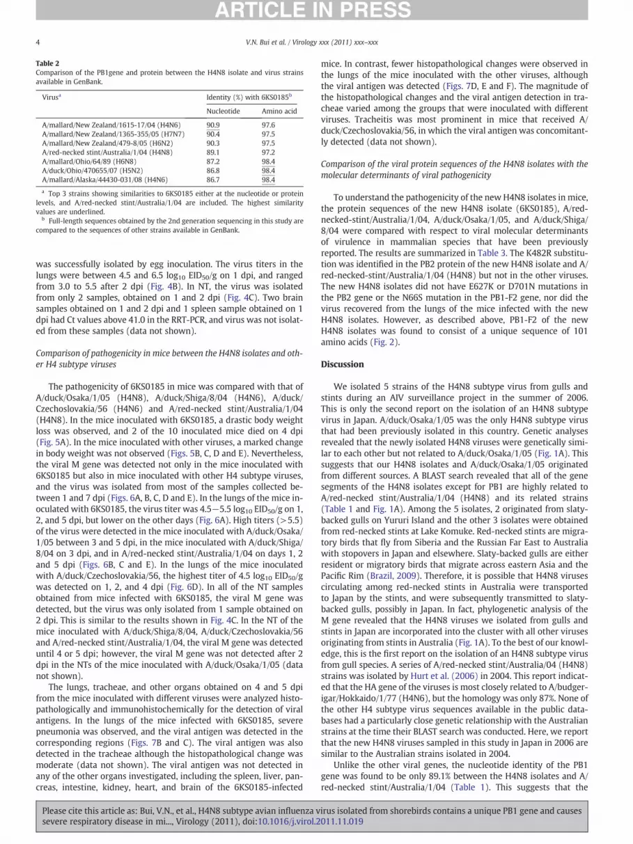
Table 2Comparison of the PB1gene and protein between the H4N8 isolate and virus strainsavailable in GenBank.
Virusa Identity (%) with 6KS0185b
Nucleotide Amino acid
A/mallard/New Zealand/1615-17/04 (H4N6) 90.9 97.6A/mallard/New Zealand/1365-355/05 (H7N7) 90.4 97.5A/mallard/New Zealand/479-8/05 (H6N2) 90.3 97.5A/red-necked stint/Australia/1/04 (H4N8) 89.1 97.2A/mallard/Ohio/64/89 (H6N8) 87.2 98.4A/duck/Ohio/470655/07 (H5N2) 86.8 98.4A/mallard/Alaska/44430-031/08 (H4N6) 86.7 98.4
a Top 3 strains showing similarities to 6KS0185 either at the nucleotide or proteinlevels, and A/red-necked stint/Australia/1/04 are included. The highest similarityvalues are underlined.
b Full-length sequences obtained by the 2nd generation sequencing in this study arecompared to the sequences of other strains available in GenBank.
4 V.N. Bui et al. / Virology xxx (2011) xxx–xxx
was successfully isolated by egg inoculation. The virus titers in thelungs were between 4.5 and 6.5 log10 EID50/g on 1 dpi, and rangedfrom 3.0 to 5.5 after 2 dpi (Fig. 4B). In NT, the virus was isolatedfrom only 2 samples, obtained on 1 and 2 dpi (Fig. 4C). Two brainsamples obtained on 1 and 2 dpi and 1 spleen sample obtained on 1dpi had Ct values above 41.0 in the RRT-PCR, and virus was not isolat-ed from these samples (data not shown).
Comparison of pathogenicity in mice between the H4N8 isolates and oth-er H4 subtype viruses
The pathogenicity of 6KS0185 in mice was compared with that ofA/duck/Osaka/1/05 (H4N8), A/duck/Shiga/8/04 (H4N6), A/duck/Czechoslovakia/56 (H4N6) and A/red-necked stint/Australia/1/04(H4N8). In the mice inoculated with 6KS0185, a drastic body weightloss was observed, and 2 of the 10 inoculated mice died on 4 dpi(Fig. 5A). In the mice inoculated with other viruses, a marked changein body weight was not observed (Figs. 5B, C, D and E). Nevertheless,the viral M gene was detected not only in the mice inoculated with6KS0185 but also in mice inoculated with other H4 subtype viruses,and the virus was isolated from most of the samples collected be-tween 1 and 7 dpi (Figs. 6A, B, C, D and E). In the lungs of the mice in-oculated with 6KS0185, the virus titer was 4.5−5.5 log10 EID50/g on 1,2, and 5 dpi, but lower on the other days (Fig. 6A). High titers (>5.5)of the virus were detected in the mice inoculated with A/duck/Osaka/1/05 between 3 and 5 dpi, in the mice inoculated with A/duck/Shiga/8/04 on 3 dpi, and in A/red-necked stint/Australia/1/04 on days 1, 2and 5 dpi (Figs. 6B, C and E). In the lungs of the mice inoculatedwith A/duck/Czechoslovakia/56, the highest titer of 4.5 log10 EID50/gwas detected on 1, 2, and 4 dpi (Fig. 6D). In all of the NT samplesobtained from mice infected with 6KS0185, the viral M gene wasdetected, but the virus was only isolated from 1 sample obtained on2 dpi. This is similar to the results shown in Fig. 4C. In the NT of themice inoculated with A/duck/Shiga/8/04, A/duck/Czechoslovakia/56and A/red-necked stint/Australia/1/04, the viral M gene was detecteduntil 4 or 5 dpi; however, the viral M gene was not detected after 2dpi in the NTs of the mice inoculated with A/duck/Osaka/1/05 (datanot shown).
The lungs, tracheae, and other organs obtained on 4 and 5 dpifrom the mice inoculated with different viruses were analyzed histo-pathologically and immunohistochemically for the detection of viralantigens. In the lungs of the mice infected with 6KS0185, severepneumonia was observed, and the viral antigen was detected in thecorresponding regions (Figs. 7B and C). The viral antigen was alsodetected in the tracheae although the histopathological change wasmoderate (data not shown). The viral antigen was not detected inany of the other organs investigated, including the spleen, liver, pan-creas, intestine, kidney, heart, and brain of the 6KS0185-infected
Please cite this article as: Bui, V.N., et al., H4N8 subtype avian influenza vsevere respiratory disease in mi..., Virology (2011), doi:10.1016/j.virol.2
mice. In contrast, fewer histopathological changes were observed inthe lungs of the mice inoculated with the other viruses, althoughthe viral antigen was detected (Figs. 7D, E and F). The magnitude ofthe histopathological changes and the viral antigen detection in tra-cheae varied among the groups that were inoculated with differentviruses. Tracheitis was most prominent in mice that received A/duck/Czechoslovakia/56, in which the viral antigen was concomitant-ly detected (data not shown).
Comparison of the viral protein sequences of the H4N8 isolates with themolecular determinants of viral pathogenicity
To understand the pathogenicity of the new H4N8 isolates in mice,the protein sequences of the new H4N8 isolate (6KS0185), A/red-necked-stint/Australia/1/04, A/duck/Osaka/1/05, and A/duck/Shiga/8/04 were compared with respect to viral molecular determinantsof virulence in mammalian species that have been previouslyreported. The results are summarized in Table 3. The K482R substitu-tion was identified in the PB2 protein of the new H4N8 isolate and A/red-necked-stint/Australia/1/04 (H4N8) but not in the other viruses.The new H4N8 isolates did not have E627K or D701N mutations inthe PB2 gene or the N66S mutation in the PB1-F2 gene, nor did thevirus recovered from the lungs of the mice infected with the newH4N8 isolates. However, as described above, PB1-F2 of the newH4N8 isolates was found to consist of a unique sequence of 101amino acids (Fig. 2).
Discussion
We isolated 5 strains of the H4N8 subtype virus from gulls andstints during an AIV surveillance project in the summer of 2006.This is only the second report on the isolation of an H4N8 subtypevirus in Japan. A/duck/Osaka/1/05 was the only H4N8 subtype virusthat had been previously isolated in this country. Genetic analysesrevealed that the newly isolated H4N8 viruses were genetically simi-lar to each other but not related to A/duck/Osaka/1/05 (Fig. 1A). Thissuggests that our H4N8 isolates and A/duck/Osaka/1/05 originatedfrom different sources. A BLAST search revealed that all of the genesegments of the H4N8 isolates except for PB1 are highly related toA/red-necked stint/Australia/1/04 (H4N8) and its related strains(Table 1 and Fig. 1A). Among the 5 isolates, 2 originated from slaty-backed gulls on Yururi Island and the other 3 isolates were obtainedfrom red-necked stints at Lake Komuke. Red-necked stints are migra-tory birds that fly from Siberia and the Russian Far East to Australiawith stopovers in Japan and elsewhere. Slaty-backed gulls are eitherresident or migratory birds that migrate across eastern Asia and thePacific Rim (Brazil, 2009). Therefore, it is possible that H4N8 virusescirculating among red-necked stints in Australia were transportedto Japan by the stints, and were subsequently transmitted to slaty-backed gulls, possibly in Japan. In fact, phylogenetic analysis of theM gene revealed that the H4N8 viruses we isolated from gulls andstints in Japan are incorporated into the cluster with all other virusesoriginating from stints in Australia (Fig. 1A). To the best of our knowl-edge, this is the first report on the isolation of an H4N8 subtype virusfrom gull species. A series of A/red-necked stint/Australia/04 (H4N8)strains was isolated by Hurt et al. (2006) in 2004. This report indicat-ed that the HA gene of the viruses is most closely related to A/budger-igar/Hokkaido/1/77 (H4N6), but the homology was only 87%. None ofthe other H4 subtype virus sequences available in the public data-bases had a particularly close genetic relationship with the Australianstrains at the time their BLAST search was conducted. Here, we reportthat the new H4N8 viruses sampled in this study in Japan in 2006 aresimilar to the Australian strains isolated in 2004.
Unlike the other viral genes, the nucleotide identity of the PB1gene was found to be only 89.1% between the H4N8 isolates and A/red-necked stint/Australia/1/04 (Table 1). This suggests that the
irus isolated from shorebirds contains a unique PB1 gene and causes011.11.019

Fig. 2. Comparison of the PB1-F2 protein sequence of the H4N8 isolates obtained in this study and the reference strains. (A) The PB1-F2 protein sequences of the H4N8 isolates(6KS0185 and 6KS0279) were aligned with sequences identified in a BLAST search. The strains of the top 6 in identity to 6KS0185 and 6KS0279 are shown. The PB1-F2 protein con-sists of 101 amino acids in the H4N8 isolates but 90 amino acids in other strains. (B) The PB1-F2 protein sequence of the H4N8 isolate 6KS0185 was aligned with other sequences ofa length of 101 amino acids, which were identified in GenBank. Asterisks represent the homologous amino acids in all genes. Identities between the 6KS0185 protein and each pro-tein are presented in the table below the sequence alignment.
5V.N. Bui et al. / Virology xxx (2011) xxx–xxx
H4N8 isolates are PB1 reassortants. Despite an extensive search in theGenBank database, we could not find any sequences that are closelyrelated to the PB1 gene of the new isolates. The PB1 genes of theH6N8, H5N2, and H4N6 subtype viruses isolated from duck speciesin North America were found to have 98.4% homology at the proteinlevel, but less than 90.0% homology at the nucleotide level. The PB1genes of the New Zealand strains of H4N6, H7N7, and H6N2 subtypes
Please cite this article as: Bui, V.N., et al., H4N8 subtype avian influenza vsevere respiratory disease in mi..., Virology (2011), doi:10.1016/j.virol.2
showed nucleotide homology slightly higher than 90.0%, but theamino acid identity was less than 98.0% (Table 2). From these results,it is concluded that the PB1 genes of the H4N8 isolates are unique,and thus, we could not identify the geographical origin of the PB1gene.
Another interesting feature was identified in PB1 from the newH4N8 isolates. None of the PB1-F2 protein sequences in GenBank
irus isolated from shorebirds contains a unique PB1 gene and causes011.11.019
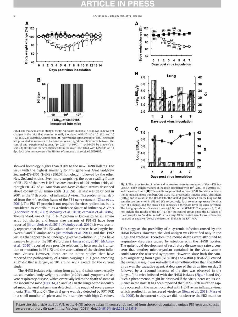
Fig. 3. The mouse infection study of the H4N8 isolate 6KS0185 (n=4). (A) Body weightchanges in the mice that were intranasally inoculated with 103 (○), 102 (□), and 10(△) TCID50 of 6KS0185. Control mice (●) received the same amount of PBS. The resultsare presented as mean±S.D. Asterisks represent significant differences between thecontrol and experimental groups. *pb0.05, **pb0.001, ***pb0.0001 by Student's t-test. (B) HI titers of the sera obtained from the mice inoculated with 6KS0185 on 14dpi. Each column represents the HI titer of a mouse that received 6KS0185.
Fig. 4. The tissue tropism in mice and mouse-to-mouse transmission of the H4N8 iso-late. (A) Body weight changes of the mice inoculated with 103 TCID50 of 6KS0185 (○)and the contact mice (■). The results are presented as mean±S.D. Numbers in paren-theses indicate mouse numbers. One sharp mark represents 1 mouse death. Virus titers(EID50) and Ct values in the RRT-PCR for the viral M gene obtained for the lung and NTsamples are presented in (B) and (C), respectively. Each column represents the virustiter of 1 mouse, and the broken line indicates a threshold level for virus detection.The line graph shows Ct values (mean±S.D.) in the RRT-PCR. The graphs (B, C) donot include the results of the RRT-PCR for the control group, since the Ct values ofthose samples are “undetermined” in the assay. All the control samples were thereforeregarded as negative (below the detection limit) in the RRT-PCR.
6 V.N. Bui et al. / Virology xxx (2011) xxx–xxx
showed homology higher than 90.0% to the new H4N8 isolates. Thevirus with the highest similarity for this gene was A/mallard/NewZealand/479-8/05 (H6N2) (90.0% homology), followed by the otherNew Zealand strains. Even more surprising, the open reading frameof PB1-F2 of the new H4N8 isolates consists of 101 amino acids, al-though PB1-F2 of all American and New Zealand strains describedabove consist of 90 amino acids (Fig. 2A). PB1-F2 was described in2001 as the 11th protein of influenza A virus. This protein is translat-ed from the +1 reading frame of the PB1 gene segment (Chen et al.,2001). The PB1-F2 protein is not required for virus replication, but isconsidered to contribute as a virulence factor of influenza A virus(Conenello et al., 2007; McAuley et al., 2010; Zamarin et al., 2006).The standard size of the PB1-F2 protein is known to be 90 aminoacids but shorter and longer size variants of PB1-F2 have beenreported (Krumbholz et al., 2011; McAuley et al., 2010). It was recent-ly reported that the PB1-F2 variants of swine viruses have lengths be-tween 8 and 90 amino acids (Krumbholz et al., 2011), and the H9N2viruses that appear to be undergoing active evolution in China havevariable lengths of the PB1-F2 protein (Huang et al., 2010). McAuleyet al. (2010) reported on a possible relationship between the trunca-tion or mutation in PB1-F2 and the attenuation of the virulent influ-enza viruses. However, there are no other studies that havereported the pathogenicity of a virus carrying a PB1 gene encodinga PB1-F2 that is longer, at 101 amino acids, except for the currentstudy.
The H4N8 isolates originating from gulls and stints unexpectedlycaused marked body weight reduction (>20%), and symptoms of se-vere respiratory disease, which eventually led to the death of some ofthe inoculated mice (Figs. 3A, 4A and 5A). In the lungs of the inoculat-ed mice, the viral antigen was detected in the region of severe pneu-monia (Figs. 7B and C). The viral gene was also detected by RRT-PCRin a small number of spleen and brain samples with high Ct values.
Please cite this article as: Bui, V.N., et al., H4N8 subtype avian influenza vsevere respiratory disease in mi..., Virology (2011), doi:10.1016/j.virol.2
This suggests the possibility of a systemic infection caused by theH4N8 isolates. However, the viral antigen was identified only in thelungs and tracheae. Therefore, the mouse deaths were attributed torespiratory disorders caused by infection with the H4N8 isolates.The quite rapid development of respiratory disease may raise a con-cern that another microorganism contained in the virus samplecould cause the observed symptoms. However, since both the sam-ples, originating from a gull (6KS0185) and a stint (6KS0279), causedthe same disease, it was unlikely that something other than the H4N8virus was the causative agent. A decrease of the virus titer on day 3followed by a rebound increase of the titer was observed in thelungs of the mice infected with the H4N8 isolates (Figs. 4B and 6A).Such a phenomenon might be observed if the virus increased its vir-ulence in the host. It has been reported that PB2 E627K mutation rap-idly occurred in the mice inoculated with H5N1 avian influenza virus,which resulted in an increased virulence (Bogs et al., 2011; Mase etal., 2006). In the current study, we did not observe the PB2 mutation
irus isolated from shorebirds contains a unique PB1 gene and causes011.11.019

Fig. 5. Body weight changes of the mice inoculated with the H4N8 isolate and the reference viruses. 103 TCID50 of 6KS0185 (A), A/duck/Osaka/1/05 (B), A/duck/Shiga/8/04 (C), A/duck/Czechoslovakia/56 (D) or A/red-necked-stint/Australia/1/04 (E) was intranasally inoculated into the mice. The results are presented as the mean±S.D. Open circles show theresults of the control mice, and filled circles show the results of the experimental mice. Numbers in parentheses indicate mouse numbers. One sharp mark represents 1 mousedeath. *pb0.05, ***pb0.0001 by Student's t-test.
7V.N. Bui et al. / Virology xxx (2011) xxx–xxx
in mice infected with the H4N8 isolates. Further study is necessary toelucidate the mechanism of the virus titer rebound in mice: thatwould include studies searching candidates of genetic mutations re-lated to the titer change and/or investigating host-related factorssuch as the immune response of the host towards the virus. It shouldbe interesting to passage the virus in mice to determine whether vir-ulence is increased by host specific adaptations. Mouse-to-mousetransmission was not observed in the H4N8 infection (Fig. 4). Sincethe mouse is not regarded as a suitable model for investigating viraltransmission (O'Donnell and Subbarao, 2011), additional studies arerequired to evaluate transmission of the H4N8 isolates among mam-malian species by using a more reliable animal model such as ferrets(Gustin et al., 2011) and guinea-pigs (Lowen et al., 2006). While thevirus we identify in this study is not considered a pandemic threat,these additional studies could help clarify the boundaries existing be-tween avian viruses normally considered low pathogenic and poten-tial pandemic strains.
Driskell et al. (2010) also studied the pathogenicity of several sub-types of viruses isolated from wild birds including ducks and shore-birds such as turnstone, sanderling, and knot. These subtypesreplicated well in MDCK cells without trypsin in a manner similar tothe replication of the H4N8 isolates observed in the current study.Among the 28 strains Driskel et al. tested, most of the viruses (71%)
Please cite this article as: Bui, V.N., et al., H4N8 subtype avian influenza vsevere respiratory disease in mi..., Virology (2011), doi:10.1016/j.virol.2
replicated efficiently in mouse lungs to attain high titers without ad-aptation and induction of histopathological lesions in the lungs. How-ever, the maximum reduction in body weight observed was less than8%, and mouse deaths were not recorded. The results of previousstudies and the current study suggest that the H4N8 viruses newlyisolated from shorebirds are more virulent in mice than are the previ-ously isolated viruses. Ability to replicate well in cell culture in the ab-sence of trypsin has been regarded as a possible factor for the virus toacquire virulence in hosts, since proteolytic cleavage of the influenzavirus hemagglutinin by protease is essential for viral infectivity andspread (Goto and Kawaoka, 1998; Tumpey et al., 2005). In the currentstudy, both the new H4N8 isolate and A/red-necked stint/Australia/1/04 replicate in MDCK to similar titers in the presence and absence oftrypsin. However, the new H4N8 isolate but not A/red-necked stint/Australia/1/04 caused disease in mice, indicating that trypsin-independent replication is not necessarily linked to the pathogenicityof this virus.
We also found that mice inoculated with A/duck/Osaka/1/05, A/duck/Shiga/8/04, and A/duck/Czechoslovakia/56 did not show anysymptoms of disease, and there were no marked body weight reduc-tions (Fig. 5). In the lungs of these mice, the viral gene was detectedwith Ct values similar to those of 6KS0185, and the virus was also iso-lated in similar titers (Fig. 6). These results indeed indicate that
irus isolated from shorebirds contains a unique PB1 gene and causes011.11.019

Fig. 6. Virus titers (EID50) and Ct values in RRT-PCR analysis of the viral M gene for the lungs obtained from the mice inoculated with the H4N8 isolate and the reference viruses. Themice were euthanized on the indicated days after inoculation (103 TCID50) with 6KS0185 (A), A/duck/Osaka/1/05 (B), A/duck/Shiga/8/04 (C), A/duck/Czechoslovakia/56 (D) or A/red-necked-stint/Australia/1/04 (E). Each column represents a virus titer of 1 mouse and the broken line indicates the threshold level for virus detection. The line graph presents theaverage of the Ct values in the RRT-PCR.
8 V.N. Bui et al. / Virology xxx (2011) xxx–xxx
6KS0185 as well as the other H4 subtype viruses tested in this studycould efficiently replicate in mouse lungs without prior adaptation.These results also indicate that the efficiency of viral replication inthe lung is not the sole determinant of the differences in virulence ob-served. There must be other factors involved in order for the virus tocause severe respiratory disease in the inoculated mice. Hurt et al.(2006) reported that strains of A/red-necked stint/Australia/2004are not pathogenic to chickens. We also found that the H4N8 viruseswe isolated are not pathogenic to chickens (data not shown). A lethalinfection of A/chicken/Alabama/7395/75 (H4N8) in chickens hasbeen reported (Slemons et al., 1991), and infection of pigs with H4subtype viruses have also been reported (Karasin et al., 2000;Ninomiya et al., 2002). However, this is the first report on diseasecaused by H4N8 virus infection in a mammalian host. Moreover, themajor differences between the new H4N8 isolates that are pathogenicin mice and the non-pathogenic A/red-necked-stint/Australia/1/04are only in the PB1 gene.
Many studies have been performed to identify the molecular de-terminants responsible for viral pathogenicity in the host, and the ac-cumulated data provide concrete evidence for many determinants, as
Please cite this article as: Bui, V.N., et al., H4N8 subtype avian influenza vsevere respiratory disease in mi..., Virology (2011), doi:10.1016/j.virol.2
described in recent review articles (Neumann et al., 2009; O'Donnelland Subbarao, 2011; Tscherne and Garcia-Sastre, 2011). The proteinsequence of the viruses examined in this study was compared tothe previously identified virulence determinants. The PB2 protein ofthe new H4N8 isolates contains the K482R substitution. However,A/red-necked-stint/Australia/1/04 also has the substitution, suggest-ing that the amino acid 482 in the PB2 protein is not likely to relateto the pathogenicity of the new H4N8 isolates. Other virulence deter-minants that have been reported, including PB2-627K, PB2-701N, andPB1-F2-66S were not identified in the proteins of the new H4N8 iso-lates (Table 3).
Amino acid positions for the receptor-binding sites of HA havebeen identified for H1, H2, and H3 subtypes, and certain positionshave been identified as predictors of the receptor specificity, i.e.,avian preference (SAα2,3) or human preference (SAα2,6)(Neumann et al., 2009; O'Donnell and Subbarao, 2011; Tscherne andGarcia-Sastre, 2011). However, no such information is currently avail-able for the H4 subtype virus.
The PB1 gene of the H4N8 isolates was found to be unique, espe-cially in PB1-F2, which has a length of 101 amino acids. Several
irus isolated from shorebirds contains a unique PB1 gene and causes011.11.019

Fig. 7. Histopathological and immunohistochemical analyses of the lungs obtained from the mice inoculated with the H4N8 isolate and the reference viruses. Hematoxylin and eosinstaining of the lung of the control mouse (A) and the mouse infected with 6KS0185 (B). Detection of the viral antigen in the lungs of the mice inoculated with 6KS0185 (C), A/duck/Osaka/1/05 (D), A/duck/Shiga/8/04 (E), and A/duck/Czechoslovakia/56 (F). The tissue samples were collected from the mice 4 or 5 days after the intranasal inoculation of the vi-ruses (103 TCID50). The results are of a representative mouse of each group (n=4). Arrows indicate the viral antigen detected by polyclonal antibody against influenza A virus. Thescale bar represents 200 μm.
9V.N. Bui et al. / Virology xxx (2011) xxx–xxx
PB1-F2 proteins of this length were identified in GenBank. The virusescontaining PB1-F2 of this length can be divided into 2 groups. The firstgroup includes viruses isolated in North America between 1988 and2001. This group includes the H3N2, H4N6, and H10N7 subtype virus-es from mallard, and many H3N2 subtype human viruses. The secondgroup includes viruses isolated in China between 2003 and 2009. Thisgroup includes H9N2 viruses isolated from humans and chickens andthe H5N1 virus from pheasants. All of the PB1-F2 proteins withlengths of 101 amino acids found in GenBank have amino acid homol-ogy lower than 80.0% to the new H4N8 viruses from stints and gulls inJapan (Fig. 2B). This result could be attributed to a shortage of aminoacid sequence data for PB1-F2 currently available in GenBank. A morediverse global representation of PB1-F2 sequence of influenza A virus-es in the public database may reveal more closely related viruses inthe future.
Table 3Comparison of the protein sequence between the H4N8 isolate and other H4 subtype virus
Protein Position Virus
6KS0185 H4N8/
Australia
H4N8
Osak
PB2 271 T T T
482 R R K
627 E E E
701 D D D
714 S S S
PB1 13 P P P
538 D D D
678 S N S
PB1-F2 66 N N N
PA 97 T T T
550 L L L
615 R R K
M1 139 T T T
NS1 92 D D (ND)c
C-terminus GSEV ESEV (ND)
aThe protein sequences of 6KS0185, A/red-necked stint/Australia/1/04 (H4N8), A/duck/Osakdeterminants of viral pathogenicity. The amino acids matched to the determinants for highbReferences: a) Bussey et al., 2010; b) Brown and Bailly, 1999; c) Subbarao et al., 1993; d)h) Rolling et al., 2009; i) Seo et al., 2002; j) Jackson et al., 2008; k) Obenauer et al., 2006.cNot determined.
Please cite this article as: Bui, V.N., et al., H4N8 subtype avian influenza vsevere respiratory disease in mi..., Virology (2011), doi:10.1016/j.virol.2
Although the exact role of the PB1-F2 in viral infection has notbeen fully elucidated, expression of PB1-F2 has been reported to en-hance viral pathogenicity in mouse models of influenza infection(Conenello et al., 2007; McAuley et al., 2007; Zamarin et al., 2006). In-creased virulence caused by PB1-F2 seems to involve mechanismssuch as: 1) mitochondrial targeting leading to apoptosis, 2) an in-creased inflammatory response, and 3) enhancement of transcriptionby an increase in the nuclear retention time (Chen et al., 2001;Conenello et al., 2007; Gibbs et al., 2003; Mazur et al., 2008;McAuley et al., 2007, 2010). Further characterization of PB1-F2 ofthe H4N8 isolates, which causes severe inflammatory disease inmice, may provide new information to improve our understandingof the role of PB1-F2 in diseases caused by the influenza A virus,and ultimately improve our understanding of the evolution of thePB1-F2 protein. Since molecular determinants of influenza virulence
es with the molecular determinants of virus pathogenicity in mammalian speciesa.
Pathogenicity
/
a
H4N6/
Shiga
Low High Ref.b
T T A a)
K K R b)
E E K c)
D D N d), e)
S S R d)
P P d)
D D G d)
S N d)
N N S f)
C T I g)
L I L h)
K K N d )
T T A b)
D D E i)
ESEV RSEV ESEV j), k)
a/1/05 (H4N8), and A/duck/Shiga/8/04 (H4N6) were compared with the molecularpathogenicity are highlighted by gray shading.
Gabriel et al., 2005; e) Li et al., 2005; f) Conenello et al., 2007; g) Song et al., 2009;
irus isolated from shorebirds contains a unique PB1 gene and causes011.11.019

10 V.N. Bui et al. / Virology xxx (2011) xxx–xxx
depend on the gene constellation and backbone, a study employingreverse genetics will be necessary to test the exact role of the PB1-F2 in the virus pathogenicity. The results obtained in these studieswill help to define the virulence determinant in H4N8 viruses origi-nating from gulls and stints.
Materials and methods
Sample collection
A total of 497 shorebird samples (369 cloacal and 128 fecal sam-ples) were collected in 2006 between June and September in easternHokkaido, Japan. The locations included Lake Komuke (GPS coordi-nates 44° 15′ N, 143° 31′ E, n=172), Kushiro Wildlife Center (43°03′ N, 144° 17′ E, n=162), and Yururi Island (43° 13′ N, 145° 36′ E,n=128). Additional samples were obtained at three other locations.Cloacal swabs were collected by licensed bird banders during thebanding procedure. The fecal samples were collected around the hab-itat area of the birds, and only fresh samples for which the species oforigin was defined were included in this study. The bird species in-cluded black-faced bunting (n=132), slaty-backed gull (n=128),red-necked stint (n=94), gray-tailed tattler (n=27), greenshank(n=15), Latham's snipe (n=15), Mongolian plover (n=11), greenwing teal duck (n=9), and other non-duck species. Following thecollection of samples in the field, each sample was stored in virustransport medium (VTM: M4RT, Remel Inc., Lenexa, KS) and kept ina cold chain followed by storage at −80 °C after transportation tothe laboratory. The VTM samples were subjected to both RRT-PCRfor detection of the M gene of influenza A virus and virus isolationusing embryonated chicken eggs as described below.
Cells and viruses
Madin-Darby canine kidney (MDCK) cells were cultured in Dul-becco's Modified Eagle's medium (DMEM: Nissui PharmaceuticalCo., Ltd., Tokyo, Japan) supplemented with 10% fetal bovine serum(FBS) and 2 mM L-glutamine. Cells were seeded onto 96-well tissueculture plates to evaluate viral titers. Upon virus inoculation, thecells were washed twice with the DMEM and the medium wasreplaced with virus growth medium (VGM) containing trypsin fol-lowing protocol in the WHO Manual on Animal Influenza Diagnosisand Surveillance (WHO/CDS/CSR/NCS/2002.5 Rev. 1: WHO Manual).The sample to be tested was serially diluted (1:10) for the titration.Based on the cytopathic effect (CPE) observed 4 days dpi, the 50% tis-sue culture infectious dose (TCID50) was calculated by the Behrens–Kärber method. The hemagglutination test using 0.5% chicken eryth-rocytes suspended in phosphate-buffered saline (PBS, pH 7.4) wasperformed on the cell culture supernatants to confirm that the ob-served CPE reflects the growth of the virus in the cells. In some exper-iments, VGM lacking trypsin was used to measure the virus titer.
A/duck/Osaka/1/05 (H4N8) and A/duck/Shiga/8/04 (H4N6) werekindly provided by the National Institute of Animal Health, Japan. A/duck/Czechoslovakia/56 (H4N6) was supplied by Dr. H. Kida at theOIE Reference Laboratory for HPAI at Hokkaido University, Japan. A/red-necked stint/Australia/1/04 (H4N8) was kindly provided by Dr.Aeron Hurt at the WHO Collaborating Centre for Reference and Re-search on Influenza and the Victorian Infectious Diseases ReferenceLaboratory. The viruses were propagated in the allantoic cavity of10-day-old embryonated chicken eggs. Aliquots of the allantoic fluidscontaining the viruses were stored at −80 °C until use.
Virus isolation
Virus isolation from cloacal/fecal samples was carried out in 10-day-old embryonated chicken eggs according to the WHO Manualwith some modifications. Prior to egg inoculation, the thawed
Please cite this article as: Bui, V.N., et al., H4N8 subtype avian influenza vsevere respiratory disease in mi..., Virology (2011), doi:10.1016/j.virol.2
samples were mixed well and centrifuged at 1000×g for 10 min at4 °C. The supernatants were supplemented with antibiotics and anti-mycotics to achieve the following final concentrations: 1000 U/mlpenicillin, 1 mg/ml streptomycin, 100 μg/ml gentamicin, and 10 μg/ml amphotericin B. These supernatants were kept at room tempera-ture for 2 h. Then, 0.1 ml of the sample was inoculated into the allan-toic cavity of each egg (2 eggs for each sample). After an incubationperiod of 4 days at 37 °C, the eggs were chilled overnight at 4 °C.Egg allantoic fluid from the initial inoculation (E1) was tested by ahemagglutination test. The E1 allantoic fluids with negative resultsin the test were used in a second egg inoculation followed by thehemagglutination test.
Allantoic fluid with hemagglutination activity were subjected tohemagglutination inhibition (HI) and neuraminidase inhibition (NI)tests performed according to the WHO Manual, to identify the influ-enza virus subtypes. The reference antisera against influenza A virus-es and the reference viruses for the HI and NI tests were also providedby Dr. H. Kida.
RT-PCR and RRT-PCR
The viral RNA of the M gene of the influenza virus in the originaland the allantoic fluid samples was detected by RRT-PCR as we previ-ously reported (Bui et al., 2011). In brief, total RNA was extractedfrom the samples by a KingFisher purification system (Thermo Scien-tific, Waltham, MA) and a Magmax-96 AI/ND Viral RNA isolation kit(Thermo Scientific). First-strand cDNA was prepared using randomhexamer primers (Invitrogen, Carlsbad, CA) and M-MLV reverse tran-scriptase (Invitrogen) under the following conditions: 25 °C for10 min, 37 °C for 50 min, and 65 °C for 10 min. Using Taqman Univer-sal PCR Master mix (Applied Biosciences, Foster City, CA) RRT-PCRwas carried out using an ABI PRISM Sequence Detection System7900HT (Applied Biosciences) as follows: stage 1, 95 °C for 10 minand stage 2, 45 cycles of 95 °C for 15 s and 60 °C for 1 min. Sampleswith Ct below 40 were regarded as M gene positive, and sampleswith Ct over 40 were considered as suspect positives.
Nucleotide sequencing and phylogenetic analysis
Total RNA extracted from allantoic fluid (E1) containing AIV wastranscribed into cDNA using the Uni12 primer (5′-agcraaagcagg-3′)and SuperScript III Reverse Transcriptase (Invitrogen) at 50 °C for60 min followed by 70 °C for 10 min. Using the cDNAs as templates,a full length of the 8 viral gene segments was amplified as describedby Hoffmann et al. (2001). The PCR products were separated by 1%agarose gel electrophoresis and purified using a QIAquick PCR Purifi-cation kit (Qiagen, Hilden, Germany). The purified PCR products weredirectly applied to the sequencing reaction using a BigDye Terminatorv3.1 cycle sequencing kit (Applied Biosystems). Nucleotide sequenc-ing was performed in an ABI PRISM 310 Genetic Analyzer (AppliedBiosystems). The primer sets amplifying the PCR products were firstused for the sequencing reaction, and then primer walking was con-ducted to read the full-length nucleotide sequence of the gene. Alter-natively, the PCR products were subcloned into the pGEM-T Easyvector (Promega, Madison, WI, USA), and the plasmids obtainedwere used as templates for sequencing. Simultaneously, the cDNAsamples were sent to JCVI as a partner submission to the National In-stitute of Allergy and Infectious Diseases-funded Influenza GenomeSequencing Project. International transportation of the cDNAs wasperformed according to the protocol permitted by the United StatesDepartment of Agriculture (USDA #108081). The sequence analysisfor the samples was performed at the JCVI using 454 FLX/Roche andSolexa/Illumina systems. The output data from the JCVI were alto uti-lized in this study.
Sequence data were analyzed by GENETYX Ver. 9 (Genetyx Corp.,Tokyo, Japan) and compared with other GenBank sequences that
irus isolated from shorebirds contains a unique PB1 gene and causes011.11.019

11V.N. Bui et al. / Virology xxx (2011) xxx–xxx
were identified in the BLAST homology searches (conducted in May2011). The nucleotide sequences were aligned by Clustal W(Thompson et al., 1994), and the evolutionary distances were com-puted using the Tamura–Nei method. Phylogenetic trees were con-structed using the neighbor-joining method (Saitou and Nei, 1987;Tamura et al., 2004) and bootstrap analysis (1000 replicates), aswell as the bootstrap interior branch test for phylogeny constructionusing Mega 4.0 software (Tamura et al., 2007).
Mouse infection study of the H4N8 isolates
To investigate the pathogenicity of the H4N8 viruses isolated inthis study, a mouse infection study was performed using 8- to 10-week-old female BALB/c mice purchased from Clea Japan Inc.(Tokyo, Japan). All mouse studies were conducted in compliancewith the institutional rules for the care and use of laboratory animals,and using protocols approved by the relevant committee at theUniversity.
First, a preliminary study was performed using 2 groups of mice (3mice/group). A group of mice received intranasal inoculations of 103
TCID50 of 6KS0185, (a H4N8 virus isolate obtained from a gull) andanother group received inoculations of 6KS0279 (a H4N8 virus isolateobtained from a stint) in 50 μl of allantoic fluid (E1) under light etheranesthesia. Body weights and clinical signs of the mice were recordeddaily afterwards. The mice were sacrificed on 3 dpi. The lungs excisedfrom the mice were weighed and homogenized in DMEM containingpenicillin (500 U/ml) and streptomycin (500 μg/ml) to prepare 10%homogenates. Lung homogenates were then subjected to RRT-PCRto amplify the M gene. Since the results obtained in the preliminarystudy were quite similar among the mice inoculated with 6KS0185and 6KS0279, subsequent studies were performed only for 6KS0185.In the next experiment, 3 groups of mice (4 mice/group) receivedan intranasal inoculation of different concentrations of 6KS0185 (10,102, or 103 TCID50), and 4 control mice received PBS. The bodyweights were monitored daily and blood samples were collectedfrom the mice prior to sacrifice on 14 dpi. The sera obtained were sub-jected to the HI test measuring antibody against the virus inoculated.
Tissue tropism and mouse-to-mouse transmission of the H4N8 isolates
Tissue tropism and transmission of the H4N8 isolates were studiedusing a total of 25 mice. The mice were assigned to 5 cages with 5mice in each cage. Three mice in each cage were intranasally inoculat-ed with 103 TCID50 of 6KS0185, and the 2 mice that did not receivethe virus were kept in the same cage. On 1, 2, 3, 4, and 7 dpi, all 5mice were sacrificed in 1 of the 5 cages, and their lungs, NTs, spleens,and brains were collected. The tissue samples were subjected to a 10%homogenate preparation, except for the NT (5%) homogenates, fol-lowed by the RRT-PCR for the viral M gene and isolation of thevirus. For virus isolation, homogenate samples were serially diluted1:10 and inoculated into eggs. The 50% egg infectious dose (EID50)was calculated by the Behrens–Kärber method. Nucleotide sequenceof the gene segments of the virus isolated in the lung samples was an-alyzed as described above.
In mice that developed symptoms of disease, the animals weremaintained alive, unless found dead, until the intended euthanizingtime points to follow the disease time course.
Comparison of the pathogenicity with other H4 subtype viruses
Six groups of 15 mice were used to compare the pathogenicity of6KS0185 with that of other H4 subtype viruses including A/duck/Osaka/1/05 (H4N8), A/duck/Shiga/8/04 (H4N6), A/duck/Czechoslo-vakia/56 (H4N6), and A/red-necked stint/Australia/1/04 (H4N8).Five groups of mice received 103 TCID50 of the different viruses viathe nasal route. Another group of mice served as controls and
Please cite this article as: Bui, V.N., et al., H4N8 subtype avian influenza vsevere respiratory disease in mi..., Virology (2011), doi:10.1016/j.virol.2
received the same amount of PBS. Body weights and clinical signs ofdisease were monitored daily for all of the mice. On 1 dpi, 1 mousein each group was sacrificed, and between 2 and 7 dpi, 1 mouse inthe control group and 2 mice in each experimental group were sacri-ficed each day. Lungs and NTs obtained from these mice were sub-jected to viral gene detection and virus isolation as described above.On 4 and 5 dpi, 2 additional mice were euthanized in each group forhistopathological and immunohistochemical analysis of the lungs,tracheae, and other major organs. The organ samples were fixed in10% neutralized buffered formalin solution. All samples were thenembedded in paraffin, sectioned at 4 μm, and stained with hematoxy-lin and eosin. The immunoperoxidase technique was used to detectinfluenza virus antigens in formalin-fixed, paraffin-embedded sam-ples. A Histofine Simple Stain MAX-PO(G) kit (Nichirei Inc., Tokyo,Japan) was used according to the manufacturer's instructions. Goatpolyclonal antibody against influenza A virus (AB1074, Gt X InfluenzaA virus, Chemicon International, Temecula, CA, USA) was used as theprimary antibody at 1:8000. After staining, sections were counter-stained with hematoxylin.
Nucleotide sequence accession numbers
The nucleotide sequences obtained in this study are available fromGenBank under accession numbers CY089478 to CY089497 (dideoxymethod), CY079283 to CY079298 and CY080223 to CY080238 (2ndgeneration method). The following published AIV sequences were re-trieved from GenBank and included in the analyses of the currentstudy: accession numbers CY028259 to CY028266, CY016625,CY039373, CY045365, CY061624, GQ923547, and HM193786.
Acknowledgments
Wewould like to thank Florian Aldehoff, University of Alaska Fair-banks for his great help on sequence analysis and data management.We also would like to thank Eric Bortz, Mount Sinai School of Medi-cine for his help on the sequence analysis. We are also grateful toSachikoMatsuda for technical assistance. This work was partially sup-ported by grants from the Program of Founding Research Centers forEmerging and Reemerging Infectious Diseases, and by a Grant-in-Aidfor Exploratory Research (19659115) from the Ministry of Education,Culture, Sports, Science and Technology (MEXT), Japan. This workwas also partially supported by the US National Institute of Allergyand Infectious Diseases (NIAID contracts HHSN266200700009C andHHSN266200700007C).
References
Bogs, J., Kalthoff, D., Veits, J., Pavlova, S., Schwemmle, M., Mänz, B., Mettenleiter, T.C.,Stech, J., 2011. Reversion of PB2-627E to -627K during replication of an H5N1Clade 2.2 virus in mammalian hosts depends on the origin of the nucleoprotein.J. Virol. 85, 10691–10698.
Brazil, M., 2009. Birds of East Asia. Princeton University Press, Princeton, New Jersey.Brown, E.G., Bailly, J.E., 1999. Genetic analysis of mouse-adapted influenza A virus iden-
tifies roles for the NA, PB1, and PB2 genes in virulence. Virus Res. 61, 63–76.Bui, V.N., Ogawa, H., Karibe, K., Matsuo, K., Nguyen, T.H., Awad, S.S.A., Minoungou, G.L.,
Xininigen, Saito, K., Watanabe, Y., Runstadler, J.A., Happ, G.M., Imai, K., 2011. Sur-veillance of avian influenza virus in migratory water birds in eastern Hokkaido,Japan. J. Vet. Med. Sci. 73, 209–215.
Bussey, K.A., Bousse, T.L., Desmet, E.A., Kim, B., Takimoto, T., 2010. PB2 residue 271plays a key role in enhanced polymerase activity of influenza A viruses in mamma-lian host cells. J. Virol. 84, 4395–4406.
Chen, W., Calvo, P.A., Malide, D., Gibbs, J., Schubert, U., Bacik, I., Basta, S., O'Neill, R.,Schickli, J., Palese, P., Henklein, P., Bennink, J.R., Yewdell, J.W., 2001. A novel influ-enza A virus mitochondrial protein that induces cell death. Nat. Med. 7,1306–1312.
Conenello, G.M., Zamarin, D., Perrone, L.A., Tumpey, T., Palese, P., 2007. A single muta-tion in the PB1-F2 of H5N1 (HK/97) and 1918 influenza A viruses contributes to in-creased virulence. PLoS Pathog. 3, 1414–1421.
Driskell, E.A., Jones, C.A., Stallknecht, D.E., Howerth, E.W., Tompkins, S.M., 2010. Avianinfluenza virus isolates from wild birds replicate and cause disease in a mousemodel of infection. Virology 399, 280–289.
irus isolated from shorebirds contains a unique PB1 gene and causes011.11.019

12 V.N. Bui et al. / Virology xxx (2011) xxx–xxx
Fang, R., Min Jou, W., Huylebroeck, D., Devos, R., Fiers, W., 1981. Complete structure ofA/duck/Ukraine/63 influenza hemagglutinin gene: animal virus as progenitor ofhuman H3 Hong Kong 1968 influenza hemagglutinin. Cell 25, 315–323.
Gabriel, G., Dauber, B., Wolff, T., Planz, O., Klenk, H.D., Stech, J., 2005. The viral polymer-ase mediates adaptation of an avian influenza virus to a mammalian host. Proc.Natl. Acad. Sci. U. S. A. 102, 18590–18595.
Garten, R.J., Davis, C.T., Russell, C.A., Shu, B., Lindstrom, S., Balish, A., Sessions, W.M., Xu,X., Skepner, E., Deyde, V., Okomo-Adhiambo, M., Gubareva, L., Barnes, J., Smith, C.B.,Emery, S.L., Hillman, M.J., Rivailler, P., Smagala, J., de Graaf, M., Burke, D.F., Fouchier,R.A., Pappas, C., Alpuche-Aranda, C.M., Lopez-Gatell, H., Olivera, H., Lopez, I., Myers,C.A., Faix, D., Blair, P.J., Yu, C., Keene, K.M., Dotson, P.D.J., Boxrud, D., Sambol, A.R.,Abid, S.H., St George, K., Bannerman, T., Moore, A.L., Stringer, D.J., Blevins, P.,Demmler-Harrison, G.J., Ginsberg, M., Kriner, P., Waterman, S., Smole, S., Guevara,H.F., Belongia, E.A., Clark, P.A., Beatrice, S.T., Donis, R., Katz, J., Finelli, L., Bridges,C.B., Shaw, M., Jernigan, D.B., Uyeki, T.M., Smith, D.J., Klimov, A.I., Cox, N.J., 2009.Antigenic and genetic characteristics of swine-origin 2009 A(H1N1) influenza vi-ruses circulating in humans. Science 325, 197–201.
Gething, M.J., Bye, J., Skehel, J., Waterfield, M., 1980. Cloning and DNA sequence ofdouble-stranded copies of haemagglutinin genes from H2 and H3 strains eluci-dates antigenic shift and drift in human influenza virus. Nature 287, 301–306.
Gibbs, J.S., Malide, D., Hornung, F., Bennink, J.R., Yewdell, J.W., 2003. The influenza Avirus PB1-F2 protein targets the inner mitochondrial membrane via a predictedbasic amphipathic helix that disrupts mitochondrial function. J. Virol. 77,7214–7224.
Goto, H., Kawaoka, Y., 1998. A novel mechanism for the acquisition of virulence by ahuman influenza A virus. Proc. Natl. Acad. Sci. U. S. A. 95, 10224–10228.
Gustin, K.M., Belser, J.A., Wadford, D.A., Pearce, M.B., Katz, J.M., Tumpey, T.M., Maines,T.R., 2011. Influenza virus aerosol exposure and analytical system for ferrets.Proc. Natl. Acad. Sci. U. S. A. 108, 8432–8437.
Hanson, B.A., Stallknecht, D.E., Swayne, D.E., Lewis, L.A., Senne, D.A., 2003. Avian influ-enza viruses in Minnesota ducks during 1998–2000. Avian Dis. 47, 867–871.
Hinshaw, V.S., Bean, W.J., Webster, R.G., Sriram, G., 1980. Genetic reassortment of influ-enza A viruses in the intestinal tract of ducks. Virology 102, 412–419.
Hoffmann, E., Stech, J., Guan, Y., Webster, R.G., Perez, D.R., 2001. Universal primer setfor the full-length amplification of all influenza A viruses. Arch. Virol. 146,2275–2289.
Huang, Y., Hu, B., Wen, X., Cao, S., Gavrilov, B.K., Du, Q., Khan, M.I., Zhang, X., 2010. Di-versified reassortant H9N2 avian influenza viruses in chicken flocks in northernand eastern China. Virus Res. 151, 26–32.
Hurt, A.C., Hansbro, P.M., Selleck, P., Olsen, B., Minton, C., Hampson, A.W., Barr, I.G.,2006. Isolation of avian influenza viruses from two different transhemispheric mi-gratory shorebird species in Australia. Arch. Virol. 151, 2301–2309.
Jackson, D., Hossain, M.J., Hickman, D., Perez, D.R., Lamb, R.A., 2008. A new influenzavirus virulence determinant: the NS1 protein four C-terminal residues modulatepathogenicity. Proc. Natl. Acad. Sci. U. S. A. 105, 4381–4386.
Karasin, A.I., Brown, I.H., Carman, S., Olsen, C.W., 2000. Isolation and characterization ofH4N6 avian influenza viruses from pigs with pneumonia in Canada. J. Virol. 74,9322–9327.
Kawaoka, Y., Krauss, S., Webster, R.G., 1989. Avian-to-human transmission of the PB1gene of influenza A viruses in the 1957 and 1968 pandemics. J. Virol. 63,4603–4608.
Kida, H., Yanagawa, R., Matsuoka, Y., 1980. Duck influenza lacking evidence of diseasesigns and immune response. Infect. Immun. 30, 547–553.
Krumbholz, A., Philipps, A., Oehring, H., Schwarzer, K., Eitner, A., Wutzler, P., Zell, R.,2011. Current knowledge on PB1-F2 of influenza A viruses. Med. Microbiol. Immu-nol. 200, 69–75.
Li, Z., Chen, H., Jiao, P., Deng, G., Tian, G., Li, Y., Hoffmann, E., Webster, R.G., Matsuoka, Y.,Yu, K., 2005. Molecular basis of replication of duck H5N1 influenza viruses in amammalian mouse model. J. Virol. 79, 12058–12064.
Lowen, A.C., Mubareka, S., Tumpey, T.M., Garcia-Sastre, A., Palese, P., 2006. The guineapig as a transmission model for human influenza viruses. Proc. Natl. Acad. Sci. U. S.A. 103, 9988–9992.
Mase, M., Tanimura, N., Imada, T., Okamatsu, M., Tsukamoto, K., Yamaguchi, S., 2006.Recent H5N1 avian influenza A virus increases rapidly in virulence to mice aftera single passage in mice. J. Gen. Virol. 87, 3655–3659.
Please cite this article as: Bui, V.N., et al., H4N8 subtype avian influenza vsevere respiratory disease in mi..., Virology (2011), doi:10.1016/j.virol.2
Mazur, I., Anhlan, D., Mitzner, D., Wixler, L., Schubert, U., Ludwig, S., 2008. The proa-poptotic influenza A virus protein PB1-F2 regulates viral polymerase activity by in-teraction with the PB1 protein. Cell. Microbiol. 10, 1140–1152.
McAuley, J.L., Hornung, F., Boyd, K.L., Smith, A.M., McKeon, R., Bennink, J., Yewdell, J.W.,McCullers, J.A., 2007. Expression of the 1918 influenza A virus PB1-F2 enhances thepathogenesis of viral and secondary bacterial pneumonia. Cell Host Microbe 2,240–249.
McAuley, J.L., Chipuk, J.E., Boyd, K.L., Van De Velde, N., Green, D.R., McCullers, J.A., 2010.PB1-F2 proteins from H5N1 and 20th century pandemic influenza viruses causeimmunopathology. PLoS Pathog. 6, e1001014.
Neumann, G., Noda, T., Kawaoka, Y., 2009. Emergence and pandemic potential ofswine-origin H1N1 influenza virus. Nature 459, 931–939.
Ninomiya, A., Takada, A., Okazaki, K., Shortridge, K.F., Kida, H., 2002. Seroepidemiologi-cal evidence of avian H4, H5, and H9 influenza A virus transmission to pigs insoutheastern China. Vet. Microbiol. 88, 107–114.
Obenauer, J.C., Denson, J., Mehta, P.K., Su, X., Mukatira, S., Finkelstein, D.B., Xu, X.,Wang, J., Ma, J., Fan, Y., Rakestraw, K.M., Webster, R.G., Hoffmann, E., Krauss, S.,Zheng, J., Zhang, Z., Naeve, C.W., 2006. Large-scale sequence analysis of avian influ-enza isolates. Science 311, 1576–1580.
O'Donnell, C.D., Subbarao, K., 2011. The contribution of animal models to the under-standing of the host range and virulence of influenza A viruses. Microbes Infect.13, 502–515.
Rolling, T., Koerner, I., Zimmermann, P., Holz, K., Haller, O., Staeheli, P., Kochs, G., 2009.Adaptive mutations resulting in enhanced polymerase activity contribute to highvirulence of influenza A virus in mice. J. Virol. 83, 6673–6680.
Saitou, N., Nei, M., 1987. The neighbor-joining method: a new method for reconstruct-ing phylogenetic trees. Mol. Biol. Evol. 4, 406–425.
Seo, S.H., Hoffmann, E., Webster, R.G., 2002. Lethal H5N1 influenza viruses escape hostanti-viral cytokine responses. Nat. Med. 8, 950–954.
Sharp, G.B., Kawaoka, Y., Wright, S.M., Turner, B., Hinshaw, V., Webster, R.G., 1993. Wildducks are the reservoir for only a limited number of influenza A subtypes. Epide-miol. Infect. 110, 161–176.
Slemons, R.D., Condobery, P.K., Swayne, D.E., 1991. Assessing pathogenicity potential ofwaterfowl-origin type A influenza viruses in chickens. Avian Dis. 35, 210–215.
Song, M.S., Pascua, P.N., Lee, J.H., Baek, Y.H., Lee, O.J., Kim, C.J., Kim, H., Webby, R.J.,Webster, R.G., Choi, Y.K., 2009. The polymerase acidic protein gene of influenza avirus contributes to pathogenicity in a mouse model. J. Virol. 83, 12325–12335.
Subbarao, E.K., London, W., Murphy, B.R., 1993. A single amino acid in the PB2 gene ofinfluenza A virus is a determinant of host range. J. Virol. 67, 1761–1764.
Tamura, K., Nei, M., Kumar, S., 2004. Prospects for inferring very large phylogenies byusing the neighbor-joining method. Proc. Natl. Acad. Sci. U. S. A. 101, 11030–11035.
Tamura, K., Dudley, J., Nei, M., Kumar, S., 2007. MEGA4: Molecular Evolutionary Genet-ics Analysis (MEGA) software version 4.0. Mol. Biol. Evol. 24, 1596–1599.
Taubenberger, J.K., Reid, A.H., Lourens, R.M., Wang, R., Jin, G., Fanning, T.G., 2005. Char-acterization of the 1918 influenza virus polymerase genes. Nature 437, 889–893.
Thompson, J.D., Higgins, D.G., Gibson, T.J., 1994. CLUSTAL W: improving the sensitivityof progressive multiple sequence alignment through sequence weighting,position-specific gap penalties and weight matrix choice. Nucleic Acids Res. 22,4673–4680.
Trifonov, V., Khiabanian, H., Rabadan, R., 2009. Geographic dependence, surveillance,and origins of the 2009 influenza A (H1N1) virus. N. Engl. J. Med. 361, 115–119.
Tscherne, D.M., Garcia-Sastre, A., 2011. Virulence determinants of pandemic influenzaviruses. J. Clin. Invest. 121, 6–13.
Tumpey, T.M., Basler, C.F., Aguilar, P.V., Zeng, H., Solorzano, A., Swayne, D.E., Cox, N.J.,Katz, J.M., Taubenberger, J.K., Palese, P., Garcia-Sastre, A., 2005. Characterizationof the reconstructed 1918 Spanish influenza pandemic virus. Science 310, 77–80.
Webster, R.G., Bean, W.J., Gorman, O.T., Chambers, T.M., Kawaoka, Y., 1992. Evolutionand ecology of influenza A viruses. Microbiol. Rev. 56, 152–179.
Webster, R.G., Shortridge, K.F., Kawaoka, Y., 1997. Influenza: interspecies transmissionand emergence of new pandemics. FEMS Immunol. Med. Microbiol. 18, 275–279.
Wright, P.F., Neumann, G., Kawaoka, Y., 2007. Orthomyxoviruses, In: Knipe, D.M., How-ley, P.M. (Eds.), Fields Virology, 5th ed. Lippincott Williams &Wilkins Co., Philadel-phia, pp. 1691–1740.
Zamarin, D., Ortigoza, M.B., Palese, P., 2006. Influenza A virus PB1-F2 protein contrib-utes to viral pathogenesis in mice. J. Virol. 80, 7976–7983.
irus isolated from shorebirds contains a unique PB1 gene and causes011.11.019


![FeoC from Klebsiella pneumoniae contains a [4Fe-4S] cluster](https://img.pdfslide.net/doc/110x75/634f5a76eb0b18f1440acd70/feoc-from-klebsiella-pneumoniae-contains-a-4fe-4s-cluster.jpg)


