Embed Size (px)
Citation preview
Acta Biomaterialia 9 (2013) 7787–7795
Contents lists available at SciVerse ScienceDirect
Acta Biomaterialia
journal homepage: www.elsevier .com/locate /actabiomat
High density type I collagen gels for tissue engineering of whole menisci
1742-7061/$ - see front matter � 2013 Acta Materialia Inc. Published by Elsevier Ltd. All rights reserved.http://dx.doi.org/10.1016/j.actbio.2013.05.002
⇑ Corresponding author at: Department of Biomedical Engineering, CornellUniversity, 149 Weill Hall, Ithaca, NY 14853, USA. Tel.: +1 607 255 9381; fax: +1607 255 1222.
E-mail address: [email protected] (L.J. Bonassar).
Jennifer L. Puetzer a, Lawrence J. Bonassar a,b,⇑a Department of Biomedical Engineering, Cornell University, Ithaca, NY 14853, USAb Mechanical and Aerospace Engineering, Cornell University, Ithaca, NY 14853, USA
a r t i c l e i n f o a b s t r a c t
Article history:Received 5 December 2012Received in revised form 25 April 2013Accepted 1 May 2013Available online 10 May 2013
Keywords:MeniscusTissue engineeringCollagenAlginateInjection molding
This study investigates the potential of high density type I collagen gels as an injectable scaffold for tissueengineering of whole menisci, and compares these results with previous strategies using alginate as aninjectable scaffold. Bovine meniscal fibrochondrocytes were mixed with collagen and injected intomicro-computed tomography-based molds to create 10 and 20 mg ml�1 menisci that were cultured forup to 4 weeks and compared with cultured alginate menisci. Contraction, histological, confocal micros-copy, biochemical and mechanical analysis were performed to determine tissue development. After4 weeks culture, collagen menisci had preserved their shape and significantly improved their biochemicaland mechanical properties. Both 10 and 20 mg ml�1 menisci maintained their DNA content while signif-icantly improving the glycosaminoglycan and collagen content, at values significantly higher than thealginate controls. Collagen menisci matched the alginate control in terms of the equilibrium modulus,and developed a 3- to 6-fold higher tensile modulus than alginate by 4 weeks. Further fibrochondrocyteswere able to reorganize the collagen gels into a more fibrous appearance similar to native menisci.
� 2013 Acta Materialia Inc. Published by Elsevier Ltd. All rights reserved.
1. Introduction
Injuries to the meniscus are most commonly tears, with themajority occurring in the avascular region, and are characterizedby slow healing [1,2]. Meniscal injuries are among the most com-mon orthopedic injuries in the knee and result in over 1 millionsurgical interventions a year in the USA [1–3]. While there havebeen improvements in suturing techniques and materials for par-tial repair [4–6], improvements are still needed in treating injuriestoo severe for repair of focal lesions. Currently the only meniscusreplacement treatment is cadaveric allograft, however donor tissueis scarce, it is difficult to match native joint architecture, preserva-tion is difficult, and it carries a risk of disease transmission [3,7].
Recently there has been considerable effort expended to gener-ate appropriately shaped tissue engineered (TE) whole meniscimade from either synthetic or natural materials to serve as analternative to allograft replacement [8–19]. Natural scaffolds oftenlack the necessary mechanical properties, while synthetic scaffoldsoften cause an immune response or degradation of the surroundingarticular cartilage [2,3,7]. None of these efforts have achieved clin-ical use, as many lack the correct shape, appropriate structure, andnative mechanical properties to withstand implantation in vivo.
Previously we developed an injection molding technique to cre-ate anatomical meniscal constructs composed of fibrochondrocyteseeded alginate [11]. We have reported that these alginate meniscigenerate a matrix and maintain shape fidelity with and withoutmechanical stimulation [10,11,15,20]. However, these menisci stillhave levels of collagen and glycosaminoglycans (GAGs) less thannative menisci, poor tensile properties, and, due to the lack of nat-ural cell- and matrix-binding ligands in alginate [21], a largeamount of the matrix produced and a large number of cells are lostto the medium during culture [10,11,15,20].
Collagen is an attractive alternative to alginate. Type I collagenis the major component of menisci and its circumferential fiberorganization prevents radial extrusion while providing resistanceto hoop stresses, thus playing a significant role in the transmissionof forces [2,22]. Due to the prevalence and significant role type Icollagen plays in the meniscus a scaffold primarily composed oftype I collagen is a very appealing option. Collagen is a naturalmaterial composed of fibers that exhibits increased tensile proper-ties, better retention of cells and matrix, and a site for cells to at-tach and remodel the matrix into a more mechanically stablematerial [23,24]. Additionally, it is believed a collagen matrix mustbe established in order to accumulate GAGs, which contribute tothe compressive properties of menisci [25].
Freeze-dried collagen matrices have been investigated in TEmeniscal constructs and are the most successful scaffold to date,however, these matrices often have high resorption withoutreplacement of organized collagen, weak mechanical properties,contraction, and are not anatomically shaped [4,24,26–28]. Due

7788 J.L. Puetzer, L.J. Bonassar / Acta Biomaterialia 9 (2013) 7787–7795
to the fabrication methods used to make these scaffolds, cells mustbe seeded onto the surface and allowed to migrate into the scaf-fold, rather than being seeded throughout [24,26]. This process ofpopulating the scaffold is challenging, since it is often difficultand time consuming for cells to penetrate the scaffolds. We pro-pose overcoming these obstacles by making an anatomical meni-scal construct using an injection molded collagen gel seeded withmeniscal fibrochondrocytes.
It has repeatedly been shown that the fiber alignment of colla-gen gels can be organized by multiple cell types, thus improvingthe mechanical properties of the gel and the overall organization[23,29,30]. Low density collagen gels (1–3 mg ml�1) have beeninvestigated for decades in many different TE applications, buthave often been avoided in orthopeadic applications due to theweak mechanical strength, low shape fidelity, and high contraction[23]. It has been well established that cell seeded collagen gels con-tract due to cell traction forces, however, this contraction has beenshown to decrease with increasing concentrations of collagen[23,31,32]. Recently high density gels (10–20 mg ml�1) wereshown to be moldable and able to carry cells without high viabilityissues [23]. Further, these high density gels have better mechanicalproperties and decreased contraction, while still allowing the cellsto proliferate and rearrange the matrix [23]. To date very few stud-ies have characterized seeded high density collagen gels, and noone has investigated meniscal fibrochondrocyte seeded high den-sity collagen gels in either anatomical or simple geometryscaffolds.
The objective of this study was to investigate the potential ofhigh density collagen gels as meniscal replacements once seeded,molded and cultured and to compare the results with our previ-ously published alginate menisci over 4 weeks culture. Further,we investigated the effect of collagen concentration. We hypothe-size that high density type I collagen gels will produce a constructwith biochemical and mechanical properties more analogous tonative menisci than alginate.
2. Materials and methods
2.1. Collagen extraction and reconstitution
Collagen was extracted and reconstituted as previously de-scribed [23,29,30]. Briefly, tendons were excised from 7–8-year-old mixed gender Sprague–Dawley rat tails and suspended in0.1% acetic acid at 150 ml g�1 of tendon for at least 48 h at 4 �C.The collagen solution was then centrifuged for 90 min at4500 rpm. at 4 �C. The clear supernatant was collected and lyoph-ilized, while the pellet was discarded. Finally, the collagen wasweighed and reconstituted in 0.1% acetic acid at 20 and 30 mg ml�1
concentrations.
2.2. Cell isolation and injection molding
Bovine meniscal fibrochondrocytes were isolated as previouslydescribed [11,15,33]. Briefly, menisci were dissected from freshlyslaughtered 1–3-day-old calf knees. The tissue was diced and di-gested overnight in 0.3% collagenase, 100 lg ml�1 penicillin, and100 lg ml�1 streptomycin in Dulbecco’s modified Eagle’s medium(DMEM). The next day the cells were filtered, washed, and counted.The cells were then suspended in medium to a final concentrationin the constructs of 25 � 106 cells ml�1. The stock collagen solu-tion was returned to pH 7.0 and maintained at 300 mOsm by mix-ing it with appropriate volumes of 1 N NaOH, 10� phosphate-buffered saline (PBS), and 1� PBS as previously described [23,29].This collagen solution was immediately mixed with the cell/med-ium solution and injected into micro-computed tomography
(micro-CT)-based ovine meniscal molds [11] using a syringe stop-cock system to obtain 10 and 20 mg ml�1 meniscal constructs. Themolds were allowed to gel for 50 min at 37 �C. After 50 min themeniscal constructs were removed from the molds and culturedin medium composed of DMEM, 10% fetal bovine serum (FBS),100 lg ml�1 penicillin, 100 lg ml�1 streptomycin, 0.1 mM non-essential amino acids, 50 lg ml�1 ascorbate, and 0.4 mM L-proline.
A total of 24 menisci per collagen concentration were culturedin 20 ml of medium each, changed every 2–3 days. Medium sam-ples were collected before each medium change for biochemicalanalysis and photographs were taken throughout culture to trackcontraction. 10 and 20 mg ml�1 collagen menisci were culturedfor up to 4 weeks and compared with previously reported staticcultured alginate menisci [15]. Briefly, alginate menisci were cre-ated by suspending cells at 50 � 106 cells ml�1 in sterile 2% w/valginate and mixing with 0.02 g ml�1 CaSO4 at a 2:1 ratio beforebeing injected into ovine meniscal molds. The molds were then al-lowed to further crosslink for 40 min in 60 mM CaCl2 before beingremoved from the molds. At 0, 2, and 4 weeks, eight samples fromeach group were removed from culture for analysis of contraction,organization, biochemical composition, and mechanical properties.
2.3. Post-culture analysis
2.3.1. Gross appearance and contractionUpon removal from culture all meniscal constructs were grossly
examined, weighed, photographed and then cut into sections foranalysis by confocal imaging, histological analysis, biochemical as-says, and mechanical testing. Photographs taken throughout theculture duration were analyzed using ImageJ software (NIH,Bethesda, MD) to calculate changes in area of the collagenconstructs.
2.3.2. Confocal image analysisCross-sections of the collagen menisci were fixed in 10% buf-
fered formalin, stored in 70% ethanol, and imaged with confocalreflectance to visualize the collagen fiber and cell organizationbased on previously described methods [29,34,35]. Imaging wasperformed with a Zeiss 710 confocal microscope on a Zeiss AxioObserver Z1 inverted stand using a LCI Plan-Apochromat 25�/0.8water immersion objective operated by ZEN Software (Carl ZeissMicroImaging, Jena, Germany). Confocal reflectance imaging wasperformed in conjunction with fluorescence imaging to visualizethe collagen organization and cells, respectively, by splitting a488 nm laser. Confocal reflectance microscopy was performed bycollecting the backscattered light from collagen fibers capturedthrough a 30 lm pinhole with a pixel dwell time of 1.58 ls at475–510 nm. Fluorescence was captured at 500–580 nm, producedby the auto-fluorescence of the cells, believed to be primarily dueto flavin-containing proteins. In the images green represents thecollagen content, while red is the cells.
2.3.3. Histological analysisCross-sections of the collagen menisci were fixed, dehydrated,
and embedded in paraffin blocks. The blocks were sectioned andstained with picrosirius red. Images were taken in brightfield andpolarized light at 40� and 200� to observe collagen localizationand collagen fiber organization. Imaging was performed with a Ni-kon Eclipse TE2000-S microscope (Nikon Instruments, Melville,NY) fitted with a SPOT RT camera (Diagnostic Instruments, SterlingHeights, MI).
2.3.4. Biochemical analysisBiochemical analysis was performed as previously described
[11,15]. Briefly, samples were collected from the center (centralregion), bottom (inferior surface) and face (superficial and lateral

J.L. Puetzer, L.J. Bonassar / Acta Biomaterialia 9 (2013) 7787–7795 7789
surfaces) of the constructs as previously described [10,15,20] anddepicted [20] (totaling six samples per meniscus). The sampleswere then weighed wet (WW), frozen, lyophilized, and weighedagain to obtain the dry weight (DW). The samples were then di-gested with 1.25 mg ml�1 papain solution overnight at 60 �C andanalyzed for DNA, GAG and collagen content via the Hoechst DNAassay [36], a modified 1,9-dimethylmethylene blue (DMMB) assay[37], and a hydroxyproline assay [38], respectively. The same assayswere used to analyze medium samples taken every 2–3 daysthroughout culture to track DNA, GAG, and collagen content re-leased into the medium. Biochemical properties are reported nor-malized to WW of the samples, total production, and percentretention. Total production of a biochemical component was calcu-lated as the summed amount of accumulation in the scaffold andmedium throughout culture, while the percentage retention wascalculated as the fraction of the biochemical component accumu-lated in the scaffold compared with the total production. Totalaccumulation within the scaffolds was determined by multiplyingthe weight of the entire construct upon removal from culture bythe concentration of a biochemical component normalized toWW, thus accounting for contraction of the collagen gels.
2.3.5. Mechanical analysisThe compressive and tensile moduli were determined as previ-
ously described [20,33,39]. Briefly, 4 � 1 mm plugs were cut fromthe center (central region in both the circumferential and radialdirections), bottom (inferior surface) and face (superficial and lat-eral surfaces) of the meniscal constructs in order to examine spa-tial and directional differences in compressive modulus anddumbbell-shaped samples were cut circumferentially for tensiletesting. All mechanical testing took place in an ElectroForce 3200System (Bose, Eden Prairie, MN). The equilibrium modulus wasdetermined via a confined compression stress relaxation test per-formed by imposing 10 � 50 lm steps on the plugs with the resul-tant loads fitted to a poroelastic model using a custom MATLABprogram [33,39]. The tensile modulus was determined by testingto failure at a strain rate of 0.75% s�1 assuming quasi-static loadingand ensuring failure in the central region. The modulus was thencalculated as the slope of the linear region of the stress–straincurve [20].
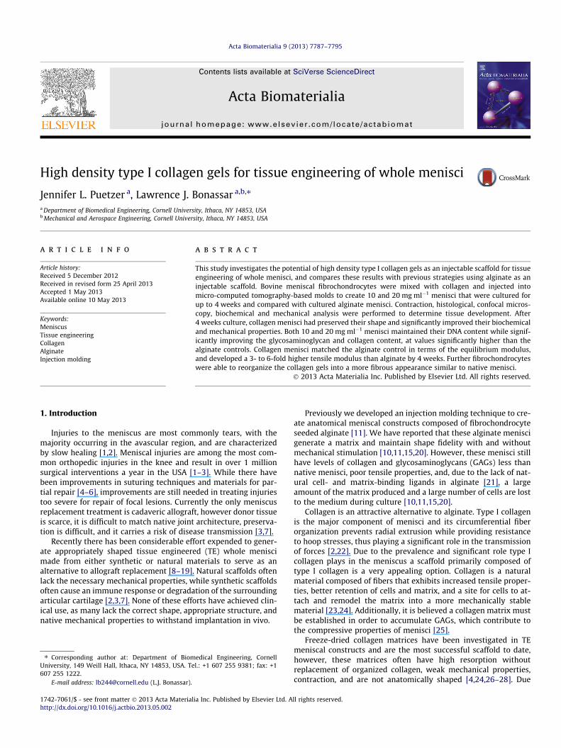
Fig. 1. Photographs of (A) constructs, (B) percent area of collagen menisci with time andbetween 10 and 20 mg ml�1; +significant difference between alginate and both collagen
2.4. Statistics
All biochemical and mechanical data were analyzed by 2- or 3-way ANOVA using Tukey’s t-test for post hoc analysis and P < 0.05as a threshold of statistical significance. All data are expressed asmeans ± SD. Correlation data were fitted to a plane and R2 valueswere determined using r-tables with a < 0.05 consideredsignificant.
3. Results
3.1. Construct shape fidelity and appearance
Gross inspection of the collagen meniscal constructs demon-strated that the overall shape was maintained throughout culture,however, menisci did contract uniformly with time (Fig. 1A). Fur-ther, 20 mg ml�1 menisci appeared to contract less than10 mg ml�1 menisci, which was confirmed by image analysis,which showed that 10 mg ml�1 collagen constructs were signifi-cantly smaller than 20 mg ml�1 constructs from 2 weeks on(Fig. 1B). Alginate scaffolds were not analyzed for changes in areasince it is well established that alginate scaffolds do not changein size with time in culture and we have previously reported thatalginate meniscal constructs maintain their volume and mass over6 weeks static culture [10]. Percent change in mass of constructsthroughout culture matched the area change and volume calcula-tions, with alginate menisci maintaining their mass while boththe 10 and 20 mg ml�1 collagen menisci showed significant de-creases in mass with time in culture (Fig. 1C). Further, by 4 weeksthe 10 and 20 mg ml�1 constructs showed �50–60% decreases inpercent area and mass. This demonstrates that the percent areacalculations are similar to those for volume, and that contractionis uniform in area to volume measurements as well.
3.2. Collagen localization and organization analysis
Brightfield low magnification images of picrosirius red stainedsamples reveal both the 10 and 20 mg ml�1 collagen samplesstained more robustly and had a more uniform distribution of
(C) percent mass of constructs throughout culture. Bar 5 mm. #Significant differenceconcentrations (P < 0.05).

Fig. 2. Brightfield picrosirius red stained images of 4 week static cultured alginate menisci, collagen menisci, and native tissue at 40� (bar 500 lm) and 200� (bar 100 lm).10 and 20 mg ml�1 collagen menisci demonstrate more robust staining, localization, and organization of collagen then alginate. Arrows point to areas of organized collagenfibers in engineered scaffolds.
7790 J.L. Puetzer, L.J. Bonassar / Acta Biomaterialia 9 (2013) 7787–7795
collagen than alginate samples after 4 weeks culture (Fig. 2). High-er magnification images demonstrate that the alginate constructshad only focal accumulation of collagen by 4 weeks, while the col-lagen constructs began to show organized collagen fibers. Due tothe apparent organization in the collagen constructs, further anal-ysis was performed with confocal reflectance and polarized light.
Confocal reflectance imaging demonstrated that the 10 and20 mg ml�1 collagen menisci began as homogeneous, unorganizedcollagen/cell mixtures at 0 weeks, which changed by 4 weeks tolarge regions of organized collagen with cells localized to non-col-lagenous regions, similar to native tissue (Fig. 3). The regions of or-ganized collagen are distinguished by the presences of distinctfibers after 4 weeks culture with 10 mg ml�1 collagen constructsforming 5–12 lm diameter single fibers and 20 mg ml�1 collagenconstructs forming 15–50 lm diameter fiber bundles. Further,there was no noticeable depletion of cells anywhere within thescaffolds, as shown by the visible autofluorescing cells.
In picrosirius red images radial cross-sections of native menisciwere characterized by smaller randomly oriented collagen fibersthat branch off larger and larger circumferential fibers. Picrosiriusred staining demonstrated a similar organization, with both con-centrations of collagen menisci forming well-defined randomlyoriented collagen fibers by 4 weeks (Fig. 3). 20 mg ml�1 meniscihad additional formation of much thicker collagen fibers. Both con-centrations of collagen menisci formed an outer coating of fibersparallel to the surface by 4 weeks.
The collagen menisci are primarily composed of type I collagensince they are created using extracted and reconstituted type I col-lagen from rat tail tendons. However, meniscal fibrochondrocytescan produce both type I and type II collagen, thus immunohisto-chemistry for type II collagen was performed. Type II collagenwas present in detectable levels after 20 days culture in the20 mg ml�1 collagen constructs (see Supplementary material andFig. S1).
3.3. Biochemical analysis
Biochemical analysis demonstrated that menisci constructedfrom 10 and 20 mg ml�1 collagen had significantly more DNA,GAG and collagen at 2 and 4 weeks than alginate menisci. Regard-
less of the scaffold, meniscal fibrochondrocytes produced the sameamount of extracellular matrix (ECM) throughout culture, with sig-nificant improvements in total GAG and collagen production withtime in culture. However, both concentrations of collagen menisciretained significantly more DNA, GAG, and hydroxyproline thanalginate menisci (Fig. 4).
Both groups of collagen menisci maintained or increased theirDNA content, while alginate menisci, seeded at double the concen-tration, lost DNA content with time in culture (P < 0.001). Through-out the entire culture period alginate menisci lost significantlymore DNA to the medium than collagen menisci (data not shown,P < 0.001). Collagen menisci retained �70–80% of their DNA con-tent throughout culture, while alginate menisci retained only20%. This increased retention in collagen menisci was not due toincreased production of DNA, as total DNA (i.e., the sum of DNAin the samples and medium) did not change dramatically through-out culture for any scaffold group (Fig. 4, row 1). Further, therewere no significant spatial differences in DNA concentration forany of the scaffolds with time in culture (power of 78%, data notshown), suggesting there were no areas of necrosis within any ofthe scaffolds.
Collagen menisci showed a 3-fold increase in GAG accumula-tion by 2 weeks (P < 0.001), which was maintained through4 weeks, while alginate menisci demonstrated no significant in-creases in GAG content throughout culture. Total production ofGAG significantly increased after 2 weeks culture for all scaffoldsand was maintained through 4 weeks, and there were no differ-ences in total GAG production between scaffolds. Throughout the4 weeks the alginate menisci lost significantly more GAG to themedium than both concentrations of collagen menisci (data notshown, P < 0.001). Alginate menisci retained �30% of their GAGcontent throughout culture, while both groups of collagen menisciretained �60–80% and �40–50% at 2 and 4 weeks, respectively(Fig. 4, row 2).
As expected, both groups of collagen menisci had significantlyhigher collagen contents than the alginate menisci (P < 0.001),and the 20 mg ml�1 menisci had a significantly higher collagencontent than the 10 mg ml�1 menisci throughout culture(P < 0.001). Additionally, both collagen menisci showed significantincreases in collagen density with time in culture (P < 0.001), while

Fig. 3. Confocal reflectance (green, collagen; red, cells; bar 50 lm) and picrosirius red stained collagen menisci samples visualized with polarized light (bar 100 lm) toobserve the collagen and cell organization, which increased with time in culture and collagen concentration to produce large regions organized similarly to native tissue.Arrows point to areas of organization, with large arrows pointing to large collagen fiber bundles and smaller arrows pointing to radial unorganized single collagen fibers. (Forinterpretation of the references to colour in this figure legend, the reader is referred to the web version of this article.)
J.L. Puetzer, L.J. Bonassar / Acta Biomaterialia 9 (2013) 7787–7795 7791
alginate showed no change in content (Fig. 4, row 3). Because ofthe significant amount of hydroxyproline in collagen-based sam-ples, total hydroxyproline accumulation was assessed by subtract-ing the amount of hydroxyproline at 0 weeks from that atsubsequent time points (Fig. 4, row 3, middle panel). Using thismethod it was clear that, regardless of scaffold type, meniscalfibrochondrocytes produced the same amount of total collagenthroughout culture, and significantly increased this productionwith time in culture. Collagen and alginate menisci lost a similaramount of collagen to the medium throughout culture until4 weeks, when alginate menisci lost significantly more than colla-gen menisci (data not shown, P < 0.03). Thus the 20 mg ml�1 me-nisci retained significantly more collagen throughout culturethan the other scaffolds, with �75% and 60% retention at 2 and4 weeks, respectively. The 10 mg ml�1 menisci retained �55%
and 40% collagen at 2 and 4 weeks, while the alginate menisci re-tained �20% and 5% at 2 and 4 weeks (Fig. 4, row 3).
3.4. Mechanical analysis
Mechanical analysis demonstrated that the equilibrium andtensile moduli of the 10 and 20 mg ml�1 menisci increased signif-icantly with time in culture (Fig. 5). Both the 10 and 20 mg ml�1
menisci had a significantly lower equilibrium modulus than algi-nate at 0 weeks, which increased to a value similar to that of algi-nate at 2 weeks. By 4 weeks both groups of collagen menisci andthe alginate menisci had significantly increased equilibrium mod-uli (P < 0.001), with the 20 mg ml�1 constructs having a signifi-cantly higher equilibrium modulus than the other constructs(P < 0.01). There were no spatial or directional differences in

Fig. 4. DNA, GAG, and collagen content in scaffolds normalized to wet weight (first column), cumulative total production found in scaffolds and media throughout culture(second column), and percent retention in scaffolds (third column). ⁄Significant difference from 0 week; +significant difference from alginate; $significant difference fromweek 2; %significant difference from bracket group (P < 0.05).
Fig. 5. Equilibrium and tensile modulus of meniscal scaffolds throughout culture. ⁄Significant difference from 0 week; +significant difference from alginate; $significantdifference from week 2; %significant difference from bracket group (P < 0.05).
7792 J.L. Puetzer, L.J. Bonassar / Acta Biomaterialia 9 (2013) 7787–7795
compressive properties for any of the constructs throughout cul-ture (data not shown). At 0 weeks the collagen menisci were ableto match the tensile modulus of an alginate meniscus static cul-tured for 6 weeks. With time in culture the collagen menisci signif-icantly improved the tensile modulus, so that by 4 weeks the 10and 20 mg ml�1 menisci showed 3- and 6-fold increases, respec-tively, in tensile modulus over the 6 week cultured alginate menis-ci (P < 0.001).
3.5. Biochemical and mechanical correlations
In order to examine the underlying connection between con-struct properties and composition three-dimensional multiple cor-relation analyses were performed with the equilibrium and tensilemoduli as the dependent variables (z-axis) and the GAG and
hydroxyproline contents as the independent variables (x- and y-axes) (Fig. 6). Both concentrations of collagen menisci were plottedtogether on a graph and a plane was fitted to the data. A significantpositive correlation (P < 0.001) was found between the GAG andcollagen contents for both the equilibrium and tensile moduli ofthe collagen menisci. For both mechanical properties an increasedcollagen content correlated well with increased mechanical mod-uli. Further, both moduli increased at a similar ratio to collagencontent, as is evident from the slopes of 7.9 and 9.2 kPa (lg mg�1)�1
for the equilibrium and tensile moduli, respectively, with respectto collagen content. Increased GAG content had little effect onthe tensile modulus, but had a much more prominent role in theequilibrium modulus. This relationship is easily visualized inthe equations for the planes, where the slope for GAG content inthe tensile plane is 13.8 kPa (lg mg�1)�1, while the slope in the

Fig. 6. Correlation of biochemical components with mechanical properties. ⁄Significant correlation as determined by comparison with critical numbers of R based on thenumbers of samples tested (P < 0.001).
J.L. Puetzer, L.J. Bonassar / Acta Biomaterialia 9 (2013) 7787–7795 7793
equilibrium modulus plane is 29.5 kPa (lg mg�1)�1. Collectivelythese correlations indicate that the GAG content makes a muchgreater contribution to the mechanical properties at lower collagencontents. For example, at 3 lg ml�1 collagen, increasing the GAGcontent from 0.3 to 1.2 lg mg�1 increased the equilibrium modu-lus by 275%, while this same change in GAG content increasedthe equilibrium modulus by only 45% when the collagen contentwas 9 lg mg�1 (Fig. 6).
4. Discussion
This study has demonstrated that TE menisci composed of highdensity type I collagen gels preserve their shape and show signifi-cantly improved biochemical and mechanical properties with timein culture. Both the 10 and 20 mg ml�1 menisci maintained theirDNA content while significantly increasing the content of ECMcomponents. The collagen menisci matched the equilibrium mod-ulus of alginate menisci, while having a 3- to 6-fold higher tensilemodulus after 4 weeks culture. Further, the 20 mg ml�1 meniscicontracted less while having improved organization, ECM andmechanical properties than the 10 mg ml�1 menisci.
Confocal reflectance and picrosirius red staining revealed thatmeniscal fibrochondrocytes are able to reorganize the collagenscaffolds, resulting in a more fibrous appearance similar to nativemenisci with time in culture. This supports a previous study thatfound that meniscal fibrochondrocytes were capable of organizingtheir surrounding matrix with time in vivo and suggested thatthese cells have an intrinsic ability to form specific meniscal tissuestructures [40]. It has been well established in the literature thatcollagen gels do not change in size or structure without the pres-ence of cells [23,31], therefore, we believe that the cells are respon-sible for this reorganization in the collagen menisci. Additionally,the collagen menisci formed an outer coating of collagen fibers
parallel to the surface, similar to the superficial zone of native me-nisci. A well-developed superficial zone will most likely be neededfor any meniscal replacement in order for the engineered tissue tofunction properly within the knee.
As discussed in the Introduction, contraction is an inherentproperty of cell seeded collagen gels, which decreases withincreasing concentrations of collagen [23,31,32]. In this study thecollagen menisci contracted significantly with time in culture,and with that time and contraction they developed significantlyimproved ECM and mechanical properties. These data suggest thatcontraction plays a significant role in the increased GAG and colla-gen concentrations when normalized to wet weight. However, con-traction is not the only reason for the improvement in properties.Total overall production of GAG and collagen in the scaffold andmedium significantly improved with time in culture, and the colla-gen menisci retained significantly more GAG and collagen than thealginate menisci throughout culture, thus demonstrating that con-traction was not the sole reason for the improved biochemicalproperties.
Further, we believe that the improved biochemical propertiesare primarily due to the collagen scaffolds retaining more matrixcomponents, thus enhancing the ability for matrix to be formed,rather than the scaffold influencing the production rate of cells.This theory is in contrast to previous studies that compared men-iscal fibrochondrocytes seeded on fibers with those encapsulatedin gels [40,41]. Aufderheide et al. [41] previously compared meni-scal fibrochondrocyte seeded fibrous poly(glycolic acid) (PGA) scaf-folds with agarose hydrogels and found significant improvementsin the biochemical composition of PGA over agarose scaffolds. Thusthey concluded that meniscal fibrochondrocytes produce moreECM when attached to fibers than when encapsulated in gels.However this study did not investigate the amount of ECM re-leased into the medium and, therefore, did not calculate totalamounts produced [41]. In the current study, when the contents

7794 J.L. Puetzer, L.J. Bonassar / Acta Biomaterialia 9 (2013) 7787–7795
of GAG and collagen in the medium and scaffold were added to-gether there were no differences in the total production of thesebiochemical components between the collagen and alginate scaf-folds. This demonstrates that fibrochondrocytes produce the sameamount of ECM whether cultured on fibers or encapsulated in gels,and that a primary advantage of collagen-based gels is their abilityto retain more of the ECM produced.
In this study bovine meniscal fibrochondrocytes were used,however, for this scaffold to be used clinically it will most likelyhave to incorporate human cells. There are a variety of cell sourcesfor meniscal repair and regeneration currently being investigated,as discussed in the review by Makris et al. [42]. A number of groupsare investigating the expansion of autologous meniscal fibrochon-drocytes (MFCs), the use of allogeneic MFCs, and the differentiationof human embryonic stem cells (hESCs) and adult mesenchymalstem cells (hMSCs) [42]. Our high density collagen scaffolds couldbe used with any of these cell sources. The advantage of allogenicMFCs would be they have the right synthetic potential, whileautologous hESCs and hMSCs have a better immune profile.
Many previous meniscus TE studies have reported loss of GAGto the culture medium with mechanical loading and have sug-gested that an established collagen matrix is necessary in orderto maintain GAG accumulation within the scaffold [15,25,41]. Datafrom the current study further support this theory, based on theobservation of significant improvements in GAG accumulation inboth concentrations of collagen menisci but no improvement inthe alginate menisci with time in culture.
Both concentrations of collagen menisci not only showed signif-icant improvements in biochemical properties but also in compres-sive and tensile mechanical properties. GAGs are thought to beresponsible for the compressive properties of cartilaginous tissues,due to their high osmotic pressure, while collagen is thought toprovide the tensile properties. In this study we performed correla-tion studies to determine the contribution of different biochemicalcomponents to the mechanical properties of the collagen menisci.A significant positive correlation was found between the GAG andcollagen contents for both the equilibrium and tensile moduli ofthe collagen menisci. We found that the amount of collagen pres-ent was positively correlated with both the compressive and ten-sile moduli. In keeping with the hypothesized mechanical role ofGAGs, GAG had a much more prominent role in the equilibriummodulus than the tensile modulus. Further, GAGs appeared to playmore of a role in the mechanical properties at lower concentrationsof collagen. This suggests that the collagen content plays a signifi-cant role in the mechanical properties of TE menisci and that onlywhen there is less collagen present does GAG play a slight role.Similarly, in a previous study on TE menisci [15], we found thatthe collagen content had a more direct relationship to the com-pressive modulus than GAG. This could be due to the increasedorganization attributed to collagen fiber accumulation and not nec-essarily the properties of collagen [41,43].
There have been many studies investigating freeze-dried colla-gen matrices in meniscus tissue engineering. To date they havebeen some of the most successful scaffolds in meniscal research,however, these matrices often have high resorption, poor mechan-ical properties, contraction, are not anatomically shaped, and aredifficult to seed with cells throughout the scaffold [4,24,26–28].Additionally, there have been many recent studies investigatingthe use of fibrous meniscal scaffolds [9,16,19,44]. The high densitycollagen gels developed in this study build on the successes of pastfreeze-dried collagen matrices and overcome many of the obstaclesassociated with collagen matrices and other fibrous scaffolds.Injection molding allows cells to be easily seeded throughout ana-tomically correct scaffolds. Further, with time collagen meniscishow improved biochemical and mechanical properties, in the ab-sence of any mechanical or chemical conditioning. Injection
molded constructs often have inferior mechanical properties to fi-brous scaffolds. However, it is believed that with time and condi-tioning, such as mechanical and chemical stimulation, injectionmolded constructs can significantly improve their mechanicalproperties, while fibrous scaffolds often lose mechanical propertieswith time as cells resorb fiber components without replacement oforganized collagen. We have previously reported matching nativecompressive properties in chemically stimulated alginate con-structs [45] and we believe the tensile properties will increase withmechanical and/or chemical stimulation.
This is the first study to our knowledge to investigate the use ofhigh density collagen gels for injection molding of living engi-neered tissues. Both 10 and 20 mg ml�1 concentrations were ableto be successfully seeded with cells and injection molded to formmeniscal scaffolds. The higher concentration 20 mg ml�1 meniscicontracted less, had greater collagen fiber formation by 4 weeks,and had better ECM and mechanical properties than the10 mg ml�1 menisci. A higher density of collagen did result in lessoverall contraction as we hypothesized, however, the 20 mg ml�1
menisci still contracted to �60% of their original size. It has beenreported that a deviation of more than 10% in meniscal size match-ing can result in detrimental loading across the joint [46], thus it isimportant we control contraction. Contraction of the collagen me-nisci was uniform and consistent, which suggests that we couldaccommodate the contraction by simply over-sizing our molds.Contraction of the collagen gels is beneficial, as it results in an in-creased collagen density, better construct biochemical andmechanical properties, and improved collagen organization. It isdifficult to create collagen meniscal constructs above 20 mg ml�1
since the collagen becomes too dense to injection mold. However,a collagen concentration of 20 mg ml�1 is still only�15% of the col-lagen concentration in native menisci. As the collagen contracts itallows the concentration to increase, reaching a collagen concen-tration of �30% of that of native menisci by 4 weeks static culture.Further, contraction allows organization of the collagen fibers.Contraction in collagen gels occurs as cells create traction forceswithin the gel, often resulting in the formation of well-defined col-lagen fibrils [29,31,32]. By 4 weeks culture we found that the 10and 20 mg ml�1 collagen gels had increased organization, withthe 10 mg ml�1 menisci developing 5–12 lm diameter single fibersand the 20 mg ml�1 menisci developing 15–50 lm diameter fiberbundles. We could attempt to control this contraction by using gly-cated collagen [47] or riboflavin crosslinking, which we have pre-viously demonstrated limited contraction in our TE intervertebraldiscs [48]. However, limiting contraction long term could decreaseor impede collagen reorganization and thus may not be the opti-mal choice for ensuring the constructs are appropriately sized.
5. Conclusions
This study has demonstrated the use of high density type I col-lagen gels to create whole meniscal constructs with mechanicaland biochemical properties similar to native menisci. The GAGand collagen concentrations increased to �20% and �15–30% thatof native tissue, respectively, while the equilibrium modulus was�50–70% that of native menisci. Although the tensile modulus im-proved 6-fold in the collagen menisci over alginate controls, this isstill 1–2 orders of magnitude lower than native menisci and doesnot mirror the anisotropic properties of native tissue. Thus,although the use of constructs seeded with bovine meniscal fibro-chondrocytes demonstrate great promise as meniscal replace-ments, future work should focus on the use of human cells andimproving the tissue organization and tensile properties to achievethe functional anisotropic organization and properties of the nativetissue.

J.L. Puetzer, L.J. Bonassar / Acta Biomaterialia 9 (2013) 7787–7795 7795
Acknowledgements
The authors would like to thank the National Science Founda-tion Graduate Research Fellowship Program, the Cornell BME NSFGK-12 program (DGE 0841291), the Microscopy and Imaging Facil-ity, Life Sciences Core Laboratories Center at Cornell University, Ed-ward Bonnevie, and the members of the Bonassar Laboratory fortheir support in this research.
Appendix A. Figures with essential colour discrimination
Certain figures in this article, particularly Figs. 1–6, is difficult tointerpret in black and white. The full colour images can be found inthe on-line version, at http://dx.doi.org/10.1016/j.actbio.2013.05.002
Appendix B. Supplementary data
Supplementary data associated with this article can be found, inthe online version, at http://dx.doi.org/10.1016/j.actbio.2013.05.002.
References
[1] Khetia EA, McKeon BP. Meniscal allografts: biomechanics and techniques.Sports Med Arthrosc 2007;15:114–20.
[2] Sweigart MA, Athanasiou KA. Toward tissue engineering of the knee meniscus.Tissue Eng 2001;7:111–29.
[3] Peters G, Wirth CJ. The current state of meniscal allograft transplantation andreplacement. Knee 2003;10:19–31.
[4] Stone KR, Steadman JR, Rodkey WG, Li ST. Regeneration of meniscal cartilagewith use of a collagen scaffold. Analysis of preliminary data. J Bone Joint SurgAm 1997;79:1770–7.
[5] Farng E, Sherman O. Meniscal repair devices: a clinical and biomechanicalliterature review. Arthroscopy 2004;20:273–86.
[6] McDermott I. Meniscal tears, repairs and replacement: their relevance toosteoarthritis of the knee. Br J Sports Med 2011;45:292–7.
[7] Buma P, Ramrattan NN, van Tienen TG, Veth RP. Tissue engineering of themeniscus. Biomaterials 2004;25:1523–32.
[8] Aufderheide AC, Athanasiou KA. Assessment of a bovine co-culture, scaffold-free method for growing meniscus-shaped constructs. Tissue Eng2007;13:2195–205.
[9] Balint E, Gatt CJ, Dunn MG. Design and mechanical evaluation of a novel fiber-reinforced scaffold for meniscus replacement. J Biomed Mater Res A2012;100:195–202.
[10] Ballyns JJ, Bonassar LJ. Dynamic compressive loading of image-guided tissueengineered meniscal constructs. J Biomech 2011;44:509–16.
[11] Ballyns JJ, Gleghorn JP, Niebrzydowski V, Rawlinson JJ, Potter HG, Maher SA,et al. Image-guided tissue engineering of anatomically shaped implants viaMRI and micro-CT using injection molding. Tissue Eng Part A2008;14:1195–202.
[12] Huey DJ, Athanasiou KA. Tension – compression loading with chemicalstimulation results in additive increases to functional properties of anatomicmeniscal constructs. PLoS One 2011;6:e27857.
[13] Kang SW, Son SM, Lee JS, Lee ES, Lee KY, Park SG, et al. Regeneration of wholemeniscus using meniscal cells and polymer scaffolds in a rabbit totalmeniscectomy model. J Biomed Mater Res A 2006;78:659–71.
[14] Kon E, Chiari C, Marcacci M, Delcogliano M, Salter DM, Martin I, et al. Tissueengineering for total meniscal substitution: animal study in sheep model.Tissue Eng Part A 2008;14:1067–80.
[15] Puetzer JL, Ballyns JJ, Bonassar LJ. The effect of the duration of mechanicalstimulation and post-stimulation culture on the structure and properties ofdynamically compressed tissue-engineered menisci. Tissue Eng Part A2012;18:1365–75.
[16] Tienen TG, Heijkants RG, de Groot JH, Schouten AJ, Pennings AJ, Veth RP, et al.Meniscal replacement in dogs. Tissue regeneration in two different materialswith similar properties. J Biomed Mater Res B Appl Biomater 2006;76:389–96.
[17] Zur G, Linder-Ganz E, Elsner JJ, Shani J, Brenner O, Agar G, et al.Chondroprotective effects of a polycarbonate–urethane meniscal implant:histopathological results in a sheep model. Knee Surg Sports TraumatolArthrosc 2011;19:255–63.
[18] Huey DJ, Athanasiou KA. Maturational growth of self-assembled, functionalmenisci as a result of TGF-beta1 and enzymatic chondroitinase-ABCstimulation. Biomaterials 2011;32:2052–8.
[19] Mandal BB, Park SH, Gil ES, Kaplan DL. Multilayered silk scaffolds for meniscustissue engineering. Biomaterials 2011;32:639–51.
[20] Ballyns JJ, Wright TM, Bonassar LJ. Effect of media mixing on ECM assemblyand mechanical properties of anatomically-shaped tissue engineeredmeniscus. Biomaterials 2010;31:6756–63.
[21] Awad HA, Wickham MQ, Leddy HA, Gimble JM, Guilak F. Chondrogenicdifferentiation of adipose-derived adult stem cells in agarose, alginate, andgelatin scaffolds. Biomaterials 2004;25:3211–22.
[22] Kawamura S, Lotito K, Rodeo SA. Biomechanics and healing response of themeniscus. Oper Techn Sports Med 2003;11:68–76.
[23] Cross VL, Zheng Y, Won Choi N, Verbridge SS, Sutermaster BA, Bonassar LJ,et al. Dense type I collagen matrices that support cellular remodeling andmicrofabrication for studies of tumor angiogenesis and vasculogenesis in vitro.Biomaterials 2010;31:8596–607.
[24] Mueller SM, Shortkroff S, Schneider TO, Breinan HA, Yannas IV, Spector M.Meniscus cells seeded in type I and type II collagen–GAG matrices in vitro.Biomaterials 1999;20:701–9.
[25] Baker BM, Shah RP, Huang AH, Mauck RL. Dynamic tensile loading improvesthe functional properties of mesenchymal stem cell-laden nanofiber-basedfibrocartilage. Tissue Eng Part A 2011;17:1445–55.
[26] Martinek V, Ueblacker P, Braun K, Nitschke S, Mannhardt R, Specht K, et al.Second generation of meniscus transplantation: in vivo study with tissueengineered meniscus replacement. Arch Orthop Trauma Surg2006;126:228–34.
[27] Stone KR, Rodkey WG, Webber R, McKinney L, Steadman JR. Meniscalregeneration with copolymeric collagen scaffolds. In vitro and in vivostudies evaluated clinically, histologically, and biochemically. Am J SportsMed 1992;20:104–11.
[28] Walsh CJ, Goodman D, Caplan AI, Goldberg VM. Meniscus regeneration in arabbit partial meniscectomy model. Tissue Eng 1999;5:327–37.
[29] Bowles RD, Williams RM, Zipfel WR, Bonassar LJ. Self-assembly of alignedtissue-engineered annulus fibrosus and intervertebral disc composite viacollagen gel contraction. Tissue Eng Part A 2010;16:1339–48.
[30] Elsdale T, Bard J. Collagen substrata for studies on cell behavior. J Cell Biol1972;54:626–37.
[31] Bell E, Ivarsson B, Merrill C. Production of a tissue-like structure by contractionof collagen lattices by human fibroblasts of different proliferative potentialin vitro. Proc Natl Acad Sci USA 1979;76:1274–8.
[32] Vernon RB, Sage EH. Contraction of fibrillar type I collagen by endothelial cells:a study in vitro. J Cell Biochem 1996;60:185–97.
[33] Chang SC, Rowley JA, Tobias G, Genes NG, Roy AK, Mooney DJ, et al. Injectionmolding of chondrocyte/alginate constructs in the shape of facial implants. JBiomed Mater Res 2001;55:503–11.
[34] Carey SP, Kraning-Rush CM, Williams RM, Reinhart-King CA. Biophysicalcontrol of invasive tumor cell behavior by extracellular matrixmicroarchitecture. Biomaterials 2012;33:4157–65.
[35] Mason BN, Starchenko A, Williams RM, Bonassar LJ, Reinhart-King CA. Tuningthree-dimensional collagen matrix stiffness independently of collagenconcentration modulates endothelial cell behavior. Acta Biomater2013;9:4635–44.
[36] Kim YJ, Sah RL, Doong JY, Grodzinsky AJ. Fluorometric assay of DNA in cartilageexplants using Hoechst 33258. Anal Biochem 1988;174:168–76.
[37] Enobakhare BO, Bader DL, Lee DA. Quantification of sulfatedglycosaminoglycans in chondrocyte/alginate cultures, by use of 1,9-dimethylmethylene blue. Anal Biochem 1996;243:189–91.
[38] Neuman RE, Logan MA. The determination of hydroxyproline. J Biol Chem1950;184:299–306.
[39] Gleghorn JP, Jones AR, Flannery CR, Bonassar LJ. Boundary mode frictionalproperties of engineered cartilaginous tissues. Eur Cell Mater 2007;14:20–8[discussion 8–9].
[40] Ibarra C, Jannetta C, Vacanti CA, Cao Y, Kim TH, Upton J, et al. Tissue engineeredmeniscus: a potential new alternative to allogeneic meniscus transplantation.Transplant Proc 1997;29:986–8.
[41] Aufderheide AC, Athanasiou KA. Comparison of scaffolds and cultureconditions for tissue engineering of the knee meniscus. Tissue Eng2005;11:1095–104.
[42] Makris EA, Hadidi P, Athanasiou KA. The knee meniscus: structure–function,pathophysiology, current repair techniques, and prospects for regeneration.Biomaterials 2011;32:7411–31.
[43] Waldman SD, Spiteri CG, Grynpas MD, Pilliar RM, Kandel RA. Long-termintermittent shear deformation improves the quality of cartilaginous tissueformed in vitro. J Orthop Res 2003;21:590–6.
[44] Fisher MB, Henning EA, Soegaard N, Esterhai JL, Mauck RL. Organizednanofibrous scaffolds that mimic the macroscopic and microscopicarchitecture of the knee meniscus. Acta Biomater 2013;9:4496–504.
[45] Puetzer JL, Brown B, Ballyns JJ, Bonassar LJ. The effect of IGF-I on anatomically-shaped tissue engineered menisci. Tissue Eng Part A 2013;19:1443–50.
[46] Dienst M, Greis PE, Ellis BJ, Bachus KN, Burks RT. Effect of lateral meniscalallograft sizing on contact mechanics of the lateral tibial plateau: anexperimental study in human cadaveric knee joints. Am J Sports Med2007;35:34–42.
[47] Roy R, Boskey AL, Bonassar LJ. Non-enzymatic glycation of chondrocyte-seeded collagen gels for cartilage tissue engineering. J Orthop Res2008;26:1434–9.
[48] Mozia, R.I., Bowels, R., Saroka, J., Gebhard, H., Hartl, R., Bonassar, L.J. Riboflavincrosslinking of composite tissue engineered intervertebral discs. AnnualMeeting of the Orthopeadic Research Society, Long Beach, CA, Poster No.0312; 2011.


