Embed Size (px)
Citation preview

ISSN 1070-3284, Russian Journal of Coordination Chemistry, 2009, Vol. 35, No. 10, pp. 731–739. © Pleiades Publishing, Ltd., 2009.Original Russian Text © V.V. Gorinchoy, V.E. Zubareva, S.G. Shova, V.N. Szafranski, J. Lipkowski, N. Stanica, Yu.A. Simonov, C.I. Turta, 2009, published in KoordinatsionnayaKhimiya, 2009, Vol. 35, No. 10, pp. 743–751.
731
The chemistry of homo- and heteropolynuclear ironcarboxylates are of current interest because they helpthe researchers to enhance their knowledge of magneticinteractions between paramagnetic ions [1] and pro-mote the development of bioinorganic chemistry [2].Compounds of this class serve as precursors of nanoox-ide and metallic particles with useful magnetic proper-ties [3] and can be used as physiologically active drugs[4], etc.
Metal complexes with acetic acid [5] and its halogenderivatives [6–12] have been comprehensively studiedto date. The structures of many such complexes wereproved by X-ray diffraction. Some studies weredevoted to the structures and properties of complexeswith hydroxy carboxylic acids (e.g., salicylic acid,H
2
Sal). According to the literature data, coordinatedsalicylic acid is fully or partially deprotonated and canact as a monodentate, chelating bidentate, or bridgingligand [13]. For instance, salicylic acid in a dimeric ter-bium complex has been found to be coordinated inthree ways [14]. In a nanonuclear manganese complexwith benzoic and salicylic acids, the anion Sal
2–
is coor-dinated in a pentadentate chelating
µ
3
-bridging fashion
[15]. Among 120 metal complexes with salicylic aciddeposited with the Cambridge Crystallographic DataCollection, we have found no iron(III)
µ
3
-oxo salicy-lates [16].
In this study, we obtained the trinuclear homo- andheterometallic iron(III) complexes[Fe
2
MgO
(
SalH
)
6
(DMAA)
0.4
(
H
2
O
)
2.6
]
· 4DMAA
(
I
),[
Fe
2
CoO
(
SalH
)
6
(
CH
3
OH
)
2
(
H
2
O
)]
· DMF ·
2.5
H
2
O
(
II
),
and
[
Fe
3
O
(
SalH
)
6
(
H
2
O
)
3
]ë
l
· DMAA ·
H
2
O
(
III
)
andexamined their structures.
EXPERIMENTAL
The starting salt Mg
(
SalH
)
2
·
3
H
2
O was preparedfrom MgO and salicylic acid. The other reagents(CH
3
OH, DMAA, DMF, and THF) were commercialchemicals.
Synthesis of complex I.
The saltMg
(ë
6
H
4
(
OH
)ë
OO
)
2
·
3
H
2
O (3.49 g, 9.89 mmol) wasadded with continuous stirring to Fe
(
NO
3
)
3
·
9
H
2
O (1 g,2.47 mmol) in methanol (25 ml). After 30 min, theresulting solution was filtered and dried on a waterbath. To a dry residue THF (18 ml) and DMAA (7 ml)
Homo- and Heteronuclear Iron Complexes {Fe
2
MO} with Salicylic Acid: Synthesis, Structures,
and Physicochemical Properties
V. V. Gorinchoy
a
, V. E. Zubareva
a
, S. G. Shova
b
, V. N. Szafranski
a
, J. Lipkowski
c
, N. Stanica
d
, Yu. A. Simonov
b
, and C. I. Turta
a
*
a
Institute of Chemistry, Academy of Sciences of Moldova, Chisinau, Moldova
b
Institute of Applied Physics, Academy of Sciences of Moldova, Chisinau, Moldova
c
Institute of Physical Chemistry, Polish Academy of Sciences, Warsaw, Poland
d
Institute of Physical Chemistry, Romanian Academy of Sciences, Bucharest, Romania*e-mail: [email protected]
Received December 17, 2008
Abstract
—A reaction of iron nitrate with magnesium salicylate and reactions of iron and cobalt chlorides withammonium salicylate in the presence of water, methanol, DMAA, and DMF gave the trinuclear heterometalliccomplexes: [hexa-
µ
-salicylato-
µ
3
-oxo-0.4-dimethylacetamide-2.6-aquadiiron(III)magnesium(II)] tetra(dime-thylacetamide), [Fe
2
MgO(SalH)
6
(DMAA)
0.4
(H
2
O)
2.6
] · 4DMAA (
I
); [hexa-
µ
]-salicylato-
µ
3
-oxo(dimetha-nol)aquadiiron(III)cobalt(II)] dimethylformamide · 2.5-hydrate, [Fe
2
CoO(SalH)
6
(CH
3
OH)
2
(H
2
O)] · DMF ·2.5H
2
O (
II
); and [hexa-
µ
-salicylato-
µ
3
-oxotriaquatriiron(III)] chloride dimethylacetamide monohydrate,[Fe
3
O(SalH)
6
(H
2
O)
3
]Cl · DMAA · H
2
O (
III
). The X-ray study revealed that the molecular structures of com-plexes
I
and
II
are [Fe
2III
M
II
(
µ
3
-O)(RCOO)
6
L
3
] ·
n
Solv. The IR and Mössbauer spectra of complexes
I
–
III
were examined; their magnetochemical and thermal properties were studied. The parameters of the Mössbauerspectra (
δ
Na+
= 0.69
±
0.03 mm/s,
∆
E
Q
= 0.76–1.08 mm/s, 300 K) suggest the high-spin state of the Fe
3+
ionsin complexes I–III (S = 5/2). The paramagnetic Fe3+ ions are involved in antiferromagnetic exchange interac-tions with the parameter J = –44 cm–1, g = 2.05 (for I). Complexes I–III are thermally unstable.
DOI: 10.1134/S1070328409100042

732
RUSSIAN JOURNAL OF COORDINATION CHEMISTRY Vol. 35 No. 10 2009
GORINCHOY et al.
were added. The mixture was stirred at room tempera-ture for 20 min, filtered, and left in air for crystalliza-tion. After four weeks, dark red crystals of complex Iformed as rectangular prisms. The yield was 2.72 g(69%).
Synthesis of complex II. A solution of CoCl2 ·6H2O (1.15 g, 4.8 mmol) in methanol (10 ml) wasadded to ammonium salicylate (1.5 g, 9.68 mmol) inmethanol (10 ml). The mixture was refluxed for ~1 h,whereupon a solution of Fe(NO3)3 · 9H2O (0.49 g,1.2 mmol) in a mixture of methanol (10 ml) and DMF(1 ml) was added. Heating was continued while stirringthe mixture until pink flakes dissolved completely.After 1 h, DMF (1 ml) and THF (1 ml) were added. Thisresulted in the formation of a fine crystalline solid,which dissolved completely on 1-h heating. The result-ing dark red solution was transferred to a warm beakerand left for crystallization. After a day, a dark brown(with a reddish tinge) coarse crystalline solid was fil-tered off, washed with methanol (2 ml) and ether (2 ml)(the solid is soluble in both solvents), and dried in air.Single crystals suitable for X-ray diffraction analysiswere obtained by recrystallization of complex II frommethanol. The yield was 0.51 g (68%).
Synthesis of complex III. A solution of FeCl3 ·6H2O (1 g, 2.7 mmol) in methanol (4 ml) and a solutionof ammonium salicylate (1.15 g, 7.4 mmol) in methanol(5 ml) were mixed to give a dark violet precipitate. Theprecipitate was stirred with DMAA (0.8 ml, 9.2 mmol).A fine crystalline red-brown solid formed. Water(10 ml) was added and the reaction mixture was thor-oughly stirred. The product was filtered off and washedwith water (2 × (15–20) ml). The yield was 1.1 g (25%).
Complexes I–III were analyzed for C, H, and N bythe elemental analysis group of the Institute of Chemis-try of the Academy of Sciences of the Moldova Repub-lic. Complexes I–III were analyzed for metals on anAAS-1N atomic absorption spectrometer (Carl Zeiss)at the Center of Metrology and Analytical Methods ofthe Academy of Sciences of the Moldova Republic.
For C59.6H74.8Fe2MgN4.4O26
anal. calc., %: C, 50.95; H, 5.36; N, 4.38.
Found, %: C, 50.78; H, 5.47; N, 4.43.
ForC47H52CoFe2NO25.5
anal. calcd, %: C, 46.67; H, 4.33; N, 1.15.
Found, %: C, 45.77; H, 4.20; N, 1.46.
For C46H47ClFe3NO24
anal. calcd, %: C, 46.01; H, 3.94; N, 1.17.
Found, %: C, 45.85; H,4.27; N, 1.18.
X-ray diffraction analysis. Experimental materialfor complexes I and II was collected at 100 K on a Non-ius Kappa CCD diffractometer (MoKα radiation, graph-ite monochromator, ω−2θ scan mode). The unit cellparameters were refined for the whole array of experi-mental data. The integration of the reflection intensitiesand their reduction to a common scale was performedwith the DENZO and SKALEPACK programs [17].Absorption correction was applied with the XEMP pro-gram [18].
Structures I and II were solved by direct methodsand refined by the least-squares method in the anisotro-pic full-matrix approximation for non-hydrogen atoms(SHELX-97) [19]. The hydrogen atoms in complex Iwere located objectively; those in complex II were cal-culated geometrically and refined isotropically usingthe rigid-body model. The refinement revealed that theposition at the M(2) atom in complex I is randomlyoccupied by water and DMAA molecules (0.6 : 0.4). Incomplex II, the hydroxy O atoms of the coordinatedHSal residues are disordered.
Crystallographic parameters and the data collectionstatistics for structures I and II are given in Table 1.Selected bond lengths and bond angles are listed inTables 2 and 3. Atomic coordinates and other parame-ters for structures I and II have been deposited with theCambridge Crystallographic Data Collection(nos. 723904 (I) and 723 903 (II)); [email protected] or http://www.ccdc.cam.ac.uk/data_request/cif.
IR spectra were recorded on a Specord M75 spec-trometer in the 200–600 cm−1 range and a Perkin-Elmer100 FT-IR spectrometer in the 400–4000 cm–1 range(suspensions in Vaseline or fluorinated oils).
Integrated thermal analysis was carried out on aPaulik–Paulik–Erdey derivatograph in air with Al2O3 asa reference. The recording conditions were 1/5 (DTG),1/10 (DTA), and 100/100 (TG), Tmax = 800°C, heatingrate 5°C/min, a sample weight 100 mg.
The magnetic properties of complexes II and IIwere studied by the Gouy method at room temperature;the temperature range for complex I was 291–136 K.The magnetic susceptibility was calibrated againstHg[Co(NCS)4]. Diamagnetic corrections were appliedfrom the Pascal constants [1, 20].
Mössbauer spectra were recorded on an electrody-namic setup with a constant acceleration at room tem-perature; 57Co in a Rh matrix was used as a radiationsource (1.0 MBq). The isomer shifts are referenced tosodium nitroprusside.
RESULTS AND DISCUSSION
Heterotrinuclear iron complexes with salicylic acidcan be obtained when mixing a solution of iron(III)nitrate with some metal salicylate, or with simple metalsalts in the presence of ammonium salicylate. It wasfound that the formation of a crystalline product is well

RUSSIAN JOURNAL OF COORDINATION CHEMISTRY Vol. 35 No. 10 2009
HOMO- AND HETERONUCLEAR IRON COMPLEXES 733
reproducible only in the presence of a small amount ofdimethylacetamide. In addition, a considerable excessof bivalent metal ions (Mg2+ and Co2+) is required toincorporate them into a trimer structure.
The best results were obtained in the study of reac-tions of Fe3+ ions with salicylates of other 2s- and 3dmetals (Mg or Co) in the CH3OH–DMAA–THF sys-tem. In this series of solvents, the affinity for coordina-tion changes as follows: THF < DMAA ~ DMF < ç2é ~ëç3éç.
Structures I and II are molecular ones. The trinu-clear complexes [Fe2MO(OOCR)6 · 3L] · nSolv containtwo iron(III) atoms and a bivalent metal atom (M = Mg(I, Fig. 1) and Co (II, Fig. 2)). The outer sphere in thepacking of large trinuclear complexes in the crystalspace is made up of solvate molecules (nSolv =4DMAA (I) and DMF · 2.5ç2é (II)).
A main problem in the study of trinuclear heterometal-lic complexes is the determination of the distribution ofmetal cations in the cluster. Their statistical distributionwas proved in [21–24]. In some cases [25–27] (e.g., forclose atomic numbers of the metal cations in the PeriodicTable), such a distribution is presumed. In other cases, astatistical distribution of cations follows from the ownsymmetry of complex ë3 [28]. In recent years, localizationof foreign cations has been proved for trimers containingmetals with different atomic radii, electronic properties,and atomic numbers in the Periodic Table. This fact wasrevealed with Fe2/Sr, Fe2/Ba, and Fe2/Ca complexes of theformula [Fe2M(Ca,Sr,Ba)O(CCl3COO)6(THF)n] [29].
The differences between the ionic radii of Fe3+ (0.78 Å),Ca2+ (1.00 Å), Sr2+ (1.18 Å), and LJ2+ (1.35 Å) make theå3 triangle substantially distorted. In physicochemicalcharacteristics, magnesium is an analog of calcium, stron-tium, and barium, but its ionic radius (0.72 Å [30]) is suf-ficiently close to that of iron(III); therefore, one can expectits incorporation into the å3 triangle in complex I.
In [24], it was found that the Mg2+ ions in
[F MgIIO(OCl3COO)6Py3] · CH3C6H5 (IV) are statis-tically distributed with iron atoms over two positions of
the trimer and those in [F MgIIO(OCl3COO)6(íçF)3]are statistically distributed over all three positions.Such a distribution is also true for metal–ligand dis-tances in coordination entities. In complex I, the M–Mdistances in the triangle are 3.288–3.234 Å and the M–O distances are normal for complexes of this type(Table 2). It is impossible to identify the positions pre-dominantly occupied by Mg or Fe atoms. An analysisof the geometrical parameters did not reveal the domi-nant occupation of the positions M(1)–M(3) by Fe3+
and Mg2+ cations. In crystal structure I, the geometry ofthe triangle å(3)(µ3-é) is typical. Each metal atom hasa slightly distorted octahedral environment with closeinteratomic distances and geometrical parameters. The
average M–O(1)oxo (1.887 ) and M–Ocarb bond
lengths (2.036 ) in complex I do not differ substan-tially from those in complex IV. The specific feature of
e2III
e2III
Å
Å
Table 1. Crystallographic parameters and the data collection statistics for structures I and II
ParameterValue
I II
M 1404.85 1209.53Space group C2/c C2/cUnit cell parametersa, Å 43.3859(15) 13.4150(3)b, Å 15.3037(5) 21.0090(6)c, Å 20.8776(6) 21.5450(5)β, deg 103.997(3) 99.920(2)V, Å3 13450.4(7) 5981.4(3)Z 8 4ρcalcd, g/cm3 1.388 1.343µMo, mm–1 0.524 0.830F(000) 5882 2496θ scan range, deg 2.01–26.00 1.82–26.00(Ranges of h, k, and l indices –53 ≤ h ≤ 53, –18 ≤ k ≤ 18, –25 ≤ l ≤ 25 –16 ≤ h ≤ 16, –25 ≤ k ≤ 25, –26 ≤ l ≤ 26Number of measured reflections: 98644 11078Number of independent reflections,I > 2σ(I)
13205 (Rint = 0.0846) 5858 (Rint = 0.0627)
Completeness of data collection for θ, % 99.9 99.5Number of parameters refined 1032 388GOOF on F2 1.129 1.038R factors (I > 2σ(I)) R1 = 0.0751, wR2 = 0.2199 R1 = 0.0997, wR2 = 0.2663R factors (for all reflections) R1 = 0.1423, wR2 = 0.2584 R1 = 0.1398, wR2 = 0.2889∆ρmax and ∆ρmin, e Å–3 0.703 and –0.929 1.264 and –0.462

734
RUSSIAN JOURNAL OF COORDINATION CHEMISTRY Vol. 35 No. 10 2009
GORINCHOY et al.
the trimer is the statistic distribution of neutral ligands.The positions at the M(1) and M(2) atoms are unambig-uously occupied by a water molecule, while the posi-tion at the M(3) atom is statistically occupied by waterand DMAA molecules (0.6 : 0.4). In crystal structure I,the outer-sphere DMAA molecules and the complexmolecules are united through a system of hydrogenbonds (Table 4). The complexes are also linked by π–πand ë–ç···π interactions.
Structure II is similar to structure I; the own sym-metries of the trimers differ. Two Fe3+ ions and a Co2+
ion are statistically distributed, making up an isoscelestriangle (M(1)–M(2), 3.287 Å; M(2)–M(3), 3.287 Å).The center of the triangle is occupied by the µ3-O atomcoplanar with the metal atoms (M(1)–O(1), 1.884 Å;M(2)–O(1), 1.895 Å). In the crystal, the M(1) and µ3-Oatoms are on the axis 2. The structure of the complex asa whole, as well as its geometrical parameters (Table 3),agrees with the literature data for related complexes[31, 32].
Each metal atom in complex II has an octahedralcoordination but a different environment. The coordi-nation sphere of each metal atom is built from four Oatoms of the carboxyl groups and the central atomµ3-oxo. The sixth (apical) position is occupied by a
methanol molecule for M(2) and a water molecule forM(1). The average M(1)–Ocarb and M(2)–Ocarb bondlengths do not differ substantially, which is due to theclose ionic radii of Co2+ (0.75 Å) and Fe3+ (0.78 Å)[30]. When solving structure II, we found that the sali-cylic acid residue and DMF molecules are randomlydisordered. For the salicylate fragments, these twopositions correlate with a rotation about the C–Ccarbbond through 180°. Solvated DMF molecules are dis-tributed with an equal probability between two posi-tions about axis 2. Crystal structure II is primarily sta-bilized by intermolecular π–π stacking and, to a lessdegree, by O–H···O hydrogen bonding (Table 4)between water molecules and the O atoms of DMFmolecules. In crystal structure II, infinite chains of tri-nuclear complexes are aligned with the crystallo-graphic axis z (Fig. 3).
The presence of π–π interactions in 1D chains isconfirmed by short distances (3.82 and 3.98 Å) betweenthe centers of the aromatic rings in the antiparallel ori-entation. The π–π interactions of other salicylic acidresidues with solvated DMF molecules unite the afore-mentioned chains into 2D layers parallel to the crystal-lographic plane xz (Fig. 3).
Table 2. Selected bond lengths and bond angles in structure I
Bond d, Å Bond d, Å Bond d, Å
M(1)–O(1) 1.919(3) M(2)–O(1) 1.880(3) M(3)–O(1) 1.862(3)M(1)–O(1w) 2.093(4) M(2)–O(2w) 2.116(3) M(3)–O(3) 2.034(4)M(1)–O(2) 2.043(3) M(2)–O(7) 2.041(4) M(3)–O(5) 2.030(3)M(1)–O(4) 2.028(4) M(2)–O(9) 2.047(3) M(3)–O(11) 2.036(4)M(1)–O(6) 2.039(4) M(2)–O(10) 2.031(3) M(3)–O(13) 2.027(3)M(1)–O(8) 2.050(3) M(2)–O(12) 2.024(3) M(3)–O(3w) 2.086(3)M(1)–M(2) 3.288(1) M(2)–M(3) 3.234(1)M(1)–M(3) 3.281(1)
Angle ω, deg Angle ω, deg Angle ω, deg
O(1)M(1)O(1w) 179.04(1) O(1)M(2)O(2w) 177.48(1) O(1)M(3)O(3w) 178.42(1)O(1)M(1)O(2) 96.76(1) O(1)M(2)O(7) 99.27(1) O(1)M(3)O(3) 97.10(1)O(1)M(1)O(4) 92.88(13) O(1)M(2)O(9) 94.57(1) O(1)M(3)O(5) 95.05(1)O(1)M(1)O(6) 93.96(1) O(1)M(2)O(10) 97.15(1) O(1)M(3)O(11) 96.36(1)O(1)M(1)O(8) 95.98(1) O(1)M(2)O(12) 93.96(1) O(1)M(3)O(13) 96.33(1)O(2)M(1)O(1w) 82.29(1) O(7)M(2)O(2w) 83.25(1) O(3)M(3)O(3w) 84.33(1)O(2)M(1)O(8) 167.04(1) O(7)M(2)O(9) 88.96(1) O(3)M(3)O(11) 166.54(1)O(4)M(1)O(1w) 87.19(1) O(9)M(2)O(2w) 85.32(1) O(5)M(3)O(3) 90.65(1)O(4)M(1)O(2) 92.31(1) O(10)M(2)O(2w) 80.33(1) O(5)M(3)O(11) 88.35(1)O(4)M(1)O(6) 173.12(1) O(10)M(2)O(7) 162.89(1) O(11)M(3)O(3w) 82.21(1)O(4)M(1)O(8) 89.41(1) O(10)M(2)O(9) 84.97(1) O(13)M(3)O(3w) 84.39(1)O(6)M(1)O(1w) 85.98(1) O(12)M(2)O(2w) 86.22(1) O(13)M(3)O(3) 87.04(1)O(6)M(1)O(2) 87.60(1) O(12)M(2)O(7) 88.24(1) O(13)M(3)O(5) 168.58(1)O(6)M(1)O(8) 89.16(1) O(12)M(2)O(9) 171.35(1) O(13)M(3)O(11) 91.30(1)O(8)M(1)O(1w) 84.97(1) O(12)M(2)O(10) 95.41(1) O(3w)M(3)O(5) 84.25(1)

RUSSIAN JOURNAL OF COORDINATION CHEMISTRY Vol. 35 No. 10 2009
HOMO- AND HETERONUCLEAR IRON COMPLEXES 735
Complexes I–III are characterized by complicatedIR spectra. Intense absorption bands at 3600 and3200 cm–1 in the IR spectra of all complexes are due towater molecules. The SalH– anion is coordinated in abridging bidentate fashion: the band ν(ëé) at1660 cm−1 due to the carboxy group COOH disappears,while two bands appear at 1590–1583 (νas(COO)) and1388–1385 cm–1 (νs(COO)) [33]. The frequency
ν(C=O)(1626–1622 cm–1) of the outer-sphere DMAAmolecules indicates the hydrogen bonding between thecarbonyl group and water molecules.
According to thermal analysis data, complexesI−III are unstable. Their thermolysis (> 50°ë) involvesmany steps and is similar for all complexes, althoughwith some individual features. Let us dwell on the ther-mal analysis data for complex I. The DTG and TGcurves suggest the first three endothermic processes at40–120, 120–155, and 155–235°ë. The correspondingweight losses are ~12–13, 19, and 42%. From the dataobtained and the composition of the complex, it seemsto be highly probable that the observed changes areattributable to the sequential elimination of (2.6ç2é +0.4DMAA), (DMAA), and (3DMAA). Subsequentexothermic processes at 235–280, 280–360, and 360–450°ë are due to the elimination of all remainingorganic ligands and the formation of products withatmospheric oxygen. The final thermolysis product is~13% of the initial weight, which corresponds to themixed oxide Fe2é3 · MgO (calculated: 14.23%).
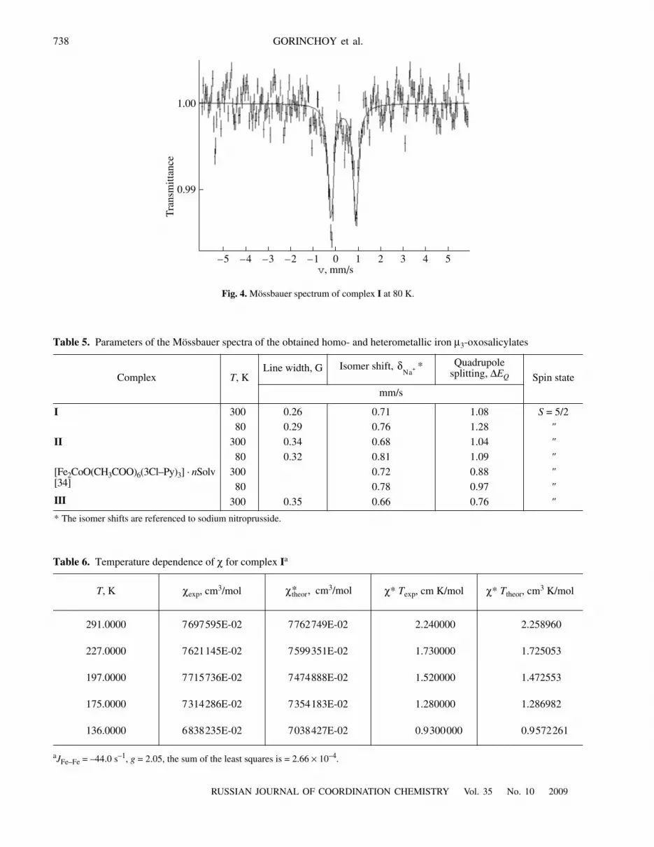
The Mössbauer spectra of complexes I–III at 300and 80 K show a doublet, its peaks being approximatelyequal in intensity; no additional absorption is detected(Fig. 4). At 80°C, the spectral pattern remainsunchanged; the parameters of the Mössbauer spectra(Table 5) are typical of high-spin iron(III) complexes(S = 5/2).
Replacement of one iron(III) ion in the triangle by acobalt(II) or magnesium(II) ion increases the quadru-pole splitting from 0.76 to 1.08 mm/s (by ~26%). Thischange is consistent with our data [24, 28, 29, 34] andthe data obtained by other researchers [35–38] and isdue to the lowering of the symmetry of the fragment{Fe2MO} in the complex from D3h to C2v. A comparisonof the parameters of the Mössbauer spectra of thecobalt-containing clusters reveals a slight increase inthe quadrupole splitting (0.88 1.04 mm/s) when theacetate ion is replaced by a salicylate one, probablybecause of the ligand contribution (Table 5).
With a decrease in the temperature, the isomer shiftincreases by 0.13–0.15 mm/s, which agrees with the lit-erature data for 57Fe [39]. This decrease reflects a
Table 3. Selected bond lengths and bond angles in structure II*
Bond d, Å Bond d, Å
M(1)–O(1) 1.884(6) M(2)–O(1) 1.895(3)
M(1)–O(1w) 2.098(8) M(2)–O(2) 2.066(5)
M(1)–O(3) 2.034(5) M(2)–O(5)1 2.030(5)
M(1)–O(8) 2.060(5) M(2)–O(6) 2.082(5)
M(1)–M(2) 3.271(3) M(2)–O(9) 2.039(5)
M(2)–O(11) 2.092(5)
M(2)–M(3) 3.287(3)
Angle ω, deg Angle ω, deg
O(1w)M(1)O(3) 84.7(1) O(1)M(2)O(2) 96.4(2)
O(1w)M(1)O(8) 84.6(1) O(1)M(2)O(5)1 94.9(2)
O(1)M(1)O(3) 95.3(1) O(1)M(2)O(6) 94.7(2)
O(1)M(1)O(8) 95.4(2) O(1)M(2)O(9) 95.0(2)
O(3)M(1)O(3)1 169.4(3) O(1)M(2)O(11) 177.5(2)
O(3)M(1)O(8) 89.6(2) O(2)M(2)O(5)1 89.4(2)
O(3)M(1)O(8)1 89.5(2) O(2)M(2)O(6) 168.8(2)
O(8)M(1)O(8)1 169.2(3) O(2)M(2)O(9) 88.7(2)
O(2)M(2)O(11) 86.2(2)
O(6)M(2)O(5)1 91.9(2)
O(6)M(2)O(9) 88.1(2)
O(6)M(2)O(11) 82.8(2)
O(9)M(2)O(5)1 170.1(2)
O(9)M(2)O(11) 84.8(2)
O(11)M(2)O(5)1 85.4(2)
* The symmetry operation code is: 1 –x, y, –z + 0.5.
Table 4. Parameters of the hydrogen bonds in structures I and II
D–H···ADistance, Å
Angle DHA, deg Symmetry operation code for AD–H H···A D···A
IO(1w)–H···O(3w) 0.91 1.83 2.62(2) 144 x, y, z
O(11)–H···O(4w) 0.97 1.65 2.59(1) 160 x, y, z
IIO(2w)–H···O(22) 0.89 1.78 2.67(1) 176 0.5 – x, 0.5 – y, 1 – z
O(3w)–H···O(24) 0.90 1.87 2.58(2) 135 x, 1 + y, z
O(3w)–H···O(23) 0.90 1.80 2.62(1) 151 0.5 – x, 0.75 – y, 1 – z

736
RUSSIAN JOURNAL OF COORDINATION CHEMISTRY Vol. 35 No. 10 2009
GORINCHOY et al.
changed total s-electron density around the Mössbauernucleus as a result of the second-order Doppler effect[40, 41].
The magnetic properties of complexes I–III weremeasured in the 291–136 K range for I and at roomtemperature for II and III. The products of the experi-mental values (χåí) for complexes I, II, and III atroom temperature are 2.24, 4.59, and 5.12 cm3 K/mol,respectively. These are substantially lower than theproducts expected for non-interacting trinuclear com-plexes with the set of paramagnetic ions under study(8.75, 9.13, and 13.13 cm3 K/mol, respectively). Forcomplex I, χåí decreases from 2.24 (291 K) to0.93 cm3 K/mol (136 K) (Table 6). The temperaturedependence of the magnetic properties of complex Iwas described in terms of the HDVV model [1, 20, 42]with the Hamiltonian of spin-spin coupling H =−2J12(S1 · S2). The resulting parameters (JFe–Fe ~~
−44 cm−1, g = 2.05, the sum of the least squares 2.66 ×10⁻⁴) suggest an antiferromagnetic exchange interac-tion between the paramagnetic ions. The exchangeparameter for complex I lies in the J range characteris-tic of other heteronuclear µ3-oxo iron complexes with adiamagnetic heteroatom [28].
Our study confirmed that the three-center system in
the clusters [F MIIO(RCOO)6 · 3L] is stable and is
found in complexes where åII is a transition metal(donor-acceptor M–O bonds) and an alkaline-earthmetal (ionic M–O bonds).
Introduction of a heteroatom into the metal trianglelowers the symmetry of the electron cloud around theiron nuclei. With an s element as a heteroatom, the anti-ferromagnetic exchange interaction between two Fe3+
ions is stronger than that in homotrinuclear clusters.
e2III
N(4)
O(22)
M(1)
M(2) M(3)O(16)
O(17)
O(9)
O(7)
O(6)
O(8)
O(2w) O(12)
O(10)
O(13)
O(1)
O(4)
O(2)
O(15)
O(1w)
O(20)
O(14)
O(11)O(19)
O(18)
O(23)
O(3w)
O(5)O(3)
O(24)
N(6x)
N(5)
N(1)
N(3)N(2)
O(21)
Fig. 1. Molecular structure of the complex [Fe2MgO(SalH)6(DMAA)0.4(H2O)2.6] · 4DMAA.

RUSSIAN JOURNAL OF COORDINATION CHEMISTRY Vol. 35 No. 10 2009
HOMO- AND HETERONUCLEAR IRON COMPLEXES 737
O(3w)
O(1w)
O(3)O(8')
O(8)O(3')
O(1)
M(2')
O(5)O(6)
O(9)
O(2)
O(11)
M(2)
M(1)
O(5')
O(4w)
Fig. 2. Molecular structure of the complex [Fe2CoO(SalH)6(CH3OH)2(H2O)] · DMF · 2.5H2O.
x
z
Fig. 3. Fragment of the crystal structure of the complex [Fe2CoO(SalH)6(CH3OH)2(H2O)] · DMF · 2.5H2O.
738
RUSSIAN JOURNAL OF COORDINATION CHEMISTRY Vol. 35 No. 10 2009
GORINCHOY et al.
–5 –4 –3 –2 –1 0 1 2 3
0.99
5
1.00
4
Tra
nsm
ittan
ce
v, mm/s
Fig. 4. Mössbauer spectrum of complex I at 80 K.
Table 5. Parameters of the Mössbauer spectra of the obtained homo- and heterometallic iron µ3-oxosalicylates
Complex T, KLine width, G Isomer shift, * Quadrupole
splitting, ∆EQ Spin state
mm/s
I 300 0.26 0.71 1.08 S = 5/2
80 0.29 0.76 1.28 "
II 300 0.34 0.68 1.04 "
80 0.32 0.81 1.09 "
[Fe2CoO(CH3COO)6(3Cl–Py)3] · nSolv [34]
III
300 0.72 0.88 "
80 0.78 0.97 "
300 0.35 0.66 0.76 "
* The isomer shifts are referenced to sodium nitroprusside.
δNa+
Table 6. Temperature dependence of χ for complex Ia
T, K χexp, cm3/mol cm3/mol χ* Texp, cm K/mol χ* Ttheor, cm3 K/mol
291.0000 7697595E-02 7762749E-02 2.240000 2.258960
227.0000 7621145E-02 7599351E-02 1.730000 1.725053
197.0000 7715736E-02 7474888E-02 1.520000 1.472553
175.0000 7314286E-02 7354183E-02 1.280000 1.286982
136.0000 6838235E-02 7038427E-02 0.9300000 0.9572261
aJFe–Fe = –44.0 s–1, g = 2.05, the sum of the least squares is = 2.66 × 10⁻⁴.
χtheor* ,

RUSSIAN JOURNAL OF COORDINATION CHEMISTRY Vol. 35 No. 10 2009
HOMO- AND HETERONUCLEAR IRON COMPLEXES 739
REFERENCES
1. Kahn, O., Molecular Magnetism, New York: Weinheim-Cambridge: VCH, 1993.
2. Harrison, P.M., Hempstead, P.D., Artymiuk, P.J., andAndrews, S.C., Metal Ions in Biological Systems, NewYork: Marcel Dekker, 1998, vol. 35, Ch. 11, p. 435.
3. Long, J.R., Molecular Cluster Magnets, Yang., P., Ed.,Hong Kong: World Scientific, 2003, p. 291.
4. Sheriff, S., Hendrickson, W.A., and Smith, J.L., J. Mol.Biol., 1987, vol. 197, no. 2, p. 273.
5. Cannon, R.D. and White, R.P., Prog. Inorg. Chem.,1988, vol. 36, p. 196.
6. Cotton, F.A., Extine, M.W., Falvello, L.R., et al., Inorg.Chem., 1986, vol. 25, no. 19, p. 3505.
7. Bond, A.M., Clark, J.H.R., Humphrey, D.G., et al., Dal-ton Trans., 1998, p. 1845.
8. Ren, X.M., Okudera, H., and Kremer, R.K., Acta Crys-tallogr, Sect. E: Structure Reports Online, 2004, vol. 60,p. 14.
9. Prodius, D.N., Cand. Sci. (Chem.) Dissertation, Chi-sinau: Institute of Chemistry, Moldova Academy of Sci-ences, 2007.
10. Cui, Y., Zheng, F.K., Yan, D.Ch., et al., Jiegou Huaxue,1998, vol. 17, no. 1, p. 5.
11. Tong, M.L., Wu, Y.L., Chen, X.M., et al., Chem. Res.Chin. Univ., 1998, vol. 14, no. 3, p. 230.
12. Voronkova, V.K., Galeev, R.T., Shova, S., et al., Appl.Mag. Res., 2003, vol. 25, no. 2, p. 227.
13. Tel’zhenskaya, P.N. and Shvarts, E.M., Koord. Khim.,1977, vol. 3, no. 9, p. 1279.
14. Ming-Cai Yin, Chang-Chun Ai, Liang-Jie Yuan, et. al., J.Mol. Struct., 2004, vol. 33, p. 691.
15. Shake, A.R., Tsai, H.L., Webb, R.J., et al., Inorg. Chem.,1994, vol. 33, no. 26, p. 6020.
16. Allen, F.N., Acta Crystallogr., Sect. B: Struct. Sci., 2002,vol. 58, no. 2, p. 380.
17. Otwinowski, Z. and Minor, W., Methods in Enzymology,Carter, C.W. and Sweet, R.M., Eds., New York: Aca-demic, 1997, vol. 276, Pt A, p. 276.
18. XEMP. Version 4.2, Siemens Analytical X-Ray Inst. Inc.,1990.
19. Sheldrick, G.M., SHELX-97. Program for Refinement ofCrystal Structures, Göttingen (Germany): Univ. of Göt-tingen, 1997.
20. Kalinnikov, V.T. and Rakitin, B.V., Vvedenie v magne-tokhimiyu. Metod staticheskoi magnitnoi vospriimchi-vosti v khimii (Introduction to Magnetochemistry. The
Method of Static Magnetic Susceptibility in Chemistry),Moscow: Mir, 1980.
21. Nakamoto, T., Hanaya, M., Katada, M., et al., Inorg.Chem., 1997, vol. 36, p. 4347.
22. Jang, H.G., Geib, S.J., Kaneko, Y., et al., J. Am. Chem.Soc., 1989, vol. 111, p. 173.
23. Woehler, S.E., Wittebort, R.J., Oh, S.M., et al., J. Am.Chem. Soc., 1987, vol. 109, p. 1063.
24. Turta, C.I., Shova, S.G., Prodius, D., et al., Inorg. Chim.Acta, 2004, vol. 357, no. 15, p. 4396.
25. Zhang, H.H. and Yu, X.F., Chin. J. Sruct. Chem. (JiegouHuaxue), 1990, vol. 9, no. 1, p. 1.
26. Batsanov, A.S., Timko, G.A., Struchkov, Yu.T., et al.,Koord. Khim., 1991, vol. 17, no. 7, p. 922.
27. Singh, B., Long, J.R., and de Biani, F.F., et al., J. Am.Chem. Soc., 1997, vol. 119, no. 30, p. 7030.
28. Turta, K.I., Shova, S.G., Zhovmir, F.K., et al., Zh. Neorg.Khim., 2003, vol. 48, no. 1, p. 80 [Russ. J. Inorg. Chem.(Engl. Transl.), vol. 48, no. 1, p. 72].
29. Prodius, D., Turta, C.I., Mereacre, V.M., et al., Polyhe-dron, 2006, vol. 25, p. 2175.
30. Wells, A.F., Structural Inorganic Chemistry, Oxford:Claredent, 1984.
31. Wang, Zh.M. and Yu, X-F., Chin. J. Sruct. Chem. (JiegouHuaxue), 1990, vol. 9, no. 1, p. 14.
32. Sato, T. and Ambe, F., Acta Crystallogr., Sect. C: Cryst.Struct. Commun., 1996, vol. 52, p. 3005.
33. Nakomoto, K., Infrared and Raman Spectra of Inorganicand Coordination Compounds, New York: Wiley, 1986.
34. Zhovmir, T.K., Turta, K.I., Shova, S.G., et al., Zh. Strukt.Khim., 1999, vol. 40, no. 6, p. 1155.
35. Long, G.J., Robinson, W.T., Tappeyer, et al., J. Chem.Soc., Dalton Trans., 1973, no. 6, p. 573.
36. Duncan, J.F., Kanekar, C.R., and Mok, K.F., J. Chem.Soc., A, 1969, no. 3, p. 480.
37. Mansurov, M.M., Semenova, G.L., Yakubov, Kh.M.,et al., Zh. Neorg. Khim., 1983, vol. 28, no. 6, p. 1460.
38. Yakubov, Kh.M., Semenov, G.L., Mansurov, M.M., Zh.Neorg. Khim., 1985, vol. 30, no. 2, p. 368.
39. Nefed’ev, A.V., Lapkina, N.D., Stukan, R.A., et al., Zh.Strukt. Khim., 1979, vol. 20, no. 5, p. 835.
40. Pound, R.V. and Rebka, G.A., Phys. Rev. Lett., 1960,vol. 4, no. 6, p. 274.
41. Josephson, B.D., Phys. Rev. Lett., 1960, vol. 4, no. 7,p. 341.
42. Van Vleck, J.H., Theory of Electric and Magnetic Sus-ceptibilities, London-Oxford: Univ., 1932.