Embed Size (px)
Citation preview
HOW TO USE MICROSCOPE AND CELL
OBSERVATION
OKTAVIA INTAN
3315116258
Fakultas Matematika dan Ilmu
Pengetahuan Alam
Universitas Negeri Jakarta
2011
I. How to Use Microscope and Cell Observation
II. Purpose :1. Learn how to preparr te materials which would be observed
with microscope.
2. Observe the living cell and nonliving cell.
3. Observed the shape of cell.
4. Observe the differences between plant cell and animal
cell.
III. Theoretical Background
We know that we can’t see everything only by naked eyes. Someone
who difficult to identify had invented the first microscope that
is optical microscope. An early microscope was made in
Netherlands. The compound microscope has two systems of lenses
for greater magnification, 1) the ocular, or eyepiece lens that
one looks into and 2) the objective lens, or the lens closest to
the object. Before purchasing or using a microscope, it is
important to know the functions of each part.
A. The function and the part of microscope
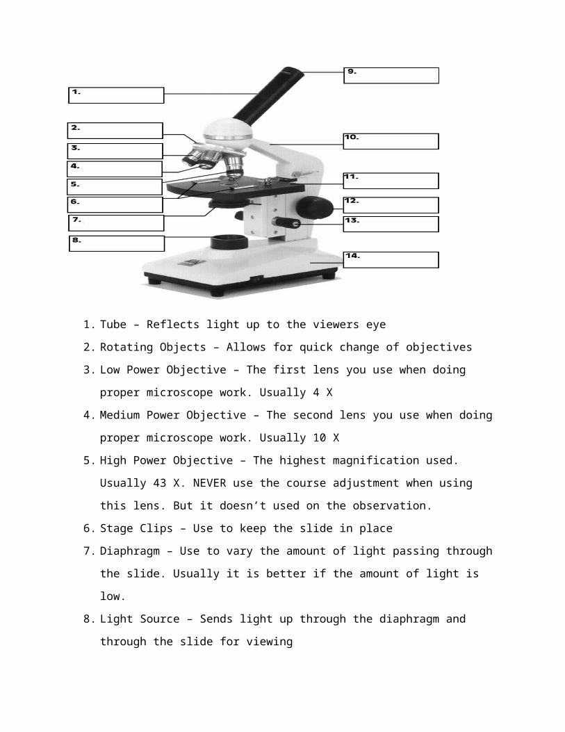
1. Tube – Reflects light up to the viewers eye
2. Rotating Objects – Allows for quick change of objectives
3. Low Power Objective – The first lens you use when doing
proper microscope work. Usually 4 X
4. Medium Power Objective – The second lens you use when doing
proper microscope work. Usually 10 X
5. High Power Objective – The highest magnification used.
Usually 43 X. NEVER use the course adjustment when using
this lens. But it doesn’t used on the observation.
6. Stage Clips – Use to keep the slide in place
7. Diaphragm – Use to vary the amount of light passing through
the slide. Usually it is better if the amount of light is
low.
8. Light Source – Sends light up through the diaphragm and
through the slide for viewing
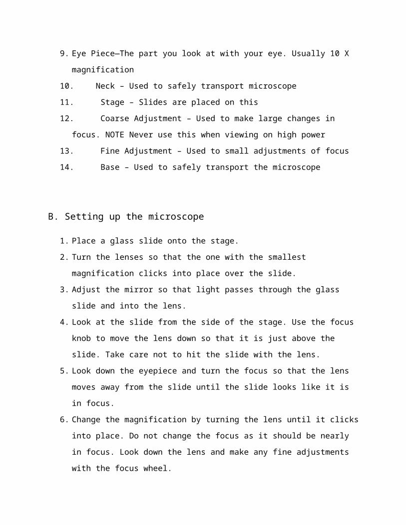
9. Eye Piece—The part you look at with your eye. Usually 10 X
magnification
10. Neck – Used to safely transport microscope
11. Stage – Slides are placed on this
12. Coarse Adjustment – Used to make large changes in
focus. NOTE Never use this when viewing on high power
13. Fine Adjustment – Used to small adjustments of focus
14. Base – Used to safely transport the microscope
B. Setting up the microscope
1. Place a glass slide onto the stage.
2. Turn the lenses so that the one with the smallest
magnification clicks into place over the slide.
3. Adjust the mirror so that light passes through the glass
slide and into the lens.
4. Look at the slide from the side of the stage. Use the focus
knob to move the lens down so that it is just above the
slide. Take care not to hit the slide with the lens.
5. Look down the eyepiece and turn the focus so that the lens
moves away from the slide until the slide looks like it is
in focus.
6. Change the magnification by turning the lens until it clicks
into place. Do not change the focus as it should be nearly
in focus. Look down the lens and make any fine adjustments
with the focus wheel.
With microscope we can see the smallest things for example
cell. There are two kinds of cell that is plant cell and animal
cell.
C. Cell
1. ANIMAL CELL
That’s the explanation about the organelles with their
function.
a. Nucleus, to holds the DNA
b. Ribosome, makes proteins and found in all cells,
prokaryotic and eukaryotic
c. Endoplasmic Reticulum (ER or Roads), The internal
delivery system of the cell. 2 Types:
1. a Rough ER: Rough appearance because it has
ribosome
Function: helps make proteins, that’s why it has
ribosome
1.b Smooth ER:NO ribosome
Function: makes fats or lipids
d. Golgi Complex (The shippers), packages, modifies, and
transports materials to different location inside/outside of
the cell
e. Lysosomes (“Clean-up Crews”): circular, but bigger than
ribosome.
Function: to break down food into particles the rest of
the cell can use and to destroy old cells
f. Mitochondria (“The Powerhouse”), Energy formation and
breaks down food to make ATP. ATP is the major fuel for all
cell activities that require energy.
2. PLANT CELL
Plant cell commonly has same organelles with animal cell.
But in plant cell there are more organelles that will be
explained
g. Vacuoles, stores water. This is what makes lettuce crisp
and when there is no water, the plant wilts
Cytoplasm
h. Chloroplasts, traps energy from the sun to produce food
for the plant cell. Green in color because of chlorophyll,
which is a green pigment
i. Cell Wall, provides support and protection to the cell
membrane. Found outside the cell membrane in plant cells
IV. Instrument and MaterialA.Instrument
1. Microscope
2. Slide
3. Cover Glass
4. Beaker Glass
5. Cutter
6. Tissue
7. Drop Forceps
8. Tooth Pick
9. Filtrating Paper
B.Materials1. Pieces of with text A
2. Dry red onion cover (Album cepa)
3. Leaf of Adam and Eva (Rhoeo discolor)
4. Leaf Hdyrillia (Hydrilla verticilata)
5. Leaf of Cocor Bebek (Kalanchoe pfflata)
6. Oral mucosal epithelium
7. HCL concentrate
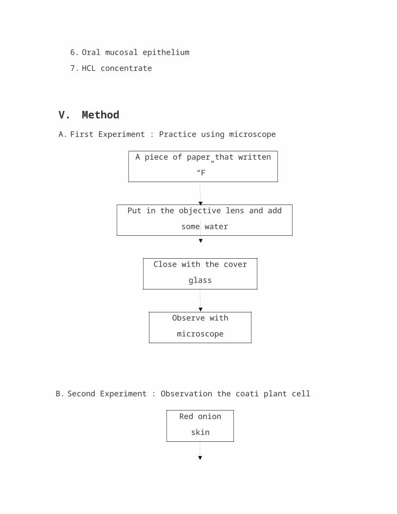
V. MethodA. First Experiment : Practice using microscope
A piece of paper that written
“F”
Put in the objective lens and add
some water
Close with the cover
glass
Observe with
microscope
B. Second Experiment : Observation the coati plant cell
Red onion
skin
Put in the objective lens and
add some water
Drop the HCL
concentrate
Close with the cover
glass
Observe with
microscope
C. Third Experiment : Observe the fresh plant cell slide
1. Leaf of Adam and Eva (Rheo discolor)
A piece of paper that written
“W”
Put in the objective lens and add
some water
Close with the cover
glass
Observe with
microscope
2. Leaf Hdyrillia (Hydrilla verticilata)
A piece of paper that written
“W”
Put in the objective lens and add
some water
Close with the cover
glass
Observe with
microscope
3. Leaf of Cocor Bebek (Kalanchoe pfflata)
A piece of paper that written
“W”
Put in the objective lens and add
some water
Close with the cover
glass
Observe with
microscope
4. Oral mucosal epithelium
A piece of paper that written
“W”
Put in the objective lens and add
some water
Close with the cover
glass
Observe with
microscope
VI. Result
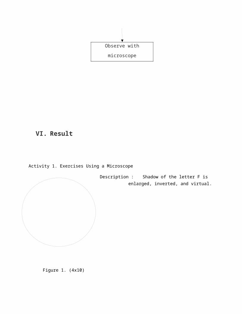
Activity 1. Exercises Using a Microscope
Description : Shadow of the letter F is enlarged, inverted, and virtual.
Figure 1. (4x10)
Activity 2. Tues Coati observed in plants
Description : cell look like space and there are shape like brown fiber.
Figure 2. (10x10)
Activity 3. Observing fresh preparations of plant cells
A. Adam Eva Leaf (Rhoeo discolor)
Description : Hexagonal-shaped. There arefibers like greenish brown rope and there are pink space.
Figure 3. (10x10)
B. Hydrilla Leaf (Hydrilla verticilata)
Description : The colour is green and there are soft green between oneof another chain.

Figure 4. (10x10)
C. Cocor Bebek Leaf (Kalanchoe pinata)
Description : irregular quadrilateral-shaped and there are greenish brown small circles.
Figure 5. (10x10)
D. Observing Fresh Preparations of Animal Cell
Description : has an irregular shape. part of the cell is not clearly visible. The color is greenish brown.
Figure 6. (10x10)
VII. DiscussionA.First Experiment : Practice using microscope
After finish the observation with the paper which written “F”
with lowest magnification. The paper of “F” look inverse, that is
because the objective lens and the ocular lens are convex lens.
It show that the shadow are virtual, inverted, and enlarge. And
ocular lens decided the last shadow. In optical microscope, the
last shadow has same properties with shadow wile, virtual,
inverted and anlarge.
B.Second Experiment : Observation the coati plant
cell
In the cover cell of red union we can see the cell wall and
cytoplasm. The function of cell wall is provides support and
protection to the cell membrane. In this observation we use HCL
concentrate because HCL able to broke the nucleus and the nucleus
mix with the cytoplasm. Because of it, only cytoplasm that look.
When the nucleus broken, the others organelles will broken too
because nucleus is the control. But HCL cannot broken the cell
wall because it has a strong organ and the cytoplasm doesn’t
broken too because it’s a liquid. The nucleus and the others
organelles that broken called nonliving cell.
C.Third Experiment : Observation the fresh plant cell
slide.1. Leaf of Adam and Eva (Rheo discolor)
In the picture there are cell wall, cytoplasm, and stomata.
Like in the second experiment, the function of cell wall is
provides support and protection to the cell membrane. And cytoplasm is a liquid that has some organelles there are Vacuole,
Endoplasmic Reticulum, Ribosome , Nucleus, Mitochondria, Golgi
Bodies, Cell Wall, Cell Membrane, and Chloroplast. And the
nucleus should look in the observation, but the nucleus doesn’t
find, maybe it is because the specimen that too tick and make the
cell that look in the microscope are collide. The Vacuole doesn’t
look as well but the stomata are look clearly. Stomata is a place
exchange between atmosphere and CO2. It show that in Adam Eva
Leaf there are chloroplast because in mesophyll there are
chloroplast that can photosynthesis.
2. Leaf Hdyrillia (Hydrilla verticilata)
In the picture there are cell wall, cytoplasm, and
chloroplast. Like the cell red union and also the leaf of Adam
Eva, in the Hydrilla there is a cell wall that provides support
and protection to the cell membrane and it show that Hydrilla
cell is plant cell. Cytoplasm is a liquid (85-90% consist of
water) that has some organelles. And chloroplast shape is like
lens. In the chloroplast there are chlorophyll. The function of
chloroplast is traps energy from the sun to produce food for the
plant cell. In this experiment, the cell wall, cytoplasm, and the
chloroplast look maybe because the specimen is thinner than the
second experiment.
3. Leaf of Cocor Bebek (Kalanchoe pfflata)
In the picture there are cell wall, cytoplasm, stomata, and
spaces between cells. All like the before experiment. In this
experiment there is thing that look like rose flower and maybe
that is Stomata. stomata look like rose flower maybe because the
specimen is too big so the stomata is collide like rose flower.
In this experiment there are spaces between cells. Spaces between
cells usually called lamela middle. Lamela middle is one of
another structure plant cell. Lamela middle consist of Pektin.
Enzyme pektinase can dissolve lamella middle so the cell will
divorce each other is called maceration. Cocor Bebek plant is
live in water and float upward, so it need smaller the density of
plant than water. With lamella middle or space between cells,
will form cell structure that distantly and density of cell
smaller than another plant.
4. Oral mucosal epithelium
In the animal cell slide experiment, we use human oral
mucosa epithelium. In the picture look cell membrane, cytoplasm,
and nucleus. Cell membrane covering or protect nucleus and
cytoplasm , cell membrane is flexible it make the shape of
animal cell is messy. Cell membrane cover organelles in the cell
and also a transportation equipment for that cell because in cell
membrane the substance that needed and unneeded can into or out
to. Cell membranes consist of protein, oligosaccharide
glycolipids and phospholipids colestrol. Cytoplasm also look in
the picture, maybe the cytoplasm structure more wide than another
part of cell and cytoplasm viscous liquid make it easier look in
the optical microscope. Nucleus or called cell center and consist
of matrix.
VIII. Suplement1.Compare the location of the shadow with the object location
that observed. That’s the location same or inverted?
Inverted. That’s the shadow is mirror images? Yes.
2.While looking the ocular lens, move the slide from the left
to the right. Where the shadow move? To the opposite direction. Where’s the shadow move if the slide move to
forward? To backward.
3. Rotate the low power objective to strong power objective.
That’s change the field of view become wide or narrow? Wide.
That is change the position of the shadow? No. And that’s
the shadow darker or brighter? Darker.
IX. References
Care and Use the microscope, http://faculty.valenciacollege.edu/smatthews/Use%20of%20the%20Microscope.pdf, October 6th 2011, 13.00 WIB.
Using the Microscope, http://krupp.wcc.hawaii.edu/BIOL100L/powerpoint/microscope.pdf, October 6th 2011, 13.45 WIB.
TeachAssist Resource Worksheets, http://www.griffin-education.co.uk/resources/pdfs/Booklet1-Ueofmicrosco.pdf , October 6th 2011, 14.00 WIB.