Embed Size (px)
Citation preview

Human Polymerase-Associated Factor complex(PAFc) connects the Super Elongation Complex(SEC) to RNA polymerase II on chromatinNanhai Hea, Caleb K. Chana, Bijan Sobhianb, Seemay Choua, Yuhua Xuea, Min Liua,c, Tom Albera,Monsef Benkiraneb, and Qiang Zhoua,1
aDepartment of Molecular and Cell Biology, University of California, Berkeley, CA 94720; bLaboratoire de Virologie Moleculaire, Institut de GenetiqueHumaine, Centre National de la Recherche Scientifique-Unités Propres de Recherche (CNRS-UPR) 1142, 34000 Montpellier, France; and cSchool ofPharmaceutical Science, Xiamen University, Xiamen 361005, China
Edited by James L. Manley, Columbia University, New York, NY, and approved July 15, 2011 (received for review May 3, 2011)
The Super Elongation Complex (SEC), containing transcription elon-gation activators/coactivators P-TEFb, ELL2, AFF4/1, ENL, and AF9,is recruited by HIV-1 Tat andmixed lineage leukemia (MLL) proteinsto activate the expression of HIV-1 and MLL-target genes, respec-tively. In the absence of Tat and MLL, however, it is unclear howSEC is targeted to RNA polymerase (Pol) II to stimulate elongationin general. Furthermore, although ENL and AF9 can bind the H3K79methyltransferase Dot1L, it is unclear whether these bindings arerequired for SEC-mediated transcription. Here, we show that thehomologous ENL and AF9 exist in separate SECs with similar butnonidentical functions. ENL/AF9 contacts the scaffolding proteinAFF4 that uses separate domains to recruit different subunits intoSEC. ENL/AF9 also exists outside SEC when bound to Dot1L, whichis found to inhibit SEC function. The YEATS domain of ENL/AF9targets SEC to Pol II on chromatin through contacting the humanPolymerase-Associated Factor complex (PAFc) complex. This findingexplains the YEATS domain’s dispensability for leukemogenesiswhen ENL/AF9 is translocated to MLL, whose interactions withPAFc and DNA likely substitute for the PAFc/chromatin-targetingfunction of the YEATS domain.
Accumulating evidence has implicated the elongation stage ofRNA polymerase II (Pol II) transcription as a major rate-
limiting step for the expression of a large number of metazoangenes, especially those that control cell growth, renewal, and dif-ferentiation (1–3). During elongation, the processivity of Pol II isregulated by a set of transcription factors, which had beenthought to exist as separate entities and impact on the Pol II elon-gation complex independently of one another. However, recentdata from us and others indicate that at least two well definedtranscription elongation factors of different classes reside ina single multisubunit complex termed SEC [super elongationcomplex, (4)] to cooperatively activate transcription (4–6).
The first elongation factor found in SEC is human positivetranscription elongation factor b (P-TEFb). Consisting of CDK9(Cyclin-dependent Kinase 9) and cyclin T1 (CycT1), P-TEFbfunctions by phosphorylating the C-terminal domain (CTD) ofthe largest subunit of Pol II and negative elongation factors DSIFand NELF. These events antagonize the actions of the negativefactors, release Pol II from promoter-proximal pausing, and trig-ger the production of full-length mRNA transcripts (7, 8). Thesecond elongation factor in SEC is ELL2, which promotes elon-gation by keeping the 3′ OH of nascent mRNA in alignment withthe catalytic site to prevent Pol II backtracking (9). BesidesP-TEFb and ELL2, SEC also contains transcription factors/coac-tivators ENL, AF9, AFF4 (AF5q31), AFF1 (AF4), and probablyothers (4–6). Among these, AFF4 is known to mediate the inter-action between ELL2 and P-TEFb and maintain the integrity ofSEC (5). While AFF1 has been shown to interact with AFF4 (10),it remains to be seen whether the interaction between these twohomologous proteins can exist in a single SEC complex.
The biological importance of SEC has been highlighted bythe recent discoveries that it is targeted by at least two sequence-specific transcriptional activators that play important roles inhuman diseases. First, the HIV-1 Tat protein is shown to interactwith and recruit SEC to the HIV-1 LTR (Long Terminal Repeat)to stimulate viral transcription in a SEC-dependent manner(5, 6). In addition, Tat also promotes SEC formation, which inturn stabilizes ELL2, an otherwise short-lived protein rapidlydegraded by the proteasome (5).
Besides HIV/AIDS, SEC is also found to be important for gen-erating certain types of acute leukemias that involve chromoso-mal translocations of the mixed lineage leukemia (MLL) gene(4, 10, 11). At least three components of SEC, namely AFF4,ENL, and AF9, are known as fusion partners of MLL (12). Whenfused to the DNA-binding domain of MLL, these proteins deliverSEC and its powerful elongation stimulatory activity to the MLL-target genes to promote leukemic transformation (4, 10, 11).
Despite the demonstrations that SEC exists in cells under con-ditions that are free of Tat and MLL-translocations and is essen-tial for metazoan transcriptional elongation in general (4–6, 10),little is known about how this complex is targeted to Pol II on achromatin template in the absence of sequence-specific recruit-ment factors like Tat and MLL. Furthermore, compared to theother subunits of SEC, the contributions of ENL and AF9 toSEC function have not been well characterized. So far, the onlyclue to how these two proteins may be involved in gene expressioncontrol arises from their reported interactions with Dot1L(13, 14), a methyltransferase responsible for histone H3 lysine79(H3K79) methylation (15, 16). As this modification is often foundin actively transcribed regions (17, 18), the associations of ENL/AF9 with P-TEFb and Dot1L are believed to recruit the latter twointo a large complex for efficient activation of transcription(12, 19), an intriguing proposition that is yet to be proven. Besidesthe uncertainty about the Dot1L-ENL/AF9 interaction, it is alsounclear whether the evolutionarily conserved YEATS domainlocated in the N-terminal regions of ENL and AF9, which areoften missing in the MLL-ENL/AF9 fusions and unimportantfor leukemogenesis (20), is required for SEC function.
Here, presenting answers to these questions, we show that thehighly homologous ENL and AF9 exist in separate SEC com-plexes that display similar but nonidentical functions. Within a
Author contributions: N.H., T.A., M.B., and Q.Z. designed research; N.H., C.K.C., B.S., S.C.,Y.X., and M.L. performed research; N.H., T.A., M.B., and Q.Z. analyzed data; and N.H. andQ.Z. wrote the paper.
The authors declare no conflict of interest.
This article is a PNAS Direct Submission.1To whom correspondence should be addressed. E-mail: [email protected].
See Author Summary on page 14719.
This article contains supporting information online at www.pnas.org/lookup/suppl/doi:10.1073/pnas.1107107108/-/DCSupplemental.
E636–E645 ∣ PNAS ∣ September 6, 2011 ∣ vol. 108 ∣ no. 36 www.pnas.org/cgi/doi/10.1073/pnas.1107107108

SEC, ENL/AF9 is connected to the rest of the complex throughthe scaffolding protein AFF4, which uses separate domains tobind to different SEC subunits. Our data further show that Dot1Ldoes not reside in or associate with SEC. Rather, Dot1L com-petes with AFF4 for binding to ENL/AF9 and inhibits SEC func-tion. Finally, in the absence of sequence-specific recruitmentfactors, the YEATS domain of ENL/AF9 targets SEC to chroma-tin through contacting the human Polymerase-Associated Factorcomplex (PAFc) transcription elongation complex (21, 22), andthrough PAFc, the elongating Pol II. This observation explainswhy the N-terminal regions of ENL and AF9 are dispensable forleukemogenesis when fused toMLL, whose abilities to bind PAFc(23) and DNA likely bypass the requirement for the PAFc/chro-matin-targeting function of the YEATS domain.
ResultsENL and AF9 Interact with P-TEFb Through the Scaffolding ProteinAFF4s. In light of our previous demonstration that AFF4 bridgesthe ELL2-P-TEFb interaction in SEC (5), we asked whether AFF4plays a similar role in mediating the interactions of ENL and AF9with P-TEFb. To answer this question, short hairpin (sh)RNA-mediated depletion of AFF4 was performed in HeLa cells to assessits impact on the associations of ENL and AF9 with P-TEFb.Although the depletion did not affect the total levels of ENL andAF9 in nuclear extracts [nuclear extracts (NE); Fig. 1A, left box],depletion of AFF4 significantly reduced the levels of ENL andAF9 bound to the immunoprecipitated CDK9 (right box).
Next, in vitro binding reactions were performed to test whetherAFF4 could directly bridge the interaction between purifiedP-TEFb and ENL/AF9 in the absence of other SEC components.All the proteins added to the reactions were highly purified fromtransfected HeLa cells under stringent conditions (1.0MKCl plus0.5% NP-40) and confirmed by SDS-PAGE followed by silver-staining to be free of any contaminating proteins [Fig. S1; (5)].As indicated in Fig. 1B, purified ENL and AF9 did not interactwith immobilized CycT1-HA/CDK9 unless AFF4 was also pre-sent in the same reactions. Together, these in vitro and in vivobinding data highlight the important role for AFF4 to serve asa molecular scaffold to mediate the interactions of P-TEFb withnot only ELL2 but also ENL and AF9.
ENL and AF9 Directly Interact with AFF4 Through Their C-TerminalRegions. Because AFF4 was shown to interconnect P-TEFb andENL/AF9 in the absence of other SEC components, we reasonedthat it must be able to make direct and simultaneous contacts withP-TEFb and ENL/AF9. In fact, a direct binding between AFF4and P-TEFb has been demonstrated previously (5). To show thatENL and AF9 can also directly contact AFF4, in vitro bindingreactions employing highly purified proteins (Fig. S1) were con-ducted. While wild-type (WT) ENL and AF9 were able to bindto immobilized HA-AFF4 directly, the C-terminally truncatedENL (1–431) and AF9 (1–480) were not (Fig. 1C). The require-ment for the C-terminal regions of ENL and AF9 for bindingto AFF4, which in turn allowed the formation of a completeSEC complex, was also confirmed in vivo in transfected cells(Fig. 1 D and E).
Separate Regions of AFF4 Are Used to Interact with Different Subunitsof SEC. Given the demonstrations that the scaffolding proteinAFF4 can directly and simultaneously contact each and everysubunit of SEC [Fig. 2, (5)], we would like to map its regions thatare involved in these interactions. A series of Flag-tagged AFF4deletion mutants were tested for their ability to coprecipitate theother components of SEC. When the levels of WT and mutantAFF4 in the antiFlag immunoprecipitates (IP) were normalized toa similar level, the mutant missing the first 300 residues, ∆1-300,was found to interact with ELL2, ENL, and AF9 normally but notCDK9 (Fig. 2, lane 3). In contrast, a short AFF4 truncationmutantcontaining just the first 300 amino acids (1–300) was able to effi-ciently pull-down CDK9 but not any other components of SEC(lane 6). Thus, the first 300 residues of AFF4 contain an indepen-dent domain that is necessary and sufficient for P-TEFb-binding.
Similarly, the region between aa 301 and 600 in AFF4 appearsto contain an ELL2-binding domain. This notion is indicated bythe demonstration that the deletion mutant ∆1-600 failed tointeract with ELL2 and P-TEFb, but still retained WT ability tobind to ENL and AF9 (Fig. 2, lane 4). On the other hand, a 300residue-long segment of AFF4 encompassing positions 301 to 600(301–600) displayed reduced but clearly above-the-backgroundlevel of binding to ELL2 but not any other components of SEC(lane 7). Finally, the region between amino acids 601 and 900 ofAFF4 likely contains an independent binding domain for ENL
vect
or
shAFF4
NE
ctl.
IgG
αCDK9 IP
AFF4
ENL
AF9
CDK9
Tubulin
F-AFF4:ENL-F:AF9-F:
Input (5%)
immobilizedCycT1-HA
/CDK9
CycT1-HA
F-AFF4ENL-FAF9-F
1 2 3 4
B
A
vect
or
shAFF4
WT:1-430:
1-480:WT:
ENL-F
AF9-F
immobilizedHA-AFF4:
Input (10%) on anti-HA beads
Flag
HA
WB
C
AFF4
CDK9
Flag
WB
ENL-F:
αFlag IP
AF9-F:
αFlag IP
AFF4
ELL2-HA
CDK9
Flag
WB
D E
ELL2-HA
WT 1-
430
WT 1-
480
onanti-HAbeads
1 559
5681
WT
WT
YEATS domain
AFF4-interacting domain
112 430
480112
1-430
1-480
ENL
AF9
Fig. 1. The scaffolding protein AFF4 directly binds to the C-terminal regions of ENL and AF9 to mediate their interactions with P-TEFb. (A). NE were preparedfromHeLa cells either containing an empty vector or expressing the AFF4-specifc shRNA (shAFF4) and subjected to immunoprecipitation (IP) with anti-CDK9 or anirrelevant rabbit IgG as a control. The isolated NE (left box) and immunoprecipitates (right box) were analyzed byWestern blotting with the indicated antibodies.(B). The indicated highly purified proteins were incubated with immobilized CycT1-HA/CDK9 in vitro and the bound proteins were eluted and analyzed byWestern blotting (right). Five percent of the input proteins were also examined by anti-FlagWestern blotting (left). (C). In vitro binding reactions were performedby incubating highly purified WTor C-terminally truncated ENL-F or AF9-F (schematic diagram on the right) with immobilized HA-AFF4. The bound proteins and10% of the soluble input proteins were analyzed by Western blotting with the indicated antibodies. (D). WT or mutant ENL and AF9, all Flag-tagged, wereexpressed in transfected HeLa cells. Anti-Flag immunoprecipitates were examined by Western blotting for the indicated ENL/AF9-associated factors.
He et al. PNAS ∣ September 6, 2011 ∣ vol. 108 ∣ no. 36 ∣ E637
BIOCH
EMISTR
YPN
ASPL
US

and AF9, as the fragment encompassing this region interactedefficiently with these two proteins but not any other subunitsof SEC (lane 8) and the deletion mutant lacking the entireN-terminal 900 amino acids (∆1-900) failed to associate withany component of SEC (lane 5). Together, these data indicate thatAFF4 uses separate domains to interact with different subunits ofSEC and serves as a platform to nucleate the assembly of SEC.
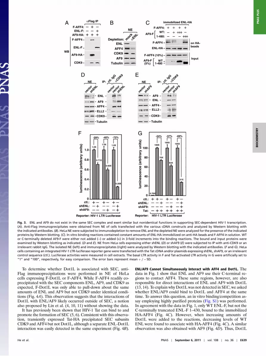
ENL and AF9 Exist in Separate SEC Complexes. ENL and AF9 arehighly homologous proteins, with their N-terminal YEATS do-mains and C-terminal coiled-coil domains showing a particularlyhigh degree of identity (82%). Given this homology and theabove demonstration that both proteins bound to the same region(aa 601–900) of AFF4 (Fig. 2), we asked whether they exist simul-taneously in a single SEC complex. To address this question, anti-Flag immunoprecipitations were performed in extracts of cellscoexpressing HA-tagged AF9 and Flag-tagged ENL or AFF4.While AF9-HA coprecipitated with F-AFF4 and CDK9 as ex-pected, it did not coprecipitate with ENL-F (Fig. 3A). The reci-procal co-IP experiment employing anti-HA beads also failed todetect the interaction between AF9-HA and ENL-F. These datasuggest that AF9 and ENL do not exist in the same complex,although each can establish independent interactions with otherSEC subunits.
To obtain further evidence in support of this notion, we per-formed immunodepletion to remove all ENL proteins fromHeLaNE (Fig. 3B). This procedure caused the codepletion of morethan 75% of AFF4, ∼30% of CDK9, but very few AF9 and theinternal control protein, α-Tubulin, from NE. This result is con-sistent with the idea that ENL and AF9 do not exist in the sameSEC complex. Because a major fraction of nuclear CDK9 is nor-mally sequestered in the 7SK snRNP (8), it is not surprising to seethat more than half of CDK9 were still present in the ENL-de-pleted NE. However, the codepletion of more than 70% of AFF4suggests that the AF9-containing SEC contains at most 30% of
total AFF4 in NE, assuming that no AFF4 exists outside of theSEC complexes. Thus, the ENL-containing SEC appears to be thepredominant form between the two SEC complexes.
ENL and AF9 Compete for Binding to AFF4. A likely reason for thefailure of ENL and AF9 to coexist in the same SEC is revealedsubsequently in an in vitro binding assay employing highly puri-fied proteins (Fig. S1). The addition of increasing levels of WTAF9-F, but not the C-terminally truncated AF9-F 1-480 that isdefective for AFF4-binding (Fig. 1C), into binding reactions pro-gressively reduced the amounts of F-AFF4 bound to the immo-bilized ENL-HA (Fig. 3C). Thus, the bindings of AF9 and ENLto AFF4, which occur through the same region (aa 601–900) ofAFF4 (Fig. 2), were mutually exclusive, which explains their fail-ure to exist in the same SEC complex.
Loss of ENL Is Compensated by Increased AF9 Expression, but Not ViceVersa. The existence of two versions of SEC with one containingAF9 and the other ENL makes it important to examine whetherthese two homologous proteins have similar or different func-tions. Toward this goal, stable shRNA-mediated depletion ofeither ENL or AF9 was performed and the effect on SEC forma-tion was assessed by anti-CDK9 immunoprecipitation followed byWestern blotting. Notably, the reduction of total and CDK9-bound ENL in HeLa cells by shENL triggered a marked increasein the amounts of AF9, but not AFF4 or ELL2, in both NE andanti-CDK9 IP (Fig. 3D). In contrast, shRNA depletion of AF9did not produce any significant change in ENL expression or se-questration into SEC (Fig. 3E). Thus, the loss of the ENL-con-taining SEC was compensated by the increased formation of theAF9-containing SEC, but not vice versa. This phenomenon islikely caused by the fact that the former complex is the predomi-nant form between the two SECs, and the compensation serves toprevent a major reduction in the overall SEC level in the cell.
AF9 and ENL Have Similar but Nonidentical Functions in MediatingSEC-Dependent Transcription. In agreement with the above demon-stration that AF9 increased expression to compensate for theloss of ENL but not vice versa, we consistently observed thatshRNA depletion of ENL had a smaller effect on HIV-1 tran-scription, which is shown to depend on the SEC function (5, 6),than did the depletion of AF9. For example, using the HIV-1LTR-driven luciferase expression as readout, shENL reducedbasal and Tat-activated HIV LTR activity by 29% and 24%, re-spectively (Fig. 3 F and G), whereas shAF9 decreased the two by40% and 37%, respectively, when the knockdown efficiency wasnormalized between the two shRNAs. Despite this difference, theoverall impact on the HIV-1 LTR by the individual depletion ofAF9 or ENL was relatively minor. Only when both proteins werecodepleted at the same time, a more drastic reduction in HIV-1transcriptions (72% reduction for basal and 69% for Tat-acti-vated transcription) was observed (Fig. 3 F and G). The fact thatthe depletion of ENL or AF9 alone only partially suppressedSEC-dependent HIV-1 transcription whereas the depletion ofboth had a much more significant effect indicates that the twoproteins have similar but not completely identical functions.
Dot1L Is Not a Component of SEC.The identification of ENL/AF9 asa subunit of SEC raises the issue of whether the methyltransfer-ase Dot1L, a well known partner of ENL and AF9 (13, 14), alsoexists and plays a key role in this complex. Dot1L is responsiblefor the methylation of H3K79 (15, 16), a modification that isoften associated with actively transcribed genes (17, 18). Becauseof their overall positive influence on transcription and commonconnection through ENL/AF9, Dot1L and the key SEC subunitsP-TEFb and ELL have been depicted in recent models as com-ponents of a larger complex for coordinated activation of tran-scription (11, 12, 19).
ENL
ELL2-HA
CDK9
Flag
F-AFF4: WT 1-
300
1-60
0
1-90
0
1-30
030
1-60
0
601-
900
αFlag IP
WB
AF9
1 2 3 4 5 6 7 8
1 300 600 900 1,163AFF4
CDK9
CycT1
ELL2 ENL AF9
Fig. 2. Separate regions of AFF4 are used to interact with different subunitsof SEC. NEs derived from HeLa cells, which were transfected with cDNA con-structs expressing either WT Flag-tagged AFF or the various deletion mutantsas indicated, were subjected to anti-Flag IP. The immunoprecipitates wereanalyzed by Western blotting with the indicated antibodies. The diagramat the bottom summarizes the findings of the binding study.
E638 ∣ www.pnas.org/cgi/doi/10.1073/pnas.1107107108 He et al.
To determine whether Dot1L is associated with SEC, anti-Flag immunoprecipitations were performed in NE of HeLacells expressing F-Dot1L or F-AFF4. While F-AFF4 readily co-precipitated with the SEC components ENL, AF9, and CDK9 asexpected, F-Dot1L was only able to pull-down about the sameamounts of ENL and AF9 but not CDK9 under identical condi-tions (Fig. 4A). This observation suggests that the interactions ofDot1L with ENL/AF9 likely occurred outside of SEC, a notionalso proposed by Lin et al. (4, 10, 11) without showing the data.
It has previously been shown that HIV-1 Tat can bind to andpromote the formation of SEC (5, 6). Consistent with this observa-tion, transiently expressed Tat-HA precipitated SEC subunitsCDK9 and AFF4 but not Dot1L, although a separate ENL-Dot1Linteraction was easily detected in the same experiment (Fig. 4B).
ENL/AF9 Cannot Simultaneously Interact with AFF4 and Dot1L. Thedata in Fig. 1 show that ENL and AF9 use their C-terminal re-gions to contact AFF4. These same regions, however, are alsoresponsible for direct interactions of ENL and AF9 with Dot1L(13, 14). To explain why Dot1L was not detected in SEC, we askedwhether ENL/AF9 could bind to Dot1L and AFF4 at the sametime. To answer this question, an in vitro binding/competition as-say employing highly purified proteins (Fig. S1) was performed.In agreement with the data in Fig. 1, only WT ENL-F, but not theC-terminally truncated ENL-F 1–430, bound to the immobilizedHA-AFF4 (Fig. 4C). However, when increasing amounts ofDot1L were added to the reactions, decreasing levels of WTENL were found to associate with HA-AFF4 (Fig. 4C). A similarobservation was also obtained with AF9 (Fig. 4D). Thus, Dot1L
F-AFF4
ENL-F
AF9-HA
CDK9
F-AFF4:ENL-F:
AF9-HA:
αFlag IP
WB
AF-AFF4:
AF9-F1-480:
WT:
immobilized ENL-HA
on HA-beads
Input
F-AFF4
ENL-HA
F-AFF4 (10%)
AF9-F(5%) 1-480
WT
AFF4ELL2
ENL
Tubulin
CDK9
AF9ve
ctor
shAF9
vect
or
vect
or
ENL
Tubulin
AFF4
CDK9
AF9
vect
or
shENL
vect
or
vect
or
NE ctl. I
gG
αCDK9
IP:
ELL2
B C
shENL
shAF9
NE ctl. I
gG
αCDK9
IP:
1.0
0.8
0.6
0.4
0.2
0
1.0X1.0X 1.0X
0.71X
0.60X
0.28X
lum
ines
cen
ce(a
rbit
rary
un
its)
ctl.:shENL:shAF9:
Reporter: HIV-1 LTR Luciferase
D
-29% -40% -72%
100
80
60
40
20
0
Reporter: HIV-1 LTR Luciferase
100X100X 100X
76X
63X
31X
-24% -37% -69%
ctl.:shENL:shAF9:
Tat:
E
0.76X
lum
ines
cen
c e(a
rbit
r ary
un
its)
F
ENL
AFF4CDK9
Depletion: mock
αENL
NE
AF9
Tubulin
G
Fig. 3. ENL and AF9 do not exist in the same SEC complex and exert similar but nonidentical functions in supporting SEC-dependent HIV-1 transcription.(A). Anti-Flag immunoprecipitates were obtained from NE of cells transfected with the various cDNA constructs and analyzed by Western blotting withthe indicated antibodies. (B). HeLa NEwere subjected to immunodepletion to remove ENL and the depleted NEwere analyzed for the presence of the indicatedproteins by Western blotting. (C). In vitro binding reactions contained constant amounts of ENL-HA immobilized on anti-HA beads and F-AFF4 in solution. WTor C-terminally deleted AF9-F were either not added (−) or added (+) in 3-fold increments into the binding reactions. The bound and input proteins wereexamined by Western blotting as indicated. (D and E). NE from HeLa cells expressing either shENL (D) or shAF9 (E) were subjected to IP with anti-CDK9 or anirrelevant rabbit IgG. The isolated NE (left) and immunoprecipitates (right) were analyzed by Western blotting with the indicated antibodies. (F and G). HeLacells containing an integrated HIV-1 LTR-luciferase reporter gene were transfected with the Tat cDNA and/or plasmids expressing shENL, shAF9, or an irrelevantcontrol sequence (ctl.). Luciferase activities were measured in cell extracts. The basal LTR activity in F and Tat-activated LTR activity in G were artificially set to“1” and “100”, respectively, for easy comparison. The error bars represent meanþ ∕ − SD.
He et al. PNAS ∣ September 6, 2011 ∣ vol. 108 ∣ no. 36 ∣ E639
BIOCH
EMISTR
YPN
ASPL
US

and AFF4 directly compete for binding to ENL and AF9, whichexplains why Dot1L was not detected in SEC. These results areconsistent with the observations by Yokoyama et al. (10) thatENL coprecipitated with Dot1L and AFF4 when coexpressedin HEK293 cells and that Dot1L and AFF4 failed to coprecipitateunder the same conditions.
Dot1L Inhibits SEC-Dependent Transcription. Even though Dot1L isnot an integral component of SEC, we asked whether it might stillbe required for basal and Tat-activated HIV-1 transcription, whichis shown to proceed in a SEC-dependent manner (5, 6). To thisend, the siRNA-mediated silencing of Dot1L expression was per-formed, which was found to also significantly reduce the nuclearlevel of H3K79me2 but not AF9 as expected (Fig. 4F). Surpris-ingly, the loss of Dot1L and H3K79me2 not only failed to inhibitthe HIV-1 LTR; it actually enhanced the luciferase productionfrom a stably integrated HIV-1 LTR-driven lucfierase reportergene under both Tat(+) and (−) conditions (Fig. 4E). It is inter-esting to note not all genes depend on Dot1L for expression. The
data above place the HIV-1 LTR among a group of actively tran-scribed genes that are known to be inhibited by Dot1L (24, 25).
The YEATS Domains of ENL and AF9 Are Unimportant for SEC Forma-tion but Essential for SEC-Dependent Transcription. ENL and AF9are two of the most frequent MLL fusion partners for inducingacute leukemias. When fused to MLL, their C-terminal regionsare necessary and sufficient for oncogenic activities of the fusionproducts (10, 20). In contrast, their N-terminal regions that con-tain the highly conserved YEATS domain (26) are largely dispen-sable for leukemic transformation.
To determine whether the YEATS domains of AF9 and ENLplay any role in SEC function, we compared WT AF9 and ENLand their mutants lacking the N-terminal YEATS domain fortheir abilities to promote luciferase gene expression driven bythe HIV-1 LTR, whose activity requires a functional SEC (5, 6).Whereas WT AF9 and ENL activated the LTR by 9.3- and 5.4-folds, respectively, the deletion mutants produced only 1.3- and2.1-fold increase, despite their stable accumulation in transfectedcells (Fig. 5A). These results reveal a critical role for the YEATS
F-Dot1LF-AFF4
ENL
CDK9
Tubulin
vect
or
F-Dot1
L
F-AFF4
vect
or
F-Dot1
L
F-AFF4
NE αFlag IP
AF9
A
Dot1L
AFF4
ENL-HA
CDK9
Tat-HA
Tubulin
ENL-HA:Tat-HA:
NE αHA IPB
F-Dot1L:
ENL-F1-430:
WT:
HA-AFF4:
ENL-F
HA-AFF4
F-Dot1L (5%)
ENL-F(10%) 1-430
WTInput
1 2 3 4
AF9-F
HA-AFF4
1 2 3 4
F-Dot1L:
AF9-F1-480:
WT:
HA-AFF4:
InputF-Dot1L (5%)
AF9-F(10%) 1-480
WT
C D
on HA-beads
on HA-beads
85.9
1.0 3.5
28.7
Reporter: HIV-1 LTR Luciferase
20
40
60
80
100
120
Tat:
0
fold
acti
vati
on
E
Dot1L
AF9
H3K79me2
ctl. siRNA:siDot1L:
ctl. siRNA:siDot1L:
Flysate
Fig. 4. Dot1L competes with AFF4 for binding to ENL/AF9 and does not exist in SEC. (A and B). HeLa cells were transfected with the indicated cDNA constructs.NE (left boxes) and immunoprecipitates isolated fromNE (right boxes) with either anti-Flag (A) or anti-HA beads (B) were examined byWestern blotting for thepresence of the indicated proteins. (C and D). In vitro binding assay was performed in reactions containing HA-AFF4 immobilized on anti-HA beads, WT, or theC-terminally truncated AF9 or ENL, and different amounts of Dot1L. The bound and input proteins were examined by Western blotting as indicated. (E). HeLacells containing a copy of integrated HIV-1 LTR-luciferase reporter construct were transfected with Dot1L-specific or control scrambled siRNA (ctl.). After sixdays of transfection, cells were transducedwith a retrovirus encoding for Tat or empty vector. Luciferase was measured 24 h post transduction. Fold activation isthe ratio between luciferase values obtained in the presence or absence of Tat for each siRNA. The graph represents mean and standard error obtained fromthree independent experiments. (F). Knock-down efficiency of siRNA treatment was measured by Western blot with specific antibodies as indicated.
E640 ∣ www.pnas.org/cgi/doi/10.1073/pnas.1107107108 He et al.

domains in mediating the SEC-dependent HIV-1 transcription.However, our subsequent co-IP experiments indicate that liketheir WT counterparts, the N-terminal deletion mutants ofAF9 and ENL were fully capable of interacting with all the othercomponents of SEC (Fig. 5 B and C). Thus, the YEATS domainscontribute to the SEC function not through maintaining theintegrity of the complex.
The YEATS Domains of ENL and AF9 Display Chromatin-Targeting Func-tion.Given the observations that the YEATS domains are crucialfor AF9/ENL to support the SEC-dependent HIV-1 transcriptionbut dispensable for MLL-AF9/ENL to cause leukemia (10, 20),we wanted to know exactly how this domain contributes to SECfunction. Because MLL confers the chromatin-targeting functionto the MLL-AF9/ENL fusions, we asked whether the YEATSdomain plays a similar role in promoting the interaction of SECwith chromatin in the absence of a sequence-specific recruitmentfactor like Tat or MLL. To this end, the chromatin immunopre-cipitation (ChIP) assay was performed to test the associationsof WT ENL and AF9 as well as their YEATS domain deletionmutants with a stably integrated HIV-1 LTR-lucfierase reportergene. While WT ENL and AF9 were detected readily on thechromatin template at the promoter, interior of the luciferaseORFand the 3′UTR regions, the deletion mutants displayed sig-nificantly reduced occupancy at these locations (Fig. 6 A and B),indicating that the YEATS domain is crucial for ENL/AF9 tointeract with the HIV-1 chromatin template.
The YEATS Domain Directly Binds to the PAF1 Subunit of PAFc to TargetSEC to Chromatin.What could be the functional target on chroma-
tin that is used by the YEATS domain to deliver SEC to Pol II?Recently, an interaction of SEC with the PAFc, which is known toassociate with Pol II during productive elongation (21, 27), hasbeen discovered in cells expressing HIV-1 Tat (6). It is yet to beshown whether this interaction also occurs under Tat(−) condi-tions and what role it may play during SEC-dependent transcrip-tion. As the first step toward answering these questions, we askedwhether the YEATS domain might be required for the SEC-PAFcinteraction. Indeed, compared to WT ENL, the mutant missingthe N-terminal YEATS domain (aa 113–559) interacted normallywith CDK9 but not PAF1, the scaffolding subunit of the multi-component PAFc (Fig. 6C). In comparison, the ENLmutant lack-ing the C-terminal AFF4/P-TEFb-binding domain (aa 1–430)showed WT interaction with PAF1 but not CDK9.
Just like ENL, AF9 also depended on the YEATS domain tointeract with PAF1 (Fig. 6D). The subsequent in vitro GST pull-down assay further reveals a direct physical interaction between re-combinant PAF1 and the ENLYEATS domain that is fused to GST(GST-ENL-N; Fig. 6E). In contrast, GST-ENL-C, which retains theC-terminal AFF4/P-TEFb-binding region, failed to bind to PAF1.
PAFc Connects SEC to Pol II on Chromatin Templates. Given that theYEATS domain mediated the interaction of SEC with PAFc,which is known to associate with Pol II during elongation (21),we postulated that this domain must also play a key role in allow-ing SEC to ultimately reach its functional target Pol II. Indeed,co-IP experiments in Fig. 7A reveal that compared to WT ENL,the YEATS-deleted mutant (aa113–559) consistently showed de-creased binding to RPB1, the largest subunit of Pol II. To ensurethat the observed interactions with endogenous PAF1 and RPB1
AF9-F
WT
113-
568
Flag
Tubulin
2
4
6
8
10
1X2.1X
9.3X
Fo
ldo
fac
tiva
tio
n
vect
or
AF9-F
WT
113-
568
Reporter: HIV-1 LTR Luciferase
ENL-F
WT
113-
559
Flag
Tubulin
AFF4
ELL2
CDK9
Flag
Tubulin
vector
ENL-F
WT113-559
NE αFlag IP
AFF4
ELL2
CDK9
Flag
Tubulin
vector
AF9-F
WT113-568
NE αFlag IP
A
B C
1.3X
5.4X
WT
113-
559
ENL-F
vect
or
vect
or
* *
Fig. 5. The YEATS domains of ENL and AF9 are not required for SEC formation but essential for SEC-dependent HIV-1 transcription. (A). HeLa cells containingan integrated HIV-1 LTR-luciferase reporter gene were transfected with the indicated expression constructs. Left box: luciferase activities were measured in cellextracts, with the activity in cells transfected with an empty vector artificially set to “1.” The error bars represent meanþ ∕ − SD. Right boxes: Western analysisof the levels of WT and N-terminally deleted AF9-F and ENL-F in transfected cells. (B and C). HeLa cells were transfected with the indicated cDNA constructs.NE (left boxes) and anti-Flag immunoprecipitates isolated from NE (right boxes) were examined byWestern blotting for the presence of the indicated proteins.A nonspecific band is indicated by asterisks (*).
He et al. PNAS ∣ September 6, 2011 ∣ vol. 108 ∣ no. 36 ∣ E641
BIOCH
EMISTR
YPN
ASPL
US

by transfected WT ENL-F and AF9-F are not a result of transientoverexpression of the latter two proteins, we expressed specificshRNAs to enable simultaneous knockdown of both ENL and
AF9, which were shown to exist in two separate SECs and displaysimilar functions (Fig. 3). Confirming the dependence on theYEATS-containing ENL/AF9 for the interactions of endogenous
PAF1CDK9
Flag
Tubulin
ENL-F: WT
113-
559
1-43
0
NE
WT
113-
559
1-43
0
αFlag IP
PAF1
AF9-F: WT
113-
568
NE
WT
113-
568
αFlag IP
CDK9
Flag
Tubulin
*
AF9-F
Flag
Tubulin
vect
or
ENL-F
113-
559
WT
Flag
Tubulin
NE
WB
vect
or
113-
568
WT
NE
WB
BA
C D E
PAF1
GST-ENL-NGST-ENL-C
PAF1:GST-ENL-N:GST-ENL-C:
GSTpull-down
0
0.005
0.01
0.015
0.02
0.025
0.03
0.035
0.04
%In
pu
t
AF9-F(WT)AF9-F(113-568)
Promoter Interior 3'UTR
vector
0
0.005
0.01
0.015
0.02
0.025
0.03
0.035
%In
pu
t
ENL-F(WT)ENL-F(113-559)
vector
Promoter Interior 3'UTR
Fig. 6. ENL/AF9 YEATS domain interacts directly with PAF1 to target SEC to a chromatin template. (A and B). ChIP with the anti-Flag antibody was performedin cells containing an integrated HIV-1 LTR-luciferase reporter gene and transiently expressing WT or N-terminally deleted AF9-F (A) or ENL-F (B). Threeregions corresponding to the promoter, interior, and 3′ UTR of the integrated reporter gene were qPCR-amplified from the precipitated and purifiedDNA and shown as percentages of the input chromatin. The error bars represent meanþ ∕ − SD. The levels of WT and mutant AF9-F or ENL-F in NE wereexamined by anti-Flag Western blotting in the right boxes. (C and D). HeLa cells were transfected with the indicated ENL-F (C) or AF9-F-expressing constructs(D). NE (left boxes) and anti-Flag immunoprecipitates isolated from NE (right boxes) were examined by Western blotting for the presence of the indicatedproteins. A nonspecific band in (D) is indicated by an asterisk (*). (E). The GST pull-down assay was performed with the indicated proteins present in thereactions. After extensive washing, the proteins bound to the GST beads were detected by silver staining. GST-ENL-N and GST-ENL-C contain amino acids 1-154and 433-559 of ENL, respectively.
RPB1
CDK9
Flag
Tubulin
NE αFlag IP
ENL-F: WT
113-
559
WT
113-
559
A
vect
or
shENL+
shAF9
NE
vect
or
vect
orctl.
IgG
αCDK9 IP
RPB1
PAF1
ENL
AF9
CDK9Tubulin
shENL+
shAF9
B C
RPB1
PAF1
ENL
AF9
CDK9-F
Tubulin
Dox
NE αFlag IP
αHA
I P
D
0
0.005
0.01
0.015
0.02
0.025
0.03
0.035
%In
pu
t
c-MycHEXIM1
PAF1
CDK9Tubulin
siPAF1:ctl. siRNA:
lysate
ctl. siRNA:siPAF1:
IP:
ctl. I
gG
ctl. I
gG
αCDK9
αCDK9
E
Fig. 7. PAFc connects SEC to Pol II. (A). HeLa cells were transfected with the indicated ENL-F-expressing constructs. NE (left boxes) and anti-Flag immuno-precipitates isolated from NE (right boxes) were examined by Western blotting for the presence of the indicated proteins. (B and C). NE from HeLa cells eithercontaining an empty vector or expressing the indicated shRNAs (B) or from the inducible shPAF1-expressing cells treated with (+) or without (−) doxycycline(Dox) to induce shPAF1 expression (C) were subjected to IP with the indicated antibodies. The isolated NE (left) and immunoprecipitates (right) were analyzedbyWestern blotting with the indicated antibodies. (D). The ChIP assay was performed in HeLa cells with either the anti-CDK9 or an irrelevant control antibody.Interior regions of the c-Myc and HEXIM1 gene were amplified by qPCR from the precipitated and purified DNA and shown as percentages of the inputchromatin. The error bars represent meanþ ∕ − SD. E. A model showing the recruitment of the SEC complex, which contains either ENL or AF9 and is assembledaround the scaffolding protein AFF4, to the elongating Pol II through the interaction of the ENL/AF9 YEATS domain with the PAF1 subunit of PAFc. Thisconfiguration allows SEC to use its P-TEFb and ELL2 functional modules to exert a multitude of effects that include the phosphorylation of the Pol II CTDand elongation factors DSIF and NELF (the latter is released upon phosphorylation) by CDK9 and the suppression of Pol II pausing by ELL2. These eventssynergistically activate productive elongation and likely also transcription-coupled mRNA 3′ processing.
E642 ∣ www.pnas.org/cgi/doi/10.1073/pnas.1107107108 He et al.

SEC with PAFs and Pol II, the combination of shENL and shAF9markedly decreased the amounts of PAF1 and RPB1 associatedwith the immunoprecipitated CDK9 (Fig. 7B).
Theoretically speaking, the above-described YEATS domain-dependent interaction between SEC and Pol II could proceed in-dependently of PAFc. To prove that PAFc indeed acts as a bridgeto interconnect SEC and Pol II, we performed shRNA-mediateddepletion of PAF1 under inducible conditions. Upon the induc-tion of PAF1 depletion by doxycycline, which activated shPAF1expression, a significant reduction in the amount of RPB1 boundto the immunoprecipitated CDK9-F, a key SEC subunit, wasobserved (Fig. 7C).
Given the important role for PAFc in mediating the interactionof SEC with elongating Pol II, we predicted that PAFc must alsobe required to target SEC to chromatin templates that encompassendogenous genes. Indeed, siRNA-mediated PAF1 knockdownin HeLa cells was found to markedly reduce the associations ofthe SEC subunit CDK9 with two endogenous gene loci c-Myc andHEXIM1 (Fig. 7D), which have been shown to depend on P-TEFbfor expression (28, 29). Very similar observations were also ob-tained in 293T cells. Taken together, the data above support amodel (Fig. 7E) that the YEATS domain of ENL/AF9 functionsas a bridge to connect SEC to PAFc, and through PAFc, the elon-gating Pol II on a chromatin template. Once positioned next toPol II, SEC applies its two functional modules, P-TEFb andELL2, to synergistically stimulate the processivity of Pol II andfacilitate elongation-coupled mRNA 3′ processing, leading to theproduction of full-length, polyadenylated mRNA transcripts in aDot1L-independent manner.
DiscussionOf all the SEC subunits, ENL and AF9 had been the least char-acterized up until this moment, which had prevented a thoroughunderstanding of the SEC functions in normal and disease-asso-ciated transcriptional elongation. The current study represents amajor effort aimed at correcting this deficiency. Our data indicatethat ENL and AF9, which display extensive sequence homology,compete for binding to the same region of the scaffolding proteinAFF4 and thus cannot reside in the same complex. The AF9-SECand ENL-SEC complexes, with the latter appearing to be morepredominant in HeLa cells, show similar but not completely iden-tical functions. It is possible that the existence of two homologousSEC complexes with complementary functions can better addressthe needs of diverse cellular and viral genes to enable efficienttranscriptional elongation under different conditions. Our datafurther show that within each SEC complex, ENL/AF9 is con-nected to the other subunits through AFF4, which acts like anassembly platform and uses separate regions to contact differentsubunits to nucleate the formation of SEC.
Notably, the notion of separate AF9-SEC and ENL-SEC com-plexes is also supported by recent glycerol gradient analysis ofisolated SEC complexes, which reveals incompletely overlappingdistributions of ENL and AF9 despite their very similar sizesand close identity (5, 6). Thus, although the two complexes sharethe common subunits AFF4, ELL2, CDK9, and Cyclin T1 (asrevealed in the current study), they may have somewhat differentcompositions or structures. Future in-depth analyses are neces-sary to identify this difference, which could be responsible for thenonidentical but complementary functions of the two complexes.
Another key finding of the current study concerns the methyl-transferase Dot1L, which is a major binding partner of ENLand AF9 and has received much attention because of its modi-fication of H3K79 and possible involvement in leukemogenesis(30). Contrary to the models proposed in several recent articles(11, 12, 19), our data indicate that Dot1L does not associate withSEC. Rather, it competes with AFF4 for binding to ENL/AF9,suggesting that the Dot1L-ENL/AF9 interactions occur outsideof the SEC complex, a conclusion that was also reached under
different experimental conditions (10). Consistent with this con-clusion, it was recently shown that the MLL-ENL fusion deliversDot1L and SEC as two separate entities to the MLL-target loci.However, contradicting with the general presumption of the field,it is the recruitment of SEC that contributes predominantly toMLL-dependent leukemogenesis (10).
The methylation of H3K79, including mono-, di- and trimethy-lation, is likely carried out exclusively by Dot1L (16). So far, thesemodification marks have displayed a complex relationship withgene transcription. For example, genome-wide analyses in Droso-phila indicate that hypermethylated H3K79 is frequently en-riched within actively transcribed genes, whereas hypomethylatedH3K79 is generally associated with inactive genes (18). A subse-quent high-resolution profiling of histone methylations in the hu-man genome has painted a more detailed picture by showing thatwhile monomethylation of H3K79 is linked to gene activation,trimethylation is associated with repression (24). In an attemptto examine the relationship between H3K79 methylation andtranscriptional control at a single gene locus, it was found thatH3K79 dimethylation marks developmental activation of theβ-globin gene but is reduced upon LCR-mediated high-level tran-scription (25). Taken together, these studies send a clear messagethat H3K79 methylation and its responsible enzyme Dot1L playcomplicated and sometimes conflicting roles in controlling geneexpression. Depending on the specific genes and conditions in-volved, H3K79 methylation and Dot1L can exert either a positiveor negative influence on transcription. The inhibitory effect ofDo1L on HIV-1 transcription observed in the current study pro-vides yet another example supporting the notion that Do1L andH3K79 methylation are not always associated with transcriptionalactivation.
The final important finding of the current study is aboutthe highly conserved YEATS domain in ENL and AF9. ManyYEATS domain-containing proteins are components of histone-modifying and transcription complexes (26). However, the func-tion of this domain itself remains poorly understood thus far.Prior to the current study, the only clue suggesting how it mightcontribute to the activity of SEC comes from the observation thatthe YEATS domain of ENL interacts with histones H3 and H1in vitro (31). These interactions, which are yet to be confirmedin vivo, could in principle play a role in attracting SEC to a chro-matin template. However, because H3 and H1 are not specificallyassociated with active transcription, their interactions with ENL/AF9 are not expected to recruit SEC to only the actively tran-scribed genes, let alone to keep SEC continuously engaged incomigrating with its functional target, Pol II, during elongation.
These tasks, while challenging for histones H3 and H1, wouldbe fairly straightforward and natural for the multisubunit andmultifunctional PAFc to accomplish. The reason is becausePAFc is well known for its association with the elongating PolII (21, 22, 32), contribution to transcriptional elongation on chro-matin templates (21) and participation in transcription-coupledmRNA 3′ processing (33). Furthermore, PAFc also interacts di-rectly with SEC, although the physiological significance of thisinteraction was unknown at the time (6). These functions makePAFc an ideal candidate for the YEATS domain of ENL/AF9 tobind and deliver SEC to Pol II on a chromatin template. Indeed,data presented here completely agree with this notion and indi-cate that the YEATS domain directly binds to the PAF1 subunit,which is a scaffolding molecule to reinforce the binary interac-tions between other subunits of PAFc (21). This interaction isshown to target SEC to Pol II and chromatin and allow SEC tostimulate productive elongation and likely also transcription-coupled mRNA polyadenylation. This latter role of SEC is sup-ported by the demonstrations that the SEC component P-TEFbplays a key role in 3′ processing and that the distribution of PAFcis concentrated toward the 3′ end of genes (34–37).
He et al. PNAS ∣ September 6, 2011 ∣ vol. 108 ∣ no. 36 ∣ E643
BIOCH
EMISTR
YPN
ASPL
US

It is interesting to note that SII/TFIIS, another well knowntranscription elongation factor that acts by inducing transcriptcleavage in arrested elongation complexes and permitting pausedPol II to proceed downstream, was recently shown to cooperatewith PAFc to bind to Pol II and stimulate elongation (21). It willbe interesting to test whether the interactions of PAFc with SIIand SEC occur simultaneously or in an exclusive manner, whichwill determine whether different elongation activities as repre-sented by SEC and SII can work on the same polymerase enzymeto achieve synergistic activation.
The discovery of the chromatin/PAFc-targeting function ofthe YEATS domain has also helped clarify a long-standing con-fusion stemming from the observations that this domain confersautonomous transactivation of the SV40 minimal promoter (31)and contributes to the SEC function (Fig. 5A) but is neverthelessdispensable for oncogenic transformation in the context of theMLL-ENL/AF9 translocations (10, 20). It is interesting to notethat the MLL portion of the fusion proteins contains multipleDNA-binding structures that enable both sequence-specificand -nonspecific bindings to the target loci (20) and also theCxxC-RD2 domain for interacting with PAFc (23). It is highlylikely that these activities of MLL, which are known to be essen-tial for transformation, can effectively substitute for the PAFc/chromatin-targeting function of the YEATS domain. BesidesENL and AF9, 132 additional proteins in 59 different eukaryotesare also known to possess the YEATS domain (26). Future stu-dies will shed light on whether the ability of this domain to targetchromatin, PAFc, and Pol II is evolutionarily conserved and howit may contribute to the biological functions of diverse YEATSdomain-containing proteins.
Experimental ProceduresAntibodies. The anti-ENL (A302-267A), -AF9 (A300-595A) and-ELL2 (A302-505A-1) antibodies were purchased from BethylLaboratories, Inc.. The anti-Dot1L (ab72454) and anti-H3 di-methyl K79 (ab3594) antibodies were purchased from Abcam.The antibodies against CDK9 and AFF4 have been describedpreviously (5).
Coimmunoprecipitation (Co-IP). All co-IPs were performed in NEsprepared fromHeLa cells transfected with either specific cDNAs,siRNAs, or shRNA-expressing constructs as indicated. The anti-Flag and anti-HA agarose beads were purchased from Sigma-Aldrich. After incubation at 4 °C for 2 h, the immunoprecipitateswere washed with buffer D0.3M (20 mM Hepes, pH 7.9, 10%glycerol, 0.3 M KCl, 0.2 mM EDTA, 0.2% NP-40, 1 mM DTT,and 0.5 mM PMSF) unless indicated otherwise. The purified ma-terials were eluted off the beads with buffers containing syntheticFlag or HA peptides as described (27) and analyzed by Westernblotting with the indicated antibodies.
In Vitro Binding Assay. Proteins used for in vitro binding assay wereaffinity-purified under highly stringent conditions (1.0 M KClplus 0.5%NP-40) to strip away their binding partners as described(5). HA-AFF4 or the CycT1-HA/CDK9 complex immobilized onanti-HA-agarose beads was isolated under similar conditions.Prior to the binding assay, all the proteins were checked by Wes-tern blotting to ensure that they were free of their normal bindingpartners. For the binding reactions, approximately 100 ng of eachindividual protein was incubated with immobilized HA-AFF4 orCycT1-HA/CDK9 isolated from 50 μL of NE. The washing andeluting conditions were as described (5).
The expression and purification of PAF1, GST-ENL-N, andGST-ENL-C from recombinant Escherichia coli were describedin detail in SI Experimental Procedures. Pull-down assays wereperformed using 10 ug of each protein in a final volume 1 mLof Buffer400 [400 mM NaCl, 20 mM Hepes, pH 8.0, 0.5 mMTCEP (tris(2-carboxyethyl)phosphine), 10% glycerol, and 0.1%
NP-40)]. Proteins were incubated on a rotator for 30 min at4 °C followed by the addition of 10 uL GST beads and furtherincubation for 30 min. Beads were then washed in Buffer400and eluted with SDS-PAGE sample buffer.
Chromatin Immunoprecipitation (ChIP) Assay. The ChIP assay wascarried out essentially as described (38) with minor modifications.After formaldehyde fixation, HeLa cells (2 × 10⁷) or a HeLa-based cell line containing a stably integrated HIV-1 LTR-Lucifer-ase reporter gene and transfected with the indicated ENL/AF9-expressing constructs were incubated in lysis buffer (10 mMHepes pH 7.9, 1.5 mM MgCl2, 10 mM KCl and 1% NP-40) onice for 10 min and the nuclei were collected and resuspendedin sonication buffer (15 mM Tris-Hcl, pH 8.0, 0.1% SDS, 1%Triton X-100, 1 mM EDTA, 150 mM NaCl). For RNAi-coupledChIP analyses, HeLa cells were treated for 48 h with specific siR-NA duplexes (Dharmacon) using INTERFERin™ (Polyplus) andthen subjected to ChIP analyses as mentioned above. Primersused to amplify the HEXIM1 gene are: forward 5′-TAACAC-CACGCAGTTCCTCATGGA-3′ and reverse 5′-TGAGCTCCT-GCTTGCTCATGTTCT-3′. For the c-Myc gene, the forward pri-mer is: 5′-ACTCGGTGCAGCCGTATTTCTACT-3′ and the re-verse primer is: 5′-GCAGCAGCTCGAATTTCTTCCAGA-3′.The nucleotide sequences of the PCR primers used to examinethe occupancy of ENL/AF9 at the HIV-1 promoter, the interiorof the luciferase coding sequence and the 3′ untranslated region(3′ UTR) were essentially as described (5).
Generation of Inducible PAF1 Knockdown Cells. The DNA oligonu-cleotide encoding the PAF1-specific shRNA (shPAF1; 5′- GAT-CAAGGTGGCAGTGACAATGATTTTCAAGAGAAATC-ATTGTCACTGCCACCTTTTTTA-3′) was cloned into thepSuperior.retro.neo þGFP construct (OligoEngine), whichwas introduced by retroviral infection into the T-RExTM-293(Invitrogen)-based cell line stably expressing CDK9-F as de-scribed previously (5). The procedures for the production ofrecombinant retroviruses, infection of cells, and generation ofneomycin-resistant colonies have been described previously (5).Single colonies were picked and screened for inducible knock-down of endogenous PAF1 upon the treatment with doxycycline(1 μg∕mL) for 2 d.
Generation of ENL and AF9 Knockdown Cells.The procedure for gen-erating a HeLa-based cell line in which the expressions of ENLand AF9 were silenced simultaneously has been described pre-viously (5). The shRNA sequences used in the current procedureare:
ENL-ctrl.:5′GATCAATCAGTGCACCGTCCAGGTGTTCAAGAGAC-
ACCTGGACGGTGCACTGATTTTTA 3′ENL-sh3:5′GATCAAGGTCTGCTTCACCTACGACTTCAAGAGAG-
TCGTAGGTGAAGCAGACCTTTTTA 3′AF9-ctrl.:5′GATCGCGGTCCGGAGCACAGTAACAGCTTCCTGTC-
ACTGTTACTGTGCTCCGGACCGCTTTTTA 3′AF9-sh10:5′GATCGAGTTACCTGGAAACATCTGGGCTTCCTGTC-
ACCCAGATGTTTCCAGGTAACTCTTTTTA 3′
siRNA-Mediated Knockdown of Dot1L Expression. HeLa cells con-taining an integrated LTR-luciferase reporter construct weretransfected twice with Dot1L-specific or control scrambled siR-NA (ctl.). After six days of transfection cells were transduced witha retrovirus encoding for Tat or empty vector. siRNA sequencesused: Ctl.: 5′-auguauuggccuguauuagtt -3′; siDot1L: 5′-uguauug-gccuguauuagtt -3′.
E644 ∣ www.pnas.org/cgi/doi/10.1073/pnas.1107107108 He et al.

ACKNOWLEDGMENTS. We thank Zichong Li for assistance in RNAi experi-ments. We also thank Dr. R. Slany of University of Erlangen, Germanyfor the ENL cDNA construct. This work is supported by grants from the
National Institutes of Health (R01AI41757 and R01AI095057) to Q.Z. anda dissertation fellowship (D09-B-301) from California HIV/AIDS ResearchProgram to N.H.
1. Core LJ, Lis JT (2008) Transcription regulation through promoter-proximal pausing ofRNA polymerase II. Science 319:1791–1792.
2. GuentherMG, Levine SS, Boyer LA, Jaenisch R, Young RA (2007) A chromatin landmarkand transcription initiation at most promoters in human cells. Cell 130:77–88.
3. Muse GW, et al. (2007) RNA polymerase is poised for activation across the genome.NatGenet 39:1507–1511.
4. Lin C, et al. (2010) AFF4, a component of the ELL/P-TEFb elongation complex and ashared subunit of MLL chimeras, can link transcription elongation to leukemia.Mol Cell 37:429–437.
5. He N, et al. (2010) HIV-1 Tat and host AFF4 recruit two transcription elongation factorsinto a bifunctional complex for coordinated activation of HIV-1 transcription.Mol Cell38:428–438.
6. Sobhian B, et al. (2010) HIV-1 Tat assembles a multifunctional transcription elongationcomplex and stably associates with the 7SK snRNP. Mol Cell 38:439–451.
7. Peterlin BM, Price DH (2006) Controlling the elongation phase of transcription withP-TEFb. Mol Cell 23:297–305.
8. Zhou Q, Yik JH (2006) The Yin and Yang of P-TEFb regulation: implications for humanimmunodeficiency virus gene expression and global control of cell growth and differ-entiation. Microbiol Mol Biol R 70:646–659.
9. Shilatifard A, et al. (1997) ELL2, a new member of an ELL family of RNA polymerase IIelongation factors. Proc Natl Acad Sci USA 94:3639–3643.
10. Yokoyama A, Lin M, Naresh A, Kitabayashi I, Cleary ML (2010) A higher-order complexcontaining AF4 and ENL family proteins with P-TEFb facilitates oncogenic and physio-logic MLL-dependent transcription. Cancer Cell 17:198–212.
11. Mueller D, et al. (2009) Misguided transcriptional elongation causes mixed lineageleukemia. PLoS Biol 7:e1000249.
12. Slany RK (2009) The molecular biology of mixed lineage leukemia. Haematologica94:984–993.
13. Mueller D, et al. (2007) A role for the MLL fusion partner ENL in transcriptional elon-gation and chromatin modification. Blood 110:4445–4454.
14. Zhang W, Xia X, Reisenauer MR, Hemenway CS, Kone BC (2006) Dot1a-AF9 complexmediates histone H3 Lys-79 hypermethylation and repression of ENaCalpha in analdosterone-sensitive manner. J Biol Chem 281:18059–18068.
15. Feng Q, et al. (2002) Methylation of H3-lysine 79 is mediated by a new family ofHMTases without a SET domain. Curr Biol 12:1052–1058.
16. Jones B, et al. (2008) The histone H3K79 methyltransferase Dot1L is essential formammalian development and heterochromatin structure. PLoS Genet 4–e1000190.
17. Morillon A, Karabetsou N, Nair A, Mellor J (2005) Dynamic lysine methylation onhistone H3 defines the regulatory phase of gene transcription. Mol Cell 18:723–734.
18. Schubeler D, et al. (2004) The histone modification pattern of active genes revealedthrough genome-wide chromatin analysis of a higher eukaryote. Genes Dev18:1263–1271.
19. Fromm G, Adelman K (2010) Taking MLL through the MudPIT: identification of novelcomplexes that bring together MLL-fusion proteins and transcription elongationfactors. Mol Cell 37:449–450.
20. Slany RK, Lavau C, Cleary ML (1998) The oncogenic capacity of HRX-ENL requires thetranscriptional transactivation activity of ENL and the DNA bindingmotifs of HRX.MolCell Biol 18:122–129.
21. Kim J, Guermah M, Roeder RG (2010) The human PAF1 complex acts in chromatintranscription elongation both independently and cooperatively with SII/TFIIS. Cell140:491–503.
22. Shi X, et al. (1996) Paf1p, an RNA polymerase II-associated factor in Saccharomycescerevisiae, may have both positive and negative roles in transcription. Mol Cell Biol16:669–676.
23. Muntean AG, et al. (2010) The PAF complex synergizes with MLL fusion proteins atHOX loci to promote leukemogenesis. Cancer Cell 17:609–621.
24. Barski A, et al. (2007) High-resolution profiling of histone methylations in the humangenome. Cell 129:823–837.
25. Sawado T, et al. (2008) H3 K79 dimethylation marks developmental activation of thebeta-globin gene but is reduced upon LCR-mediated high-level transcription. Blood112:406–414.
26. Schulze JM, Wang AY, Kobor MS (2009) YEATS domain proteins: a diverse family withmany links to chromatin modification and transcription. Biochem Cell Biol 87:65–75.
27. Chen Y, et al. (2009) DSIF, the Paf1 complex, and Tat-SF1 have nonredundant, coop-erative roles in RNA polymerase II elongation. Genes Dev 23:2765–2777.
28. He N, Pezda AC, Zhou Q (2006) Modulation of a P-TEFb functional equilibrium for theglobal control of cell growth and differentiation. Mol Cell Biol 26:7068–7076.
29. Kanazawa S, Soucek L, EvanG, Okamoto T, Peterlin BM (2003) c-Myc recruits P-TEFb fortranscription, cellular proliferation and apoptosis. Oncogene 22:5707–5711.
30. Okada Y, et al. (2005) hDOT1L links histone methylation to leukemogenesis. Cell121:167–178.
31. Zeisig DT, et al. (2005) The eleven-nineteen-leukemia protein ENL connects nuclearMLL fusion partners with chromatin. Oncogene 24:5525–5532.
32. Wade PA, et al. (1996) A novel collection of accessory factors associatedwith yeast RNApolymerase II. Protein Expres Purif 8:85–90.
33. Nagaike T, et al. (2011) Transcriptional activators enhance polyadenylation of mRNAprecursors. Mol Cell 41:409–418.
34. Ahn SH, Kim M, Buratowski S (2004) Phosphorylation of serine 2 within the RNA poly-merase II C-terminal domain couples transcription and 3′ end processing. Mol Cell13:67–76.
35. Ni Z, Schwartz BE, Werner J, Suarez JR, Lis JT (2004) Coordination of transcription, RNAprocessing, and surveillance by P-TEFb kinase on heat shock genes. Mol Cell 13:55–65.
36. Rahl PB, et al. (2010) c-Myc regulates transcriptional pause release. Cell 141:432–445.37. Saunders A, Core LJ, Lis JT (2006) Breaking barriers to transcription elongation.Nat Rev
Mol Cell Biol 7:557–567.38. Boyer LA, et al. (2005) Core transcriptional regulatory circuitry in human embryonic
stem cells. Cell 122:947–956.
He et al. PNAS ∣ September 6, 2011 ∣ vol. 108 ∣ no. 36 ∣ E645
BIOCH
EMISTR
YPN
ASPL
US