Embed Size (px)
Citation preview
©Polish Histochemical et Cytochemical SocietyFolia Histochem Cytobiol. 2010:48(2): 178 (171-177) 10.2478/v10042-010-0022-2
Introduction The course of Graves' disease (GD) is associated withthe inflow of lymphocytes to the thyroid gland anddysregulation of the immune system characterized byreaction to thyroid antigens (peroxidase, thyroglobu-lin, TSH receptors and Na+/I- symporter). After acti-vation they shift to the inflamed thyroid gland, thus
leading to the production of cytokines which can stim-ulate activity of thyrocytes and increase expression onintracellular proapoptotic markers such as TIAR andTIA-1.
T-cell intracellular antigen 1 (TIA-1) and TIA-1-related protein (TIAR) are the RNA-binding proteins.They consist of three RNA recognition motifs (RRMs)and a glutamine-rich carboxyl-terminal domain [1].
Both proteins play role in nuclear and cytoplasmicRNA metabolism, in pre-mRNA splicing and mRNAtranslation. TIA-1 has been identified as an importantsplicing regulator in mammals. It was proven thatTIA-1 regulates the alternative pre-mRNA splicing ofvarious human and Drosophila genes (FGFR-2, msl-2,
FOLIA HISTOCHEMICAET CYTOBIOLOGICAVol. 48, No. 2, 2010pp. 178-184
Identification of chosen apoptotic (TIAR and TIA-1)markers expression in thyroid tissues from adolescentswith immune and non-immune thyroid diseases
A. Bossowski1, B. Czarnocka2, K. Bardadin3, A. Moniuszko4, A. £yczkowska2, J. Czerwinska3, J Dadan5, A. Bossowska6
1Department of Paediatrics, Endocrinology, Diabetology with Cardiology Division, 51st Department ofGeneral Surgery, Medical University in Bialystok, Poland 2Department of Biochemistry and Molecular Biology, 3Department of Patomorphology, Medical Center ofPostgraduate Education, Warsaw, Poland 4Department of Infectious Diseases and Neuroinfections, Medical University in Bialystok, Poland 6Division of Cardiology, Internal Affairs and Administration Ministry Hospital in Bia³ystok, Poland
Abstract: The aim of this study was to estimate sodium iodide symporter (NIS) and thyroid peroxidase (TPO) expressionin thyrocytes from patients with GD and no-toxic multinodular goitre (NTMG) in relationship with apoptotic (TIAR andTIA-1) markers. The investigation was performed on thyroid cells isolated from postoperation thyroid tissues from 15 patients aged 12-21 years old with GD and 15 cases aged 13-21 years old with NTMG. Detection of NIS and TPO wasperformed by immunohistochemistry. Analysis of apoptotic markers in thyroid tissues was performed using antibodies toTIAR and TIA-1 by Western Blot and immunohistochemistry. Identification of proapoptotic TIAR and TIA-1 molecules inthe thyroid tissues revealed a higher expression of both proteins in patients with Graves' disease (+++; +, respectively) incomparison to patients with NTNG (+; 0). In addition, TIAR expression was detected in three bands [p50, p42, p38 (kDa)]and TIA-1 in two bands [p22, p17 (kDa)]. using Western Blot test in patients with thyroid autoimmune diseases. In patientswith NTNG expression of both apoptotic proteins was lower and identified in single bands: 42 (kDa) for TIAR and 17 (kDa)for TIA-1. The analysis of expression of NIS and TPO in thyroid follicular cells was higher in patients with Graves' diseasein compared to their detection in patients with NTMG. In addition, degree of thyroid antigen expression positive correlatedwith amount of proapoptotic markers (TIAR, p<0.001; TIA-1, p<0.025 for NIS; TIAR, p<0.012 for TPO). We conclude thatelevated expression of NIS and TPO in Graves' disease is associated with higher stimulation and activation of apoptosis inthyroid follicular cells during autoimmune process.
Key words: thyrocytes, apoptosis, Graves' disease, TIAR
Correspondence: A. Bossowski, Dept. of Paediatrics,Endocrinology, Diabetology with Cardiology Division, Medical University of Bia³ystok, Poland, Waszyngtona Str. 17,15-274 Bia³ystok, Poland; tel.: (+4885) 7450730, fax.: (+4885) 7450730, e-mail: [email protected]
TIAR, cystic fibrosis transmembrane conductance reg-ulator and Fas) through binding to U-rich stretches,facilitating atypical 5-splice site recognition by U1small nuclear ribonucleoprotein [2,3].
TIA-1 has also been well characterized as a transla-tional regulator. TIA-1 and TIAR are both able to bindto the 3-untranslated regions of the translational regu-latory AU-rich elements of tumor necrosis factor,human matrix metalloproteinases-13, cyclooxygenase-2 , 2-adrenergic receptor, mitochondrial cytochrome c,GADD45 and -F1-ATPase mRNAs [4-10].
These both protein may also promote cellular andvirus-induced apoptosis, to be implicated in viral repli-cation and to be required for DT40 cell viability. TIA-1 plays also important functions in apoptotic cell deathand in adapting the cellular response to metabolicstress and inflammation. TIAR is translocated from thenucleus to the cytoplasm during Fas- mediated apopto-sis. TIA-1 is a specific substrate for the Fas- activatedprotein serine/ threonine kinase [10].
The isoforms of both mTIA-1 and mTIAR are pre-dominantly expressed in brain, spleen and testis andmTIAR is also expressed in liver and lung. mTIA-1and mTIAR are not expressed or only very weakly, inthe other tissues tested such as heart, skeletal muscleand kidney [11].
The aim of this study was to estimate sodium iodidesymporter (NIS) and thyroid peroxidase (TPO) expres-sion in thyrocytes from patients with GD and no-toxicmultinodular goiter (NTMG) in relation to the apop-totic markers.
Material and methods Patients and study material. The study was performed in a groupof 30 adolescent patients (8 boys and 22 girls) aged 8-21 years withGD (n=15, mean age 13.9±3.5 years) and nontoxic nodular goiter(NTNG; n=20, mean age 15.8±2.2 years) hospitalized in theDepartment of Pediatrics, Endocrinology, Diabetology with Cardiology Division, Medical University of Bia³ystok and in theDepartment of Pediatric Endocrinology and Diabetology, PoznañUniversity of Medical Sciences. The patients underwent total orsubtotal thyroidectomy in the 1st Department of General Surgery,Medical University of Bia³ystok or in the Department of PediatricSurgery, Poznañ University of Medical Sciences.
The diagnosis was established based on clinical examinationsconfirmed by laboratory, ultrasonographic and scintigraphic inves-tigations with the use of I131 (in case of nodular goiter with symp-toms of hyperthyroidism). Additionally, fine-needle aspirationbiopsies of nodular goiter were performed in the Department ofPathological Anatomy, Medical University of Bia³ystok. The qual-ifying criteria for patients with GD were as follows: large goiter,presence of ophthalmopathy, antibodies against receptor for thy-roid stimulating hormone (TRAb) >5, positive titers of antithyroidperoxidase (anti-TPO) and anti-thyroglobulin (anti-TG) antibodiespersisting over 2-3 months since the diagnosis of thyroid-stimulat-ing hormone (TSH) <0.45.
Methimazole therapy, at the initial dose of 0.5-1.0 mg/kg/day,was used in combination with propranolol 0.5-1.0 mg/kg/day totreat hyperthyroidism in the course of GD and toxic nodular goiter.A further reduction in methimazole dose and obtaining euthyrosis
prior to surgery depended on clinical-biochemical parameters.Average daily doses of this antithyroid drug were 10-15 mg. Someof the GD patients underwent combined treatment (antithyroiddrugwith thyroxine at a dose of 50-100 μg/day).
The function of the thyroid gland in patients with GD (as wellas in nodular goiter) was assessed at the time of diagnosis and priorto surgery. Thyroid function was evaluated based on thyroid hor-mones and TSH tests performed jointly with the measurement oftiters of antithyroid antibodies (ATPO, ATG, TRAK). The expres-sion of proapoptotic proteins was identified in tissue materialobtained from patients with immune and nonimmune disorders.
Determination of the antithyroid antibody titers and thyroidhormone concentration. Blood for analysis was collected onempty stomach in the morning hours from the basilic vein andcentrifuged for 10 min at 2,000 rotations/min. Sera were stored at -20°C until the required number was collected. Immunodiagnos-tic test Varelisa (Variable Enzyme Linked Immno Sorbent Assay,Pharmacia Upjohn Diagnostics,GmbH & Co.KG., Freiburg, Ger-many) was used to determine anti-peroxidase antibodies (anti-TPO) and anti-TG antibodies in the sera, using human microso-mal antigen and human thyroglobulin, respectively. The resultswere read on a photometer (STAT FAX 303 PLUS, ANALCO-GBG), at 450 nm of light wavelength for which absorption val-ues were proportional to the level of anti-TPO or anti-TG anti-bodies. The radioreceptor method (TRAK-human, Brahms Diag-nostica, GmbH, Berlin, Germany) was employed to assess TRAbin blood serum. The TRAb level was negative at the values<1 U/l, doubtful between 1.0 and 1.5 U/l (grey zone) and positiveabove 1.5 U/l.
Determination of serum thyrotropin hormone (TSH) was doneusing a mini-analyzer VIDAS (bioMérieux) and VIDAS TSH test,being a combination of the immunoenzymatic method and finalfluorescence measurement (ELFA). In the permanent phase, anti-TSH mouse monoclonal antibodies were used. Normal values forTSH ranged between 0.32 and 5.0 μIU/ml. Serum levels of freethyroxine (fT4) and free triiodothyronine (fT3) were determinedon a mini-analyzer VIDAS based on VIDAS fT4 and VIDAS fT3tests that combine the immunoenzymatic method with the finalmeasurement of fluorescence (ELFA).
Normal values ranged between 0.71 and 1.55 ng/dl for fT4 andbetween 2.6 and 5.4 ng/dl for fT3.
SDS-PAGE and Western Blotting. Tissue samples werehomogenized in an ice-cold buffer (250 mM sucrose, 20 mMTris-HCl, pH 7.4, 1 mM EDTA) containing a cocktail of proteaseinhibitors (Roche Diagnostics, GmbH, Mannheim, Germany).Homogenates were centrifuged at 1,000 g for 15 min at 4°C, thenat 100,000 g for 60 min at 4°C. The resulting pellets containingparticulate fractions were recovered, resuspended in 20 mM Tris-HCl, pH 7.4 with 1 μg/ml phenyl-methylsulphonyl fluoride andkept at -80°C. The protein concentrations were evaluated by thebicinchronic acid protein assay reagent (Pierce Chemical, Co.,Rockford, Ill., USA).
A total of 75 μg of crude membrane proteins was mixed with a protein buffer (0.25 mM Tris-HCl, pH 6.8, 20% glycerol, 4%SDS, and 0.1% bromophenol blue) and incubated with 0.125 Mdithiothreitol for 30 min at 37°C. Each sample was loaded intoindividual wells and electrophoresed on a 9% acrylamide, usingthe SDS-PAGE method. Proteins were electrotransferred toimmunoblot PVDF membranes (Bio-Rad Laboratories, Hercules,Calif., USA), which were then saturated with 5% powdered milkin PBS-Tween. Western blotting experiments were subsequentlycarried out by incubating the blotted membranes with mouse mon-oclonal anti-human TIAR and TIA-1 antibodies (BD BiosciencesPharmingen) diluted according to the manufacturer's suggestionovernight at 4°C. After three washings in PBSTween for 10 min,
179TIAR and TIA-1 expression thyroid in immune and non-immune thyroid diseases
©Polish Histochemical et Cytochemical SocietyFolia Histochem Cytobiol. 2010:48(2): 179 (171-177) 10.2478/v10042-010-0022-2
180 A. Bossowski et al.
©Polish Histochemical et Cytochemical SocietyFolia Histochem Cytobiol. 2010:48(2): 180 (171-177) 10.2478/v10042-010-0022-2
the membranes were incubated with affinity purified anti-mouseantibody labeled with horseradish peroxidase (JacksonImmunoResearch Laboratories, West Grove, Pa.,USA) for 1 h atroom temperature under shaking. After extensive washing, mem-branes were developed with a SuperSignal West Pico (Pierce). Thevisualization of proteins was performed by chemiluminescencedetection procedure – exposure to BioMax Ms film (Sigma-Aldrich, Corp., St. Louis, Mo., USA). The secondary antibodycontrols were included in the series of Western blots. Immunoblotswere reprobed with β-actin antibody 1:10,000 (mouse monoclonalantibody; Sigma) for normalization.
Immunohistochemistry. Tissue sections of 3 μm were mountedon silane-coated glass slides, deparaffinized in xylene, and rehy-drated via graded ethanols to water. Then the antigen was heatretrieved at 95-99°C for 20 min in TRS pH 6.0 and after endoge-nous peroxidase block slides were incubated with anti-NIS, anti-TPO#47, anti-TIAR and anti-TIA-1 (at a concentration accordingto the manufacture formula) overnight at 4°C; after washing sec-tions were incubated with LSAB+ kit (DAKO).
All primary antibodies used were commercially available(BD Biosciences). The reaction was developed with DAB chromo-gene. The slides were counterstained with Mayer's hematoxylin.The specificity of the immunostaining was checked by omission ofsingle steps in the protocol, replacement of the primary antibodywith preimmune serum and peptide competition tests. Slides thatshowed no staining were considered negative, all other slides withstaining were considered positive. Two pathologists independentlyevaluated reaction on randomly numbered slides. Expression indexwas created by classifying the samples into three categories basedon the percentage of positive cells in the total number of cellscounted per field. Grade I (+) included samples with less than 10%positive cells, grade II (++) samples had 10-50% positive cells, andgrade III (+++) samples showed more than 50% positive cells.
Ethical issues. Our study was approved by the Committee forEthics and Supervision on Human and Animal Research of theMedical University of Bia³ystok.
Statistical analysis. The results were analyzed using Statistica 8.0software. The mean values of immune parameters between groupswere evaluated using Student's t test, U Mann-Whitney's test orFisher's exact probability test. Correlation was assessed by Spear-man's signed ranks test. P<0.05 was considered significant.
ResultsTable 1 presents the characteristics and laboratoryfindings of patients with GD (prior to methimazoletherapy and during clinical-biochemical euthyreosisbefore surgery) or with NTMG (before surgery).Patients with nodular lesions above 1 cm in size seenon ultrasonography underwent fine-needle aspirationbiopsy, which revealed benign changes in the form of'colloid nodular goiter'. Scintigraphy additionally per-formed in 3 patients with nodular goiter and clinicalsymptoms of hyperthyroidism showed a selectiveincrease in the accumulation of radioactive iodinewithin the area of single nodular lesions. Postoperativetissue material was used to identify proapoptotic regu-latory proteins of the TIAR and TIA family by meansof immunohistochemistry and Western blot analysis.
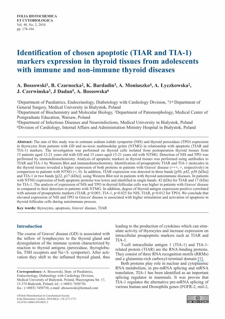
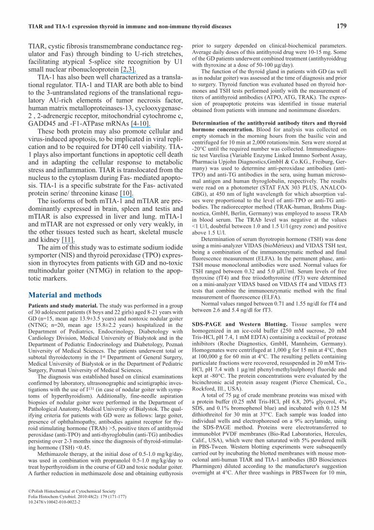
Identification of proapoptotic TIAR (TIA-1 relatedprotein) molecule in the thyroid tissues revealed a higher expression of this protein in patients withGraves' disease (+++) in comparison to patients withNTNG (+) (Figs. 1 and 2). The elevated expression ofTIAR in the thyroid tissue of patients with GD wasobserved mainly in hyperfunctional thyrocytes (withhigher epithelium). The expression of TIA-1 moleculewas detected only in patients with Graves' disease (+)in lymph nodes with germinal centers.
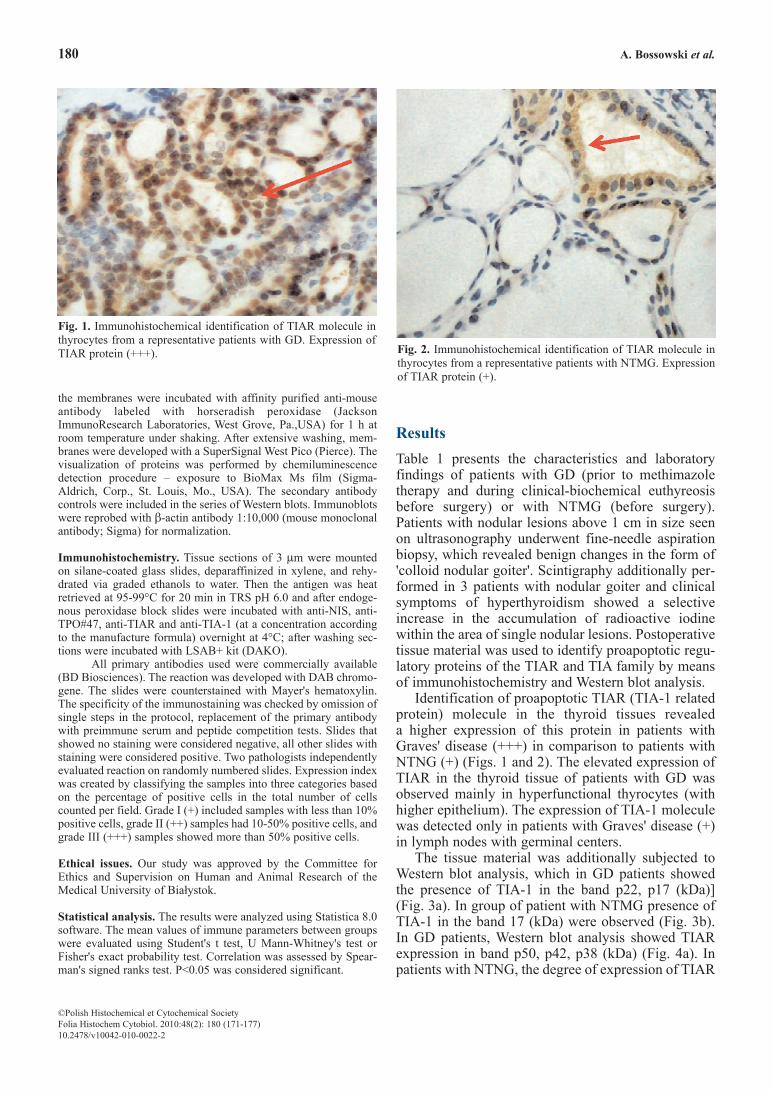
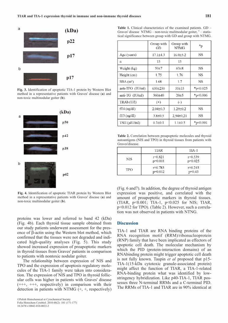
The tissue material was additionally subjected toWestern blot analysis, which in GD patients showedthe presence of TIA-1 in the band p22, p17 (kDa)](Fig. 3a). In group of patient with NTMG presence ofTIA-1 in the band 17 (kDa) were observed (Fig. 3b).In GD patients, Western blot analysis showed TIARexpression in band p50, p42, p38 (kDa) (Fig. 4a). Inpatients with NTNG, the degree of expression of TIAR
Fig. 1. Immunohistochemical identification of TIAR molecule inthyrocytes from a representative patients with GD. Expression ofTIAR protein (+++). Fig. 2. Immunohistochemical identification of TIAR molecule in
thyrocytes from a representative patients with NTMG. Expressionof TIAR protein (+).
181TIAR and TIA-1 expression thyroid in immune and non-immune thyroid diseases
©Polish Histochemical et Cytochemical SocietyFolia Histochem Cytobiol. 2010:48(2): 181 (171-177) 10.2478/v10042-010-0022-2
proteins was lower and referred to band 42 (kDa) (Fig. 4b). Each thyroid tissue sample obtained fromour study patients underwent assessment for the pres-ence of β-actin using the Western blot method, whichconfirmed that the tissues were not degraded and indi-cated high-quality analyses (Fig. 5). This studyshowed increased expression of proapoptotic markersin thyroid tissues from Graves' patients in comparisonto patients with nontoxic nodular goiter.
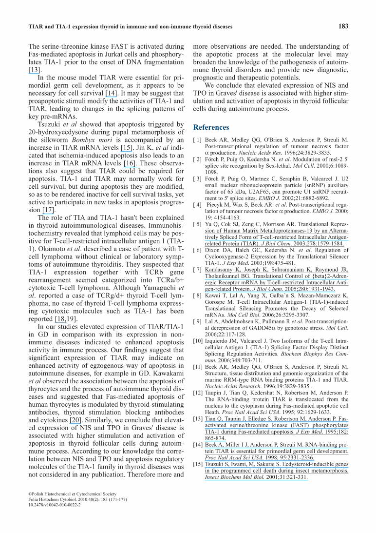
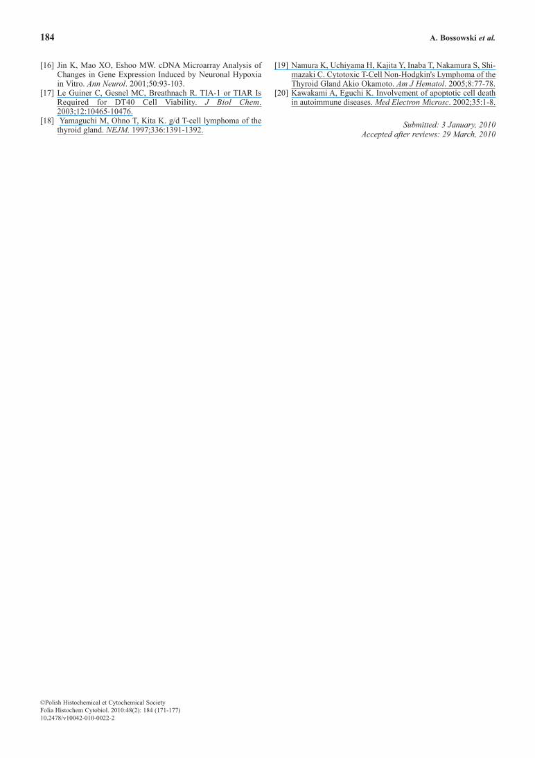
The relationship between expression of NIS andTPO and the expression of apoptosis regulatory mole-cules of the TIA-1 family were taken into considera-tion. The expression of NIS and TPO in thyroid follic-ular cells was higher in patients with Graves' disease(+++, +++, respectively) in comparison with theirdetection in patients with NTMG (+, +, respectively)
(Fig. 6 and7). In addition, the degree of thyroid antigenexpression was positive, and correlated with theamount of proapoptotic markers in thyroid tissues.(TIAR, p<0.001; TIA-1, p<0.025 for NIS; TIAR,p<0.012 for TPO). (Table 2). However, such a correla-tion was not observed in patients with NTNG.
DiscussionTIA-1 and TIAR are RNA binding proteins of theRNA recognition motif (RRM)/ribonucleoprotein(RNP) family that have been implicated as effectors ofapoptotic cell death. The molecular mechanism bywhich the PID (protein-interaction domains) of anRNAbinding protein might trigger apoptotic cell deathis not fully known. Taupin et al proposed that p15-TIA-1(15-kDa cytotoxic granule-associated protein)might affect the function of TIAR, a TIA-1-relatedRNA-binding protein what was identified by low-stringency hybridization. Like p40-TIA-1, TIAR pos-sesses three N-terminal RRMs and a C-terminal PID.The RRMs of TIA-1 and TIAR are in 90% identical at
Table 1. Clinical characteristics of the examined patients. GD –Graves' disease NTMG – non-toxic mulitnodular goiter, * – statis-tical significance between group with GD and group with NTMG.
Table 2. Correlation between proapoptotic molecules and thyroidautoantigens (NIS and TPO) in thyroid tissues from patients withGraves'disease.
Fig. 3. Identification of apoptotic TIA-1 protein by Western Blotmethod in a representative patients with Graves' disease (a) andnon-toxic multinodular goiter (b).
Fig. 4. Identification of apoptotic TIAR protein by Western Blotmethod in a representative patients with Graves' disease (a) andnon-toxic multinodular goiter (b).
182 A. Bossowski et al.
©Polish Histochemical et Cytochemical SocietyFolia Histochem Cytobiol. 2010:48(2): 182 (171-177) 10.2478/v10042-010-0022-2
the amino acid level, and the PIDs are 50% identical.Like TIA-1, TIAR triggers DNA fragmentation in per-meabilized thymocytes, suggesting its possibleinvolvement in apoptosis. Results of their study showthat TIAR is a ubiquitously expressed nuclear proteinthat rapidly moves to the cytoplasm in response toexogenous triggers of apoptosis. TIAR is normally
confined to the nucleus of cells, but during Fas-medi-ated apoptosis, it is rapidly translocated to the cyto-plasm. Cytoplasmic redistribution precedes the onsetof DNA fragmentation and the nuclear architecturalchanges that facilitate histone extraction [12].
Similar studies were conducted by Tain et al, whoproved that TIA-1 itself has been linked to apoptosis.
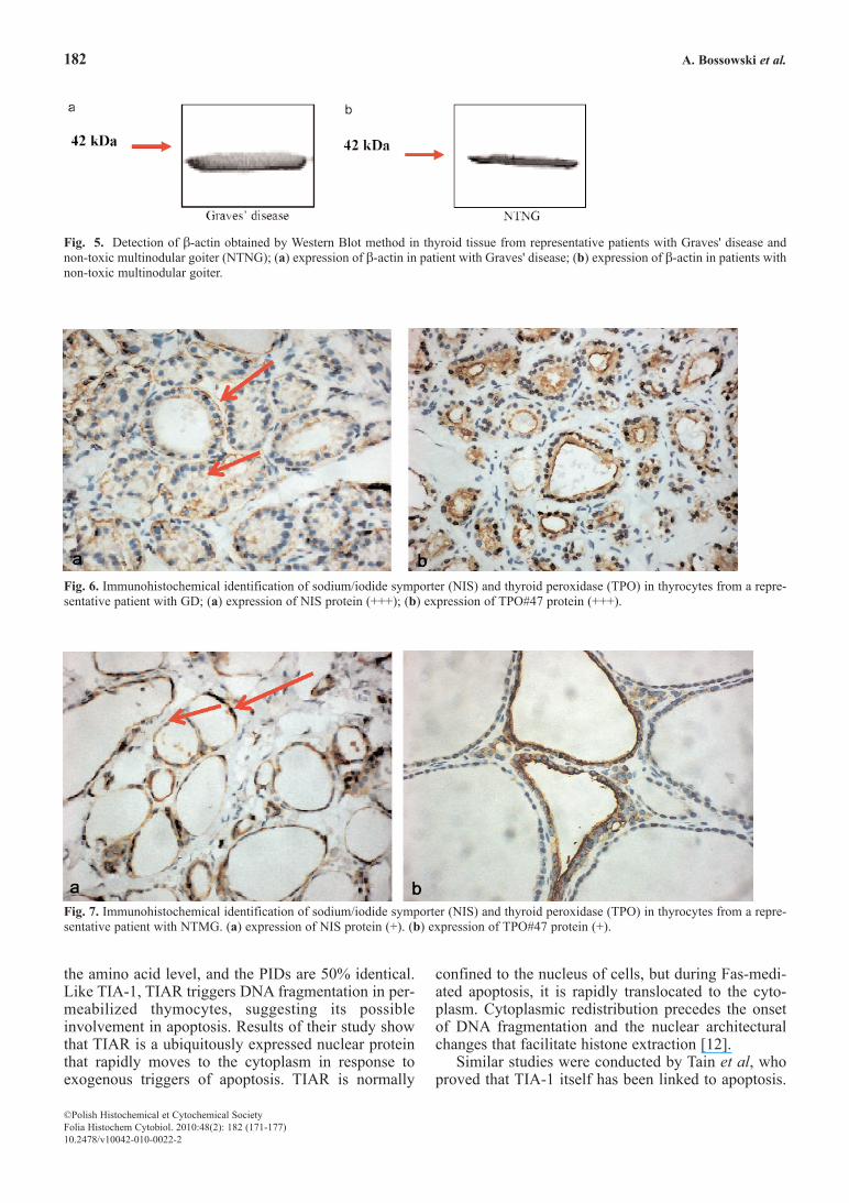
Fig. 5. Detection of β-actin obtained by Western Blot method in thyroid tissue from representative patients with Graves' disease and non-toxic multinodular goiter (NTNG); (a) expression of β-actin in patient with Graves' disease; (b) expression of β-actin in patients withnon-toxic multinodular goiter.
Fig. 6. Immunohistochemical identification of sodium/iodide symporter (NIS) and thyroid peroxidase (TPO) in thyrocytes from a repre-sentative patient with GD; (a) expression of NIS protein (+++); (b) expression of TPO#47 protein (+++).
Fig. 7. Immunohistochemical identification of sodium/iodide symporter (NIS) and thyroid peroxidase (TPO) in thyrocytes from a repre-sentative patient with NTMG. (a) expression of NIS protein (+). (b) expression of TPO#47 protein (+).
The serine-threonine kinase FAST is activated duringFas-mediated apoptosis in Jurkat cells and phosphory-lates TIA-1 prior to the onset of DNA fragmentation[13].
In the mouse model TIAR were essential for pri-mordial germ cell development, as it appears to benecessary for cell survival [14]. It may be suggest thatproapoptotic stimuli modify the activities of TIA-1 andTIAR, leading to changes in the splicing patterns ofkey pre-mRNAs.
Tsuzuki et al showed that apoptosis triggered by20-hydroxyecdysone during pupal metamorphosis ofthe silkworm Bombyx mori is accompanied by anincrease in TIAR mRNA levels [15]. Jin K. et al indi-cated that ischemia-induced apoptosis also leads to anincrease in TIAR mRNA levels [16]. These observa-tions also suggest that TIAR could be required forapoptosis. TIA-1 and TIAR may normally work forcell survival, but during apoptosis they are modified,so as to be rendered inactive for cell survival tasks, yetactive to participate in new tasks in apoptosis progres-sion [17].
The role of TIA and TIA-1 hasn't been explainedin thyroid autoimmunological diseases. Immunohis-tochemistry revealed that lymphoid cells may be pos-itive for T-cell-restricted intracellular antigen 1 (TIA-1). Okamoto et al. described a case of patient with T-cell lymphoma without clinical or laboratory symp-toms of autoimmune thyroiditis. They suspected thatTIA-1 expression together with TCRb generearrangement seemed categorized into TCRa/b+cytotoxic T-cell lymphoma. Although Yamaguchi etal. reported a case of TCRg/d+ thyroid T-cell lym-phoma, no case of thyroid T-cell lymphoma express-ing cytotoxic molecules such as TIA-1 has beenreported [18,19].
In our studies elevated expression of TIAR/TIA-1in GD in comparison with its expression in non-immune diseases indicated to enhanced apoptosisactivity in immune process. Our findings suggest thatsignificant expression of TIAR may indicate onenhanced activity of egzogenous way of apoptosis inautoimmune diseases, for example in GD. Kawakamiet al observed the association between the apoptosis ofthyrocytes and the process of autoimmune thyroid dis-eases and suggested that Fas-mediated apoptosis ofhuman thyrocytes is modulated by thyroid-stimulatingantibodies, thyroid stimulation blocking antibodiesand cytokines [20]. Similarly, we conclude that elevat-ed expression of NIS and TPO in Graves' disease isassociated with higher stimulation and activation ofapoptosis in thyroid follicular cells during autoim-mune process. According to our knowledge the corre-lation between NIS and TPO and apoptosis regulatorymolecules of the TIA-1 family in thyroid diseases wasnot considered in any publication. Therefore more and
more observations are needed. The understanding ofthe apoptotic process at the molecular level maybroaden the knowledge of the pathogenesis of autoim-mune thyroid disorders and provide new diagnostic,prognostic and therapeutic potentials.
We conclude that elevated expression of NIS andTPO in Graves' disease is associated with higher stim-ulation and activation of apoptosis in thyroid follicularcells during autoimmune process.
References [ 1] Beck AR, Medley QG, O'Brien S, Anderson P, Streuli M.
Post-transcriptional regulation of tumour necrosis factor α production. Nucleic Acids Res. 1996;24:3829-3835.
[ 2] Förch P, Puig O, Kedersha N. et al. Modulation of msl-2 5'splice site recognition by Sex-lethal. Mol Cell. 2000;6:1089-1098.
[ 3] Förch P, Puig O, Martnez C, Seraphin B, Valcarcel J. U2small nuclear ribonucleoprotein particle (snRNP) auxiliaryfactor of 65 kDa, U2AF65, can promote U1 snRNP recruit-ment to 5' splice sites. EMBO J. 2002;21:6882-6892.
[ 4] Piecyk M, Wax S, Beck AR. et al. Post-transcriptional regu-lation of tumour necrosis factor α production. EMBO J. 2000;19: 4154-4163.
[ 5] Yu Q, Cok SJ, Zeng C, Morrison AR. Translational Repres-sion of Human Matrix Metalloproteinases-13 by an Alterna-tively Spliced Form of T-cell-restricted Intracellular Antigen-related Protein (TIAR). J Biol Chem. 2003;278:1579-1584.
[ 6] Dixon DA, Balch GC, Kedersha N. et al. Regulation ofCyclooxygenase-2 Expression by the Translational SilencerTIA-1. J Exp Med. 2003;198:475-481.
[ 7] Kandasamy K, Joseph K, Subramaniam K, Raymond JR,Tholanikunnel BG. Translational Control of {beta}2-Adren-ergic Receptor mRNA by T-cell-restricted Intracellular Anti-gen-related Protein. J Biol Chem. 2005;280:1931-1943.
[ 8] Kawai T, Lal A, Yang X, Galba´n S, Mazan-Mamczarz K,Gorospe M. T-cell Intracellular Antigen-1 (TIA-1)-inducedTranslational Silencing Promotes the Decay of SelectedmRNAs. Mol Cell Biol. 2006;26:3295-3307.
[ 9] Lal A, Abdelmohsen K, Pullmann R et al. Post-transcription-al derepression of GADD45α by genotoxic stress. Mol Cell.2006;22:117-128.
[10] Izquierdo JM, Valcarcel J. Two Isoforms of the T-cell Intra-cellular Antigen 1 (TIA-1) Splicing Factor Display DistinctSplicing Regulation Activities. Biochem Biophys Res Com-mun. 2006;348:703-711.
[11] Beck AR, Medley QG, O'Brien S, Anderson P, Streuli M.Structure, tissue distribution and genomie organization of themurine RRM-type RNA binding proteins TIA-1 and TIAR.Nucleic Acids Research. 1996;19:3829-3835 .
[12] Taupin J, Tian Q, Kedershat N, Robertson M, Anderson P.The RNA-binding protein TIAR is translocated from thenucleus to the cytoplasm during Fas-mediated apoptotic cellHeath. Proc Natl Acad Sci USA. 1995; 92:1629-1633.
[13] Tian Q, Taupin J, Elledge S, Robertson M, Anderson P. Fas-activated serine/threonine kinase (FAST) phosphorylatesTIA-1 during Fas-mediated apoptosis. J Exp Med. 1995;182:865-874.
[14] Beck A, Miller I J, Anderson P, Streuli M. RNA-binding pro-tein TIAR is essential for primordial germ cell development.Proc Natl Acad Sci USA. 1998; 95:2331-2336.
[15] Tsuzuki S, Iwami, M, Sakurai S. Ecdysteroid-inducible genesin the programmed cell death during insect metamorphosis.Insect Biochem Mol Biol. 2001;31:321-331.
183TIAR and TIA-1 expression thyroid in immune and non-immune thyroid diseases
©Polish Histochemical et Cytochemical SocietyFolia Histochem Cytobiol. 2010:48(2): 183 (171-177) 10.2478/v10042-010-0022-2
184 A. Bossowski et al.
©Polish Histochemical et Cytochemical SocietyFolia Histochem Cytobiol. 2010:48(2): 184 (171-177) 10.2478/v10042-010-0022-2
[16] Jin K, Mao XO, Eshoo MW. cDNA Microarray Analysis ofChanges in Gene Expression Induced by Neuronal Hypoxiain Vitro. Ann Neurol. 2001;50:93-103.
[17] Le Guiner C, Gesnel MC, Breathnach R. TIA-1 or TIAR IsRequired for DT40 Cell Viability. J Biol Chem.2003;12:10465-10476.
[18] Yamaguchi M, Ohno T, Kita K. g/d T-cell lymphoma of thethyroid gland. NEJM. 1997;336:1391-1392.
[19] Namura K, Uchiyama H, Kajita Y, Inaba T, Nakamura S, Shi-mazaki C. Cytotoxic T-Cell Non-Hodgkin's Lymphoma of theThyroid Gland Akio Okamoto. Am J Hematol. 2005;8:77-78.
[20] Kawakami A, Eguchi K. Involvement of apoptotic cell deathin autoimmune diseases. Med Electron Microsc. 2002;35:1-8.
Submitted: 3 January, 2010Accepted after reviews: 29 March, 2010


