Embed Size (px)
Citation preview

Identification of Neutral Cholesterol Ester Hydrolase, a KeyEnzyme Removing Cholesterol from Macrophages*□S
Received for publication, April 7, 2008, and in revised form, September 8, 2008 Published, JBC Papers in Press, September 9, 2008, DOI 10.1074/jbc.M802686200
Hiroaki Okazaki‡1, Masaki Igarashi‡1, Makiko Nishi‡, Motohiro Sekiya‡, Makiko Tajima‡, Satoru Takase‡,Mikio Takanashi‡, Keisuke Ohta‡, Yoshiaki Tamura‡, Sachiko Okazaki‡, Naoya Yahagi‡, Ken Ohashi‡,Michiyo Amemiya-Kudo‡§, Yoshimi Nakagawa¶, Ryozo Nagai�, Takashi Kadowaki‡, Jun-ichi Osuga‡,and Shun Ishibashi‡**2
From the ‡Departments of Metabolic Diseases and �Cardiovascular Diseases, Graduate School of Medicine, University of Tokyo,7-3-1 Hongo, Bunkyo-ku, Tokyo 113-8655, §Okinaka Memorial Institute for Medical Research and Toranomon Hospital,Tokyo 105-0001, ¶Metabolism, Endocrinology, and Atherosclerosis, Institute of Clinical Medicine, University of Tsukuba,Ibaraki 305-8575, and **Division of Endocrinology and Metabolism, Department of Medicine, School of Medicine, Jichi MedicalUniversity, Tochigi 329-0498, Japan
Unstable lipid-rich plaques in atherosclerosis are character-ized by the accumulation of macrophage foam cells loaded withcholesterol ester (CE). Although hormone-sensitive lipase andcholesteryl ester hydrolase (CEH) have been proposed to medi-ate the hydrolysis of CE in macrophages, circumstantial evi-dence suggests the presence of other enzymes with neutral cho-lesterol ester hydrolase (nCEH) activity. Here we show that themurine orthologue of KIAA1363, designated as neutral choles-terol ester hydrolase (NCEH), is a microsomal nCEH with highexpression in murine and human macrophages. The effect ofvarious concentrations of NaCl on its nCEH activity resemblesthat on endogenous nCEHactivity ofmacrophages. RNA silenc-ing of NCEH decreases nCEH activity at least by 50%; con-versely, its overexpression inhibits the CE formation in macro-phages. Immunohistochemistry reveals that NCEH is expressedin macrophage foam cells in atherosclerotic lesions. These dataindicate that NCEH is responsible for a major part of nCEHactivity in macrophages and may be a potential therapeutic tar-get for the prevention of atherosclerosis.
Atherosclerotic cardiovascular diseases are the leadingcauses ofmortality in industrialized countries, despite advancesin the management of coronary risk factors. Heart attacks arisefrom thrombotic occlusion of coronary arteries following therupture of plaques. Lipid-rich plaques, which are characterizedby a plethora of CE3-ladenmacrophage foam cells, are prone to
rupture (1). Thus, it is important to clarify the mechanism thateliminates CE from macrophage-derived foam cells.Foam cells are generated by the unlimited uptake ofmodified
lipoproteins through scavenger receptors (2). Cholesterol in thelipoproteins is stored in lipid droplets as CE after re-esterifica-tion by acyl-CoA:cholesterol acyltransferase 1 (ACAT1) (3).Hydrolysis of CE is the initial step toward elimination of cho-lesterol from foam cells (4). Free cholesterol thus generated isre-esterified or is released from the cells primarily throughATP-binding cassette transporters (5). Thus, the balancebetween synthesis andhydrolysis of CE conceivably governs thelevel of CE in macrophages.Hydrolysis of CE inmacrophages has been known for over 40
years (6). However, its molecular mechanism has yet to be fullyunderstood. Circumstantial evidence suggests that the hydrol-ysis of CE in foam cell macrophages is mediated by hormone-sensitive lipase (HSL), a multifunctional enzyme that catalyzesthe hydrolysis of triacylglyerol (TG), diacylglycerol, CE, andretinyl ester in various organs such as adipose tissue, muscle,and testis (7, 8). This belief is supported by the following facts.First, it has been demonstrated that various lines of macro-phages express HSL (9–12). Second, HSL expression is regu-lated coordinately with nCEH activity in murine macrophages(13). Third, we (14) and others (15) demonstrated that overex-pression of HSL is associated with increased hydrolysis of CEstores in THP-1 and RAW264.7 macrophages.However, recent studies by us (16) and others (17) have chal-
lenged this notion by finding that peritoneal macrophages iso-lated fromHSL-deficientmice retained almost a normal level ofnCEH activity, suggesting that enzyme(s) other than HSL areresponsible for nCEH activity in macrophages. Moreover, HSLis expressed at a low level (11, 12) or is undetectable (17) inhumanmacrophages. Recently, Ghosh et al. (18) reported clon-ing of cholesteryl ester hydrolase (CEH) which mediates thehydrolysis of CE in human monocyte-derived macrophages.They have further shown that macrophage-specific transgenicexpression of CEH significantly reduced atherosclerosis in low
* This work was supported by a grant-in-aid for scientific research from theMinistry of Education, Science, and Culture and the Program for Promotionof Fundamental Studies in Health Sciences of the National Institute of Bio-medical Innovation and Takeda Science Foundation. The costs of publica-tion of this article were defrayed in part by the payment of page charges.This article must therefore be hereby marked “advertisement” in accord-ance with 18 U.S.C. Section 1734 solely to indicate this fact.
□S The on-line version of this article (available at http://www.jbc.org) containssupplemental Figs. S1 and S2.
1 Both authors contributed equally to this work.2 To whom correspondence should be addressed: Division of Endocrinology
and Metabolism, Dept. of Medicine, Jichi Medical University, 3311-1Yakushiji, Shimotsuke, Tochigi 329-0498, Japan. Tel.: 81-285-58-7355; Fax:81-285-40-6035; E-mail: [email protected].
3 The abbreviations used are: CE, cholesterol ester; HSL, hormone-sensitivelipase; CEH, cholesteryl ester hydrolase; nCEH, neutral cholesteryl esterhydrolase; Ad, adenovirus; BSA, bovine serum albumin; LDL, low density
lipoprotein; acLDL, acetylated LDL; DMEM, Dulbecco’s modified Eagle’smedium; MPM, murine peritoneal macrophage; shRNA, short hairpin RNA;PBS, phosphate-buffered saline; TGH, triacylglycerol hydrolase; GST, gluta-thione S-transferase; m.o.i., multiplicity of infection; TG, triacylglycerol.
THE JOURNAL OF BIOLOGICAL CHEMISTRY VOL. 283, NO. 48, pp. 33357–33364, November 28, 2008© 2008 by The American Society for Biochemistry and Molecular Biology, Inc. Printed in the U.S.A.
NOVEMBER 28, 2008 • VOLUME 283 • NUMBER 48 JOURNAL OF BIOLOGICAL CHEMISTRY 33357
at Indiana University Libraries on January 1, 2009
ww
w.jbc.org
Dow
nloaded from
http://www.jbc.org/cgi/content/full/M802686200/DC1Supplemental Material can be found at:
density lipoprotein receptor knock-outmice (19). However, theCEH gene is identical to the human orthologue of TGH, whichwas originally reported as amicrosomal TG lipase in rat liver byLehner et al. (20). In our hands, both the mouse orthologue ofTGH, designated as TGH-1, and its paralogue with 70.4% iden-tity to TGH-1, designated as TGH-2, have negligible nCEHactivity (21). These considerations have prompted us to employa bioinformatic approach to identify the enzyme responsible fornCEH activity in macrophages.
EXPERIMENTAL PROCEDURES
Materials—Phorbol 12-myristate 13-acetate, triolein, leci-thin, bovine serum albumin fraction V (BSA), p-nitrophenylbutyrate, and leupeptin were purchased from Sigma. Dexam-ethasone, 3-isobutyl-2-methylxanthine, fatty acid-free BSA,cholesterol esterase fromPseudomonas sp., cholesterol oxidase,horseradish peroxidase, p-hydroxyphenylacetic acid, andsodium taurocholate were purchased from Wako Pure Chem-icals (Osaka, Japan). Pioglitazone was provided by TakedaPharmaceutical (Osaka, Japan). Tri[3H]oleoylglycerol, choles-terol [1-14C]oleate, and [1-14C]oleic acid were purchased fromGE Healthcare. LDL was isolated by ultracentrifugation andacetylated to prepare acetylated LDL (acLDL) as described pre-viously (14).Cells—HEK293 or RAW264.7 cells were cultured in Dulbec-
co’s modified Eagle’s medium (DMEM) high glucose contain-ing 10% (v/v) fetal bovine serum without or with 1 mM sodiumpyruvate, respectively. THP-1 cells were cultured in RPMI 1640medium containing 10% (v/v) fetal bovine serum and differen-tiated to THP-1 macrophages by treatment with 100 nM phor-bol 12-myristate 13-acetate for 48 h. Murine peritoneal macro-phages (MPM) were harvested as described previously (16). Inbrief, 1 ml of 5% thioglycolate broth was injected into the peri-toneal cavities ofmice. After 4 days, the peritoneal cavities werelavaged with 16 ml of ice-cold saline. The cells were washedthree times with PBS and resuspended in DMEM to give a con-centration of 106 cells per ml, and 10 ml per dish was plated in10-cm dishes. After incubation at 37 °C for 2 h, the nonadher-ent cells were removed by washing three times with warmedPBS. Cells were incubated in DMEM containing 10% fetal calfserum for 24 h and harvested for the experiments. 3T3-L1 cellswere cultured in medium A (DMEM containing 10% calfserum, supplemented with calcium pantothenate and biotin).To induce differentiation of 3T3-L1 cells into adipocytes, 2 daypostconfluent preadipocytes (day 0) were incubated with 1 �Mdexamethasone and 0.5 mM isobutylmethylxanthine, 5 �g/mlinsulin, 1 �M pioglitazone for 48 h, following treatment with 5�g/ml insulin and 1 �M pioglitazone for 48 h. After the incuba-tion period, cells were switched to medium A, and the mediumwas renewed every other day. Mononuclear cells were isolatedfrom a normal volunteer using Lymphoprep (NYCOMED,Roskilde, Denmark), and cells that attached to plastic disheswere cultured in RPMI 1640 medium containing 10% fetal calfserum and used as human monocyte-derived macrophages.Mice—C57BL/6J and apoE knock-out mice were purchased
from Clea (Tokyo, Japan) and The Jackson Laboratories (BarHarbor,ME), respectively. Mice weremaintained and cared foraccording to the regulations of the Animal Care Committee of
the University of Tokyo. For the preparation of atheroscleroticaortas, mice were caged separately with 12-h light/dark cyclesand given free access to standard chow diet containing 0.075%cholesterol (MF; Oriental Yeast Co., Ltd.; Osaka, Japan).Data Base Search and Amino Acid Sequence Analysis—The
search for a novel lipase containing lipase consensusmotifs and�/�-hydrolase folds was performed in the GENES protein database of Kyoto Encyclopedia ofGenes andGenomes by using theMOTIF search program (22), followed by prediction of second-ary structure by PSIPRED and the PREDICT PROTEIN PHD-sec program (23). The search for orthologues or paralogues ofNCEH was performed by Sequence Similarity Database in theKyoto Encyclopedia of Genes and Genomes Database (22).Alignment of the deduced protein sequence with other lipases,calculation of the percent identity, and generation of a phylo-genetic tree were performed using the ClustalW program. Pre-diction of the transmembrane domain was performed usingSOSUI and the PREDICT PROTEIN PHDhtm program (23).cDNA Cloning for the Expression of Recombinant Proteins—
Total RNA isolated from murine peritoneal macrophages wasreverse-transcribed into cDNA using reverse transcriptase fol-lowing the manufacturer’s protocol (Thermoscript RNaseH-reverse transcriptase (Invitrogen)). The coding sequences ofmurineNCEHwere amplified by PCR frommacrophage cDNAusing cDNA polymerase (AmpliTaq DNA polymerase (RocheApplied Science)). The primers used for PCR were as follows:murine NCEH forward 5�-CCGCCTGGGGACAATGAG-3�and murine NCEH reverse 5�-TCTGAGGAGTTTCCTCCG-TCA-3�.
The construction of cDNA for HSL was described previ-ously. The products containing the complete open readingframe were ligated into compatible sites of pGEM T easyvector (Promega).Recombinant Adenovirus for NCEHandHSL Expression (Ad-
NCEH, Ad-HSL)—Murine NCEH or HSL cDNA fragment wasamplified by PCR from mouse cDNA containing plasmids(described above) using forward and reverse primers. LacZcDNAwas amplified by PCR from SV40 �-galactosidase vector(Promega). The PCR products were ligated to pENTER4 vector(Invitrogen). Subsequently, murine NCEH or HSL cDNA frag-ment was subcloned into pAd/CMV/V5-DEST vector by Gate-way Technology (Invitrogen). The adenoviral vectors were lin-earized using restriction enzyme and transfected into HEK293using Superfect reagent (Promega). Large scale production ofhigh titer recombinant Ad-NCEH, Ad-HSL, or Ad-LacZ wasperformed as described elsewhere (14). The purified viruseswere stored in 10% (v/v) glycerol/phosphate-buffered saline(PBS) at �80 °C.Recombinant Adenovirus Expressing Short Hairpin (sh) RNA
Directed against Murine NCEH—Recombinant adenovirusexpressing shRNA for NCEH (Ad-shNCEH) was produced bythe Gateway Technology (Invitrogen). The sense and antisensetemplates of NCEH shRNA were produced from nucleotides136 to 154 of the NCEH coding sequence.The following oligonucleotides were used: shRNA for NCEH
sense template, 5�-GT GAG TAA TCT GAT ACG TTA CGTGTG CTG TCC GTA ATG TAT CAG GTT ACT CAC-3�;shRNA for NCEH antisense template, 5�-GT GAG TAA CCT
Neutral Cholesterol Ester Hydrolase in Macrophages
33358 JOURNAL OF BIOLOGICAL CHEMISTRY VOLUME 283 • NUMBER 48 • NOVEMBER 28, 2008
at Indiana University Libraries on January 1, 2009
ww
w.jbc.org
Dow
nloaded from

GAT ACA TTA CGG ACA GCA CAC GTA ACG TAT CAGATTACTCAC-3�. An adenovirus containing shRNA for�-ga-lactosidase (Ad-shLacZ) was used as a control.Murine peritoneal macrophages were seeded in 6-well plates
at a cell density of 1.7 � 106 cells/well. After transduction withAd-shNCEH or Ad-shLacZ for 48 h at 37 °C, cells were har-vested, and whole cell lysates were assayed for enzymaticactivities.Generation of Rabbit Polyclonal Antibodies against Murine
NCEH—To prepare polyclonal anti-murine NCEH antiserum,amino acid residues containing the catalytic domain of murineNCEH (amino acids 99–250) were expressed in bacteria as aglutathione S-transferase (GST) fusion protein, which waspurified by glutathione affinity chromatography, and used forimmunization of rabbits according to a standard protocol asdescribed previously (24). Serum of rabbits before immuniza-tion was used as a control. From serum samples with high anti-body titers, IgG fractions were isolated using a protein G col-umn (GE Healthcare).Northern Blot Analyses—Total RNA was isolated from vari-
ous tissues or cultured cells using TRIzol reagent according tothe manufacturer’s protocol (Invitrogen). RNA (10 �g) wassubjected to formaldehyde/agarose gel electrophoresis andblotted to a Hybond-N membrane (GE Healthcare). Aftercross-linking, mouse NCEH, HSL, and TGH-1 mRNA weredetected using 32P-labeled probes. Probes for murine NCEHwere originally prepared by reverse transcription-PCR frommouse peritoneal macrophage mRNA. Probes for HSL (exon 8probe), TGH-1were constructed from cDNA fragments ampli-fied by reverse transcription-PCR using cDNA obtained frommouse adipose tissue as a template (14). After hybridization andwashing, signals were visualized by exposure to a Phosphor-Imager Screen (FUJIFILM) and analyzed using BASTATION(FUJIFILM) software.Western Blot Analysis—Cells were sonicated in buffer A and
centrifuged at 40,000 or 100,000 � g for 45 min at 4 °C. Thesupernatant was used as S-40 or S-100 cytosolic fraction, andthe precipitates were resuspended and used as the microsomalfraction (14). Ten micrograms of proteins of various cellularfractions were separated on a 10% SDS-PAGE and transferredto a nitrocellulosemembrane. For detection of the proteins, themembranes were incubated with either anti-murine NCEH,anti-murine HSL antiserum, monoclonal anti-mouse ACAT1antibody (25), or anti-F4/80 antibody (AbD serotec) at a dilu-tion of 1:100–1,000. Specifically bound immunoglobulins weredetected in a second reaction with a horseradish peroxidase-labeled IgG conjugate and visualized by enhanced chemilumi-nescence detection (ECL Plus, GE Healthcare) using a Kodakimage system, and the intensity of immunoreactive bands werequantified by NIH-image software.Assays for CE Hydrolase and TG Lipase Activities—Whole
cell lysates were prepared from transfected HEK293, murineperitonealmacrophages, orTHP1 cells andused for the enzymeactivity assays. These cellswere sonicated in bufferA (50mmol/liter Tris-HCl, pH 7.0, 250 mmol/liter sucrose, 1 mmol/literEDTA, 2�g/ml leupeptin) and centrifuged at 100,000� g for 45min at 4 °C. The supernatant was used as S-100 cytosolic frac-tion, respectively. The precipitates at 100,000 � g were resus-
pended andused as themicrosomal fraction (14). nCEHactivitywasmeasured essentially as described byHajjar et al. (26), usinga reactionmixture containing 6.14�Mcholesterol [1-14C]oleate(48.8�Ci/�mol; 1�Ci� 37 kBq). Triacylglycerol lipase activitywas measured according to a modification of the method ofHajjar et al. (26). In brief, the samples were incubated at 37 °Cfor 30 min in a final volume of 200 �l of a reaction mixturecontaining 105 �M tri[3H]oleoylglycerol (99.4 �Ci/�mol), 23.7�M lecithin, 12.5�M sodium taurocholate, 1 MNaCl, and 85mM
potassium phosphate, pH 7.0. The high concentration of NaClwas included to inactivate lipoprotein lipase.In Vitro Adenovirus Experiments—Two days after THP-1
cells were treatedwith phorbol 12-myristate 13-acetate, THP-1macrophages were incubated in RPMI 1640 medium contain-ing 5mg/ml BSA and transducedwith recombinant adenoviruscarrying NCEH, HSL, or LacZ as a control. After 24 h, acLDLwas added to the medium at a final concentration of 100 �g/mlwithout or with 1% [14C]oleate-BSA complex (14), and cellswere incubated for 48 h at 37 °C. Cellular lipids were extractedusing hexane/isopropyl alcohol (3:2), and cholesterol contentwas determined by enzymatic fluorometric microassay accord-ing to the method of Heider and Boyett (27), with minor mod-ifications (25). The radioactivity of intracellular CE formedfrom [14C]oleate was determined as described (14).Immunohistochemical Localization—Cells were fixed with
3% paraformaldehyde after washing with PBS. Tissue sampleswere fixed in 10% formalin, pH 7.0, embedded in paraffin, cutinto sections, and placed on glass slides. Tissue samples weredeparaffinized and rehydrated before use. The samples wereincubated in 3%H2O2 inPBS to quench endogenous peroxidaseactivity and washed with PBS, and nonspecific binding siteswere blocked by incubation with 1% goat serum. The sectionswere then incubated with affinity-purified anti-NCEH poly-clonal antibody. After washing with PBS, the sections wereincubated with a biotinylated goat anti-rabbit antibody (1:200,Vector Laboratories, Burlingame, CA). The antibody-specificstaining was visualized with a Vecstatin ABC reagent (VectorLaboratories) and diamine benzidine, resulting in a brown pre-cipitate. Cell nuclei were counterstained with methyl green.Combined immunohistochemical staining was performed toidentify the cell types expressingNCEH as described previouslywith minor modifications (28). NCEH was first localized byusing the protocol above. Subsequently, the sections weretreated with 0.1% pepsin (Roche Applied Science) in 0.1 N HClfor 30 min and incubated with rat monoclonal anti-murineF4/80 (1:10, serotec) for 30 min. The respective sections wereincubated with a biotinylated rabbit anti-rat antibody (1:200,Vector Laboratories) for 30 min, followed by a 30-min incuba-tion with an alkaline phosphatase-coupled ABC reagent (1:200,Vector Laboratories). Alkaline phosphatase activity was visual-ized by using the Alkaline Phosphatase Kit III (Vector Labora-tories), resulting in a blue precipitate.Statistical Analyses—Results are presented as means � S.E.
Student’s t test was employed to compare the means. All calcu-lations were performed with STAT view version 5.0 forMacin-tosh (SAS Institute Inc.).
Neutral Cholesterol Ester Hydrolase in Macrophages
NOVEMBER 28, 2008 • VOLUME 283 • NUMBER 48 JOURNAL OF BIOLOGICAL CHEMISTRY 33359
at Indiana University Libraries on January 1, 2009
ww
w.jbc.org
Dow
nloaded from
RESULTS AND DISCUSSION
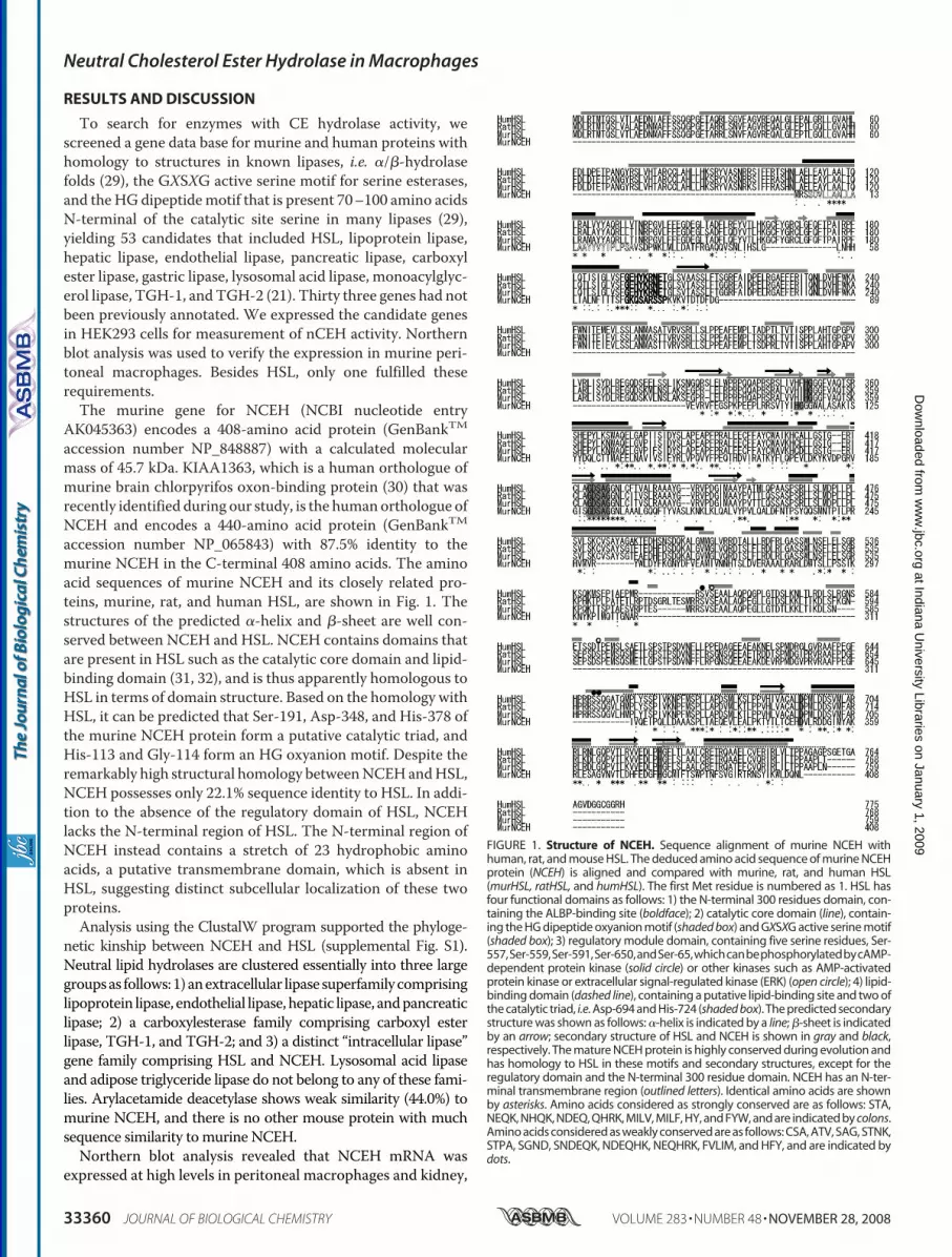
To search for enzymes with CE hydrolase activity, wescreened a gene data base for murine and human proteins withhomology to structures in known lipases, i.e. �/�-hydrolasefolds (29), the GXSXG active serine motif for serine esterases,and theHG dipeptidemotif that is present 70–100 amino acidsN-terminal of the catalytic site serine in many lipases (29),yielding 53 candidates that included HSL, lipoprotein lipase,hepatic lipase, endothelial lipase, pancreatic lipase, carboxylester lipase, gastric lipase, lysosomal acid lipase, monoacylglyc-erol lipase, TGH-1, andTGH-2 (21). Thirty three genes had notbeen previously annotated. We expressed the candidate genesin HEK293 cells for measurement of nCEH activity. Northernblot analysis was used to verify the expression in murine peri-toneal macrophages. Besides HSL, only one fulfilled theserequirements.The murine gene for NCEH (NCBI nucleotide entry
AK045363) encodes a 408-amino acid protein (GenBankTMaccession number NP_848887) with a calculated molecularmass of 45.7 kDa. KIAA1363, which is a human orthologue ofmurine brain chlorpyrifos oxon-binding protein (30) that wasrecently identified during our study, is the humanorthologue ofNCEH and encodes a 440-amino acid protein (GenBankTMaccession number NP_065843) with 87.5% identity to themurine NCEH in the C-terminal 408 amino acids. The aminoacid sequences of murine NCEH and its closely related pro-teins, murine, rat, and human HSL, are shown in Fig. 1. Thestructures of the predicted �-helix and �-sheet are well con-served between NCEH and HSL. NCEH contains domains thatare present in HSL such as the catalytic core domain and lipid-binding domain (31, 32), and is thus apparently homologous toHSL in terms of domain structure. Based on the homology withHSL, it can be predicted that Ser-191, Asp-348, and His-378 ofthe murine NCEH protein form a putative catalytic triad, andHis-113 and Gly-114 form an HG oxyanion motif. Despite theremarkably high structural homology betweenNCEHandHSL,NCEH possesses only 22.1% sequence identity to HSL. In addi-tion to the absence of the regulatory domain of HSL, NCEHlacks the N-terminal region of HSL. The N-terminal region ofNCEH instead contains a stretch of 23 hydrophobic aminoacids, a putative transmembrane domain, which is absent inHSL, suggesting distinct subcellular localization of these twoproteins.Analysis using the ClustalW program supported the phyloge-
netic kinship between NCEH and HSL (supplemental Fig. S1).Neutral lipid hydrolases are clustered essentially into three largegroupsas follows:1)anextracellular lipase superfamilycomprisinglipoprotein lipase, endothelial lipase,hepatic lipase, andpancreaticlipase; 2) a carboxylesterase family comprising carboxyl esterlipase, TGH-1, and TGH-2; and 3) a distinct “intracellular lipase”gene family comprising HSL and NCEH. Lysosomal acid lipaseand adipose triglyceride lipase do not belong to any of these fami-lies. Arylacetamide deacetylase shows weak similarity (44.0%) tomurine NCEH, and there is no other mouse protein with muchsequence similarity to murine NCEH.Northern blot analysis revealed that NCEH mRNA was
expressed at high levels in peritoneal macrophages and kidney,
FIGURE 1. Structure of NCEH. Sequence alignment of murine NCEH withhuman, rat, and mouse HSL. The deduced amino acid sequence of murine NCEHprotein (NCEH) is aligned and compared with murine, rat, and human HSL(murHSL, ratHSL, and humHSL). The first Met residue is numbered as 1. HSL hasfour functional domains as follows: 1) the N-terminal 300 residues domain, con-taining the ALBP-binding site (boldface); 2) catalytic core domain (line), contain-ing the HG dipeptide oxyanion motif (shaded box) and GXSXG active serine motif(shaded box); 3) regulatory module domain, containing five serine residues, Ser-557,Ser-559,Ser-591,Ser-650,andSer-65,whichcanbephosphorylatedbycAMP-dependent protein kinase (solid circle) or other kinases such as AMP-activatedprotein kinase or extracellular signal-regulated kinase (ERK) (open circle); 4) lipid-binding domain (dashed line), containing a putative lipid-binding site and two ofthe catalytic triad, i.e. Asp-694 and His-724 (shaded box). The predicted secondarystructure was shown as follows: �-helix is indicated by a line; �-sheet is indicatedby an arrow; secondary structure of HSL and NCEH is shown in gray and black,respectively. The mature NCEH protein is highly conserved during evolution andhas homology to HSL in these motifs and secondary structures, except for theregulatory domain and the N-terminal 300 residue domain. NCEH has an N-ter-minal transmembrane region (outlined letters). Identical amino acids are shownby asterisks. Amino acids considered as strongly conserved are as follows: STA,NEQK, NHQK, NDEQ, QHRK, MILV, MILF, HY, and FYW, and are indicated by colons.Amino acids considered as weakly conserved are as follows: CSA, ATV, SAG, STNK,STPA, SGND, SNDEQK, NDEQHK, NEQHRK, FVLIM, and HFY, and are indicated bydots.
Neutral Cholesterol Ester Hydrolase in Macrophages
33360 JOURNAL OF BIOLOGICAL CHEMISTRY VOLUME 283 • NUMBER 48 • NOVEMBER 28, 2008
at Indiana University Libraries on January 1, 2009
ww
w.jbc.org
Dow
nloaded from

and to a lesser degree in heart and adrenal tissue (Fig. 2, Aand B). Compared with other enzymes with nCEH activityexpressed in macrophages, such as HSL and TGH-1, a murineorthologue of CEH reported by Ghosh (18), the expression ofNCEH was the most abundant and specific for macrophages(Fig. 2A). Although NCEH expression was barely detectable infreshly isolated humanmonocytes, it was robustly induced dur-ing the differentiation to mature macrophages (Fig. 2C).To determine the subcellular localization of endogenous
nCEHactivity andNCEHprotein, we isolated S-100 andmicro-somal fractions from murine peritoneal macrophages, meas-ured the nCEH activity (Fig. 2D), and performed Western blotanalysis for either NCEH or HSL (Fig. 2E). The nCEH activitywas distributed preferentially in the microsomal fraction.NCEH proteins, duplets withmolecular mass of 45 and 50 kDa,were also distributed preferentially in the microsomal fraction.HSL was more equally distributed between the two fractions.Thus, the subcellular distribution pattern of nCEH activity iscloser to that of NCEH protein than that of the HSL protein.We compared the amounts of NCEH protein with those of
HSL protein inmurine peritonealmacrophages andRAW264.7cells using recombinant proteins as standards for calculation(Fig. 2F and see supplemental Fig. S2). Here again, two bandswere reacted with anti-NCEH antibody. Subsequent studyshowed that both bands are products of glycation of the 40-kDa
protein.4 The ratio of NCEH proteinto HSL protein was calculated to be11 in murine peritoneal macro-phages and 1 in RAW264.7 cells,respectively.The preferential localization of
HSL and NCEH in the cytosol andmicrosomal fractions, respectively,was also confirmed by measure-ment of the enzymatic activity ofeach fraction in cells transientlytransfected with plasmids express-ing HSL or NCEH. Although mostof the nCEH activity of HSL-trans-fected cells was recovered in theS-40 fraction, nCEH activity wasbarely detectable in the S-40 frac-tion of NCEH-transfected cells aswell as murine peritoneal macro-phages (data not shown). This sub-cellular localization pattern iscompatible with the microsomaldistribution of NCEH and its pre-ponderance in murine peritonealmacrophages. These results areapparently contrary to those re-ported by Khoo et al. (33), whoshowed that 60% of the CE hydro-lase activity in the homogenate ofJ774 cells was recovered in the S-40fraction; this difference could beattributed to the high level ofexpression of HSL in J774 cells (34)
and the high level of expression of NCEH in murine peritonealmacrophages.To further determine enzymological characteristics of
NCEH, we overexpressed NCEH in HEK293 cells (Fig. 3A) andmeasured enzymatic activities of their whole cell lysate at vari-ous concentrations of NaCl (Fig. 3B), pH (Fig. 3D), and concen-trations of cholesterol oleate (Fig. 3E) or triolein (Fig. 3F) assubstrates.The nCEH activity in the whole cell lysates from NCEH-
transfected cells was slightly stimulated by low concentrationsof sodiumchloride (6.5% at 0.05M), and inhibited only by 31% athigher concentrations (1 M) (Fig. 3B). This pattern closelyresembled that of murine peritoneal macrophages (Fig. 3C).The CE hydrolase activity of NCEH-transfected cells was opti-mal at neutral pH (7.2) (Fig. 3D), which is close to the optimalpH for HSL, suggesting that both enzymes are not active or atleast not fully functional in lysosomes.The whole cell lysate of HSL-transfected cells exhibited
increased hydrolase activities for both TG and CE, with appar-ent Km of 11.7 and 22.8 �M, respectively. On the other hand,whole cell lysate of NCEH-transfected cells hydrolyzed TG andCE with apparent Km of 30.3 and 324 �M, respectively.
4 M. Igarashi, manuscript in preparation.
FIGURE 2. Tissue and subcellular distribution of NCEH expression. Total RNA (10 �g) from adipose tissuesand 3T3-L1 adipocytes at various stages of differentiation and murine peritoneal macrophages (A), variousmurine tissues (B), and human monocytes/monocyte-derived macrophages (C) were subjected to Northernblot analysis. Specific mRNAs were detected with radiolabeled murine cDNAs for NCEH, HSL, and TGH-1.Ethidium bromide staining of the gels is shown. Abbreviations used are as follows: WAT, white adipose tissue;BAT, brown adipose tissue; MPM, murine peritoneal macrophages; ed, epididymal; sc, subcutaneous. Subcel-lular distribution of nCEH activity and NCEH and HSL proteins. MPM were sonicated and centrifuged at100,000 � g and the supernatant (S-100) or microsomal (Ms) fractions along with whole cell lysate (whole) weresubjected to the measurements of nCEH activity (D) and Western blot analysis (E) using anti-HSL and anti-NCEHantisera. F, quantification of NCEH or HSL proteins in macrophages. Ten micrograms of proteins of whole celllysates from RAW264.7 and MPM were separated by SDS-PAGE on the same gel as the indicated amounts ofGST fusion proteins (supplemental Fig. S2). Immunoblotting was performed, and the densities of bands fromHSL or NCEH of RAW264.7 or MPM were quantified using NIH Image. Blots for ACAT1 and F4/80 are shown ascontrols. The moles of HSL or NCEH in 10 �g of protein from RAW264.7 or MPM were calculated from equationsrelating moles and densities of GST fusion proteins.
Neutral Cholesterol Ester Hydrolase in Macrophages
NOVEMBER 28, 2008 • VOLUME 283 • NUMBER 48 JOURNAL OF BIOLOGICAL CHEMISTRY 33361
at Indiana University Libraries on January 1, 2009
ww
w.jbc.org
Dow
nloaded from

To determine more directly how much of the nCEH activ-ity is accounted for by NCEH in murine peritoneal macro-phages, we used RNA silencing technology (Fig. 4). Infectionwith increasing doses of Ad-shLacZ nonspecifically reducedthe protein expression of both HSL and NCEH (Fig. 4A) aswell as nCEH activities in the infected cells (Fig. 4B).Whereas infection with 300 and 600 m.o.i. of Ad-shNCEHdid not specifically reduce the protein expression of HSL, itreduced the protein expression of NCEH by 51 and
41%, respectively, compared withAd-shLacZ. In parallel with theinhibition of NCEH, nCEH activi-ties were reduced by 49 and 42% byinfection with 300 and 600 m.o.i.of Ad-shNCEH, respectively (Fig.4B). These results indicate that atleast half of the nCEH activity ismediated by NCEH in murineperitoneal macrophages.To further examine whether
NCEH inhibits accumulation ofCE in macrophages, we infectedTHP-1 cells, which had beeninduced to differentiate to maturemacrophages by incubation withphorbol ester, with Ad-NCEH,Ad-HSL, or Ad-LacZ, and incu-bated the cells with acetyl-LDL,and compared the amounts of CE inthe cells (Fig. 5C) and CE formationfrom [14C]oleate (Fig. 5D). The cellsinfected with Ad-HSL or Ad-NCEHexpressed HSL or NCEH proteins,respectively, in a dose-dependentmanner (Fig. 5A) and showed dose-dependent increases of nCEH activ-ity in whole cell lysates (Fig. 5B). At300 m.o.i., the nCEH activity in Ad-NCEH-infected cells was 14% that
inAd-HSL-infected cells (Fig. 5B). This increase in nCEHactiv-ity in cells overexpressing HSL or NCEH was associated withdecreases in the intracellular CE content (Fig. 5C) as well as inthe rate of CE formation from [14C]oleate (Fig. 5D). More pro-nounced inhibition of CE accumulation by Ad-HSL than byAd-NCEH may be explained by the higher nCEH activityattained by Ad-HSL.To determine whether NCEH protein is expressed in ath-
erosclerotic lesions, we performed immunohistochemistry(Fig. 6). Cultured peritoneal macrophages were positivelystained with anti-NCEH antibody (brown, Fig. 6B) but notwith nonimmune IgG (Fig. 6A). Next, we stained tissue sec-tions of aorta from apoE�/� mice (Fig. 6C), which containedadvanced atherosclerotic lesions filled with cholesterol cleft.Double staining with F4/80, a macrophage-specific antibody(blue, Fig. 6E), showed prominent expression of NCEH pro-tein (brown, Fig. 6,D and E) in macrophages surrounding thecholesterol cleft in the subintimal space of apoE�/� aorta.
In summary, NCEH is substantially expressed in macro-phages in atherosclerotic plaques and significantly contrib-utes to nCEH activity of murine macrophages. This study isthe first demonstration of the molecular identity of the dom-inant nCEH in macrophages, a long unidentified enzyme cata-lyzing the counter-reaction of acyl-CoA:cholesterol acyltrans-ferase. Although our data do not exclude the possibility thatHSL or possibly other enzymes account at least in part for thenCEH activity in macrophages, it is tempting to speculate thatHSL evolves to have a more specialized role in adipocyte lipol-
FIGURE 3. Enzymological characteristics of NCEH. HEK293 cells were infected with Ad-LacZ, Ad-HSL, orAd-NCEH and used for the experiments. A, expression of NCEH or HSL was confirmed by Western blot analysis.Effects of various concentrations of NaCl on the nCEH activity of whole cell lysate from cells infected withAd-NCEH (B) or of MPM (C). D, effects of various pH on the nCEH activity of whole cell lysate from cells infectedwith Ad-NCEH using 50 mM acetate buffer (pH � 5), 50 mM phosphate buffer (5 � pH � 7.6), or 50 mM Tris-HClbuffer (pH � 7.6). Shown are the effects of various concentrations of cholesterol oleate (E) or triolein (F) on thenCEH or triglyceride lipase activities, respectively, of whole cell lysate from cells infected with Ad-NCEH orAd-HSL. Values for Ad-LacZ were subtracted from those for Ad-NCEH or Ad-HSL and plotted against the con-centrations of substrates. Km values were calculated by fitting lines in Lineweaver-Burk plots.
FIGURE 4. Effects of RNA interference of NCEH to inhibit protein expres-sion and nCEH activity in murine peritoneal macrophages. Recombinantadenoviruses coding for shRNA against LacZ (Ad-shLacZ) or NCEH (Ad-shNCEH) were used to infect murine peritoneal macrophages at 300 and600 m.o.i. Two days after infection, whole cell lysates were subjected to West-ern blotting (A) and the measurement of nCEH activity (B). This is a represent-ative result of two independent experiments.
Neutral Cholesterol Ester Hydrolase in Macrophages
33362 JOURNAL OF BIOLOGICAL CHEMISTRY VOLUME 283 • NUMBER 48 • NOVEMBER 28, 2008
at Indiana University Libraries on January 1, 2009
ww
w.jbc.org
Dow
nloaded from

ysis and spermatogenesis (16), whereas NCEH serves in mac-rophages to hydrolyze CE or possibly other esters to main-tain basic macrophage functions as scavengers. Asmentionedabove, the CEH gene is identical to the TGH gene in humans.Based on the negligible nCEH activity of overexpressed TGH-1(21) and its marginal expression in murine peritoneal macro-phages (Fig. 2A), it is unlikely that CEH plays amore significantrole in CE hydrolysis than HSL or NCEH at least in mice. Fur-
ther studies are needed to define theprecise in vivo function of NCEHand its role in the development ofatherosclerosis.Given the relatively high expres-
sion of NCEH in human mono-cyte-derived macrophages (Fig.2C), it is plausible that NCEHaccounts for a major part of the CEhydrolysis in human macro-phages, thereby contributing tothe development of atherosclero-sis. Although HSL (11) and CEH(18) have been reported to beexpressed in human macrophages,there is no direct evidence fortheir relative contribution to theendogenous nCEH activity inhuman macrophages. It is neces-sary to clarify which enzymes aremore relevant in human macro-phages, before translating the cur-rent findings to clinical settings.Thorough characterization ofthese lipases in human macro-phages, including relative expres-sion and relative contribution toendogenous nCEH activity, de-serves further study and is ongoingin our laboratory. Resolving thiscontroversial issue would pave a
way to the development of a new therapy targeted for theprevention and treatment of atherosclerosis.
Acknowledgments—We thank Drs. Joseph L. Goldstein, Michael S.Brown, David W. Russell, Hitoshi Shimano, and Nobuhiro Yamadafor helpful discussions.
REFERENCES1. Fuster, V., Moreno, P. R., Fayad, Z. A., Corti, R., and Badimon, J. J. (2005)
J. Am. Coll. Cardiol. 46, 937–9542. Greaves, D. R., and Gordon, S. (2005) J. Lipid Res. 46, 11–203. Chang, T. Y., Chang, C. C., Ohgami, N., and Yamauchi, Y. (2006) Annu.
Rev. Cell Dev. Biol. 22, 129–1574. Brown, M. S., Ho, Y. K., and Goldstein, J. L. (1980) J. Biol. Chem. 255,
9344–93525. Oram, J. F., and Vaughan, A. M. (2006) Circ. Res. 99, 1031–10436. Goodman, D. S. (1965) Physiol. Rev. 45, 747–8397. Holm, C., Kirchgessner, T. G., Svenson, K. L., Fredrikson, G., Nilsson, S.,
Miller, C. G., Shively, J. E., Heinzmann, C., Sparkes, R. S., Mohandas, T.,et al. (1988) Science 241, 1503–1506
8. Holm, C., Osterlund, T., Laurell, H., and Contreras, J. A. (2000)Annu. Rev.Nutr. 20, 365–393
9. Khoo, J. C., Reue, K., Steinberg, D., and Schotz, M. C. (1993) J. Lipid Res.34, 1969–1974
10. Small, C. A., Goodacre, J. A., and Yeaman, S. J. (1989) FEBS Lett. 247,205–208
11. Reue, K., Cohen, R. D., and Schotz, M. C. (1997) Arterioscler. Thromb.Vasc. Biol. 17, 3428–3432
12. Harte, R. A., Hulten, L.M., Lindmark, H., Reue, K., Schotz,M. C., Khoo, J.,
FIGURE 5. Effects of overexpression of NCEH on cholesterol ester accumulation or cholesterol esterformation in THP-1 macrophages. Recombinant adenoviruses coding for LacZ (Ad-LacZ), HSL (Ad-HSL),or NCEH (Ad-NCEH) were used to infect THP-1 macrophages, and experiments were performed 3 daysafter infection. The cells were sonicated and used for Western blot analysis (A) and measurement of nCEHactivity (B). Twenty four h after THP-1 macrophages were infected with recombinant adenovirus carryingNCEH, HSL, or LacZ as a control, the cells were incubated with 100 �g/ml acLDL. On day 3 after infection,intracellular content of CE (C) and CE formation from [14C]oleate (D) were measured. Data are presented asmeans � S.E. of four measurements (C and D) (*, p � 0.001, Ad-HSL versus Ad-LacZ, or Ad-NCEH versusAd-LacZ).
FIGURE 6. Expression of NCEH in murine peritoneal macrophages andin foamy macrophages in atherosclerotic plaques. Expression of NCEHprotein in MPM (A and B) and the aorta of apoE�/� mice (C and D) waslocalized by immunohistochemistry using affinity-purified anti-NCEH rab-bit IgG (visualized in brown) (B and D) or by combined immunohistochem-ical staining for NCEH (brown) and F4/80 (in blue, for macrophages) (E).Peroxidase activity was visualized using 3,3�-diaminobenzidine (brown) orVector blue (blue), and sections were counterstained with 3% methylgreen. As a control experiment, staining of MPM (A) and the aorta (C) withpreimmune rabbit IgG as a primary antibody did not produce any horse-radish peroxidase cross-reactivity. Objective magnifications are �400 (Aand B) and �100 (C–E).
Neutral Cholesterol Ester Hydrolase in Macrophages
NOVEMBER 28, 2008 • VOLUME 283 • NUMBER 48 JOURNAL OF BIOLOGICAL CHEMISTRY 33363
at Indiana University Libraries on January 1, 2009
ww
w.jbc.org
Dow
nloaded from

and Rosenfeld, M. E. (2000) Atherosclerosis 149, 343–35013. Jepson, C. A., Harrison, J. A., Kraemer, F. B., and Yeaman, S. J. (1996)
Biochem. J. 318, 173–17714. Okazaki, H., Osuga, J., Tsukamoto, K., Isoo, N., Kitamine, T., Tamura, Y.,
Tomita, S., Sekiya, M., Yahagi, N., Iizuka, Y., Ohashi, K., Harada, K.,Gotoda, T., Shimano, H., Kimura, S., Nagai, R., Yamada, N., and Ishibashi,S. (2002) J. Biol. Chem. 277, 31893–31899
15. Escary, J. L., Choy, H. A., Reue, K., and Schotz, M. C. (1998) Arterioscler.Thromb. Vasc. Biol. 18, 991–998
16. Osuga, J., Ishibashi, S., Oka, T., Yagyu,H., Tozawa, R., Fujimoto, A., Shion-oiri, F., Yahagi, N., Kraemer, F. B., Tsutsumi, O., and Yamada, N. (2000)Proc. Natl. Acad. Sci. U. S. A. 97, 787–792
17. Contreras, J. A. (2002) Biochem. Biophys. Res. Commun. 292, 900–90318. Ghosh, S. (2000) Physiol Genomics 2, 1–819. Zhao, B., Song, J., Chow,W. N., St Clair, R. W., Rudel, L. L., and Ghosh, S.
(2007) J. Clin. Investig. 117, 2983–299220. Lehner, R., and Vance, D. E. (1999) Biochem. J. 343, 1–1021. Okazaki, H., Igarashi, M., Nishi, M., Tajima, M., Sekiya, M., Okazaki, S.,
Yahagi, N., Ohashi, K., Tsukamoto, K., Amemiya-Kudo, M., Matsuzaka,T., Shimano, H., Yamada, N., Aoki, J., Morikawa, R., Takanezawa, Y., Arai,H., Nagai, R., Kadowaki, T., Osuga, J., and Ishibashi, S. (2006)Diabetes 55,2091–2097
22. Kanehisa, M., Goto, S., Kawashima, S., and Nakaya, A. (2002) NucleicAcids Res. 30, 42–46
23. Rost, B., and Sander, C. (1993) J. Mol. Biol. 232, 584–599
24. Okazaki, H., Osuga, J., Tamura, Y., Yahagi, N., Tomita, S., Shionoiri, F.,Iizuka, Y., Ohashi, K., Harada, K., Kimura, S., Gotoda, T., Shimano, H.,Yamada, N., and Ishibashi, S. (2002) Diabetes 51, 3368–3375
25. Yagyu, H., Kitamine, T., Osuga, J., Tozawa, R., Chen, Z., Kaji, Y., Oka, T.,Perrey, S., Tamura, Y., Ohashi, K., Okazaki, H., Yahagi, N., Shionoiri, F.,Iizuka, Y., Harada, K., Shimano, H., Yamashita, H., Gotoda, T., Yamada,N., and Ishibashi, S. (2000) J. Biol. Chem. 275, 21324–21330
26. Hajjar, D. P., Minick, C. R., and Fowler, S. (1983) J. Biol. Chem. 258,192–198
27. Heider, J. G., and Boyett, R. L. (1978) J. Lipid Res. 19, 514–51828. Faber, B. C., Cleutjens, K. B., Niessen, R. L., Aarts, P. L., Boon, W., Green-
berg, A. S., Kitslaar, P. J., Tordoir, J. H., andDaemen,M. J. (2001)Circ. Res.89, 547–554
29. Ollis, D. L., Cheah, E., Cygler, M., Dijkstra, B., Frolow, F., Franken, S. M.,Harel, M., Remington, S. I., Silman, I., Schrag, J., et al. (1992) Protein Eng.5, 197–211
30. Nomura, D. K., Leung, D., Chiang, K. P., Quistad, G. B., Cravatt, B. F., andCasida, J. E. (2005) Proc. Natl. Acad. Sci. U. S. A. 102, 6195–6200
31. Kraemer, F. B., and Shen, W. J. (2002) J. Lipid Res. 43, 1585–159432. Osterlund, T. (2001) Eur. J. Biochem. 268, 1899–190733. Khoo, J. C., Mahoney, E. M., and Steinberg, D. (1981) J. Biol. Chem. 256,
12659–1266134. Contreras, J. A., and Lasuncion, M. A. (1994) Arterioscler. Thromb. 14,
443–452
Neutral Cholesterol Ester Hydrolase in Macrophages
33364 JOURNAL OF BIOLOGICAL CHEMISTRY VOLUME 283 • NUMBER 48 • NOVEMBER 28, 2008
at Indiana University Libraries on January 1, 2009
ww
w.jbc.org
Dow
nloaded from


