Embed Size (px)
Citation preview
Molecular Cell, Vol. 4, 299–308, September, 1999, Copyright 1999 by Cell Press
Identification of the Major IntestinalFatty Acid Transport Protein
fructose, enter the epithelial cells via facilitated diffusion(Thorens, 1993). Glucose entry into the enterocyte ismediated by SGLT1, a sodium-coupled glucose trans-
Andreas Stahl,* David J. Hirsch,*†
Ruth E. Gimeno,‡ Sandhya Punreddy,‡ Pei Ge,‡Nicki Watson,* Shraddha Patel,* Mariana Kotler,*Alejandra Raimondi,‡ Louis A. Tartaglia,‡ porter in the brush border membrane of enterocytes
(Hediger et al., 1987). Most of the glucose taken up byand Harvey F. Lodish*†§
*The Whitehead Institute for Biomedical Research enterocytes is not modified and exits the cell followinga concentration gradient through Glut 2, a facilitative9 Cambridge Center
Cambridge, Massachusetts 02142 glucose transporter expressed basolaterally (Thorens,1992). Subsequent glucose transport in other organs†Department of Biology
Massachusetts Institute of Technology such as liver, muscle, and kidney is then mediated byvarious members of the Glut family (Thorens, 1996).‡Millennium Pharmaceuticals, Incorporated
640 Memorial Drive Although there are well characterized examples oftransporter families for amphipathic molecules such asCambridge, Massachusetts 02139bile acids (Suchy et al., 1997; Shneider, 1998), it wasinitially believed that LCFAs are absorbed by the intesti-nal epithelial cells through mere diffusion (Green andSummaryRiley, 1981; Ling et al., 1989). However, there is nowample evidence that, in addition to this diffusion compo-While intestinal transport systems for metabolites
such as carbohydrates have been well characterized, nent, the intestine (Stremmel, 1988b; Gore et al., 1994),liver (Stremmel, 1989), heart (Sorrentino et al., 1988;the molecular mechanisms of fatty acid (FA) transport
across the apical plasmalemma of enterocytes have Stremmel, 1988a), adipose tissue (Schaffer and Lodish,1994), and other organs express a saturable and com-remained largely unclear. Here, we show that FATP4,
a member of a large family of FA transport proteins petable LCFA transport system (Abumrad et al., 1998).Using an expression cloning strategy, our lab had(FATPs), is expressed at high levels on the apical side
of mature enterocytes in the small intestine. Further, previously identified a membrane protein, fatty acidtransport protein (FATP), from murine adipocytes thatoverexpression of FATP4 in 293 cells facilitates uptake
of long chain FAs with the same specificity as entero- facilitates the uptake of LCFAs (Schaffer and Lodish,1994). Subsequently, we reported the discovery of acytes, while reduction of FATP4 expression in primary
enterocytes by antisense oligonucleotides inhibits FA large family of FATPs characterized by the presence ofa FATP signature sequence (Faergeman et al., 1997;uptake by 50%. This suggests that FATP4 is the princi-
pal fatty acid transporter in enterocytes and may con- Hirsch et al., 1998). Human and mouse FATPs haveunique expression patterns and are found in major or-stitute a novel target for antiobesity therapy.gans of fatty acid metabolism such as adipose tissue,liver, heart, and kidney (Hirsch et al., 1998). So far, fivedistinct FATPs in mice and six different FATPs in humansIntroductionhave been identified and designated mmFATP1 throughmmFATP5 and hsFATP1 thorough hsFATP6, respec-Fats, mainly in the form of di- and triglycerides, contrib-
ute over 40% of the caloric content of western diet tively (Hirsch et al., 1998). Here, we show that a memberof this novel family, FATP4, mediates the efficient uptake(Clandinin et al., 1991). Efficient intraluminal digestion
and absorption predominantly in the jejunum and also of fatty acids by enterocytes.ileum allow less than 5% of the ingested lipids to escapewith feces (Carey et al., 1983). Lipases, mainly pancre- Resultsatic lipase, liberate fatty acids from the lipid droplets inthe small intestine (Verger et al., 1996), which then form FATP4 Is a Functional Fatty Acid Transportermixed micelles with bile acids. Long chain fatty acids Full-length hsFATP1 and hsFATP4 cDNAs were identi-(LCFAs) are absorbed by the epithelial cells of the small fied by searching Millennium’s databases using theintestinal villi termed enterocytes (Windler and Greten, BlastX algorithm (Altschul et al., 1990). A full-length1989), reesterified, and incorporated into chylomicrons mmFATP4 was amplified by PCR from a liver library.as trigylceride. Chylomicrons are formed in the ER of Alignments of human and mouse FATP1 and FATP4enterocytes and undergo exocytosis at the basolateral showed 91% identity between hs- and mmFATP4, whileside of the cell where they subsequently enter the lym- the homology to the closest related gene, FATP1, inphatic system (Tso and Balint, 1986). the same species was significantly less (62% identity),
The uptake of many nutrients including amino acids, clearly demonstrating that hsFATP4 is indeed the homo-di- and tripeptides, and many vitamins and minerals is log of mmFATP4. During the preparation of this paper,mediated by energy-coupled transporters on the apical another group (Fitscher et al., 1998) independentlyside of the enterocyte while other molecules, such as cloned hsFATP4 and reported an amino acid sequence
identical to ours.Using a mammalian expression vector, we generated§ To whom correspondence should be addressed (e-mail: lodish@
wi.mit.edu). 40 stable 293 cell lines expressing hsFATP4 and 20 cell
Molecular Cell300
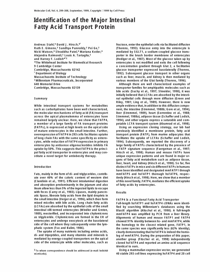
Figure 1. FATP4 Mediates LCFA Uptake
(A) Mammalian expression constructs con-taining either hsFATP4 (squares and trian-gles) or empty control vector (circles) werestably transfected into 293 cells. Short-termuptake of Bodipy-FA in the presence of BSAwas determined by FACS. The mean fluores-cence of the viable cell population is ex-pressed in arbitrary fluorescence units. FATP4protein expression was determined by densi-tometry of anti-FATP4 Western blots, shownin the insert, and is expressed in arbitraryunits.(B) Short-term uptake of Bodipy-palmitate (1mM), either by control cells (black bars) orFATP4-expressing cells (hatched bars), wasmeasured in the presence of 0, 10, or 100 mMunlabeled palmitate. FA uptake was quanti-fied by FACS and expressed in arbitrary fluo-rescence units.(C) The rate of [3H]palmitate uptake by 293cells, which were stably transfected with aconstruct for either human FATP4 (diamonds)or an empty vector (circles), was comparedto that of isolated enterocytes (squares).
lines transfected with a control plasmid. The ability of and lipid-soluble vitamins and hormones. Both satu-rated and nonsaturated fatty acids containing 10–26 Cthe different cell lines to take up FA, as assessed by
uptake assays using the fluorescently labeled Bodipy- atoms strongly competed for uptake of Bodipy-palmi-tate (Figure 1B and Table 2) and thus are presumed topalmitate, correlated well with their FATP4 expression
levels determined by Western blotting (Figure 1A). All be substrates of FATP4. In contrast, fatty acids witheight or fewer C atoms did not compete and thus are20 vector control clones showed amounts of Bodipy-
FA uptake similar to each other and to untransfected presumed not to be FATP4 substrates. Similarly, estersof long chain FAs and other hydrophobic molecules293 cells. In contrast, among the 40 FATP4-transfected
clones, a large number (z20) showed an approximately tested had no effect on uptake of Bodipy-palmitate.2-fold increase in Bodipy-FA uptake compared to anyof the vector controls, and three had a 5- to 10-foldincrease in Bodipy-FA uptake. FATP4 Is Strongly Expressed by the Enterocytes
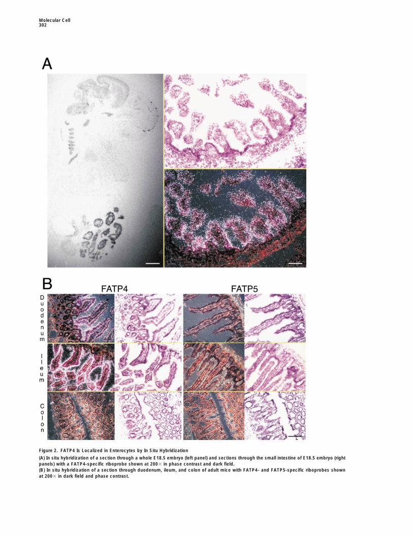
of the Small IntestineSeveral of the cell lines with the highest amount ofBodipy-FA uptake as well as isolated primary entero- To determine the cellular distribution of FATP4 in E18.5
C57/B16 embryos, in situ hybridization with a non-cytes were used to measure the uptake of radiolabeledFAs. Short-term uptake by 293 cells and enterocytes of cross-reacting probe was used. On embryo whole
mounts, strong mmFATP4 expression could be seen inall FAs tested was linear (Figure 1C). hsFATP4 expres-sion enhanced the rate of palmitate uptake approxi- the small intestine (Figure 2A, left panel). Microscopy of
counterstained sections showed that FATP4 mRNA wasmately 3-fold over 293 cells transfected with vectoralone (Figure 1C) and also accelerated the uptake of present at high levels in the enterocytes of the intestinal
villi (Figure 2A, right panels). In situ hybridization of crossoleate but not of linolate, arachidonate, octanoate, buty-rate, or cholesterol (Table 1). Isolated primary entero- sections of the ileum and duodenum from 8-week-old
adult mice also demonstrated a strong expression ofcytes showed a similar preference for palmitate andoleate and absence of transport of arachidonate, octa- FATP4 in enterocytes (Figure 2B). The highest expres-
sion levels of FATP4 were seen in the enterocytes of thenoate, and butyrate, but displayed a more robust trans-port of linolate and cholesterol than the transfected 293 jejunum and ileum, and lower, but significant, amounts
were detected in the epithelial cells of the duodenum.cells.To further characterize the substrate specificity of However, FATP4 mRNA was undetectable in any other
cell type of the small intestine such as mesenchymal,FATP4, we measured the uptake by stably transfected293 cells of 5 mM Bodipy-FA in the presence of a 20- endothelial, and smooth muscle cells and was com-
pletely absent from the colon (Figure 2B, left six panels).fold molar excess (i.e., 100 mM) of FAs, FA derivatives,
Identifying the Intestinal Fatty Acid Transporter301
Table 1. Uptake of Different Substrates by FATP4-Expressing Cell Lines and Enterocytes
293 Cells
Fatty Acid Controla Stably Expressing FATP4a FATP4 Specifica Enterocytesa
Palmitate 564 1695 1131 3036Oleate 662 1122 459 117Linolate 640 673 33 116Arachidonate 3 5 2 0Octanoate 0 0 0 5Butyrate 0 50 50 73Cholesterol 319 345 26 531
Uptake of different substrates by enterocytes and by control and stable FATP4-expressing 293 cells. The rates of uptake for the indicatedfatty acids was measured over 4 min taking measurements every 30 s. All fatty acids were at a concentration of 10 mM in HBS containing 5mM taurocholate.aUptake measured as pmol/min 106 cells.
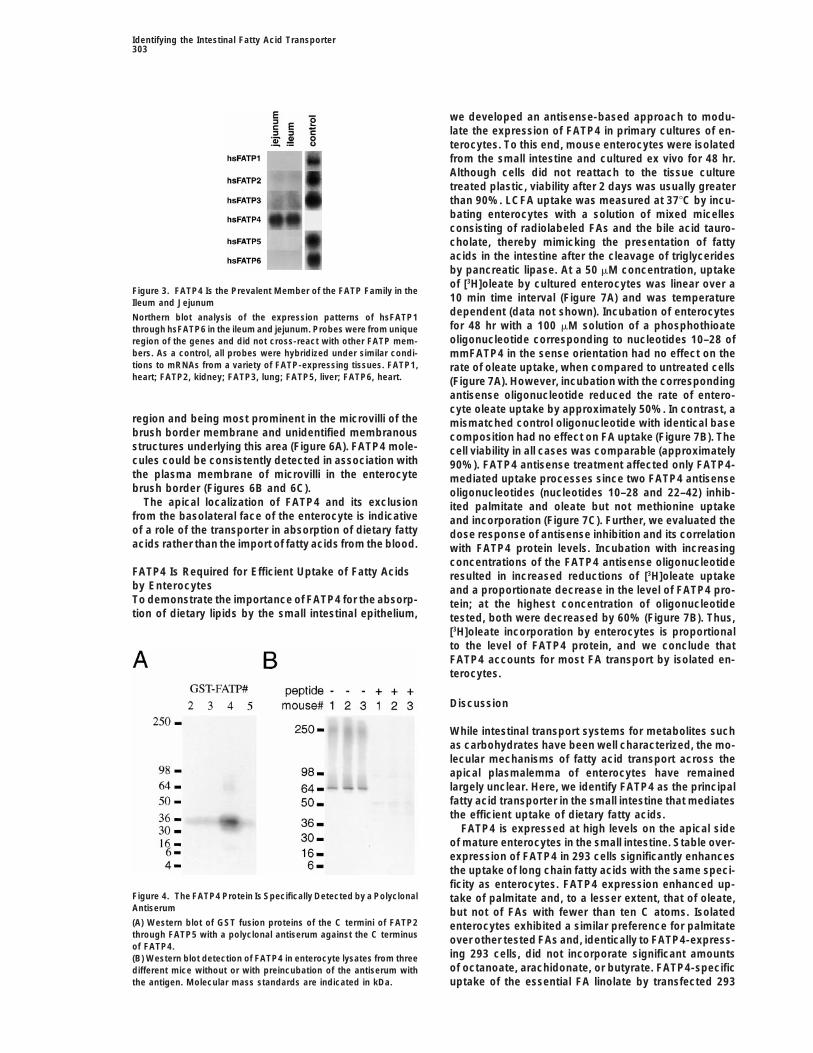
Interestingly, while the expression of FATP4 in the ma- tissues (Figure 2B, right six panels). mmFATP2 was de-tected by in situ hybridization at low levels in the epithe-ture enterocytes of the villi was exceptionally high, it
was low or undetectable in the undifferentiated precur- lial cells of the ileum, jejunum, and duodenum. Northernblot analysis of hsFATP1 through hsFATP6 in humansor cells in the crypts between the villi (Figures 2A, right
panel, and 2B, upper left four panels). No signals above ileum and jejunum confirmed the notion from the murinedata that only FATP4 is expressed at appreciable levelsbackground were detected for mmFATP1, mmFATP3
(data not shown), and mmFATP5 in any of the intestinal in the small intestine (Figure 3).
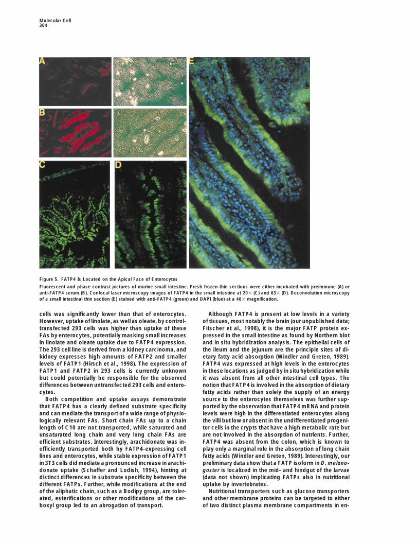
The FATP4 Protein Is Localized to the Apical Sideof EnterocytesTable 2. Competition of Bodipy-FA Uptake by FATP4-
Expressing Cells To further characterize the FATP4 protein and its local-ization, we raised a polyclonal antiserum against a GSTFatty Acids Formula Competitionfusion protein of the C terminus of mmFATP4 in rabbits.
Butyric acid C4H8O2 2 The antiserum showed only weak cross-reactivity withCaproic acid C6H12O2 2
the GST fusion proteins of C termini of other FATP familyCaprylic acid C8H16O2 2members in Western blot experiments (Figure 4A). InCapric acid C10H20O2 11Western blot experiments with lysates from isolated en-Lauric acid C12H24O2 11
Myristic acid C14H28O2 11 terocytes from three different adult Balb C mice, thePalmitic acid C16H32O2 11 antiserum exclusively recognized a single approxi-Stearic acid C18H36O2 1 mately 70 kDa band, which is in accordance with theOleic acid C18H34O2 11
predicted molecular weight of 72 kDa for mmFATP4Linoleic acid C18H32O2 11(Figure 4B). This signal was specific for FATP4 sinceArachidic acid C20H40O2 11none was obtained with preimmune serum (data notLignoceric acid C24H48O2 11
Cerotic acid C26H52O2 11 shown), and, more importantly, it could be abolished bypreincubation of the anit-FATP4 serum with FATP4-GST
Fatty Acid Derivatives fusion protein (Figure 4B).Palmitic acid methyl ester C17H34O2 2 Immunofluorescence microscopy of fresh frozen un-Stearic acid methyl ester C19H38O2 2 fixed sections of adult mouse small intestine with theOleic acid ethyl ester C20H38O2 2 FATP4-specific antiserum confirmed the expression ofOleic acid oley ester C36H68O2 2
FATP4 in the epithelial cell layer of the villi (Figure 5B),Oleoyl CoA C39H68N7O17P3S 2while incubation with a preimmune serum from the sameCholesteryl oleate C45H78O2 2rabbit demonstrated only weak background fluores-
Lipid-Soluble Vitamins and Hormones cence (Figure 5A). At higher magnifications (Figures 5CRetinoic acid (pro-vitamin A) C20H28O2 6 and 5D), it was apparent that FATP4 is preferentiallyErgocalciferol (vitamin D2) C28H44O 2 localized to the apical side of the enterocyte that facesTocopherol (vitamin E) C29H50O2 2 the lumen of the small intestine. Similar observations3-Phytylmenadione (vitamin K1) C31H46O2 2
were made for the localization of hsFATP4 in the humanProstaglandin E2 C20H32O5 2ileum and jejunum (data not shown). Further analysis of
Competition for bodipy-FA uptake by FATP4-expressing cells by the subcellular localization of FATP4 in enterocytes bydifferent hydrophobic compounds. The uptake of 5 mM Bodipy-FA,
deconvolution microscopy confirmed that the trans-C1-Bodipy-C12, was measured in the presence a 20-fold molarporter is localized to the apical side of the enterocyteexcess (i.e., 100 mM) of the indicated fatty acids or fatty acid deriva-including the brush border membrane (Figure 5E).tives. The maximal 100% inhibition was defined as the amount of
Bodipy-FA incorporated in the presence of 200 mM lauric acid which Immunoelectron microscopy of fresh frozen sectionswas on average 18% 6 5% that of untreated cells. 2, 0%–30% through the small intestine using a FATP4-specific poly-inhibition by the indicated substance; 6, 30–50% inhibition; 1, 50%– clonal antiserum showed a gradient of protein distribu-70% inhibition; 11, 70%–100% inhibition.
tion starting from the apical side of the paranuclear
Molecular Cell302
Figure 2. FATP4 Is Localized in Enterocytes by In Situ Hybridization
(A) In situ hybridization of a section through a whole E18.5 embryo (left panel) and sections through the small intestine of E18.5 embryo (rightpanels) with a FATP4-specific riboprobe shown at 2003 in phase contrast and dark field.(B) In situ hybridization of a section through duodenum, ileum, and colon of adult mice with FATP4- and FATP5-specific riboprobes shownat 2003 in dark field and phase contrast.
Identifying the Intestinal Fatty Acid Transporter303
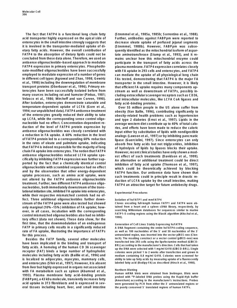
we developed an antisense-based approach to modu-late the expression of FATP4 in primary cultures of en-terocytes. To this end, mouse enterocytes were isolatedfrom the small intestine and cultured ex vivo for 48 hr.Although cells did not reattach to the tissue culturetreated plastic, viability after 2 days was usually greaterthan 90%. LCFA uptake was measured at 378C by incu-bating enterocytes with a solution of mixed micellesconsisting of radiolabeled FAs and the bile acid tauro-cholate, thereby mimicking the presentation of fattyacids in the intestine after the cleavage of triglyceridesby pancreatic lipase. At a 50 mM concentration, uptakeof [3H]oleate by cultured enterocytes was linear over a
Figure 3. FATP4 Is the Prevalent Member of the FATP Family in the10 min time interval (Figure 7A) and was temperatureIleum and Jejunumdependent (data not shown). Incubation of enterocytesNorthern blot analysis of the expression patterns of hsFATP1for 48 hr with a 100 mM solution of a phosphothioatethrough hsFATP6 in the ileum and jejunum. Probes were from uniqueoligonucleotide corresponding to nucleotides 10–28 ofregion of the genes and did not cross-react with other FATP mem-
bers. As a control, all probes were hybridized under similar condi- mmFATP4 in the sense orientation had no effect on thetions to mRNAs from a variety of FATP-expressing tissues. FATP1, rate of oleate uptake, when compared to untreated cellsheart; FATP2, kidney; FATP3, lung; FATP5, liver; FATP6, heart. (Figure 7A). However, incubation with the corresponding
antisense oligonucleotide reduced the rate of entero-cyte oleate uptake by approximately 50%. In contrast, a
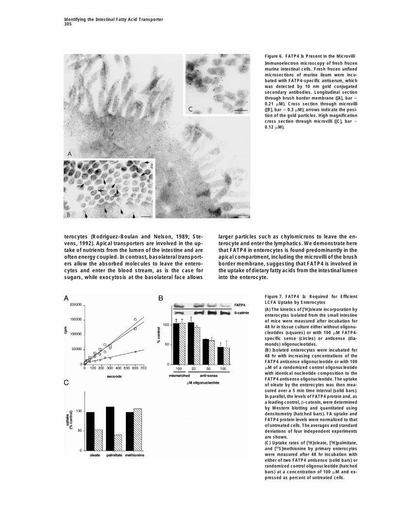
region and being most prominent in the microvilli of the mismatched control oligonucleotide with identical basebrush border membrane and unidentified membranous composition had no effect on FA uptake (Figure 7B). Thestructures underlying this area (Figure 6A). FATP4 mole- cell viability in all cases was comparable (approximatelycules could be consistently detected in association with 90%). FATP4 antisense treatment affected only FATP4-the plasma membrane of microvilli in the enterocyte mediated uptake processes since two FATP4 antisensebrush border (Figures 6B and 6C). oligonucleotides (nucleotides 10–28 and 22–42) inhib-
The apical localization of FATP4 and its exclusion ited palmitate and oleate but not methionine uptakefrom the basolateral face of the enterocyte is indicative and incorporation (Figure 7C). Further, we evaluated theof a role of the transporter in absorption of dietary fatty dose response of antisense inhibition and its correlationacids rather than the import of fatty acids from the blood. with FATP4 protein levels. Incubation with increasing
concentrations of the FATP4 antisense oligonucleotideFATP4 Is Required for Efficient Uptake of Fatty Acids resulted in increased reductions of [3H]oleate uptakeby Enterocytes and a proportionate decrease in the level of FATP4 pro-To demonstrate the importance of FATP4 for the absorp- tein; at the highest concentration of oligonucleotidetion of dietary lipids by the small intestinal epithelium, tested, both were decreased by 60% (Figure 7B). Thus,
[3H]oleate incorporation by enterocytes is proportionalto the level of FATP4 protein, and we conclude thatFATP4 accounts for most FA transport by isolated en-terocytes.
Discussion
While intestinal transport systems for metabolites suchas carbohydrates have been well characterized, the mo-lecular mechanisms of fatty acid transport across theapical plasmalemma of enterocytes have remainedlargely unclear. Here, we identify FATP4 as the principalfatty acid transporter in the small intestine that mediatesthe efficient uptake of dietary fatty acids.
FATP4 is expressed at high levels on the apical sideof mature enterocytes in the small intestine. Stable over-expression of FATP4 in 293 cells significantly enhancesthe uptake of long chain fatty acids with the same speci-ficity as enterocytes. FATP4 expression enhanced up-
Figure 4. The FATP4 Protein Is Specifically Detected by a Polyclonal take of palmitate and, to a lesser extent, that of oleate,Antiserum but not of FAs with fewer than ten C atoms. Isolated(A) Western blot of GST fusion proteins of the C termini of FATP2 enterocytes exhibited a similar preference for palmitatethrough FATP5 with a polyclonal antiserum against the C terminus over other tested FAs and, identically to FATP4-express-of FATP4.
ing 293 cells, did not incorporate significant amounts(B) Western blot detection of FATP4 in enterocyte lysates from threeof octanoate, arachidonate, or butyrate. FATP4-specificdifferent mice without or with preincubation of the antiserum with
the antigen. Molecular mass standards are indicated in kDa. uptake of the essential FA linolate by transfected 293
Molecular Cell304
Figure 5. FATP4 Is Located on the Apical Face of Enterocytes
Fluorescent and phase contrast pictures of murine small intestine. Fresh frozen thin sections were either incubated with preimmune (A) oranti-FATP4 serum (B). Confocal laser microscopy images of FATP4 in the small intestine at 203 (C) and 633 (D). Deconvolution microscopyof a small intestinal thin section (E) stained with anti-FATP4 (green) and DAPI (blue) at a 403 magnification.
cells was significantly lower than that of enterocytes. Although FATP4 is present at low levels in a varietyof tissues, most notably the brain (our unpublished data;However, uptake of linolate, as well as oleate, by control-
transfected 293 cells was higher than uptake of these Fitscher et al., 1998), it is the major FATP protein ex-pressed in the small intestine as found by Northern blotFAs by enterocytes, potentially masking small increases
in linolate and oleate uptake due to FATP4 expression. and in situ hybridization analysis. The epithelial cells ofthe ileum and the jejunum are the principle sites of di-The 293 cell line is derived from a kidney carcinoma, and
kidney expresses high amounts of FATP2 and smaller etary fatty acid absorption (Windler and Greten, 1989).FATP4 was expressed at high levels in the enterocyteslevels of FATP1 (Hirsch et al., 1998). The expression of
FATP1 and FATP2 in 293 cells is currently unknown in these locations as judged by in situ hybridization whileit was absent from all other intestinal cell types. Thebut could potentially be responsible for the observed
differences between untransfected 293 cells and entero- notion that FATP4 is involved in the absorption of dietaryfatty acids rather than solely the supply of an energycytes.
Both competition and uptake assays demonstrate source to the enterocytes themselves was further sup-ported by the observation that FATP4 mRNA and proteinthat FATP4 has a clearly defined substrate specificity
and can mediate the transport of a wide range of physio- levels were high in the differentiated enterocytes alongthe villi but low or absent in the undifferentiated progeni-logically relevant FAs. Short chain FAs up to a chain
length of C10 are not transported, while saturated and tor cells in the crypts that have a high metabolic rate butare not involved in the absorption of nutrients. Further,unsaturated long chain and very long chain FAs are
efficient substrates. Interestingly, arachidonate was in- FATP4 was absent from the colon, which is known toplay only a marginal role in the absorption of long chainefficiently transported both by FATP4-expressing cell
lines and enterocytes, while stable expression of FATP1 fatty acids (Windler and Greten, 1989). Interestingly, ourpreliminary data show that a FATP isoform in D. melano-in 3T3 cells did mediate a pronounced increase in arachi-
donate uptake (Schaffer and Lodish, 1994), hinting at gaster is localized in the mid- and hindgut of the larvae(data not shown) implicating FATPs also in nutritionaldistinct differences in substrate specificity between the
different FATPs. Further, while modifications at the end uptake by invertebrates.Nutritional transporters such as glucose transportersof the aliphatic chain, such as a Bodipy group, are toler-
ated, esterifications or other modifications of the car- and other membrane proteins can be targeted to eitherof two distinct plasma membrane compartments in en-boxyl group led to an abrogation of transport.
Identifying the Intestinal Fatty Acid Transporter305
Figure 6. FATP4 Is Present in the Microvilli
Immunoelectron microscopy of fresh frozenmurine intestinal cells. Fresh frozen unfixedmicrosections of murine ileum were incu-bated with FATP4-specific antiserum, whichwas detected by 10 nm gold conjugatedsecondary antibodies. Longitudinal sectionthrough brush border membrane ([A], bar 5
0.21 mM). Cross section through microvilli([B], bar 5 0.3 mM); arrows indicate the posi-tion of the gold particles. High magnificationcross section through microvilli ([C], bar 5
0.12 mM).
terocytes (Rodriguez-Boulan and Nelson, 1989; Ste- larger particles such as chylomicrons to leave the en-terocyte and enter the lymphatics. We demonstrate herevens, 1992). Apical transporters are involved in the up-
take of nutrients from the lumen of the intestine and are that FATP4 in enterocytes is found predominantly in theapical compartment, including the microvilli of the brushoften energy coupled. In contrast, basolateral transport-
ers allow the absorbed molecules to leave the entero- border membrane, suggesting that FATP4 is involved inthe uptake of dietary fatty acids from the intestinal lumencytes and enter the blood stream, as is the case for
sugars, while exocytosis at the basolateral face allows into the enterocyte.
Figure 7. FATP4 Is Required for EfficientLCFA Uptake by Enterocytes
(A) The kinetics of [3H]oleate incorporation byenterocytes isolated from the small intestineof mice were measured after incubation for48 hr in tissue culture either without oligonu-cleotides (squares) or with 100 mM FATP4-specific sense (circles) or antisense (dia-monds) oligonucleotides.(B) Isolated enterocytes were incubated for48 hr with increasing concentrations of theFATP4 antisense oligonucleotide or with 100mM of a randomized control oligonucleotidewith identical nucleotide composition to theFATP4 antisense oligonucleotide. The uptakeof oleate by the enterocytes was then mea-sured over a 5 min time interval (solid bars).In parallel, the levels of FATP4 protein and, asa loading control, b-catenin, were determinedby Western blotting and quantitated usingdensitometry (hatched bars). FA uptake andFATP4 protein levels were normalized to thatof untreated cells. The averages and standarddeviations of four independent experimentsare shown.(C) Uptake rates of [3H]oleate, [3H]palmitate,and [35S]methionine by primary enterocyteswere measured after 48 hr incubation witheither of two FATP4 antisense (solid bars) orrandomized control oligonucleotide (hatchedbars) at a concentration of 100 mM and ex-pressed as percent of untreated cells.
Molecular Cell306
The fact that FATP4 is a functional long chain fatty (Stremmel et al., 1985a, 1985b; Sorrentino et al., 1988).acid transporter highly expressed on the apical side of Further, antibodies against FABPpm were reported toenterocytes in the small intestine strongly suggests that decrease oleate uptake of perfused jejunal segmentsit is involved in the transporter-mediated uptake of di- (Stremmel, 1988b). However, FABPpm was subse-etary fatty acids. However, the overall contribution of quently identified as the mitochondrial isoform of aspar-FATP4 to the absorption of dietary lipids could not be tate aminotransferase (Stump et al., 1993), and it re-concluded from these data alone. Therefore, we used an mains unclear how this mitochondrial enzyme couldantisense oligonucleotide–based approach to modulate participate in the transport of fatty acids across theFATP4 expression in primary enterocytes. Phosphothi- plasma membrane. FATP4 expression correlates closelyoate-modified oligonucleotides have been successfully with FA uptake in 293 cells and enterocytes, and FATP4employed to modulate expression of a number of genes can mediate the uptake of all physiological long chainin different cell types (Agrawal and Zhao, 1998; Gewirtz FAs tested, demonstrating that FATP4 is the major FAet al., 1998) including the downregulation of membrane transporter in the small intestine. However, it is likelytransport proteins (Oberbauer et al., 1996). Primary en- that efficient FA uptake requires many components up-terocytes have been successfully isolated before from stream as well as downstream of FATPs, possibly in-many sources including rat and hamster (Pinkus, 1981; cluding extracellular scavenger receptors such as CD36,Velasco et al., 1986; Mircheff and van Corven, 1990). and intracellular molecules, like LCFA CoA ligases andAfter isolation, enterocytes demonstrate saturable and fatty acid–binding proteins.temperature-dependent uptake of LCFA (Gore et al., Over 55 million people in the US alone suffer from1994; our unpublished data). FATP4 antisense treatment obesity (Van Itallie, 1996), contributing significantly toof the enterocytes greatly reduced their ability to take obesity-related health problems such as hypertensionup LCFA, while the corresponding sense control oligo- and type 2 diabetes (Ernst et al., 1997). Lipids in thenucleotide had no effect. Concentration-dependent re- average western diet contribute up to 40% of total calo-duction of the level of FATP4 protein expression by ries, and efforts have been made to reduce the caloricantisense oligonucleotides was closely correlated with input either by substitution of lipids with nondigestiblea reduction in FA uptake. A 60% reduction in the level analogs (Lawson et al., 1997) or by inhibiting pancreaticof FATP4 protein led to a corresponding 60% reduction lipase (Guerciolini, 1997). Since enterocytes can onlyin the rates of oleate and palmitate uptake, indicating absorb free fatty acids but not triglycerides, inhibitionthat FATP4 is indeed responsible for the majority of long of hydrolysis of lipids by lipases blocks their uptake.chain FA uptake into enterocytes. The notion that FATP4 However, recent clinical studies have only shown a mod-antisense oligonucleotides reduced LCFA uptake spe- est effect of such treatments (Davidson et al., 1999).cifically by inhibiting FATP4 expression was further sup- An alternative or additional treatment could be directported by the fact that a chemically identical control inhibition of fatty acid uptake (Thomson et al., 1997),oligonucleotide with scrambled sequence had no effect which could be theoretically achieved by blockingand by the observation that other energy-dependent FATP4 function. Our antisense data have shown thatuptake processes, such as amino acid uptake, were such treatments could in principle result in drastic re-not altered by the FATP4 antisense oligonucleotide duction of LCFA uptake by the small intestine, makingtreatment. Further, two different FATP4 antisense oligo- FATP4 an attractive target for future antiobesity drugs.nucleotides, both immediately downstream of the trans-lational initiation site, inhibited FA uptake into enterocytes, Experimental Procedureswhile their respective mismatched controls had no ef-fect. Three additional oligonucleotides further down- Isolation of hsFATP1 and mmFATP4
Clones encoding full-length human FATP1 and FATP4 were ob-stream of the FATP4 gene were also tested but showedtained from a heart and a spleen cDNA library, respectively, byonly marginal (10%–15%) inhibition of FA uptake; how-searching Millennium databases for sequences similar to murineever, in all cases, incubation with the correspondingFATP1–5 coding regions using the BlastX algorithm (Altschul et al.,control mismatched oligonucleotides also had no inhibi-1990).
tory effect (data not shown). These data show, for thefirst time, that the downmodulation of an endogenous Generation of Cell Lines Stably Expressing hsFATP4FATP in primary cells results in a significantly reduced A DNA fragment containing the entire hsFATP4-coding sequence,
as well as 100 nucleotides of the 59 and 50 nucleotides of the 39rate of FA uptake, illustrating the importance of FATPsuntranslated region, was inserted into the vector pIRES-neo (Clon-for this process.tech). The resulting construct or a vector control (pIRES-neo) wasBesides the FATP family, several other moleculestransfected into 293 cells using the lipofectamine method (GIBCO-have been implicated in the binding and transport ofBRL) according to the manufacturer’s direction. Cells that had taken
fatty acids. A homolog of the human CD 36 scavenger up the DNA were selected with 1 mg/ml G418 (GIBCO-BRL). Singlereceptor (FAT) binds a large number of hydrophobic colonies were picked 1 to 2 weeks after transfection and grown inmolecules including fatty acids (Baillie et al., 1996) and medium containing 0.8 mg/ml G418. Colonies were screened for
ability to take up fatty acids by measuring uptake of a fluorescentlyis localized to adipocytes, myocytes, mammary cells,labeled fatty acid (Bodipy-FA) as described (Hirsch et al., 1998).and enterocytes (Sfeir et al., 1997). However, it is absent
from liver but highly expressed in organs not associatedNorthern Blottingwith FA metabolism such as spleen (Abumrad et al.,Human mRNA blots were obtained from Invitrogen. Blots were
1993). Plasma membrane fatty acid–binding protein probed with 32P-labeled DNA probes using the Rapid-Hyb buffer(FABPpm), a 43 kDa molecule, transiently increases fatty (Amersham) according to the manufacturer’s instructions. Probesacid uptake in 3T3 fibroblasts and is expressed in sev- were generated by PCR from either the 39 untranslated regions or
the poorly conserved 59 translated regions of human FATPs.eral tissues including heart, liver, and small intestine
Identifying the Intestinal Fatty Acid Transporter307
In Situ Hybridization LCFA Uptake AssaysBodipy-FA uptake assays using FACS were performed as describedTissues were collected from 8-week-old C57/Bl6 mice. Tissues were
fresh frozen, cut on a cryostat at 10 mm thickness, and mounted on previously (Hirsch et al. 1998) and also adapted to a 96-well format.LCFA uptake assays with enterocytes or with stably transfected 293Superfrost Plus slides (VWR). Sections were air dried for 20 min and
then incubated with ice-cold 4% paraformaldehyde (PFA)/phos- cells were done as follows. Mixed micelles of radiolabeled FA (NEN)and taurocholate (Sigma) in HBS were generated by brief sonicationphate-buffered saline (PBS) for 10 min. Slides were washed twice
for 5 min each with PBS, incubated with 0.25% acetic anhydride/1 at 378C. Equal volumes of cells and micelle solution were mixed,resulting in a final FA concentration of 25 mM for antisense assaysM triethanolamine for 10 min, washed with PBS for 5 min, and
dehydrated with 70%, 80%, 95%, and 100% ethanol for 1 min each. and 10 mM for substrate specificity assays. Final taurocholate con-centration was 5 mM. Cells were incubated for the indicated amountSections were incubated with chloroform for 5 min. Hybridizations
were performed with 35S-radiolabeled (5 3 107 cpm/ml) cRNA probes of time at 378C. The assay was stopped by transferring the cellsonto filter paper followed by extensive washes with ice-cold HBSgenerated from the 39 untranslated regions of mouse FATPs by PCR
followed by in vitro transcription in the presence of 50% formamide, containing 0.1% BSA using a cell harvester (Brandell). Incorporatedoleate was then determined by b-scintillation counting (Beckman).10% dextran sulfate, 13 Denhardt’s solution, 600 mM NaCl, 10
mM DTT, 0.25% SDS, and 100 mg/ml tRNA for 18 hr at 558C. Afterhybridization, slides were washed with 10 mM Tris-HCl (pH 7.6), 500 AcknowledgmentsmM NaCl, 1 mM EDTA (TNE) for 10 min, incubated in 40 mg/mlRNase A in TNE at 378C for 30 min, washed in TNE for 10 min, We thank Marlene Dembski for help with generating stable cell lines,
John Keilty for help with sequencing, Matthew Dube for assistanceincubated once in 23 SSC at 608C for 1 hr, once in 0.23 SSC at608C for 1 hr, and once in 0.23 SSC at 658C for 1 hr, and dehydrated with Bodipy-FA uptake assays, and Chris Groves for help with FACS
analysis. This work was supported by a Program of Excellence inwith 50%, 70%, 80%, 95%, and 100% ethanol. Localization of mRNAtranscripts was detected by dipping slides in Kodak NBT-2 pho- Molecular Biology grant to H. F. L. from the National Heart, Lung,
and Blood Institute (HL41484) and NIH grant DK 47618. A. S. wastoemulsion and exposing for 7 days at 48C, followed by developmentwith Kodak Dektol developer. Slides were counterstained with in part supported by a postdoctoral fellowship from the Studiensti-
fung des Deutschen Volkes and D. H. by the NIH grant 5 T32 CAhaemotoxylin and eosin and photographed. Controls for the in situhybridization experiments included the use of a sense probe that 09541. Part of this work was conducted utilizing the W. M. Keck
Foundation Biological Imaging Facility at the Whitehead Institute.showed no signal above background in all cases.
Received March 1, 1999; revised June 14, 1999.Immunofluorescence and Immunogold Electron MicroscopyUnfixed mouse small intestine was washed with Hanks’ buffered
Referencessalt solution containing 1 mM EDTA, infused with 2.3 M sucrosesolution, and embedded in O.C.T., 4583 compound. The material
Abumrad, N., El-Maghrabi, M.R., Amri, E.Z., Lopez E., and Grimaldi,was thick sectioned (15 mm–40 mm). The sections were washed inP.A. (1993). Cloning of a rat adipocyte membrane protein implicatedPBS containing 1% BSA and 0.075% glycine to block nonspecificin binding or transport of long-chain fatty acids that is inducedbinding. Primary and secondary antibodies were diluted in PBS withduring preadipocyte differentiation. J. Biol. Chem. 268, 17665–10% FCS and incubated for 1 hr. The sections were mounted in17668.90% glycerol/PBS containing 1 mg/ml paraphenylinediamine andAbumrad, N., Harmon, C., and Ibrahimi, A. (1998). Membrane trans-examined with a Bio-Rad MRC 600 confocal, mounted on a Zeissport of long-chain fatty acids: evidence for a facilitated process. J.Axioscop.Lipid Res. 39, 2309–2318.For the immunogold labeling, the tissue was fixed with 2% para-
formaldehyde in PBS for 10 min, after which it was cryoprotected Agrawal, S., and Zhao, Q. (1998). Antisense therapeutics. Curr. Opin.by infiltration with 2.3 M sucrose in 0.1 M phosphate buffer (pH Chem. Biol. 2, 519–528.7.4), containing 20% polyvinylpyrrolidone, and then mounted on Altschul, S.F., Gish, W., Miller, W., Myers, E.W., and Lipman, D.J.aluminum cryo nails and frozen in liquid nitrogen (Tokuyasu, 1986). (1990). Basic local alignment search tool. J. Mol. Biol. 215, 403–410.Ultrathin sections were collected on carbon/formvar-coated nickel
Baillie, A.G.S., Coburn, C.T., and Abumrad, N.A. (1996). Reversiblegrids. The primary antibody (anti-FATP4) was diluted in 10% FCSbinding of long-chain fatty acids to purified FAT, the adipose CD36in PBS and incubated overnight at 48C, followed by donkey anti-homolog. J. Membr. Biol. 153, 75–81.rabbit IgG-gold (12 nm) (Jackson labs) for 1 hr. The sections wereCarey, M.C., Small, D.M., and Bliss, C.M. (1983). Lipid digestion andstained in 2% neutral uranyl acetate (20 min) and absorption stainedabsorption. Annu. Rev. Physiol. 45, 651–677.with 2% uranyl acetate in 0.2% methylcellulose containing 3.2%
polyvinyl alcohol. The sections were examined with a Philips EM Clandinin, M.T., Cheema, S., Field, C.J., Garg, M.L., Venkatraman,J., and Clandinin, T.R. (1991). Dietary fat: exogenous determination410 electron microscope.of membrane structure and cell function. FASEB J. 5, 2761–2769.
Davidson, M.H., Hauptman, J., DiGirolamo, M., Foreyt, J.P., Halsted,Enterocyte Isolation and Antisense Oligonucleotide TreatmentC.H., Heber, D., Heimburger, D.C., Lucas, C.P., Robbins, D.C.,Enterocytes from male and female 2- to 8-month-old BALB/c miceChung, J., and Heymsfield, S.B. (1999). Weight control and riskwere isolated following standard procedures (Pinkus, 1981). In brief,factor reduction in obese subjects treated for 2 years with orlistat:small intestines (duodenum, ileum, and jejunum) were removed,a randomized controlled trial. JAMA 281, 235–242.rinsed with Hanks’ buffered salt solution (HBS; GIBCO-BRL), cut
into approximately 1 cm long sections, and incubated in 50 ml HBS Ernst, N.D., Obarzanek, E., Clark, M.B., Briefel, R.R., Brown, C.D.,containing 0.1 M sucrose (Bio-Rad) and 20 mM EDTA. Intestinal and Donato, K. (1997). Cardiovascular health risks related to over-sections were gently stirred for 10 min. The detached enterocytes weight. J. Am. Diet. Assoc. 97, S47–S51.were filtered through sterile cheesecloth (VWR) and pelleted by cen- Faergeman, N.J., Di Russo, C.C., Elberger, A., Knudsen, J., andtrifugation. The cells were then gently resuspended in RPMI 1640 Black, P.N. (1997). Disruption of the Saccharomyces cerevisiae ho-(GIBCO-BRL) containing 10% FCS and 0.01 mg/ml transferrin. Con- mologue to the murine fatty acid transport protein impairs uptakecentrated solutions of oligonucleotides were added to the entero- and growth on long-chain fatty acids. J. Biol. Chem. 272, 8531–8538.cytes to yield the indicated final concentrations and the cells were
Fitscher, B.A., Riedel, H.D., Young, K.C., and Stremmel, W. (1998).incubated for 48 hr at 378C, 5% CO2. Two antisense oligonucleotidesTissue distribution and cDNA cloning of a human fatty acid transportwith their respective controls were used showing comparable re-protein (hsFATP4). Biochim. Biophys. Acta 1443, 381–385.sults. The sequences of the phosphothioate oligonucleotides were:Gewirtz, A.M., Sokol, D.L., and Ratajczak, M.Z. (1998). Nucleic acidmmFATP4-S1, GGAGCCTCTCTGGTGGGGG; mmFATP4-AS1, CCCtherapeutics: state of the art and future prospects. Blood 92,CCACCAGAGAGGCTCC; mmFATP4-Control1, CCACCCCCGGAAAG712–736.CCTGC; mmFATP4-AS2, GGAGAACAGTAGCGCCCCCAC; mmFATP4-
Control2, GAGCCCGCCACCGTAGAGACA. Gore, J., Hoinard, C., and Couet, C. (1994). Linoleic acid uptake by
Molecular Cell308
isolated enterocytes: influence of alpha-linolenic acid on absorption. Thorens, B. (1992). Molecular and cellular physiology of GLUT-2, ahigh-Km facilitated diffusion glucose transporter. Int. Rev. Cytol.Lipids 29, 701–706.137, 209–238.Green, P.H., and Riley, J.W. (1981). Lipid absorption and intestinal
lipoprotein formation. Aust. N. Z. J. Med. 11, 84–90. Thorens, B. (1993). Facilitated glucose transporters in epithelialcells. Annu. Rev. Physiol. 55, 591–608.Guerciolini, R. (1997). Mode of action of orlistat. Int. J. Obes. Relat.
Metab. Disord. 21, S12–S23. Thorens, B. (1996). Glucose transporters in the regulation of intesti-nal, renal, and liver glucose fluxes. Am. J. Physiol. 270, G541–G553.Hediger, M.A., Coady, M.J., Ikeda, T.S., and Wright, E.M. (1987).
Expression cloning and cDNA sequencing of the Na1/glucose co- Tokuyasu, K.T. (1986). Application of cryoultramicrotomy to immu-nocytochemistry. J. Microsc. 143, 139–149.transporter. Nature 330, 379–381.
Hirsch, D., Stahl, A., and Lodish, H.F. (1998). A family of fatty acid Tso, P., and Balint, J.A. (1986). Formation and transport of chylomi-crons by enterocytes to the lymphatics. Am. J. Physiol. 250, G715–transporters conserved from mycobacterium to man. Proc. Natl.
Acad. Sci. USA 95, 8625–8629. G726.
Van Itallie, T.B. (1996). Prevalence of obesity. Endocrinol. Metab.Lawson, K.D., Middleton, S.J., and Hassall, C.D. (1997). Olestra, anonabsorbed, noncaloric replacement for dietary fat: a review. Drug Clin. North Am. 25, 887–905.Metab. Rev. 29, 651–703. Velasco, G., Dominguez, P., Shears, S.B., and Lazo, P.S. (1986).
Permeability properties of isolated enterocytes from rat small intes-Ling, K.Y., Lee, H.Y., and Hollander, D. (1989). Mechanisms of linoleicacid uptake by rabbit small intestinal brush border membrane vesi- tine. Biochim. Biophys. Acta 889, 361–365.cles. Lipids 24, 51–55. Verger, R., Aoubala, M., Carriere, F., Ransac, S., Dupuis, L., De Caro,
J., Ferrato, F., Douchet, I., Laugier, R., and De Caro, A. (1996).Mircheff, A.K., and van Corven, E.J. (1990). Isolation of enterocytemembranes. Methods Enzymol. 192, 341–354. Regulation of lumen fat digestion: enzymic aspects. Proc. Nutr. Soc.
55, 5–18.Oberbauer, R., Schreiner, G.F., Biber, J., Murer, H., and Meyer, T.W.(1996). In vivo suppression of the renal Na1/Pi cotransporter by Windler, E., and Greten, H. (1989). Intestinal lipid and lipoprotein
metabolism (Munchen: W. Zuckschwerdt Verlag).antisense oligonucleotides. Proc. Natl. Acad. Sci. USA 93, 4903–4906.
Pinkus, L.M. (1981). Separation and use of enterocytes. MethodsEnzymol. 77, 154–162.
Rodriguez-Boulan, E., and Nelson, W.J. (1989). Morphogenesis ofthe polarized epithelial cell phenotype. Science 245, 718–725.
Schaffer, J.E., and Lodish, H.F. (1994). Expression cloning and char-acterization of a novel adipocyte long chain fatty acid transportprotein. Cell 79, 427–436.
Sfeir, Z., Ibrahimi, A., Amri, E., Grimaldi, P., and Abumrad, N. (1997).Regulation of FAT/CD36 gene expression: further evidence in sup-port of a role of the protein in fatty acid binding/transport. Prosta-glandins Leukot. Essent. Fatty Acids 57, 17–21.
Shneider, B.L. (1998). A new era in bile acid transport pathophysiol-ogy. J. Pediatr. Gastroenterol. Nutr. 26, 236–237.
Sorrentino, D., Stump, D., Potter, B.J., Robinson, R.B., White, R.,Kiang, C.L., and Berk, P.D. (1988). Oleate uptake by cardiac myo-cytes is carrier mediated and involves a 40-kD plasma membranefatty acid binding protein similar to that in liver, adipose tissue, andgut. J. Clin. Invest. 82, 928–935.
Stevens, B.R. (1992). Vertebrate intestine apical membrane mecha-nisms of organic nutrient transport. Am. J. Physiol. 263, R458–R463.
Stremmel, W. (1988a). Fatty acid uptake by isolated rat heart myo-cytes represents a carrier-mediated transport process. J. Clin. In-vest. 81, 844–852.
Stremmel, W. (1988b). Uptake of fatty acids by jejunal mucosal cellsis mediated by a fatty acid binding membrane protein. J. Clin. Invest.82, 2001–2010.
Stremmel, W. (1989). Mechanism of hepatic fatty acid uptake. J.Hepatol. 9, 374–382.
Stremmel, W., Lotz, G., Strohmeyer, G., and Berk, P.D. (1985a).Identification, isolation, and partial characterization of a fatty acidbinding protein from rat jejunal microvillous membranes. J. Clin.Invest. 75, 1068–1076.
Stremmel, W., Strohmeyer, G., Borchard, F., Kochwa, S., and Berk,P.D. (1985b). Isolation and partial characterization of a fatty acidbinding protein in rat liver plasma membranes. Proc. Natl. Acad.Sci. USA 82, 4–8.
Stump, D.D., Zhou, S.L., and Berk, P.D. (1993). Comparison ofplasma membrane FABP and mitochondrial isoform of aspartateaminotransferase from rat liver. Am. J. Physiol. 265, G894–G902.
Suchy, F.J., Sippel, C.J., and Ananthanarayanan, M. (1997). Bile acidtransport across the hepatocyte canalicular membrane. FASEB J.11, 199–205.
Thomson, A.B., De Pover, A., Keelan, M., Jarocka-Cyrta, E., andClandinin, M.T. (1997). Inhibition of lipid absorption as an approachto the treatment of obesity. Methods Enzymol. 286, 3–44.


