Embed Size (px)
Citation preview
Review
From fatty-acid sensing to chylomicron synthesis: Role of intestinallipid-binding proteins
Marjorie Buttet 1, Véronique Traynard 1, Thi Thu Trang Tran, Philippe Besnard,Hélène Poirier, Isabelle Niot*
Physiologie de la Nutrition et Toxicologie Team (NUTox), UMR U866 INSERM, Université de Bourgogne, AgroSup Dijon, 1 Esplanade Erasme,21000 Dijon, France
a r t i c l e i n f o
Article history:Received 15 April 2013Accepted 5 August 2013Available online 16 August 2013
Keywords:Lipid-binding proteinsCD36Intestinal adaptationIntestinal lipid sensingChylomicron
a b s t r a c t
Today, it is well established that the development of obesity and associated diseases results, in part, fromexcessive lipid intake associated with a qualitative imbalance. Among the organs involved in lipid ho-meostasis, the small intestine is the least studied even though it determines lipid bioavailability andlargely contributes to the regulation of postprandial hyperlipemia (triacylglycerols (TG) and free fattyacids (FFA)). Several Lipid-Binding Proteins (LBP) are expressed in the small intestine. Their supposedintestinal functions were initially based on what was reported in other tissues, and took no account ofthe physiological specificity of the small intestine. Progressively, the identification of regulating factors ofintestinal LBP and the description of the phenotype of their deletion have provided new insights intocellular and molecular mechanisms involved in fat absorption. This review will discuss the physiologicalcontribution of each LBP in the main steps of intestinal absorption of long-chain fatty acids (LCFA):uptake, trafficking and reassembly into chylomicrons (CM). Moreover, current data indicate that thesmall intestine is able to adapt its lipid absorption capacity to the fat content of the diet, especiallythrough the coordinated induction of LBP. This adaptation requires the existence of a mechanism ofintestinal lipid sensing. Emerging data suggest that the membrane LBP CD36 may operate as a lipidreceptor that triggers an intracellular signal leading to the modulation of the expression of LBP involvedin CM formation. This event could be the starting point for the optimized synthesis of large CM, whichare efficiently degraded in blood. Better understanding of this intestinal lipid sensing might provide newapproaches to decrease the prevalence of postprandial hypertriglyceridemia, which is associated withcardiovascular diseases, insulin resistance and obesity.
! 2013 Published by Elsevier Masson SAS.
1. Introduction
Dietary fat accounts for 35e40% of the calories ingested inWestern countries [1]. TG, compounds in which three fatty acids(FA), mainly long-chain fatty acids (LCFA), are bound to one glycerolmolecule, are the predominant form of dietary fat (around 95%). FAare crucial for health because they constitute the major metabolicsubstrate for energy production by cells (9 kcal/g) and are also,themselves or their derivatives (eicosanoids), essential modulatorsof cellular functions such as differentiation, proliferation and theproduction of hormones and cytokines, especially through their cell-signaling capacity. These important functions suggest that intestinal
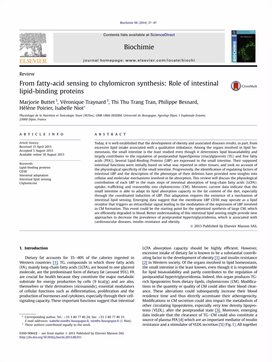
LCFA absorption capacity should be highly efficient. However,excessive intake of dietary fat is known to be a substantial contrib-uting factor to the development of obesity [1] and insulin resistance[2] in Western society. Of the organs involved in lipid homeostasis,the small intestine is the least known, even though it is responsiblefor lipid bioavailability and partly contributes to the regulation ofpostprandial hypertriglyceridemia. Indeed, this organ produces TG-rich lipoproteins from dietary lipids, chylomicrons (CM). Modifica-tions in the quantity or quality of CM could alter their blood clear-ance. These alterations could subsequently increase their bloodresidence time and thus directly accentuate their atherogenicity.Modifications in CM secretion could also impact the metabolism ofother circulating lipoproteins, especially very low density lipopro-teins (VLDL), after the postprandial state [3]. Moreover, emergingdata indicate that the clearance of TGeCM could also constitute asource of plasma FFA [4] which are an important mediator of insulinresistance and a stimulator of VLDL secretion [5] (Fig.1). All together
* Corresponding author. Tel.: !33 3 80 77 40 24; fax: !33 3 80 77 40 33.E-mail addresses: [email protected], [email protected] (I. Niot).
1 These authors contributed equally to the work.
Contents lists available at ScienceDirect
Biochimie
journal homepage: www.elsevier .com/locate/biochi
0300-9084/$ e see front matter ! 2013 Published by Elsevier Masson SAS.http://dx.doi.org/10.1016/j.biochi.2013.08.011
Biochimie 96 (2014) 37e47

the effects mentioned above explain the resurgence of interest in therole of CM in dyslipidemia and in associated diseases such as car-diovascular diseases and insulin resistance, which is often associatedwith obesity. Therefore, understanding the molecular mechanismsof LCFA absorption and its regulation may provide new strategies tofight against these metabolic disorders. This review will thus focuson the intestinal fate of LCFA.
2. Cellular and molecular mechanisms of intestinal LCFAabsorption
TG are naturally hydrophobic, but become soluble and are hy-drolyzed in the digestive tract (mouth, stomach and mainly in theintestinal lumen) by various lipases to yield 2 FA and 1 mono-acylglycerol. These lipid products are made soluble by bile salts toform mixed micelles in the proximal intestinal lumen (Fig. 2). Un-der normal conditions, LCFA absorption is known to occurmainly inthe jejunum, inwell-differentiated enterocytes located in the upper2/3 of the villi [6]. A high-fat diet/meal may trigger an overflow ofdietary fat absorption capacity in the proximal part of intestine andthus trigger the recruitment of the distal part of the intestine fortheir absorption [7].
Despite the significant strides made over the last three decadesin understanding themechanisms of intestinal LCFA absorption/CMformation, additional effort is still necessary to identify the mo-lecular and cellular events involved in the process. Intestinal LCFAabsorption requires three successive steps: cellular uptake, cellulartrafficking and the synthesis and secretion of lipoproteins (Fig. 2)[8]. This review examines the LBP involved in the cellular andmolecular aspects of this mechanism and the recent data demon-strating that dietary FA are able to regulate CM formation probablyvia cell signaling and the subsequent use of dietary LCFA.
2.1. LCFA uptake in enterocytes: contribution of membrane LBP?
Enterocytes are highly polarized cells. The apical membrane ofthese cells displays specific properties which distinguish it from thebasolateral membrane. Indeed, the presence of microvilli in theapical side of enterocytes, named the Brush Border Membrane(BBM), greatly enhances the absorptive surface of the small intes-tine. These microvilli (in addition to the mucus and glycocalyx)contribute to the generation of a unique microclimate due to thetrapping of water molecules: the unstirred water layer. This weakrenewal compartment has a low pH generated by an efficient H!/Na! antiport exchange system located in the BBM. This acidicmicroclimate leads to the micellar dissociation and the protonationof LCFA [9] once the local pH becomes lower than that of LCFA pKa.It has been demonstrated that LCFA protonation facilitates theirsubsequent cellular uptake mainly by passive diffusion (for review[8]). All together, these features may explain how intestinal ab-sorption remains efficient despite of intermittent and dramaticchanges in daily fat supply: passive diffusion which is a high ca-pacity low affinity transport system. However, it raises the questionof the physiological and quantitative contribution of membraneLBP, such as Fatty Acid Binding-Protein Plasma Membrane(FABPpm), Fatty Acid Transport Protein 4 (FATP4), CD36 and Cav-eolin 1 (Cav-1) (Fig. 2) in the intestinal LCFA absorptionmechanism.
2.1.1. FABPpmFABPpm is expressed in organs with high lipid requirements
[10]. FABPpm is located at the level of the BBM but also in the lateralmembranes of differentiated and undifferentiated enterocytes. Theapparent dissociation constant (Kd) for LCFA was evaluated at80 nM [11]. Its involvement in LCFA uptake was initially suggestedby the fact that the treatment of jejunal explants with a large
Fig. 1. Postprandial fate of dietary LCFA. 1 e Sensing of dietary lipids triggers the efficient synthesis of chylomicrons. 10 e Depending on the quantity of lipid ingested, a portion offatty acid (FA) is transiently stored in enterocytes as triacylglycerol (TG). This store seems to contribute to the early secretion of chylomicrons (CM) for the next meal. 2 e In theblood, CMeTG are hydrolyzed by lipoprotein lipase (LPL) located in extra hepatic tissues and the long chain fatty acids (LCFA) provided are directly taken up by these tissues. 20 e Aportion of dietary FA escapes uptake by extra hepatic tissues (dietary fatty acid “spillover”) and enters the circulating free fatty acid (FFA) pool. 3 e The remaining particles, theremnant, are taken up by the liver to be degraded to form, more slowly, very low density lipoproteins (VLDL). 30 e FFA from this source could be taken up by the liver to form VLDL,but could also contribute to insulin resistance by favoring ectopic fat deposition in other tissues. FI: food intake.
M. Buttet et al. / Biochimie 96 (2014) 37e4738

quantity of polyclonal antibodies raised against FABPpm, led to areduction in LCFA uptake [12]. More recently, the peptidic sequenceof FABPpm was demonstrated to be identical to that of mitochon-drial aspartate amino-transferase (mAspAT), which catalyzes thetransamination reaction at the level of the inner membrane ofmitochondria [13] suggesting another function for this protein inthe maintenance of the cytoplasmic/mitochondrial NADH/NADratio. All together, these data indicate that the dual location ofFABPpm may reflect a possible link between fatty acid uptake andredox potential as proposed by Ref. [14]. Further experiments arerequired to understand the physiological role of FABPpm in intes-tinal LCFA absorption, especially the generation of mice deleted inFABPpm specifically in the small intestine.
2.1.2. FATP4FATP4 is a membrane protein belonging to the FATP family,
which contains five rodent isoforms and six human isoforms. Eachmember displays a specific tissue distribution [15]. In the smallintestine, FATP4 is mainly expressed in the highly differentiatedenterocytes located in the jejunum. Its cellular location is acontroversial issue: it was initially found at the level of the BBM[16], but more recent data have shown that it is preferentiallyconfined in the Endoplasmic Reticulum (ER) (Fig. 2) [17,18]. Dataobtained with isolated enterocytes frommice, characterized by theabsence of the unstirred water layer and thus displaying a lowcapacity of FA uptake by passive diffusion, have shown that LCFAuptake efficiency is positively correlated with the expression levelof FATP4 [16,19]. However, the tridimensional structure of FATP4reveals that the protein is mainly present at the level of cytoplasmic
side of the BBMwith only a short extracellular sequence at the levelof the N-terminal tail, which does not contain a putative LCFAbinding domain [20,21]. These properties, in addition to the cellularlocation of FATP4, have raised doubts about its function as an apicalplasma membrane FA transporter or translocase in enterocytes.More recent findings have demonstrated that FATP4 has anendogenous acyl-CoA synthetase (ACS) activity for LCFA and veryLCFA [22]. This latter capacity may lead to the vectorial acylation ofFA, which could in turn finally maintain a low FA concentrationwithin the enterocyte, favoring their entrance by passive diffusion[18,22]. This function may consequently explain the contributionof FATP4 in LCFA uptake, but indirectly by transforming FA intoactivated intermediates subsequently used in downstream meta-bolism [23].
Despite of this function, FATP4 deletion in the small intestine ofmice does not modify the total net LCFA uptake and lipid fecal loss.Moreover, this deletion does not confer protection from weightgain induced by a high-fat diet. Only a slight accumulation of TGwas observed in enterocytes from intestinal FATP4-deficient micefed a high fat diet [24], suggesting that FATP4 participates in TGprocessing, probably via LCFA acylation [18]. To date, the contri-bution of FATP4-ACS activity in the total LCFA activation rate in thejejunum is not known because total ACS activity has not beenestimated in FATP4-null mice [24]. However, a recent finding hasindicated that FATP4 and acyl-CoA synthetase long chain 5 (ACSL5)are the two major proteins with ACS activity expressed in the smallintestine among 13 genes [25]. These authors also reported that thedeletion of ACSL5 in mice led to a 60% reduction in the jejunal LCFAactivation rate, but as shown for FATP4 deletion, LCFA intestinal net
Fig. 2. Main steps in the intestinal absorption of long-chain fatty acids. 1 e Passive diffusion of LCFA; 10 e caveolae mediated FA transport; 2 e LCFA Sensing. 2-MG: 2-mono-glyceride; ApoAIV: apolipoprotein AIV; ApoB48: apolipoprotein B48; ApoCII: apolipoprotein CII; ApoCIII: apolipoprotein CIII; BA: biliary acids; CD36: fatty acid transporter; CM:chylomicron; DGAT: Acyl-CoA: diacylglycerol acyltransferase; ER: endoplasmic reticulum; FATP4: fatty acid transport protein 4; I-FABP: intestinal fatty acid binding protein; LCFA:long chain fatty acids; L-FABP: liver fatty acid binding protein; MGAT: Acyl-CoA: monoacylglycerol acyltransferase; MTP: microsomal triglyceride transfer protein; P-ERK: phos-phorylated extracellular signal regulated kinases; PCTV: prechylomicron transport vesicle; TG: triacylglycerol.
M. Buttet et al. / Biochimie 96 (2014) 37e47 39

absorption is not affected by this deletion. In summary, FATP4 isdispensable for efficient intestinal lipid absorption, but couldimpact the synthesis and the secretion of TG-rich lipoproteins by itsACSL activity. According to this hypothesis, a decrease in intestinalFATP4 expression in mice [26] and Gly-209-Ser polymorphism inhuman FATP4 protein [20] result in a reduction in postprandialhypertriglyceridemia. However, the function of intestinal FATP4 isstill under research because, recently, FATP4 has also beendemonstrated to play a key role in mediating FA-induced glucagon-like peptide-1 (GLP-1) secretion from endocrine cells found in thedistal small intestine [27]. These new data open new perspectivessince it has been shown that GLP-1 analogs reduce postprandiallipemia by reducing intestinally-derived TG and food intake [28].
2.1.3. CD36CD36 is a multifunctional glycoprotein first identified in adipo-
cytes but ubiquitously expressed, especially in organswith high lipidmetabolism (i.e. adipocytes and myocytes). CD36 is able to bindionized LCFA with an affinity in the nanomolar range and a stoichi-ometry of threemoles of FA per onemole of protein. CD36 also bindscollagen, anionic phospholipids, oxidized LDL, Plasmodium falcipa-rum, thrombospondin-1 and b-amyloid [29] (for review see Refs.[30e32]), explaining its multifunctionality and thus its classificationas a scavenger receptor. In the small intestine, CD36 is mainlyexpressed in the BBM of enterocytes located in the upper 2/3 of thevilli from the proximal part of the small intestine in rodents andhumans (Fig. 2) [33,34]. The level of its intestinal expression corre-lates positively with the lipid content of the diet: a low-fat diet isassociatedwith low CD36 intestinal expression [35] whereas a high-fat diet leads to an increase in CD36 expression [34]. By contrast tothe contribution of CD36 in LCFA uptake in adipocytes or myocytes,its contribution to LCFA uptake in the intestine appears to be minor.
According to the authors, CD36 deletion in isolated enterocytesis reported either to decrease LCFA uptake [36] or to have no impact[37]. More recently, our data using an in situ isolated intestinalsegment, an almost physiological model that keeps the unstirredwater intact, have demonstrated that CD36 deletion in mice doesnot affect intestinal lipid uptake [38]. These data are in agreementwith the fact that lipid elimination in feces in CD36 knocked-outmice was no higher except for very LCFA [36,39,40]. It may reflecttwo intestinal specificities. Firstly, enterocytes are subjected to veryhigh levels of lipids during postprandial periods, and luminal LCFAlevels would greatly exceed CD36 binding capacity since the pro-tein functions at nanomolar concentrations of LCFA. Secondly, theunstirred water layer maintains an efficient intestinal fat absorp-tion probably obviating the need of protein facilitated transfer.Despite of CD36 dispensable role in LCFA uptake during the dietarylipid absorption, its deletion significantly impairs the production ofCM which contain less TG and are thus smaller [40,41]. These al-terations are associated with TG retention in enterocytes, especiallyin high fat diet. A mechanistic explanation of these alterations hasbeen recently proposed. Indeed, in addition to its presence in BBM,CD36 expression has been also demonstrated in intestinal ER-budding complex which generates the intestinal-specific largevesicles able to carry the preCM from ER to Golgi (prechylomicrontransfer vesicles: PCTV). Although the precise function of CD36 inthis step has not been elucidated, its contribution is physiologicallyrelevant, because the generation of PCTV is inhibited in CD36knocked-out mice [42]. All together, these data indicate that CD36is a key element in the mechanism of LCFA absorption through theactivation of CM formation/secretion, but not directly as an efficientLCFA transporter. However, in relation to its binding affinity, theinvolvement of CD36 in the uptake of very small quantities of LCFAis not excluded, especially at the beginning of lipid absorptionbefore themassive influx of LCFA [39]. As described for FATP4, CD36
has recently been demonstrated to be found in endocrine cellslocated in the proximal part of the intestine and to play a major rolein the FA-induced release of cholecystokinin (CCK) and secretin[43]. This last function provides evidence of another importantcontribution of intestinal CD36 in the mechanism of lipid absorp-tion and also in food intake.
2.1.4. Caveolin 1Caveolins (Cav) are small integrated plasma membrane proteins
(21e24 kDa) associated with lipid rafts (dynamic assemblies ofsphingolipids and cholesterol) that do not extend to the extracel-lular milieu. There are three family members, caveolin 1e3, each ofwhich is expressed in the intestine [44]. Caveolae are flask-shapedinvaginations of the plasma membrane that play multiple roles incell physiology, including serving as signaling platforms fornumerous pathways and as clathrin-independent routes of endo-cytosis [45]. Caveolae-mediated endocytosis may constitute an FAendocytic carrier in enterocytes similarly to what has been shownin adipocytes [46]. Such a function is supported by several argu-ments: caveolae are significantly present in the BBM [47], caveolaeare detergent-resistant membrane domains and thus able toconcentrate large amounts of FA at the level of the BBM, especiallyin the postprandial state [48], Cav-1, which plays an essential role inthe formation of caveolae [45,49], has been shown to be an FA-binding protein [50] and Cav-1 is found in caveolae endocyticvesicles (CEV) in the cytosol (Fig. 2) [51]. These findings explainwhy Cav-1 null mice are resistant to diet-induced obesity, and showfasted hypertriglyceridemia with adipocyte abnormalities [52].Moreover, these Cav-1 deficient mice have elevated postprandialtriglyceridemia which is not due to modifications in lipoproteinlipase, which also suggests that Cav-1/caveolae contributes to in-testinal FA absorption. According to this idea, a recent paper [51]demonstrated that intestinal uptake of dietary FA (after an acutelipid load) is dependent on caveolae, which are subsequentlyendocytosed and transported into the cell via Cav1-containing CEV.These findings are physiologically relevant since Cav-1 null micehave more steatorrhea in comparison to wild type mice in high-fatdiet conditions (23% fat w:w) [51]. It has been suggested thatmembrane LBP contribute to the caveolae-mediated endocytosis ofFA, but this hypothesis remains questionable. Indeed, at least infibroblasts, Cav-1 is required for the presence of CD36 at the level oflipid rafts [53] and, moreover, CD36 is associated with BBM cav-eolae of enterocytes [51,54]. However, the transfection of caveolinhas been demonstrated to be sufficient in cells lacking CD36 (suchas HEK293) to significantly increase FA uptake [55], and CD36 is notintegrated into intestinal internalized caveolae (CEV) [51].
In conclusion, the findings to date suggest that the presence ofthe unstirred water layer at the apical side of enterocytes, inaddition to the structural and functional properties of membraneLBP and the existence of CEV do not contribute greatly to theprotein-mediated transport of LCFA during dietary lipid absorption.This is not the case for caveolin, but the involvement of caveolin inFA uptake is probably not directly due to its function as an LBP butrather its crucial role in the formation of caveolae. To date, the datahighlight that CD36, which has long been considered an efficientLCFA transporter despite the fact that it binds small amounts of FA,probably displays another essential function in intestinal lipidmetabolism as described in paragraph 4. By contrast, the passivediffusion of LCFA, a transport system with high capacity and lowaffinity, would make intestinal LCFA absorption more efficient(Fig. 2). It is noteworthy that the efficiency of passive diffusiondepends on the cell’s capacity to maintain a favorable LCFA con-centration gradient between the extra and intracellular compart-ments. The abundance of soluble cytoplasmic LBP and the efficiency
M. Buttet et al. / Biochimie 96 (2014) 37e4740

of subsequent LCFA metabolism may contribute maintaining lowLCFA concentrations within enterocytes. In addition to passivediffusion, the contribution of caveolae-mediated FA transport hasto be taken into account. By protecting the plasma membrane fromthe detergent effect of excess FA, and by targeting the FA to theesterification site, this mechanism may also allow the cellulartransport of large amounts of FA (Fig. 2) [47]. This endocytic vesicle-mediated transport of dietary LCFA is not in conflict with passivediffusion and their respective contribution probably depends onthe quantity of dietary FA and the moment of LCFA absorption.Together these two mechanisms prevent intestinal FA uptake frombecoming limiting after a meal.
2.2. Intracellular trapping/trafficking of LCFA: contribution of twosoluble LBP?
As described above, once LCFA are in the cytoplasm, theymay beeither packaged into CEV or bound by cytosolic FABP (intestinal andliver-FABP, I and L-FABP) before being targeted to the different LCFAmetabolic sites. The small intestine expresses two different FABP atrelatively high and similar concentrations (3e5% of cytosolic pro-teins), at least in rodents, but with higher L-FABP expression inhumans [56]. The precise functions of the respective FABP were nottotally elucidated until recently. The binding of LCFA in the cyto-plasm by these two soluble proteins contributes to the mainte-nance of an LCFA concentration gradient that favors their uptake by
passive diffusion [8]. However, it is noteworthy that the bindingcapacity of the two FABP (nM range, Table 1, [57]) is largely underthe FA concentration found in enterocytes in the postprandialperiod, which is in mM range [51,58]. These findings suggest theexistence of another FA sequestration system in the cytosol espe-cially in CEV. FABP have been shown to facilitate the subsequentmetabolism of FA and to be at the origin of an intracellular pool ofnon-esterified FA (for review see Ref. [59]) (Fig. 2). The findingshave shown that when LCFA are delivered to the apical side ofenterocytes, they are preferentially bound to I-FABP, which requiresa collision mechanism with membrane phospholipids to transferLCFA. These two properties suggest that I-FABP, which is only foundin the intestine, is involved in the vectorial transfer of LCFA to ER,where LCFA is re-esterified into TG [60]. This function may explainwhy a mutation that increases the affinity of this protein for FA isassociated with hypertriglyceridemia and insulin resistance inhumans [61] and also with the overproduction of CM when humanintestinal explants are used [62]. By contrast, FA transfer kineticsstudies showed that L-FABP LCFA exchange with the membraneoccurs by aqueous diffusion, that’s why L-FABP was initiallyconsidered to be at the origin of the formation of an intracellular“waiting” pool of LCFA. However, besides its non-collisional LCFAtransfer mechanism, L-FABP was recently shown to interact withmembrane phospholipids. This apparent discrepancy may beexplained by the fact that the membrane interaction occurs at thelevel of its ligand binding cavity [63].
Table 1Functional properties of LBP and impact of their deficiency on intestinal long-chain fatty acid absorption.
LBP Location Stoichiometryand LCFAaffinity
Impact onLCFA uptake
Intestinal regulation Deficiency impact
On net lipidabsorption
On intestinal TGretention
On intestinal TGsecretion inlipoproteins
FABPpm BBM and lateralmembrane [11]
Binding siteAffinity:80 nM [11]
[(intestinalexplants) [12]
? ? ? ?
FATP-4 BBM and/or ER[16,18]
e [(isolated enterocytes)[16,19]
[In HF diet (mouse)[26,100]
No effect in STDand HF diets(mouse) [19,24]
Tend to increase(mouse) [24]
Y(human and mouse)[20,26]
CD36 BBM and PCTV[33,34]
3 mol LCFA by1 mol proteinAffinity: nM[29]
No effect (isolatedenterocytes [37] andin situ intestinalloops [38])
[in HF diet (mouse)[2,34,77,92]YFast after a single lipidload (mouse) [38]
No effect in STDand HF diets (mouse)[36,39,40]
Retention in HFdiet (mouse)[36,37]
YSecretion of smallchylomicrons after alipid load (mouse andhuman) [37,41]
Cav-1 BBM and CEV[47]
High affinity[50]
[[51]
? Steatorrhea inHFD (mouse) [51]
? [[51]
I-FABP Cytoplasm [58] 1 Binding siteAffinity (oleicacid): 39 nM [57]
[(Cell culture)[132e134]
[in HF diet (mouse)[77,100]
No effect in STDand HF diets(mouse) [135]
? YOnly in male(mouse) [135][Secretion if Ala-54-Thrmutation (human) [61]
L-FABP Cytoplasm andPCTV andnucleus [42,58]
2 Binding sitesAffinity (oleicacid): 9 and62 nM [57]
[(Cell culture)[132e134]
[in HF diet (mouse)[77,100] and 2 h aftera lipid load (mouse) [38]
No effect in STDand HF diets(mouse) [65,136]
No retention inSTD dietRetention aftera single lipidload (mouse)[65]
YSecretion after a singlelipid load (mouse) [65]
MTP ER and PCTV[137,138]
e ? [in HF diet [92,100,139]
Steatorrhea(mouse) [140]
Important retention(mouse) [83]
Y(mouse) [83]
BBM: brush border membrane; Cav-1: caveolin 1; CEV: caveolae endocytic vesicule; ER: endoplasmic reticulum; FABPpm: fatty acid binding protein plasma membrane;FATP4: fatty acid transport protein 4; HF: high fat; I-FABP: intestinal fatty acid binding protein; LCFA: long chain fatty acids; L-FABP: liver fatty acid binding protein; MTP:microsomal triglyceride transfer protein; PCTV: prechylomicron transport vesicle; STD: standard; TG: triacylglycerol.
M. Buttet et al. / Biochimie 96 (2014) 37e47 41

In accordance with this finding, data obtained in hepatocytesdemonstrated that L-FABP is found in membrane micro-domainspreferentially in those poor in cholesterol. Its role at this level issignificant because its absence affects both the lipid compositionand the levels of several membrane LBP, FATP4 and Scavenger Re-ceptor Class B type 1 (SRB1). If such localization/function exists inthe enterocyte BBM, L-FABP could be directly involved in intestinalFA uptake [64].
However, in the small intestine, the findings have shown that L-FABP is not associated with BBM caveolae [51] but is present at thelevel of ER membrane (ER budding complex) of enterocytes [42],where, by contrast to I-FABP, it plays a role as a budding initiatorprotein for PCTV. As detailed below, L-FABP is thus consideredcrucial for the synthesis/secretion of CM. In accordance with thisfinding, L-FABP knocked out mice subjected to a single lipid loaddisplay lower secretion of TG in the blood, and this is associatedwith TG retention in enterocytes [65] and absence of generation ofPCTV [42]. To finish, L-FABP has also been demonstrated to be aperoxisome proliferator activated receptor a (PPAR a) partner atleast in hepatocyte [66]. Indeed L-FABP regulates PPAR a tran-scriptional activity through direct interaction with this nuclearreceptor [67] and could thus participate in the FA-mediated regu-lation of numerous proteins involved in lipid metabolism includingLBP [8].
2.3. Synthesis and secretion of chylomicrons: two LBP are requiredin the rate limiting steps
In enterocytes, LCFA processing is initiated by their rapid acti-vation into acyl-CoA. During the postprandial period, acyl-CoA arepreferentially transformed into TG at the level of the membrane ofthe smooth ER via esterification of the 2-MG generated duringluminal TG hydrolysis (for review see Ref. [8]). This metabolicpathway is specific to the small intestine and represents around80% of newly synthesized TG after a meal [68] (Fig. 2) and is veryrapid since within 30 s, 79% of the absorbed FA are converted intoTG in the rat intestine [69]. Acyl-CoA: mono and diacylglycerolacyltransferase (MGAT and DGAT) are required for the successiveformation of DG and TG. Among the three MGAT isoforms onlyMGAT2 is expressed in the main site of FA intestinal absorption[70]. MGAT2 deletion in mice leads to reduced FA absorption andincreased intestinal FA oxidation, which partly explains why thesemice are resistant to a high-fat diet [71]. Two DGAT isoforms,DGAT1 and DGAT2, are expressed in enterocytes. They havedifferent amino acid sequences, different structures and differentsubcellular locations although their expression pattern along theintestine is similar (like that for MGAT2, with a higher level in theproximal intestine) (for review see Ref. [72]). DGAT1 and DGAT2 areboth ER-membrane-bound enzymes and DGAT1 is reported torepresent around 85% of the total DGAT activity in the small in-testine [73]. However, DGAT2 is also found at the level of cyto-plasmic lipid droplets [74,75]. The existence of these organellesprovides evidence of a new characteristic of intestinal lipid pro-cessing, the capacity to store TG. Indeed, under certain conditions,high-fat diet/meal, enterocytes are able to store TG from the mealtransiently within lipid cytoplasmic droplets. The expression of atleast two lipases in enterocytes (triacylglycerol hydrolase andadipocyte triglyceride lipase) supports the existence of a lipolysis/re-esterification cycle in enterocytes. These lipases also providesubstrates for lipoprotein formation (Fig. 1).
In the absence of DGAT1, DGAT2 is sufficient to ensure lipidabsorption in mice on a normolipidic diet, but not in those on ahigh-fat diet (HFD) during which a reduction in secreted CM(smaller in size) and an increase in intestinal TG storage areobserved [76,77]. This abnormal metabolism is not due to reduced
total activity of DGAT because this intestinal TG storage phenotyperemains when DGAT1 knocked out mice are crossed with miceoverexpressing DGAT2 specifically in the intestine [74]. To date, thefindings indicate that these two isoforms work together, and arecomplementary in the intestinal processing of dietary fat, by con-trolling the balance between intestinal TG storage and secretioninto lipoproteins. In recent years, significant progress has beenmade in the understanding of molecular events at the origin of CMassembly, trafficking and secretion (for review see Refs. [8,78,79]).CM are particles with a density lower than 1.006 g/ml and are thelargest lipoproteins. The size varies and can range from 80 to1000 nm [80]. To summarize, the assembly of pre-CM requires twosteps [81]. A part of the newly synthesized TG are translocated intothe lumen of the ER with phospholipids and associated with oneapolipoprotein B48 (ApoB48) by interacting with microsomal tri-glyceride transfer protein (MTP) to form a primordial lipoprotein inthe lumen of the ER. The lipidation of ApoB48, a non-exchangeabletruncated isoform of ApoB100, only expressed in the small intestineat least in humans and hamsters, allows the correct folding of thisapolipoprotein. The primordial lipoprotein is further enriched in TGby a second-step fusion with lipid droplets (containing TG, phos-pholipids and cholesterol esters) generated independently in thesmooth ER also through the MTP.
MTP, another LBP, thus plays a crucial role in CM formation sincea deficiency in theMTP gene in humans ormice (due tomutation ortransgenesis) leads to abetalipoproteinemia, a defect associatedwith fat malabsorption, steatorrhea and TG retention in enterocytes[82,83]. The above explains whyMTP is considered the first limitingstep in intestinal lipid absorption. Another apolipoprotein, ApoAIV,is also important in this step since it facilitates ApoB48 coreexpansion by stabilizing the pre-CM and/or ApoB48 [79,84]. Itseems to affect the size of secreted CM [84]. Pre-CM are then tar-geted to the Golgi apparatus via vesicles generated by the ER, thePCTV which are large COP II-like vesicles only found in the smallintestine (Fig. 2). Finally, in the Golgi, the lipoprotein biogenesisinvolves a maturation step before their secretion into the lymphaticcirculation: glycosylation and addition of different apolipoproteinssuch as ApoCII, CIII, and A1. This vectorial transfer constitutes thesecond limiting step, which requires different proteins: amongthem the Smad anchor for receptor activation 2 (Sar 1B GTPase) andmore specifically L-FABP for the budding of PCTV from ER [85],CD36 and Vesicle Associated Membrane Protein 7 (VAMP7) forcorrect vesicle targeting and fusion with the Golgi membrane.
CM are then secreted into the lymph to reach a TG concentrationpeak in blood after 3e4 h, whereas TG from VLDL remain constant[86] (Fig. 1). Thus, the intestine constitutes the main modulator ofpostprandial triglyceridemia. In addition to this contribution, it hasbeen shown that TG stored in enterocytes are secreted and pack-aged into CM at the very onset of the next meal (found in the bloodafter 10e30 min) (for review see Ref. [87]). This release of CMbefore ingestion of a meal suggests a taste-gut-brain axis since itcould be triggered when fat or glucose are only tasted but notswallowed [88,89]. The physiological role of this phenomenon hasto be established. Several hypotheses may be proposed: early signalindicating to the body that energy is coming in or preparation of thedigestive tract for incoming lipid load to avoid spillover of intestinalsynthesis capacity of CM.
According to this last hypothesis, deficiencies of several LBP butalso ApoAIV [90] are often associated with excess intestinal TGstorage/retention (Table 1; Fig. 1) and with higher postprandialtriglyceridemia, which thus appears paradoxical and remains un-explained. These two intriguing events are also found in cases ofboth diet induced-obesity and ob/ob mice, in which reducedsecretion of TG from the intestinal mucosa into the circulation wasdemonstrated [91,92]. All together these findings indicate that an
M. Buttet et al. / Biochimie 96 (2014) 37e4742

excess ability to store TGmay also be associated with the metabolicdysfunction of enterocytes.
2.4. Clearance of chylomicrons: a step partly dependent onintestinal lipid metabolism
The release of FA from CMeTG occurs through the activityof lipoprotein lipase (LPL), which is found in the capillary endo-thelium in different organs such as adipose tissue [93], muscle,heart [94], but also brain [95] where FA are metabolized. Then,dietary FA, carried in CM, are cleared in many peripheral tissues.This contributes to the short half-life of CM, around 5 min, in theblood. However, it is now recognized that a portion of the CM-derived FA could re-enter the circulation and contribute to theplasma FFA pool in a process referred to as “spillover” (contrib-uting 5e35% of FFA to the plasma after a meal). This phenomenonseems to depend not only on the efficiency of dietary fat storagein adipose tissue, but also on the quantity and quality of dietarylipids [87]. These FFA are then rapidly taken up by the liver toform VLDL, but could also contribute to ectopic fat deposition intissues such as pancreas and play a role in insulin resistance(Fig. 1).
Then CM remnants containing a smaller quantity of TG aremainly taken up by the liver and their TG are re-packaged intoVLDL. Thus, in addition to the level of CM secreted by the smallintestine, LPL is also an important regulator of postprandial lipemia(TG and FFA). LPL activity is regulated by its expression level (forexample, upregulated by insulin in adipose tissue, and thus higherin the postprandial state (for review see Ref. [95])). However, itsactivity is also regulated by the ratio of ApoCII/ApoCIII found in thelipoprotein. Indeed, ApoCII is an activator of LPL while ApoCIII is aninhibitor [96]. Although the level of these apolipoproteins may bemodulated by exchanges with hepatic-derived lipoproteins in theblood, the level acquired by CM in enterocytes also determines theefficiency of LPL activity [97]. Moreover, clearance also depends onthe number and the size of secreted CM. Indeed, LPL activity ispositively linked to CM size [98] and is more efficient on a smallnumber of large CM than on a large number of small CM [99](Fig. 1). It is noteworthy that these properties are dependent onthe efficiency of the TG lipidation of ApoB48 in the enterocyte.Interestingly, the efficiency of TG core expansion of the pre-CMseems to be dependent on the quantity [100] and on the qualityof dietary FA (saturated or poly-unsaturated) [101].
CM clearance needs to be understood more clearly since there isa resurgence of interest in the role of postprandial lipemia (TG andFFA) in the development of cardiovascular diseases, insulin resis-tance [102,103] and obesity. Indeed, humans spend the majority ofthe day in a state of postprandial metabolism. Moreover, it has beendemonstrated that, in the postprandial state, FA are preferentiallytaken up by adipose tissue and, to a lesser extent, by muscle[104,105] (Fig. 1).
In conclusion, except for MTP, LBP are dispensable for FA ab-sorption. However, they facilitate it at different levels such as thesynthesis and/or the secretion of large CM, which are efficientlydegraded in the blood. These findings explain why LBP deficienciesdo not alter the net lipid absorption capacity (no alteration in thefecal lipid loss) even in HFD, but rather alter TG secretion into thecirculation and consequently postprandial hypertriglyceridemia(Table 1) [8] and putatively dietary lipid partitioning.
3. Do dietary fatty acids regulate the mechanism of intestinallipid absorption?
The other question to solve is to know how the small intestine,which secretes VLDL-like lipoproteins in the fasting or
interprandial state, shifts to the secretion of larger CMparticles, andthus becomes able to absorb large quantities of lipids after a meal.Does this result from an innate property of the intestine or from themetabolic adaptation of enterocytes to the lipid content of the diet?In the second hypothesis, dysregulation of this adaptation mayimpact the lipid absorption capacity (TG retention, fecal lipid loss)and postprandial lipemia.
Several factors support the existence of this lipid-dependentmetabolic adaptation. Firstly, it is widely accepted that in humansand mice, an increase in the lipid content of the meal leads to anincrease in the size of CM, but not to an increase in their number,suggesting that the ApoB48 lipidation capacity adapts to the lipidcontent of the diet [101,106]. Secondly, it has been demonstratedin vitro, that LCFA are able to up-regulate the expression levels ofseveral LBP (at least MTP, CD36 and L-FABP) [107e109], suggestingthat CM processing is modulated by dietary fat. Thirdly, the addi-tion of FA to BBM potentiates the endocytosis of the BBM caveolae[110]. Fourthly, it has been reported that PCTV (not present duringfasting) become especially abundant after a high-fat meal [42,111].Finally, our work has confirmed the relevance of all these data,in vivo, because an in situ isolated segment of jejunum from micefed with a chronic high-fat diet is able to absorb more FA than asegment from mice fed with a normolipidic diet (enhancement ofthe uptake capacity and of TG synthesis and secretion without TGintestinal retention) [100]. These findings explain why in vivo achronic high-fat diet does not lead to an increase in lipid fecal loss.These intestinal lipid-dependent adaptations result from twomechanisms: the induction of both intestinal proliferation andexpression of LBP genes and proteins involved in CM formation(Table 1) (CD36, FATP4, I and L-FABP, MTP, ApoAIV and ApoCII)[100,112]. This dietary lipid-dependent regulation was observed inthe proximal part but also in the distal part of intestine where MTPand L-FABP are induced after a high-fat diet [7] probably, inresponse to the large amount of dietary fat that reaches the ileum[113]. It is interesting to note that these intestinal modifications arereversible and their importance is positively linked to the lipidcontent of the diet. Indeed, the work of de Wit et al. [2] hasdemonstrated that the number of genes regulated by dietary lipidsand the intensity of their regulation are both positively linked to thelipid content of the diet (45% of lipids (% w/w) compared to 10, 20and 30%). Moreover, mRNA levels of ApoCII are also increased in ahigh-fat diet while those of ApoCIII are decreased [100,112]. Thus,the higher ApoCII/ApoCIII ratio and the larger CM possibly explainthe paradoxically lower postprandial hypertriglyceridemia foundafter a high-fat diet (rich in unsaturated FA) in both rodents [100]and humans [114].
In conclusion, this intestinal metabolic adaptation to the lipidcontent of the diet could also affect postprandial lipemia (TG andFFA). Moreover, at least in mice, these lipid-dependent regulationscontribute to lipid homeostasis [2,112] since their impairment isassociated with diet-induced obesity [112].
Interestingly, our recent work, confirmed by others, stronglysuggests that the gene regulation observed after a high-fat diet isalso found after a single lipid load [38,77]. These last findingsindicate that these metabolic adaptations are not secondary to achronic high-fat diet, but are possibly directly dependent on theamount of dietary lipids in intestinal lumen. Indeed, a lipid loadwith LCFA-rich oil triggers the up-regulation of L-FABP and MTP,two crucial proteins for the formation of PCTV and large CM, afterless than two hours [38,77]. To be effective, this postprandialadaptation requires the existence of a mechanism of dietary lipidsensing at the level of the enterocyte. Among the membrane LBP,the structural and functional features of CD36, in addition to itslipid-dependent regulation, suggest that CD36 could be a lipidsensor.
M. Buttet et al. / Biochimie 96 (2014) 37e47 43

4. A mechanism of dietary lipid sensing in enterocytes:emerging role of CD36
Dietary lipids not only lead to the induction of L-FABP andMTP inrodents soon after ameal [38], but they also drive a rapid decrease inthe level of CD36 protein only one hour or even less after theiringestion. Immunohistochemistry has demonstrated that thisdecrease in CD36 protein levels corresponds to the partial disap-pearance of CD36 from the BBM of enterocytes after ingestion of ameal containing lipids. Interestingly, the same immunostainingpattern persisted when fasted rats were fed a fat-free diet,demonstrating that CD36 removal from the luminal side of enter-ocytes is a physiological event related to dietary lipids. This dietarylipid-dependent CD36 modification is not associated with CD36recovery in the intracellular compartment, but results fromthe proteasomal degradation of CD36 triggered by its poly-ubiquitination. Our in vitro data have shown that diglycerides, butnot TG, and/or the FA derived from gastric and pancreatic digestion(CD36 ligands) lead to CD36 ubiquitination [38]. This fast degrada-tion of CD36 in the intestine suggests that it does not participateeffectively in LCFA uptake during the postprandial period asdemonstrated with CD36 knocked out mice. However, such aligand-induced down-regulation of a protein is generally observedfor cell surface receptor associated with cell signaling, duringexposure to a persistent and large supply of ligand, because it servesas a feedback regulator (desensitizationof the cell). According to thisconcept, there is a large body of evidence to support the function ofCD36 in transducing intracellular signals after binding a number ofligands [30] such as thrombospondin-1 in endothelial cells, oxidizedLDL in macrophages and LCFA in taste bud cells [115,116]. Moreover,CD36 is located in plasma membrane detergent-resistant micro-domains known to be involved in cell signaling [54].
Our work has recently confirmed such a function of CD36 in theintestine. Indeed, we have shown in vivo that dietary LCFAmediatesextracellular signal-regulated kinases 1/2 (ERK1/2) activation dur-ing intestinal absorption and that this effect is dependent on thelevel of CD36 protein. Moreover, ex vivo, using isolated intestinalsegments from wild-type and CD36 knocked out mice, we docu-mented that CD36 lipid-dependent modulation of ERK1/2 associ-ates a fast (withinminutes) sequential up-regulation of ApoB48 andMTP, two proteins required for the assembly of large CM. Theseex vivo data are physiologically relevant since the postprandial up-regulation of MTP and L-FABP (which, respectively, facilitate theformation of large CM and large vesicles to transport them)observed after a lipid load in wild-type mice is blunted in CD36knocked outmice. A link betweenmitogen-activated protein kinase(MAPK) activation and lipoprotein synthesis has already been re-ported. Indeed, ERK1/2 are involved in regulating ApoB48 and MTPprotein levels [117,118] and in regulating the size of ApoB-containinglipoproteins secreted by the liver [119]. The phosphorylation ofERK1/2 induced by FA binding with CD36 may subsequently lead tothe activation of one or several proteins involved in the formation ofthe ER-budding complex initiated by L-FABP [120,121] and thus theformation of PCTV. This hypothesismay explainwhy both CD36 andL-FABP knocked out mice are not able to produce PCTV [42].
Overall, these new observations, together with previously pub-lished data [37], suggest that CD36 operates as a lipid sensor,responsible for absorption during the early postprandial stages, andfor transducing signals related to the dietary lipid content and thusoptimizing the formation of large CM [38]. This intestinal CD36function may explain why lipoprotein synthesis and/or secretion areseverely impaired in enterocytes fromCD36knockedoutmice leadingto the secretion of smaller CM. The postprandial hypertriglyceridemiafound in both CD36-deficient mice and humans may be the conse-quence of the lower activity of LPL on these smaller particles [37,41].
It is noteworthy that another protein of the scavenger receptorfamily, SRB1, also expressed in the BBM, may have a similar func-tion. SRB1, generally reported to bind cholesterol [122], triggersERK1/2 activation after its binding with mixed micelles [123] in aCaco2 cell line, which does not express CD36. Such a function mayexplain why intestinal overexpression of SRB1 in mice leads toaccelerated TG absorption [124]. Together, these cell-signalingpathways may allow the processing of larger amounts of dietarylipids (LCFA and cholesterol), thus optimizing the formation of largeCM which can be used by peripheral tissues more efficiently.
In conclusion, the fact that CD36 signaling in the proximal in-testine also plays a key role in the release of CCK and secretin, twointestinal peptides that affect fat absorption, reinforces our hy-pothesis that CD36 and its associated signalingmay be the first stepof the adaptation of intestinal lipid metabolism toTG content of thediet [43].
5. Conclusions and future directions
In conclusion, significant progress has been made during thethree past decades in the understanding of molecular events at theorigin of dietary LCFA intestinal absorption. Given the functionalproperties of membrane LBP and the physiological specificities ofthe small intestine, it is unlikely that LBP play a role as efficientLCFA transporters during the mechanism of lipid absorption.However, they may indirectly facilitate LCFA uptake by optimizingtheir subsequent intracellular utilization. Considerable progresshas been made in the understanding of the functions of L-FABP andI-FABP, especially in the formation of PCTV, a limiting step in in-testinal dietary fat absorption.
This review has also summarized three recent developments inthe study of dietary lipid absorption: the caveolae mediated up-take/transport of FA, intestinal TG storage and postprandial dietaryFA spillover into the FFA circulating pool. Although the physiolog-ical relevance of these two last phenomena is not known (beneficialor detrimental) theymay be themselves depend on the efficiency ofthe intestinal CMmetabolism. In linewith this idea, themost recentdata concern the existence of lipid sensing in enterocytes. Thissensing may be involved in adapting intestinal lipid metabolism tothe lipid content of the meal. As discussed in this review, in theearly stages of dietary lipid absorption, these functions may bemediated by CD36. Given this hypothesis, it is now necessary tounderstand the factors that regulate or alter CD36 expression and/or its signaling pathways to determine their contribution to theintestinal metabolism of dietary lipids and the subsequent post-prandial lipemia and portioning of dietary-TG in the different or-gans. These future directions are particularly relevant for insulin,which is known to affect CM secretion [125], CD36 protein regu-lation and the regulation of proteins required for the efficient for-mation of CM and identified as targets of CD36 signaling (ApoB48,L-FABP, MTP) [126]. Indeed, Psammomys obesus insulin-resistantand diabetic sand rats, for instance, contain increased amounts ofL-FABP and show elevated ApoB48 biogenesis [127] in the post-prandial state, while streptozotocin-induced diabetic ratsexpressed more MTP [128] and Zucker fatty diabetic rats absorbmore TG and assemble more CM [129]. These alterations are alsofound in insulin-resistant and diabetic humans, who showincreased intestinal lipoprotein production [130].
Until now, in the intestine, dietary FA sensing was onlydescribed in entero-endocrine cells, which secrete hormones thatregulate not only intestinal digestion and absorption but also foodintake. A recent interesting finding is that two LBP (FATP4and CD36) are involved in intestinal LCFA sensing in these twotypes of cells.
M. Buttet et al. / Biochimie 96 (2014) 37e4744

Therefore, intestinal sensing and intestinal metabolic responseto dietary lipids open the way for future therapeutic and/or nutri-tional approaches for the treatment and prevention of dyslipidemia[37,41], insulin resistance, and more generally obesity [2,112] bytargeting the intestine. Indeed, more and more, findings indicatethat it is not only the amount of fat in the diet, but also the effi-ciency of its postprandial handling that determines the rate atwhich they are delivered to the blood and to other tissues, and thatthese factors contribute to disturbances in lipid homeostasis [131].Since CD36 and other LBP play a role in the balance between TGstorage/retention and TG secretion by enterocytes (Table 1), theseproteins could constitute pertinent therapeutic targets.
Acknowledgments
This work was supported, in whole or in part, by a FrenchGovernment grant managed by the National Research Agency(ANR) under the program “Investissements d’Avenir” with refer-ence ANR-11-LABEX-0021/LipSTIC Labex, and the program “Sen-soFAT 2”. Marjorie Buttet and Véronique Traynard are financed byFonds Unique Interministériel (FUI SYMPPA) and Ministère del’Enseignement Supérieur et de la Recherche, respectively.
References
[1] G.A. Bray, S. Paeratakul, B.M. Popkin, Dietary fat and obesity: a reviewof animal, clinical and epidemiological studies, Physiol. Behav. 83 (2004)549e555.
[2] N.J. de Wit, M.V. Boekschoten, E.M. Bachmair, G.J. Hooiveld, P.J. de Groot,I. Rubio-Aliaga, H. Daniel, M. Muller, Dose-dependent effects of dietary fat ondevelopment of obesity in relation to intestinal differential gene expressionin C57BL/6J mice, PLoS ONE 6 (2011) e19145.
[3] S. Mora, N. Rifai, J.E. Buring, P.M. Ridker, Fasting compared with nonfastinglipids and apolipoproteins for predicting incident cardiovascular events,Circulation 118 (2008) 993e1001.
[4] G. Boden, X. Chen, J. Ruiz, J.V. White, L. Rossetti, Mechanisms of fatty acid-induced inhibition of glucose uptake, J. Clin. Invest. 93 (1994) 2438e2446.
[5] G.F. Lewis, K.D. Uffelman, L.W. Szeto, B. Weller, G. Steiner, Interaction be-tween free fatty acids and insulin in the acute control of very low densitylipoprotein production in humans, J. Clin. Invest. 95 (1995) 158e166.
[6] J.M. Mariadason, C. Nicholas, K.E. L’Italien, M. Zhuang, H.J. Smartt,B.G. Heerdt, W. Yang, G.A. Corner, A.J. Wilson, L. Klampfer, D. Arango,L.H. Augenlicht, Gene expression profiling of intestinal epithelial cell matu-ration along the crypt-villus axis, Gastroenterology 128 (2005) 1081e1088.
[7] N. de Wit, M. Derrien, H. Bosch-Vermeulen, E. Oosterink, S. Keshtkar,C. Duval, J. de Vogel-van den Bosch, M. Kleerebezem, M. Muller, R. van derMeer, Saturated fat stimulates obesity and hepatic steatosis and affects gutmicrobiota composition by an enhanced overflow of dietary fat to the distalintestine, Am. J. Physiol. Gastrointest. Liver Physiol. 303 (2012) G589eG599.
[8] I. Niot, H. Poirier, T.T. Tran, P. Besnard, Intestinal absorption of long-chainfatty acids: evidence and uncertainties, Prog. Lipid Res. 48 (2009) 101e115.
[9] Y.F. Shiau, P. Fernandez, M.J. Jackson, S. McMonagle, Mechanisms main-taining a low-pH microclimate in the intestine, Am. J. Physiol. 248 (1985)G608eG617.
[10] B.J. Potter, D. Stump, W. Schwieterman, D. Sorrentino, L.N. Jacobs, C.L. Kiang,J.H. Rand, P.D. Berk, Isolation and partial characterization of plasma mem-brane fatty acid binding proteins from myocardium and adipose tissue andtheir relationship to analogous proteins in liver and gut, Biochem. Biophys.Res. Commun. 148 (1987) 1370e1376.
[11] W. Stremmel, G. Lotz, G. Strohmeyer, P.D. Berk, Identification, isolation, andpartial characterization of a fatty acid binding protein from rat jejunalmicrovillous membranes, J. Clin. Invest. 75 (1985) 1068e1076.
[12] W. Stremmel, Uptake of fatty acids by jejunal mucosal cells is mediated by afatty acid binding membrane protein, J. Clin. Invest. 82 (1988) 2001e2010.
[13] D.D. Stump, S.L. Zhou, P.D. Berk, Comparison of plasma membrane FABP andmitochondrial isoform of aspartate aminotransferase from rat liver, Am. J.Physiol. 265 (1993) G894eG902.
[14] N.A. Abumrad, N.O. Davidson, Role of the gut in lipid homeostasis, Physiol.Rev. 92 (2012) 1061e1085.
[15] H. Doege, A. Stahl, Protein-mediated fatty acid uptake: novel insights fromin vivo models, Physiol. (Bethesda) 21 (2006) 259e268.
[16] A. Stahl, D.J. Hirsch, R.E. Gimeno, S. Punreddy, P. Ge, N. Watson, S. Patel,M. Kotler, A. Raimondi, L.A. Tartaglia, H.F. Lodish, Identification of the majorintestinal fatty acid transport protein, Mol. Cell 4 (1999) 299e308.
[17] C. Garcia-Martinez, M. Marotta, R. Moore-Carrasco, M. Guitart, M. Camps,S. Busquets, E. Montell, A.M. Gomez-Foix, Impact on fatty acid metabolismand differential localization of FATP1 and FAT/CD36 proteins delivered in
cultured human muscle cells, Am. J. Physiol. Cell Physiol. 288 (2005) C1264eC1272.
[18] K. Milger, T. Herrmann, C. Becker, D. Gotthardt, J. Zickwolf, R. Ehehalt,P.A. Watkins, W. Stremmel, J. Fullekrug, Cellular uptake of fatty acids drivenby the ER-localized acyl-CoA synthetase FATP4, J. Cell Sci. 119 (2006) 4678e4688.
[19] R.E. Gimeno, D.J. Hirsch, S. Punreddy, Y. Sun, A.M. Ortegon, H. Wu, T. Daniels,A. Stricker-Krongrad, H.F. Lodish, A. Stahl, Targeted deletion of fatty acidtransport protein-4 results in early embryonic lethality, J. Biol. Chem. 278(2003) 49512e49516.
[20] K. Gertow, M. Bellanda, P. Eriksson, S. Boquist, A. Hamsten, M. Sunnerhagen,R.M. Fisher, Genetic and structural evaluation of fatty acid transport protein-4 in relation to markers of the insulin resistance syndrome, J. Clin. Endo-crinol. Metab. 89 (2004) 392e399.
[21] A. Stahl, R.E. Gimeno, L.A. Tartaglia, H.F. Lodish, Fatty acid transport proteins:a current view of a growing family, Trends Endocrinol. Metab. 12 (2001)266e273.
[22] A.M. Hall, B.M. Wiczer, T. Herrmann, W. Stremmel, D.A. Bernlohr, Enzymaticproperties of purified murine fatty acid transport protein 4 and analysis ofacyl-CoA synthetase activities in tissues from FATP4 null mice, J. Biol. Chem.280 (2005) 11948e11954.
[23] J.M. Ellis, J.L. Frahm, L.O. Li, R.A. Coleman, Acyl-coenzyme A synthetases inmetabolic control, Curr. Opin. Lipidol. 21 (2010) 212e217.
[24] J. Shim, C.L. Moulson, E.P. Newberry, M.H. Lin, Y. Xie, S.M. Kennedy,J.H. Miner, N.O. Davidson, Fatty acid transport protein 4 is dispensable forintestinal lipid absorption in mice, J. Lipid Res. 50 (2009) 491e500.
[25] N. Meller, M.E. Morgan, W.P. Wong, J.B. Altemus, E. Sehayek, Targeting ofAcyl-CoA synthetase 5 decreases jejunal fatty acid activation with no effecton dietary long-chain fatty acid absorption, Lipids Health Dis. 12 (2013) 88.
[26] V. Frochot, M. Alqub, A.L. Cattin, V. Carriere, A. Houllier, F. Baraille, L. Barbot,S. Saint-Just, A. Ribeiro, M. Lacasa, P. Cardot, J. Chambaz, M. Rousset,J.M. Lacorte, The transcription factor HNF-4alpha: a key factor of the intes-tinal uptake of fatty acids in mouse, Am. J. Physiol. Gastrointest. Liver Physiol.(2012).
[27] M.A. Poreba, C.X. Dong, S.K. Li, A. Stahl, J.H. Miner, P.L. Brubaker, Role of fattyacid transport protein 4 in oleic acid-induced glucagon-like peptide-1secretion from murine intestinal L cells, Am. J. Physiol. Endocrinol. Metab.303 (2012) E899eE907.
[28] I. Eleftheriadou, P. Grigoropoulou, N. Katsilambros, N. Tentolouris, The effectsof medications used for the management of diabetes and obesity on post-prandial lipid metabolism, Curr. Diabetes Rev. 4 (2008) 340e356.
[29] A. Ibrahimi, Z. Sfeir, H. Magharaie, E.Z. Amri, P. Grimaldi, N.A. Abumrad,Expression of the CD36 homolog (FAT) in fibroblast cells: effects on fatty acidtransport, Proc. Natl. Acad. Sci. U. S. A. 93 (1996) 2646e2651.
[30] C. Martin, M. Chevrot, H. Poirier, P. Passilly-Degrace, I. Niot, P. Besnard, CD36as a lipid sensor, Physiol. Behav. 105 (2011) 36e42.
[31] R.L. Silverstein, M. Febbraio, CD36, a scavenger receptor involved in immu-nity, metabolism, angiogenesis, and behavior, Sci. Signal. 2 (2009) re3.
[32] X. Su, N.A. Abumrad, Cellular fatty acid uptake: a pathway under construc-tion, Trends Endocrinol. Metab. 20 (2009) 72e77.
[33] M.V. Lobo, L. Huerta, N. Ruiz-Velasco, E. Teixeiro, P. de la Cueva, A. Celdran,A. Martin-Hidalgo, M.A. Vega, R. Bragado, Localization of the lipid receptorsCD36 and CLA-1/SR-BI in the human gastrointestinal tract: towards theidentification of receptors mediating the intestinal absorption of dietarylipids, J. Histochem. Cytochem. 49 (2001) 1253e1260.
[34] H. Poirier, P. Degrace, I. Niot, A. Bernard, P. Besnard, Localization and regu-lation of the putative membrane fatty-acid transporter (FAT) in the smallintestine. Comparison with fatty acid-binding proteins (FABP), Eur. J. Bio-chem. 238 (1996) 368e373.
[35] I. Sukhotnik, A.S. Gork, M. Chen, R.A. Drongowski, A.G. Coran, C.M. Harmon,Effect of low fat diet on lipid absorption and fatty-acid transport followingbowel resection, Pediatr. Surg. Int. 17 (2001) 259e264.
[36] F. Nassir, B. Wilson, X. Han, R.W. Gross, N.A. Abumrad, CD36 is important forfatty acid and cholesterol uptake by the proximal but not distal intestine,J. Biol. Chem. 282 (2007) 19493e19501.
[37] V.A. Drover, M. Ajmal, F. Nassir, N.O. Davidson, A.M. Nauli, D. Sahoo, P. Tso,N.A. Abumrad, CD36 deficiency impairs intestinal lipid secretion and clear-ance of chylomicrons from the blood, J. Clin. Invest 115 (2005) 1290e1297.
[38] T.T.T. Tran, H. Poirier, L. Clement, F. Nassir, M.M. Pelsers, V. Petit, P. Degrace,M.C. Monnot, J.F. Glatz, N.A. Abumrad, P. Besnard, I. Niot, Luminal lipidregulates CD36 levels and downstream signaling to stimulate chylomicronsynthesis, J. Biol. Chem. 286 (2011) 25201e25210.
[39] V.A. Drover, D.V. Nguyen, C.C. Bastie, Y.F. Darlington, N.A. Abumrad,J.E. Pessin, E. London, D. Sahoo, M.C. Phillips, CD36 mediates both cellularuptake of very long chain fatty acids and their intestinal absorption in mice,J. Biol. Chem. 283 (2008) 13108e13115.
[40] A.M. Nauli, F. Nassir, S. Zheng, Q. Yang, C.M. Lo, S.B. Vonlehmden, D. Lee,R.J. Jandacek, N.A. Abumrad, P. Tso, CD36 is important for chylomicron for-mation and secretion and may mediate cholesterol uptake in the proximalintestine, Gastroenterology 131 (2006) 1197e1207.
[41] D. Masuda, K. Hirano, H. Oku, J.C. Sandoval, R. Kawase, M. Yuasa-Kawase,Y. Yamashita, M. Takada, K. Tsubakio-Yamamoto, Y. Tochino, M. Koseki,F. Matsuura, M. Nishida, T. Kawamoto, M. Ishigami, M. Hori, I. Shimomura,S. Yamashita, Chylomicron remnants are increased in the postprandial statein CD36 deficiency, J. Lipid Res. 50 (2009) 999e1011.
M. Buttet et al. / Biochimie 96 (2014) 37e47 45

[42] S. Siddiqi, U. Saleem, N.A. Abumrad, N.O. Davidson, J. Storch, S.A. Siddiqi,C.M. Mansbach 2nd, A novel multiprotein complex is required to generatethe prechylomicron transport vesicle from intestinal ER, J. Lipid Res. 51(2010) 1918e1928.
[43] S. Sundaresan, R. Shahid, T.E. Riehl, R. Chandra, F. Nassir, W.F. Stenson,R.A. Liddle, N.A. Abumrad, CD36-dependent signaling mediates fatty acid-induced gut release of secretin and cholecystokinin, FASEB J. 27 (2013)1191e1202.
[44] W.P. Li, P. Liu, B.K. Pilcher, R.G. Anderson, Cell-specific targeting of caveolin-1to caveolae, secretory vesicles, cytoplasm or mitochondria, J. Cell Sci. 114(2001) 1397e1408.
[45] P. Lajoie, I.R. Nabi, Lipid rafts, caveolae, and their endocytosis, Int. Rev. CellMol. Biol. 282 (2010) 135e163.
[46] P.F. Pilch, T. Meshulam, S. Ding, L. Liu, Caveolae and lipid trafficking in adi-pocytes, Clin. Lipidol. 6 (2011) 49e58.
[47] F.J. Field, E. Born, S. Murthy, S.N. Mathur, Caveolin is present in intestinalcells: role in cholesterol trafficking? J. Lipid Res. 39 (1998) 1938e1950.
[48] P.F. Pilch, R.P. Souto, L. Liu, M.P. Jedrychowski, E.A. Berg, C.E. Costello,S.P. Gygi, Cellular spelunking: exploring adipocyte caveolae, J. Lipid Res. 48(2007) 2103e2111.
[49] B. Razani, S.E. Woodman, M.P. Lisanti, Caveolae: from cell biology to animalphysiology, Pharmacol. Rev. 54 (2002) 431e467.
[50] B.L. Trigatti, R.G. Anderson, G.E. Gerber, Identification of caveolin-1 as a fattyacid binding protein, Biochem. Biophys. Res. Commun. 255 (1999) 34e39.
[51] S. Siddiqi, A. Sheth, F. Patel, M. Barnes, C.M. Mansbach 2nd, Intestinalcaveolin-1 is important for dietary fatty acid absorption, Biochim. Biophys.Acta 1831 (2013) 1311e1321.
[52] B. Razani, T.P. Combs, X.B. Wang, P.G. Frank, D.S. Park, R.G. Russell, M. Li,B. Tang, L.A. Jelicks, P.E. Scherer, M.P. Lisanti, Caveolin-1-deficient mice arelean, resistant to diet-induced obesity, and show hypertriglyceridemia withadipocyte abnormalities, J. Biol. Chem. 277 (2002) 8635e8647.
[53] A. Ring, S. Le Lay, J. Pohl, P. Verkade, W. Stremmel, Caveolin-1 is required forfatty acid translocase (FAT/CD36) localization and function at the plasmamembrane of mouse embryonic fibroblasts, Biochim. Biophys. Acta 1761(2006) 416e423.
[54] R. Ehehalt, R. Sparla, H. Kulaksiz, T. Herrmann, J. Fullekrug, W. Stremmel,Uptake of long chain fatty acids is regulated by dynamic interaction of FAT/CD36 with cholesterol/sphingolipid enriched microdomains (lipid rafts),BMC Cell Biol. 9 (2008) 45.
[55] T. Meshulam, J.R. Simard, J. Wharton, J.A. Hamilton, P.F. Pilch, Role ofcaveolin-1 and cholesterol in transmembrane fatty acid movement,Biochemistry 45 (2006) 2882e2893.
[56] M.M. Pelsers, Z. Namiot, W. Kisielewski, A. Namiot, M. Januszkiewicz,W.T. Hermens, J.F. Glatz, Intestinal-type and liver-type fatty acid-bindingprotein in the intestine. Tissue distribution and clinical utility, Clin. Bio-chem. 36 (2003) 529e535.
[57] G.V. Richieri, R.T. Ogata, A.M. Kleinfeld, Equilibrium constants for the bindingof fatty acids with fatty acid-binding proteins from adipocyte, intestine,heart, and liver measured with the fluorescent probe ADIFAB, J. Biol. Chem.269 (1994) 23918e23930.
[58] D.H. Alpers, N.M. Bass, M.J. Engle, K. DeSchryver-Kecskemeti, Intestinal fattyacid binding protein may favor differential apical fatty acid binding in theintestine, Biochim. Biophys. Acta 1483 (2000) 352e362.
[59] J. Storch, B. Corsico, The emerging functions and mechanisms of mammalianfatty acid-binding proteins, Annu. Rev. Nutr. 28 (2008) 73e95.
[60] B. Corsico, D.P. Cistola, C. Frieden, J. Storch, The helical domain of intestinalfatty acid binding protein is critical for collisional transfer of fatty acids tophospholipid membranes, Proc. Natl. Acad. Sci. U. S. A. 95 (1998) 12174e12178.
[61] L.J. Baier, J.C. Sacchettini, W.C. Knowler, J. Eads, G. Paolisso, P.A. Tataranni,H. Mochizuki, P.H. Bennett, C. Bogardus, M. Prochazka, An amino acid sub-stitution in the human intestinal fatty acid binding protein is associated withincreased fatty acid binding, increased fat oxidation, and insulin resistance,J. Clin. Invest. 95 (1995) 1281e1287.
[62] E. Levy, D. Menard, E. Delvin, S. Stan, G. Mitchell, M. Lambert, E. Ziv, J.C. Feoli-Fonseca, E. Seidman, The polymorphism at codon 54 of the FABP2 gene in-creases fat absorption in human intestinal explants, J. Biol. Chem. 276 (2001)39679e39684.
[63] L.J. Falomir-Lockhart, G.R. Franchini, M.X. Guerbi, J. Storch, B. Corsico,Interaction of enterocyte FABPs with phospholipid membranes: clues forspecific physiological roles, Biochim. Biophys. Acta 1811 (2011) 452e459.
[64] A.L. McIntosh, B.P. Atshaves, S.M. Storey, K.K. Landrock, D. Landrock,G.G. Martin, A.B. Kier, F. Schroeder, Loss of liver FA binding protein signifi-cantly alters hepatocyte plasma membrane microdomains, J. Lipid Res. 53(2012) 467e480.
[65] E.P. Newberry, Y. Xie, S.M. Kennedy, J. Luo, N.O. Davidson, Protection againstWestern diet-induced obesity and hepatic steatosis in liver fatty acid-binding protein knockout mice, Hepatology 44 (2006) 1191e1205.
[66] H. Huang, O. Starodub, A. McIntosh, A.B. Kier, F. Schroeder, Liver fatty acid-binding protein targets fatty acids to the nucleus. Real time confocal andmultiphoton fluorescence imaging in living cells, J. Biol. Chem. 277 (2002)29139e29151.
[67] H.A. Hostetler, A.L. McIntosh, B.P. Atshaves, S.M. Storey, H.R. Payne, A.B. Kier,F. Schroeder, L-FABP directly interacts with PPARalpha in cultured primaryhepatocytes, J. Lipid Res. 50 (2009) 1663e1675.
[68] P. Tso, Intestinal lipid absorption, in: Physiology of the Gastrointestinal Tract,third ed., 1994, pp. 1867e1907.
[69] C.M. Mansbach 2nd, P. Nevin, Intracellular movement of triacylglycerols inthe intestine, J. Lipid Res. 39 (1998) 963e968.
[70] J. Cao, J. Lockwood, P. Burn, Y. Shi, Cloning and functional characterization ofa mouse intestinal acyl-CoA:monoacylglycerol acyltransferase, MGAT2,J. Biol. Chem. 278 (2003) 13860e13866.
[71] T. Tsuchida, S. Fukuda, H. Aoyama, N. Taniuchi, T. Ishihara, N. Ohashi, H. Sato,K. Wakimoto, M. Shiotani, A. Oku, MGAT2 deficiency ameliorates high-fatdiet-induced obesity and insulin resistance by inhibiting intestinal fat ab-sorption in mice, Lipids Health Dis. 11 (2012) 75.
[72] Q. Liu, R.M. Siloto, R. Lehner, S.J. Stone, R.J. Weselake, Acyl-CoA:diacylglycerolacyltransferase: molecular biology, biochemistry and biotechnology, Prog.Lipid Res. 51 (2012) 350e377.
[73] S.J. Smith, S. Cases, D.R. Jensen, H.C. Chen, E. Sande, B. Tow, D.A. Sanan,J. Raber, R.H. Eckel, R.V. Farese Jr., Obesity resistance and multiple mecha-nisms of triglyceride synthesis in mice lacking Dgat, Nat. Genet. 25 (2000)87e90.
[74] A. Uchida, M.N. Slipchenko, T. Eustaquio, J.F. Leary, J.X. Cheng, K.K. Buhman,Intestinal acyl-CoA:diacylglycerol acyltransferase 2 overexpression enhancespostprandial triglyceridemic response and exacerbates high fat diet-inducedhepatic triacylglycerol storage, Biochim. Biophys. Acta 1831 (2013) 1377e1385.
[75] F. Wilfling, H. Wang, J.T. Haas, N. Krahmer, T.J. Gould, A. Uchida, J.X. Cheng,M. Graham, R. Christiano, F. Frohlich, X. Liu, K.K. Buhman, R.A. Coleman,J. Bewersdorf, R.V. Farese Jr., T.C. Walther, Triacylglycerol synthesis enzymesmediate lipid droplet growth by relocalizing from the ER to lipid droplets,Dev. Cell 24 (2013) 384e399.
[76] K.K. Buhman, S.J. Smith, S.J. Stone, J.J. Repa, J.S. Wong, F.F. Knapp Jr., B.J. Burri,R.L. Hamilton, N.A. Abumrad, R.V. Farese Jr., DGAT1 is not essential for in-testinal triacylglycerol absorption or chylomicron synthesis, J. Biol. Chem.277 (2002) 25474e25479.
[77] A. Uchida, M.C. Whitsitt, T. Eustaquio, M.N. Slipchenko, J.F. Leary, J.X. Cheng,K.K. Buhman, Reduced triglyceride secretion in response to an acute dietaryfat challenge in obese compared to lean mice, Front. Physiol. 3 (2012) 26.
[78] M.M. Hussain, S. Fatma, X. Pan, J. Iqbal, Intestinal lipoprotein assembly, Curr.Opin. Lipidol. 16 (2005) 281e285.
[79] C.M. Mansbach 2nd, F. Gorelick, Development and physiological regulation ofintestinal lipid absorption. II. Dietary lipid absorption, complex lipid syn-thesis, and the intracellular packaging and secretion of chylomicrons, Am. J.Physiol. Gastrointest. Liver Physiol. 293 (2007) G645eG650.
[80] M.J. Chapman, Animal lipoproteins: chemistry, structure, and comparativeaspects, J. Lipid Res. 21 (1980) 789e853.
[81] J. Iqbal, M.M. Hussain, Intestinal lipid absorption, Am. J. Physiol. Endocrinol.Metab. 296 (2009) E1183eE1194.
[82] N. Berriot-Varoqueaux, A.H. Dannoura, A. Moreau, N. Verthier, A. Sassolas,G. Cadiot, A. Lachaux, A. Munck, J. Schmitz, L.P. Aggerbeck, M.E. Samson-Bouma, Apolipoprotein B48 glycosylation in abetalipoproteinemia andAnderson’s disease, Gastroenterology 121 (2001) 1101e1108.
[83] Y. Xie, J. Luo, S. Kennedy, N.O. Davidson, Conditional intestinal lipotoxicity inApobec-1"/"Mttp-IKO mice: a survival advantage for mammalian intestinalapolipoprotein B mRNA editing, J. Biol. Chem. 282 (2007) 33043e33051.
[84] R.B. Weinberg, J.W. Gallagher, M.A. Fabritius, G.S. Shelness, ApoA-IV modu-lates the secretory trafficking of apoB and the size of triglyceride-rich lipo-proteins, J. Lipid Res. 53 (2012) 736e743.
[85] I. Neeli, S.A. Siddiqi, S. Siddiqi, J. Mahan, W.S. Lagakos, B. Binas, T. Gheyi,J. Storch, C.M. Mansbach 2nd, Liver fatty acid-binding protein initiatesbudding of pre-chylomicron transport vesicles from intestinal endoplasmicreticulum, J. Biol. Chem. 282 (2007) 17974e17984.
[86] B.O. Schneeman, L. Kotite, K.M. Todd, R.J. Havel, Relationships between theresponses of triglyceride-rich lipoproteins in blood plasma containing apo-lipoproteins B-48 and B-100 to a fat-containing meal in normolipidemichumans, Proc. Natl. Acad. Sci. U. S. A. 90 (1993) 2069e2073.
[87] J.E. Lambert, E.J. Parks, Postprandial metabolism of meal triglyceride inhumans, Biochim. Biophys. Acta 1821 (2012) 721e726.
[88] R.D. Mattes, Brief oral stimulation, but especially oral fat exposure, elevatesserum triglycerides in humans, Am. J. Physiol. Gastrointest. Liver Physiol. 296(2009) G365eG371.
[89] M.D. Robertson, M. Parkes, B.F. Warren, D.J. Ferguson, K.G. Jackson,D.P. Jewell, K.N. Frayn, Mobilisation of enterocyte fat stores by oral glucose inhumans, Gut 52 (2003) 834e839.
[90] A.B. Kohan, F. Wang, X. Li, S. Bradshaw, Q. Yang, J.L. Caldwell, T.M. Bullock,P. Tso, Apolipoprotein A-IV regulates chylomicron metabolism-mechanismand function, Am. J. Physiol. Gastrointest. Liver Physiol. 302 (2012) G628eG636.
[91] P. Degrace, C. Caselli, A. Bernard, Long-term adaptation to high-fat dietsmodifies the nature and output of postprandial intestinal lymph fatty acid inrats, J. Nutr. 128 (1998) 185e192.
[92] J.D. Douglass, N. Malik, S.H. Chon, K. Wells, Y.X. Zhou, A.S. Choi, L.B. Joseph,J. Storch, Intestinal mucosal triacylglycerol accumulation secondary todecreased lipid secretion in obese and high fat fed mice, Front. Physiol. 3(2012) 25.
[93] A.B. Kohan, A.E. Vandersall, Q. Yang, M. Xu, R.J. Jandacek, P. Tso, The transportof DDT from chylomicrons to adipocytes does not mimic triacylglyceroltransport, Biochim. Biophys. Acta 1831 (2012) 300e305.
M. Buttet et al. / Biochimie 96 (2014) 37e4746

[94] M. Harrison, N.M. Moyna, T.W. Zderic, D.J. O’Gorman, N. McCaffrey,B.P. Carson, M.T. Hamilton, Lipoprotein particle distribution and skeletalmuscle lipoprotein lipase activity after acute exercise, Lipids Health Dis. 11(2012) 64.
[95] H. Wang, R.H. Eckel, Lipoprotein lipase: from gene to obesity, Am. J. Physiol.Endocrinol. Metab. 297 (2009) E271eE288.
[96] M.C. Jong, M.H. Hofker, L.M. Havekes, Role of ApoCs in lipoprotein meta-bolism e functional differences between ApoC1, ApoC2, and ApoC3, Arte-rioscler. Thromb. Vasc. Biol. 19 (1999) 472e484.
[97] G. Olivecrona, U. Beisiegel, Lipid binding of apolipoprotein CII is required forstimulation of lipoprotein lipase activity against apolipoprotein CII-deficientchylomicrons, Arterioscler. Thromb. Vasc. Biol. 17 (1997) 1545e1549.
[98] S.Q. Xiang, K. Cianflone, D. Kalant, A.D. Sniderman, Differential binding oftriglyceride-rich lipoproteins to lipoprotein lipase, J. Lipid Res. 40 (1999)1655e1663.
[99] I.J. Martins, B.C. Mortimer, J. Miller, T.G. Redgrave, Effects of particle size andnumber on the plasma clearance of chylomicrons and remnants, J. Lipid Res.37 (1996) 2696e2705.
[100] V. Petit, L. Arnould, P. Martin, M.C. Monnot, T. Pineau, P. Besnard, I. Niot,Chronic high-fat diet affects intestinal fat absorption and postprandial tri-glyceride levels in the mouse, J. Lipid Res. 48 (2007) 278e287.
[101] I.J. Cartwright, J.A. Higgins, Increased dietary triacylglycerol markedly en-hances the ability of isolated rabbit enterocytes to secrete chylomicrons:an effect related to dietary fatty acid composition, J. Lipid Res. 40 (1999)1858e1866.
[102] J. López-Miranda, P. Pérez-Martínez, C. Marín, J.A. Moreno, P. Gómez,F. Pérez-Jiménez, Postprandial lipoprotein metabolism, genes and risk ofcardiovascular disease, Curr. Opin. Lipidol. 17 (2006) 132e138.
[103] D. Lairon, Macronutrient intake and modulation on chylomicron productionand clearance, Atheroscler. Suppl. 9 (2008) 45e48.
[104] A.S. Bickerton, R. Roberts, B.A. Fielding, L. Hodson, E.E. Blaak,A.J. Wagenmakers, M. Gilbert, F. Karpe, K.N. Frayn, Preferential uptake ofdietary fatty acids in adipose tissue and muscle in the postprandial period,Diabetes 56 (2007) 168e176.
[105] C. Couillard, N. Bergeron, D. Prud’homme, J. Bergeron, A. Tremblay,C. Bouchard, P. Mauriege, J.P. Despres, Postprandial triglyceride response invisceral obesity in men, Diabetes 47 (1998) 953e960.
[106] F. Karpe, T. Olivecrona, A. Hamsten, M. Hultin, Chylomicron/chylomicronremnant turnover in humans: evidence for margination of chylomicrons andpoor conversion of larger to smaller chylomicron remnants, J. Lipid Res. 38(1997) 949e961.
[107] C. Ameen, U. Edvardsson, A. Ljungberg, L. Asp, P. Akerblad, A. Tuneld,S.O. Olofsson, D. Linden, J. Oscarsson, Activation of peroxisome proliferator-activated receptor alpha increases the expression and activity of microsomaltriglyceride transfer protein in the liver, J. Biol. Chem. 280 (2005) 1224e1229.
[108] C. Bastie, D. Holst, D. Gaillard, C. Jehl-Pietri, P.A. Grimaldi, Expression ofperoxisome proliferator-activated receptor PPARdelta promotes induction ofPPARgamma and adipocyte differentiation in 3T3C2 fibroblasts, J. Biol. Chem.274 (1999) 21920e21925.
[109] H. Poirier, I. Niot, M.C. Monnot, O. Braissant, C. Meunier-Durmort, P. Costet,T. Pineau, W. Wahli, T.M. Willson, P. Besnard, Differential involvement ofperoxisome-proliferator-activated receptors alpha and delta in fibrate andfatty-acid-mediated inductions of the gene encoding liver fatty-acid-bindingprotein in the liver and the small intestine, Biochem. J. 355 (2001) 481e488.
[110] R. Lipowsky, Domain-induced budding of fluid membranes, Biophys. J. 64(1993) 1133e1138.
[111] H. Hayashi, K. Fujimoto, J.A. Cardelli, D.F. Nutting, S. Bergstedt, P. Tso, Fatfeeding increases size, but not number, of chylomicrons produced by smallintestine, Am. J. Physiol. 259 (1990) G709eG719.
[112] H. Kondo, Y. Minegishi, Y. Komine, T. Mori, I. Matsumoto, K. Abe, I. Tokimitsu,T. Hase, T. Murase, Differential regulation of intestinal lipid metabolism-related genes in obesity-resistant A/J vs. obesity-prone C57BL/6J mice, Am.J. Physiol. Endocrinol. Metab. 291 (2006) E1092eE1099. E1092eE1099.
[113] H. Poirier, I. Niot, P. Degrace, M.C. Monnot, A. Bernard, P. Besnard, Fatty acidregulation of fatty acid-binding protein expression in the small intestine,Am. J. Physiol. 273 (1997) G289eG295.
[114] C. Defoort, S. Vincent-Baudry, D. Lairon, Effects of 3-month Mediterranean-type diet on postprandial TAG and apolipoprotein B48 in the Medi-RIVAGEcohort, Public Health Nutr. 14 (2011) 2302e2308.
[115] A. El-Yassimi, A. Hichami, P. Besnard, N.A. Khan, Linoleic acid induces cal-cium signaling, Src kinase phosphorylation, and neurotransmitter release inmouse CD36-positive gustatory cells, J. Biol. Chem. 283 (2008) 12949e12959.
[116] F. Laugerette, P. Passilly-Degrace, B. Patris, I. Niot, M. Febbraio,J.P. Montmayeur, P. Besnard, CD36 involvement in orosensory detection ofdietary lipids, spontaneous fat preference, and digestive secretions, J. Clin.Invest. 115 (2005) 3177e3184.
[117] L.M. Federico, M. Naples, D. Taylor, K. Adeli, Intestinal insulin resistance andaberrant production of apolipoprotein B48 lipoproteins in an animal modelof insulin resistance and metabolic dyslipidemia: evidence for activation ofprotein tyrosine phosphatase-1B, extracellular signal-related kinase, andsterol regulatory element-binding protein-1c in the fructose-fed hamsterintestine, Diabetes 55 (2006) 1316e1326.
[118] W.S. Au, H.F. Kung, M.C. Lin, Regulation of microsomal triglyceride transferprotein gene by insulin in HepG2 cells: roles of MAPKerk and MAPKp38,Diabetes 52 (2003) 1073e1080.
[119] J. Tsai, W. Qiu, R. Kohen-Avramoglu, K. Adeli, MEK-ERK inhibition correctsthe defect in VLDL assembly in HepG2 cells: potential role of ERK inVLDL-ApoB100 particle assembly, Arterioscler. Thromb. Vasc. Biol. 27 (2007)211e218.
[120] S. Siddiqi, C.M. Mansbach 2nd, Phosphorylation of Sar1b protein releasesliver fatty acid-binding protein from multiprotein complex in intestinalcytosol enabling it to bind to endoplasmic reticulum (ER) and bud the pre-chylomicron transport vesicle, J. Biol. Chem. 287 (2012) 10178e10188.
[121] S.A. Siddiqi, C.M. Mansbach 2nd, PKC zeta-mediated phosphorylation con-trols budding of the pre-chylomicron transport vesicle, J. Cell Sci. 121 (2008)2327e2338.
[122] M. Van Eck, M. Pennings, M. Hoekstra, R. Out, T.J. Van Berkel, Scavengerreceptor BI and ATP-binding cassette transporter A1 in reverse cholesteroltransport and atherosclerosis, Curr. Opin. Lipidol. 16 (2005) 307e315.
[123] O. Beaslas, C. Cueille, F. Delers, D. Chateau, J. Chambaz, M. Rousset,V. Carriere, Sensing of dietary lipids by enterocytes: a new role for SR-BI/CLA-1, PLoS ONE 4 (2009) e4278.
[124] F. Bietrix, D. Yan, M. Nauze, C. Rolland, J. Bertrand-Michel, C. Comera,S. Schaak, R. Barbaras, A.K. Groen, B. Perret, F. Terce, X. Collet, Acceleratedlipid absorption in mice overexpressing intestinal SR-BI, J. Biol. Chem. 281(2006) 7214e7219.
[125] M. Haidari, N. Leung, F. Mahbub, K.D. Uffelman, R. Kohen-Avramoglu,G.F. Lewis, K. Adeli, Fasting and postprandial overproduction of intestinallyderived lipoproteins in an animal model of insulin resistance. Evidence thatchronic fructose feeding in the hamster is accompanied by enhanced in-testinal de novo lipogenesis and ApoB48-containing lipoprotein over-production, J. Biol. Chem. 277 (2002) 31646e31655.
[126] J. Smith, X. Su, R. El-Maghrabi, P.D. Stahl, N.A. Abumrad, Opposite regulationof CD36 ubiquitination by fatty acids and insulin: effects on fatty acid uptake,J. Biol. Chem. 283 (2008) 13578e13585.
[127] M. Zoltowska, E. Ziv, E. Delvin, D. Sinnett, R. Kalman, C. Garofalo, E. Seidman,E. Levy, Cellular aspects of intestinal lipoprotein assembly in Psammomysobesus: a model of insulin resistance and type 2 diabetes, Diabetes 52 (2003)2539e2545.
[128] S. Lally, D. Owens, G.H. Tomkin, Genes that affect cholesterol synthesis,cholesterol absorption, and chylomicron assembly: the relationship betweenthe liver and intestine in control and streptozotosin diabetic rats, Meta-bolism 56 (2007) 430e438.
[129] M. Blay, J. Peinado-Onsurbe, J. Julve, V. Rodriguez, J.A. Fernandez-Lopez,X. Remesar, M. Alemany, Anomalous lipoproteins in obese Zucker rats,Diabetes Obes. Metab. 3 (2001) 259e270.
[130] H. Duez, M. Pavlic, G.F. Lewis, Mechanism of intestinal lipoprotein over-production in insulin resistant humans, Atheroscler. Suppl. 9 (2008) 33e38.
[131] C.L. Yen, M.L. Cheong, C. Grueter, P. Zhou, J. Moriwaki, J.S. Wong, B. Hubbard,S. Marmor, R.V. Farese Jr., Deficiency of the intestinal enzyme acyl CoA:monoacylglycerol acyltransferase-2 protects mice from metabolic disordersinduced by high-fat feeding, Nat. Med. 15 (2009) 442e446.
[132] B.P. Atshaves, W.B. Foxworth, A. Frolov, J.B. Roths, A.B. Kier, B.K. Oetama,J.A. Piedrahita, F. Schroeder, Cellular differentiation and I-FABP proteinexpression modulate fatty acid uptake and diffusion, Am. J. Physiol. 274(1998) C633eC644.
[133] E.L. Holehouse, M.L. Liu, G.W. Aponte, Oleic acid distribution in small in-testinal epithelial cells expressing intestinal-fatty acid binding protein, Bio-chim. Biophys. Acta 1390 (1998) 52e64.
[134] D.R. Prows, F. Schroeder, Metallothionein-IIA promoter induction alters ratintestinal fatty acid binding protein expression, fatty acid uptake, andlipid metabolism in transfected L-cells, Arch. Biochem. Biophys. 340 (1997)135e143.
[135] G. Vassileva, L. Huwyler, K. Poirier, L.B. Agellon, M.J. Toth, The intestinal fattyacid binding protein is not essential for dietary fat absorption in mice, FASEBJ. 14 (2000) 2040e2046.
[136] E.P. Newberry, Y. Xie, S. Kennedy, X. Han, K.K. Buhman, J. Luo, R.W. Gross,N.O. Davidson, Decreased hepatic triglyceride accumulation and altered fattyacid uptake in mice with deletion of the liver fatty acid-binding protein gene,J. Biol. Chem. 278 (2003) 51664e51672.
[137] L.L. Swift, A. Jovanovska, B. Kakkad, D.E. Ong, Microsomal triglyceridetransfer protein expression in mouse intestine, Histochem. Cell Biol. 123(2005) 475e482.
[138] D.M. Wong, J.P. Webb, P.M. Malinowski, E. Xu, J. Macri, K. Adeli, Proteomicprofiling of intestinal prechylomicron transport vesicle (PCTV)-associatedproteins in an animal model of insulin resistance (94 char), J. Proteomics 73(2010) 1291e1305.
[139] M.C. Lin, C. Arbeeny, K. Bergquist, B. Kienzle, D.A. Gordon, J.R. Wetterau,Cloning and regulation of hamster microsomal triglyceride transfer protein.The regulation is independent from that of other hepatic and intestinalproteins which participate in the transport of fatty acids and triglycerides,J. Biol. Chem. 269 (1994) 29138e29145.
[140] Y. Xie, E.P. Newberry, S.G. Young, S. Robine, R.L. Hamilton, J.S. Wong, J. Luo,S. Kennedy, N.O. Davidson, Compensatory increase in hepatic lipogenesis inmice with conditional intestine-specific Mttp deficiency, J. Biol. Chem. 281(2006) 4075e4086.
M. Buttet et al. / Biochimie 96 (2014) 37e47 47


