Embed Size (px)
Citation preview

G
A
Im
SJa
b
c
Kd
1e
f
a
ARRAA
KASSCh
1
(nif
o5
0h
ARTICLE IN PRESS Model
PSUSC-26020; No. of Pages 11
Applied Surface Science xxx (2013) xxx– xxx
Contents lists available at SciVerse ScienceDirect
Applied Surface Science
j ourna l ho me page: www.elsev ier .com/ locate /apsusc
mmobilization of sericin molecules via amorphous carbon plasmaodified-polystyrene dish for serum-free culture
omruthai Tunmaa,b, Doo-Hoon Songc, Si-Eun Kimc,d, Kyoung-Nam Kimc,d,eon-Geon Hane, Dheerawan Boonyawanb,f,∗
The Graduate School, Chiang Mai University, 239 Huay Kaew Road, Muang District, Chiang Mai 50200, ThailandThailand Center of Excellence in Physics (ThEP), 239 Huay Kaew Road, Muang District, Chiang Mai 50200, ThailandResearch Center for Orofacial Hard Tissue Regeneration, College of Dentistry, Yonsei University, 50 Yonsei-ro, Seodaemun-gu, Seoul 120-752, Republic oforeaDepartment and Research Institute of Dental Biomaterials and Bioengineering, College of Dentistry, Yonsei University, 50 Yonsei-ro, Seodaemun-gu, Seoul20-752, Republic of KoreaCenter for Advanced Plasma Surface Technology, Sungkyunkwan University, 300 Chunchun-dong, Jangan-gu, Suwon 440-746, Republic of KoreaDepartment of Physics and Materials Science, Faculty of Science, Chiang Mai University, 239 Huay Kaew Road, Muang District, Chiang Mai 50200, Thailand
r t i c l e i n f o
rticle history:eceived 5 March 2013eceived in revised form 9 June 2013ccepted 11 July 2013vailable online xxx
eywords:morphous carbon filmsericinerum-free conditionsovalent graftingBM-MSCs
a b s t r a c t
In this study, we focused on sericin hydrolysates, originating from silkworm used in serum-free humanbone marrow-derived mesenchymal stem cells (hBM-MSCs) culture. We reported the effect of a covalentlinkage between a bioactive protein molecule and polystyrene dish surface via a carbon intermediatelayer which can slow down the release rate of protein compounds into the phosphate buffer saline (PBS)solution. Films of amorphous carbon (a-C) and functionalized-carbon were deposited on PS culture dishsurfaces by using a DC magnetron sputtering system and RF PECVD system. We found that a-C based-films can increase the hydrophilicity and biocompatibility of polystyrene (PS) dishes, especially a-C filmsand a-C:N2 films showed good attachment of hBM-MSCs at 24 h. However, in the case of silica surface(a-C:SiOx films), the cells showed a ragged and unattached boundary resulting from the presence ofsurface silanol groups. For the UV–vis absorbance, all carbon modified-PS dishes showed a lower releaserate of sericin molecules into PBS solution than PS control. This revealed that the functionalized carboncould be enhanced by specific binding properties with given molecules. The carbon-coated PS dishesgrafting with sericin protein were used in a serum-free condition. We also found that hBM-MSCs have
higher percentage of proliferated cells at day 7 for the modified dishes with carbon films and coatedwith sericin than the PS control coated with sericin. The physical film properties were measured byatomic force microscopy (AFM), scanning electron microscope (SEM) and contact angle measurement. Thepresence of NH2 groups of sericin compounds on the PS dish was revealed by Fourier transform infraredspectroscopy (FTIR). The stability of covalent bonds of sericin molecules after washing out ungraftedsericin was confirmed by X-ray photoelectron spectroscopy (XPS).© 2013 Elsevier B.V. All rights reserved.
. Introduction
The development of cell therapy using mesenchymal stem cellsMSCs) can be achieved by transplantation of cells in sufficient
Please cite this article in press as: S. Tunma, et al., Immobilization of sericinfor serum-free culture, Appl. Surf. Sci. (2013), http://dx.doi.org/10.1016/j.a
umbers and quality to treat many human diseases. Nowadays,n vitro mammalian cell culture, mammal-derived factors includingetal bovine serum (FBS) are often used as a source of nutrients and
∗ Corresponding author at: Department of Physics and Materials Science, Facultyf Science, Chiang Mai University, 239 Huay Kaew Road, Muang District, Chiang Mai0200, Thailand. Tel.: +66 53943379; fax: +66 53222776.
E-mail address: [email protected] (D. Boonyawan).
169-4332/$ – see front matter © 2013 Elsevier B.V. All rights reserved.ttp://dx.doi.org/10.1016/j.apsusc.2013.07.046
growth factors in the media which causes concern about the risk ofzoonosis such as abnormal prions and various viruses [1,2]. There-fore, serum- and mammal-free culture is strongly required. Wefocused on sericin hydrolysates, a water-soluble protein extractedfrom the glue of cocoons, with the alternative strategy of beingused as a supplement in the culture media. Silk proteins wereadded to culture media used as serum free media for cell culture[1,2]. Additionally, it was coated onto Petri dishes which resultedin enhanced attachment of cultured human skin fibroblasts [3].
molecules via amorphous carbon plasma modified-polystyrene dishpsusc.2013.07.046
However, when substrates coated with sericin were applied to cellculture, they were immediately removed from the surface uponexposure to the culture media which could not support the cellgrowth for a prolonged time. In comparison with bovine serum

IN PRESSG Model
A
2 rface Science xxx (2013) xxx– xxx
ata
mniccaeaaaocppt[
twwtbbTlti
ccel
idwssioPcc
2
oupa
Table 1Sputtering parameters.
Parameters Conditions
Base pressure About 2.0 × 10⁻⁵ TorrSputtering pressure
(1) a-C1 1.2 × 10⁻¹ Torr(2) a-C2 1.3 × 10⁻¹ Torr
DC power 0.8 kWSputter time 10 min
Table 2Deposition parameters.
Parameters Conditions
Base pressure About 3.0 × 10⁻² TorrDeposition pressure
(1) SiOx deposition 1.0 × 10⁻¹ TorrTop electrode (RF, 13.56 MHz) 140 WBottom electrode (RF, 13.56 MHz) 100 W
(2) O2 plasma treatment 100 W(3)N2 plasma treatment 80 W
Deposition time 1 min
Fl
ARTICLEPSUSC-26020; No. of Pages 11
S. Tunma et al. / Applied Su
lbumin (BSA), sericin had an equivalent effect on the prolifera-ion of the hybridomas with BSA and the activity of sericin was notffected by autoclaving [2].
Polystyrene (PS) has been used as a popular culture vessel foricrobes due to its excellent durability, good optical properties and
on-toxicity. The tissue culture polystyrene (TCPS), as a standardn vitro cell culture substrate, is unsuitable for serum free cellulture medium since it normally occurs in a poor and insuffi-iently reproducible manner [4]. Therefore, surface modificationsre required to optimize cell adhesion and accelerate the cell prolif-ration. Amorphous carbon (a-C) is currently attractive in biologicalpplications because it can be prepared relatively inexpensively for
wide variety of low-cost precursors. It is typically biocompatiblend quite chemically stable under nonoxidizing conditions. More-ver, it has low density, high thermal conductivity, good electricalonductivity, mechanical stability and is non cytotoxic [5–7]. Therominent feature of the carbon film including a high specific-area,orous carbon is a more binding active molecule and more resistanto structural change by hydrolytic effects in aqueous environments6].
It is well-known that attachment in the first stage is con-rolled by the interaction between cell and surface materials,hile the longer term adhesion and proliferation are associatedith the presence of specific biological molecules and/or pro-
eins [8,9]. Mechanism of immobilization biomolecules by covalentond offers several advantages by providing the most stable bondetween the biomolecules and the functionalized polymer surface.he surface-immobilized biomolecules with chemical bond couldead to permanent or long-term retention. For covalent bindingo an inert solid polymer surface, the surface must first be mod-fied to provide reactive groups (e.g. OH, NH2, COOH, SH orCH CH2) for the subsequent immobilization step [10,11]. For cellulture applications, a covalent linkage ensure that the bioactiveompound will not be suddenly removed from the surface whenxposed to the culture media or migrate to the culture media for aong period of time.
This review therefore selectively focuses on a new method ofmmobilization of bioactive compounds on polymer surface viaeposited-carbon films (Fig. 1). Many types of carbon-based filmsere grown by physical vapor deposition in a DC magnetron plasma
puttering system and a RF plasma-enhanced chemical vapor depo-ition (PECVD) system. The deposited-carbon films were used as anntermediate layer for sericin grafting on PS surface. The objectivef this study is to investigate and discuss the interaction betweenS surface–carbon intermediate layer–silk coating and attempt toontrol and slow down the release rate of coated-silk molecules byhemical bond between protein molecules and PS surface.
. Experimental
The manufacture of immobilizing silk protein by methods based
Please cite this article in press as: S. Tunma, et al., Immobilization of sericinfor serum-free culture, Appl. Surf. Sci. (2013), http://dx.doi.org/10.1016/j.a
n the formations of covalent bonds are among the most widelysed. The immobilizing methods can be divided into two mainrocesses: (1) creating amorphous carbon (a-C) films and function-lization by addition of a reactive functional group to produce an
Polymer surface a-Carbon crea ted on polymer surface
ig. 1. Concept of biomolecules immobilization onto modified surface using two main payer and (2) immobilization of sericin molecules.
Temperature Room temperatureDistance between top electrode and substrate 70 mm
activated group (as described in Section 2.1) and (2) immobiliza-tion (as described in Section 2.3) by specific adsorption which ismainly based on physical and chemical adsorption to create thestable bonding between bioactive protein molecules and activatedpolymer surface, as shown in Fig. 1.
2.1. Physical vapor deposition (PVD) sputtering system andplasma-enhanced chemical vapor deposition (PECVD) system
Commercialized TCPS dishes (Nunclon®, Denmark;Cat#153066), 35 mm in diameter, were used as the materialmodel in this study. The bottom part of each dish was used in thisexperiment as a modified PS dish with amorphous carbon-basedintermediate layer.
To manipulate the amorphous carbon-based films, we used a DCmagnetron sputtering system (Center for Advanced Plasma SurfaceTechnology; CAPST, Sungkyunkwan University, Korea) attached toa high vacuum chamber (base pressure 2.0 × 10⁻⁵ Torr) using a 4-in. diameter high purity carbon target cathode and argon (purity99.999%) gas as a sputter gas. The a-C films deposited onto the PSbottom part was used for the SiOx deposition as a-C:SiOx film andwas used for the nitrogen plasma treatment as a-C:N2 film by usinga RF plasma-enhanced chemical vapor deposition (PECVD) system[12]. Table 1 lists the sputtering deposition parameters.
To functionalize the carbon film surfaces with SiOx plasma usingoctamethylcyclotetrasiloxane (OMCTS) as a precursor and oxygenas a carrier gas, we used oxygen plasma treated on SiOx films to
molecules via amorphous carbon plasma modified-polystyrene dishpsusc.2013.07.046
improve the hydrophilicity and used as the a-C:SiOx films. In case ofa-C:N2 films, we used nitrogen plasma treated on a-C films. Table 2lists the deposition parameters.
Functi onalized carb on on polymer surface
Biomolecules immobili zat ion onto polymer
rocesses; (1) creating and nitrogen-, oxygen- and silicon-functionalization of a-C

ING Model
A
rface S
2
wttlmtdfs
wmt1
sts
dpUTtweyr
2a
2
w6spcf
2s
V3w1
2
2
(TK(UUthac
materials and were spread over a large area. At the same time,ECM proteins were adsorbed in a more flexible form, which allowsthem to be rearranged by the cells and thus provides access for celladhesion receptors to the adhesion motifs on these molecules.
Table 3Films thickness and contact angle.
Plasma modified-PS surfaces Thickness anddeposition (nm) rate
Contact angle(◦) ± SD
PS control – 82 ± 1.4PS + a-C film 35 43 ± 2.5
ARTICLEPSUSC-26020; No. of Pages 11
S. Tunma et al. / Applied Su
.2. Evaluation
Contact angle was obtained using the sessile drop techniqueith a DI water droplet of 20 �l. A micropipette was used to drop
he water on the surface of PS-membrane samples. The image ofhe water droplet was then captured and exported to image ana-yzing software to determine the contact angle. Water contact angle
easurement demonstrates the relationship between the proper-ies and chemistry of a surface by wettability. The contact angle (inegrees) is the angle at which a liquid interface meets a solid sur-ace. The greater the angle is the higher the hydrophobicity of theurface [13].
An atomic force microscope (SHIMADZU SPM-9500 J2, Japan)as used to observe surface topography of the carbon baseodified-PS surfaces. The image measurement was operated in
apping mode. The images were collected at a fixed scan rate of Hz in air. The sampling rate was 256 samples/line.
An ATR-unit of a Nicolet OMNI-Sampler Smart Accessory FTIRpectrophotometer was used to investigate chemical bonding onhe sample surfaces. The spectra were collected by averaging 64cans at a resolution of 4 cm−1 from 400 to 4000 cm−1.
X-ray photoelectron spectroscopy (XPS) was carried out toetermine the quantitative and qualitative elemental surface com-osition, using an Ultra DLD spectrometer (Kratos Analytical Ltd,K) with a monochromatized Al-K� X-ray source (h� = 1486.6 eV).he anode voltage and current used were 15 kV and 10 mA, respec-ively. Survey spectra were collected using a pass energy of 160 eVith 1 eV/step, while region scans were collected with a pass
nergy of 40 eV, at a rate of 0.1 eV/step. The pressure in the anal-sis chamber was maintained at 7 × 10⁻⁷ Pa. Binding energy waseferenced to the C1s neutral carbon peak at 284.6 eV.
.3. The sericin release rate detection using ultraviolet (UV)bsorption
.3.1. Immobilization methodSericin powder (Thailand Institute of Nuclear Technology; TINT)
as dissolved in DI water as a 5%w/v solution and stirred at0–70 ◦C for 30 min. 100 �l of sericin solution was dropped andpread into each PS dish and let dry in air for 24 h. After that, 7 ml ofhosphate buffer saline (PBS) solution was poured into each sericinoated-PS dish and an immersion time of 1, 3, 5 and 7 days (n = 4or each).
.3.2. Measurement of the UV absorbance of the released-sericinolution
UV spectra of the released-sericin solution were taken on a UV-IS-NIR spectrophotometer (UV-3600, Shimadzu, Tokyo, Japan).
ml of released-sericin solution was transferred to a quartz cuvetteith 1 mm pathlength and scanned with a scanning range between
90 and 450 nm.
.4. Cell behavior study
.4.1. Cell culture and seedingThe human bone marrow-derived mesenchymal stem cells
hBM-MSCs) were purchased from Lonza Group Ltd., Switzerland.he cells were cultured on 75 cm2 PS cultured flasks (SPL®,orea; cat#70025) in DMEM (Dulbecco’s modified Eagle’s medium)
Gibco, USA) containing 20% (v/v) FBS (fetal bovine serum) (Gibco,SA) and 1% penicillin/streptomycin (Pen Strep, Gibco, California,SA) for serum condition and FBS omitted for serum-free condi-
Please cite this article in press as: S. Tunma, et al., Immobilization of sericinfor serum-free culture, Appl. Surf. Sci. (2013), http://dx.doi.org/10.1016/j.a
ion. Cultures were maintained at 37 ◦C, 5% CO2 and 95% relativeumidity. The culture medium was refreshed every 2 days beforechieving 80% cell-population confluence. On detaching from theulture flasks, they were exposed to 0.05% (v/v) trypsin-EDTA
PRESScience xxx (2013) xxx– xxx 3
(Gibco, USA) for 3 min. The cells were collected and washed withphosphate buffer saline (PBS) and centrifuged and re-suspended inthe medium prior to cell seeding.
2.4.2. Cell attachment efficiency and proliferation assayAttachment efficiency and proliferation assay of hBM-MSCs
were quantitatively evaluated by using the WST-1 reagent (Roche,USA). Cells were plated at a density of 1 × 10⁴ cells per well. Accord-ing to the manufacture’s instruction, 150 �l of WST-1 solution wasadded to each dish and incubated at 37 ◦C for 4 h. The colorimet-ric optical density (OD) of liquid in each dish was obtained byspectrophotometry at 450 nm using Epoch (Biotek, USA). The pro-liferation assay was analyzed using the OD450 value from each PSdish sample at day 1 and day 7 in comparison to the control at day1, which was presented as a percentage of attached and prolifer-ated cells. A higher percentage of cell attachment and proliferationcorresponded to a higher number of viable cells.
2.4.3. Cell morphologyA scanning electron microscope (SEM) (S-800, Hitachi, Ltd.,
Japan) was also used to observe the fine details of the attachmentmechanism of cells on the surfaces.
3. Results
3.1. Surface characterization
The surface hydrophilicity of modified PS dish was improved byamorphous carbon (a-C) deposition and was significantly improvedby using SiOx deposition and nitrogen plasma treatment. A differenta-C films; variation of DC power and Ar pressure, had a lower con-tact angle of less than 50◦ (compared with 82◦ for the PS-control)and a-C:N2 films had a lower contact angle of 25◦. A-C:SiOx filmshad a much lower contact angle of less than 10◦, indicating goodspreading of water on the material surface and low hydrophobicityof the material surface (Table 3). The deposited SiOx and nitrogentreatment resulted in more polar groups (oxidized structures e.g.carbonyl, carboxyl and ester groups and Si O, NH2) being graftedonto the PS surface during the plasma process. Nonpolar moleculesare exhibited by London forces because of the correlated move-ments of the electrons in interacting molecules. These forces revealweak intermolecular forces. If the material is too hydrophobic,molecules of extracellular matrix (ECM) are absorbed in a dena-tured and rigid state. This geometrical appearance is unsuitablefor binding to cells, since specific sites on these molecules are lessaccessible to cell adhesion receptors, e.g. integrin. The polar com-ponent of surface energy consists of all other interactions due tonon-London forces. Polar molecules interact through dipole–dipoleintermolecular forces and hydrogen bonds [14]. On hydrophilicsurfaces, cells adhered in higher numbers to more hydrophilic
molecules via amorphous carbon plasma modified-polystyrene dishpsusc.2013.07.046
1
PS + a-C2 film 25 46 ± 2.0PS + a-C1:SiOx film 157 14 ± 2.0PS + a-C2:SiOx film 94 19 ± 2.0PS + a-C2:N2 film 110 25 ± 1.5
IN PRESSG Model
A
4 rface Science xxx (2013) xxx– xxx
o1TatfiOnh
fibsst
ppt(satt(isFfiTR
Table 4XPS surface elements analysis of PS control and PS coated with sericin after washingout.
Atomic mole fraction (%) Atomic ratio N/C
C O N
PS control 91.69 5.90 – –PS control + sericin 72.50 16.04 7.57 0.10PS + a-C1 + sericin 67.40 20.10 9.97 0.15
Ffi
ARTICLEPSUSC-26020; No. of Pages 11
S. Tunma et al. / Applied Su
In these experiments, the a-C1 and a-C2 films thicknessf approximately 35 nm and 25 nm, respectively (sputter time:0 min) were measured by surface profiler Alpha-step IQ (Table 3).he a-C2:N2 film thickness was about 110 nm (sputter time: 10 minnd N2 treatment time: 1 min). In the case of a-C:SiOx films, thehicknesses were 157 nm for a-C1:SiOx film and 95 nm for a-C2:SiOx
lm (sputter time: 10 min and SiOx treatment time: 1 min and2 treatment 1 min). It was also shown that SiOx deposition anditrogen plasma treatment can increase the film thickness and theydrophilicity of carbon films.
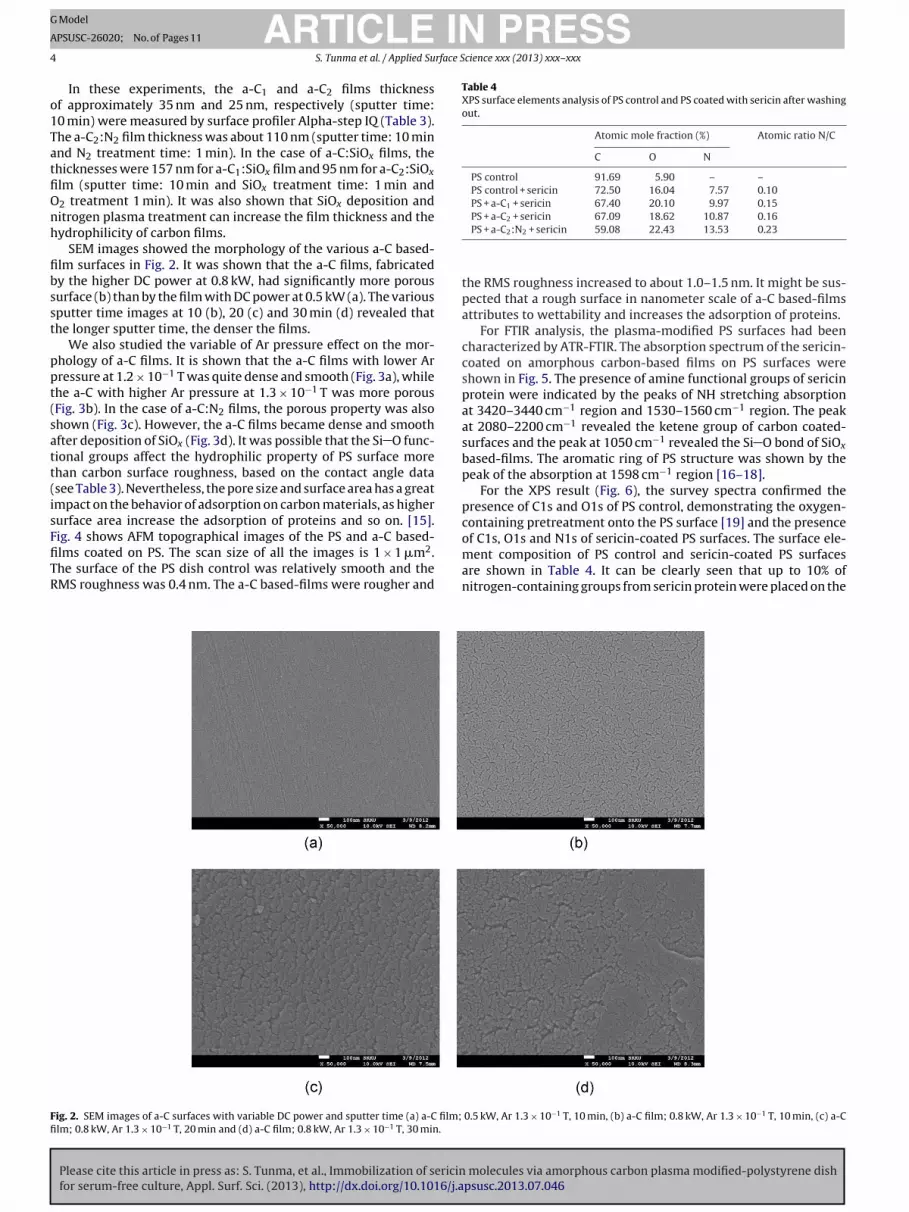
SEM images showed the morphology of the various a-C based-lm surfaces in Fig. 2. It was shown that the a-C films, fabricatedy the higher DC power at 0.8 kW, had significantly more porousurface (b) than by the film with DC power at 0.5 kW (a). The variousputter time images at 10 (b), 20 (c) and 30 min (d) revealed thathe longer sputter time, the denser the films.
We also studied the variable of Ar pressure effect on the mor-hology of a-C films. It is shown that the a-C films with lower Arressure at 1.2 × 10⁻¹ T was quite dense and smooth (Fig. 3a), whilehe a-C with higher Ar pressure at 1.3 × 10⁻¹ T was more porousFig. 3b). In the case of a-C:N2 films, the porous property was alsohown (Fig. 3c). However, the a-C films became dense and smoothfter deposition of SiOx (Fig. 3d). It was possible that the Si O func-ional groups affect the hydrophilic property of PS surface morehan carbon surface roughness, based on the contact angle datasee Table 3). Nevertheless, the pore size and surface area has a greatmpact on the behavior of adsorption on carbon materials, as higherurface area increase the adsorption of proteins and so on. [15].
Please cite this article in press as: S. Tunma, et al., Immobilization of sericinfor serum-free culture, Appl. Surf. Sci. (2013), http://dx.doi.org/10.1016/j.a
ig. 4 shows AFM topographical images of the PS and a-C based-lms coated on PS. The scan size of all the images is 1 × 1 �m2.he surface of the PS dish control was relatively smooth and theMS roughness was 0.4 nm. The a-C based-films were rougher and
ig. 2. SEM images of a-C surfaces with variable DC power and sputter time (a) a-C film;lm; 0.8 kW, Ar 1.3 × 10⁻¹ T, 20 min and (d) a-C film; 0.8 kW, Ar 1.3 × 10⁻¹ T, 30 min.
PS + a-C2 + sericin 67.09 18.62 10.87 0.16PS + a-C2:N2 + sericin 59.08 22.43 13.53 0.23
the RMS roughness increased to about 1.0–1.5 nm. It might be sus-pected that a rough surface in nanometer scale of a-C based-filmsattributes to wettability and increases the adsorption of proteins.
For FTIR analysis, the plasma-modified PS surfaces had beencharacterized by ATR-FTIR. The absorption spectrum of the sericin-coated on amorphous carbon-based films on PS surfaces wereshown in Fig. 5. The presence of amine functional groups of sericinprotein were indicated by the peaks of NH stretching absorptionat 3420–3440 cm−1 region and 1530–1560 cm−1 region. The peakat 2080–2200 cm−1 revealed the ketene group of carbon coated-surfaces and the peak at 1050 cm−1 revealed the Si O bond of SiOx
based-films. The aromatic ring of PS structure was shown by thepeak of the absorption at 1598 cm−1 region [16–18].
For the XPS result (Fig. 6), the survey spectra confirmed thepresence of C1s and O1s of PS control, demonstrating the oxygen-containing pretreatment onto the PS surface [19] and the presenceof C1s, O1s and N1s of sericin-coated PS surfaces. The surface ele-
molecules via amorphous carbon plasma modified-polystyrene dishpsusc.2013.07.046
ment composition of PS control and sericin-coated PS surfacesare shown in Table 4. It can be clearly seen that up to 10% ofnitrogen-containing groups from sericin protein were placed on the
0.5 kW, Ar 1.3 × 10⁻¹ T, 10 min, (b) a-C film; 0.8 kW, Ar 1.3 × 10⁻¹ T, 10 min, (c) a-C

ARTICLE IN PRESSG Model
APSUSC-26020; No. of Pages 11
S. Tunma et al. / Applied Surface Science xxx (2013) xxx– xxx 5
F 0−1 T,
−1
1
atlPr((Cd2bCio(2Cpwaspg1
3s
wa
ig. 3. SEM images of different types of a-C surfaces (a) a-C1 film; 0.8 kW, Ar 1.2 × 1 min and (d) a-C2 10 min and SiOx + O2 1 min.
morphous carbon-coated surface after PBS solution washing outhe ungrafted molecules. On the XPS analysis, the stability of cova-ent grafting of sericin molecules onto amorphous carbon film andS surface after washing out was discussed. Fig. 7 shows the high-esolution C1s spectra of PS control (Fig. 7a), PS control + sericinFig. 7b), PS + a-C1 (Fig. 7c), PS + a-C1 + sericin (Fig. 7d), PS + a-C2Fig. 7e), PS + a-C2 + sericin (Fig. 7f), PS + a-C2:N2 (Fig. 7g) and PS + a-2:N2 + sericin (Fig. 7h). The C1s peak of PS control (Fig. 7a) waseconvoluted into 6 peaks with binding energies of 284.6, 285.2,85.9, 286.7, 288.5 and 292–293 eV, which were attributed to car-ons in C H aromatic (C C), C H aliphatic (C C), C H2, C O or
OH, C O and �–�*, respectively [20,21]. The �–�* C1s peaks due to “shake-up” excitations taking place in the � orbitalsn the benzene rings [21]. Sericin-coated onto PS control surfaceFig. 7b) produced three new peaks with binding energies of 287.6,89.2 and 290.3 eV, which were attributed to C NH2 (amine) or
O C or C OH, C O, and O CO O groups, respectively. Amor-hous carbon (a-C1 and a-C2) (Fig. 7c and e) produced the new peakith binding energy of 286.2 eV, which attributed to C O C group
nd increased the intensity of C O peak at 288 eV especially, a-C2howed the COOH peak at 289.3 eV. In the case of a-C2:N2, the neweak of amide group (N C O) at 288.5 eV was shown. With sericin-rafted, C main peak was rearranged however can be grouped into;. C NH2, C O C, C OH, 2. C O, N C O and 3. O CO O.
.2. The release rate detection of sericin molecules into PBSolution
Please cite this article in press as: S. Tunma, et al., Immobilization of sericinfor serum-free culture, Appl. Surf. Sci. (2013), http://dx.doi.org/10.1016/j.a
UV absorbance of the released-sericin solution was scannedith the scanning range 190–450 nm as shown in Fig. 8. The
mino acids composition of sericin contained a high serine
10 min, (b) a-C2 film; 0.8 kW, Ar 1.3 × 10 T, 10 min, (c) a-C2 10 min and N2 plasma
content, accounting for about 27.3% of the 18 kinds of amino acids.Serine has strongly polar hydroxyl groups, and is possibly related tothe functional and physiochemical properties of sericin [3]. It wasalso found that the aspartic acid and glycine content accountingfor 18.8% and 10.7%, respectively, indicated that aspartic acid andglycine were also important amino acids attributed to the func-tions of sericin. In addition, the hydrophilic amino acid amounts upto 70% of the 18 kinds of amino acids, which could account for thegood solubility and water absorbability of sericin. It was also foundthat the amount of aromatic amino acids in sericin was very low(accounting for 6.6% of the 18 kinds of amino acids) compared withother proteins, and this was confirmed by the ultraviolet absorptionspectrum. As shown in Fig. 8, the maximal absorption wavelengthwas at 214 nm which indicated that peptide bonds were the majorabsorbing group for sericin in the ultraviolet region, not at 280 nmwhich is assigned to aromatic amino acids absorption wavelength.
For the release rate detection of released-sericin solution, it wasfound that all of sericin coating on modified-PS culture dishes had alower release rate of sericin compounds into the PBS solution thanPS control for the immersed time at day 1 (Fig. 8a), day 3 (Fig. 8b),day 5 (Fig. 8c) and day 7 (Fig. 8d).
3.3. Cell behavior study
Cellular behavior on any biomaterial is an important indicationto determine its biocompatibility. The whole process of adhesion
molecules via amorphous carbon plasma modified-polystyrene dishpsusc.2013.07.046
and spreading of cells after contacting with biomaterials consistsof cell attachment, filopodia growth, cytoplasmic webbing andflattening cell mass, and the ruffling peripheral cytoplasm, whichprogress in a sequential fashion [22].

ARTICLE IN PRESSG Model
APSUSC-26020; No. of Pages 11
6 S. Tunma et al. / Applied Surface Science xxx (2013) xxx– xxx
a-C1 c
3
hw7wwfw
Fig. 4. AFM images of a-C based-films (a) PS control, (b)
.3.1. Serum condition (20% FBS)Cell behavior experiments were performed over 7 days using
BM-MSCs. Fig. 9 shows the proliferation pattern of hBM-MSCsith serum condition (20% FBS and 5% sericin) at day 1 and day
. Day 1 shows the percentage of attached cells on the PS surfaces
Please cite this article in press as: S. Tunma, et al., Immobilization of sericinfor serum-free culture, Appl. Surf. Sci. (2013), http://dx.doi.org/10.1016/j.a
hen cultured for 24 h. compared with PS-control. Less adhesionas observed on the three groups of a-C:Si Ox-based (silica sur-
ace) films as shown in Fig. 10. The cells also appeared roundedhich indicated the incomplete attachment of hBM-MSC with
oated-PS, (c) a-C2 coated-PS and (d) a-C2:N2 coated-PS.
ragged cytoplasmic boundary. The silica surface has strong elec-trostatic attraction between surface silanol groups and the surfacecharge of non-prosthetic residues of protein molecules may alterthe damage to the coherent structure and the native state of theprotein [15]. Moreover, silica has weak stability upon prolonged
molecules via amorphous carbon plasma modified-polystyrene dishpsusc.2013.07.046
exposure to aqueous solutions which remains a major problem inthe case of cell culture. It might be concluded that the biologicalactivity and stability of the sericin protein may change upon inter-action with a silica surface. In this case, hBM-MSCs cultured in 20%

ARTICLE IN PRESSG Model
APSUSC-26020; No. of Pages 11
S. Tunma et al. / Applied Surface Science xxx (2013) xxx– xxx 7
and m
Flas
3
fFd
Fig. 5. Infrared spectra of the PS control
BS and sericin coated dish, had the proliferation pattern (Fig. 9)ower than the PS control which cultured with only 20% FBS, it canlso be implied that the cell culture with sericin plus FBS was notupporting the cell growth rate of hBM-MSCs.
.3.2. Serum-free condition (silk sericin protein)
Please cite this article in press as: S. Tunma, et al., Immobilization of sericinfor serum-free culture, Appl. Surf. Sci. (2013), http://dx.doi.org/10.1016/j.a
Cell behavior experiments of serum-free conditions were per-ormed over 7 days using hBM-MSCs with silk sericin coatings.ig. 11 shows the proliferation pattern of hBM-MSCs at day 1 anday 7 with a mixture of 5% sericin coatings and 5% FBS and 5%
Fig. 6. XPS scan spectra of PS control and mo
odified-PS coated with sericin surface.
sericin coatings without FBS conditions. It was shown that PS con-trol dish with 10% FBS has the highest percentage of proliferatedcells at day 7 while at day 1, no differences were detected. Sericinprotein on modified-dish promotes cell attachment like using theFBS alone. The carbon-based with sericin-coated surfaces have thehigher proliferation rate with a mixture of 5% FBS than without FBS
molecules via amorphous carbon plasma modified-polystyrene dishpsusc.2013.07.046
condition and also higher than the PS control coated with sericinat day 7 especially, in the case of a-C2 surface. However, PS disheswith a mixture of 5% FBS and sericin showed the higher prolifer-ated cells than that used with only sericin in the same condition.
dified-PS coated with sericin surfaces.

Please cite this article in press as: S. Tunma, et al., Immobilization of sericin molecules via amorphous carbon plasma modified-polystyrene dishfor serum-free culture, Appl. Surf. Sci. (2013), http://dx.doi.org/10.1016/j.apsusc.2013.07.046
ARTICLE IN PRESSG Model
APSUSC-26020; No. of Pages 11
8 S. Tunma et al. / Applied Surface Science xxx (2013) xxx– xxx
Fig. 7. High resolution peaks of C1s for PS control and modified-PS coated with sericin surfaces (a) C1s of PS control, (b) C1s of PS control + sericin coated, (c) C1s of a-C1
coated-PS, (d) C1s of a-C1 coated-PS + sericin coated, (e) C1s of a-C2 coated-PS, (f) C1s of a-C2 coated-PS + sericin coated, (g) C1s of a-C2:N2 coated-PS and (h) C1s of a-C2:N2
coated-PS + sericin coated.

ARTICLE IN PRESSG Model
APSUSC-26020; No. of Pages 11
S. Tunma et al. / Applied Surface Science xxx (2013) xxx– xxx 9
F t 1 daa
Atmf
Fs
ig. 8. UV absorption spectrum of the released-sericin solution (a) immersed time at 7 days.
Please cite this article in press as: S. Tunma, et al., Immobilization of sericinfor serum-free culture, Appl. Surf. Sci. (2013), http://dx.doi.org/10.1016/j.a
ccording to SEM images in Fig. 12, the attached cell on PS con-rol (Fig. 12a) showed the ragged boundary (red arrows) indicating
ore incomplete attachments than cells on the carbon-based sur-aces (Fig. 12c–e).
ig. 9. Cell proliferation patterns of hBM-MSCs on PS surfaces when cultured inerum condition with 20% FBS and 5% sericin coating at day 1 and day 7.
y, (b) immersed time at 3 days, (c) immersed time at 5 days and (d) immersed time
4. Discussion
To study the adsorption of proteins on hydrophilic and roughsurfaces, the chemical functional groups and the physical expla-nation of surface were discussed. On hydrophilic surfaces, cellsadhered in higher numbers to more hydrophilic materials andwere spread over a large area. At the same time, ECM proteinswere adsorbed in a more flexible form, which allows them to berearranged by the cells and thus provides access for cell adhesionreceptors to the adhesion motifs on these molecules. The physi-cal explanation for the increase in adsorption as surface roughnesshad to be provided. Basically, a protein has a definite size and shape,more like a colloidal particle, whereas a simple polymer in solutionbehaves like a random coil. So the studies of simple polymers withhigh values of surface roughness enhanced adsorption because thepolymer can bind several places on a rough surface without losingtoo much conformation entropy, do not necessarily apply to pro-teins [19]. Several experimental investigation of protein adsorptionon rough substrates exist [17,18]. All these conclusions were thatthe height of the nanostructures (the RMS roughness) should be1–2 nm in order to enhance protein adsorption compared to a flatsurface. For higher values of 4 nm, adsorption decreases again [18].This is in agreement with AFM images which showed the surfaceroughness of a-C based-films are about 1.0–1.5 nm, as shown in
molecules via amorphous carbon plasma modified-polystyrene dishpsusc.2013.07.046
Fig. 4.In combination of FTIR and XPS results, it was shown that a-
C based films have a higher percentage of N atomic mole fraction

Please cite this article in press as: S. Tunma, et al., Immobilization of sericinfor serum-free culture, Appl. Surf. Sci. (2013), http://dx.doi.org/10.1016/j.a
ARTICLE IN PRESSG Model
APSUSC-26020; No. of Pages 11
10 S. Tunma et al. / Applied Surface Science xxx (2013) xxx– xxx
Fig. 10. Examples of SEM micrographs of attac
Fig. 11. Cell proliferation patterns of hBM-MSCs on PS surfaces when cultured inserum-free condition at days 1 and 7.
Fig. 12. Examples of SEM micrographs of attac
hed hBM-MSCs on silica-based surfaces.
than PS control, especially in the case of a-C2:N2 and a-C2 films.A-C2 and a-C2:N2 films have porous structures and nanometerroughness surfaces which lead to enhancement of the adsorptionof sericin proteins. It was revealed that functionalized carbon canbe enhanced by specific molecules. Moreover, due to the nitrogen-containing groups such as amine group ( NH2) of a-C2:N2 film,being positively charged which is highly reactive, it was believedthat it can covalently couple with protein molecules in aqueousenvironments [9,16,23]. Corresponding with FTIR result, a-C basedfilms produced the peak of ketene groups (C C O) at wavenumberof 2080–2200 cm−1. Ketene and aminoketene ((NH2)C C O) are ahighly reactive and transient intermediary in a variety of reactions[24].
From the UV-absorption peaks of the released sericin solution, itwas implied that the covalent bonds interaction between the a-C1,a-C2 as intermediate layer on PS surfaces and sericin coatings can
molecules via amorphous carbon plasma modified-polystyrene dishpsusc.2013.07.046
slow down the release rate of sericin compound into the immersed-solution, due to the porous structures and the reactivity functionalgroups surfaces. This supports the idea that carbon material is moreattractive for protein adsorption due to their inherent stability and
hed hBM-MSCs in serum-free condition.

ING Model
A
rface S
ssi
wstcTCtcps
5
mPmgfabcsauctatoi
A
CUcTKw
R
[
[
[
[
[
[
[
[
[
[
[
[
[
[
ARTICLEPSUSC-26020; No. of Pages 11
S. Tunma et al. / Applied Su
urface properties such as high surface area. Besides the poroustructure, the surface functional groups of the carbon are also anntriguing parameter that should be taken into account [15].
Additionally, carbon coated-PS culture dish and sericin coatingere used in hBM-MSCs culture with serum-free system and pre-
ented the higher proliferation rate with the mixture of 5% FBShan without FBS condition and also higher than the PS controloated with sericin at day 7 especially, in the case of a-C2 surface.his suggested that a-C based films with N2 plasma treatment (a-2:N2) have higher specific binding with sericin molecules, due toheir nitrogen-containing groups. However, it cannot support theell growth for a prolonged time, while a-C2 films show significantroliferated cell numbers at day 7 due to their roughness and thetability function.
. Conclusions
In this study, amorphous carbon based films used as inter-ediate layers for covalent grafting between sericin protein and
S surface successfully slowed down the release rate of sericinolecules into PBS solution. The Si O and nitrogen-functional
roups revealed a great affect to the hydrophilic property of sur-ace more than the roughness of a-C based films. The Si O groupslso influenced the surface by making the smooth surface of car-on film and disfavor with hBM-MSCs adherence. The stability ofarbon based films with sericin grafting which is more resistant totructural change by hydrolytic effects in aqueous environmentsnd biocompatibility, has been proven and could be potentiallysed in hBM-MSCs culture in serum-free condition. Amorphousarbon is an attractive material for biomolecules adsorption dueo its high specific surface area and porous structure which lead ton increment in the total amount of sericin protein absorbed andhe significant proliferated-cell numbers at a prolonged time. Ourbservations suggest that sericin grafting to a-C surfaces will havemportant uses in a wide range of biotechnological areas.
cknowledgements
We thank the Graduate School Chiang Mai University, Thailandenter of Excellence in Physics (ThEP), the National Researchniversity (NRU) Project under Thailand’s Office of the Higher Edu-ation Commission (OHEC), Center for Advanced Plasma Surfaceechnology (CAPST), College of Dentistry at Yonsei University andwangwoon University for financial and scholarship support. Alsoe wish to thank M. Rhodes for correcting the manuscript.
eferences
[1] S. Wataru, F. Ken, Y. Kana, S. Masahiro, K. Yoshihiro, T. Satoshi, Mitogenic effect
Please cite this article in press as: S. Tunma, et al., Immobilization of sericinfor serum-free culture, Appl. Surf. Sci. (2013), http://dx.doi.org/10.1016/j.a
of sericin on mammalian cells, BioMed Central’s Biomedical Journal 5 (8) (2011)121–123.
[2] T. Satoshi, N. Taeko, S. Masahiro, Y. Hideyuki, M. Masao, Sericin a protein derivedfrom silkworms, accelerates the proliferation of several mammalian cell linesincluding a hybridoma, Cytotechnology 40 (2002) 3–12.
[
PRESScience xxx (2013) xxx– xxx 11
[3] T. Kozo, I. Yumiko, T. Yoko, Y. Hiromi, Sericin enhances attachment of culturedhuman skin fibroblasts, Bioscience, Biotechnology, and Biochemistry 69 (2)(2005) 403–405.
[4] W. Kristian, S. Karsten, L. Ulrike, O. Andreas, Plasma-based modification ofpolystyrene surfaces for serum-free culture of osteoblast cell lines, PlasmaProcesses and Polymers 3 (2006) 524–531.
[5] S.E. Rodil, R. Olivares, H. Arzate, S. Muhl, Properties of carbon films and theirbiocompatibility using in-vitro tests, Diamond and Related Materials 12 (2003)931–937.
[6] S. Andreas, W. Zhiyong, A.F. Melissa, Functionalization of porous carbon mate-rials with designed pore architecture, Advanced Materials 21 (2009) 265–293.
[7] O. Soon-Eng, Z. Sam, D. Hejun, S.W. Yong, M. Lwin-Lwin, In-vitro cellularbehavior on amorphous carbon containing silicon, Thin Solid Films 516 (2008)5152–5156.
[8] N. Ming, H.T. Wen, C. Deepak, A.R. Nur Nida, I. Ciprian, Y. Hanry, Cell cultureon MEMS platforms: a review, International Journal of Molecular Sciences 10(2009) 5411–5441.
[9] L. Hyun-Uk, J. Ye-Sul, J. Se-Young, P. So-Young, B. Jong-Seoung, K. Hyun-Gyu, C.Chae-Ryoung, Role of reactive gas in atmospheric plasma for cell attachmentand proliferation on biocompatible poly �-caprolactone film, Applied SurfaceScience 254 (2008) 5700–5705.
10] J.M. Goddard, J.H. Hotchkiss, Polymer surface modification for the attachmentof bioactive compounds, Progress in Polymer Science 32 (2007) 698–725.
11] S.B. Hari, D. Jagannath, D.P. Chowdhury, A.V.R. Reddy, C.G. Uday, K.S. Arvind, R.R.Nihar, Covalent immobilization of protein onto a functionalized hydrogenateddiamond-like carbon substrate, Langmuir 26 (22) (2010) 17413–17418.
12] B.J. Su, S.C. Yoon, G.H. Jeon, Surface energy modification of SiOxCyHz film usingPECVD by controlling the plasma processes for OMCTS (Si4O4C8H24) precursor,Thin Solid Films 519 (2011) 6763–6768.
13] B.P. Binks, S.O. Lumsdon, Influence of particle wettability on the type and sta-bility of surfactant-free emulsions, Langmuir 16 (2000) 8622–8631.
14] B. Lucie, F.F. Elena, P. Martin, R. Tomas, S. Vaclav, Modulation of cell adhe-sion, proliferation and differentiation on materials designed for body implants,Biotechnology Advances 29 (6) (2011) 739–767.
15] V. Munusami, C. Roger, A. Karine, G. Camelia, V.G. Cathie, O. Hironori, K. Takahi,I. Sumlak, The influence of surface chemistry and pore size in the adsorption ofproteins on nanostructured carbon materials, Advanced Functional Materials20 (2010) 2489–2499.
16] W. Jin-Hong, W. Zhang, X. Shi-Ying, Preparation and characterization of sericinpowder extracted from silk industry wastewater, Food Chemistry 103 (2007)1255–1262.
17] F. Birgit, L. Frank, S. Karsten, D.M. Petra, B. Claudia, F. Marion, O. Andreas,J.N. Barbara, The effect of positively charged plasma polymerization on ini-tial osteoblastic focal adhesion on titanium surfaces, Biomaterials 28 (2007)4521–4534.
18] Y. Lizhen, L. Juan, W. Zhenduo, L. Zhongwei, C. Qiang, Calibration of aminedensity measurement on plasma grafting PET surface and its cell adsorptionbehavior, Surface and Coatings Technology 205 (2010) S345–S348.
19] P.C. Schamberger, J.I. Abes, J.A. Gardella Jr., Surface chemical studies of aging andsolvent extraction effects on plasma-treated polystyrene, Colloids and SurfacesB: Biointerfaces 3 (1994) 203–215.
20] S. Yasushi, M. Natsuko, K. Shin-ichi, K. Masayuki, Introduction of carboxyl grouponto polystyrene surface using plasma techniques, Surface and Coatings Tech-nology 202 (2008) 5724–5727.
21] M. Delphine, P. Claude, B. Patrick, S. Michele, R. Francois, Synthesis ofpolystyrene thin films by means of an atmospheric-pressure plasma torch and adielectric barrier discharge, IEEE Transactions on Plasma Sciences 37 (6) (2009)951–960.
22] M. Dadsetan, H. Mirzadeh, N. Sharifi, M. Daliri, Cell behavior on laser surface-modified polyethylene terephthalate in vitro, Journal of Biomedical MaterialsResearch Part A 257 (2007) 183–189.
23] K. Jinmo, J. Donggeun, P. Yongsup, K. Yongki, W.M. Dae, G.L. Tae, Quantita-tive analysis of surface amine groups on plasma-polymerized ethylenediaminefilms using UV–visible spectroscopy compared to chemical derivatization with
molecules via amorphous carbon plasma modified-polystyrene dishpsusc.2013.07.046
FT-IR spectroscopy, XPS and TOF-SIMS, Applied Surface Science 253 (2007)4112–4118.
24] V.P. Gupta, S. Archna, S.G. Agrawat, Conformations, chemical, reactivities andspectroscopic characteristic of some di-substituted ketenes: an ab initio study,The Bulletin of the Korean Chemical Society 27 (9) (2006) 1297–1304.


