Embed Size (px)
Citation preview
Dental Medicine Research 30(3):228⊖236, 2010228
The skeletal system, characterized by a hard tissue component, is an extraordinarily dynamic system with disparate functions such as structural support, movement, soft-organ protection and the maintenance of calcium homeostasis. In addition, bone houses definitive hematopoiesis. During vertebrate ontogeny, hematopoiesis is established sequentially in different anatomical sites. Around birth time, hematopoietic site shifts from fetal liver to the forming bone marrow cavity which is the major site of hematopoiesis during adult life.1~3)
Vertebrate bones are formed by two different types of ossification systems, endochondral and intramembranous ossification. Intramembranous bone formation is mediated by osteoblasts differentiated from the periosteal osteogenic cells, which form bone without the mediation of cartilage, while, in the case of endochondral bone formation, osteoblasts form bone on a calcified cartilage scaffold after epiphyseal and physeal cartilage have shaped and elongated.4~6) In both of ossification systems, hematopoietic tissues are located inside the forming bones.
Original
Immunohistochemical Comparison of Ontogenic Development ofBone Marrow Hematopoiesis in Two Different Ossification Systems
Minako Ikeda*,**, Hirotada Ohtsuka*, Yukikatsu IwasakI*,***,Mariko Ikeda*,**, Kazuyoshi BaBa** and Masanori NakaMura*
*Department of Oral Anatomy and Developmental Biology, Showa University School of Dentistry1⊖5⊖8 Hatanodai, Shinagawa-ku, Tokyo, 142⊖8555 Japan
(Chief : Prof. Masanori Nakamura)**Department of Prosthodontics, Showa University School of Dentistry
2⊖1⊖1 Kitasenzoku, Ohta-ku, Tokyo, 145⊖8515 Japan(Chief : Prof. Kazuyoshi Baba)
***Department of Clinical Cariology and Endodontology, Showa University School of Dentistry2⊖1⊖1 Kitasenzoku, Ohta-ku, Tokyo, 145⊖8515 Japan
Abstract: It is well known that the body skeleton is formed by two different types of ossification systems, endochondoral and intramembranous ossification. Bone marrow is the main site of active hematopoiesis after the formation of the bone marrow cavities. However, it is unclear whether the hematopoiesis in the bone marrow of two types of ossification is regulated by the same system or not. In this study, we focused on the ontogenic development of bone marrow hematopoiesis in two different ossification systems using mouse humeral bones and palatal process of maxillary bones. Immunohistochemical and RT-PCR analyses were performed to examine the development of hematopoiesis and the expression of cytokines related to hematopoiesis in the forming bone marrow (16-days gestation stage to 1-day postnatal stage). Immunohistochemical studies showed the sequential difference of hematopoiesis between two different ossification systems. In humeral bone marrow, granulopoiesis appeared first at E16, followed by erythropoiesis from E17. On the contrary, erythropoiesis preceded one day in the maxillary bone marrow at E18, one day before the detection of granulopoiesis. GSCF and GMCSF were expressed at every examined stage in both types of bones while MCSF was not expressed in the humeral bone marrow at E16. Erythropoietin was detetcted in the endothelial cells and its expression was coincident with the onset of erythopoiesis. These results suggest the time kinetic and sequential differences of hematopoiesis in two different ossification systems, which might relate to the differences of hematopoietic microenvironment.
Key words: bone marrow, hematopoiesis, endochondoral ossification, intramembranous ossification.
(Received September 7, 2010; Accepted September 14, 2010)
Bone Marrow Hematopoiesis During Bone Development 229Dental Med Res. 30
It has been indicated that the bone marrow micro-environment provides essential regulatory role in hematopoietic system. The hematopoietic micro-environment includes stromal cells and the cytokines and extracellular matrix which they secrete.7) The bone marrow stromal cells include macrophages, endothelial cells, adipocytes, fibroblasts and osteoblasts.8, 9) Erythroblastic islands consist of a central macrophage surrounded by a ring of erythroblasts that undergo terminal maturation leading to enucleation. It has been reported that erythroid cells mature to the late erythroblast stage but fail to enucleate in macrophage-depleted cultures.10) Erythropoietin (EPO), a major regulator of erythropoiesis, has also been detected in the macrophages of fetal liver blood islands.11, 12) These results suggest that erythroblast-macrophage contact promotes proliferation and terminal maturation of erythroid cells. Osteoblasts are differentiated stromal cell type unique to the bone marrow environment. It has been indicated that osteoblasts play the central and direct role in myelopoiesis by secreting numerous hematopoietic cytokines which support the terminal neutrophil matu-ration and the in vitro expansion of primitive long-term culture-initiating cells (LTCICs).13~15)
Osteoblasts are differentiated from a multipotent mesenchymal stem cell (MMSCs), which have the ability to commit to osteogenesis, chondrogenesis, adipogenesis and myogenesis by the defined developmental pro-grams.16) It has been proposed that once MMSCs commit to osteogenesis the cells differentiate into a putative committed osteoprogenitor cells, then progress to preosteoblasts and osteoblasts.17) Although the precise mechanisms for the regulation of this process are poorly understood, several studies indicate the inherent and/or functional heterogeneity of osteoblasts.18, 19) These results lead to suggest the heterogeneity of hematopoietic microenvironment in the bone marrow during the bone development and between two different osiification sysytems. Therefore, in this study, we compared the ontogenic
development of hematopoiesis, erythropoiesis and granulopoiesis, in the bone marrow of two different ossification systems to know the heterogeneity of hematopoietic microenvironment in two different ossification systems.
Materials and Methods
Animals and antibodies Pregnant BALB/c mice were purchased from Saitama Breeding Laboratory, Saitama, Japan. Mice from 15day gestation (E15) to 1 day postnatal (1DPN) stage were used in this study. Monoclonal antibodies, Gr1 and TER119, were purchased from BD Biosciences (San Jose, CA) and F4/80 was purchased from UKserotec Ltd. (Oxfordshire, UK). A polyclonal antibody, goat antihuman Erythropoietin (EPO) was purchased from Santa Cruz Biotechnology (Santa Cruz, CA). Alexa Fluor 488labeled rat antimouse Gr1 antibody was purchased from Biolegend (San Diego, CA), and Alexa Fluor 594labeled goat antirat IgG antibody was purchased from Invitrogen (Carlsbad, CA). Biotinylated goat antirat IgG and rabbit antigoat IgG antibodies were from VECTOR, Burlingame, CA. Tissue preparation The experimental protocol was approved by the Animal Care Committee of Showa University. Humeral long bones and maxillary bones from E15 to 1DPN stage were dissected and fixed with 4% paraformaldehyde in PBS. After decalcification by 10% EDTA in PBS, the specimens were washed with 20% sucrosePBS, embedded in CRYOform (International Equipment Co., Needham, MA), and quickfrozen in a mixture of acetone and dry ice. Some of the humeral and maxillary bones were processed for RT-PCR. Immunohistochemical procedure Frozen sections (8 µm thick) were cut, placed on poly-l-lysine-coated glass slides and air dried. After incubation in 0.3% H2O2methanol for 30 min, the sections were incubated with 5% normal goat serum in PBS containing 5% bovine serum albumin and
M. Ikeda and others230 Dental Med Res. 30
0.025% Triton X100, followed by incubation with each of the monoclonal antibodies. After several rinses, the sections were incubated with biotinylated goat antirat IgG antibody and then with avidinbiotinhorseradish peroxidase complex. After washing, the sections were transferred to and incubated with a mixture of 3,3’-diaminobenzidine tetrahydrochloride (0.5 mg/ml) (WAKO, Osaka, Japan) and H2O2 at a final concentration of 0.03% in 0.1 M TrisHCl buffer at pH7.6. Counterstaining was produced by methylgreen. As control experiments, sections were incubated with either a normal rat serum or PBS instead of the primary antibody. For the double labeling of granulopoiesis and erythropoiesis, sections were stained first with antiTER119 antibody coupled with Alexa Flour 594labeled goat antirat IgG antibody, and then with Alexa Flour 488labeled Gr1. After several rinses with PBS, sections were mounted with AquaMount (Polysciences, Warrington, PA). For the detection of erythropoietin, sections were stained with goat anti-erythropoietin antibody after the pretreatment described above. After thorough rinse with PBS, sections were incubated with biotinylated rabbit antigoat IgG antibody and processed for the visualization of the immunoreaction products. Control sections were incubated with normal goat serum instead of the primary antibody. RT-PCR analysis Humeral and maxillary bones were isolated and homogenized. Total RNA was extracted from each sample using RNeasy Mini kit (QIAGEN, Tokyo, Japan). Four micrograms total RNA served as template for reverse transcriptase polymerase chain reaction (RTPCR), using GeneAmp Gold RNA PCR (Applied
Biosystems, Tokyo, Japan) with mMCSF, mGCSF and mGMCSF sequence aligned primers. Primers used in this study were listed in Table 1. Amplification conditions were as follows: RT reaction was performed at 25 for 10 min and at 42 for 12 min; PCR reaction start at 95 for 5 min, and then the 35 cycles were consisted of denaturing at 95 for 10 s, annealing at 60 for 30 s and extension at 72 for 2 min. The final extension was at 72 for 7 min. PCR products were mixed with 6×loading dye (TOYOBO Biologics, Osaka, Japan) at the ratio 6:1 (v/v) and separated by electrophoresis at 100 V for 20 min on a 1.0% agarose gel.
Results
Immunohistochemical detection of erythropoiesis and granulopoiesis during bone development
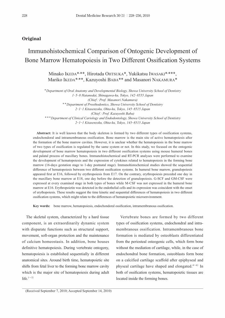
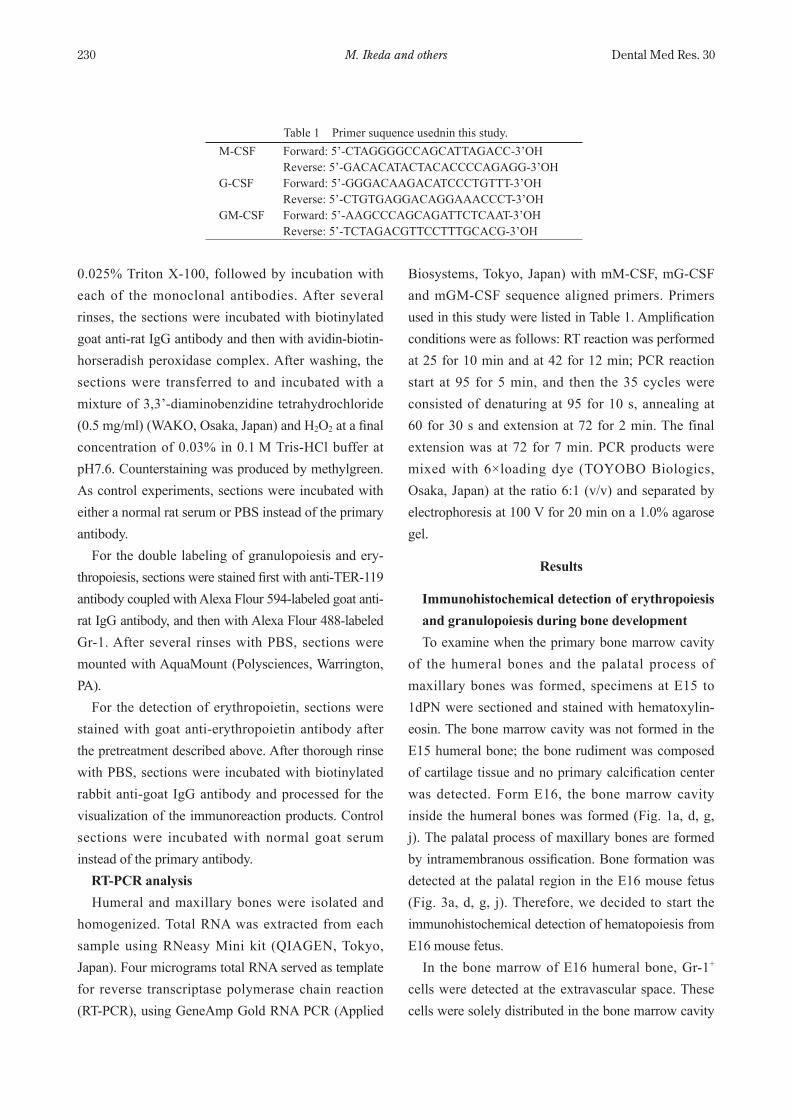
To examine when the primary bone marrow cavity of the humeral bones and the palatal process of maxillary bones was formed, specimens at E15 to 1dPN were sectioned and stained with hematoxylineosin. The bone marrow cavity was not formed in the E15 humeral bone; the bone rudiment was composed of cartilage tissue and no primary calcification center was detected. Form E16, the bone marrow cavity inside the humeral bones was formed (Fig. 1a, d, g, j). The palatal process of maxillary bones are formed by intramembranous ossification. Bone formation was detected at the palatal region in the E16 mouse fetus (Fig. 3a, d, g, j). Therefore, we decided to start the immunohistochemical detection of hematopoiesis from E16 mouse fetus. In the bone marrow of E16 humeral bone, Gr1+
cells were detected at the extravascular space. These cells were solely distributed in the bone marrow cavity
Table 1 Primer suquence usednin this study.MCSF Forward: 5’CTAGGGGCCAGCATTAGACC3’OH
Reverse: 5’GACACATACTACACCCCAGAGG3’OHGCSF Forward: 5’GGGACAAGACATCCCTGTTT3’OH
Reverse: 5’CTGTGAGGACAGGAAACCCT3’OHGMCSF Forward: 5’AAGCCCAGCAGATTCTCAAT3’OH
Reverse: 5’TCTAGACGTTCCTTTGCACG3’OH
Bone Marrow Hematopoiesis During Bone Development 231Dental Med Res. 30
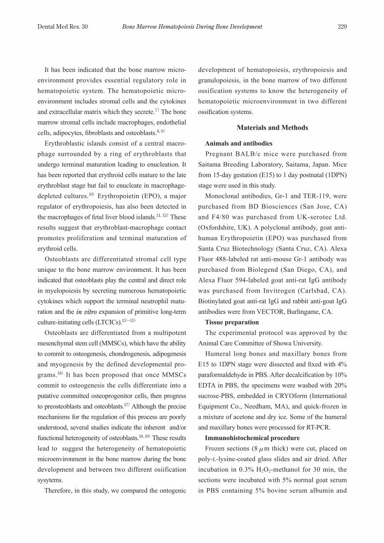
(Fig. 1c). The number of Gr1+ cells was increased with the development and formed the clusters near to the bone trabecular surfac (Fig. 1f, i, l). In the case of erythropoiesis in the humeral bone marrow, TER119+ nucleated erythrocyte-precursor cells were detected from E17 (Fig. 1e). With bone development, these cells formed clusters like Gr1+ cells, which were mainly located adjacent to the vasculature (Fig. 1e, h, k). These results indicated the difference of sequential development of erythropoiesis and granulopoiesis in two different ossification systems and the difference of the hematopoietic sites between erythropoiesis and granulopoiesis in the bone marrow. Double staining GR1+ and TER119+ cells confirmed clearly the different hematopoietic loci of erythropoiesis and granulopoiesis in the bone marrow. Gr1+ cells were mainly located along the bone surface while TER119 cells were detected in more central region of the bone
marrow (Fig. 2). In the palatal process of the maxillary bone, TER119+ nucleated cells were first detected at E17 when Gr1+ cells were not yet detected (Fig. 3b, c). With bone development, the number of TER119+ and Gr1+ cells were increased and formed clusters, respectively (Fig. 3h, i, k, l). Hematopoietic loci of TER119+ and Gr1+
E16j k l
E16d e f
E16g h i
E16a b c
H-E TER-119 Gr-1
Fig. 1 Ontogenic development of endochondral ossifica-tion and hematopoiesis in humeral bones at E16 (a, b, c), E17 (d, e, f), E18 (g, h, i) and 0DPN (j, k, l). Histological features of bone development (a, d, g, i) and immunohistochemical localization of TER119+ cells (b, e, h, k) and Gr1+ cells (c, f, i, l) were represented. Bars=50 µm (a, d, g, h) and 25 µm (b, c, e, f, h, i, k, l).
ba
Fig. 2 Double staining of granulopoiesis and erythro-poiesis in the humeral bone marrow at 0DPN. (a) A differential-interference image of the bone marrow. (b) Double staining of Gr1+ cells (green) and TER119+ cells (red). Bars=25 µm.
E16j k l
E16d e f
E16g h i
E16a b c
H-E TER-119 Gr-1
Fig. 3 Ontogenic development of intramembranous ossification and hematopoiesis in the palatal processes of maxillary bones at E16 (a, b, c), E17 (d, e, f), E18 (g, h, i) and 0DPN (j, k, l). Histological features of bone development (a, d, g, i) and immunohistochemical localization of TER119+ (b, e, h, k) and Gr1+ cells (c, f, i, l) were represented. Bars=100 µm (a, d, g, h) and 25 µm (b, c, e, f, h, i, k, l).
M. Ikeda and others232 Dental Med Res. 30
cells showed same distribution pattern with the blood formation in the humeral bone marrow. Immunohistochemical detection of macrophages
and erythropoietin in the bone marrow Bone marrow resident macrophages and erythro-
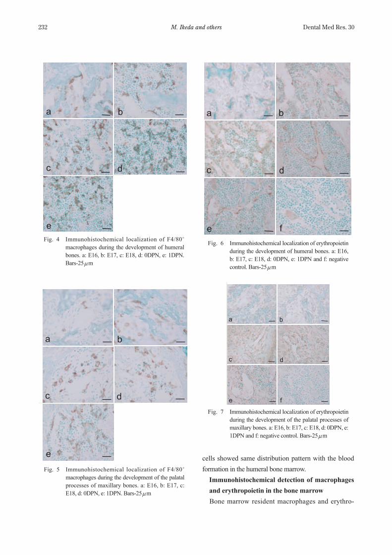
Fig. 4 Immunohistochemical localization of F4/80+ macrophages during the development of humeral bones. a: E16, b: E17, c: E18, d: 0DPN, e: 1DPN. Bars25 µm
a b
c d
e
a b
c d
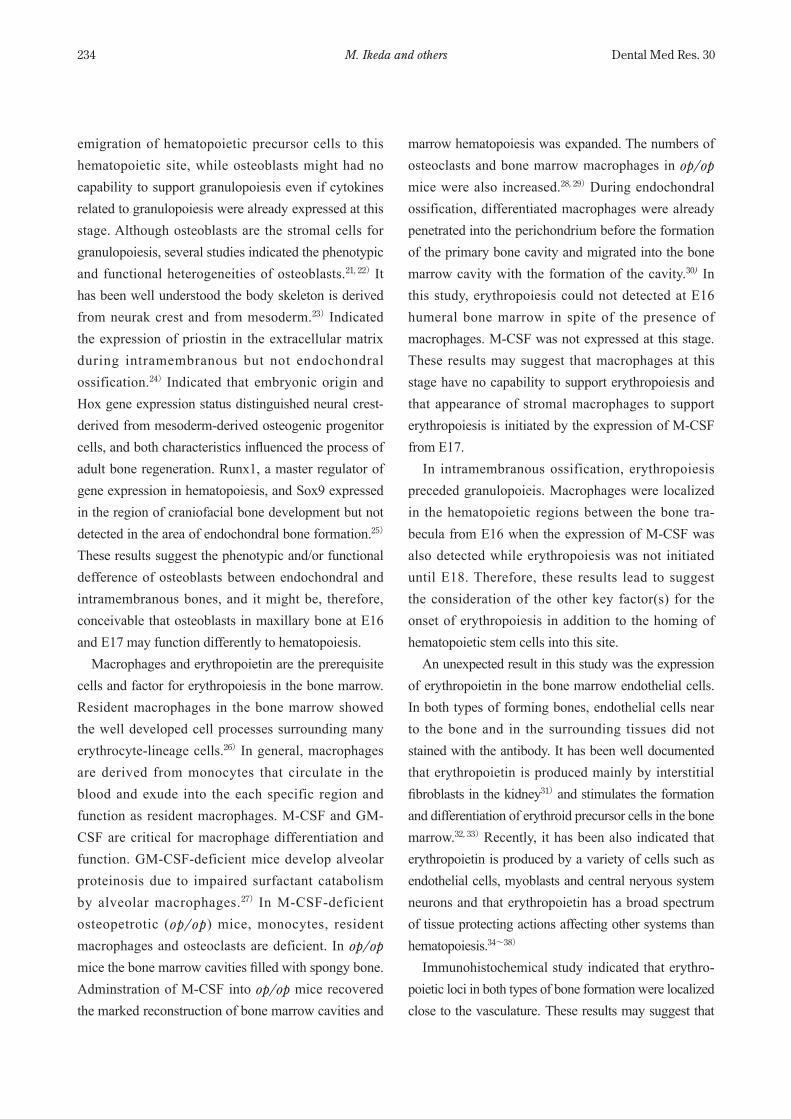
eFig. 5 Immunohistochemical localization of F4/80+
macrophages during the development of the palatal processes of maxillary bones. a: E16, b: E17, c: E18, d: 0DPN, e: 1DPN. Bars25 µm
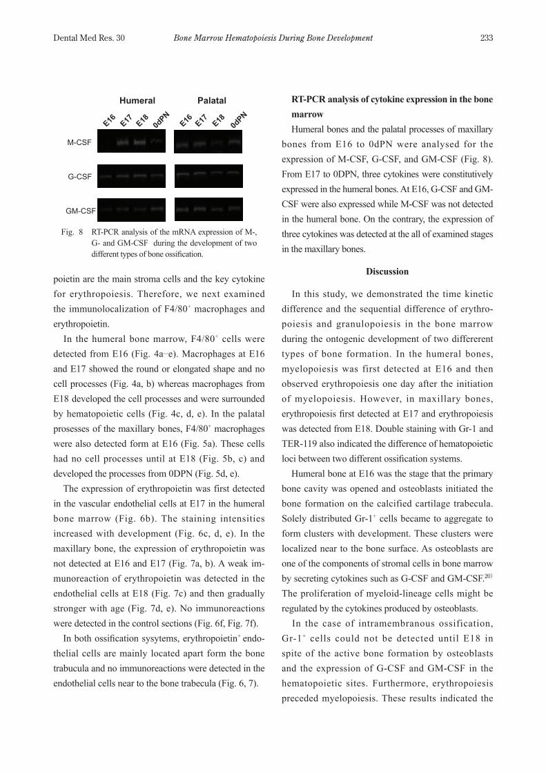
Fig. 6 Immunohistochemical localization of erythropoietin during the development of humeral bones. a: E16, b: E17, c: E18, d: 0DPN, e: 1DPN and f: negative control. Bars25 µm
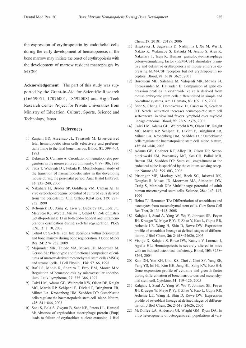
Fig. 7 Immunohistochemical localization of erythropoietin during the development of the palatal processes of maxillary bones. a: E16, b: E17, c: E18, d: 0DPN, e: 1DPN and f: negative control. Bars25 µm
a b
c d
fe
a b
c d
e f
Bone Marrow Hematopoiesis During Bone Development 233Dental Med Res. 30
poietin are the main stroma cells and the key cytokine for erythropoiesis. Therefore, we next examined the immunolocalization of F4/80+ macrophages and erythropoietin. In the humeral bone marrow, F4/80+ cells were detected from E16 (Fig. 4a⊖e). Macrophages at E16 and E17 showed the round or elongated shape and no cell processes (Fig. 4a, b) whereas macrophages from E18 developed the cell processes and were surrounded by hematopoietic cells (Fig. 4c, d, e). In the palatal prosesses of the maxillary bones, F4/80+ macrophages were also detected form at E16 (Fig. 5a). These cells had no cell processes until at E18 (Fig. 5b, c) and developed the processes from 0DPN (Fig. 5d, e). The expression of erythropoietin was first detected in the vascular endothelial cells at E17 in the humeral bone marrow (Fig. 6b). The staining intensities increased with development (Fig. 6c, d, e). In the maxillary bone, the expression of erythropoietin was not detected at E16 and E17 (Fig. 7a, b). A weak immunoreaction of erythropoietin was detected in the endothelial cells at E18 (Fig. 7c) and then gradually stronger with age (Fig. 7d, e). No immunoreactions were detected in the control sections (Fig. 6f, Fig. 7f). In both ossification sysytems, erythropoietin+ endo-thelial cells are mainly located apart form the bone trabucula and no immunoreactions were detected in the endothelial cells near to the bone trabecula (Fig. 6, 7).
RT-PCR analysis of cytokine expression in the bone marrow
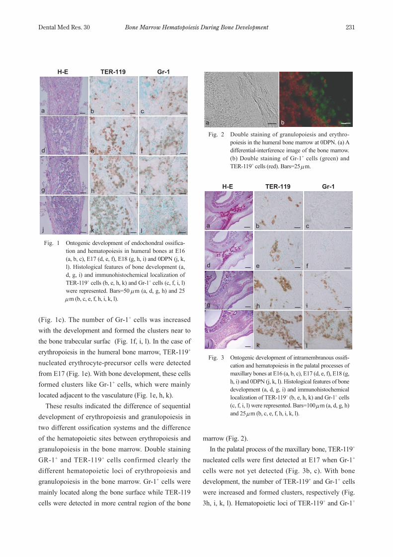
Humeral bones and the palatal processes of maxillary bones from E16 to 0dPN were analysed for the expression of MCSF, GCSF, and GMCSF (Fig. 8). From E17 to 0DPN, three cytokines were constitutively expressed in the humeral bones. At E16, GCSF and GMCSF were also expressed while MCSF was not detected in the humeral bone. On the contrary, the expression of three cytokines was detected at the all of examined stages in the maxillary bones.
Discussion
In this study, we demonstrated the time kinetic difference and the sequential difference of erythro-poiesis and granulopoiesis in the bone marrow during the ontogenic development of two differerent types of bone formation. In the humeral bones, myelopoiesis was first detected at E16 and then observed erythropoiesis one day after the initiation of myelopoiesis. However, in maxillary bones, erythropoiesis first detected at E17 and erythropoiesis was detected from E18. Double staining with Gr1 and TER119 also indicated the difference of hematopoietic loci between two different ossification systems. Humeral bone at E16 was the stage that the primary bone cavity was opened and osteoblasts initiated the bone formation on the calcified cartilage trabecula. Solely distributed Gr1+ cells became to aggregate to form clusters with development. These clusters were localized near to the bone surface. As osteoblasts are one of the components of stromal cells in bone marrow by secreting cytokines such as GCSF and GMCSF.20) The proliferation of myeloid-lineage cells might be regulated by the cytokines produced by osteoblasts. In the case of intramembranous ossification, Gr1+ cells could not be detected until E18 in spite of the active bone formation by osteoblasts and the expression of GCSF and GMCSF in the hematopoietic sites. Furthermore, erythropoiesis preceded myelopoiesis. These results indicated the
Fig. 8 RTPCR analysis of the mRNA expression of M, G and GMCSF during the development of two different types of bone ossification.
E16E17E180dPN
M-CSF
G-CSF
GM-CSF
Humeral Palatal
E16E17E180dPN
M. Ikeda and others234 Dental Med Res. 30
emigration of hematopoietic precursor cells to this hematopoietic site, while osteoblasts might had no capability to support granulopoiesis even if cytokines related to granulopoiesis were already expressed at this stage. Although osteoblasts are the stromal cells for granulopoiesis, several studies indicated the phenotypic and functional heterogeneities of osteoblasts.21, 22) It has been well understood the body skeleton is derived from neurak crest and from mesoderm.23) Indicated the expression of priostin in the extracellular matrix during intramembranous but not endochondral ossification.24) Indicated that embryonic origin and Hox gene expression status distinguished neural crest-derived from mesoderm-derived osteogenic progenitor cells, and both characteristics influenced the process of adult bone regeneration. Runx1, a master regulator of gene expression in hematopoiesis, and Sox9 expressed in the region of craniofacial bone development but not detected in the area of endochondral bone formation.25) These results suggest the phenotypic and/or functional defference of osteoblasts between endochondral and intramembranous bones, and it might be, therefore, conceivable that osteoblasts in maxillary bone at E16 and E17 may function differently to hematopoiesis. Macrophages and erythropoietin are the prerequisite cells and factor for erythropoiesis in the bone marrow. Resident macrophages in the bone marrow showed the well developed cell processes surrounding many erythrocyte-lineage cells.26) In general, macrophages are derived from monocytes that circulate in the blood and exude into the each specific region and function as resident macrophages. MCSF and GMCSF are critical for macrophage differentiation and function. GMCSFdeficient mice develop alveolar proteinosis due to impaired surfactant catabolism by alveolar macrophages.27) In MCSFdeficient osteopetrotic (op/op) mice, monocytes, resident macrophages and osteoclasts are deficient. In op/op mice the bone marrow cavities filled with spongy bone. Adminstration of MCSF into op/op mice recovered the marked reconstruction of bone marrow cavities and
marrow hematopoiesis was expanded. The numbers of osteoclasts and bone marrow macrophages in op/op mice were also increased.28, 29) During endochondral ossification, differentiated macrophages were already penetrated into the perichondrium before the formation of the primary bone cavity and migrated into the bone marrow cavity with the formation of the cavity.30) In this study, erythropoiesis could not detected at E16 humeral bone marrow in spite of the presence of macrophages. MCSF was not expressed at this stage. These results may suggest that macrophages at this stage have no capability to support erythropoiesis and that appearance of stromal macrophages to support erythropoiesis is initiated by the expression of MCSF from E17. In intramembranous ossification, erythropoiesis preceded granulopoieis. Macrophages were localized in the hematopoietic regions between the bone tra-becula from E16 when the expression of MCSF was also detected while erythropoiesis was not initiated until E18. Therefore, these results lead to suggest the consideration of the other key factor(s) for the onset of erythropoiesis in addition to the homing of hematopoietic stem cells into this site. An unexpected result in this study was the expression of erythropoietin in the bone marrow endothelial cells. In both types of forming bones, endothelial cells near to the bone and in the surrounding tissues did not stained with the antibody. It has been well documented that erythropoietin is produced mainly by interstitial fibroblasts in the kidney31) and stimulates the formation and differentiation of erythroid precursor cells in the bone marrow.32, 33) Recently, it has been also indicated that erythropoietin is produced by a variety of cells such as endothelial cells, myoblasts and central neryous system neurons and that erythropoietin has a broad spectrum of tissue protecting actions affecting other systems than hematopoiesis.34~38)
Immunohistochemical study indicated that erythro-poietic loci in both types of bone formation were localized close to the vasculature. These results may suggest that
Bone Marrow Hematopoiesis During Bone Development 235Dental Med Res. 30
the expression of erythropoietin by endothelial cells during the early development of hematopoiesis in the bone marrow may initiate the onset of erythropoiesis with the development of marrow resident macrophages by MCSF.
Acknowledgement The part of this study was sup-ported by the GrantinAid for Scientific Research (16659051, 17076001, 18592008) and HighTech Research Center Project for Private Universities from Ministry of Education, Culture, Sports, Science and Technology, Japan.
References
1) Zanjani ED, Ascensao JL, Tavassoli M: Liverderived fetal hematopoetic stem cells selectively and preferen-tially hime to the fatal bone marrow. Blood, 81: 399⊖404, 1993
2) Delassus S, Cumano A: Circulation of hematopoietic pro-genitors in the mouse embryo. Immunity, 4: 97⊖106, 1996
3) Tada T, Widayati DT, Fukuta K: Morphological study of the transition of haematopoietic sites in the developing mouse during the perinatal period. Anat Histol Embryol, 35: 235⊖240, 2006
4) Nakahara H, Bruder SP, Goldberg VM, Caplan AI: In vivo osteochondrogenic potential of cultured cells derived from the periosteum. Clin Orthop Relat Res, 259: 223⊖232, 1990
5) Behonick DJ, Xing Z, Lieu S, Buckley JM, Lotz JC, Marcucio RS, Werb Z, Miclau T, Colnot C: Role of matrix metalloproteinase 13 in both endochondral and intramem-branous ossification during skeletal regeneration. PLoS ONE, 2: 1⊖10, 2007
6) Colnot C: Skeletal cell fate decisions within periosteum and bone marrow during bone regeneration. J Bone Miner Res, 24: 274⊖282, 2009
7) Majumdar MK, Thiede MA, Mosca JD, Moorman M, Gerson SL: Phenotypic and functional comparison of cul-tures of marrowderived mesenchymal stem cells (MSCs) and stromal cells. J Cell Physiol, 176: 57⊖66, 1998
8) Rafii S, Mohle R, Shapiro F, Frey BM, Moore MA: Regulation of hematopoiesis by microvascular endothe-lium. Leuk Lymphoma, 27: 375⊖386, 1997
9) Calvi LM, Adams GB, Weibrecht KW, Olson DP, Knight MC, Martin RP, Schipani E, Divieti P, Bringhurst FR, Milner LA, Kronenberg HM, Scadden DT: Osteoblastic cells regulate the haematopoietic stem cell niche. Nature, 425: 841⊖846, 2003
10) Soni S, Bala S, Gwynn B, Sahr KE, Peters LL, Hanspal M: Absence of erythroblast macrophage protein (Emp) leads to failure of erythroblast nuclear extrusion. J Biol
Chem, 29: 20181⊖20189, 2006 11) Hisakawa H, Sugiyama D, Nishijima I, Xu M, Wu H,
Nakao K, Watanabe S, Katsuki M, Asano S, Arai K, Nakahara T, Tsuji K: Human granulocytemacrophage colonystimulating factor (hGMCSF) stimulates primi-tive and definitive erythropoiesis in mouse embryos ex-pressing hGMCSF receptors but not erythropoietin re-ceptors. Blood, 98: 3618⊖3625, 2001
12) Boroujeni MB, Salehnia M, Valojerdi MR, Mowla SJ, Forouzandeh M, Hajizadeh E: Comparison of gene ex-pression profiles in erythroid-like cells derived from mouse embryonic stem cells differentiated in simple and coculture systems. Am J Hemato, 83: 109⊖115, 2008
13) Stier S, Cheng T, Dombkowski D, Carlesso N, Scadden DT: Notch1 activation increases hematopoietic stem cell self-renewal in vivo and favors lymphoid over myeloid lineage outcome. Blood, 99: 2369⊖2378, 2002
14) Calvi LM, Adams GB, Weibrecht KW, Olson DP, Knight MC, Martin RP, Schipani E, Divieti P, Bringhurst FR, Milner LA, Kronenberg HM, Scadden DT: Osteoblastic cells regulate the haematopoietic stem cell niche. Nature, 425: 841846, 2003
15) Adams GB, Chabner KT, Alley IR, Olson DP, Szczepiorkowski ZM, Poznansky MC, Kos CH, Pollak MR, Brown EM, Scadden DT: Stem cell engraftment at the endosteal niche is specified by the calcium-sensing recep-tor. Nature 439: 599⊖603, 2006
16) Pittenger MF, Mackay AM, Beck SC, Jaiswal RK, Douglas R, Mosca JD, Moorman MA, Simonetti DW, Craig S, Marshak DR: Multilineage potential of adult human mesenchymal stem cells. Science, 284: 143⊖147, 1999
17) Heino TJ, Hentunen TA: Differetiation of osteoblasts and osteocytes from mesenchymal stem cells. Curr Stem Cell Res Ther, 3: 131⊖145, 2008
18) Kalajzic I, Staal A, Yang W, Wu Y, Johnson SE, Feyen JH, Krueger W, Maye P, Yu F, Zhao Y, Kuo L, Gupta RR, Achenie LE, Wang H, Shin D, Rowe DW: Expression profile of osteoblast lineage at defined stages of differen-tiation. J Biol Chem, 26: 24618⊖24626, 2005
19) Visnijc D, Kalajzic Z, Rowe DW, Katavic V, Lorenzo J, Aguila HL: Hematopoiesis is severely altered in mice with an induced osteoblast deficiency. Blood, 103: 3258⊖3264, 2004
20) Kim DH, Yoo KH, Choi KS, Choi J, Choi SY, Yang SE, Yang YS, Im HJ, Kim KH, Jung HL, Sung KW, Koo HH: Gene expression profile of cytokine and growth factor during differentiation of bone marrow-derived mesenchy-mal stem cell. Cytokine, 31: 119⊖126, 2005
21) Kalajzic I, Staal A, Yang W, Wu Y, Johnson SE, Feyen JH, Krueger W, Maye P, Yu F, Zhao Y, Kuo L, Gupta RR, Achenie LE, Wang H, Shin D, Rowe DW: Expression profile of osteoblast lineage at defined stages of differen-tiation. J Biol Chem, 26: 24618⊖24626, 2005
22) McDuffee LA, Anderson GI, Wright GM, Ryan DA: In vitro heterogeneity of osteogenic cell populations at vari-
M. Ikeda and others236 Dental Med Res. 30
ous equine skeletal sites. Can J Vet Res, 70: 277⊖284, 2006
23) Kashima TG, Nishiyama T, Shimazu K, Shimazaki M, Kii I, Grigoriadis AE, Fukayama M, Kudo A: Periostin, a novel marker of intramembranous ossification, is ex-pressed in fibrous dysplasia and in cFosoverexpressing bone lesions. Human Pathol, 40: 226⊖237, 2009
24) Leucht P, Kim JB, Amasha R, James AW, Girod S, Helms JA: Embryonic origin and Hox status determine progeni-tor cell fate during adult bone regeneration. Development, 135: 2845⊖2854, 2008
25) Yamashiro T, Wang X, Li Z, Oya S, Aberg T, Fukunaga T, Kamioka H, Speck NA, Takanoyamamoto T, Thesleff I: Possible roles of Runx1 and Sox9 in incipient intramembranous ossification. J Bone Miner Res, 19: 1671⊖1677, 2004
26) Nakamura M, Yagi H, Endo Y, Kosugi H, Ishi T, Itoh T: A time kinetic study of the effect of aminobisphosphonate on murine haemopoiesis. Br J Haematol, 107: 779⊖790, 1999
27) Naito M: Macrophage differentiation and function in health and disease. Path Int, 58: 143⊖155, 2008
28) Niida S, Amizuka N, Hara F, Ozawa H, Kodama H: Expression of Mac2 antigen in the preosteoclast and os-teoclast identified in the op/op mouse injected with mac-rophage colonystimulating factor. J Bone Miner Res, 9: 873⊖881, 1994
29) Umeda S, Takahashi K, Shultz LD, Naito M, Takagi K: Effects of macrophage colonystimulating factor on mac-rophages and their related cell populations in the osteope-trosis mouse defective in production of functional mac-rophage colonysyimulating factor protein. Am J Pathol, 149: 559⊖574, 1996
30) Sminia T, Dijkstra CD: The origin of osteoclasts: an im-munohistochemical study on macrophages and osteoclasts
in embryonic rat bone. Calcif Tissue Int, 39: 263⊖266, 1986
31) Ozüyaman B, Ding Z, Buchheiser A, Koszalka P, Braun N, Gödecke A, Decking UK, Zimmermann H, Schrader J: Adenosine produced via the CD73/ecto5’uncleotidase pathway has no impact on erythropoietin production but is associated eith reduced kidney weight. Pflugers Arch, 452: 324⊖331, 2006
32) Marmont AM: Erythropoietin: biochemical characteris-tics, biologic effects, indications and results of use in he-matology. Tumori, 83: S3⊖S15, 1997
33) Mide SM, Huygens P, Bozzini CE, Fernandez Pol JA: Effects of human recombinant erythropoietin on differ-entiation and distribution of erythroid progenitor cells on murine medullary and splenic erythropoiesis during hy-poxia and post-hypoxia. In Vivo, 15: 125⊖132, 2001
34) Brines M, Cerami A: Discovering erythropoietin’s extra-hematopoietic functions: biology and clinical promise. Kidney Int, 70: 246⊖250, 2006
35) Anagnostou A, Lee ES, Kessimian N, Levinson R, Steiner M: Erythropoietin has a mitogenic and positive chemotac-tic effect on endothelial cells. Proc Natl Acad Sci USA, 87: 5978⊖5982, 1990
36) Digicaylioglu M, Bichet S, Marti HH, Wenger RH, Rivas LA, Bauer C, Gassmann M: Localization of specific erythropoietin binding sites in defined areas of the mouse brain. Proc Natl Acad Sci USA, 25; 92: 3717⊖3720, 1995
37) Masuda S, Okano M, Yamagishi K, Nagao M, Ueda M, Sasaki R: A novel of erythropoietin production. Oxygendependent production in cultured rat astrocytes. J Biol Chem, 269: 19488⊖19493, 1994
38) Noguchi CT, Asavaritikrai P, Teng R, Jia Y: Role of eryth-ropoietin in the brain. Crit Rev Oncol Hematol, 64: 159⊖171, 2007


