Embed Size (px)
Citation preview

molecules
Article
Immunological Analysis of Isothiocyanate-Modifiedα-Lactalbumin Using High-Performance ThinLayer Chromatography
Jenny Spöttel 1, Johannes Brockelt 1, Svenja Badekow 1 and Sascha Rohn 1,2,*
�����������������
Citation: Spöttel, J.; Brockelt, J.;
Badekow, S.; Rohn, S. Immunological
Analysis of Isothiocyanate-Modified
α-Lactalbumin Using
High-Performance Thin Layer
Chromatography. Molecules 2021, 26,
1842. https://doi.org/10.3390/
molecules26071842
Academic Editors: Tuba Esatbeyoglu
and Banu Bayram
Received: 1 March 2021
Accepted: 22 March 2021
Published: 25 March 2021
Publisher’s Note: MDPI stays neutral
with regard to jurisdictional claims in
published maps and institutional affil-
iations.
Copyright: © 2021 by the authors.
Licensee MDPI, Basel, Switzerland.
This article is an open access article
distributed under the terms and
conditions of the Creative Commons
Attribution (CC BY) license (https://
creativecommons.org/licenses/by/
4.0/).
1 Institute of Food Chemistry, Hamburg School of Food Science, University of Hamburg, Grindelallee 117,20146 Hamburg, Germany; [email protected] (J.S.); [email protected] (J.B.);[email protected] (S.B.)
2 Department of Food Chemistry and Analysis, Institute of Food Technology and Food Chemistry,Technische Universität Berlin, TIB 4/3-1, Gustav-Meyer-Allee 25, 13355 Berlin, Germany
* Correspondence: [email protected]; Tel.: +49-30-314-72583
Abstract: Undirected modifications between food proteins and secondary plant metabolites can occurduring food processing. The results of covalent interactions can alter the functional and biologicalproperties of the proteins. The present work studied the extent of which covalent conjugation ofthe bioactive metabolite benzyl isothiocyanate (BITC; a glucosinolate breakdown product) to thewhey protein α-lactalbumin affects the protein’s allergenicity. Additional to the immunologicalanalysis of native untreated and BITC-modified α-lactalbumin, the analysis of antigenic propertiesof proteolytically digested protein derivatives was also performed by high performance thin layerchromatography and immunostaining. As a result of the chemical modifications, structural changesin the protein molecule affected the allergenic properties. In this process, epitopes are destroyedor inactivated, but at the same time, buried epitopes can be exposed or newly formed, so that thenet effect was an increase in allergenicity, in this case. Results from the tryptic hydrolysis suggestthat BITC conjugation sterically hindered the cleavage sites for the enzyme, resulting in reduceddigestibility and allergenicity. Residual antigenicity can be still present as short peptide fragmentsthat provide epitopes. The desire to make food safer for allergy sufferers and to protect sensitizedindividuals from an allergenic reaction makes it clear that the detection of food antigens is mandatory;especially by considering protein interactions.
Keywords: whey proteins; allergenicity; α-lactalbumin; isothiocyanates; protein modifications;protein antigenicity; peptide antigenicity; HPTLC immunostaining; food processing
1. Introduction
Interactions between proteins and secondary plant metabolites occur frequently innature, and can also arise during food production and processing. The effects of covalentprotein modifications are diverse and can influence protein folding and structure as well asvarious biological (hydrolysis, antigenic and antimicrobial activity), and technofunctionaland functional properties such as viscosity, gelation, foaming, solubility, emulsification,color, odor, and taste [1]. From a physiological point of view, a conjugation of secondaryplant metabolites or their degradation products with dietary proteins may reduce the avail-ability of the health-promoting secondary metabolites or decrease the bioavailability ofessential amino acids [2]. However, covalent addition of natural, hydrophobic, electrophilicplant compounds is also considered a promising method to specifically affect protein func-tionality [3–6]. In this respect, a bunch of studies have already studied the functional andbiological properties of proteins by their modification with secondary plant metabolites. Forexample, it was reported that a covalent interaction of selected polyphenols with the wheyprotein β-lactoglobulin (β-LG) altered its functional properties and reduced its allergenic
Molecules 2021, 26, 1842. https://doi.org/10.3390/molecules26071842 https://www.mdpi.com/journal/molecules

Molecules 2021, 26, 1842 2 of 19
activity [7]. Rade-Kukic et al. (2011) concluded that binding of isothiocyanates to β-LGchanged the protein’s folding and structure, improving its technofunctional properties suchas heat aggregation, foam formation, and emulsification [3]. Almost a handful of studiesconfirmed the change in the secondary and tertiary structure of proteins as a result ofisothiocyanate (ITC) conjugation [6,8–11]. ITC are degradation products of glucosinolates,which are mainly found in Brassicales plants such as broccoli, cauliflower, brussels sprouts,and cabbage and are associated with various health-promoting properties (e.g., antibacte-rial, anti-inflammatory, and antidiabetogenic activity) [2,4,6,12–16]. Due to their functionalgroup, isothiocyanates possess a high electrophilicity at the carbon atom, which makes areaction with nucleophiles conceivable. The electrophilic center of ITC reacts with thiol andamino groups in the side chains of proteins to form thiocarbamates and thioureas [17–19].Keppler et al. (2017) showed that covalent conjugation of allyl isothiocyanate (being adegradation of the glucosinolate sinigrin) to a whey protein isolate significantly affectedthe physicochemical properties such as charge, aggregation, surface hydrophobicity, andsecondary structural features of the protein, depending on pH value [6]. It was also shownthat a modification of these proteins with allyl isothiocyanate had no significant effect onthe antibacterial activity of this protein against different strains of Staphyolococcus aureusand Eschericha coli [6].
The reason for an intensified research on whey proteins is because of their very highnutritional quality, being due to their high content of branched, sulfur-containing, andessential amino acids in an advantageous composition. Besides their high biological value,whey proteins are characterized by their extraordinary functional properties and theirsolubility over a wide pH value range, making them valuable food ingredients [3,20–23].Whey proteins include various albumins and globulins, among which α-lactalbumin (α-LA) is the second most common protein in cow’s milk, accounting for 2–5% [20]. α-LA is asmall, acidic protein, consisting of 123 amino acids and a molecular weight of 14.2 kDa.In addition, it is an important Ca2+-binding model protein and a classic example of themolten globule state. It is a component of lactose synthase [20,24]. For example, α-LA andits hydrolysates have been found to have an antihypertensive effect in hypertension [25], tocontribute to stress reduction [26], and to regulate cell growth [27]. It further provides an-timicrobial [28] and immunostimulatory properties [29]. Besides to the positive propertiesof α-LA, it is one of the main allergens in cow’s milk, along with β-LG and casein [30].
In general, food allergies are mostly type 1 (immediate-type) allergies that are medi-ated by specific IgE antibodies bound on mast cells. Binding of the antibody to the antigenactivates the mast cells and stimulates them to degranulate. There is a release of histamineand a number of other mediators of allergic inflammation [31]. Type I allergenicity of theimmediate reaction is a particular immunogenicity. While immunogenicity describes theability to induce a humoral or cellular immune response, antigenicity describes the abilityto be specifically recognized by antibodies produced as a result of an immune response tothe given substance or molecule [32]. A substance that is recognized by the organism asan antigen is capable of eliciting an immune response and therefore has an immunogenicpotential, the extent of which depends, among other things, on the molecular size andchemical structure. In the literature it is described that immunogenic substances usuallymust have a molecular weight higher than 5000 Daltons and contain antigenic regions in thesecondary and tertiary structure, so-called epitopes. Generally, various proteins in food cantherefore have an allergenic potential, because the epitopes can be specifically recognizedby antibodies and subsequently trigger an allergic reaction. The extent of immunogenicitydepends on the abundance and density of the epitopes. These epitope structures of proteinscan be formed as linear (sequential and continuous) or conformational (discontinuous)epitopes [33–37]. The latter are formed by the folding of spatial structures such as thesecondary or tertiary conformational arrangements, while sequential epitopes are shortsections of the primary structure of proteins [31,38].
Despite cow’s milk being a valuable food source for humans and especially for infants,these food allergens can cause allergic reactions in sensitive people. However, there

Molecules 2021, 26, 1842 3 of 19
is a growing desire to make food safer for allergic sufferers and to further investigatethe problem of milk allergy. As food allergens are a widespread health problem, manyinvestigations have been made to modify milk ingredients to reduce or eliminate theirallergenic potential [39–41]. It is well known that immune-influencing properties arecharacterized by immunogenicity and antigenicity, being related to protein structures.Therefore, it can be assumed that modifications of proteins influence the immune propertiesof the native protein. In the past, the influence of various food processing such as lactic acidfermentation, glycation, heat treatment, hydrostatic pressure and enzymatic hydrolysiswas studied [42]. The results showed that proteins can aggregate, denature, or bind tolipid structures by the aforementioned technologies. They can also be glycosylated orglycated [43]. Obviously, these processing-induced structural and chemical alterations ofthe proteins are accompanied by a change in immunogenicity and allergenicity [43,44]. Alarge part of the current state of knowledge about the influence of the food processing onprotein’s structure and function is based on numerous studies of model foods, in particularwhey proteins from cow’s milk [45]. Not all food processing has the potential to reducemilk allergenicity. Increasingly, it is reported that allergenicity may be decreased, increased,or even remain unchanged by food techniques [46]. Although knowledge in this field isconstantly improving, strategies to control milk allergy have not yet been satisfactorilysolved. As the processing of milk proteins can affect the protein’s structure and functionin various ways and thus, its allergenicity, this topic should be an important focus offuture research considering a change of the allergenicity of milk proteins. In addition,there is a need for a robust and thoroughly evaluated and validated method for foodallergenicity risk assessment that considers both protein digestion and protein analysis [47].In fact, many studies focused on the investigation of the covalent interaction betweenwhey proteins and isothiocyanates and its effect on the structural, functional, and selectednutritional properties of the proteins [3,4,6,8–11], while the studies regarding the influenceon allergenic properties are insufficient [1].
However, analysis of undirected protein modifications and their implications for aller-genicity is quite challenging. In contrast to other separation techniques, high performancethin layer chromatography (HPTLC) offers several advantages to study the influence ofundirected posttranslational modifications that significantly affect the polarity, solubility,and respective properties of the protein. With little effort, it is possible to use differentsolvent systems, allowing a wide polarity range to be covered. This is supported by thevariety of available chromatographic stationary phases. By varying the mobile and sta-tionary phases, HPTLC can respond quite easily and quickly to the separation problemat hand. Biller et al. (2015) succeeded in developing a concept for the development ofsolvent systems, enabling intact proteins to be analyzed by HPTLC and providing a basisfor the development of specific detections [48]. As the potential BITC-protein adductsdescribed above can heavily influence the separation behavior, HPTLC separation mightbe advantageous. Even when not separated and remaining at the starting point, it ispossible to recognize the behavior of the (modified) proteins. In a high-performance liquidchromatography (HPLC) approach, such compounds are discriminated, as they get stuckat the beginning of the analytical column or already in the pre-column. Another advantageof HPTLC compared to other separation techniques is the variety of compatible detectionmethods. For example, a chromatogram can be evaluated with different staining reagents,with coupling to mass spectrometry, or with a bioactivity-driven and effect-directed analy-sis [49–51]. The combined strategy of HPTLC followed by effect-directed detection via thespecific antigen-antibody reaction, known as HPTLC immunostaining (HPTLC-IS) [52], canbe used to analyze the biological function of allergenicity of proteins. The direct applicationof the bioassay to the analytical plate eliminates the time-consuming transfer of analytes,such as in immunoblotting, thereby further increasing the applicability of the method.Several studies already described antibody-based detection methods coupled to HPTLC:Meisen et al. (2011) succeeded in detecting glycosphingolipids [53], while Morschheuseret al. (2016) presented the detection of phosphorylated peptides using antibodies [54].

Molecules 2021, 26, 1842 4 of 19
Another study by Morschheuser et al. (2017) dealt with the immunological investigation ofproteins in milk after chromatographic separation [52].
A scheme of the principle of immunostaining is shown in Figure 1. After the chromato-graphic separation of the samples, the analytes are detected by a protein antigenicity assaydirectly on the HPTLC plate. The immunological staining procedure used is very similarto the indirect enzyme immunoassay. The post-chromatographic detection starts with ablocking step to avoid non-specific binding of the detection antibodies on the surface ofthe separation material. Afterwards, the incubation is performed with a primary antibodythat has an affinity for the target protein. The primary antibody used in the present workwas a polyclonal anti-α-LA antibody from the organism rabbit. The secondary antibodyneeds to be specific for the host animal of the primary antibody and is conjugated with anenzyme, here, horseradish peroxidase (HRP). For detection, an enzymatic conversion ofthe chromogenic substrate tetramethylbenzidine (TMB) into a colored precipitate is finallyperformed, making the first antibody visible.
Molecules 2021, 26, x FOR PEER REVIEW 4 of 19
of analytes, such as in immunoblotting, thereby further increasing the applicability of the method. Several studies already described antibody-based detection methods coupled to HPTLC: Meisen et al. (2011) succeeded in detecting glycosphingolipids [53], while Morschheuser et al. (2016) presented the detection of phosphorylated peptides using an-tibodies [54]. Another study by Morschheuser et al. (2017) dealt with the immunological investigation of proteins in milk after chromatographic separation [52].
A scheme of the principle of immunostaining is shown in Figure 1. After the chroma-tographic separation of the samples, the analytes are detected by a protein antigenicity assay directly on the HPTLC plate. The immunological staining procedure used is very similar to the indirect enzyme immunoassay. The post-chromatographic detection starts with a blocking step to avoid non-specific binding of the detection antibodies on the sur-face of the separation material. Afterwards, the incubation is performed with a primary antibody that has an affinity for the target protein. The primary antibody used in the pre-sent work was a polyclonal anti-α-LA antibody from the organism rabbit. The secondary antibody needs to be specific for the host animal of the primary antibody and is conju-gated with an enzyme, here, horseradish peroxidase (HRP). For detection, an enzymatic conversion of the chromogenic substrate tetramethylbenzidine (TMB) into a colored pre-cipitate is finally performed, making the first antibody visible.
The aim of the present work was to study adducts of the whey protein α-LA and a bioactive metabolite from Brassicaceae vegetables, exemplarily benzyl isothiocyanate (BITC), to subsequently estimate and compare the antigenicity of the BITC-treated and untreated proteins. In addition to the analysis of protein derivatives, the study of residual antigenicity of peptides after enzymatic hydrolysis, which may still carry epitopes, should not be underestimated and was also be investigated in the present work. For this purpose, a combined strategy of HPTLC and immunostaining was applied.
Figure 1. Schematic representation of the principle of an indirect antibody-based detection of pro-teins after chromatographic separation (HPTLC-IS). The primary antibody having affinity for the target protein is associated with the HRP-conjugated secondary antibody specific for the host of the primary antibody. Visualization of the ligated antibodies is achieved by the formation of a colored precipitate derived from a chromogenic substrate.
2. Results 2.1. HPTLC-Immunostaining (HPTLC-IS) of Intact Proteins and Its Validation
Primarily, following the work of Morschheuser et al. [52], the HPTLC method cou-pled with antibody-based detection was applied to the whey protein α-LA and subse-quently validated.
Figure 1. Schematic representation of the principle of an indirect antibody-based detection of proteinsafter chromatographic separation (HPTLC-IS). The primary antibody having affinity for the targetprotein is associated with the HRP-conjugated secondary antibody specific for the host of the primaryantibody. Visualization of the ligated antibodies is achieved by the formation of a colored precipitatederived from a chromogenic substrate.
The aim of the present work was to study adducts of the whey protein α-LA anda bioactive metabolite from Brassicaceae vegetables, exemplarily benzyl isothiocyanate(BITC), to subsequently estimate and compare the antigenicity of the BITC-treated anduntreated proteins. In addition to the analysis of protein derivatives, the study of residualantigenicity of peptides after enzymatic hydrolysis, which may still carry epitopes, shouldnot be underestimated and was also be investigated in the present work. For this purpose,a combined strategy of HPTLC and immunostaining was applied.
2. Results2.1. HPTLC-Immunostaining (HPTLC-IS) of Intact Proteins and Its Validation
Primarily, following the work of Morschheuser et al. [52], the HPTLC method coupledwith antibody-based detection was applied to the whey protein α-LA andsubsequently validated.
Initially, the linear correlation of the method used was confirmed by analyzing thestandard solutions in a concentration range between 0.2 µg and 3 µg α-LA. The linearitywas verified by means of Mandel’s fitting test. These results obtained are comparable to

Molecules 2021, 26, 1842 5 of 19
the linearity ranges of other HPTLC applications. For example, the HPTLC method ofthe antimalarial substance artemisinin showed linearity in the concentration range of 0.03–0.120 µg [55]. Reim et al. (2015) presented an HPTLC analysis of pea saponins that showeda linear correlation in the range of 1.25–6.25 µg [56]. For proteins, Morschheuser et al. (2016,2017) reported a linear correlation for the range of 0.1–1 µg lysozyme using aptamers asdetecting agents [57] and a linearity between 0.075–2 µg β-LG using an antibody-baseddetection [52].
Additionally, in the present study, the LOD and LOQ were also determined based onDIN standards using the calibration method (Table 1). Compared to other semi-quantitativeHPTLC analyses, the results obtained were comparable. For example, Reim et al. (2015)calculated an LOD of 0.6 µg ± 17% and an LOQ of 2.1 µg ± 5% [56]. Morschheuser et al.(2016) showed a LOD and LOQ of 0.063 µg ± 19% and 0.112 µg ± 19%, when usingHPTLC-aptastaining (HPTLC-AS) [57]. When using HPTLC-IS, they determined an LODof 0.062 µg ± 24% and an LOQ of 0.093 µg ± 22% [52]. The precision of the HPTLC-ISmethod was within the accepted range of 15% according to the FDA guidelines.
Table 1. Statistical parameters of HPTLC-IS using anti-bovine α-LA antibodies.
Synonym Value RSD
Limit of detection 0.040 µg 0.74%Limit of quantification 0.177 µg 3.27%
Accuracy 99.93% 4.27%Precision 8.55% -
Coefficient of determination 0.956 1.52%
2.2. HPTLC-Immunostaining of BITC-Modified and Non-Modified α-Lactalbumin
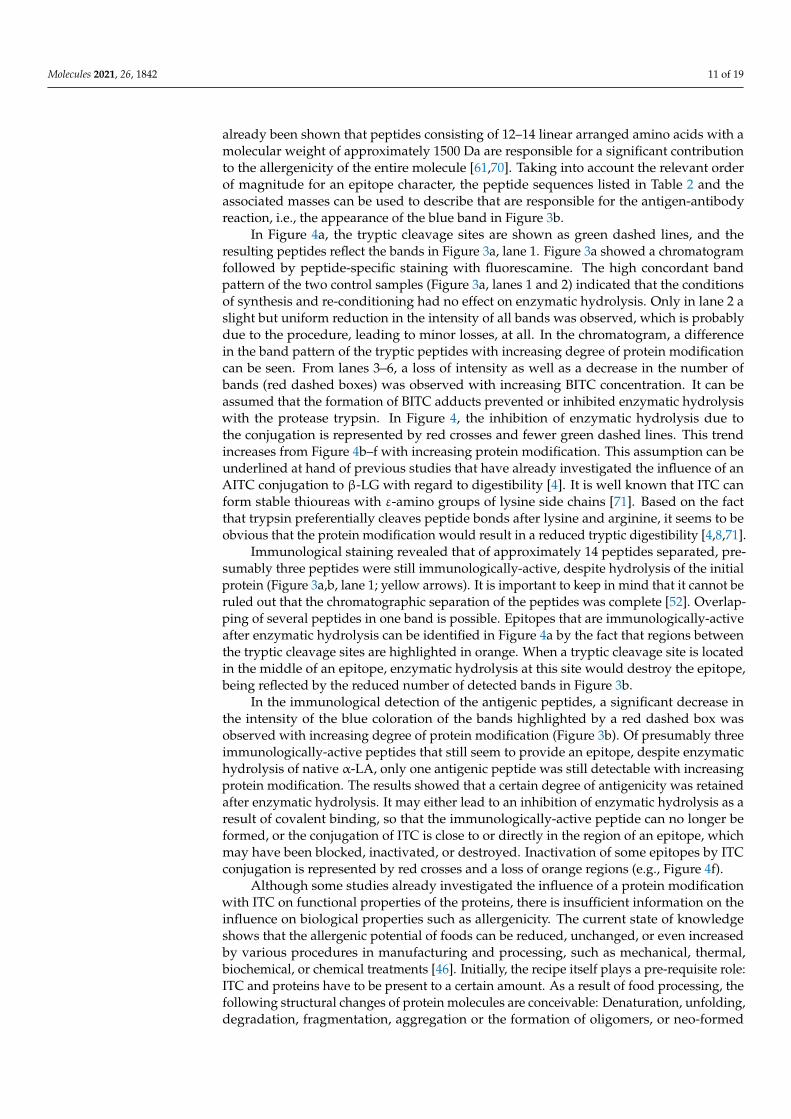
In the following, the influence of a conjugation between α-LA and BITC on the al-lergenic properties is presented. For this purpose, treated and untreated proteins weredetected after chromatographic separation using the specific antigen-antibody reaction.Figure 2 shows a “twin plate” (one plate cut into two similar plates after the chromato-graphic separation) stained with the protein-specific dye fluorescamine (Figure 2a), detect-ing all proteins present. Figure 2b shows the chromatogram obtained from the immunolog-ical staining protocol.
Molecules 2021, 26, x FOR PEER REVIEW 6 of 19
Figure 2. HPTLC analysis of BITC-α-LA derivatives as a function of the concentration of BITC. (a) Protein-specific staining with fluorescamine (UV-light; λ = 366 nm); (b) Immunostaining with antibodies (white light) for the exclusive detection of the antigen. (1) α-LA control (freshly prepared), (2) α-LA control (treated similar as BITC-protein derivatives), (3) BITC-α-LA derivative “low” (cBITC = 3.8 mM), (4) BITC-α-LA derivative “moderate” (cBITC = 38 mM), (5) BITC-α-LA derivative “high” (cBITC = 75 mM); (6) BITC-α-LA derivative “very high” (cBITC = 113 mM).
With increasing concentrations of BITC, a steady decrease in intensity of the bands of the control (I, II and III) was observed, until they could not be detected anymore (Figure 2a,b; lanes 4-6). Similarly, a continuous increase in the intensity of new bands (green ar-rows) was observed with increasing BITC concentration (Figure 2a,b; lanes 4–6). The new bands had higher Rf values and appeared more intense and partially broadened. The trend obvious from Figure 2a supported the assumption that the ITC-protein adducts formed depending on BITC concentration to a certain extent.
In both figures, the formation of smeary streaks was pronounced at higher BITC con-centrations (lanes 5 and 6, yellow arrows), which is probably due to an increase in the degree of modification. It is likely that there were numerous different protein modifica-tions formed that impaired the chromatographic separation. Moreover, after the protein-specific staining with fluorescamine, another band can be detected attributable to an ex-cess of residual BITC (red arrows). This identification was made by comparison with a standard BITC solution (comparison not shown here).
2.3. HPTLC-Immunostaining of Tryptic Peptides The present study showed that untreated and BITC-modified α-LA can be analyzed
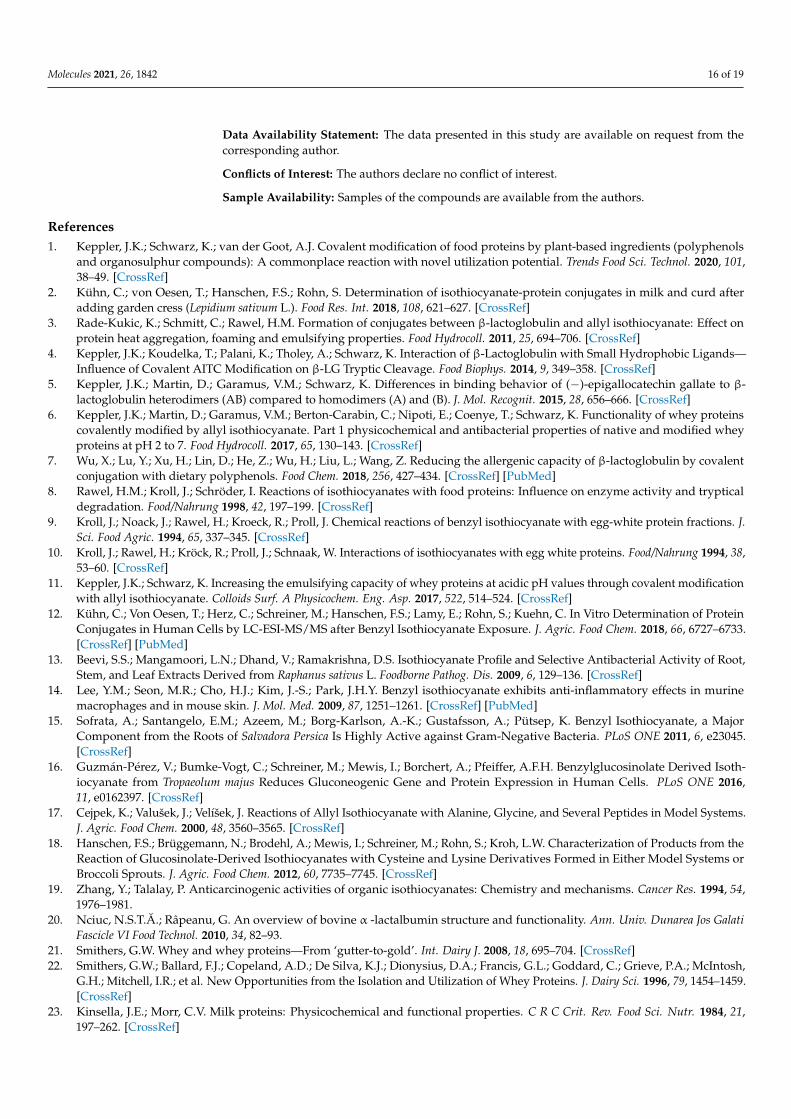
with regard to its antigenic properties using HPTLC-IS. In addition to the native protein structure, fragments/parts of the protein structure may also retain their antigenicity after proteolytic hydrolysis, so that antigenic analysis of corresponding peptides must not be neglected. Due to the low molecular weight of the peptides, their analysis by traditional methods such as Western blotting after gel electrophoresis is limited. Consequently, an immunological detection after HPTLC separation of the peptides resulting from the tryp-tic digestion of α-LA was also performed in the present work. For this purpose, α-LA was derivatized with BITC and subsequently hydrolyzed with the protease trypsin. Immuno-logical detection by HPTLC-IS enabled the identification of the presence of residual epitopes in the hydrolysate. Figure 3 shows the HPTLC chromatograms of tryptic pep-tides after peptide/protein staining with fluorescamine (Figure 3a) and after im-munostaining (Figure 3b). Antigenic and non-antigenic peptides can be differentiated.
Figure 2. HPTLC analysis of BITC-α-LA derivatives as a function of the concentration of BITC. (a) Protein-specific stainingwith fluorescamine (UV-light; λ = 366 nm); (b) Immunostaining with antibodies (white light) for the exclusive detection ofthe antigen. (1) α-LA control (freshly prepared), (2) α-LA control (treated similar as BITC-protein derivatives), (3) BITC-α-LAderivative “low” (cBITC = 3.8 mM), (4) BITC-α-LA derivative “moderate” (cBITC = 38 mM), (5) BITC-α-LA derivative “high”(cBITC = 75 mM); (6) BITC-α-LA derivative “very high” (cBITC = 113 mM).

Molecules 2021, 26, 1842 6 of 19
In Figure 2, an α-LA solution freshly prepared in water was applied at position 1of the plate. Lane 2 shows the α-LA control treated like the modifications, but withoutthe addition of BITC. This control was intended to represent the effect of synthesis andre-conditioning of the protein. Lanes 3-6 show the protein derivatives “low”, “moderate”,“high”, and “very high” produced with increasing amounts of BITC.
The control samples applied as lanes 1 and 2 showed three bands (I, II, and III) forboth staining procedures. When comparing the band sharpness between both detectionmethods, it was obvious that the band sharpness of HPTLC-IS slightly decreased. Whilethe bands of the BITC-α-LA derivatives “low” and “moderate” were clearly separatedfrom each other when stained with fluorescein (Figure 2a, lanes 3 and 4), overlappingand broadening of the bands already occurred in these samples during immunostaining(Figure 2b, lanes 3 and 4). This could be attributed to diffusion effects during the incubationin aqueous solutions [52].
When using a low concentration of BITC for producing the BITC-α-LA derivative“low”, both staining methods (Figure 2a,b; lane 3) showed bands that were assigned tonon-modified, residual protein (I, II, III) as well as new additional bands (green arrows)with higher Rf values. For this protein derivative, both detection methods still showed ahigh agreement with the band pattern of the control sample (Figure 2a,b; lane 2).
With increasing concentrations of BITC, a steady decrease in intensity of the bandsof the control (I, II and III) was observed, until they could not be detected anymore(Figure 2a,b; lanes 4–6). Similarly, a continuous increase in the intensity of new bands(green arrows) was observed with increasing BITC concentration (Figure 2a,b; lanes 4–6).The new bands had higher Rf values and appeared more intense and partially broadened.The trend obvious from Figure 2a supported the assumption that the ITC-protein adductsformed depending on BITC concentration to a certain extent.
In both figures, the formation of smeary streaks was pronounced at higher BITCconcentrations (lanes 5 and 6, yellow arrows), which is probably due to an increase in thedegree of modification. It is likely that there were numerous different protein modificationsformed that impaired the chromatographic separation. Moreover, after the protein-specificstaining with fluorescamine, another band can be detected attributable to an excess ofresidual BITC (red arrows). This identification was made by comparison with a standardBITC solution (comparison not shown here).
2.3. HPTLC-Immunostaining of Tryptic Peptides
The present study showed that untreated and BITC-modified α-LA can be analyzedwith regard to its antigenic properties using HPTLC-IS. In addition to the native proteinstructure, fragments/parts of the protein structure may also retain their antigenicity afterproteolytic hydrolysis, so that antigenic analysis of corresponding peptides must not beneglected. Due to the low molecular weight of the peptides, their analysis by traditionalmethods such as Western blotting after gel electrophoresis is limited. Consequently, animmunological detection after HPTLC separation of the peptides resulting from the tryp-tic digestion of α-LA was also performed in the present work. For this purpose, α-LAwas derivatized with BITC and subsequently hydrolyzed with the protease trypsin. Im-munological detection by HPTLC-IS enabled the identification of the presence of residualepitopes in the hydrolysate. Figure 3 shows the HPTLC chromatograms of tryptic peptidesafter peptide/protein staining with fluorescamine (Figure 3a) and after immunostaining(Figure 3b). Antigenic and non-antigenic peptides can be differentiated.

Molecules 2021, 26, 1842 7 of 19Molecules 2021, 26, x FOR PEER REVIEW 7 of 19
Figure 3. HPTLC analysis of tryptically digested α-LA derivatives as a function of BITC concentration. (a) Protein staining using fluorescamine as a derivation reagent (UV-light; λ = 366 nm), (b) Immunological staining (white light). (1) α-LA control (freshly prepared), (2) α-LA control (treated similar as BITC-protein derivatives), (3) BITC-α-LA derivative “low” (cBITC = 3.8 mM), (4) BITC-α-LA derivative “moderate” (cBITC = 38 mM), (5) BITC-α-LA derivative “high” (cBITC = 75 mM); (6) BITC-α-LA derivative “very high” (cBITC = 113 mM).
Figure 3a shows a high agreement in the band pattern of both control samples (lanes 1 and 2). The number of bands detected was consistent. Only a slight, but uniform reduc-tion in the intensity of all bands in lane 2 was observed. By comparing the band patterns of the control sample (lane 2) with those of the BITC-protein derivatives (lanes 3–6), a steady decrease in intensity was observed with increasing BITC concentration. Exempla-rily, some bands, whose intensity decreased with increasing BITC concentration have been highlighted by red dashed boxes. In the case of an excess of BITC, some bands showed such a strong loss of intensity that some of them could no longer be detected (lanes 5 and 6; red dashed boxes). In Figure 3b, peptides with residual epitopes appear as distinct blue bands on a lighter background (yellow arrows). Based on this chromatogram, conclusions can be drawn about the residual antigenicity of the peptides. After enzymatic hydrolysis of the native α-LA using trypsin as protease, three blue bands were detected in the immunological assay (Figure 3b, lane 1; yellow arrows). The immunological stain-ing showed a reduction in the number and a decrease in intensity of the blue band (red dashed box) with increasing modification with BITC (Figure 3b). Furthermore, a signifi-cant antigen-antibody reaction was observed for the initial application position of the sample (Figure 3b, lane 1, blue arrow). The intense blue coloration could be attributed to residual non-hydrolyzed and non-separated α-LA.
3. Discussion In the following, the previously described results from Figures 2 and 3 will be illus-
trated and explained at hand of Figure 4. This figure serves to facilitate understanding and schematically depicts the influence of ITC conjugation on the structure, on the epitopes, and on the potential tryptic cleavage sites of the protein, when being succes-sively modified with ITC, and depending on the degree of α-LA modification. It should be noted that only different model scenarios are illustrated, and that the discussion is based on a hypothetical approach for describing the impact of protein modifications on its antigenicity (creation and destruction of epitopes) in accordance with the current liter-ature. Protein structure, the locations of epitopes, disulfide bridges, and tryptic cleavage sites are not depicted realistically, but only schematically for illustrating the different out-comes from a certain degree of modification possible.
Figure 3. HPTLC analysis of tryptically digested α-LA derivatives as a function of BITC concentration. (a) Protein stainingusing fluorescamine as a derivation reagent (UV-light; λ = 366 nm), (b) Immunological staining (white light). (1) α-LAcontrol (freshly prepared), (2) α-LA control (treated similar as BITC-protein derivatives), (3) BITC-α-LA derivative “low”(cBITC = 3.8 mM), (4) BITC-α-LA derivative “moderate” (cBITC = 38 mM), (5) BITC-α-LA derivative “high” (cBITC = 75 mM);(6) BITC-α-LA derivative “very high” (cBITC = 113 mM).
Figure 3a shows a high agreement in the band pattern of both control samples (lanes 1and 2). The number of bands detected was consistent. Only a slight, but uniform reductionin the intensity of all bands in lane 2 was observed. By comparing the band patternsof the control sample (lane 2) with those of the BITC-protein derivatives (lanes 3–6), asteady decrease in intensity was observed with increasing BITC concentration. Exemplarily,some bands, whose intensity decreased with increasing BITC concentration have beenhighlighted by red dashed boxes. In the case of an excess of BITC, some bands showed sucha strong loss of intensity that some of them could no longer be detected (lanes 5 and 6; reddashed boxes). In Figure 3b, peptides with residual epitopes appear as distinct blue bandson a lighter background (yellow arrows). Based on this chromatogram, conclusions can bedrawn about the residual antigenicity of the peptides. After enzymatic hydrolysis of thenative α-LA using trypsin as protease, three blue bands were detected in the immunologicalassay (Figure 3b, lane 1; yellow arrows). The immunological staining showed a reductionin the number and a decrease in intensity of the blue band (red dashed box) with increasingmodification with BITC (Figure 3b). Furthermore, a significant antigen-antibody reactionwas observed for the initial application position of the sample (Figure 3b, lane 1, bluearrow). The intense blue coloration could be attributed to residual non-hydrolyzed andnon-separated α-LA.
3. Discussion
In the following, the previously described results from Figures 2 and 3 will be illus-trated and explained at hand of Figure 4. This figure serves to facilitate understanding andschematically depicts the influence of ITC conjugation on the structure, on the epitopes,and on the potential tryptic cleavage sites of the protein, when being successively modifiedwith ITC, and depending on the degree of α-LA modification. It should be noted that onlydifferent model scenarios are illustrated, and that the discussion is based on a hypotheticalapproach for describing the impact of protein modifications on its antigenicity (creationand destruction of epitopes) in accordance with the current literature. Protein structure,the locations of epitopes, disulfide bridges, and tryptic cleavage sites are not depicted

Molecules 2021, 26, 1842 8 of 19
realistically, but only schematically for illustrating the different outcomes from a certaindegree of modification possible.
Molecules 2021, 26, x FOR PEER REVIEW 8 of 19
Figure 4. Schematic illustration of the influence of a protein modification on protein structure, epitopes, and enzymatic hydrolysis. While potential reaction sites for the ITC are represented by gray dots, ITC conjugation is highlighted by the red crosses. Intact epitopes are shown as orange regions, tryptic cleavage sites by green dashed lines, and disulfide bridges by black dashed lines. (a) Native protein with the potential reaction sites for ITC and its intact epitopes; (b)–(f) increasing degree of protein modification and the resulting change in protein structure, epitope, and enzymatic hydrolysis. The pro-tein structure, locations of epitopes, disulfide bridges, and tryptic cleavage sites are not realistically depicted.
In Figure 4a, the native protein is shown with potential reaction sites suitable for a reaction with ITC (gray dots) and with intact epitopes (orange regions). Here, no distinc-tion is made between linear (sequential and continuous) or conformational (discontinu-ous) epitopes. The green dashed lines show the naturally occurring tryptic cleavage sites (trypsin cleaves exclusively C-terminal to arginine and lysine residues [58]), and the black dashed lines show the disulfide bridges in the protein [59]. In the native protein, the epitopes are intact and accessible to the antibody reaction [33,60–63], as reflected by the three blue bands in Figure 2b (lane 1, bands I, II, III). The high concordance of band pat-terns between control samples suggested that the synthesis and re-conditioning had no effect on the protein and its allergenicity (Figure 2a,b; lanes 1 and 2). The three bands detected in the native protein solution in lane 1 can be attributed either to the purity of the protein (≥85%) or to the three genetic variants of α-LA. It is noteworthy that the protein has two predominant variants A and B, and a third variant has been described, but was not yet approved [64].
The potential reaction sites, shown as gray dots in the figure, are replaced by red crosses in the case of a covalent bond between the nucleophilic groups of α-LA and the BITC. Previous studies confirmed the conjugation of ITC with the whey protein β-LG, determined by a decreased content of free amino and thiol groups in the protein, suggest-ing that ITC bind covalently to these protein side chains [3].
Figure 4. Schematic illustration of the influence of a protein modification on protein structure, epitopes, and enzymatichydrolysis. While potential reaction sites for the ITC are represented by gray dots, ITC conjugation is highlighted by the redcrosses. Intact epitopes are shown as orange regions, tryptic cleavage sites by green dashed lines, and disulfide bridgesby black dashed lines. (a) Native protein with the potential reaction sites for ITC and its intact epitopes; (b–g) increasingdegree of protein modification and the resulting change in protein structure, epitope, and enzymatic hydrolysis. The proteinstructure, locations of epitopes, disulfide bridges, and tryptic cleavage sites are not realistically depicted.
In Figure 4a, the native protein is shown with potential reaction sites suitable for areaction with ITC (gray dots) and with intact epitopes (orange regions). Here, no distinctionis made between linear (sequential and continuous) or conformational (discontinuous)epitopes. The green dashed lines show the naturally occurring tryptic cleavage sites(trypsin cleaves exclusively C-terminal to arginine and lysine residues [58]), and the blackdashed lines show the disulfide bridges in the protein [59]. In the native protein, theepitopes are intact and accessible to the antibody reaction [33,60–63], as reflected by thethree blue bands in Figure 2b (lane 1, bands I, II, III). The high concordance of band patternsbetween control samples suggested that the synthesis and re-conditioning had no effecton the protein and its allergenicity (Figure 2a,b; lanes 1 and 2). The three bands detectedin the native protein solution in lane 1 can be attributed either to the purity of the protein(≥85%) or to the three genetic variants of α-LA. It is noteworthy that the protein has twopredominant variants A and B, and a third variant has been described, but was not yetapproved [64].

Molecules 2021, 26, 1842 9 of 19
The potential reaction sites, shown as gray dots in the figure, are replaced by redcrosses in the case of a covalent bond between the nucleophilic groups of α-LA and theBITC. Previous studies confirmed the conjugation of ITC with the whey protein β-LG,determined by a decreased content of free amino and thiol groups in the protein, suggestingthat ITC bind covalently to these protein side chains [3].
Using a low concentration of BITC for the preparation of the protein conjugates, a highagreement in the band pattern compared to the control sample (Figure 2a,b, lanes 2 and3) can be seen in both detection techniques. It can be assumed that the use of a low BITCconcentration resulted in a small number of protein modifications having no significantinfluence on the protein and its allergenicity. Despite the protein modifications with BITC,the epitopes appear to remain immunologically-active (Figure 2b, lane 3). This situationcan be illustrated as in Figure 4b. It is likely that when using a low BITC concentration,only comparatively more easily accessible reaction sites in the outer part or the surfacesof the native protein structure were conjugated primarily, not noticeable influencing thewhole protein structure with its epitopes.
For both detection techniques, two differences in the band pattern are highlightedwith increasing concentration of BITC in Figure 2. On the one hand, with increasing BITCconcentration (lanes 4–6), a strong intensity decrease was noted (band I), while on the otherhand, a clear intensity increase of a new band (green arrows) could be seen. Immunologicaldetection showed certain differences with increasing concentration of BITC, indicatinga change of the allergenic properties induced by ITC conjugation (Figure 2b). The bluecoloration resulted from an antigen-antibody reaction, so that an increase in the colorintensity could be equated with an increase in the immunological reaction. Conversely, adecrease in intensity of a band indicated an inhibition of the antibody response.
It is conceivable that the amino acid residues that covalently interact with ITC aresometimes located in an epitope. When amino acids in an epitope are conjugated with ITCor an ITC-protein bond is directly in the region or close to a linear epitope, the epitope canbe blocked, inactivated, or destroyed (Figure 4c, loss of orange regions). The epitope isthen no longer accessible for an antibody reaction [7], as reflected by a decrease in intensityof the blue coloration of the bands in Figure 2b (band I).
In addition to the shielding/masking of the epitopes by the covalent binding of ITC,the irreversible structural change of the protein molecule induced by ITC conjugation couldcause the decrease in the intensity of blue staining, i.e., a decreased binding to antibodies.
A change in the structural properties of proteins as a result of the conjugation withITC has been already observed in previous studies. Thus, a loosening of protein foldingand a change in secondary and tertiary structure could be noted for an ITC conjugation toa whey protein isolate [6] and pure β-LG [3]. In addition, the results suggested that ITCcould also play a role in the cleavage of disulfide bridges in β-LG [2,4,65,66]. Similar effectsof ITC conjugation are conceivable for the protein α-LA.
As the conformation of the epitopes of α-LA is associated with its secondary or tertiarystructure, which play an extraordinary role in the protein antigenicity, it is conceivable thata structural change induced by covalent ITC conjugation could presumably change some ofthe epitope structures and thus, affecting the allergenicity of the protein [60,63,67,68]. Basedon these assumptions, a loosening of the protein molecule was mapped in Figure 4d (lossof orange regions), which may lead to the destruction or inactivation of conformational(discontinuous) epitopes under certain circumstances. A more significant change in theprotein structure is shown in Figure 4e. Here, the fact that the ITC could cleave the disulfidebridges is pictorially illustrated. The result is a significant unfolding of the protein structure,corresponding to a certain extent of secondary structure transformation, but least strongtransformation of the tertiary structure. These unfolding and denaturation processes mayalso inactivate and destroy conformational (discontinuous) and/or linear (sequential andcontinuous) epitopes (Figure 4d, further loss of orange regions), which could explain thereduction in intensity or disappearance of the bands in Figure 2b, lanes 4–6 (band I).

Molecules 2021, 26, 1842 10 of 19
In addition to the destruction or inactivation of epitopes, it can be seen that as aresult of the structural change, neo-formed epitopes occur or epitopes that were previouslyhidden inside (“buried”) the protein structure can become more accessible and exposedthrough unfolding and denaturation processes (Figure 4d,e, new orange regions), therebyincreasing the immunological response. This effect of ITC conjugation on the antigenicitycould explain the significant increase in intensity of the blue coloration of the band markedwith green arrows in Figure 2b, lanes 5 and 6.
A purely hypothetical approach is shown in Figure 4f. Here, it is assumed that theprotein is fully derivatized and completely fold over, after being exposed to an excessiveBITC-modification. Furthermore, it is not known, whether epitopes can be completelydestroyed or partially preserved in this state.
In summary, epitopes located on the surface of protein molecules can react with anti-bodies, while amino acids hidden inside cannot be recognized in the first moment. Externalinfluences and post-translational modifications such as the covalent binding of BITC as inthis case, can cause the protein to denature and unfold, and consequently affecting proteinstructure with its physicochemical and biological properties. Irreversible structural changesof proteins resulting from covalent modifications can lead to antigenic epitopes either be-ing blocked, destroyed, remodeled, or of even improved accessibility, thereby affectingthe allergenic properties. On the one hand, the formation of neo-formed epitopes or theexposure of epitopes, initially buried inside the protein can lead to the formation of new,potentially immunogenic sequences or conformations. On the other hand, epitopes may beblocked or inactivated by ITC conjugation or conformational (discontinuous) epitopes maybe destroyed by the structural change, resulting in a decreased antigen-antibody response.
In the following, the influence of ITC conjugation on enzymatic hydrolysis and on theantigenicity of tryptic peptides is discussed. Initially, the theoretically generated peptidesupon enzymatic hydrolysis of α-LA with trypsin can be calculated along with the massesand positions of the peptides in the protein, using the PeptideMass tool available atwww.expasy.org (accessed on 20 March 2021) (Table 2), The protein sequence of α-LA wastaken from the UniProtKB database (http://www.uniprot.org/ (accessed on 20 March2021); under the UniProt entry name LALBA_BOVIN, and file number P00711).
Table 2. Masses, positions in the protein and sequences of the calculated peptides after tryptic hydrolysis using thePeptideMass tool available at www.expasy.org (accessed on 20 March 2021).
Mass Position Peptide Sequence
618.35 20–24 EQLTK653.31 25–29 CEVFR389.24 30–32 ELK375.22 33–35 DLK
4654.15 36–77 GYGGVSLPEWVCTTFHTSGYDTQAIVQNNDSTEYGLFQINNK549.29 78–81 IWCK
1889.78 82–98 DDQNPHSSNICNISCDK1642.73 99–112 FLDDDLTDDIMCVK147.11 113–113 K488.31 114–117 ILDK
1220.65 118–127 VGINYWLAHK650.32 128–133 ALCSEK
1034.50 134–141 LDQWLCEK132.10 142–142 L
Comparison of protein-specific staining with fluorescamine and immunological stain-ing using antibodies allows differentiation and assigning of antigenic and non-antigenicpeptides. It should be emphasized that a specific antibody can bind to residual epitopesconsisting of only a few residues of the initial antigen. For this purpose, the sequentialepitope must consist of at least five linear arranged amino acids and can be up to a multiplenumber, when the linear arranged amino acids are coiled in the polypeptide [69]. It has
Molecules 2021, 26, 1842 11 of 19
already been shown that peptides consisting of 12–14 linear arranged amino acids with amolecular weight of approximately 1500 Da are responsible for a significant contributionto the allergenicity of the entire molecule [61,70]. Taking into account the relevant orderof magnitude for an epitope character, the peptide sequences listed in Table 2 and theassociated masses can be used to describe that are responsible for the antigen-antibodyreaction, i.e., the appearance of the blue band in Figure 3b.
In Figure 4a, the tryptic cleavage sites are shown as green dashed lines, and theresulting peptides reflect the bands in Figure 3a, lane 1. Figure 3a showed a chromatogramfollowed by peptide-specific staining with fluorescamine. The high concordant bandpattern of the two control samples (Figure 3a, lanes 1 and 2) indicated that the conditionsof synthesis and re-conditioning had no effect on enzymatic hydrolysis. Only in lane 2 aslight but uniform reduction in the intensity of all bands was observed, which is probablydue to the procedure, leading to minor losses, at all. In the chromatogram, a differencein the band pattern of the tryptic peptides with increasing degree of protein modificationcan be seen. From lanes 3–6, a loss of intensity as well as a decrease in the number ofbands (red dashed boxes) was observed with increasing BITC concentration. It can beassumed that the formation of BITC adducts prevented or inhibited enzymatic hydrolysiswith the protease trypsin. In Figure 4, the inhibition of enzymatic hydrolysis due tothe conjugation is represented by red crosses and fewer green dashed lines. This trendincreases from Figure 4b–f with increasing protein modification. This assumption can beunderlined at hand of previous studies that have already investigated the influence of anAITC conjugation to β-LG with regard to digestibility [4]. It is well known that ITC canform stable thioureas with ε-amino groups of lysine side chains [71]. Based on the factthat trypsin preferentially cleaves peptide bonds after lysine and arginine, it seems to beobvious that the protein modification would result in a reduced tryptic digestibility [4,8,71].
Immunological staining revealed that of approximately 14 peptides separated, pre-sumably three peptides were still immunologically-active, despite hydrolysis of the initialprotein (Figure 3a,b, lane 1; yellow arrows). It is important to keep in mind that it cannot beruled out that the chromatographic separation of the peptides was complete [52]. Overlap-ping of several peptides in one band is possible. Epitopes that are immunologically-activeafter enzymatic hydrolysis can be identified in Figure 4a by the fact that regions betweenthe tryptic cleavage sites are highlighted in orange. When a tryptic cleavage site is locatedin the middle of an epitope, enzymatic hydrolysis at this site would destroy the epitope,being reflected by the reduced number of detected bands in Figure 3b.
In the immunological detection of the antigenic peptides, a significant decrease inthe intensity of the blue coloration of the bands highlighted by a red dashed box wasobserved with increasing degree of protein modification (Figure 3b). Of presumably threeimmunologically-active peptides that still seem to provide an epitope, despite enzymatichydrolysis of native α-LA, only one antigenic peptide was still detectable with increasingprotein modification. The results showed that a certain degree of antigenicity was retainedafter enzymatic hydrolysis. It may either lead to an inhibition of enzymatic hydrolysis as aresult of covalent binding, so that the immunologically-active peptide can no longer beformed, or the conjugation of ITC is close to or directly in the region of an epitope, whichmay have been blocked, inactivated, or destroyed. Inactivation of some epitopes by ITCconjugation is represented by red crosses and a loss of orange regions (e.g., Figure 4f).
Although some studies already investigated the influence of a protein modificationwith ITC on functional properties of the proteins, there is insufficient information on theinfluence on biological properties such as allergenicity. The current state of knowledgeshows that the allergenic potential of foods can be reduced, unchanged, or even increasedby various procedures in manufacturing and processing, such as mechanical, thermal,biochemical, or chemical treatments [46]. Initially, the recipe itself plays a pre-requisite role:ITC and proteins have to be present to a certain amount. As a result of food processing, thefollowing structural changes of protein molecules are conceivable: Denaturation, unfolding,degradation, fragmentation, aggregation or the formation of oligomers, or neo-formed

Molecules 2021, 26, 1842 12 of 19
conformations. These changes in protein structure can also affect the epitope structuresand thus, the allergenicity of the proteins [72–74]. The molecular basis for the change inallergenic properties is the modification of epitopes. Inactivation or destruction of epitopes,formation of new epitopes, or improved accessibility of previously hidden epitopes mayoccur [45,46,75]. Previous studies reported that the effect of heat treatment on proteinsmay lead to protein denaturation or aggregation. Consequently, the process-induceddenaturation leads to the loss of the organized protein structure, which does not necessarilyresult in a reduced allergenicity. Rather, it is even conceivable that the formation ofaggregates increases the allergenic potential, as illustrated in Figure 4g [61]. It is wellknown that aggregated proteins are considered to have high immunogenicity, because theycan be readily taken up by antigen-presenting cells [76].
Furthermore, the influence of the food matrix during processing must be considered,because allergenic food proteins can interact chemically with other food components.Previous studies reported that new epitope structures can be formed, when proteins areheated in the presence of carbohydrates, polyphenols, or oxidized lipids [39,41,45,77].Stanic-Vucinic et al. (2013) reported that chemical modification of β-LG is associatedwith structural and functional modification of proteins, which in turn may increase theirallergenic potential by exposing hidden epitopes or creating new epitopes [78]. Whilethe reaction of β-LG with lactose in milk showed increased allergenic activity [79], theallergenicity could be reduced by the covalent modification of ovalbumin [80] and thewhey protein β-LG [7,81] with polyphenols in vitro.
In summary, chemical modifications of food proteins are often accompanied by proteinunfolding and aggregation. Therefore, a loss of some structural elements and a change inthe tertiary and secondary structure of the proteins can be expected [45].
In one study, the effect of heat exposure on the model whey protein α-LA was inves-tigated by NMR. It was shown that the protein was partially unfolded, and the surfacehydrophobicity of the protein changed. Residues previously buried in the hydrophobiccore of the native protein thus reached the protein surface. Such structural changes havethe potential to affect protein digestibility and allergenicity [45,82].
Apart from this, biochemical processes are known to potentially support the manufac-turing of hypoallergenic foods, as hydrolysis of proteins showed reduced allergenicity [46].
Depending on the enzyme applied and the degree of hydrolysis, controversial ob-servations are described in the literature. In case of incomplete hydrolysis of proteins,residual allergenicity could be attributed to the presence of native protein remaining orlarge fragments thereof. In the case of advanced hydrolysis, residual allergenic activitymay be induced by short peptide fragments. Thus, short peptide fragments can accountfor a considerable amount of the allergenic activity of the entire protein. In native proteins,these peptide fragments are located in the hydrophobic core of the protein and are onlyexposed after protein denaturation or enzymatic hydrolysis. Thus, the remaining proteincan still act as an allergen after hydrolysis [61].
4. Materials and Methods4.1. Materials
1,4-Dioxane, 3,3′,5,5′-tetramethylbenzidine, acetonitrile, ammonia, polyethylene-sorbitanmonolaurate (Tween20), sodium hydrogen carbonate, tris-HCl, tris(hydroxymethyl)-aminomethane, and dialysis membranes (regenerated cellulose, molecular weight cutoff <3.5 kDa) were obtained from Carl Roth GmbH & Co. KG, Karlsruhe, Germany. Sodiumchloride and tert-butanol were purchased from VWR International GmbH, Darmstadt, Ger-many. Bovine α-lactalbumin (α-LA) as model protein, benzyl isothiocyanate (98%), citricacid monohydrate, dioctyl sulfosuccinate sodium salt, dithiothreitol (DTT), fluorescamine,hydrogen peroxide, and trypsin from porcine pancreas were purchased from Sigma-AldrichGmbH, Steinheim, Germany. The polyclonal primary antibodies were raised in rabbitagainst bovine α-LA with a concentration of 1g/L and obtained from antibodies-onlineGmbH, Aachen, Germany. A polyclonal goat anti-rabbit antibody, conjugated to HRP, was

Molecules 2021, 26, 1842 13 of 19
used as secondary antibody (Dako GmbH, Glostrup, Denmark). The concentration of thepurified antibodies was 0.25 g/L. Ethanol, formic acid, hydrochloric acid, pyridine, urea,and HPTLC silica gel 60 were purchased from Merck KGaA, Darmstadt, Germany. Thestationary phase materials were glass-backed and had a size of 10 × 20 cm. C18 solid phaseextraction cartridges (1 mL, 100 mg) were purchased from Macherey-Nagel GmbH & Co.KG, Düren, Germany. All solvents were of HPLC grade, otherwise, ACS grade was used.Water was double distilled (ddH2O).
4.2. Methods4.2.1. Preparation of ITC Protein Conjugates
The protein solutions were prepared by dissolving α-LA in water (0.714 mM). Proteinmodification was conducted with four different BITC concentrations, ranging from 0 and113 mM. BITC dissolved in 1,4-dioxane was added to the protein solutions and stirredfor 20 h at 37 ◦C. After the incubation was finished, the reaction mixtures were dialyzedovernight and subsequently lyophilized for removing as much as residual BITC. For thelyophilization, all samples were frozen in liquid nitrogen to prevent cold denaturationand freeze-dried using a laboratory freeze dyer (Christ RVC 2−25 CDplus, Martin ChristGefriertrocknungsanlagen GmbH, Osterode am Harz, Germany). The freeze-dried sampleswere dissolved in ACN/water (60/40; v/v), stored at −20 ◦C until analysis. For one of thecontrol samples, the protein solution was treated in the same way as the modifications,except that BITC was not added.
4.2.2. Tryptic Digestion of α-Lactalbumin
For the tryptic digestion, the freeze-dried ITC-modified and non-modified proteinsamples were dissolved in 50 µL 6 M aqueous urea. Subsequently, 100 mM DTT (in100 mM sodium hydrogen carbonate) were added and incubated for 10 min at 60 ◦C tocleave any disulfide bridges. The samples were diluted with 425 µL 100 mM sodiumhydrogen carbonate (in water). Tryptic digestion was started by adding trypsin solution(1 mg/mL in 0.1 mM HCl) to the protein solution at a ratio of 1:100. The incubation wasperformed for 16 h at 37 ◦C. After the incubation period, the reaction was quenched byadding 0.2% formic acid. For purification of the digested samples, a solid phase extractionwas performed. Therefore, the chromatography material was conditioned with 60% ACNin water and equilibrated with 0.2% formic acid in water. After the application of thesamples, rinsing was performed with 0.2% formic acid in water, followed by elution with60% ACN. Finally, the samples were dried using gaseous nitrogen and re-dissolved in adefined volume of 0.2% formic acid for further analysis.
4.3. High-Performance Thin-Layer Chromatography-Immunostaining (HPTLC-IS)4.3.1. Protein Derivatives
Primarily, silica gel HPTLC plates were pre-washed with methanol and activated at100 ◦C for 10 min. Sample application was done using an HPTLC autosampler (ATS4,CAMAG AG, Muttenz, Switzerland). A total sample volume of 10 µL was applied as 6 mmbands. Each sample was applied onto the plate in duplicate so that it could be cut in halfafter chromatographic separation, resulting in two identical plates (“twin plates”). Thedevelopment of the HPTLC plates was performed in a saturated twin-trough chamberusing a solvent system consisting of 2-butanol/pyridine/ammonia/water (39/15/10/36;v/v/v/v) adapted from Biller et al. [48]. After the solvent front reached a height of 80 mm,the plate was removed, and residual solvents were evaporated overnight.
4.3.2. Tryptic Peptides
Analogously to the chromatographic analysis of the protein derivatives, the separationof the digested protein derivatives was performed. By using an HPTLC autosampler,samples were applied onto the plates and developed in a twin-trough chamber filled

Molecules 2021, 26, 1842 14 of 19
with the mobile phase consisting of 2-butanol/pyridine/ammonia/water (39/34/10/26;v/v/v/v) [52]. Finally, residual solvents were evaporated from the plates.
4.3.3. Protein-/Peptide-Specific Staining
After the residual solvents were evaporated overnight, proteins and peptides werestained with fluorescamine. Visualization of the fluorescent proteins/peptides was per-formed using a photodocumentation system (TLC-Visualizer, CAMAG AG, Muttenz,Switzerland) under ultraviolet light (UV 366 nm). The proteins/peptides appeared asfluorescent bands on a dark background. These plates were used as twin plates to comparewith the immunostained plates.
4.3.4. High-Performance Thin-Layer Chromatography-Immunostaining (HPTLC-IS)
Specific detection of proteins and peptides were performed using antibodies accordingto the procedure described by Morschheuser et al. [52]. Following the evaporation of themobile phase overnight, one half of the HPTLC plate was transferred to a small vessel.
First, the HPTLC plate was incubated two times for 15 min with a buffer containingTween20 as a blocking reagent. This prevents the primary antibody from binding non-specifically to the plate surface. For preparing the blocking solution, 0.9% sodium chloride,0.6% tris(hydroxymethyl)aminomethane, and 0.5% Tween20 was used. Subsequently, theblocking buffer was removed and replaced by the primary antibody solution (1:20,000 forintact proteins and 1:12,000 for peptides, both in blocking buffer). The incubation withprimary antibodies lasted for 2 h and was followed by three washing steps with the blockingsolution. Coating with the secondary antibody was performed for 1 h (diluted 1:2000 inblocking solution). The secondary antibody exhibited specificity against the primaryantibody and was HRP-conjugated. After the plates were washed three times with theblocking buffer, pH was lowered by incubation for 1 min with an acidic solution containing0.06% Tris-HCl (pH 6.0). The staining solution consists of the following substances: 0.06%3,3′,5,5′-tetramethylbenzidine, 0.2% dioctyl sulfosuccinate sodium salt, 0.7% citric acidmonohydrate, 1.8% sodium hydrogen phosphate dihydrate, 25% ethanol. Just beforestaining, 0.15% hydrogen peroxide was added. The TLC plate was incubated in thestaining solution for 10 min until blue bands appeared. Upon oxidation, TMB formsa water-soluble blue reaction product that can be measured spectrophotometrically at650 nm. All incubations were executed at room temperature and on an orbital shakerat 70 rpm. Finally, the analytes were detected at white light and documented using aphotodocumentation system (TLC visualizer, CAMAG AG, Muttenz, Switzerland).
4.4. Statistical Analysis
Validation of the HPTLC-IS methodology was performed with regard to followingparameters: linearity, precision, accuracy, limit of detection (LOD), and limit of quantifi-cation (LOQ). For this purpose, native α-LA was used as the standard compound andtreated like the modification except for the addition of BITC for excluding any effect oftreatment and reconditioning. Subsequently, a calibration series was prepared from thetreated non-modified α-LA stock solution. The standard solutions were separated on silicagel and then detected by immunological reaction with antibodies.
Following the documentation of the plates, densitometric evaluation was performedat 650 nm using a TLC Scanner 4 (CAMAG AG, Muttenz, Switzerland). Densitogramsrecorded with the TLC scanner can be used for quantitative analysis. Linearity wasvalidated using a 15-point calibration curve (n = 3) and was verified by means of Mandel’sfitting test. Homogeneity of variance was assigned regarding all values obtained [83,84].
Limit of detection (LOD) and limit of quantification (LOQ) were determined usingthe calibration method DIN 32645:2008-11 (n = 3) via the calibration curve (concentrationsbetween 0.2–3 µg). Accuracy and precision were determined by analyzing the close-ness of individual densitometric measurements of the band (same concentration of α-LA,c = 1.8 µg, n = 12).

Molecules 2021, 26, 1842 15 of 19
5. Conclusions
In summary, immunological staining of native untreated and BITC-modified α-LAcould be successfully performed directly on the plate after their separation using HPTLC.As the HPTLC-IS procedure does not destroy the tertiary and secondary structure, it waspossible to estimate the antigenic properties of the modified and non-modified proteins.In the present work, even highly degraded samples such as denatured or proteolyticallydigested proteins could be detected. It is obvious that due to the chemical modificationof α-LA with BITC, structural changes of the protein molecule potentially influence theallergenic properties. Consequently, the chemical modifications could have destroyed orinactivated epitopes, but at the same time buried or newly formed epitopes can be exposed,so that the net effect was an increase in allergenicity. It can be concluded that despiteprotein modification, (some of the) epitopes remained immunologically-active, at least.
Regarding the enzymatic hydrolysis with trypsin of the BITC-modified proteins, it canbe concluded that the conjugation of BITC with free amino groups of lysine protein sidechains probably sterically hindered the cleavage sites for the enzyme and consequently ledto a reduced digestibility and altered peptide patterns. While for the native and untreatedα-LA three of the 14 separated peptides could be detected immunologically, the BITC-treated samples showed only one antigenic peptide. However, it is possible that not everyband can be assigned to a single peptide, but several peptides may overlap in one band.
For this purpose, coupling with mass spectrometry of HPTLC-IS could provide excel-lent features for the advanced studies of antigens. It has to be considered that the blockingreagent Tween20 can interfere with the signals in mass spectrometry. Therefore, the im-munostaining procedure has to be optimized with regard to blocking agents. Other majoradvantages of HPTLC-IS are its ease of use, low cost compared to alternative methods suchas gel electrophoretic separation on membranes and staining afterwards, and the reduceduse of toxic substances such as acrylamide in SDS-PAGE. In addition, antibody-baseddetection is characterized by high affinity and specificity to target proteins. Due to the highvariability of the HPTLC analysis, e.g., with regard to the composition of the mobile phases,the conditions of the HPLC-IS can be individually adapted to the analytical problem athand. Furthermore, the methodology profits from a two-dimensional development, whichcan increase the chromatographic resolution. Here, the sample of interest is developedin two directions with different composition of the mobile phase. Initial experiments oftwo-dimensional HPTLC-IS have already been performed, which seem promising andshould therefore be further optimized in future research.
It is of particular importance to protect sensitized individuals from an allergenicreaction. For this purpose, a method that ensures the identification of allergenic proteins ismandatory. As only an estimation of the antigenicity of the native and the processed proteinis obtained with the method described here, future studies need to evaluate and comparethe immunogenicity and allergenicity with other analytical methods such as enzyme-linkedimmunosorbent assays, radio-immunoprecipitate assays, electrochemiluminescence, bead-based assays, surface plasmon resonance or bio-layer interferometry. In addition, specificassays for the determination of protein antigenicity are available [85,86]. In addition, thereis no clear tendency to what extent the various food processing influences the allergenicproperties [45], so furthermore exhaustive studies are necessary. For example, processingtechniques may reduce or increase the bovine allergen in cow’s milk or leave it unchangedat all [46]. The evaluation of such non-directed posttranslational modifications that canoccur frequently in foods remains challenging. In addition to in vitro studies, it is alsonecessary to verify, whether the findings obtained can be observed in vivo.
Author Contributions: Conceptualization, J.S. and S.R.; validation, J.S., S.B. and J.B.; formal analysis,J.S., S.B. and J.B.; investigation, J.S.; resources, S.R.; writing—original draft preparation, J.S.; writing—review and editing, J.S. and S.R.; visualization, J.S.; supervision, S.R. All authors have read andagreed to the published version of the manuscript.
Funding: This research received no external funding.
Molecules 2021, 26, 1842 16 of 19
Data Availability Statement: The data presented in this study are available on request from thecorresponding author.
Conflicts of Interest: The authors declare no conflict of interest.
Sample Availability: Samples of the compounds are available from the authors.
References1. Keppler, J.K.; Schwarz, K.; van der Goot, A.J. Covalent modification of food proteins by plant-based ingredients (polyphenols
and organosulphur compounds): A commonplace reaction with novel utilization potential. Trends Food Sci. Technol. 2020, 101,38–49. [CrossRef]
2. Kühn, C.; von Oesen, T.; Hanschen, F.S.; Rohn, S. Determination of isothiocyanate-protein conjugates in milk and curd afteradding garden cress (Lepidium sativum L.). Food Res. Int. 2018, 108, 621–627. [CrossRef]
3. Rade-Kukic, K.; Schmitt, C.; Rawel, H.M. Formation of conjugates between β-lactoglobulin and allyl isothiocyanate: Effect onprotein heat aggregation, foaming and emulsifying properties. Food Hydrocoll. 2011, 25, 694–706. [CrossRef]
4. Keppler, J.K.; Koudelka, T.; Palani, K.; Tholey, A.; Schwarz, K. Interaction of β-Lactoglobulin with Small Hydrophobic Ligands—Influence of Covalent AITC Modification on β-LG Tryptic Cleavage. Food Biophys. 2014, 9, 349–358. [CrossRef]
5. Keppler, J.K.; Martin, D.; Garamus, V.M.; Schwarz, K. Differences in binding behavior of (−)-epigallocatechin gallate to β-lactoglobulin heterodimers (AB) compared to homodimers (A) and (B). J. Mol. Recognit. 2015, 28, 656–666. [CrossRef]
6. Keppler, J.K.; Martin, D.; Garamus, V.M.; Berton-Carabin, C.; Nipoti, E.; Coenye, T.; Schwarz, K. Functionality of whey proteinscovalently modified by allyl isothiocyanate. Part 1 physicochemical and antibacterial properties of native and modified wheyproteins at pH 2 to 7. Food Hydrocoll. 2017, 65, 130–143. [CrossRef]
7. Wu, X.; Lu, Y.; Xu, H.; Lin, D.; He, Z.; Wu, H.; Liu, L.; Wang, Z. Reducing the allergenic capacity of β-lactoglobulin by covalentconjugation with dietary polyphenols. Food Chem. 2018, 256, 427–434. [CrossRef] [PubMed]
8. Rawel, H.M.; Kroll, J.; Schröder, I. Reactions of isothiocyanates with food proteins: Influence on enzyme activity and trypticaldegradation. Food/Nahrung 1998, 42, 197–199. [CrossRef]
9. Kroll, J.; Noack, J.; Rawel, H.; Kroeck, R.; Proll, J. Chemical reactions of benzyl isothiocyanate with egg-white protein fractions. J.Sci. Food Agric. 1994, 65, 337–345. [CrossRef]
10. Kroll, J.; Rawel, H.; Kröck, R.; Proll, J.; Schnaak, W. Interactions of isothiocyanates with egg white proteins. Food/Nahrung 1994, 38,53–60. [CrossRef]
11. Keppler, J.K.; Schwarz, K. Increasing the emulsifying capacity of whey proteins at acidic pH values through covalent modificationwith allyl isothiocyanate. Colloids Surf. A Physicochem. Eng. Asp. 2017, 522, 514–524. [CrossRef]
12. Kühn, C.; Von Oesen, T.; Herz, C.; Schreiner, M.; Hanschen, F.S.; Lamy, E.; Rohn, S.; Kuehn, C. In Vitro Determination of ProteinConjugates in Human Cells by LC-ESI-MS/MS after Benzyl Isothiocyanate Exposure. J. Agric. Food Chem. 2018, 66, 6727–6733.[CrossRef] [PubMed]
13. Beevi, S.S.; Mangamoori, L.N.; Dhand, V.; Ramakrishna, D.S. Isothiocyanate Profile and Selective Antibacterial Activity of Root,Stem, and Leaf Extracts Derived from Raphanus sativus L. Foodborne Pathog. Dis. 2009, 6, 129–136. [CrossRef]
14. Lee, Y.M.; Seon, M.R.; Cho, H.J.; Kim, J.-S.; Park, J.H.Y. Benzyl isothiocyanate exhibits anti-inflammatory effects in murinemacrophages and in mouse skin. J. Mol. Med. 2009, 87, 1251–1261. [CrossRef] [PubMed]
15. Sofrata, A.; Santangelo, E.M.; Azeem, M.; Borg-Karlson, A.-K.; Gustafsson, A.; Pütsep, K. Benzyl Isothiocyanate, a MajorComponent from the Roots of Salvadora Persica Is Highly Active against Gram-Negative Bacteria. PLoS ONE 2011, 6, e23045.[CrossRef]
16. Guzmán-Pérez, V.; Bumke-Vogt, C.; Schreiner, M.; Mewis, I.; Borchert, A.; Pfeiffer, A.F.H. Benzylglucosinolate Derived Isoth-iocyanate from Tropaeolum majus Reduces Gluconeogenic Gene and Protein Expression in Human Cells. PLoS ONE 2016,11, e0162397. [CrossRef]
17. Cejpek, K.; Valušek, J.; Velíšek, J. Reactions of Allyl Isothiocyanate with Alanine, Glycine, and Several Peptides in Model Systems.J. Agric. Food Chem. 2000, 48, 3560–3565. [CrossRef]
18. Hanschen, F.S.; Brüggemann, N.; Brodehl, A.; Mewis, I.; Schreiner, M.; Rohn, S.; Kroh, L.W. Characterization of Products from theReaction of Glucosinolate-Derived Isothiocyanates with Cysteine and Lysine Derivatives Formed in Either Model Systems orBroccoli Sprouts. J. Agric. Food Chem. 2012, 60, 7735–7745. [CrossRef]
19. Zhang, Y.; Talalay, P. Anticarcinogenic activities of organic isothiocyanates: Chemistry and mechanisms. Cancer Res. 1994, 54,1976–1981.
20. Nciuc, N.S.T.Ă.; Râpeanu, G. An overview of bovine α -lactalbumin structure and functionality. Ann. Univ. Dunarea Jos GalatiFascicle VI Food Technol. 2010, 34, 82–93.
21. Smithers, G.W. Whey and whey proteins—From ‘gutter-to-gold’. Int. Dairy J. 2008, 18, 695–704. [CrossRef]22. Smithers, G.W.; Ballard, F.J.; Copeland, A.D.; De Silva, K.J.; Dionysius, D.A.; Francis, G.L.; Goddard, C.; Grieve, P.A.; McIntosh,
G.H.; Mitchell, I.R.; et al. New Opportunities from the Isolation and Utilization of Whey Proteins. J. Dairy Sci. 1996, 79, 1454–1459.[CrossRef]
23. Kinsella, J.E.; Morr, C.V. Milk proteins: Physicochemical and functional properties. C R C Crit. Rev. Food Sci. Nutr. 1984, 21,197–262. [CrossRef]

Molecules 2021, 26, 1842 17 of 19
24. Permyakov, E.A.; Berliner, L.J. α-Lactalbumin: Structure and function. FEBS Lett. 2000, 473, 269–274. [CrossRef]25. Fitzgerald, R.J.; Murray, B.A.; Walsh, D.J. Hypotensive Peptides from Milk Proteins. J. Nutr. 2004, 134, 980S–988S. [CrossRef]26. Markus, C.R.; Olivier, B.; Panhuysen, G.E.M.; Van Der Gugten, J.; Alles, M.S.; Tuiten, A.; Westenberg, H.G.M.; Fekkes, D.;
Koppeschaar, H.F.; De Haan, E.E.H.F. The bovine protein α-lactalbumin increases the plasma ratio of tryptophan to the otherlarge neutral amino acids, and in vulnerable subjects raises brain serotonin activity, reduces cortisol concentration, and improvesmood under stress. Am. J. Clin. Nutr. 2000, 71, 1536–1544. [CrossRef]
27. Sternhagen, L.G.; Allen, J.C. Growth rates of a human colon adenocarinoma cell line are regulated by the milk proteins alpha-lactalbumin. Adv. Exp. Med. Biol. 2001, 501, 115–120.
28. Pellegrini, A.; Thomas, U.; Bramaz, N.; Hunziker, P.; Von Fellenberg, R. Isolation and identification of three bactericidal domainsin the bovine α–lactalbumin molecule. Biochim. Biophys. Acta Gen. Subj. 1999, 1426, 439–448. [CrossRef]
29. Cross, M.L.; Gill, H.S. Immunomodulatory Properties of Milk. Br. J. Nutr. 2000, 84, 81–89. [CrossRef]30. Villa, C.; Costa, J.; Oliveira, M.B.P.P.; Mafra, I. Bovine Milk Allergens: A Comprehensive Review. Compr. Rev. Food Sci. Food Saf.
2017, 17, 137–164. [CrossRef]31. Paschke, A. Proteine als Lebensmittelallergene. Nachr. Chem. 2008, 56, 1005–1009. [CrossRef]32. Ilinskaya, A.N.; Dobrovolskaia, M.A. Understanding the immunogenicity and antigenicity of nanomaterials: Past, present and
future. Toxicol. Appl. Pharmacol. 2016, 299, 70–77. [CrossRef] [PubMed]33. Li, X.; Yuan, S.; Huang, M.; Gao, J.; Wu, Z.; Tong, P.; Yang, A.; Chen, H. Identification of IgE and IgG epitopes on native Bos d 4
allergen specific to allergic children. Food Funct. 2016, 7, 2996–3005. [CrossRef] [PubMed]34. Gershoni, J.M.; Roitburd-Berman, A.; Siman-Tov, D.D.; Freund, N.T.; Weiss, Y. Epitope Mapping. BioDrugs 2007, 21, 145–156.
[CrossRef] [PubMed]35. Willison, L.A.N.; Zhang, Q.; Su, M.; Teuber, S.S.; Sathe, S.K.; Roux, K.H. Conformational epitope mapping of Pru du 6, a major
allergen from almond nut. Mol. Immunol. 2013, 55, 253–263. [CrossRef] [PubMed]36. Pomés, A. Relevant B Cell Epitopes in Allergic Disease. Int. Arch. Allergy Immunol. 2010, 152, 1–11. [CrossRef]37. Sampson, H.A. Update on food allergy. J. Allergy Clin. Immunol. 2004, 113, 805–819. [CrossRef]38. Matissek, R.; Steiner, G.; Fischer, M. Lebensmittelanalytik; Bd. fünfte, vo; Springer: Berlin/Heidelberg, Germany, 2014;
ISBN 978-3-642-34828-0.39. Davis, P.J.; Smales, C.M.; James, D.C. How can thermal processing modify the antigenicity of proteins? Allergy 2001, 56, 56–60.
[CrossRef]40. Li, Z.; Luo, Y.; Feng, L. Effects of Maillard reaction conditions on the antigenicity of α-lactalbumin and β-lactoglobulin in whey
protein conjugated with maltose. Eur. Food Res. Technol. 2011, 233, 387–394. [CrossRef]41. Jiménez-Saiz, R.; Belloque, J.; Molina, E.; López-Fandiño, R. Human Immunoglobulin E (IgE) Binding to Heated and Glycated
Ovalbumin and Ovomucoid before and after in Vitro Digestion. J. Agric. Food Chem. 2011, 59, 10044–10051. [CrossRef]42. Bu, G.; Luo, Y.; Chen, F.; Liu, K.; Zhu, T. Milk processing as a tool to reduce cow’s milk allergenicity: A mini-review. Dairy Sci.
Technol. 2013, 93, 211–223. [CrossRef]43. Verhoeckx, K.C.M.; Vissers, Y.M.; Baumert, J.L.; Faludi, R.; Feys, M.; Flanagan, S.; Herouet-Guicheney, C.; Holzhauser, T.; Shimojo,
R.; van der Bolt, N.; et al. Food processing and allergenicity. Food Chem. Toxicol. 2015, 80, 223–240. [CrossRef]44. Teodorowicz, M.; Van Neerven, J.; Savelkoul, H. Food Processing: The Influence of the Maillard Reaction on Immunogenicity and
Allergenicity of Food Proteins. Nutrients 2017, 9, 835. [CrossRef]45. Mills, E.N.C.; Sancho, A.I.; Rigby, N.M.; Jenkins, J.A.; Mackie, A.R. Impact of food processing on the structural and allergenic
properties of food allergens. Mol. Nutr. Food Res. 2009, 53, 963–969. [CrossRef] [PubMed]46. Paschke, A.; Besler, M. Stability of bovine allergens during food processing. Ann. Allergy Asthma Immunol. 2002, 89, 16–20.
[CrossRef]47. Fiocchi, A.; Bouygue, G.R.; Sarratud, T.; Terracciano, L.; Martelli, A.; Restani, P. Clinical tolerance of processed foods. Ann. Allergy
Asthma Immunol. 2004, 93, S38–S46. [CrossRef]48. Biller, J.; Morschheuser, L.; Riedner, M.; Rohn, S. Development of optimized mobile phases for protein separation by high
performance thin layer chromatography. J. Chromatogr. A 2015, 1415, 146–154. [CrossRef]49. Morlock, G.; Schwack, W. Hyphenations in planar chromatography. J. Chromatogr. A 2010, 1217, 6600–6609. [CrossRef]50. Morlock, G.; Schwack, W. Coupling of planar chromatography to mass spectrometry. TrAC Trends Anal. Chem. 2010, 29, 1157–1171.
[CrossRef]51. Morlock, G.E.; Klingelhöfer, I. Liquid Chromatography-Bioassay-Mass Spectrometry for Profiling of Physiologically Active Food.
Anal. Chem. 2014, 86, 8289–8295. [CrossRef] [PubMed]52. Morschheuser, L.; Mink, K.; Horst, R.; Kallinich, C.; Rohn, S. Immunological analysis of food proteins using high-performance
thin-layer chromatography-immunostaining. J. Chromatogr. A 2017, 1526, 157–166. [CrossRef]53. Meisen, I.; Mormann, M.; Müthing, J. Thin-layer chromatography, overlay technique and mass spectrometry: A versatile triad
advancing glycosphingolipidomics. Biochim. Biophys. Acta Mol. Cell Biol. Lipids 2011, 1811, 875–896. [CrossRef]54. Morschheuser, L.; Mükusch, S.; Riedner, M.; Seitz, H.; Rohn, S. High-performance thin-layer chromatography as a fast screening
tool for phosphorylated peptides. J. Chromatogr. B Anal. Technol. Biomed. Life Sci. 2016, 1008, 198–205. [CrossRef]55. Widmer, V.; Handloser, D.; Reich, E. Quantitative HPTLC Analysis of Artemisinin in Dried Artemisia annua L.: A Practical
Approach. J. Liq. Chromatogr. Relat. Technol. 2007, 30, 2209–2219. [CrossRef]
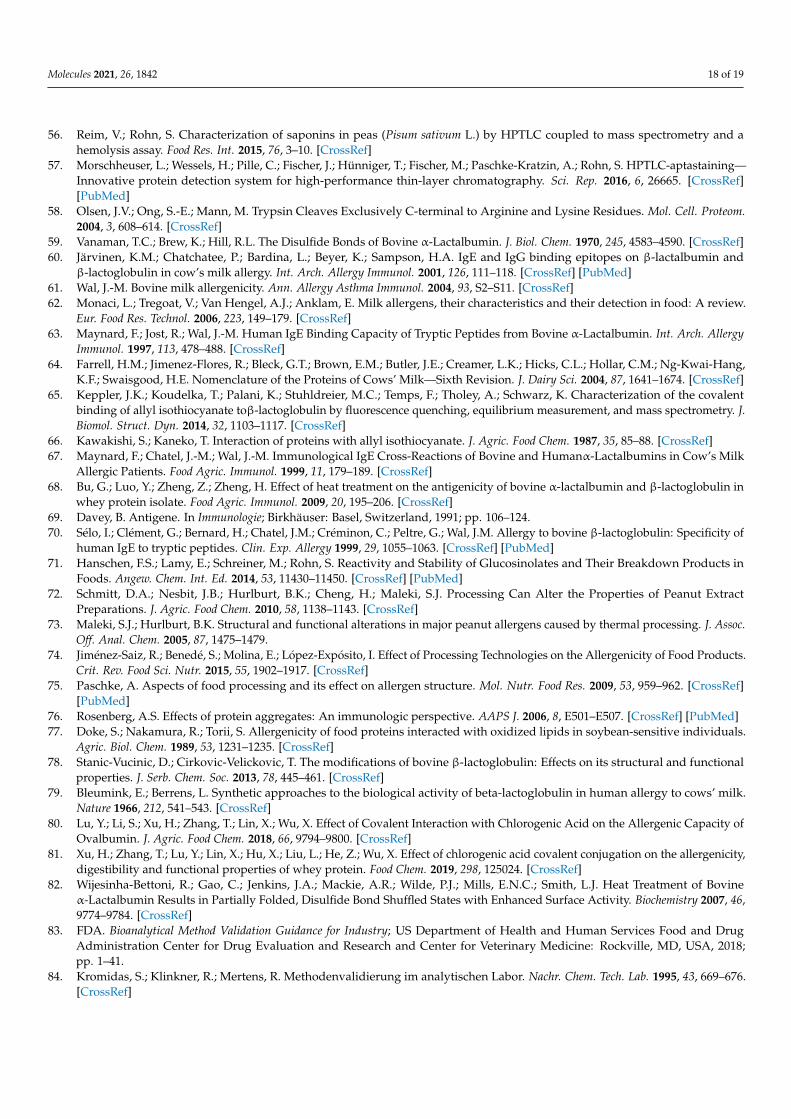
Molecules 2021, 26, 1842 18 of 19
56. Reim, V.; Rohn, S. Characterization of saponins in peas (Pisum sativum L.) by HPTLC coupled to mass spectrometry and ahemolysis assay. Food Res. Int. 2015, 76, 3–10. [CrossRef]
57. Morschheuser, L.; Wessels, H.; Pille, C.; Fischer, J.; Hünniger, T.; Fischer, M.; Paschke-Kratzin, A.; Rohn, S. HPTLC-aptastaining—Innovative protein detection system for high-performance thin-layer chromatography. Sci. Rep. 2016, 6, 26665. [CrossRef][PubMed]
58. Olsen, J.V.; Ong, S.-E.; Mann, M. Trypsin Cleaves Exclusively C-terminal to Arginine and Lysine Residues. Mol. Cell. Proteom.2004, 3, 608–614. [CrossRef]
59. Vanaman, T.C.; Brew, K.; Hill, R.L. The Disulfide Bonds of Bovine α-Lactalbumin. J. Biol. Chem. 1970, 245, 4583–4590. [CrossRef]60. Järvinen, K.M.; Chatchatee, P.; Bardina, L.; Beyer, K.; Sampson, H.A. IgE and IgG binding epitopes on β-lactalbumin and
β-lactoglobulin in cow’s milk allergy. Int. Arch. Allergy Immunol. 2001, 126, 111–118. [CrossRef] [PubMed]61. Wal, J.-M. Bovine milk allergenicity. Ann. Allergy Asthma Immunol. 2004, 93, S2–S11. [CrossRef]62. Monaci, L.; Tregoat, V.; Van Hengel, A.J.; Anklam, E. Milk allergens, their characteristics and their detection in food: A review.
Eur. Food Res. Technol. 2006, 223, 149–179. [CrossRef]63. Maynard, F.; Jost, R.; Wal, J.-M. Human IgE Binding Capacity of Tryptic Peptides from Bovine α-Lactalbumin. Int. Arch. Allergy
Immunol. 1997, 113, 478–488. [CrossRef]64. Farrell, H.M.; Jimenez-Flores, R.; Bleck, G.T.; Brown, E.M.; Butler, J.E.; Creamer, L.K.; Hicks, C.L.; Hollar, C.M.; Ng-Kwai-Hang,
K.F.; Swaisgood, H.E. Nomenclature of the Proteins of Cows’ Milk—Sixth Revision. J. Dairy Sci. 2004, 87, 1641–1674. [CrossRef]65. Keppler, J.K.; Koudelka, T.; Palani, K.; Stuhldreier, M.C.; Temps, F.; Tholey, A.; Schwarz, K. Characterization of the covalent
binding of allyl isothiocyanate toβ-lactoglobulin by fluorescence quenching, equilibrium measurement, and mass spectrometry. J.Biomol. Struct. Dyn. 2014, 32, 1103–1117. [CrossRef]
66. Kawakishi, S.; Kaneko, T. Interaction of proteins with allyl isothiocyanate. J. Agric. Food Chem. 1987, 35, 85–88. [CrossRef]67. Maynard, F.; Chatel, J.-M.; Wal, J.-M. Immunological IgE Cross-Reactions of Bovine and Humanα-Lactalbumins in Cow’s Milk
Allergic Patients. Food Agric. Immunol. 1999, 11, 179–189. [CrossRef]68. Bu, G.; Luo, Y.; Zheng, Z.; Zheng, H. Effect of heat treatment on the antigenicity of bovine α-lactalbumin and β-lactoglobulin in
whey protein isolate. Food Agric. Immunol. 2009, 20, 195–206. [CrossRef]69. Davey, B. Antigene. In Immunologie; Birkhäuser: Basel, Switzerland, 1991; pp. 106–124.70. Sélo, I.; Clément, G.; Bernard, H.; Chatel, J.M.; Créminon, C.; Peltre, G.; Wal, J.M. Allergy to bovine β-lactoglobulin: Specificity of
human IgE to tryptic peptides. Clin. Exp. Allergy 1999, 29, 1055–1063. [CrossRef] [PubMed]71. Hanschen, F.S.; Lamy, E.; Schreiner, M.; Rohn, S. Reactivity and Stability of Glucosinolates and Their Breakdown Products in
Foods. Angew. Chem. Int. Ed. 2014, 53, 11430–11450. [CrossRef] [PubMed]72. Schmitt, D.A.; Nesbit, J.B.; Hurlburt, B.K.; Cheng, H.; Maleki, S.J. Processing Can Alter the Properties of Peanut Extract
Preparations. J. Agric. Food Chem. 2010, 58, 1138–1143. [CrossRef]73. Maleki, S.J.; Hurlburt, B.K. Structural and functional alterations in major peanut allergens caused by thermal processing. J. Assoc.
Off. Anal. Chem. 2005, 87, 1475–1479.74. Jiménez-Saiz, R.; Benedé, S.; Molina, E.; López-Expósito, I. Effect of Processing Technologies on the Allergenicity of Food Products.
Crit. Rev. Food Sci. Nutr. 2015, 55, 1902–1917. [CrossRef]75. Paschke, A. Aspects of food processing and its effect on allergen structure. Mol. Nutr. Food Res. 2009, 53, 959–962. [CrossRef]
[PubMed]76. Rosenberg, A.S. Effects of protein aggregates: An immunologic perspective. AAPS J. 2006, 8, E501–E507. [CrossRef] [PubMed]77. Doke, S.; Nakamura, R.; Torii, S. Allergenicity of food proteins interacted with oxidized lipids in soybean-sensitive individuals.
Agric. Biol. Chem. 1989, 53, 1231–1235. [CrossRef]78. Stanic-Vucinic, D.; Cirkovic-Velickovic, T. The modifications of bovine β-lactoglobulin: Effects on its structural and functional
properties. J. Serb. Chem. Soc. 2013, 78, 445–461. [CrossRef]79. Bleumink, E.; Berrens, L. Synthetic approaches to the biological activity of beta-lactoglobulin in human allergy to cows’ milk.
Nature 1966, 212, 541–543. [CrossRef]80. Lu, Y.; Li, S.; Xu, H.; Zhang, T.; Lin, X.; Wu, X. Effect of Covalent Interaction with Chlorogenic Acid on the Allergenic Capacity of
Ovalbumin. J. Agric. Food Chem. 2018, 66, 9794–9800. [CrossRef]81. Xu, H.; Zhang, T.; Lu, Y.; Lin, X.; Hu, X.; Liu, L.; He, Z.; Wu, X. Effect of chlorogenic acid covalent conjugation on the allergenicity,
digestibility and functional properties of whey protein. Food Chem. 2019, 298, 125024. [CrossRef]82. Wijesinha-Bettoni, R.; Gao, C.; Jenkins, J.A.; Mackie, A.R.; Wilde, P.J.; Mills, E.N.C.; Smith, L.J. Heat Treatment of Bovine
α-Lactalbumin Results in Partially Folded, Disulfide Bond Shuffled States with Enhanced Surface Activity. Biochemistry 2007, 46,9774–9784. [CrossRef]
83. FDA. Bioanalytical Method Validation Guidance for Industry; US Department of Health and Human Services Food and DrugAdministration Center for Drug Evaluation and Research and Center for Veterinary Medicine: Rockville, MD, USA, 2018;pp. 1–41.
84. Kromidas, S.; Klinkner, R.; Mertens, R. Methodenvalidierung im analytischen Labor. Nachr. Chem. Tech. Lab. 1995, 43, 669–676.[CrossRef]

Molecules 2021, 26, 1842 19 of 19
85. Wadhwa, M.; Knezevic, I.; Kang, H.-N.; Thorpe, R. Immunogenicity assessment of biotherapeutic products: An overview ofassays and their utility. Biologicals 2015, 43, 298–306. [CrossRef] [PubMed]
86. Nowatzke, W.L.; Oliver, K.G.; Cho, C.Y.; Rallabhandi, P.; Garber, E.A.E. Single-Laboratory Validation of the Multiplex xMAPFood Allergen Detection Assay with Incurred Food Samples. J. Agric. Food Chem. 2019, 67, 484–498. [CrossRef] [PubMed]


